⌘ KStart free
0%
Skip to lesson

Biochemistry

Glycolysis and Gluconeogenesis: Carbon, Redox, and Clinical Decisions

Trace glucose through ATP production and glucose synthesis, then distinguish red-cell disease, fasting disorders, alcohol effects, and laboratory pitfalls.

A red cell needs glucose even when oxygen delivery is excellent. A fasting liver can make glucose while using fat for its own energy. Those facts fit together once you separate three questions: who needs ATP, who needs circulating glucose, and where the electrons can go? By the end, you should be able to trace carbon, count ATP and redox changes, and use a clinical trigger to identify the disrupted reaction.

Follow the carbon, then count the payments

Glycolysis occurs in the cytosol. One six-carbon glucose becomes two three-carbon pyruvates. The early reactions spend two ATP; the later reactions generate four ATP and two NADH. Net direct yield is two ATP per glucose. This calculation describes glycolysis itself, not the additional ATP obtained when mitochondrial pathways oxidize pyruvate and reoxidize NADH. Oxygen is not a glycolytic substrate, so the pathway can operate without it. Continued flux still requires a supply of oxidized NAD.

One glucose, followed through the two accounting phases
  1. Invest: glucose → glucose 6-phosphate → fructose 6-phosphate → fructose 1,6-bisphosphate. Phosphoglucose isomerase catalyzes the middle rearrangement. Hexokinase or glucokinase spends one ATP; PFK-1 spends the other.
  2. Split: aldolase produces glyceraldehyde 3-phosphate and dihydroxyacetone phosphate. Triose phosphate isomerase converts the latter into a second glyceraldehyde 3-phosphate.
  3. Collect twice: glyceraldehyde 3-phosphate → 1,3-bisphosphoglycerate → 3-phosphoglycerate → 2-phosphoglycerate → phosphoenolpyruvate → pyruvate.

Everything after the split occurs twice per original glucose. This is why two ATP-producing reactions give four ATP gross, and GAPDH gives two NADH; subtracting the two ATP invested leaves two ATP net. [1]

Glyceraldehyde 3-phosphate dehydrogenase, abbreviated GAPDH, incorporates inorganic phosphate while reducing NAD to NADH. Its product is 1,3-bisphosphoglycerate. Phosphoglycerate kinase then transfers a phosphate to ADP. Phosphoglycerate mutase relocates the remaining phosphate, enolase removes water, and pyruvate kinase transfers the phosphate from phosphoenolpyruvate to ADP. The two ATP-generating enzymes are phosphoglycerate kinase and pyruvate kinase. GAPDH generates the high-energy substrate for the first payment; it does not itself make ATP. [1]

The names bisphosphate and diphosphate are not interchangeable here: fructose 1,6-bisphosphate has phosphates attached at two different carbon positions. Fructose 2,6-bisphosphate is a separate regulatory molecule. It is not the six-carbon substrate cleaved by aldolase. Keeping those two molecules distinct prevents a regulation question from turning into an enzyme-name guessing exercise.

Trace one glucose with two tokens for the investment phase. After aldolase, duplicate every remaining carbon path. The visible consequence is that everything after the split occurs twice, producing four ATP gross but only two ATP net. Transfer this rule to an erythrocyte that converts both pyruvates to lactate.

Try it here · Checkpoint 1 of 3

Make your prediction before reading the choices. A first attempt is just a starting point.

Case 28

A physiology team follows one glucose through glycolysis in an erythrocyte. No carbon enters the 2,3-BPG branch, both pyruvates become lactate, and the glycolytic NADH is reoxidized. Which net direct yield is expected?

Show answer and explanations for case 28
  1. A. Two ATP and two lactates, with NAD recycled (Best answer)

    Four ATP are generated after two are invested, and LDH recycles the two NADH.

    Reasoning steps for option A
    1. How much ATP does glycolysis make and spend per glucose?

      It makes four ATP and spends two, for two ATP net.

    2. What happens to NADH when both pyruvates become lactate?

      LDH reoxidizes both NADH, recycling NAD.

    3. How many lactates form?

      Two, one from each three-carbon pyruvate.

  2. B. Four ATP and two lactates, with NAD recycled (Why this does not fit)

    Four is gross production and omits the two ATP investment.

    Reasoning steps for option B
    1. Why might four ATP seem the answer?

      Glycolysis does make four ATP.

    2. Why is four wrong for net yield?

      Hexokinase and phosphofructokinase-1 each consume one ATP before any is made, so subtracting those two from the four produced leaves two.

  3. C. Two ATP and one lactate, with NAD recycled (Why this does not fit)

    One six-carbon glucose produces two three-carbon lactates.

    Reasoning steps for option C
    1. Why might one lactate seem right?

      One glucose enters the pathway.

    2. How many lactates does one glucose give?

      Two, because the six-carbon sugar splits into two three-carbon units.

  4. D. Two ATP and two lactates, with two NADH retained (Why this does not fit)

    LDH consumes the glycolytic NADH when both pyruvates become lactate.

    Reasoning steps for option D
    1. Why might NADH seem to remain?

      GAPDH makes two NADH per glucose.

    2. What happens to that NADH here?

      LDH uses it to reduce both pyruvates, leaving no net NADH.

Takeaway: Specify both the tissue and whether the accounting is gross or net.

Case sources: [1]

The same glucose meets different tissue priorities

Hexokinase in many tissues has high affinity for glucose and is inhibited by glucose 6-phosphate. Glucokinase in hepatocytes and pancreatic β cells responds over a higher glucose range and is not inhibited directly by its glucose 6-phosphate product. In the liver, this favors glucose processing after meals; in β cells, glucose metabolism contributes to glucose sensing. Neither enzyme commits glucose exclusively to glycolysis: glucose 6-phosphate can also enter glycogen metabolism or the pentose phosphate pathway. [19]

PFK-1 makes the committed glycolytic intermediate fructose 1,6-bisphosphate. AMP and fructose 2,6-bisphosphate favor its activity; abundant ATP and citrate oppose it. The regulatory meaning matters more than memorizing a list: low cellular energy favors ATP production, while a plentiful energy supply reduces the need to consume another glucose molecule. Muscle acidity also restrains PFK-1 during intense exertion.

Fed liver: insulin favors the hepatic PFK-2 activity of the bifunctional PFK-2/FBPase-2 protein. Fructose 2,6-bisphosphate rises, promoting PFK-1 and inhibiting fructose 1,6-bisphosphatase.

Fasting liver: glucagon signaling through cAMP and protein kinase A favors hepatic FBPase-2 activity. Fructose 2,6-bisphosphate falls, allowing gluconeogenesis to predominate. Phosphorylation also inhibits liver pyruvate kinase.

Working muscle: local ATP demand, AMP, calcium-linked signaling and substrate supply guide fuel use. A hepatic glucagon diagram cannot be copied onto every muscle isoenzyme.

Fed and fasting hepatocyte panels connect insulin or glucagon to fructose 2,6-bisphosphate, PFK-1, FBPase-1, and hepatic pyruvate kinase.
Predict the direction of hepatic glycolysis and gluconeogenesis from hormonal state. [1] [2]

Fructose 1,6-bisphosphate activates pyruvate kinase by feed-forward regulation, helping match the pathway's late capacity to its early flux. ATP and alanine restrain pyruvate kinase. Do not translate this into “phosphorylation activates catabolism”: hepatic pyruvate kinase is a direct counterexample. Cortisol and growth hormone also cannot be substituted for glucagon in a cAMP diagram; their receptor signaling and time courses differ. [1]

Compare a fed hepatocyte with a fasting hepatocyte before naming an enzyme. Raising fructose 2,6-bisphosphate favors PFK-1, whereas lowering it releases fructose 1,6-bisphosphatase from inhibition. Now predict what hepatic glucagon does to pyruvate kinase at the same time.

NAD recycling explains lactate and red-cell vulnerability

GAPDH stops if NAD becomes unavailable. Lactate dehydrogenase reduces pyruvate to lactate and reoxidizes NADH, sustaining glycolysis. This reaction adds no ATP. In a mature erythrocyte, the absence of mitochondria makes glycolytic substrate-level phosphorylation essential even in oxygenated arterial blood. In muscle, lactate formation can increase when glycolytic flux exceeds mitochondrial oxidation capacity; its presence is not proof that the entire tissue contains no oxygen.

Lactate is usable carbon. Other tissues can oxidize it, and the liver and kidney can return its carbon to glucose. In the Cori cycle, peripheral glycolysis gains two ATP while hepatic synthesis of glucose from two lactates spends six high-energy phosphate equivalents. The body transfers an energy burden between tissues; it does not create free energy. Alanine provides a second connection: muscle transfers an amino group onto pyruvate, exporting both carbon and nitrogen for hepatic handling. [2]

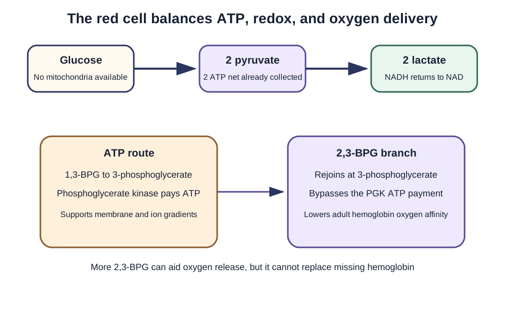
The erythrocyte has a tradeoff at 1,3-bisphosphoglycerate. The Rapoport-Luebering pathway produces 2,3-bisphosphoglycerate, which favors oxygen release from adult hemoglobin. Returning through 3-phosphoglycerate bypasses the phosphoglycerate kinase ATP payment. Fetal hemoglobin binds 2,3-BPG less strongly, contributing to its higher oxygen affinity. [20] Increased 2,3-BPG can partly support oxygen delivery in chronic anemia; it cannot restore the missing hemoglobin mass.

Red-cell pathway map shows lactate recycling of NAD and the 2,3-BPG branch bypassing the phosphoglycerate kinase ATP payment.
Connect red-cell ATP dependence, NAD recycling, and the oxygen-affinity tradeoff. [1] [3] [18] [20]

PKLR-related pyruvate kinase deficiency impairs red-cell ATP supply, membrane maintenance and survival. Expect congenital or chronic hemolysis with variable severity, rather than a literal absence of every ATP molecule. Mitapivat, an oral allosteric activator of red-cell pyruvate kinase, was approved in the United States in 2022 for hemolytic anemia in adults with pyruvate kinase deficiency. [25] PFKM-related disease can combine exertional muscle symptoms with hemolysis. [24] By comparison, the pentose phosphate pathway provides NADPH for antioxidant defense, not the NADH used in glycolytic energy accounting.

An oxidant-triggered hemolytic pattern points toward impaired antioxidant protection, while lifelong nonspherocytic hemolysis warrants evaluation of erythrocyte enzymes and other inherited causes. A negative direct antiglobulin test supports this direction but does not identify a particular enzyme. [3] [4]

Stored erythrocytes lose 2,3-BPG, but an affinity change alone does not establish a worse clinical outcome for every recipient. Randomized evidence did not show a survival advantage from routinely using fresher rather than standard-issue red cells in critically ill adults. Product handling and licensed rejuvenation processes are transfusion-service decisions, not a universal “add inosine and extend the expiration” rule. [12]

In G6PD deficiency, reduced pentose phosphate NADPH availability compromises glutathione recycling and oxidant protection. Infections, certain drugs or fava beans can precipitate hemolysis; the exposure and red-cell findings matter. This is a distinct problem from the ATP shortage of pyruvate kinase deficiency, although both disorders may shorten red-cell survival. [18]

Follow 1,3-bisphosphoglycerate through either phosphoglycerate kinase or the erythrocyte branch. Choosing the 2,3-BPG route sacrifices one ATP opportunity while favoring oxygen release from adult hemoglobin. Apply the tradeoff to chronic anemia without claiming that it restores missing hemoglobin.

Try it here · Checkpoint 2 of 3

Make your prediction before reading the choices. A first attempt is just a starting point.

Case 12

A patient with chronic anemia has increased erythrocyte 2,3-BPG, a right-shifted oxygen dissociation curve, and slightly reduced red-cell ATP yield per glucose. Which biochemical cost explains the ATP change?

Show answer and explanations for case 12
  1. A. Bypassing an ATP payment at phosphoglycerate kinase (Best answer)

    The branch rejoins at 3-phosphoglycerate and sacrifices this direct ATP opportunity.

    Reasoning steps for option A
    1. Where does the 2,3-BPG branch start and rejoin?

      It starts at 1,3-bisphosphoglycerate and rejoins at 3-phosphoglycerate.

    2. Which ATP step does this route skip?

      The phosphoglycerate kinase reaction, so that ATP is not made.

    3. How does this fit the patient?

      More 2,3-BPG right-shifts the curve at a small cost in ATP per glucose.

  2. B. Bypassing the pyruvate kinase ATP payment (Why this does not fit)

    The 2,3-BPG route rejoins before pyruvate kinase and specifically bypasses phosphoglycerate kinase.

    Reasoning steps for option B
    1. Why might pyruvate kinase seem to be the missed payment?

      Pyruvate kinase is the other ATP-producing step.

    2. Why is it still collected?

      The branch rejoins at 3-phosphoglycerate, before pyruvate kinase.

  3. C. Consumption of NADPH during the BPG-mutase reaction (Why this does not fit)

    The defining energetic tradeoff is the missed substrate-level ATP reaction, not NADPH consumption.

    Reasoning steps for option C
    1. Why might NADPH seem involved in red-cell metabolism?

      Red cells depend on NADPH for antioxidant defense.

    2. Does BPG mutase consume NADPH?

      No; the cost is a missed ATP-producing step.

  4. D. Higher hemoglobin affinity with reduced peripheral oxygen release (Why this does not fit)

    Increased 2,3-BPG favors oxygen release by lowering adult hemoglobin affinity.

    Reasoning steps for option D
    1. Why might an affinity change be the expected answer?

      2,3-BPG does change hemoglobin oxygen affinity.

    2. Why is the direction of that affinity change wrong?

      2,3-BPG lowers affinity and increases release, matching the right shift.

Takeaway: Red cells trade some ATP yield for modulation of oxygen affinity.

Case sources: [1]

Gluconeogenesis needs bypasses and an energy source

Glucose synthesis is not glycolysis read backward. Hexokinase, PFK-1 and pyruvate kinase operate far from equilibrium in their usual cellular settings. Four enzymes bypass those three reactions: pyruvate carboxylase, phosphoenolpyruvate carboxykinase, fructose 1,6-bisphosphatase and glucose 6-phosphatase. Starting with two pyruvates costs four ATP, two GTP and two NADH. The ATP and GTP together represent six high-energy phosphate equivalents.

The compartment map for making blood glucose
  1. Mitochondrial matrix: biotin-dependent pyruvate carboxylase uses ATP and bicarbonate to form oxaloacetate. Acetyl-CoA activates it.
  2. Across the inner membrane: oxaloacetate cannot simply diffuse out. Carbon can travel through malate, aspartate or phosphoenolpyruvate routes according to cellular conditions and enzyme distribution.
  3. Cytosolic reactions: PEPCK consumes GTP in the oxaloacetate-to-PEP reaction; fructose 1,6-bisphosphatase bypasses PFK-1. PEPCK also has a mitochondrial isoform.
  4. Endoplasmic reticulum: the glucose 6-phosphatase system permits release of free glucose in glucose-producing organs.

Malate dehydrogenase interconverts malate and oxaloacetate. Aspartate aminotransferase transfers an amino group between aspartate and α-ketoglutarate, yielding oxaloacetate and glutamate in one direction. It does not interconvert malate and an amino acid. [2]

Liver and renal cortex both contribute to systemic gluconeogenesis, with proportions changing during fasting and acid-base disturbances. Skeletal muscle lacks the glucose 6-phosphatase capacity needed for substantial export of free glucose. The adrenal cortex is not the standard second glucose-producing organ. Glycogen use, gluconeogenesis, lipolysis and ketogenesis overlap during fasting; they do not begin at rigid successive hour marks. Brain ketone use increases with adaptation while red cells continue to require glucose. [2]

Lactate, alanine and glycerol provide gluconeogenic carbon. Glycerol enters at the triose phosphate level and does not require the pyruvate-to-PEP bypass. Even-carbon fatty acids provide ATP and acetyl-CoA to support the process but do not provide net glucose carbon in humans. Pyruvate dehydrogenase is irreversible, and the TCA cycle does not create a net gain of oxaloacetate from acetyl-CoA. The glycerol backbone of a triglyceride is therefore metabolically different from its even-carbon fatty acids.

Sort lactate, alanine, glycerol, and palmitate by their first gluconeogenic entry point. Glycerol enters at the triose level, while even-carbon fatty acids provide energy but no net glucose carbon. Use the same routing rule to explain why two pyruvates require four bypass enzymes.

Use the trigger to identify the disrupted reaction

A fasting child with hypoglycemia, hepatomegaly and elevated lactate raises concern for glycogen storage disease I: both glycogen-derived and newly synthesized glucose encounter the impaired final glucose 6-phosphatase system. G6PC1 affects the catalytic enzyme; SLC37A4 affects the transporter. A child who becomes ill after fruit, sucrose or sorbitol instead suggests hereditary fructose intolerance.

Aldolase B deficiency traps phosphate in fructose 1-phosphate and compromises energy-dependent glucose production. Avoid a diagnostic fructose challenge; biochemical and molecular evaluation are safer. Fasting hypoglycemia with lactic acidosis can also reflect fructose 1,6-bisphosphatase deficiency. The trigger, organ findings and confirmatory testing separate these overlapping biochemical consequences. [5] [6] [14]

Ethanol oxidation increases the hepatic NADH-to-NAD ratio. [22] Lactate formation and oxaloacetate reduction are favored, making gluconeogenesis difficult when glycogen is depleted. In a malnourished person with symptomatic hypoglycemia, give glucose promptly and provide thiamine when indicated; do not postpone rescue glucose to enforce a vitamin-first sequence. Thiamine supports pyruvate dehydrogenase and other reactions, while niacin provides precursors for NAD and NADP. Photosensitive dermatitis, diarrhea and neuropsychiatric changes suggest pellagra; deficient intake or impaired tryptophan availability can contribute. [7] [11] [13]

Separate laboratory chemistry from bedside diagnosis. Fluoride inhibits enolase, but fluoride alone does not immediately prevent the early fall in glucose in an unseparated blood sample. Prompt processing or an appropriate rapidly effective glycolysis-inhibiting collection system is needed. [8] Arsenate can uncouple the GAPDH/phosphoglycerate kinase energy-conserving sequence experimentally; [21] arsenite inhibits lipoate-dependent complexes. [15] A history of mercury exposure does not establish a selective clinical GAPDH defect. Toxic exposures require exposure-specific assessment, not an enzyme mnemonic used as a diagnostic test.

Glucose uptake on FDG PET reflects transport and phosphorylation of a glucose analogue; increased uptake also occurs in inflammation. [16] Aerobic glycolysis in cancer supports biosynthesis and redox needs as well as ATP production. It does not mean all tumors have nonfunctional mitochondria or one identical PKM2 state. Metformin reduces hepatic glucose production, but presenting one proposed molecular target as its exclusive mechanism overstates the evidence. [9] Valproate-associated confusion warrants consideration of hyperammonemia even when liver tests are normal, not an assumed selective PEPCK inhibition. [10] Do not infer a glucose-infusion indication from myocardial fuel biochemistry alone.

NICE distinguishes monitored treatment of hyperglycemia in acute coronary syndromes from routine intensive insulin/glucose therapy, which it does not recommend unless clinically indicated. [23]

Hartnup disease impairs transport of neutral amino acids in intestine and kidney, including tryptophan. Reduced precursor availability can produce pellagra-like episodes during nutritional stress, with aminoaciduria distinguishing the transport problem from low niacin intake alone. The biochemical connection is impaired support for niacin-derived cofactors, not a mutation of GAPDH itself. [17]

Start with the trigger before selecting an enzyme: fruit exposure, fasting, ethanol, oxidant stress, or delayed specimen processing. The trigger plus the organ pattern separates phosphate trapping, a blocked glucose exit, a redox shift, antioxidant failure, and continued ex vivo glycolysis. Apply the comparison to a new patient before reading the choices.

Try it here · Checkpoint 3 of 3

Make your prediction before reading the choices. A first attempt is just a starting point.

Case 6

An infant tolerates milk formula but develops vomiting, sweating, and lethargy shortly after pear puree is introduced. Glucose and phosphate are low, lactate is increased, and hepatomegaly appears. Which paired intracellular change best explains the dietary trigger?

Show answer and explanations for case 6
  1. A. Fructose 1-phosphate rises as free phosphate falls (Best answer)

    Aldolase B deficiency traps dietary fructose as fructose 1-phosphate, depleting free phosphate and ATP while impairing glycogenolysis and gluconeogenesis.

    Reasoning steps for option A
    1. What does illness after pear puree but not formula suggest?

      A fructose-triggered disorder, since pears contain fructose and sorbitol.

    2. What does aldolase B deficiency do to fructose 1-phosphate?

      It traps dietary fructose as fructose 1-phosphate, which accumulates.

    3. How does that explain low phosphate and glucose?

      Trapped phosphate depletes free phosphate and ATP, impairing glycogenolysis and gluconeogenesis.

  2. B. Fructose 6-phosphate rises as ATP production increases (Why this does not fit)

    This does not describe phosphate trapping and would not account for hypophosphatemia after fructose exposure.

    Reasoning steps for option B
    1. Why might a hexose phosphate rise seem plausible after fruit?

      Fructose enters sugar-phosphate metabolism, so a fructose phosphate rise sounds relevant.

    2. Why is this pattern wrong?

      It does not trap phosphate and would not cause hypophosphatemia or reduced energy.

  3. C. Galactose 1-phosphate rises while phosphate remains available (Why this does not fit)

    Galactose 1-phosphate accumulation follows galactose exposure, not the fruit-triggered fructose pattern, and it does not match the stated phosphate depletion.

    Reasoning steps for option C
    1. Why could galactose metabolism be considered in an infant with liver disease?

      Galactosemia also causes vomiting, hepatomegaly, and hypoglycemia in infants.

    2. Which detail excludes galactosemia here?

      The infant tolerates milk formula, and symptoms follow fructose-containing fruit.

  4. D. Sorbitol accumulates in the lens while blood glucose rises (Why this does not fit)

    Sorbitol accumulation is associated with hyperglycemic tissue injury, not acute fructose-triggered hypoglycemia and hepatic phosphate trapping.

    Reasoning steps for option D
    1. Why might sorbitol be linked to pear puree?

      Pears contain sorbitol, which is converted to fructose.

    2. Why does lens sorbitol accumulation not fit?

      It is a chronic hyperglycemic complication, whereas this infant has acute hypoglycemia.

Takeaway: Hereditary fructose intolerance causes fructose 1-phosphate trapping, phosphate depletion, and secondary inhibition of hepatic glucose release after fructose exposure.

Case sources: [5]

Apply the pathway to a patient

These original educational scenarios ask you to identify the reaction, tissue priority or management distinction. Each answer includes a reason for accepting or rejecting every option.

Case 1

A 6-year-old has lifelong direct-antiglobulin-test-negative hemolytic anemia, splenomegaly, and echinocytes. Episodes are not linked to oxidant exposure; G6PD activity and glutathione reduction are normal. Erythrocyte pyruvate kinase activity is markedly reduced. Which paired metabolic change is most likely?

Show answer and explanations for case 1
  1. A. Decreased ATP with increased 2,3-BPG (Best answer)

    Pyruvate kinase deficiency removes a glycolytic ATP-producing step in mitochondria-free erythrocytes, while upstream carbon can be diverted toward 2,3-BPG.

    Reasoning steps for option A
    1. What does lifelong DAT-negative hemolysis with low pyruvate kinase activity point to?

      Pyruvate kinase deficiency, an inherited defect in the last glycolytic ATP-producing step.

    2. Why does ATP fall in these red cells?

      Mature erythrocytes lack mitochondria, so losing the pyruvate kinase payment removes a major ATP source.

    3. Why does 2,3-BPG rise at the same time?

      Glycolytic intermediates back up above the block and more carbon is diverted through the 2,3-BPG branch.

  2. B. Decreased NADPH with increased Heinz-body formation (Why this does not fit)

    This is the antioxidant pattern expected with G6PD deficiency, but G6PD activity, glutathione reduction, and the absence of oxidant-linked episodes argue against it.

    Reasoning steps for option B
    1. Why is an NADPH defect tempting in a child with hemolysis?

      G6PD deficiency is the most familiar enzyme cause of nonimmune hemolysis.

    2. Which findings exclude the antioxidant pattern?

      G6PD activity and glutathione reduction are normal and episodes are not oxidant-linked.

  3. C. Increased ATP with decreased lactate production (Why this does not fit)

    Reduced pyruvate kinase can lower downstream pyruvate and lactate, but it cannot increase ATP because the blocked step normally produces ATP.

    Reasoning steps for option C
    1. Why might lower lactate output seem expected here?

      A pyruvate kinase block reduces pyruvate available for lactate formation.

    2. Why can ATP not rise?

      The blocked enzyme normally makes ATP, so its loss lowers rather than raises ATP.

  4. D. Increased mitochondrial ATP with decreased reticulocytosis (Why this does not fit)

    Mature erythrocytes have no mitochondria, and ongoing hemolysis raises rather than lowers the reticulocyte response.

    Reasoning steps for option D
    1. Why might a cell compensate with oxidative ATP?

      Most cells can shift to mitochondrial ATP when glycolysis falters.

    2. Why is that impossible here, and what happens to reticulocytes?

      Mature red cells have no mitochondria, and ongoing hemolysis raises the reticulocyte count.

Takeaway: Pyruvate kinase deficiency limits erythrocyte ATP and can increase upstream 2,3-BPG; normal antioxidant testing separates it from G6PD deficiency.

Case sources: [1] [3]

Case 2

A 19-year-old develops early cramps and myoglobinuria during brief intense exercise. Forearm testing shows no normal lactate rise, and muscle biopsy shows excess glycogen. Mild chronic hemolysis with reticulocytosis is also present between episodes. Which reaction is most likely impaired?

Show answer and explanations for case 2
  1. A. Pyruvate reduction to lactate (Why this does not fit)

    An isolated lactate dehydrogenase defect could blunt lactate formation but would not explain the combined glycogen-rich myopathy and chronic erythrocyte hemolysis.

    Reasoning steps for option A
    1. Why is a lactate-forming defect tempting with a flat forearm lactate?

      The failed lactate rise could suggest lactate dehydrogenase is the missing step.

    2. Which findings does an isolated LDH defect fail to explain?

      Glycogen accumulation in muscle together with chronic erythrocyte hemolysis.

  2. B. Fructose 1-phosphate cleavage (Why this does not fit)

    Aldolase B deficiency produces fructose-triggered hepatic illness rather than an exertional muscle plus erythrocyte phenotype.

    Reasoning steps for option B
    1. Why might a fructose-cleaving defect come up in a glycolysis question?

      Aldolase B deficiency is a classic inherited carbohydrate disorder.

    2. Why does it not fit this patient?

      It causes fructose-triggered liver illness, not exertional myopathy with hemolysis.

  3. C. Fructose 6-phosphate phosphorylation (Best answer)

    Muscle PFK deficiency blocks the PFK-1 step, explaining low exercise lactate, glycogen accumulation, exertional symptoms, and hemolysis in erythrocytes that express the muscle isoform.

    Reasoning steps for option C
    1. What do early cramps, myoglobinuria, and no lactate rise indicate?

      Muscle cannot run glycolysis from glycogen during intense exercise.

    2. Why does glycogen accumulate?

      A block at PFK-1 traps upstream sugar phosphates, so glycogen builds up.

    3. Why is there also hemolysis?

      Red cells express the muscle PFK subunit, so the same PFKM defect shortens their survival.

  4. D. Glucose 6-phosphate hydrolysis (Why this does not fit)

    The glucose 6-phosphatase system supports hepatic and renal glucose export; its failure causes fasting metabolic disease, not this muscle and erythrocyte combination.

    Reasoning steps for option D
    1. Why might a glucose 6-phosphate step be considered with excess glycogen?

      Glycogen storage disease I is a well-known cause of glycogen accumulation.

    2. Why does the glucose 6-phosphatase system not fit?

      Its failure causes fasting hypoglycemia with liver enlargement, not exercise-induced muscle symptoms.

Takeaway: Exercise intolerance with a blunted lactate rise plus chronic hemolysis localizes the defect to muscle PFK and its fructose 6-phosphate phosphorylation step.

Case sources: [1] [4] [24]

Case 3

A patient in shock has severe tissue hypoperfusion, rising lactate, and limited mitochondrial oxidation. Cytosolic glycolysis continues despite the redox burden. Which immediate function of pyruvate-to-lactate conversion permits that continued flux?

Show answer and explanations for case 3
  1. A. Direct phosphorylation of ADP by lactate dehydrogenase (Why this does not fit)

    LDH supplies no additional ATP; its NAD recycling permits other glycolytic reactions to continue.

    Reasoning steps for option A
    1. Why might lactate formation seem like an ATP-producing step?

      Lactate rises when cells depend on glycolysis for ATP.

    2. Does lactate dehydrogenase make ATP?

      No; it only recycles NAD, which lets the ATP-producing steps continue.

  2. B. Regeneration of NADPH for the pentose phosphate pathway (Why this does not fit)

    The pyruvate/lactate pair uses NADH and NAD, not the NADPH antioxidant pair.

    Reasoning steps for option B
    1. Why might an antioxidant cofactor seem relevant in shock?

      Oxidative stress is prominent in hypoperfused tissues.

    2. Which cofactor pair does LDH actually use?

      NADH and NAD, not the NADPH pair of the pentose phosphate pathway.

  3. C. Oxidation of lactate to provide matrix NADH in the hypoperfused muscle (Why this does not fit)

    The stated increase is net lactate production, which reduces pyruvate and regenerates cytosolic NAD.

    Reasoning steps for option C
    1. Why could lactate be seen as a fuel here?

      Lactate can be oxidized as fuel by well-perfused tissues.

    2. What does rising lactate in hypoperfused tissue indicate instead?

      Net pyruvate reduction to lactate, which regenerates cytosolic NAD rather than making matrix NADH.

  4. D. Regeneration of NAD from NADH (Best answer)

    GAPDH needs oxidized NAD; LDH supplies it while reducing pyruvate.

    Reasoning steps for option D
    1. Which glycolytic enzyme needs NAD to keep working?

      GAPDH, which oxidizes glyceraldehyde 3-phosphate while reducing NAD to NADH.

    2. Why can mitochondria not supply that NAD in shock?

      Limited mitochondrial oxidation leaves NADH unoxidized in the cytosol.

    3. How does pyruvate-to-lactate conversion help?

      LDH reduces pyruvate and regenerates NAD from NADH, so glycolytic flux continues.

Takeaway: Lactate formation supports cytosolic redox recycling, not an additional ATP payment.

Case sources: [1]

Case 4

A patient with malnutrition and heavy alcohol use is confused and diaphoretic, with a glucose concentration of 31 mg/dL. Intravenous access is available, and thiamine will arrive shortly. What is the best immediate action?

Show answer and explanations for case 4
  1. A. Treat hypoglycemia promptly and give thiamine as soon as available. (Best answer)

    Urgent glucose must not be delayed; thiamine can be given concurrently or in either order.

    Reasoning steps for option A
    1. What does a glucose of 31 mg/dL with confusion and sweating require?

      Immediate treatment of symptomatic hypoglycemia.

    2. How should thiamine be timed?

      Give it as soon as available, concurrently or in either order, without delaying glucose.

  2. B. Wait for thiamine before giving any glucose. (Why this does not fit)

    This prolongs dangerous hypoglycemia without support from the cited ASAM guidance.

    Reasoning steps for option B
    1. Why does waiting for thiamine seem cautious in this patient?

      Glucose given to thiamine-deficient patients has been linked to Wernicke encephalopathy.

    2. Why is delay wrong here?

      Severe hypoglycemia is immediately dangerous, and guidance allows glucose and thiamine together.

  3. C. Give insulin to reduce lactate production. (Why this does not fit)

    Insulin would worsen the documented severe hypoglycemia.

    Reasoning steps for option C
    1. Why might insulin be suggested in a patient with high lactate?

      Insulin can shift metabolism away from lactate-producing states.

    2. Why would insulin be harmful here?

      It would lower an already dangerous glucose of 31 mg/dL even further.

  4. D. Use thiamine alone to correct the glucose concentration. (Why this does not fit)

    Thiamine supports metabolism but does not supply the urgently missing circulating substrate.

    Reasoning steps for option D
    1. Why might thiamine alone seem to address the metabolic problem?

      Thiamine deficiency impairs carbohydrate metabolism in malnourished drinkers.

    2. Why can thiamine not correct the hypoglycemia?

      It is a cofactor, not a source of glucose, so the missing substrate must be given.

Takeaway: Do not turn a useful thiamine precaution into a delay in glucose rescue.

Case sources: [7]

Case 5

After 24 hours without food, a person drinks substantial ethanol and develops hypoglycemia, increased lactate, and a high lactate-to-pyruvate ratio. Which hepatic change best connects the exposure to reduced glucose synthesis?

Show answer and explanations for case 5
  1. A. Inhibition of gluconeogenesis by a reduced hepatic NADH-to-NAD ratio (Why this does not fit)

    Ethanol oxidation increases rather than decreases this ratio.

    Reasoning steps for option A
    1. Why does a redox explanation sound correct here?

      Ethanol does alter the hepatic NADH-to-NAD ratio.

    2. What is wrong with the direction in this statement?

      Ethanol oxidation raises the NADH-to-NAD ratio rather than lowering it.

  2. B. A high NADH-to-NAD ratio favors pyruvate reduction and oxaloacetate reduction. (Best answer)

    These redox shifts reduce substrates available for gluconeogenesis when glycogen support is limited.

    Reasoning steps for option B
    1. What does ethanol oxidation do to hepatic redox state?

      Alcohol and acetaldehyde dehydrogenases generate NADH and raise the NADH-to-NAD ratio.

    2. How does a high NADH-to-NAD ratio affect gluconeogenic substrates?

      It pushes pyruvate toward lactate and oxaloacetate toward malate, reducing their availability.

    3. Why does the fast matter?

      After 24 hours without food, glycogen is low, so glucose depends on gluconeogenesis.

  3. C. Net conversion of ethanol-derived acetyl-CoA into glucose (Why this does not fit)

    Acetyl units cannot provide net glucose carbon through the human TCA cycle.

    Reasoning steps for option C
    1. Why might ethanol seem able to feed glucose synthesis?

      Ethanol is metabolized to acetyl-CoA, which enters central metabolism.

    2. Why can acetyl-CoA not supply net glucose?

      Pyruvate dehydrogenase is irreversible and the TCA cycle gives no net oxaloacetate from acetyl units.

  4. D. Phosphate trapping in fructose 1-phosphate (Why this does not fit)

    That mechanism fits fructose exposure in aldolase B deficiency, not the alcohol-triggered redox change.

    Reasoning steps for option D
    1. Why is phosphate trapping tempting for hypoglycemia with lactate?

      Hereditary fructose intolerance also causes hypoglycemia and lactic acidosis.

    2. Which trigger fails to fit phosphate trapping?

      The trigger here is ethanol after fasting, not fructose exposure.

Takeaway: Interpret alcohol-associated hypoglycemia through redox state and fasting context.

Case sources: [2] [22]

Case 7

An infant has fasting hypoglycemia, hepatomegaly, hypertriglyceridemia, hyperuricemia, and lactic acidemia. During a supervised glucagon challenge, blood glucose barely changes while lactate rises further. Which bottleneck best explains both the baseline pattern and the challenge response?

Show answer and explanations for case 7
  1. A. Blocked hydrolysis of glucose 6-phosphate (Best answer)

    Glycogenolysis and gluconeogenesis both converge on glucose 6-phosphate, so failure of its final hydrolysis prevents glucose export and diverts accumulated carbon toward lactate and lipid.

    Reasoning steps for option A
    1. Why does glucagon fail to raise glucose in this infant?

      Glycogen breaks down to glucose 6-phosphate but cannot be released as free glucose.

    2. Why does lactate rise further during the challenge?

      The trapped glucose 6-phosphate is diverted into glycolysis, producing more lactate.

    3. Which bottleneck explains both findings?

      Blocked hydrolysis of glucose 6-phosphate, the shared exit for glycogenolysis and gluconeogenesis.

  2. B. Failure to release glucose 1-phosphate from glycogen (Why this does not fit)

    A glycogen-phosphorylase problem would impair glycogenolysis but would not also block glucose made through gluconeogenesis at their shared final exit.

    Reasoning steps for option B
    1. Why might a glycogen-release defect explain a poor glucagon response?

      Phosphorylase defects also blunt glycogen breakdown.

    2. Which feature separates this from a phosphorylase problem?

      Glucose made by gluconeogenesis also fails to reach blood, pointing to their shared final step.

  3. C. Failure to convert pyruvate to oxaloacetate (Why this does not fit)

    Pyruvate carboxylase failure impairs gluconeogenesis but does not explain why glucagon-driven glycogen breakdown also fails to release free glucose.

    Reasoning steps for option C
    1. Why might pyruvate carboxylase be considered in fasting hypoglycemia with lactic acidemia?

      Its failure impairs gluconeogenesis and raises lactate.

    2. Why does it fail to explain the glucagon result?

      Pyruvate carboxylase deficiency does not stop glucagon-driven glycogen breakdown from releasing glucose.

  4. D. Failure to phosphorylate fructose after meals (Why this does not fit)

    Fructokinase deficiency is generally benign and cannot explain severe fasting disease, lactic acidemia, or the failed glucagon response.

    Reasoning steps for option D
    1. Why could a fructose-handling defect enter the differential?

      Fructose metabolism disorders affect the liver in infancy.

    2. Why is fructokinase deficiency wrong?

      It is benign and causes no fasting hypoglycemia, lactic acidemia, or failed glucagon response.

Takeaway: Glycogen storage disease I blocks the final glucose 6-phosphate exit shared by glycogenolysis and gluconeogenesis, so glucagon cannot restore blood glucose.

Case sources: [2] [6]

Case 8

A school-age child has recurrent fasting hypoglycemia and lactic acidosis during febrile illnesses but normal growth and no persistent hepatomegaly. Early in a supervised fast, glucagon raises glucose; after glycogen is depleted, glucose falls while lactate rises. Which reaction is most likely defective?

Show answer and explanations for case 8
  1. A. Fructose 1,6-bisphosphate hydrolysis (Best answer)

    Preserved early glycogen mobilization with later failure of glucose synthesis points to fructose 1,6-bisphosphatase, which bypasses PFK-1 during gluconeogenesis.

    Reasoning steps for option A
    1. What does an early glucose rise with glucagon show?

      Glycogen breakdown and the final glucose exit are intact.

    2. What happens after glycogen is gone?

      Glucose falls while lactate rises, so the problem is making new glucose.

    3. Which gluconeogenic enzyme fits that pattern?

      Fructose 1,6-bisphosphatase, the bypass for PFK-1.

  2. B. Fructose 6-phosphate phosphorylation (Why this does not fit)

    PFK-1 deficiency impairs glycolysis and produces an exertional muscle phenotype rather than isolated late-fasting gluconeogenic failure.

    Reasoning steps for option B
    1. Why might PFK-1 seem linked to this fructose-phosphate step?

      PFK-1 acts at the same fructose phosphate point in the opposite direction.

    2. Why is a PFK-1 defect wrong?

      It impairs glycolysis and causes exercise intolerance, not late-fasting hypoglycemia.

  3. C. Glycerol phosphorylation (Why this does not fit)

    Loss of glycerol kinase affects one precursor route but would not explain broad failure to use lactate and other pyruvate-derived substrates after glycogen depletion.

    Reasoning steps for option C
    1. Why could glycerol phosphorylation be relevant to fasting glucose?

      Glycerol is a gluconeogenic precursor during fasting.

    2. Why is glycerol kinase deficiency too narrow?

      It blocks only glycerol entry, not the use of lactate and other pyruvate-derived substrates.

  4. D. Pyruvate oxidation to acetyl-CoA (Why this does not fit)

    Pyruvate dehydrogenase directs carbon into oxidation, not the gluconeogenic bypass needed to convert fasting lactate into glucose.

    Reasoning steps for option D
    1. Why might pyruvate dehydrogenase be considered with lactic acidosis?

      PDH deficiency also raises lactate.

    2. Why does PDH not explain the hypoglycemia?

      PDH sends pyruvate toward oxidation, not toward glucose synthesis.

Takeaway: A normal early glycogen response followed by hypoglycemia and lactic acidosis after glycogen depletion localizes disease to fructose 1,6-bisphosphatase.

Case sources: [2] [14]

Case 9

A lean adolescent and two successive generations of relatives have stable mild fasting hyperglycemia, only a small glucose rise after an oral glucose load, and no diabetes complications. Autoantibodies are absent, and fasting insulin is not elevated. Which paired physiologic change best explains this pattern?

Show answer and explanations for case 9
  1. A. Higher insulin-release threshold and reduced hepatic glucose trapping (Best answer)

    Reduced glucokinase activity raises the pancreatic β-cell threshold for glucose-stimulated insulin release and decreases hepatic phosphorylation and trapping of glucose.

    Reasoning steps for option A
    1. What does a stable, mild, multigenerational fasting hyperglycemia suggest?

      A dominant glucokinase variant causing mild lifelong hyperglycemia.

    2. How does reduced glucokinase affect the β cell?

      It raises the glucose threshold for insulin release.

    3. How does it affect the liver?

      Hepatocytes phosphorylate and trap less glucose after meals.

  2. B. Lower insulin-release threshold and increased hepatic glucose trapping (Why this does not fit)

    These changes would favor lower fasting glucose rather than stable lifelong mild hyperglycemia.

    Reasoning steps for option B
    1. Why does this pairing seem to involve the right enzyme functions?

      It names the β-cell threshold and hepatic trapping, the two roles of glucokinase.

    2. Why is the direction wrong?

      A lower threshold and more trapping would lower fasting glucose, not raise it.

  3. C. Loss of skeletal-muscle GLUT4 and excessive hepatic glycogen synthesis (Why this does not fit)

    Marked peripheral insulin resistance usually raises fasting insulin and produces a larger metabolic syndrome phenotype, neither of which is supplied here.

    Reasoning steps for option C
    1. Why might insulin resistance be considered for high fasting glucose?

      Insulin resistance is the most common cause of hyperglycemia.

    2. Which findings argue against insulin resistance?

      The patient is lean and fasting insulin is not elevated.

  4. D. Impaired erythrocyte glycolysis and reduced pancreatic glucagon release (Why this does not fit)

    Erythrocyte glycolysis does not set the fasting glucose threshold, and reduced glucagon cannot explain this dominant multigenerational pattern.

    Reasoning steps for option D
    1. Why might a red-cell or glucagon change seem relevant to glucose?

      Red cells consume glucose and glucagon raises it.

    2. Why does this pairing fail?

      Red-cell glycolysis does not set fasting glucose, and less glucagon would lower glucose.

Takeaway: Glucokinase-related mild hyperglycemia reflects a higher β-cell glucose-sensing threshold plus reduced hepatic glucose phosphorylation, not severe insulin resistance.

Case sources: [19]

Case 10

After an overnight fast, a patient's hepatocytes show increased cAMP and protein kinase A activity in response to glucagon. Which paired response reduces futile cycling while supporting blood glucose?

Show answer and explanations for case 10
  1. A. Higher fructose 2,6-bisphosphate and activated liver pyruvate kinase (Why this does not fit)

    These favor glycolysis rather than the glucose-producing fasting response.

    Reasoning steps for option A
    1. Why might this pairing appear in a fasting question?

      It names the right regulators, fructose 2,6-bisphosphate and liver pyruvate kinase.

    2. Which state does this pattern describe?

      The fed state, which favors glycolysis over glucose production.

  2. B. Lower fructose 2,6-bisphosphate and activated liver pyruvate kinase (Why this does not fit)

    The first change fits, but glucagon-associated phosphorylation inhibits hepatic pyruvate kinase.

    Reasoning steps for option B
    1. Which half of this pairing is correct?

      Lower fructose 2,6-bisphosphate is the fasting response to glucagon.

    2. What does glucagon do to liver pyruvate kinase?

      PKA phosphorylation inhibits it, so it is not activated.

  3. C. Lower fructose 2,6-bisphosphate and inhibited liver pyruvate kinase (Best answer)

    Both changes reduce hepatic glycolytic competition with glucose synthesis.

    Reasoning steps for option C
    1. What does glucagon-driven PKA do to fructose 2,6-bisphosphate?

      It shifts the bifunctional enzyme toward FBPase-2, lowering fructose 2,6-bisphosphate.

    2. What does PKA do to liver pyruvate kinase?

      It phosphorylates and inhibits the enzyme.

    3. How do both changes reduce futile cycling?

      Less PFK-1 and pyruvate kinase activity limits glycolysis while glucose synthesis proceeds.

  4. D. Higher fructose 2,6-bisphosphate and inhibited fructose 1,6-bisphosphatase (Why this does not fit)

    This coordinated pattern favors the fed-state glycolytic direction rather than fasting gluconeogenesis.

    Reasoning steps for option D
    1. Why might inhibiting fructose 1,6-bisphosphatase seem related to control?

      Fructose 2,6-bisphosphate does inhibit that enzyme.

    2. Why is this the wrong state for fasting?

      High fructose 2,6-bisphosphate favors glycolysis and blocks gluconeogenesis, the fed pattern.

Takeaway: Hormonal regulation is tissue- and isoenzyme-specific.

Case sources: [1] [2]

Case 11

During an intense sprint, a runner has falling muscle ATP, rising AMP, and abundant glucose 6-phosphate. Which regulatory response helps increase local glycolytic ATP production?

Show answer and explanations for case 11
  1. A. Inhibition of PFK-1 by AMP (Why this does not fit)

    AMP signals low energy and favors PFK-1 activity.

    Reasoning steps for option A
    1. Why might AMP be linked to PFK-1 inhibition?

      AMP is a key PFK-1 regulator, so a direct link is familiar.

    2. What does AMP actually do to PFK-1?

      It signals low energy and activates PFK-1.

  2. B. Activation of fructose 1,6-bisphosphatase by AMP (Why this does not fit)

    AMP instead inhibits the opposing gluconeogenic enzyme.

    Reasoning steps for option B
    1. Why could AMP seem to act on fructose 1,6-bisphosphatase?

      AMP does regulate that enzyme.

    2. In which direction does AMP act on it?

      AMP inhibits fructose 1,6-bisphosphatase, opposing glucose synthesis.

  3. C. Increased allosteric inhibition of PFK-1 by ATP (Why this does not fit)

    ATP is falling during the sprint; its inhibitory signal does not explain increased glycolytic demand.

    Reasoning steps for option C
    1. Why might ATP inhibition of PFK-1 come to mind?

      ATP is a well-known allosteric inhibitor of PFK-1.

    2. Why does ATP inhibition not apply here?

      Muscle ATP is falling during the sprint, which weakens its inhibitory signal.

  4. D. Activation of PFK-1 by AMP (Best answer)

    The rising low-energy signal helps increase glycolytic flux when substrate is available.

    Reasoning steps for option D
    1. Which energy signals change during the sprint?

      Muscle ATP falls and AMP rises.

    2. How does rising AMP affect PFK-1?

      It allosterically activates PFK-1, relieving ATP inhibition.

    3. What is the result with abundant glucose 6-phosphate?

      Glycolytic flux and local ATP production increase.

Takeaway: Falling ATP and rising AMP allosterically activate PFK-1, increasing glycolytic ATP production in working muscle.

Case sources: [1] [2]

Case 13

Cord blood and maternal blood are exposed to the same 2,3-BPG concentration, yet the fetal sample has higher oxygen affinity. Which property of HbF best explains the difference?

Show answer and explanations for case 13
  1. A. HbF binds 2,3-BPG more strongly than HbA. (Why this does not fit)

    Stronger binding would favor the low-affinity state, opposite to the fetal oxygen-affinity advantage.

    Reasoning steps for option A
    1. Why might stronger 2,3-BPG binding seem to explain a difference?

      Differences in 2,3-BPG binding do explain the result.

    2. Why is stronger binding the wrong direction?

      Stronger binding would lower HbF affinity, not raise it.

  2. B. HbF binds 2,3-BPG less strongly. (Best answer)

    Reduced binding limits its affinity-lowering effect on fetal hemoglobin.

    Reasoning steps for option B
    1. Why does HbF have higher oxygen affinity despite equal 2,3-BPG?

      Its γ chains bind 2,3-BPG less strongly than the β chains of HbA.

    2. What does weaker binding do?

      It limits the affinity-lowering effect of 2,3-BPG, leaving HbF with higher affinity.

    3. Why does this help the fetus?

      Higher affinity lets fetal blood take up oxygen from maternal blood.

  3. C. HbF has a lower intrinsic affinity that improves uptake from maternal blood. (Why this does not fit)

    Lower oxygen affinity would not explain the stated advantage in fetal uptake.

    Reasoning steps for option C
    1. Why might lower affinity seem to aid oxygen transfer?

      Lower affinity favors release to tissues.

    2. Why does this not fit the observation?

      The fetal sample has higher affinity, which is what favors uptake from maternal blood.

  4. D. The fetal effect requires conversion of heme iron to its ferric state. (Why this does not fit)

    Ferric methemoglobin does not provide the normal functional oxygen binding responsible for this relationship.

    Reasoning steps for option D
    1. Why might a change in heme iron be considered?

      Iron state changes oxygen binding.

    2. Why is ferric iron wrong?

      Ferric methemoglobin cannot bind oxygen normally.

Takeaway: Globin composition changes the response to an erythrocyte metabolite.

Case sources: [20]

Case 14

A stable critically ill adult receives standard-issue red cells. A trainee notes that stored units have reduced 2,3-BPG and concludes that fresher units must improve survival, although a large randomized comparison found no mortality advantage. Which interpretation is best supported?

Show answer and explanations for case 14
  1. A. Fresh units should routinely replace standard-issue units to reduce 90-day mortality (Why this does not fit)

    The cited adult randomized trial did not demonstrate this mortality benefit.

    Reasoning steps for option A
    1. Why is replacing standard units with fresh units tempting?

      Stored units lose 2,3-BPG, which suggests poorer oxygen delivery.

    2. What did the randomized trial find?

      Fresher units did not reduce 90-day mortality in critically ill adults.

  2. B. Standard-issue units were proven equivalent for every possible recipient population (Why this does not fit)

    Failure to demonstrate benefit in critically ill adults is not proof for every population or outcome.

    Reasoning steps for option B
    1. Why might a negative trial seem to settle the question for all?

      No mortality difference sounds like proof of equivalence.

    2. Why is that conclusion too broad?

      Failure to show benefit in one population does not prove equivalence everywhere.

  3. C. The biochemical storage change does not establish a routine survival benefit from fresher units (Best answer)

    The trial compared fresh with standard-issue cells in critically ill adults and found no significant 90-day mortality benefit.

    Reasoning steps for option C
    1. What storage change prompted the trainee's reasoning?

      Stored red cells lose 2,3-BPG over time.

    2. What did the large trial show about outcomes?

      Fresher units gave no survival advantage in critically ill adults.

    3. What is the supported interpretation?

      The biochemical storage change does not establish a routine survival benefit from fresher units.

  4. D. The trial showed that 2,3-BPG concentration never changes during storage (Why this does not fit)

    The trial tested clinical outcomes; it did not negate the biochemical storage phenomenon.

    Reasoning steps for option D
    1. Why might a negative trial seem to deny the storage lesion?

      It found no clinical difference between fresh and older units.

    2. What did the trial actually test?

      Clinical outcomes, not whether 2,3-BPG changes during storage.

Takeaway: Distinguish a laboratory storage lesion from an established clinical outcome.

Case sources: [12]

Case 15

A bedside glucose measurement is 112 mg/dL, but a fluoride-tube specimen left unseparated for two hours reports 85 mg/dL. No glucose-lowering treatment occurred between samples. Which preanalytic explanation should the laboratory consider?

Show answer and explanations for case 15
  1. A. The measured value necessarily reflects the glucose at venipuncture (Why this does not fit)

    Unseparated cells may consume glucose during the delay.

    Reasoning steps for option A
    1. Why does a result from a proper tube seem trustworthy?

      Fluoride tubes are intended to preserve glucose.

    2. Why can the value differ from the time of venipuncture?

      Cells in the unseparated sample kept consuming glucose during the two-hour delay.

  2. B. Fluoride suppresses glycolysis rapidly enough to eliminate all early glucose loss (Why this does not fit)

    Fluoride alone does not promptly suppress the early reactions.

    Reasoning steps for option B
    1. Why might fluoride seem to stop glycolysis immediately?

      Fluoride is a glycolysis inhibitor.

    2. How quickly does fluoride act?

      Slowly; it inhibits enolase, and the earlier reactions continue for some time.

  3. C. Delayed separation should cause glucose to rise as cells lyse (Why this does not fit)

    Continued cellular glycolysis produces the relevant downward bias.

    Reasoning steps for option C
    1. Why might cell lysis seem to raise glucose?

      Lysed cells release intracellular contents into the sample.

    2. What is the actual direction of the bias?

      Downward, because intact cells keep consuming glucose.

  4. D. Continued cellular glycolysis before full fluoride inhibition can lower the result (Best answer)

    The 2023 laboratory guideline recommends rapid inhibition or prompt validated separation.

    Reasoning steps for option D
    1. What happened between the two measurements?

      Two hours of unseparated storage in a fluoride tube.

    2. Why did glucose fall?

      Blood cells kept using glucose before fluoride fully inhibited glycolysis.

    3. What does current laboratory guidance recommend?

      Rapid glycolysis inhibition or prompt validated separation.

Takeaway: A collection additive is not a substitute for validated specimen handling.

Case sources: [8]

Case 16

A patient with severe dietary restriction develops photosensitive dermatitis, diarrhea, and cognitive changes. The same deficiency reduces availability of the electron acceptor used by GAPDH. Which cofactor is directly limited?

Show answer and explanations for case 16
  1. A. Low niacin-derived NAD (Best answer)

    GAPDH requires NAD as its electron acceptor; the syndrome also supports pellagra.

    Reasoning steps for option A
    1. What do dermatitis, diarrhea, and cognitive change suggest?

      Pellagra from niacin deficiency.

    2. Which GAPDH cofactor comes from niacin?

      NAD, the electron acceptor that GAPDH reduces to NADH.

    3. Why does that link the syndrome to glycolysis?

      Low niacin limits NAD, the same cofactor GAPDH needs.

  2. B. Reduced biotin available to GAPDH (Why this does not fit)

    Biotin participates in carboxylation, not the GAPDH oxidation.

    Reasoning steps for option B
    1. Why might biotin seem relevant to a glucose pathway enzyme?

      Biotin is a cofactor in glucose metabolism through pyruvate carboxylase.

    2. Why does biotin not serve GAPDH?

      It supports carboxylation, not the oxidation that GAPDH performs.

  3. C. Reduced vitamin K available to GAPDH (Why this does not fit)

    Vitamin K-dependent carboxylation of proteins is unrelated to this reaction.

    Reasoning steps for option C
    1. Why might vitamin K appear among cofactors?

      Vitamin K is a cofactor for protein carboxylation.

    2. Why is vitamin K unrelated here?

      It does not act in GAPDH oxidation or cause pellagra.

  4. D. Reduced cobalamin available to GAPDH (Why this does not fit)

    B12 supports other reactions and is not its direct redox cofactor.

    Reasoning steps for option D
    1. Why might cobalamin be considered with cognitive change?

      B12 deficiency causes neurologic and cognitive symptoms.

    2. Why is cobalamin not the GAPDH cofactor?

      B12 serves other reactions and is not an electron acceptor for GAPDH.

Takeaway: Connect the deficiency syndrome to the specific cofactor reaction.

Case sources: [1] [11]

Case 17

A malnourished patient has confusion, gait instability, increased lactate, pyruvate, and alanine, and normal arterial oxygenation. After labeled glucose is given, labeled pyruvate forms normally but labeled acetyl-CoA is reduced. Which enzyme-cofactor pair is most directly impaired?

Show answer and explanations for case 17
  1. A. Pyruvate dehydrogenase and thiamine pyrophosphate (Best answer)

    The tracer reaches pyruvate but not acetyl-CoA, localizing the block to pyruvate dehydrogenase; thiamine pyrophosphate is one of its required cofactors.

    Reasoning steps for option A
    1. Where does the labeled carbon stop?

      It reaches pyruvate normally but forms little acetyl-CoA.

    2. Which enzyme converts pyruvate to acetyl-CoA?

      Pyruvate dehydrogenase, which requires thiamine pyrophosphate.

    3. How do the clinical findings fit?

      Malnutrition, confusion, and ataxia suggest thiamine deficiency, with pyruvate and lactate backing up.

  2. B. PFK-1 and biotin (Why this does not fit)

    A PFK-1 block would reduce formation of pyruvate from glucose, contradicting normal labeled pyruvate production, and PFK-1 does not use biotin.

    Reasoning steps for option B
    1. Why might PFK-1 seem responsible for impaired glucose metabolism?

      It is the committed glycolytic step.

    2. Which tracer result excludes PFK-1?

      Labeled pyruvate forms normally, so glycolysis through PFK-1 is intact; PFK-1 also does not use biotin.

  3. C. Lactate dehydrogenase and flavin adenine dinucleotide (Why this does not fit)

    Lactate dehydrogenase uses NADH rather than FAD, and its failure would not specifically block labeled pyruvate from becoming acetyl-CoA.

    Reasoning steps for option C
    1. Why might LDH be suspected with high lactate?

      LDH converts pyruvate to lactate.

    2. Why does LDH not explain the block?

      It uses NADH, not FAD, and its failure would not stop pyruvate becoming acetyl-CoA.

  4. D. Pyruvate kinase and pyridoxal phosphate (Why this does not fit)

    Pyruvate kinase acts before the normally formed labeled pyruvate and does not require pyridoxal phosphate.

    Reasoning steps for option D
    1. Why might pyruvate kinase be considered in a pyruvate question?

      Pyruvate kinase is the enzyme that makes pyruvate.

    2. Why is pyruvate kinase not the site?

      Labeled pyruvate forms normally, so the step that makes it works; it also does not use pyridoxal phosphate.

Takeaway: Normal glycolytic pyruvate formation with reduced acetyl-CoA labeling localizes the defect to thiamine-dependent pyruvate dehydrogenase beyond glycolysis.

Case sources: [13]

Case 18

After an overnight fast, labeled glycerol is infused. Label appears in newly synthesized glucose without first appearing in pyruvate, and hepatic glycerol kinase activity is normal. Which next reaction places that carbon into the gluconeogenic sequence above the pyruvate bypass?

Show answer and explanations for case 18
  1. A. Glycerol 3-phosphate oxidation to DHAP (Best answer)

    After glycerol kinase forms glycerol 3-phosphate, its oxidation produces DHAP, allowing glycerol carbon to enter at the triose-phosphate level without pyruvate carboxylase.

    Reasoning steps for option A
    1. What does glycerol kinase make from glycerol?

      Glycerol 3-phosphate.

    2. Which reaction comes next?

      Glycerol 3-phosphate is oxidized to DHAP, a triose phosphate.

    3. Why does this carbon avoid pyruvate?

      DHAP enters gluconeogenesis above the pyruvate carboxylase and PEPCK bypasses.

  2. B. Pyruvate carboxylation to oxaloacetate (Why this does not fit)

    The tracer specifically bypasses pyruvate, so entry through pyruvate carboxylase contradicts the supplied labeling pattern.

    Reasoning steps for option B
    1. Why might pyruvate carboxylase seem to be the entry point?

      Most gluconeogenic carbon enters through pyruvate carboxylase.

    2. Which tracer result excludes this route?

      The label reaches glucose without first appearing in pyruvate.

  3. C. Acetyl-CoA condensation with oxaloacetate (Why this does not fit)

    Citrate formation supports oxidation and biosynthesis but does not provide a net route from glycerol carbon into glucose.

    Reasoning steps for option C
    1. Why might citrate formation seem to route carbon into glucose?

      It is the entry point of the TCA cycle, which feeds oxaloacetate.

    2. Why is citrate synthase wrong here?

      Acetyl-CoA condensation gives no net glucose carbon and is not where glycerol enters.

  4. D. Glucose 6-phosphate hydrolysis in muscle (Why this does not fit)

    Skeletal muscle does not substantially export free glucose, and this late reaction does not explain glycerol's initial triose-level entry.

    Reasoning steps for option D
    1. Why might glucose 6-phosphate hydrolysis seem to be the final step?

      It is the last reaction before glucose release.

    2. Why is this choice wrong?

      Muscle does not export much glucose, and a late step does not explain glycerol's triose entry.

Takeaway: Glycerol enters gluconeogenesis as DHAP after glycerol phosphorylation and glycerol 3-phosphate oxidation, bypassing pyruvate carboxylase.

Case sources: [2]

Case 19

During fasting, labeled glycerol appears in newly synthesized glucose, whereas labeled palmitate increases ketone production without supplying net glucose carbon. Which statement best distinguishes the roles of these triglyceride components?

Show answer and explanations for case 19
  1. A. Both substrates supply glucose carbon in proportion to their carbon counts (Why this does not fit)

    This overlooks the inability of even-carbon acetyl units to produce net glucose in humans.

    Reasoning steps for option A
    1. Why might both components seem to supply glucose carbon?

      Both come from the same triglyceride and both are carbon fuels.

    2. Why do fatty acid carbons not yield glucose?

      Even-carbon acetyl units give no net oxaloacetate, so no net glucose.

  2. B. Palmitate supplies glucose carbon, while glycerol supplies only oxidative energy (Why this does not fit)

    This reverses the distinction between a triose precursor and even-carbon acetyl units.

    Reasoning steps for option B
    1. Why might palmitate seem the larger carbon source?

      Palmitate carries far more carbon than glycerol.

    2. Why is this reversed?

      Glycerol is the glucose precursor, and palmitate provides energy and ketones.

  3. C. Neither substrate can help maintain glucose because neither is a carbohydrate (Why this does not fit)

    Glycerol supplies precursor carbon and fatty acid oxidation supports the energy requirements of gluconeogenesis.

    Reasoning steps for option C
    1. Why might neither lipid seem able to help glucose balance?

      Neither is a carbohydrate.

    2. How does each actually help?

      Glycerol supplies carbon, and fatty acid oxidation powers gluconeogenesis.

  4. D. Glycerol supplies glucose carbon; palmitate oxidation supplies energy without net glucose carbon (Best answer)

    Glycerol enters at the triose level, whereas even-carbon acetyl units cannot provide net glucose carbon.

    Reasoning steps for option D
    1. Where does labeled glycerol enter?

      At the triose phosphate level, becoming glucose carbon.

    2. What happens to labeled palmitate?

      It is oxidized to acetyl-CoA and ketones without net glucose carbon.

    3. Which statement separates their roles?

      Glycerol supplies glucose carbon, and palmitate supplies energy only.

Takeaway: Do not equate the triglyceride backbone with its fatty acids.

Case sources: [2]

Case 20

After 60 hours of fasting, hepatic glycogen is nearly depleted and renal venous sampling shows net glucose release that was minimal after an overnight fast. Which tissue accounts for this additional systemic gluconeogenesis?

Show answer and explanations for case 20
  1. A. Renal cortex (Best answer)

    The renal cortex contributes to gluconeogenesis, with flux varying by nutritional and acid-base state.

    Reasoning steps for option A
    1. What does renal venous glucose release after 60 hours show?

      The kidney is exporting newly made glucose.

    2. Which part of the kidney makes glucose?

      The renal cortex, whose contribution grows during prolonged fasting.

  2. B. Adrenal cortex (Why this does not fit)

    This nearby anatomical name should not replace the renal glucose-producing tissue.

    Reasoning steps for option B
    1. Why might the adrenal cortex seem involved in fasting glucose?

      It makes cortisol, which supports gluconeogenesis.

    2. Why is the adrenal cortex wrong?

      It is not a major site of glucose release; the sample was from renal veins.

  3. C. Skeletal muscle (Why this does not fit)

    Muscle uses glucose and glycogen locally but lacks substantial glucose 6-phosphatase-dependent export.

    Reasoning steps for option C
    1. Why might muscle seem able to contribute glucose?

      Muscle stores large amounts of glycogen.

    2. Why can muscle not export free glucose?

      It lacks the glucose 6-phosphatase capacity needed for substantial release.

  4. D. Adipose tissue (Why this does not fit)

    Adipose supplies glycerol and fatty acids but is not the principal additional organ releasing newly synthesized glucose.

    Reasoning steps for option D
    1. Why might adipose tissue seem relevant in fasting?

      It releases glycerol and fatty acids during fasting.

    2. Why is adipose not the glucose-releasing organ?

      It supplies substrates but does not release newly made glucose.

Takeaway: Glucose production is shared principally by liver and kidney.

Case sources: [2]

Case 21

A hepatic tracer study follows two pyruvate molecules into one glucose while ATP, GTP, and reducing equivalents are measured separately. Excluding transport-dependent variation, which direct nucleotide expenditure is expected?

Show answer and explanations for case 21
  1. A. No ATP because all glycolytic reactions simply reverse (Why this does not fit)

    Irreversible glycolytic reactions require energy-consuming bypasses.

    Reasoning steps for option A
    1. Why might reversing glycolysis seem to cost nothing?

      Most glycolytic reactions are reversible.

    2. Why is energy still required?

      Three glycolytic steps are irreversible and need energy-consuming bypasses.

  2. B. Four ATP and two GTP (Best answer)

    These are the six high-energy phosphate equivalents required from two pyruvates; two NADH are also consumed.

    Reasoning steps for option B
    1. Which bypass steps consume nucleotides from two pyruvates?

      Pyruvate carboxylase uses 2 ATP, PEPCK uses 2 GTP, and phosphoglycerate kinase uses 2 ATP.

    2. What is the total direct nucleotide cost?

      Four ATP and two GTP, six high-energy phosphate equivalents.

    3. Which reducing cost is also paid?

      Two NADH at the GAPDH step.

  3. C. Six ATP and two GTP (Why this does not fit)

    This double-counts two phosphate expenditures and overstates the standard cost.

    Reasoning steps for option C
    1. Why might six ATP seem right?

      Six high-energy phosphate equivalents is a familiar total.

    2. Why is six ATP plus two GTP an overcount?

      It double-counts the two GTP, which are already part of the six.

  4. D. Two ATP and no GTP (Why this does not fit)

    This resembles glycolytic net yield rather than gluconeogenic expenditure.

    Reasoning steps for option D
    1. Why might two ATP seem to be the cost?

      Two ATP is the familiar net yield of glycolysis.

    2. Why is it wrong for glucose synthesis?

      Glycolytic yield is not the same as gluconeogenic cost, which also includes GTP.

Takeaway: Count ATP molecules and ATP equivalents separately.

Case sources: [2]

Case 22

Mitochondrial oxaloacetate cannot cross the inner membrane directly. In a preparation that transfers its carbon as malate, which enzyme uses NADH to catalyze the required first conversion?

Show answer and explanations for case 22
  1. A. Pyruvate kinase (Why this does not fit)

    It makes pyruvate from PEP in glycolysis.

    Reasoning steps for option A
    1. Why might a PEP-related enzyme seem relevant?

      Oxaloacetate becomes PEP later in the pathway.

    2. Why is pyruvate kinase wrong?

      It works in glycolysis, turning PEP into pyruvate, not oxaloacetate into malate.

  2. B. Glucose 6-phosphatase (Why this does not fit)

    It releases free glucose in the terminal glucose-production system.

    Reasoning steps for option B
    1. Why might glucose 6-phosphatase be listed for gluconeogenesis?

      It is the final gluconeogenic enzyme.

    2. Why is it wrong for oxaloacetate export?

      It acts at the end in the endoplasmic reticulum, not at the mitochondrial membrane.

  3. C. Malate dehydrogenase (Best answer)

    It couples the oxaloacetate/malate pair to NADH/NAD redox chemistry.

    Reasoning steps for option C
    1. How can oxaloacetate carbon leave the mitochondrion?

      It is reduced to malate, which can cross the inner membrane.

    2. Which enzyme uses NADH for that conversion?

      Malate dehydrogenase, which interconverts oxaloacetate and malate.

  4. D. Aspartate aminotransferase (Why this does not fit)

    AST interconverts oxaloacetate and aspartate by transferring an amino group.

    Reasoning steps for option D
    1. Why might AST seem to handle oxaloacetate export?

      AST also converts oxaloacetate for transport.

    2. Why is AST wrong for a malate route?

      AST makes aspartate by transamination, not malate by reduction.

Takeaway: Reduction and transamination are distinct ways to process oxaloacetate.

Case sources: [2]

Case 23

During prolonged fasting, skeletal muscle transfers amino groups to pyruvate and exports alanine to the liver. A selective skeletal-muscle alanine aminotransferase inhibitor is then given. Which paired change is most likely?

Show answer and explanations for case 23
  1. A. Less alanine export and less carbon for hepatic glucose (Best answer)

    Blocking muscle alanine aminotransferase reduces transfer of both the amino group and pyruvate-derived carbon as alanine, limiting that substrate contribution to hepatic gluconeogenesis.

    Reasoning steps for option A
    1. What does muscle alanine aminotransferase do?

      It transfers amino groups to pyruvate, forming alanine for export.

    2. What happens when it is inhibited?

      Less alanine leaves muscle, so less carbon reaches the liver for glucose.

  2. B. More alanine export and greater nitrogen delivery without carbon (Why this does not fit)

    Alanine formation requires the inhibited transamination, and alanine always carries a carbon skeleton together with its amino group.

    Reasoning steps for option B
    1. Why might more nitrogen export seem possible?

      Muscle still has amino groups to dispose of.

    2. Why is this impossible here?

      Alanine formation needs the inhibited enzyme, and alanine always carries carbon with nitrogen.

  3. C. Unchanged alanine export because albumin carries the amino group (Why this does not fit)

    Albumin transports fatty acids and proteins but does not replace alanine as the coupled nitrogen and carbon carrier in this cycle.

    Reasoning steps for option C
    1. Why might albumin seem to carry nitrogen?

      Albumin is a major plasma transport protein.

    2. Why can albumin not replace alanine?

      It does not carry amino groups as a coupled carbon and nitrogen carrier.

  4. D. Less fatty-acid oxidation because alanine supplies acetyl-CoA (Why this does not fit)

    Alanine contributes pyruvate-derived gluconeogenic carbon; it is not the required fuel that permits muscle fatty-acid oxidation during fasting.

    Reasoning steps for option D
    1. Why might alanine seem needed for fat oxidation?

      Alanine and fatty acids both fuel fasting metabolism.

    2. What is alanine's actual role?

      It supplies pyruvate-derived carbon for liver glucose, not fuel for fat oxidation.

Takeaway: The glucose-alanine cycle couples muscle nitrogen disposal to delivery of pyruvate-derived carbon for hepatic glucose production.

Case sources: [2]

Case 24

A patient with type 2 diabetes starts metformin and later has lower fasting glucose without direct stimulation of insulin release. Which established physiologic description is supported without assigning one disputed molecular target as the exclusive cause?

Show answer and explanations for case 24
  1. A. Reduced hepatic glucose output and improved insulin sensitivity (Best answer)

    These physiological effects are stated in the label.

    Reasoning steps for option A
    1. What does metformin do to fasting glucose?

      It lowers it without directly stimulating insulin release.

    2. Which physiologic effects are well established?

      Reduced hepatic glucose output and improved insulin sensitivity, without one proven exclusive target.

  2. B. Direct stimulation of pancreatic insulin release (Why this does not fit)

    That better describes an insulin secretagogue; metformin does not primarily work this way.

    Reasoning steps for option B
    1. Why might insulin secretion seem to explain lower glucose?

      Many diabetes drugs work by releasing insulin.

    2. Why does this not describe metformin?

      Metformin is not a secretagogue, as the stem notes.

  3. C. Increased urinary glucose loss through SGLT2 inhibition (Why this does not fit)

    This is the mechanism of SGLT2 inhibitors, not metformin.

    Reasoning steps for option C
    1. Why might urinary glucose loss seem plausible?

      SGLT2 inhibitors lower fasting glucose.

    2. Why is SGLT2 inhibition wrong here?

      It is a separate drug class, not metformin.

  4. D. Delayed carbohydrate digestion through α-glucosidase inhibition (Why this does not fit)

    This is the target of α-glucosidase inhibitors rather than metformin.

    Reasoning steps for option D
    1. Why might slower digestion seem to lower glucose?

      α-glucosidase inhibitors reduce carbohydrate absorption.

    2. Why is this not metformin's action?

      That is the action of acarbose and related drugs.

Takeaway: Use established physiological effects when molecular mechanisms remain multifactorial.

Case sources: [9]

Case 25

A patient taking valproate develops vomiting and new lethargy. Glucose is 98 mg/dL, sodium is 139 mmol/L, and liver transaminases are normal. Which metabolic complication still warrants prompt evaluation?

Show answer and explanations for case 25
  1. A. Hypoglycemia (Why this does not fit)

    The measured glucose does not support this explanation.

    Reasoning steps for option A
    1. Why might hypoglycemia be considered with lethargy?

      Hypoglycemia commonly causes lethargy and vomiting.

    2. Which measured value excludes hypoglycemia?

      Glucose is 98 mg/dL, which is normal.

  2. B. Hyperammonemia (Best answer)

    The label calls for ammonia assessment with these symptoms even when liver tests are normal.

    Reasoning steps for option B
    1. What complication should be checked in valproate users with lethargy?

      Hyperammonemia, which can cause encephalopathy.

    2. Why do normal liver tests not rule it out?

      Valproate can raise ammonia even when transaminases are normal.

  3. C. Hyponatremic encephalopathy (Why this does not fit)

    The measured sodium does not support this explanation.

    Reasoning steps for option C
    1. Why might hyponatremia be considered with vomiting and lethargy?

      Low sodium commonly causes confusion.

    2. Which laboratory value excludes low sodium?

      Sodium is 139 mmol/L, which is normal.

  4. D. Diabetic ketoacidosis (Why this does not fit)

    There is no stated diabetes, ketonemia or acidosis; the medication and symptoms specifically warrant ammonia testing.

    Reasoning steps for option D
    1. Why might ketoacidosis be considered with vomiting?

      Diabetic ketoacidosis causes vomiting and lethargy.

    2. Why is it unlikely here?

      There is no diabetes, ketosis, or acidosis, and glucose is normal.

Takeaway: Normal liver tests do not exclude valproate-associated hyperammonemia.

Case sources: [10]

Case 26

FDG PET shows uptake in a lung lesion and in an inflamed postoperative wound, with substantial tracer retention at both sites. What prevents interpreting glucose-analogue uptake alone as proof of malignancy?

Show answer and explanations for case 26
  1. A. FDG uptake directly measures complete glucose oxidation to carbon dioxide (Why this does not fit)

    The measured retention reflects uptake and phosphorylation rather than completion of glucose oxidation.

    Reasoning steps for option A
    1. Why might FDG seem to measure full glucose oxidation?

      FDG is a glucose analogue used to assess metabolism.

    2. What does FDG retention actually reflect?

      Transport and phosphorylation, which trap FDG as FDG 6-phosphate.

  2. B. Postoperative FDG uptake by itself establishes recurrent malignancy (Why this does not fit)

    Healing and inflammation can also produce uptake in the stated setting.

    Reasoning steps for option B
    1. Why might wound uptake suggest recurrent tumor?

      The patient had surgery, and tumor recurrence is a concern.

    2. What else causes uptake in a postoperative wound?

      Healing and inflammation also produce FDG uptake.

  3. C. Activated inflammatory tissues can also transport and phosphorylate FDG (Best answer)

    The tracer signal is not specific to malignant cells and needs anatomical and clinical interpretation.

    Reasoning steps for option C
    1. Which cells in a wound take up FDG?

      Activated inflammatory cells, which increase glucose transport and phosphorylation.

    2. Why does that limit FDG as proof of cancer?

      Tumor and inflamed tissue trap FDG by the same route, so uptake at both sites cannot by itself tell them apart; the lesion's anatomy and the clinical history must decide.

  4. D. The intensity of uptake alone distinguishes infection from sterile inflammation (Why this does not fit)

    Both may be FDG-avid; uptake alone does not establish their cause.

    Reasoning steps for option D
    1. Why might intensity seem to separate infection from sterile inflammation?

      Stronger uptake can seem to indicate a more active process.

    2. Why can intensity not decide the cause?

      Both infection and sterile inflammation can be strongly FDG avid.

Takeaway: Metabolic avidity must be interpreted with anatomy and clinical context.

Case sources: [16]

Case 27

An adult admitted with an acute coronary syndrome has glucose of 244 mg/dL. A trainee recommends routine intensive insulin plus glucose infusion solely to alter myocardial fuel use. Which approach matches the cited clinical guidance?

Show answer and explanations for case 27
  1. A. Use routine intensive insulin/glucose therapy for all ACS patients with hyperglycemia (Why this does not fit)

    NICE does not recommend this routinely unless clinically indicated.

    Reasoning steps for option A
    1. Why might intensive insulin with glucose seem protective?

      It shifts myocardial fuel use toward glucose.

    2. What does NICE advise about routine use?

      It does not recommend routine intensive insulin with glucose unless clinically indicated.

  2. B. Leave glucose unmonitored because the acute coronary syndrome takes priority (Why this does not fit)

    NICE recommends glucose management with monitoring and avoidance of hypoglycemia.

    Reasoning steps for option B
    1. Why might glucose seem a lower priority in ACS?

      Coronary reperfusion is the main priority.

    2. What does NICE recommend for glucose?

      Monitoring and management of hyperglycemia while avoiding hypoglycemia.

  3. C. Target biochemical ketosis to replace myocardial glucose use (Why this does not fit)

    The guideline does not recommend inducing ketosis as ACS glucose management.

    Reasoning steps for option C
    1. Why might ketosis seem to help the heart?

      Ketones are an alternative myocardial fuel.

    2. Is inducing ketosis part of ACS glucose management?

      No; guidance does not recommend inducing ketosis.

  4. D. Manage hyperglycemia with monitoring while distinguishing it from routine intensive insulin/glucose therapy (Best answer)

    NICE advises keeping glucose below 11 mmol/L while avoiding hypoglycemia, considering dose-adjusted insulin, and avoiding routine intensive insulin/glucose therapy unless clinically indicated.

    Reasoning steps for option D
    1. What glucose level does NICE target in ACS?

      Keeping glucose below 11 mmol/L while avoiding hypoglycemia.

    2. How does this differ from the trainee's plan?

      It is monitored treatment, not routine intensive insulin with glucose for fuel effects.

Takeaway: A pathway preference is not by itself a treatment indication.

Case sources: [23]

Case 29

A cell-free glycolysis preparation contains arsenate instead of inorganic phosphate at the GAPDH reaction. NADH still forms and carbon reaches 3-phosphoglycerate, but the acyl arsenate intermediate hydrolyzes rapidly. Which ATP-producing reaction loses its usual substrate?

Show answer and explanations for case 29
  1. A. Loss of the ATP payment at pyruvate kinase only (Why this does not fit)

    The unstable substitute interrupts the earlier GAPDH/PGK energy-conservation sequence.

    Reasoning steps for option A
    1. Why might the loss seem to be at pyruvate kinase?

      Pyruvate kinase is the final ATP-producing step.

    2. Where is the energy actually lost?

      Earlier, at the GAPDH and phosphoglycerate kinase sequence.

  2. B. Loss of the usual ATP payment at phosphoglycerate kinase (Best answer)

    Carbon can proceed without conservation of that phosphate-transfer energy.

    Reasoning steps for option B
    1. What does arsenate replace at GAPDH?

      Inorganic phosphate, forming 1-arseno-3-phosphoglycerate.

    2. What happens to that intermediate?

      It hydrolyzes spontaneously to 3-phosphoglycerate.

    3. Which ATP payment is lost?

      The phosphoglycerate kinase ATP, since its substrate never forms.

  3. C. Failure to form NADH at any glycolytic reaction (Why this does not fit)

    The scenario describes lost phosphate-energy conservation, not necessarily loss of the GAPDH redox reaction.

    Reasoning steps for option C
    1. Why might NADH production seem affected?

      Arsenate acts at the NADH-producing enzyme.

    2. Does NADH still form?

      Yes; the stem says NADH forms, and only phosphate-energy conservation is lost.

  4. D. An extra substrate-level ATP payment at phosphoglycerate mutase (Why this does not fit)

    Phosphoglycerate mutase relocates phosphate and does not supply a compensating ATP payment.

    Reasoning steps for option D
    1. Why might a phosphate shift seem to compensate?

      Phosphoglycerate mutase moves a phosphate group.

    2. Does phosphoglycerate mutase make ATP?

      No; it only relocates the phosphate.

Takeaway: Arsenate replaces phosphate at GAPDH, so 1-arseno-3-phosphoglycerate hydrolyzes and the phosphoglycerate kinase ATP is lost while NADH still forms.

Case sources: [1] [21]

Case 30

A patient with weight loss, diabetes, and necrolytic migratory erythema has a pancreatic neuroendocrine tumor that secretes excess glucagon. Which paired hepatic enzyme state most directly supports the hyperglycemia?

Show answer and explanations for case 30
  1. A. Low F2,6BP; inhibited hepatic pyruvate kinase (Best answer)

    Glucagon signaling lowers hepatic fructose 2,6-bisphosphate and inhibits the liver pyruvate kinase isoenzyme, reducing glycolytic competition while favoring glucose production.

    Reasoning steps for option A
    1. What does glucagon do to hepatic fructose 2,6-bisphosphate?

      It lowers it through PKA acting on the bifunctional enzyme.

    2. What does glucagon signaling do to the liver pyruvate kinase isoenzyme?

      It inhibits it by phosphorylation.

    3. How does this cause hyperglycemia in glucagonoma?

      Less glycolysis and more gluconeogenesis raise hepatic glucose output.

  2. B. High F2,6BP; activated hepatic pyruvate kinase (Why this does not fit)

    This is a fed-state combination that promotes hepatic glycolysis rather than glucagon-driven glucose output.

    Reasoning steps for option B
    1. Why might this pairing seem like an active liver?

      It names high hepatic metabolic activity.

    2. Which state does it describe?

      The fed state, which favors glycolysis rather than glucose output.

  3. C. Low F2,6BP; activated hepatic PFK-1 (Why this does not fit)

    Low fructose 2,6-bisphosphate removes a major PFK-1 activator, so PFK-1 would not be activated by this state.

    Reasoning steps for option C
    1. Which half of this pairing fits glucagon?

      Low fructose 2,6-bisphosphate.

    2. Why is activated PFK-1 wrong?

      Low fructose 2,6-bisphosphate removes PFK-1 activation.

  4. D. High F2,6BP; activated hepatic FBPase-1 (Why this does not fit)

    High fructose 2,6-bisphosphate inhibits fructose 1,6-bisphosphatase and therefore opposes gluconeogenesis.

    Reasoning steps for option D
    1. Why might activated FBPase-1 fit glucagon?

      Glucagon does favor FBPase-1 activity.

    2. Why is high fructose 2,6-bisphosphate wrong?

      It inhibits FBPase-1, so it cannot pair with an active enzyme.

Takeaway: Excess glucagon lowers hepatic fructose 2,6-bisphosphate and inhibits liver pyruvate kinase, coordinating increased glucose production with reduced glycolysis.

Case sources: [1] [2]

Case 31

A teenager develops jaundice and dark urine during an infection. The blood film shows bite cells and Heinz bodies, and the direct antiglobulin test is negative. Between episodes, erythrocyte ATP production is normal. Which paired biochemical abnormality best explains the episodic hemolysis?

Show answer and explanations for case 31
  1. A. Low NADPH with impaired reduced-glutathione regeneration (Best answer)

    G6PD deficiency limits pentose-phosphate-pathway NADPH, so erythrocytes cannot efficiently regenerate reduced glutathione during oxidant stress despite preserved glycolytic ATP.

    Reasoning steps for option A
    1. What do bite cells and Heinz bodies after infection suggest?

      Oxidant damage to hemoglobin, as in G6PD deficiency.

    2. What is limited in G6PD deficiency?

      NADPH from the pentose phosphate pathway.

    3. Why does low NADPH cause hemolysis?

      Red cells cannot regenerate reduced glutathione during oxidant stress.

  2. B. Low ATP with increased 2,3-BPG from pyruvate kinase deficiency (Why this does not fit)

    Pyruvate kinase deficiency causes chronic ATP-limited hemolysis rather than the oxidant-triggered Heinz-body pattern with normal ATP between episodes.

    Reasoning steps for option B
    1. Why might pyruvate kinase deficiency be considered?

      It is another inherited red-cell enzyme cause of DAT-negative hemolysis.

    2. Which detail excludes it?

      ATP is normal between episodes, and hemolysis is oxidant-triggered, not chronic.

  3. C. Low NAD with failure of lactate formation during fasting (Why this does not fit)

    Failure to regenerate cytosolic NAD would impair glycolysis broadly and does not specifically produce oxidant-triggered Heinz bodies and bite cells.

    Reasoning steps for option C
    1. Why might an NAD problem seem relevant to red-cell survival?

      Red cells depend on NAD for glycolysis.

    2. Why does low NAD not fit?

      It would impair glycolysis broadly and not produce oxidant Heinz bodies.

  4. D. Low glucose export from absent hepatic glucose 6-phosphatase (Why this does not fit)

    A hepatic glucose-export defect causes fasting hypoglycemia and hepatomegaly, not isolated oxidant erythrocyte injury.

    Reasoning steps for option D
    1. Why might a hepatic glucose defect be considered?

      It is another enzyme disorder in the glucose pathway.

    2. Why does it not fit?

      It causes fasting hypoglycemia and hepatomegaly, not oxidant red-cell injury.

Takeaway: G6PD deficiency preserves glycolytic ATP but limits NADPH-dependent glutathione regeneration, producing oxidant-triggered Heinz bodies, bite cells, and hemolysis.

Case sources: [1] [18]

Case 32

A child has episodic photosensitive rash and ataxia during poor intake. Neutral amino acids, including tryptophan, are increased in urine, and symptoms improve with higher protein intake. Which intervention and metabolic link best fit the disorder?

Show answer and explanations for case 32
  1. A. Nicotinamide replacement because tryptophan can supply niacin (Best answer)

    Hartnup disease reduces neutral amino acid absorption and renal reuptake; loss of tryptophan can limit endogenous niacin synthesis, so nicotinamide can treat pellagra-like episodes.

    Reasoning steps for option A
    1. What does neutral aminoaciduria with pellagra-like episodes indicate?

      Hartnup disease, with loss of tryptophan in intestine and kidney.

    2. What does tryptophan supply?

      It is a precursor for niacin synthesis.

    3. Which treatment fits?

      Nicotinamide, which replaces niacin for NAD.

  2. B. Thiamine replacement because tryptophan supplies TPP (Why this does not fit)

    Tryptophan is not a precursor for thiamine pyrophosphate, and thiamine deficiency does not explain neutral aminoaciduria.

    Reasoning steps for option B
    1. Why might thiamine be considered with ataxia?

      Thiamine deficiency causes ataxia.

    2. Why is thiamine wrong?

      Tryptophan does not make thiamine, and thiamine deficiency does not cause aminoaciduria.

  3. C. Riboflavin replacement because tryptophan supplies FAD (Why this does not fit)

    Tryptophan does not supply riboflavin or FAD, and this choice fails to connect the amino acid transport defect to the pellagra-like skin and neurologic findings.

    Reasoning steps for option C
    1. Why might riboflavin be considered with skin changes?

      Riboflavin deficiency causes skin and mucosal changes.

    2. Why is riboflavin wrong?

      Tryptophan does not make riboflavin or FAD.

  4. D. Pantothenate replacement because tryptophan supplies coenzyme A (Why this does not fit)

    Tryptophan is not the precursor for pantothenate or coenzyme A, so this does not explain the dietary sensitivity and neutral aminoaciduria.

    Reasoning steps for option D
    1. Why might pantothenate be considered?

      Pantothenate forms coenzyme A, a key metabolic cofactor.

    2. Why is pantothenate wrong?

      Tryptophan is not its precursor, and it does not explain the aminoaciduria.

Takeaway: Hartnup disease can cause pellagra-like episodes because urinary and intestinal tryptophan loss limits niacin synthesis; protein support and nicotinamide address that link.

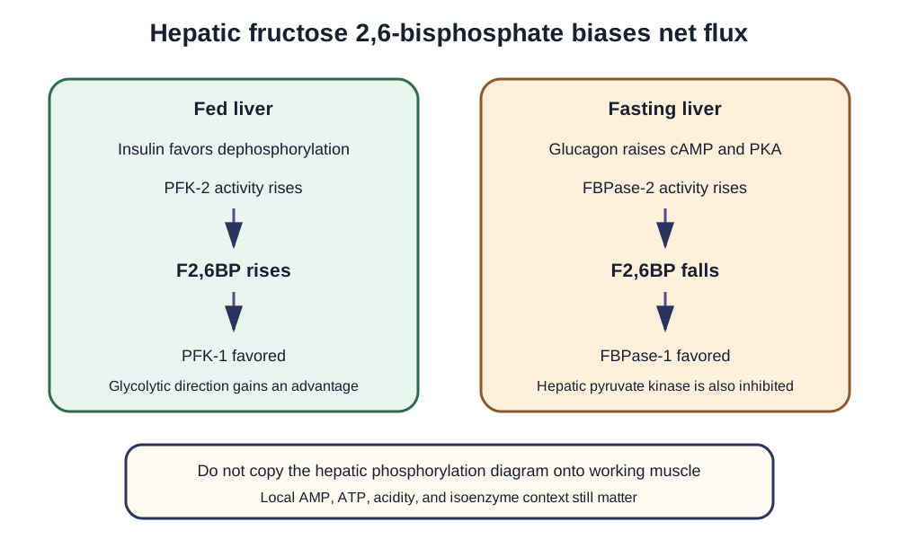
Case sources: [11] [17]

Search Bone Wizardry

Quick links