
Cardiology · Valve Disease

Mitral Regurgitation

The valve that forgets how to stay closed. Blood leaks backward, the left atrium stretches, AFib shows up uninvited, and the heart slowly drowns in its own volume.

Opening Challenge

A 52-year-old woman presents with 6 months of progressively worsening shortness of breath on exertion and occasional palpitations. On exam her pulse is irregular at 118 bpm. You hear a grade 4/6 holosystolic murmur loudest at the apex, radiating to the left axilla. The murmur gets louder when she squeezes your hand as hard as she can. Echo shows a dilated left atrium. What is the most likely diagnosis?

A. Aortic Stenosis
B. Mitral Regurgitation
C. HOCM
D. Ventricular Septal Defect
1 / 5

💡 Section 01 · The Basics

What Actually Goes Wrong

A leaky door that blood exploits every heartbeat
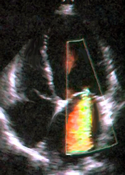
Echocardiogram showing mitral regurgitation
📷 Echocardiogram · 4-chamber view showing MR · tap to expand

Think of the mitral valve like a one-way door between the left atrium (LA) and left ventricle (LV). During systole, the LV squeezes and that door is supposed to snap shut, forcing all the blood out through the aorta. In mitral regurgitationRegurgitation = backward flow. The valve does not seal, so blood leaks backward into the LA instead of forward to the aorta during every heartbeat., the door does not close properly. Every squeeze of the LV sends some blood backward into the LA instead of forward into the body.

The result: the LV has to pump the same blood twice. Over time this volume overload stretches the LV and LA. The LA dilates. A dilated LA is a chaotic electrical nightmare, which is exactly how AFib sets up camp.

💡 MR creates a pressure escape hatch. The LV leaks into a low-pressure LA instead of the high-pressure aorta. This initially protects the LV from pressure overload, but the volume penalty accumulates silently for years.

Because the LV ejects blood in two directions (aorta plus back into LA), the ejection fractionEF = stroke volume divided by end-diastolic volume. In MR, EF is falsely elevated because the LV ejects into both the aorta AND the LA. An EF of 55% in MR is actually worse than it sounds. looks falsely preserved for a long time. This is the sneakiest thing about chronic MR: the patient can be asymptomatic with a "normal" EF of 60% while the LV is quietly decompensating.

🧬 Section 02 · Etiology

Why Does the Valve Leak?

Four causes, four different mechanisms, one broken valve

Click a cause to see what breaks

MVP Mitral Valve Prolapse Rheumatic Post-strep scarring Ischemic Papillary dysfunction Dilated CM Ring dilates, leaflets gap Click a cause above to see the mechanism

Each cause breaks the valve through a different mechanism

Cause Trace It clinical medicine Clue
MVP Myxomatous degeneration, leaflets billow into LA Most common cause in developed countries. Young women. Mid-systolic click.
Rheumatic Post-strep immune attack scars and fuses leaflet tips History of RF. Often co-occurs with MS. Still #1 worldwide.
Ischemic Papillary muscle ischemia or rupture after MI Inferior MI (RCA territory). Acute or subacute onset. Classic post-MI complication.
Dilated CM Annular dilation stretches valve ring, leaflets cannot meet Functional MR: the valve itself is structurally normal, the ring just got too big.

Board Trap: Collagen Diseases

The PASS textbook also lists SBE (subacute bacterial endocarditis) and collagen diseases (Marfan syndrome, Ehlers-Danlos) as causes of MR. MVP is the structural culprit in collagen disorders because floppy connective tissue equals floppy valve leaflets. If you see Marfan plus a murmur in clinical practice, think both MVP and aortic root dilation.

2 / 5

🔊 Section 03 · The Sound

What You Hear and Why

The murmur tells the whole story if you know how to listen

S1 SYSTOLE HOLOSYSTOLIC murmur (blowing) S2 DIASTOLE (quiet) Radiates: Apex to Left Axilla / Back

MR fills the entire systolic gap between S1 and S2

Here is why MR is holosystolic (meaning it runs all the way from S1 to S2 without a break): as soon as systole starts, the LV pressure shoots above LA pressure. Since the mitral valve does not close properly, blood starts leaking backward immediately at the start of systole and keeps leaking until the aortic valve closes at S2. The pressure gradient between LV and LA exists for the entire duration of systole, so the murmur fills the entire window.

Contrast this with aortic stenosis, which only gets loud once the LV builds enough pressure to force blood through the narrowed aortic valve, creating a crescendo-decrescendo (diamond) shape.

🎤 Holosystolic = pressure gradient exists ALL of systole. MR, TR, and VSD are the three holosystolic murmurs. They do not ramp up and down, they just blast.

Radiation: MR radiates to the left axilla (and sometimes the back) because the regurgitant jet squirts backward and lateral, following the direction of the jet. It does NOT radiate to the carotids (that is AS).

Intensity: Graded 1 through 6. Grade 4 and above has a palpable thrill. S3 gallopS3 = a third heart sound in early diastole. In MR, so much blood rushes from the overfilled LA into the LV that it causes a low-pitched thud. S3 in MR = volume overload is severe. is often heard in significant MR because a huge volume of blood rushes from the overfilled LA into the LV in early diastole.

🎯 Section 04 · Maneuvers

Turning MR Up and Down

The board's favorite test within a test

Hand Grip ✋
Valsalva 💨
Squatting 🦟
Standing 🚶

Hand Grip INCREASES MR murmur.

Here is the chain: squeezing your fist increases systemic vascular resistance (afterload). Higher afterload makes it harder for blood to leave through the aortic valve. So more blood takes the path of least resistance, which is the leaky mitral valve, back into the LA. More backflow equals louder murmur.

Rule: Anything that increases afterload (hand grip, squatting) increases MR.

Hand grip = close your fist = clamp down the pipes = blood takes the detour through the leaky mitral valve. MR louder.

Valsalva DECREASES MR murmur.

Valsalva (bearing down, like you are constipated) increases intrathoracic pressure, which squeezes the veins and reduces venous return to the heart. Less blood in the LV means less volume to leak backward. Less volume = quieter murmur. Simple.

Rule: Anything that decreases preload (Valsalva, standing) decreases MR.

💨 Valsalva = reduced venous return = less blood in the heart = less to leak backward. MR quieter.

Board Trap: Valsalva does the OPPOSITE for HOCM

Valsalva makes HOCM louder (less preload = more outflow obstruction). Valsalva makes MR quieter (less preload = less backflow). This is the most common murmur maneuver trap in clinical practice. Know this cold.

Squatting INCREASES MR murmur.

Squatting does two things: it increases venous return (more preload) AND increases afterload (compressed leg veins plus increased SVR). Both factors increase the amount of blood in the LV and make it harder for blood to get out through the aorta, pushing more through the leaky mitral valve.

Rule: Squat increases preload AND afterload, so MR gets louder.

Standing DECREASES MR murmur.

Suddenly standing causes blood to pool in the legs, reducing venous return to the heart. Less blood in the LV = less volume to leak backward = quieter murmur. Afterload also drops slightly. Standing is like Valsalva lite for MR.

Rule: Standing decreases preload, MR gets quieter.

ManeuverEffect on PreloadEffect on AfterloadMR Murmur
Hand GripNC↑ increasesLouder
Valsalva↓ decreases↓ decreasesSofter
Squatting↑ increases↑ increasesLouder
Standing↓ decreasesNCSofter
Amyl Nitrite↓ decreases↓ decreasesSofter

NC = no change. Compare to HOCM: opposite effect from Valsalva and standing.

🔑 MR gets louder when more blood is Made to stay (high afterload). MR gets softer when blood is Missing (low preload).
3 / 5

⚠️ Section 05 · Complications

Why AFib Moves In

Volume overload, LA dilation, and the electrical chaos it creates

LA dilation is not just a size problem. A stretched left atrium becomes electrically chaotic. Think of a guitar string: when it is tuned to the right tension, it vibrates at one clean frequency. Stretch it unevenly and it produces noise instead of notes. The LA works the same way. Stretch the walls enough and the electrical signals that coordinate atrial contraction become a disorganized mess, and AFibAtrial fibrillation: loss of coordinated atrial contraction. The atria quiver uselessly while the AV node lets through irregular impulses to the ventricles. Results in irregular pulse, reduced cardiac output, and thrombus risk. sets in.

Once AFib is present, things get worse fast. Without coordinated atrial kick, the LV loses the "last top-off" of blood before systole, reducing preloadPreload = the volume of blood in the LV at the end of diastole. Atrial kick contributes about 20% of LV filling. In AFib, this is lost. In MR, the LV is already volume-overloaded, so this loss matters less initially but accelerates decompensation. by about 20%.

The dilated LA also creates a clot factory: stagnant blood pools in the appendage, forms thrombus, and that thrombus can embolize to the brain. AFib in MR means anticoagulation conversation is required.

MR chain: leaky valve → LA volume overload → LA dilation → AFib → irregular pulse, reduced CO, and stroke risk. The irregular pulse at 118 bpm in our opening case was the AFib alarm.

⏰ Section 06 · Timeline Matters

Acute vs Chronic MR

Same broken valve, completely different emergency level

Acute MR

  • 📸 Cause: papillary muscle rupture post-MI, acute endocarditis
  • ⚡ Onset: sudden, hours to days
  • 🚫 LA has NO time to dilate
  • 💥 LA pressure spikes instantly
  • 🌧 Pulmonary edema FAST
  • 😷 Hemodynamic collapse, cardiogenic shock
  • 🚑 Needs EMERGENT surgery

Chronic MR

  • 🏭 Cause: MVP, rheumatic, dilated CM
  • 🕐 Onset: months to years, slow
  • ⬆ LA compensates, dilates over time
  • 💰 Eccentric hypertrophy of LV
  • 👀 Asymptomatic for years
  • 🔍 EF falsely elevated early on
  • 🔔 Surgery when EF falls below 60% or LVESD >40mm

Acute MR is like suddenly punching a hole in a water pipe. The water (blood) has nowhere to go but back into the LA, which has not had time to stretch. Pressure skyrockets. Pulmonary edema follows within hours. This patient is crashing and needs emergency surgery.

Chronic MR is like a slow, small leak in the same pipe. The LA gradually enlarges over years, accommodating more and more volume without raising pressure dramatically. The patient is asymptomatic for a long time. The LV undergoes eccentric hypertrophy, meaning it adds sarcomeres in series (gets bigger in volume capacity, not wall thickness), to handle the extra load. Eventually, even this adaptation fails.

🔥 Acute MR is a medical emergency. Chronic MR is a quiet time bomb. in clinical practice: post-MI new systolic murmur at apex plus flash pulmonary edema = acute MR from papillary muscle rupture. Call the surgeon.

🩺 Section 07 · What Happens Inside

Volume Overload and the Heart's Response

How the heart tries to cope and why it eventually stops working

NORMAL LV Normal wall CHRONIC Compensated Eccentric hypertrophy Bigger chamber, thin walls DECOMPENSATED EF drops below 60% LVESD > 40mm Surgical window closing

Eccentric hypertrophy compensates initially. When the LV can no longer maintain forward output, it decompensates fast.

In chronic MR, the LV handles a constantly elevated end-diastolic volume. To cope, it undergoes eccentric hypertrophy: sarcomeres are added in series (lengthened), which increases chamber size without proportionally thickening the wall. The LV becomes a big, stretchy, compliant chamber that can hold more blood without a dramatic rise in filling pressures.

This sounds like a win. But there is a critical threshold: when the LV begins to decompensate, the EF falls below 60% (remember, in MR the EF is inflated because the LV ejects into the low-resistance LA, so 60% in MR is actually like 50% in a normal heart) and the left ventricular end-systolic diameter (LVESD) exceeds 40mm. Once these thresholds are crossed, surgery must happen before irreversible LV dysfunction sets in.

4 / 5

🩸 Section 08 · When to Cut

Surgical Indications

The numbers that change everything

Watchful Waiting

Asymptomatic MR, EF > 60%, LVESD < 40mm. Echo every 6 to 12 months. No intervention needed yet.

Medical Management

Afterload reduction (vasodilators like hydralazine plus nitrates) to reduce regurgitant fraction. Diuretics for volume overload. Beta-blockers or rate-control for AFib. Note: ACE inhibitors in asymptomatic MR are controversial; they do not delay surgery in patients with normal EF.

Surgery Indicated (Elective)

Any of: EF 30 to 60%, LVESD ≥ 40mm, or symptoms with preserved EF, or new-onset AFib/pulmonary hypertension from MR. Prefer repair over replacement when anatomy allows.

Emergency Surgery

Acute MR with papillary muscle rupture post-MI or acute infective endocarditis with hemodynamic instability. These patients crash fast. Bridge with intra-aortic balloon pump (IABP) if needed, but get them to the OR.

Board Trap: EF Threshold in MR

In a normal patient, EF below 55% is abnormal. In MR, the EF is inflated because the LV can eject into the low-resistance LA. So EF below 60% in MR equals effectively reduced function. Surgery should happen BEFORE EF falls below 60%, not after. If you wait until EF is 45%, the LV has already sustained irreversible damage.

🎮 Section 09 · Differential Game

Which Systolic Murmur?

Four systolic murmurs enter. One leaves. You pick them off with each clue.

A patient has a systolic murmur. The clues come one at a time. Each clue eliminates one diagnosis. Click the diagnosis each clue kills off.

HOCM
Dynamic obstruction
MR
Mitral Regurgitation
VSD
Septal defect
AS
Aortic Stenosis
Loading first clue...

Prove It

Clinical Vignettes

Six patients just walked into your clinic. They did not make an appointment. Deal with it.

Medically reviewed by Kaitlyn Cocuzzo, MD and Fatima Ali, DO · Last updated July 1, 2026 at 10:03 PM ET
Bone Wizardry is an independent educational resource for visual learning in the medical sciences. It is not affiliated with, endorsed by, or sponsored by any licensing or examination board, contains no real or recalled examination questions, and does not guarantee any educational or examination outcome.
5 / 5