Trace pancreatic bud rotation, adult territories and duct drainage, then distinguish annular pancreas, divisum and accessory splenic tissue.
A newborn has an obstructed duodenum. An adult has two pancreatic drainage routes. A patient has a splenic nodule inside the pancreatic tail. These findings become easier to interpret when three questions stay separate: where did the tissue begin, where did it settle, and where does its secretion go?
Follow the bud beside the bile duct
The pancreas begins as dorsal and ventral outgrowths of foregut endoderm. Human specimens show distinct buds during approximately the fifth week after conception. The larger dorsal contribution develops toward the dorsal mesentery. The ventral contribution begins beside the developing extrahepatic biliary system. Their epithelial progenitors ultimately produce both endocrine islet cells and exocrine acinar and duct cells. This does not make every supporting vessel or connective-tissue cell in the pancreas endodermal. [1]
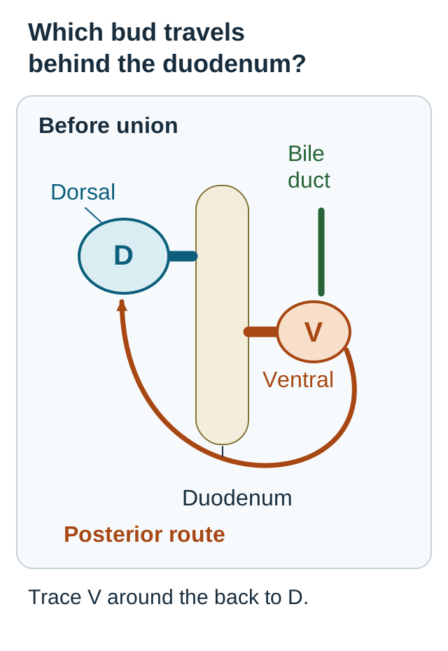
During rightward rotation of the duodenum, the ventral bud and its biliary attachment travel posteriorly around the duodenum toward the dorsal bud. The territories unite around the seventh developmental week, and their duct systems normally establish a connection. These week labels refer to approximate post-conception development, not an exact deadline in obstetric gestational dating. Human developmental stages are better anchors than pretending that every embryo completes each event on the same day. [1][2]
Try the spatial model: identify the bud beside the bile duct in the rotation figure. Trace its curved path behind the duodenum, not across the front. Now point to the tissue that can meet it on the posterior side. The consequence is a single pancreatic organ assembled from two territories. Tissue union and duct communication are related events, but a gland can contain both territories even when the ducts remain separate. [2]
Follow V beside the bile duct, then the curved posterior route toward D. This is a topological route model, not a cross-section or a claim about clockwise direction from every viewpoint. Tissue union is followed by duct remodeling. [1][2]
A description of clockwise rotation is incomplete without a specified viewing direction. The useful relationship is posterior passage of the ventral bud around the duodenum. Neither the spleen nor the pancreatic tail travels through the duodenal lumen.
Predict what could remain abnormal after the tissue is together.
The ducts may fail to establish their usual communication. That predicts separate pancreatic outlets without requiring loss of the body and tail. This is the distinction that will explain pancreas divisum.
Read adult anatomy back to its origin
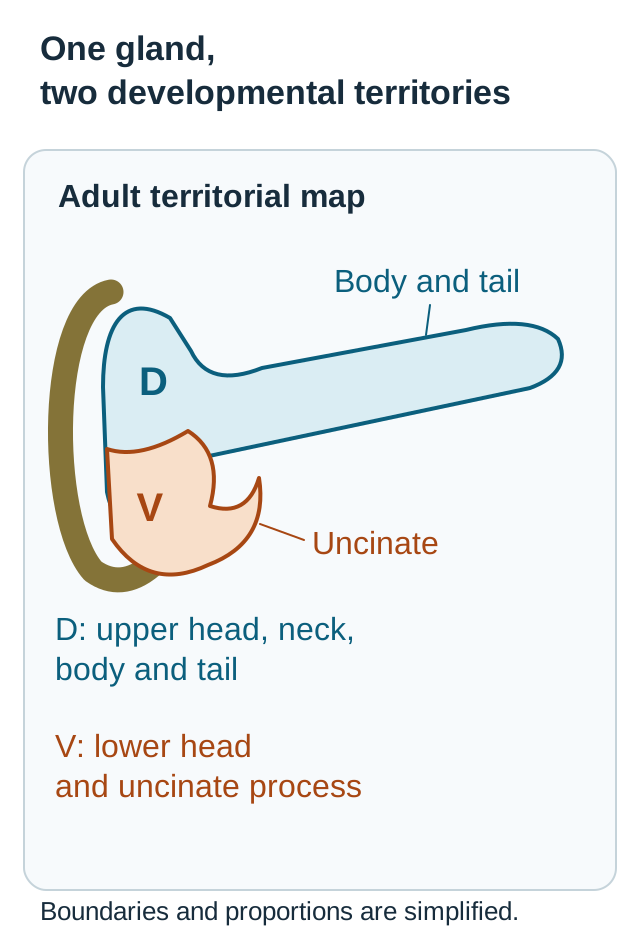
The ventral bud contributes the uncinate process and the inferior or posterior pancreatic head. The dorsal bud contributes the upper or anterior head, neck, body and tail. The head is therefore composite. Saying that the entire head is ventral discards a useful anatomical distinction. The boundary is a developmental map, not a sharp surgical plane visible on every scan. [2]
The uncinate process is the hook extending from the lower head behind the superior mesenteric vessels. A lesion described as pancreatic tissue posterior to the superior mesenteric artery and vein is localized first to that process. Only after that localization should its ventral origin be assigned. By contrast, a lesion extending from the neck through the body toward the splenic hilum occupies dorsal-derived tissue.
Use two landmarks: in the derivative figure, compare the hooked region beside the duodenum with the long body-tail region extending toward the spleen. Point to the territory remaining after a distal pancreatectomy. Much of the head, including the uncinate process, can remain even when much of the dorsal territory has been resected.
Compare the two labeled territories within the head before assigning a bud. The dorsal territory is larger, but this drawing is not a measured volume map or a surgical dissection plane. Both territories contain endocrine and exocrine tissue. [1][2]
These origins do not divide the gland into an endocrine bud and an exocrine bud. Islets, acini and ducts occur within pancreatic parenchyma. A substantial distal resection can reduce both glucose-regulating and digestive capacity; neither effect follows solely from the name of the bud. Similarly, dorsal agenesis can reduce endocrine reserve, but anatomy alone does not determine every patient's measured function. [1][2]
Compare two inflammatory foci: upper head and uncinate process.
The upper-head focus belongs to the dorsal contribution. The uncinate focus belongs to the ventral contribution. Both are in the head, so the word head alone is insufficient for this comparison.
Separate the splenic neighborhood from splenic ancestry
The spleen develops in mesenchyme on the left side of the dorsal mesogastrium, the mesentery associated with the developing stomach. It is a mesodermal organ, not an outgrowth of foregut epithelium. Direct human embryonic observations identify a single circumscribed splenic primordium in this region. The pancreatic epithelium and the splenic primordium therefore have different origins despite their close adult relationship. [1][3]
The celiac arterial system supplies the spleen through the splenic artery, but an arterial territory is not a germ-layer label. Likewise, finding pancreatic and splenic tissue beside one another does not establish a common epithelial ancestor. Keep identity, position and blood supply as separate observations.
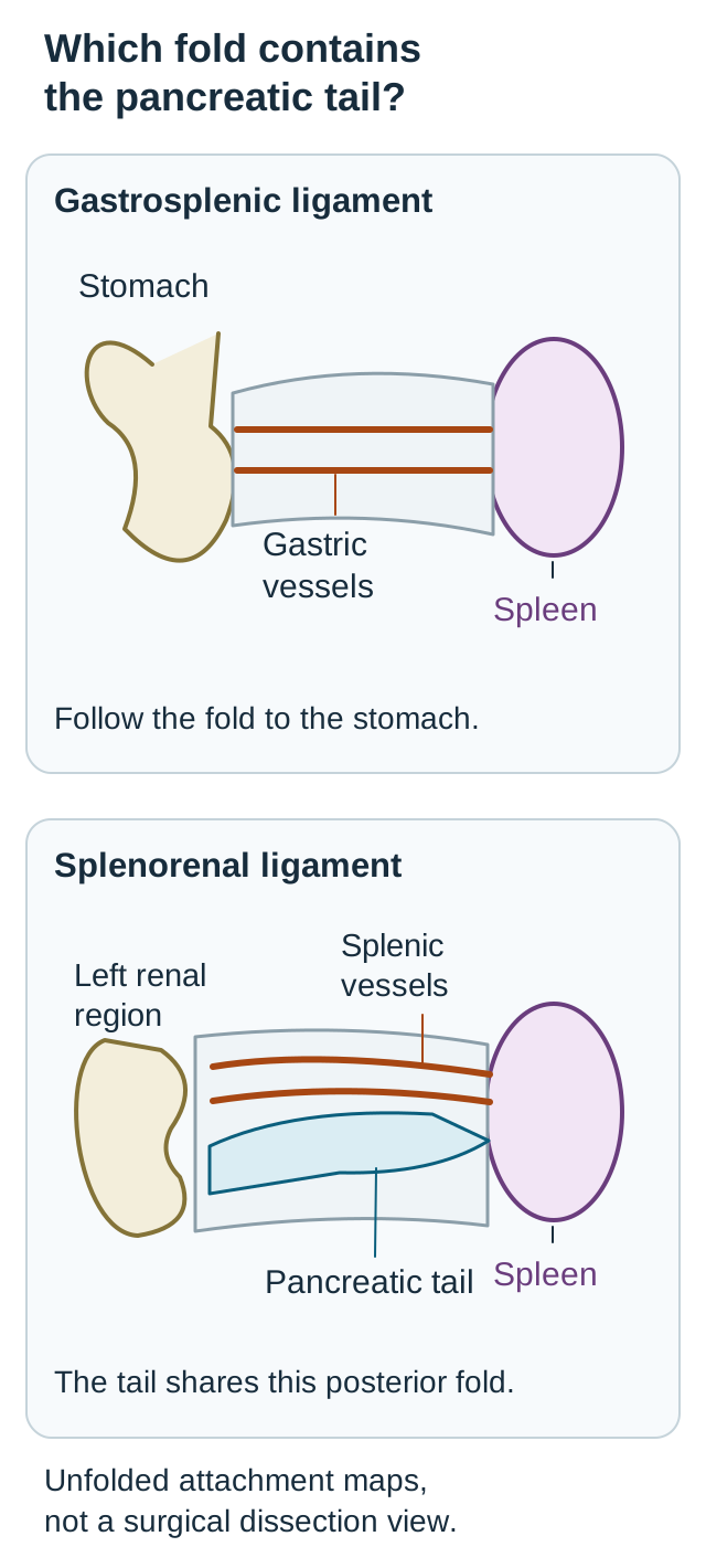
The spleen is intraperitoneal. Much of the pancreas becomes secondarily retroperitoneal as posterior peritoneal surfaces adhere during development. Its tail is the important exception in the usual anatomical description: it extends toward the splenic hilum within the splenorenal ligament, accompanying distal splenic vessels. The fold reaches the posterior abdominal wall near the anterior surface of the left kidney. The relationship explains why hilar dissection can injure pancreatic tissue. [4][5]
The gastrosplenic ligament instead connects the spleen to the greater curvature of the stomach and carries short gastric and left gastro-omental vessels. The names describe endpoints. A fold leading toward the stomach is not interchangeable with a fold leading toward the left renal region.
Trace both attachments: start at the spleen in the attachment figure. Follow the stomach-directed fold, then return and follow the posterior renal-directed fold. Only the second contains the pancreatic tail. If pancreatic tissue is injured there, an amylase-rich collection can develop near the splenic hilum. A vessel-only model would miss that risk. [5]
Trace each fold from the spleen to its other endpoint. The splenorenal attachment reaches the posterior abdominal wall near the anterior left kidney, not the renal collecting system. The two unfolded maps simplify three-dimensional peritoneal anatomy and vascular branching. [4][5]
If the normal supporting attachments are inadequate, an intact spleen may lie elsewhere on a long vascular pedicle. This is a wandering spleen, not necessarily an additional spleen. Its pedicle can twist, threatening perfusion. A newly painful displaced spleen with a twisted pedicle and reduced enhancement warrants urgent assessment for torsion. [4]
A pelvic spleen enhances normally. Does that eliminate future risk?
No. Current enhancement shows current perfusion. Inadequate fixation and a long pedicle can still permit a later torsion event. Compare anatomy with the patient's new symptoms rather than treating one earlier image as permanent reassurance.
Trace the outlet, not just the duct name
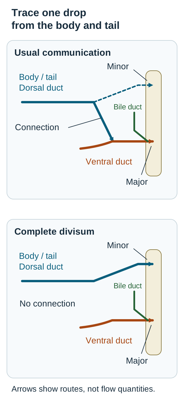
With the usual duct connection, secretion from the body and tail follows an upstream dorsal-derived duct segment into the ventral-derived outlet and reaches the major duodenal papilla. This composite route is the main pancreatic duct. A residual dorsal outlet may persist as the accessory duct of Santorini and reach the minor papilla. The major papilla also receives the common bile duct. Named ducts and their precise configurations vary; destination and demonstrated communication are more useful than memorizing an isolated name. [2]
In complete pancreas divisum, the dorsal and ventral duct systems do not communicate. The body, tail and dorsal portion of the head drain predominantly through the dorsal duct to the minor papilla. The uncinate and ventral head drain through the ventral duct to the major papilla. Both tissue territories are present. Incomplete variants have a small communication; a patent accessory outlet alone does not establish complete divisum. [2]
Change one connection: trace a drop of pancreatic secretion from the body in the upper duct diagram to the major papilla. Then cover the joining segment with a finger or imagine that it is absent. Trace the lower diagram instead. The dorsal stream now ends at the minor papilla, while the ventral stream still reaches the major papilla. The figure depicts routes, not measured flow rates or pressure.
Start at the body-tail duct in each map. Cover the upper joining segment mentally, then compare with the lower map: the principal dorsal stream now reaches the minor papilla. The dashed upper route is a possible patent accessory duct. Routes are simplified; line width and arrows do not measure secretion or pressure. [2]
This produces a testable prediction: in complete divisum, isolated minor-papilla narrowing directly burdens the dorsal route. It does not automatically obstruct the separate common bile duct. Conversely, two patent papillae can coexist with a normal communication, so two outlets do not prove nonfusion.
MRCP can evaluate the duct map without the procedural risks of diagnostic ERCP. Most people with divisum are asymptomatic. When pancreatitis is present, assess other causes rather than assigning causation from the congenital variant alone. Anatomical plausibility is not proof that opening a papilla improves outcomes. [2]
A 2026 sham-controlled randomized trial studied 148 adults with otherwise unexplained recurrent acute pancreatitis and divisum. Minor papillotomy did not significantly reduce recurrent pancreatitis after 30 days: the adjusted hazard ratio was 0.83, with a 95% confidence interval of 0.49 to 1.41. This does not support presenting routine papillotomy as proven prevention in the studied population. It also does not prove that no individual or narrowly defined subgroup could benefit. Decisions about intervention require specialist assessment of the complete clinical problem. [6]
What separates divisum from complete dorsal agenesis?
In divisum, the dorsal tissue and its long duct are present but follow a separate route. In complete dorsal agenesis, that territory and its duct are absent. Ask whether a route is disconnected or whether the tissue supplying it never developed.
Distinguish a tissue ring from a drainage variant
Annular pancreas is pancreatic tissue partly or completely surrounding the second duodenal segment, usually continuous with the head. It reflects abnormal ventral pancreatic development and positioning. Several developmental explanations exist, including abnormal attachment and persistence of ventral tissue; a simple bifid-bud story should not be presented as a directly proven sequence in every patient. The dependable observation is the pancreatic band around the bowel. [2][7]
The band may narrow the lumen and cause neonatal feeding intolerance or later postprandial fullness and vomiting. It can also be incidental. Trisomy 21 and other congenital abnormalities are associated with pediatric cases, but that association is not diagnostic. Vomiting may be bilious or nonbilious depending on the level and anatomy of the obstruction; absence of bile does not exclude an annular pancreas. [7]
Dilation of the stomach and proximal duodenum produces a double-bubble pattern. That pattern supports proximal obstruction, not a unique cause. Duodenal atresia, a web and other anatomical problems can produce a similar pattern. A newborn with concerning vomiting needs urgent evaluation rather than a diagnosis assigned from a mnemonic. Demonstrating surrounding pancreatic tissue distinguishes annular anatomy from an intraluminal membrane. [7]
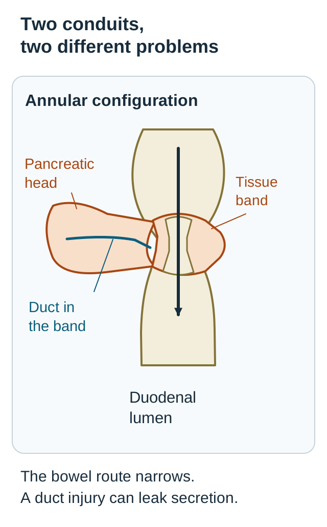
Compare the two routes through the ring figure: trace the bowel lumen vertically, then trace the pancreatic tissue surrounding it. Narrowing the first restricts intestinal passage. A disconnected pancreatic duct instead changes secretion drainage. They are different conduits, and either abnormality can exist without the other. A complete circular ring need not be visible on CT for incomplete annular tissue to cause narrowing. [2]
Trace intestinal passage through the central lumen, then locate the separate pancreatic duct. Annular tissue can obstruct the bowel and can contain ducts vulnerable to transection. This cutaway model is not an operative map; ring completeness, duct course and degree of narrowing vary. [2][7][9]
For symptomatic obstruction, surgery generally bypasses the narrowed segment rather than simply dividing the pancreatic band. Functioning ducts may traverse that band, and transection can cause a pancreatic leak or fistula. This is an anatomical reason for caution, not a procedural plan for an individual patient. [7][9]
Dorsal agenesis supplies a different comparison: a preserved uncinate region with an absent body, tail and dorsal duct favors failed dorsal development. Prior resection, acquired atrophy and fatty replacement must be considered before calling absent-appearing tissue congenital. Ectopic or heterotopic pancreas is another distinct finding: pancreatic tissue outside the native gland without anatomical or vascular continuity. A gastric antral nodule containing acini, ducts and islets fits that identity, but an imaging nodule without tissue-specific evidence has a broader differential. [2]
Identify extra splenic tissue before assigning its cause
An accessory spleen is congenitally separate splenic tissue. It is often near the splenic hilum and may lie beside or within the pancreatic tail. Its enhancement and MRI signal can match the spleen. When characterization remains necessary, uptake on a technetium-99m heat-damaged red-cell study supports functioning splenic tissue. A hypervascular pancreatic-tail nodule alone is not enough to distinguish an accessory spleen from a pancreatic neuroendocrine tumor. [4]
Compare like with like: in a multiphase study, compare the nodule with the native spleen on each matching phase, not with the pancreas on one image. Matching behavior across phases plus splenic-specific functional evidence is more informative than the single description bright nodule. Imaging findings must still be interpreted in their clinical setting.
The traditional explanation says that accessory spleens represent splenic buds that failed to fuse. It is widely repeated, but direct human observations challenge a normal multifocal origin: a 2023 study examined serial sections from 22 embryos and found a single splenic primordium. Alternative processes, such as separation during development, have been proposed but are not established for every accessory spleen. The useful clinical distinction is congenital separate tissue versus acquired implantation, not certainty about an unobserved fusion event. [3]
Splenosis is acquired implantation of splenic fragments after rupture or surgery. Nodules may be numerous and obtain blood from local vessels. A combined splenic and left diaphragmatic injury can permit implants on the left pleural surface. By contrast, accessory spleens commonly have a splenic arterial branch and may be visible on images obtained before any trauma or surgery. A tissue-specific scan identifies splenic function; history and anatomy help determine how the tissue arrived. [4]
After splenectomy for an immune cytopenia, residual functioning splenic tissue can contribute to recurrence. Human splenic macrophages clear antibody-coated blood cells. Thus, a tracer-positive nodule that predates surgery can be clinically relevant rather than merely an incidental shape variant. It does not establish the only cause of a recurrent low platelet count or automatically justify another operation. [4][8]
Two patients have splenic nodules. Which history changes the interpretation?
A single tail nodule documented before surgery supports an accessory spleen. Numerous peritoneal nodules after traumatic splenic rupture support splenosis. Both can function as splenic tissue, but their origins differ.
Return to the three opening questions: identify the tissue, reconstruct its position, and trace its outlet or blood supply. This separates the pancreatic ring from the disconnected ducts and distinguishes a congenital splenic neighbor from a later implant.
Apply the anatomy
Case 1
Show answer and explanations for case 1
A. Dorsal pancreatic bud (Why this does not fit)
The dorsal bud supplies the upper head, neck, body and tail. The hook behind those vessels is the uncinate process, a ventral derivative rather than the dorsal body or tail.
Reasoning steps for option A
Which pancreatic regions arise from the dorsal bud?
The dorsal bud supplies the upper head, neck, body and tail.
How does the relationship to the mesenteric vessels localize this lesion?
The hook behind those vessels is the uncinate process, a ventral derivative rather than the dorsal body or tail.
B. Dorsal mesogastric mesenchyme (Why this does not fit)
The splenic primordium forms in this mesenchyme. It is continuous with the lower pancreatic head behind the mesenteric vessels, not the splenic hilum.
Reasoning steps for option B
Which nearby organ forms within dorsal mesogastric mesenchyme?
The splenic primordium forms in this mesenchyme.
Does the described lesion occupy the splenic region?
It is continuous with the lower pancreatic head behind the mesenteric vessels, not the splenic hilum.
C. Ventral pancreatic bud (Best answer)
The uncinate process extends from the lower head behind these vessels. The ventral bud contributes the uncinate process and inferior or posterior part of the head.
Reasoning steps for option C
Which part of the gland lies behind the superior mesenteric vessels?
The uncinate process extends from the lower head behind these vessels.
Which bud contributes this process?
The ventral bud contributes the uncinate process and inferior or posterior part of the head.
D. Ventral mesogastric mesenchyme (Why this does not fit)
The lesser omentum and falciform ligament are ventral mesogastric derivatives. A lesion within the pancreatic uncinate process is localized to pancreatic glandular tissue rather than those peritoneal folds.
Reasoning steps for option D
What adult peritoneal structures derive from the ventral mesogastrium?
The lesser omentum and falciform ligament are ventral mesogastric derivatives.
Why does that not explain the lesion?
A lesion within the pancreatic uncinate process is localized to pancreatic glandular tissue rather than those peritoneal folds.
Takeaway: First identify the uncinate process by its vascular relationship, then assign the ventral bud. [1] [2]
A. Failure of dorsal pancreatic development (Best answer)
The neck, body, tail and their ductal system arise from the dorsal bud. It favors dorsal agenesis rather than a drainage variant with preserved tissue.
Reasoning steps for option A
Which absent structures normally share one pancreatic bud?
The neck, body, tail and their ductal system arise from the dorsal bud.
What does simultaneous absence of parenchyma and its duct favor?
It favors dorsal agenesis rather than a drainage variant with preserved tissue.
B. Failure of dorsal and ventral duct fusion (Why this does not fit)
Most pancreatic tissue remains present in pancreas divisum. The dorsal parenchyma and its duct are absent, not merely disconnected from the ventral duct.
Reasoning steps for option B
What is preserved in a typical duct-fusion defect?
Most pancreatic tissue remains present in pancreas divisum.
Why is divisum insufficient here?
The dorsal parenchyma and its duct are absent, not merely disconnected from the ventral duct.
C. Abnormal ventral tissue around the duodenum (Why this does not fit)
Pancreatic tissue partly or completely surrounds the second duodenal segment. No ring is described, and the missing dorsal territory requires a different developmental defect.
Reasoning steps for option C
What structural finding characterizes annular pancreas?
Pancreatic tissue partly or completely surrounds the second duodenal segment.
Does a ring explain the absent body and tail?
No ring is described, and the missing dorsal territory requires a different developmental defect.
D. Failure of ventral pancreatic development (Why this does not fit)
It would affect the uncinate process and inferior or posterior head. The uncinate process and inferior head are preserved while the dorsal territory is absent.
Reasoning steps for option D
Which territory would ventral agenesis affect most directly?
It would affect the uncinate process and inferior or posterior head.
Which supplied finding argues against that event?
The uncinate process and inferior head are preserved while the dorsal territory is absent.
Takeaway: Missing tissue plus a missing corresponding duct is different from two ducts that fail to connect. [2]
A. Dorsal duct throughout its entire course (Why this does not fit)
The body and tail contribute the upstream dorsal duct segment. The final pancreatic segment is ventral, so the whole route is not exclusively dorsal.
Reasoning steps for option A
Which part of the main route is dorsal in origin?
The body and tail contribute the upstream dorsal duct segment.
What is the origin of the final outlet to the major papilla?
The final pancreatic segment is ventral, so the whole route is not exclusively dorsal.
B. Ventral duct throughout its entire course (Why this does not fit)
The ventral duct provides the final outlet toward the major papilla. Those territories are dorsal derivatives and supply an upstream dorsal segment.
Reasoning steps for option B
Which part of the main route is ventral in origin?
The ventral duct provides the final outlet toward the major papilla.
Can that origin explain the duct in the body and tail?
Those territories are dorsal derivatives and supply an upstream dorsal segment.
C. Ventral duct plus the dorsal minor-papilla outlet (Why this does not fit)
The proximal dorsal remnant can reach the minor papilla as an accessory route. It ends at the major papilla, requiring the upstream body-tail segment rather than the minor-papilla remnant.
Reasoning steps for option C
Where does the residual dorsal outlet lead?
The proximal dorsal remnant can reach the minor papilla as an accessory route.
Which papilla terminates the route actually described?
It ends at the major papilla, requiring the upstream body-tail segment rather than the minor-papilla remnant.
D. Upstream dorsal duct plus the ventral outlet (Best answer)
That upstream portion is derived from the dorsal duct. The connected ventral outlet completes the principal pancreatic drainage route.
Reasoning steps for option D
Which embryonic duct drains the body and tail before the connection?
That upstream portion is derived from the dorsal duct.
What carries its secretion to the major papilla after normal fusion?
The connected ventral outlet completes the principal pancreatic drainage route.
Takeaway: Name duct segments by their course and destination rather than treating the entire main duct as one bud. [2]
A. Persistence of a communicating accessory duct outlet (Why this does not fit)
Yes. A minor-papilla route may remain patent after dorsal and ventral duct communication is established. The long dorsal duct has no connection to the separate short ventral duct, which favors complete divisum rather than a communicating accessory route.
Reasoning steps for option A
Can an accessory dorsal outlet remain after the ducts connect?
Yes. A minor-papilla route may remain patent after dorsal and ventral duct communication is established.
Which finding requires a different explanation in this patient?
The long dorsal duct has no connection to the separate short ventral duct, which favors complete divisum rather than a communicating accessory route.
B. Failure of dorsal and ventral ducts to unite (Best answer)
The long dorsal duct carries body and tail secretion to the minor papilla. The ventral duct remains separate when the duct systems do not unite, producing complete divisum.
Reasoning steps for option B
Which route drains most of this gland?
The long dorsal duct carries body and tail secretion to the minor papilla.
Why does the uncinate region have a separate major-papilla route?
The ventral duct remains separate when the duct systems do not unite, producing complete divisum.
C. Failure of dorsal glandular tissue to develop (Why this does not fit)
The dorsal tissue and its long duct are absent. A long duct traversing a present body and tail is directly demonstrated.
Reasoning steps for option C
What duct pattern accompanies complete dorsal agenesis?
The dorsal tissue and its long duct are absent.
Which observation excludes that explanation here?
A long duct traversing a present body and tail is directly demonstrated.
D. Persistence of pancreatic tissue around the duodenum (Why this does not fit)
A band of pancreatic tissue around the duodenum would establish annular anatomy. The described abnormality is separation of duct drainage without a surrounding tissue band.
Reasoning steps for option D
What would establish an annular configuration?
A band of pancreatic tissue around the duodenum would establish annular anatomy.
What is demonstrated instead?
The described abnormality is separation of duct drainage without a surrounding tissue band.
Takeaway: In complete divisum, most pancreatic secretion exits through the minor papilla while the ventral region retains its major-papilla route. [2]
A. Uncinate region with the distal bile duct (Why this does not fit)
Both reach the major papilla through their respective ducts. The major papilla is patent, so the isolated minor scar does not directly obstruct these routes.
Reasoning steps for option A
Which outlet serves the uncinate region and bile duct?
Both reach the major papilla through their respective ducts.
Is that outlet narrowed in this case?
The major papilla is patent, so the isolated minor scar does not directly obstruct these routes.
B. Entire gland with the distal bile duct (Why this does not fit)
A major-papilla obstruction can affect connected drainage pathways. Complete divisum separates the dominant dorsal route from the patent ventral and biliary outlet.
Reasoning steps for option B
When could a shared outlet affect pancreatic and biliary drainage?
A major-papilla obstruction can affect connected drainage pathways.
Why is that pattern not predicted here?
Complete divisum separates the dominant dorsal route from the patent ventral and biliary outlet.
C. Body, tail and the dorsal part of the head (Best answer)
Most dorsal tissue, including the body, tail and upper head, drains through that outlet. Resistance increases along the dorsal route without directly blocking the separate ventral or biliary route.
Reasoning steps for option C
Which pancreatic tissue drains through the minor papilla in complete divisum?
Most dorsal tissue, including the body, tail and upper head, drains through that outlet.
What follows from narrowing that outlet alone?
Resistance increases along the dorsal route without directly blocking the separate ventral or biliary route.
D. Inferior head with the uncinate process (Why this does not fit)
The ventral bud supplies the inferior head and uncinate process. It drains through the patent major papilla, not the narrowed minor papilla.
Reasoning steps for option D
Which bud supplies this paired region?
The ventral bud supplies the inferior head and uncinate process.
Where does that bud drain in complete divisum?
It drains through the patent major papilla, not the narrowed minor papilla.
Takeaway: Predict the affected tissue from the obstructed outlet and the actual duct connections. [2]
A. Divisum establishes causation and the procedure has proven benefit (Why this does not fit)
It establishes a ductal configuration, not the cause of every pancreatitis episode. No. The confidence interval includes no difference, and the trial did not demonstrate a significant reduction.
Reasoning steps for option A
What does the MRCP establish?
It establishes a ductal configuration, not the cause of every pancreatitis episode.
Does the trial establish preventive benefit?
No. The confidence interval includes no difference, and the trial did not demonstrate a significant reduction.
B. Divisum defines drainage but routine preventive benefit was not demonstrated (Best answer)
It identifies predominantly dorsal drainage through the minor papilla. It did not demonstrate that routine minor papillotomy prevents recurrent attacks in the studied adults; anatomy alone cannot supply that missing benefit.
Reasoning steps for option B
What question does the anatomical diagnosis answer?
It identifies predominantly dorsal drainage through the minor papilla.
What additional question did the randomized comparison answer?
It did not demonstrate that routine minor papillotomy prevents recurrent attacks in the studied adults; anatomy alone cannot supply that missing benefit.
C. Divisum excludes other causes and the procedure has uncertain benefit (Why this does not fit)
No. Other causes can coexist with a congenital drainage variant. The treatment result is uncertain, but excluding all other causes simply because divisum is present is unjustified.
Reasoning steps for option C
Does finding divisum rule out another pancreatitis cause?
No. Other causes can coexist with a congenital drainage variant.
Which part of this interpretation conflicts with the evidence?
The treatment result is uncertain, but excluding all other causes simply because divisum is present is unjustified.
D. Divisum defines drainage and benefit is excluded in every subgroup (Why this does not fit)
It did not demonstrate the proposed benefit within the precision of the trial. No. Failure to demonstrate an overall benefit is not proof of zero effect in every possible subgroup.
Reasoning steps for option D
What does a nonsignificant result say about the overall studied comparison?
It did not demonstrate the proposed benefit within the precision of the trial.
Can it prove that every individual or narrowly defined subgroup has zero benefit?
No. Failure to demonstrate an overall benefit is not proof of zero effect in every possible subgroup.
Takeaway: An anatomical explanation and evidence that an intervention improves outcomes are separate requirements. [6]
A. The dorsal duct is obstructed because all pancreatic secretion uses the major papilla (Why this does not fit)
Most secretion exits at the minor papilla through the dorsal duct. The dorsal outlet is patent and its duct is not dilated; the biliary obstruction is elsewhere.
Reasoning steps for option A
Where does most secretion exit in complete divisum?
Most secretion exits at the minor papilla through the dorsal duct.
Which finding opposes direct obstruction of that route here?
The dorsal outlet is patent and its duct is not dilated; the biliary obstruction is elsewhere.
B. The ductal variant proves that the biliary stone is incidental (Why this does not fit)
Yes. Identifying one does not invalidate direct evidence of the other. The lodged stone and upstream dilation demonstrate a genuine biliary obstruction requiring clinical assessment.
Reasoning steps for option B
Can a congenital variant and an acquired biliary problem coexist?
Yes. Identifying one does not invalidate direct evidence of the other.
What makes dismissal of the stone inappropriate?
The lodged stone and upstream dilation demonstrate a genuine biliary obstruction requiring clinical assessment.
C. The biliary dilation establishes failure of dorsal pancreatic development (Why this does not fit)
Absence of dorsal pancreatic tissue and its duct supports that diagnosis. No. A long dorsal duct with a patent minor-papilla outlet is demonstrated.
Reasoning steps for option C
What anatomical evidence supports dorsal agenesis?
Absence of dorsal pancreatic tissue and its duct supports that diagnosis.
Is the dorsal system absent in this patient?
No. A long dorsal duct with a patent minor-papilla outlet is demonstrated.
D. The biliary obstruction is real, while the dorsal outflow remains separate (Best answer)
There is distal biliary obstruction. The dominant dorsal route remains anatomically separate, so divisum alone should not displace evaluation of the documented acquired abnormality.
Reasoning steps for option D
Which abnormality is directly demonstrated by the stone and dilation?
There is distal biliary obstruction.
How does complete divisum change interpretation of the pancreatic duct map?
The dominant dorsal route remains anatomically separate, so divisum alone should not displace evaluation of the documented acquired abnormality.
Takeaway: Interpret each demonstrated route and abnormality; do not assign every episode to an incidental developmental variant. [2] [6]
A. Abnormal ventral pancreatic development around the duodenum (Best answer)
Pancreatic tissue continuous with the head surrounds the second duodenal segment. Abnormal ventral pancreatic development and positioning can create the band that constricts the bowel; a specific embryonic theory need not be assumed for this patient.
Reasoning steps for option A
Which operative finding distinguishes annular pancreas from other proximal obstructions?
Pancreatic tissue continuous with the head surrounds the second duodenal segment.
How is this related to development?
Abnormal ventral pancreatic development and positioning can create the band that constricts the bowel; a specific embryonic theory need not be assumed for this patient.
B. Lack of communication between the two pancreatic ducts (Why this does not fit)
It produces divisum with separate drainage outlets. Duct separation alone does not create pancreatic tissue surrounding and narrowing the duodenum.
Reasoning steps for option B
What does failure of duct communication produce?
It produces divisum with separate drainage outlets.
Why does that not explain the operative finding?
Duct separation alone does not create pancreatic tissue surrounding and narrowing the duodenum.
C. Failure of the pancreatic body and tail to develop (Why this does not fit)
The body and tail and their ductal structures are partly or wholly absent. The obstructing tissue is a surrounding band continuous with the pancreatic head.
Reasoning steps for option C
What is the characteristic consequence of dorsal agenesis?
The body and tail and their ductal structures are partly or wholly absent.
Which operative observation requires a different explanation?
The obstructing tissue is a surrounding band continuous with the pancreatic head.
D. Persistence of an intraluminal duodenal membrane (Why this does not fit)
A web can impede passage within the bowel lumen and cause proximal dilation. The operation identifies an external pancreatic band, not an intraluminal membrane.
Reasoning steps for option D
How could a duodenal membrane cause similar symptoms?
A web can impede passage within the bowel lumen and cause proximal dilation.
What directly contradicts a web as the supplied explanation?
The operation identifies an external pancreatic band, not an intraluminal membrane.
Takeaway: The surrounding pancreatic tissue, not trisomy 21 or a double-bubble pattern alone, establishes annular anatomy. [2] [7]
A. Division could interrupt duodenal arterial branches (Why this does not fit)
Yes. The pancreatic head and duodenum have closely related arterial branches. The identified pancreatic duct supplies a direct route for pancreatic leakage if transected; an arterial interruption is not the demonstrated finding.
Reasoning steps for option A
Can surgery near the pancreatic head endanger blood supply to the duodenum?
Yes. The pancreatic head and duodenum have closely related arterial branches.
Which structure specifically demonstrated within the band most directly determines the stated concern?
The identified pancreatic duct supplies a direct route for pancreatic leakage if transected; an arterial interruption is not the demonstrated finding.
B. Division could disrupt pancreatic ducts and cause a leak (Best answer)
The band contains pancreatic tissue and a branch of the pancreatic duct system. Pancreatic secretion can escape through the disrupted duct, producing a leak or fistula; bypass avoids assuming the band is safe to divide.
Reasoning steps for option B
What is contained within the obstructing band?
The band contains pancreatic tissue and a branch of the pancreatic duct system.
What consequence follows if that duct is cut?
Pancreatic secretion can escape through the disrupted duct, producing a leak or fistula; bypass avoids assuming the band is safe to divide.
C. Division could injure the adjacent common bile duct (Why this does not fit)
The distal common bile duct passes near the pancreatic head and enters the duodenum. The supplied imaging identifies a pancreatic duct in the band, making pancreatic leakage the directly supported consequence.
Reasoning steps for option C
Why is bile-duct injury an anatomically plausible concern in this region?
The distal common bile duct passes near the pancreatic head and enters the duodenum.
Is the duct identified inside the band biliary or pancreatic?
The supplied imaging identifies a pancreatic duct in the band, making pancreatic leakage the directly supported consequence.
D. Division could create a full-thickness duodenal defect (Why this does not fit)
The band lies closely around the duodenum, and wall injury could cause an intestinal leak. The demonstrated pancreatic duct would leak pancreatic secretion if divided, independently of whether the bowel wall remains intact.
Reasoning steps for option D
Why must the duodenal wall be protected during dissection?
The band lies closely around the duodenum, and wall injury could cause an intestinal leak.
Which specific tissue pathway supports the answer here?
The demonstrated pancreatic duct would leak pancreatic secretion if divided, independently of whether the bowel wall remains intact.
Takeaway: A bowel-obstructing band may contain active pancreatic ducts; relieving obstruction is not the same as safely dividing pancreatic tissue. [2] [9]
A. Dorsal agenesis accounts for the narrowed duodenum (Why this does not fit)
The neck, body and tail should be absent along with the dorsal duct system. The body and tail are present, and the abnormal tissue is beside the duodenum.
Reasoning steps for option A
What tissue should be missing in complete dorsal agenesis?
The neck, body and tail should be absent along with the dorsal duct system.
Which supplied finding makes that interpretation unsuitable?
The body and tail are present, and the abnormal tissue is beside the duodenum.
B. A complete ring must be visible before considering annular pancreas (Why this does not fit)
Yes. Some cases do not show a complete ring on cross-sectional imaging. The anterior and posterior continuity with pancreatic tissue supports annular anatomy even without a fully visible circle.
Reasoning steps for option B
Can annular tissue be incomplete or partly incorporated into the duodenal wall?
Yes. Some cases do not show a complete ring on cross-sectional imaging.
How should the demonstrated tissue and narrowing be interpreted?
The anterior and posterior continuity with pancreatic tissue supports annular anatomy even without a fully visible circle.
C. Pancreas divisum is established by the tissue configuration (Why this does not fit)
The ductal connections and their papillary outlets must be assessed. No. It describes parenchymal configuration and luminal narrowing rather than duct nonfusion.
Reasoning steps for option C
What information is needed to establish divisum?
The ductal connections and their papillary outlets must be assessed.
Does pancreatic tissue around D2 provide that information?
No. It describes parenchymal configuration and luminal narrowing rather than duct nonfusion.
D. An incomplete annular configuration can explain the obstruction (Best answer)
Pancreatic tissue borders both sides of the narrowed duodenal segment and is continuous with the head. No. Incomplete annular tissue or a subtle intramural component can produce this configuration and obstruction.
Reasoning steps for option D
Which two observations connect pancreatic tissue to the symptoms?
Pancreatic tissue borders both sides of the narrowed duodenal segment and is continuous with the head.
Must a complete visible circle be present?
No. Incomplete annular tissue or a subtle intramural component can produce this configuration and obstruction.
Takeaway: Annular pancreas can be incomplete; assess pancreatic continuity and duodenal narrowing rather than demanding a perfect circle. [2]
A. Duodenal web, then annular pancreas (Best answer)
A membrane inside the lumen with normal external pancreas supports a duodenal web. A pancreatic band around the second segment supports annular pancreas, despite the similar proximal dilation.
Reasoning steps for option A
Which finding identifies the obstruction in patient A?
A membrane inside the lumen with normal external pancreas supports a duodenal web.
Which different structure narrows patient B's duodenum?
A pancreatic band around the second segment supports annular pancreas, despite the similar proximal dilation.
B. Annular pancreas, then duodenal web (Why this does not fit)
It surrounds the duodenum rather than forming only an intraluminal membrane. No. It reverses the internal membrane in A and the external pancreatic band in B.
Reasoning steps for option B
Where would annular tissue be expected?
It surrounds the duodenum rather than forming only an intraluminal membrane.
Does this proposed order match the two operative findings?
No. It reverses the internal membrane in A and the external pancreatic band in B.
C. Duodenal web, then complete divisum (Why this does not fit)
The operation identifies an intraluminal membrane. No. Complete divisum concerns separate drainage routes rather than pancreatic tissue encircling the bowel.
Reasoning steps for option C
Why does a web fit patient A?
The operation identifies an intraluminal membrane.
Can duct nonfusion alone explain patient B's band?
No. Complete divisum concerns separate drainage routes rather than pancreatic tissue encircling the bowel.
D. Complete divisum, then annular pancreas (Why this does not fit)
Pancreatic tissue continuous with the head surrounds the narrowed duodenum. The obstructing lesion in A is a bowel membrane, not a demonstrated pancreatic duct arrangement.
Reasoning steps for option D
Why does annular pancreas fit patient B?
Pancreatic tissue continuous with the head surrounds the narrowed duodenum.
Why does divisum not explain patient A?
The obstructing lesion in A is a bowel membrane, not a demonstrated pancreatic duct arrangement.
Takeaway: A characteristic obstruction pattern localizes a problem but does not identify the surrounding tissue. [2] [7]
A. A congenital splenic nodule within the gastric wall (Why this does not fit)
Splenic architecture or tissue-specific splenic uptake would support that diagnosis. Pancreatic acini, ducts and islets identify pancreatic rather than splenic tissue.
Reasoning steps for option A
What tissue would support an accessory spleen?
Splenic architecture or tissue-specific splenic uptake would support that diagnosis.
What does the sample actually contain?
Pancreatic acini, ducts and islets identify pancreatic rather than splenic tissue.
B. An accessory lobe extending from the main pancreas (Why this does not fit)
It has a connection with the main pancreatic gland and a ductal relationship. The nodule has no anatomical or vascular continuity with the native pancreas.
Reasoning steps for option B
What feature defines an accessory pancreatic lobe?
It has a connection with the main pancreatic gland and a ductal relationship.
Which finding argues against that configuration?
The nodule has no anatomical or vascular continuity with the native pancreas.
C. Heterotopic pancreatic tissue separate from the main gland (Best answer)
Acini, ducts and islets identify pancreatic tissue. It occupies the gastric wall without continuity with the native pancreas or its vessels.
Reasoning steps for option C
What establishes the type of tissue in the nodule?
Acini, ducts and islets identify pancreatic tissue.
Why is it called heterotopic rather than an accessory lobe?
It occupies the gastric wall without continuity with the native pancreas or its vessels.
D. An annular extension around the second duodenal segment (Why this does not fit)
Annular tissue is continuous with the pancreatic head around the duodenum. A separate gastric antral nodule does not have that duodenal configuration.
Reasoning steps for option D
What location and continuity characterize an annular extension?
Annular tissue is continuous with the pancreatic head around the duodenum.
Do the nodule location and lack of continuity fit?
A separate gastric antral nodule does not have that duodenal configuration.
Takeaway: Identify the tissue first, then use anatomical continuity to distinguish an accessory lobe from heterotopic pancreas. [1] [2]
A. Short gastric vessels within the gastrosplenic ligament (Why this does not fit)
A vascular injury can cause bleeding or impair gastric perfusion. An amylase-rich collection near the hilum after dissection near the kidney suggests pancreatic injury rather than isolated short gastric vessel injury.
Reasoning steps for option A
Which complication would an injured gastric vessel most directly produce?
A vascular injury can cause bleeding or impair gastric perfusion.
Why is it less explanatory for this collection?
An amylase-rich collection near the hilum after dissection near the kidney suggests pancreatic injury rather than isolated short gastric vessel injury.
B. Uncinate process within the duodenal curve (Why this does not fit)
It extends from the pancreatic head behind the superior mesenteric vessels. No. The operative field is near the pancreatic tail, not the uncinate process.
Reasoning steps for option B
Where is the uncinate process located?
It extends from the pancreatic head behind the superior mesenteric vessels.
Does that match the left-sided hilar dissection?
No. The operative field is near the pancreatic tail, not the uncinate process.
C. Pancreatic tail within the splenorenal ligament (Best answer)
The tail travels with distal splenic vessels in the splenorenal ligament. Disruption of pancreatic tissue or its duct can cause an amylase-rich leak into the nearby operative bed.
Reasoning steps for option C
Which pancreatic part reaches the splenic hilum in the named fold?
The tail travels with distal splenic vessels in the splenorenal ligament.
How does injury there explain the laboratory finding?
Disruption of pancreatic tissue or its duct can cause an amylase-rich leak into the nearby operative bed.
D. Common bile duct within the hepatoduodenal ligament (Why this does not fit)
It runs in the hepatoduodenal ligament toward the pancreatic head. The operative location implicates the tail rather than the distant bile duct route.
Reasoning steps for option D
Where is the common bile duct carried before entering the pancreas?
It runs in the hepatoduodenal ligament toward the pancreatic head.
Does that location explain this left hilar complication?
The operative location implicates the tail rather than the distant bile duct route.
Takeaway: The splenorenal ligament contains more than vessels: the pancreatic tail can be injured during hilar dissection. [5]
A. Gastric vessels in the first; pancreatic tail and splenic vessels in the second (Best answer)
The gastrosplenic ligament carries short gastric and left gastro-omental vessels. The splenorenal ligament contains the pancreatic tail and distal splenic vessels.
Reasoning steps for option A
Which fold links the spleen to the greater curvature?
The gastrosplenic ligament carries short gastric and left gastro-omental vessels.
What does the kidney-directed fold contain?
The splenorenal ligament contains the pancreatic tail and distal splenic vessels.
B. Pancreatic tail in the first; common bile duct in the second (Why this does not fit)
The tail accompanies distal splenic vessels within the splenorenal ligament. It assigns the tail to the stomach-directed fold and the bile duct to a left renal attachment, neither of which matches normal anatomy.
Reasoning steps for option B
What normally accompanies the tail at the splenic hilum?
The tail accompanies distal splenic vessels within the splenorenal ligament.
Why is this allocation incorrect?
It assigns the tail to the stomach-directed fold and the bile duct to a left renal attachment, neither of which matches normal anatomy.
C. Common bile duct in the first; gastric vessels in the second (Why this does not fit)
The hepatoduodenal ligament carries the extrahepatic bile duct toward the duodenum. Neither connects the liver to the duodenum, so the common bile duct does not belong in this allocation.
Reasoning steps for option C
Which fold carries the common bile duct?
The hepatoduodenal ligament carries the extrahepatic bile duct toward the duodenum.
Do either of the two splenic attachments match that course?
Neither connects the liver to the duodenum, so the common bile duct does not belong in this allocation.
D. Splenic vessels in the first; pancreatic head in the second (Why this does not fit)
The head lies within the duodenal curve on the right side of the gland. The tail, not the head, reaches that attachment with the distal splenic vessels.
Reasoning steps for option D
Where is the pancreatic head positioned?
The head lies within the duodenal curve on the right side of the gland.
Which pancreatic part reaches the left renal-directed splenic attachment?
The tail, not the head, reaches that attachment with the distal splenic vessels.
Takeaway: Identify each fold by its endpoints before assigning vessels or pancreatic tissue. [4] [5]
A. Both glandular epithelia originate in foregut endoderm (Why this does not fit)
It is derived from foregut endoderm. The spleen is a mesodermal organ developing in mesogastric mesenchyme, not a digestive epithelial bud.
Reasoning steps for option A
What germ layer gives rise to pancreatic glandular epithelium?
It is derived from foregut endoderm.
Why does shared arterial supply not extend that answer to splenic tissue?
The spleen is a mesodermal organ developing in mesogastric mesenchyme, not a digestive epithelial bud.
B. Pancreatic epithelium is endodermal; splenic tissue is mesodermal (Best answer)
Ducts, acini and islets identify pancreatic glandular tissue. It develops as a mesenchymal splenic primordium in the dorsal mesogastrium; celiac supply does not change its mesodermal origin.
Reasoning steps for option B
What identifies the endodermal gland in the sample?
Ducts, acini and islets identify pancreatic glandular tissue.
How is the neighboring splenic tissue different?
It develops as a mesenchymal splenic primordium in the dorsal mesogastrium; celiac supply does not change its mesodermal origin.
C. Pancreatic epithelium is mesodermal; splenic tissue is endodermal (Why this does not fit)
No. Pancreatic endocrine and exocrine epithelial lineages both arise from pancreatic endoderm. No. It forms in mesogastric mesenchyme, so this comparison reverses both assignments.
Reasoning steps for option C
Does pancreatic endocrine function imply mesodermal origin?
No. Pancreatic endocrine and exocrine epithelial lineages both arise from pancreatic endoderm.
Is the spleen a foregut epithelial outgrowth?
No. It forms in mesogastric mesenchyme, so this comparison reverses both assignments.
D. Both tissues originate in the neural crest (Why this does not fit)
The pancreatic glandular epithelium is endodermal rather than neural crest-derived. No. Its primordium is mesenchymal and mesodermal; nearby autonomic nerves do not determine organ lineage.
Reasoning steps for option D
Which embryonic lineage supplies the pancreatic glandular epithelium?
The pancreatic glandular epithelium is endodermal rather than neural crest-derived.
Does the spleen share a neural crest epithelial origin?
No. Its primordium is mesenchymal and mesodermal; nearby autonomic nerves do not determine organ lineage.
Takeaway: Position, arterial territory and germ layer answer different anatomical questions. [1] [3]
A. Pancreatic neuroendocrine tumor (Why this does not fit)
It can be a vascular pancreatic nodule. Matching the spleen across phases plus uptake of heat-damaged red cells supports splenic tissue rather than relying on vascularity alone.
Reasoning steps for option A
Why might a neuroendocrine tumor initially be considered?
It can be a vascular pancreatic nodule.
Which paired findings favor a different tissue identity?
Matching the spleen across phases plus uptake of heat-damaged red cells supports splenic tissue rather than relying on vascularity alone.
B. Heterotopic pancreatic tissue (Why this does not fit)
Pancreatic tissue would lie outside the normal gland without continuity. The uptake pattern identifies functioning splenic tissue, not pancreatic acini or ducts.
Reasoning steps for option B
What would characterize heterotopic pancreas?
Pancreatic tissue would lie outside the normal gland without continuity.
What does the functional study identify in this nodule?
The uptake pattern identifies functioning splenic tissue, not pancreatic acini or ducts.
C. Reactive peripancreatic lymph node (Why this does not fit)
A rounded nodule near the pancreas can represent lymphatic tissue. Concordance with spleen on multiphase imaging and the red-cell study strongly supports splenic rather than nodal tissue.
Reasoning steps for option C
Why might a nearby lymph node be in the initial differential?
A rounded nodule near the pancreas can represent lymphatic tissue.
Why does that not best fit the full assessment?
Concordance with spleen on multiphase imaging and the red-cell study strongly supports splenic rather than nodal tissue.
D. Intrapancreatic accessory spleen (Best answer)
Congenitally separate splenic tissue can occur in or adjacent to the tail near the splenic hilum. Spleen-matching signal and enhancement together with heat-damaged red-cell uptake support an accessory spleen.
Reasoning steps for option D
How can splenic tissue appear within the pancreatic tail?
Congenitally separate splenic tissue can occur in or adjacent to the tail near the splenic hilum.
Which evidence supports that tissue identity here?
Spleen-matching signal and enhancement together with heat-damaged red-cell uptake support an accessory spleen.
Takeaway: A vascular tail nodule is not automatically a pancreatic tumor; compare it with the spleen and use tissue-specific evidence. [4]
A. Persistent accessory spleen clears antibody-coated platelets (Best answer)
It favors a congenital accessory spleen rather than a new operative implant. Splenic macrophages can clear antibody-coated blood cells; persistent splenic function can contribute to recurrence without excluding other causes.
Reasoning steps for option A
What does the nodule predating surgery suggest about its origin?
It favors a congenital accessory spleen rather than a new operative implant.
How could functioning tissue contribute to recurrent immune thrombocytopenia?
Splenic macrophages can clear antibody-coated blood cells; persistent splenic function can contribute to recurrence without excluding other causes.
B. New postoperative splenic implants retain active macrophages (Why this does not fit)
Yes. Viable implants can retain splenic functions. The same discrete nodule existed before surgery, so the operation did not create it.
Reasoning steps for option B
Could splenosis preserve splenic function after tissue disruption?
Yes. Viable implants can retain splenic functions.
Why does the timing favor an accessory spleen instead?
The same discrete nodule existed before surgery, so the operation did not create it.
C. Treatment-related marrow injury reduces platelet production (Why this does not fit)
Yes. Treatment-related marrow injury can impair blood-cell production. It does not explain that tissue evidence, which directly supports residual splenic function in this patient.
Reasoning steps for option C
Can impaired marrow production cause thrombocytopenia?
Yes. Treatment-related marrow injury can impair blood-cell production.
Does it account for the functioning nodule documented before surgery?
It does not explain that tissue evidence, which directly supports residual splenic function in this patient.
D. Portal venous obstruction causes congestive platelet sequestration (Why this does not fit)
It can promote splenic congestion and sequestration. The evidence identifies a preexisting functioning accessory spleen; portal obstruction or congestion is not demonstrated.
Reasoning steps for option D
How can portal hypertension contribute to thrombocytopenia?
It can promote splenic congestion and sequestration.
What evidence is actually provided here?
The evidence identifies a preexisting functioning accessory spleen; portal obstruction or congestion is not demonstrated.
Takeaway: A presurgical splenic nodule can persist after therapeutic splenectomy and contribute to hematologic recurrence. [4] [8]
A. Congenital duplication of the dorsal pancreatic bud (Why this does not fit)
It would produce pancreatic rather than splenic glandular tissue. The nodules have splenic tracer uptake and appeared in a patient with prior splenic disruption.
Reasoning steps for option A
What tissue would duplication of pancreatic development produce?
It would produce pancreatic rather than splenic glandular tissue.
Which observations identify a different origin?
The nodules have splenic tracer uptake and appeared in a patient with prior splenic disruption.
B. Congenital accessory spleens along splenic arterial branches (Why this does not fit)
They commonly receive branches from the splenic artery. Multiple peritoneal nodules with local blood supply after splenic rupture fit splenosis better than the typical congenital pattern.
Reasoning steps for option B
How do accessory spleens commonly receive blood?
They commonly receive branches from the splenic artery.
What combination favors acquired implants instead?
Multiple peritoneal nodules with local blood supply after splenic rupture fit splenosis better than the typical congenital pattern.
C. Autotransplantation of splenic tissue after traumatic disruption (Best answer)
Dispersed fragments can implant on other surfaces and acquire a local blood supply. The trauma history, numerous peritoneal nodules and splenic tracer uptake support acquired splenosis.
Reasoning steps for option C
How can viable splenic fragments survive after rupture?
Dispersed fragments can implant on other surfaces and acquire a local blood supply.
Which findings support that process here?
The trauma history, numerous peritoneal nodules and splenic tracer uptake support acquired splenosis.
D. A single spleen displaced by lax supporting ligaments (Why this does not fit)
An intact spleen is displaced on a long vascular pedicle because its normal fixation is inadequate. No. Multiple tracer-positive nodules after rupture fit dispersed tissue rather than a displaced intact organ.
Reasoning steps for option D
What defines a wandering spleen?
An intact spleen is displaced on a long vascular pedicle because its normal fixation is inadequate.
Does that explain numerous small peritoneal implants?
No. Multiple tracer-positive nodules after rupture fit dispersed tissue rather than a displaced intact organ.
Takeaway: Splenosis is acquired implantation; an accessory spleen is a congenital separate focus. [4]
A. Prenatal herniation through a congenital diaphragmatic defect (Why this does not fit)
Yes. Herniation can place abdominal structures above the diaphragm. Remote rupture of the spleen and diaphragm followed by several pleural nodules favors implanted fragments rather than a prenatally herniated intact organ.
Reasoning steps for option A
Could a congenital diaphragmatic defect permit abdominal tissue in the thorax?
Yes. Herniation can place abdominal structures above the diaphragm.
Which temporal and structural evidence favors another route here?
Remote rupture of the spleen and diaphragm followed by several pleural nodules favors implanted fragments rather than a prenatally herniated intact organ.
B. Hematogenous spread from an occult abdominal malignancy (Why this does not fit)
A malignancy can spread to pleural sites. Heat-damaged red-cell uptake identifies functioning splenic tissue, and the prior combined injuries provide a route.
Reasoning steps for option B
Why might multiple pleural nodules prompt a malignancy assessment?
A malignancy can spread to pleural sites.
Which specific finding favors splenic implants in this case?
Heat-damaged red-cell uptake identifies functioning splenic tissue, and the prior combined injuries provide a route.
C. Direct extension of an intact spleen through a persistent defect (Why this does not fit)
An intact organ or direct continuity through the diaphragm would be expected. The native spleen is absent and several separate pleural nodules follow prior traumatic rupture.
Reasoning steps for option C
What would direct herniation of an intact spleen imply?
An intact organ or direct continuity through the diaphragm would be expected.
Why does this patient fit dispersed fragments instead?
The native spleen is absent and several separate pleural nodules follow prior traumatic rupture.
D. Implantation of splenic fragments through a traumatic defect (Best answer)
Splenic disruption occurred together with a tear in the left diaphragm. They confirm functioning splenic fragments on the pleural surface, supporting thoracic splenosis.
Reasoning steps for option D
What anatomical route was opened by the two injuries?
Splenic disruption occurred together with a tear in the left diaphragm.
How do the functional findings complete the explanation?
They confirm functioning splenic fragments on the pleural surface, supporting thoracic splenosis.
Takeaway: A combined splenic and diaphragmatic injury can explain left thoracic splenosis years later. [4]
A. An unchanged marginal cleft with preserved enhancement (Why this does not fit)
Yes. Surface clefts can occur in otherwise normal splenic tissue. It does not show compromised blood flow or twisting of the long vascular pedicle.
Reasoning steps for option A
Can a splenic cleft be an anatomical variant?
Yes. Surface clefts can occur in otherwise normal splenic tissue.
Why would an unchanged cleft not explain the new acute episode?
It does not show compromised blood flow or twisting of the long vascular pedicle.
B. A twisted vascular pedicle with reduced parenchymal enhancement (Best answer)
Its long pedicle can twist when the spleen changes position. A twisted or whirled pedicle with reduced splenic enhancement supports this acute complication.
Reasoning steps for option B
What risk accompanies an intact spleen with inadequate fixation?
Its long pedicle can twist when the spleen changes position.
What imaging combination supports torsion with compromised perfusion?
A twisted or whirled pedicle with reduced splenic enhancement supports this acute complication.
C. A small hilar nodule with enhancement matching the spleen (Why this does not fit)
It can represent accessory splenic tissue. No. It does not establish twisting or impaired perfusion of the displaced organ.
Reasoning steps for option C
What can a spleen-matching hilar nodule represent?
It can represent accessory splenic tissue.
Does that finding alone demonstrate an acute complication of the pelvic spleen?
No. It does not establish twisting or impaired perfusion of the displaced organ.
D. A stable simple cyst with unchanged surrounding blood flow (Why this does not fit)
Yes. An unchanged simple cyst can coexist with other splenic anatomy. The sudden pain and long pedicle raise concern for torsion, which requires assessment of the pedicle and perfusion.
Reasoning steps for option D
Could a cyst be present incidentally in splenic tissue?
Yes. An unchanged simple cyst can coexist with other splenic anatomy.
Why is the unchanged cyst less explanatory than a vascular abnormality?
The sudden pain and long pedicle raise concern for torsion, which requires assessment of the pedicle and perfusion.
Takeaway: A wandering spleen is an intact displaced organ; its long pedicle creates a torsion risk. [4]
A. Dorsal bud passes posteriorly, then the pancreatic buds unite (Why this does not fit)
The dorsal pancreatic bud occupies that position. The ventral bud accompanies the rotating duodenum posteriorly, so this option assigns the major positional change to the wrong bud.
Reasoning steps for option A
Which bud initially lies in the dorsal mesentery?
The dorsal pancreatic bud occupies that position.
Which bud normally travels around the duodenum to meet it?
The ventral bud accompanies the rotating duodenum posteriorly, so this option assigns the major positional change to the wrong bud.
B. Ventral bud passes anteriorly, then the pancreatic buds unite (Why this does not fit)
The ventral bud has that relationship. It passes posteriorly rather than across the anterior surface.
Reasoning steps for option B
Which bud is adjacent to the biliary outgrowth?
The ventral bud has that relationship.
On which side of the duodenum does its normal route pass?
It passes posteriorly rather than across the anterior surface.
C. Ventral bud passes posteriorly, then the pancreatic buds unite (Best answer)
It lies adjacent to the extrahepatic biliary outgrowth. It carries the ventral bud behind the duodenum toward the dorsal bud, allowing union and duct remodeling.
Reasoning steps for option C
How is the ventral bud identified in this specimen?
It lies adjacent to the extrahepatic biliary outgrowth.
What does duodenal rotation permit?
It carries the ventral bud behind the duodenum toward the dorsal bud, allowing union and duct remodeling.
D. Pancreatic ducts unite first, then the ventral bud passes posteriorly (Why this does not fit)
The two pancreatic territories are brought into proximity as the ventral bud rotates posteriorly. It places duct union before the anatomical approximation that permits it.
Reasoning steps for option D
What developmental relationship precedes a shared duct network?
The two pancreatic territories are brought into proximity as the ventral bud rotates posteriorly.
Why is this order unsuitable?
It places duct union before the anatomical approximation that permits it.
Takeaway: Identify the biliary-associated bud, follow its posterior route, and only then infer tissue and duct union. [1] [2]
A. Ventral territory; loss of both functional compartments (Why this does not fit)
Yes. Both functions reside within pancreatic parenchyma. The body and tail are dorsal rather than ventral derivatives.
Reasoning steps for option A
Could loss of pancreatic parenchyma affect both endocrine and exocrine function?
Yes. Both functions reside within pancreatic parenchyma.
Which part of this answer is inconsistent with the operation?
The body and tail are dorsal rather than ventral derivatives.
B. Dorsal territory; loss of both functional compartments (Best answer)
The dorsal bud provides that territory. The resected parenchyma contains endocrine islets and exocrine acini and ducts, reducing both reserves.
Reasoning steps for option B
Which bud provides most of the body and tail?
The dorsal bud provides that territory.
Why can this resection cause both reported deficits?
The resected parenchyma contains endocrine islets and exocrine acini and ducts, reducing both reserves.
C. Dorsal territory; loss limited to the exocrine compartment (Why this does not fit)
The body and tail are dorsal derivatives. That tissue also contains islets, and the new diabetes demonstrates impaired endocrine function.
Reasoning steps for option C
Why is the dorsal territory correctly identified?
The body and tail are dorsal derivatives.
Why is the functional restriction incorrect?
That tissue also contains islets, and the new diabetes demonstrates impaired endocrine function.
D. Ventral territory; loss limited to the endocrine compartment (Why this does not fit)
It supplies the uncinate process and inferior or posterior head. The distal dorsal territory was resected, and both digestive enzyme output and glucose regulation were affected.
Reasoning steps for option D
What tissue is supplied by the ventral bud?
It supplies the uncinate process and inferior or posterior head.
Why does this not fit the operation or outcome?
The distal dorsal territory was resected, and both digestive enzyme output and glucose regulation were affected.
Takeaway: Dorsal versus ventral labels describe developmental territory, not an endocrine-versus-exocrine division. [1] [2]
The upper head adjoining the neck belongs to the dorsal contribution. The hook behind the mesenteric vessels is the ventral-derived uncinate process.
Reasoning steps for option A
Where is the first focus relative to the pancreatic territories?
The upper head adjoining the neck belongs to the dorsal contribution.
How does the second focus localize?
The hook behind the mesenteric vessels is the ventral-derived uncinate process.
B. Ventral bud, then dorsal bud (Why this does not fit)
It supplies the inferior or posterior head and uncinate process. No. The upper-head focus is dorsal, and the uncinate focus is ventral, reversing this proposed assignment.
Reasoning steps for option B
What is the usual contribution of the ventral bud to the head?
It supplies the inferior or posterior head and uncinate process.
Does that match the proposed order?
No. The upper-head focus is dorsal, and the uncinate focus is ventral, reversing this proposed assignment.
C. Dorsal bud, then dorsal bud (Why this does not fit)
The upper head and neck are dorsal derivatives. The uncinate process behind the mesenteric vessels is a ventral derivative.
Reasoning steps for option C
Why is a dorsal origin reasonable for the first focus?
The upper head and neck are dorsal derivatives.
Which feature prevents the same answer for the second?
The uncinate process behind the mesenteric vessels is a ventral derivative.
D. Ventral bud, then ventral bud (Why this does not fit)
The uncinate process is a ventral derivative. The head is composite; the upper region near the neck includes a dorsal contribution.
Reasoning steps for option D
Why is a ventral origin reasonable for the second focus?
The uncinate process is a ventral derivative.
Why does this not apply to the entire pancreatic head?
The head is composite; the upper region near the neck includes a dorsal contribution.
Takeaway: The pancreatic head contains contributions from both buds; localization within the head matters. [2]
A. Complete divisum with independent pancreatic outlets (Why this does not fit)
Yes. The accessory duct may persist and reach the minor papilla. The study directly demonstrates a connection between the dorsal and ventral systems.
Reasoning steps for option A
Can an accessory dorsal outlet remain after successful duct communication?
Yes. The accessory duct may persist and reach the minor papilla.
Which observation excludes complete noncommunication?
The study directly demonstrates a connection between the dorsal and ventral systems.
B. Dorsal agenesis with absent dorsal tissue (Why this does not fit)
It would lack dorsal pancreatic tissue and its ductal system. A present body-tail duct and preserved tissue demonstrate the dorsal territory.
Reasoning steps for option B
What would complete dorsal agenesis lack?
It would lack dorsal pancreatic tissue and its ductal system.
How does the study contradict that diagnosis?
A present body-tail duct and preserved tissue demonstrate the dorsal territory.
C. Annular tissue surrounding the duodenal lumen (Why this does not fit)
Pancreatic tissue encircles part or all of the second duodenal segment. No. Two patent outlets do not demonstrate tissue surrounding the duodenum.
Reasoning steps for option C
What structural finding defines annular pancreas?
Pancreatic tissue encircles part or all of the second duodenal segment.
Can outlet number establish that configuration?
No. Two patent outlets do not demonstrate tissue surrounding the duodenum.
D. Fused ducts with a patent accessory outlet (Best answer)
The body-tail duct communicates with the ventral outlet to the major papilla. A dorsal accessory outlet can persist despite that connection; it does not by itself establish complete divisum.
Reasoning steps for option D
What is the significance of the observed connection?
The body-tail duct communicates with the ventral outlet to the major papilla.
How should the additional minor-papilla route be interpreted?
A dorsal accessory outlet can persist despite that connection; it does not by itself establish complete divisum.
Takeaway: A minor-papilla outlet can persist after fusion; establish communication before diagnosing complete divisum. [2]
A. Acquired splenic implants; normal unifocal development proves splenosis (Why this does not fit)
Prior disruption of splenic tissue supports splenosis. No. The nodule predates any trauma or surgery, so these developmental observations do not make it an acquired implant.
Reasoning steps for option A
What history supports acquired splenic implantation?
Prior disruption of splenic tissue supports splenosis.
Does a single observed normal primordium supply such a history?
No. The nodule predates any trauma or surgery, so these developmental observations do not make it an acquired implant.
B. Congenital splenic tissue; multifocal fusion remains an unproven explanation (Best answer)
It supports a congenital accessory spleen. They challenge the traditional multifocal failed-fusion explanation without disproving the existence of congenital accessory tissue.
Reasoning steps for option B
What does the presurgical, pretraumatic nodule support?
It supports a congenital accessory spleen.
What do the embryonic observations change?
They challenge the traditional multifocal failed-fusion explanation without disproving the existence of congenital accessory tissue.
C. Ectopic pancreatic tissue; unifocal development excludes accessory spleens (Why this does not fit)
No. Tissue identity depends on the nodule itself and its imaging or histology. It challenges a developmental explanation for accessory spleens, not their splenic identity.
Reasoning steps for option C
Does embryonic unifocal origin identify the adult nodule as pancreatic tissue?
No. Tissue identity depends on the nodule itself and its imaging or histology.
What conclusion can the study support instead?
It challenges a developmental explanation for accessory spleens, not their splenic identity.
D. Adult splenic fragmentation; the embryonic observations establish its mechanism (Why this does not fit)
It observed developing embryos and compared splenic morphology across ages. No. Alternative developmental mechanisms remain under investigation; an adult mechanism was not established.
Reasoning steps for option D
What did the study directly observe?
It observed developing embryos and compared splenic morphology across ages.
Did it demonstrate adult fragmentation as the cause of accessory tissue?
No. Alternative developmental mechanisms remain under investigation; an adult mechanism was not established.
Takeaway: Separate an observed anatomical variant from a traditional explanation that direct human developmental evidence challenges. [3]