Microcytic Anemias
Small cells = not enough hemoglobin packed inside. The cell wanted to be bigger, but the hemoglobin ran out before it could finish. 🔑 TAILSTAILS - Thalassemia, Anemia of chronic disease*, Iron deficiency, Lead poisoning, Sideroblastic. (*ACD can be micro or normo)
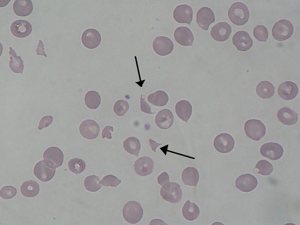
Iron Deficiency - The #1 Anemia on Earth
Most common anemia worldwide. The body doesn't have enough iron to make hemoglobin, so the cells come out small and pale.
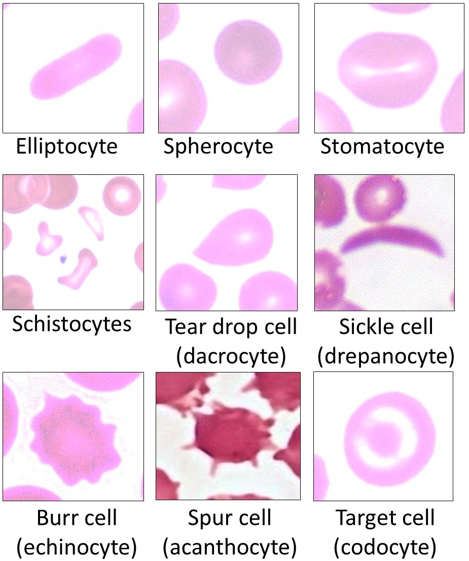
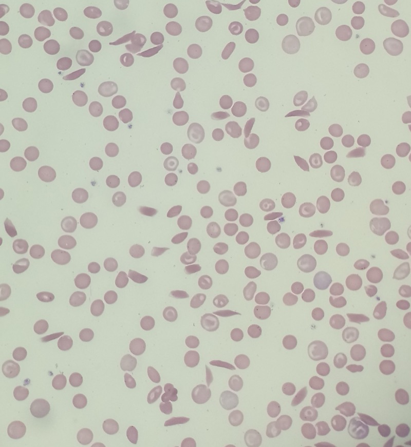
- Smear: microcytic, hypochromic (pencil cells = ovalocytes flattened by low hemoglobin) 🔑Pencil cells = pencil thin on iron - they're writing a letter asking for more
- Iron studies: ↓ ferritinFerritin = storage form of iron. Low ferritin = empty warehouse. Most specific single test for iron deficiency., ↓ serum iron, ↑ TIBCTotal Iron Binding Capacity - how many empty seats are on the transferrin bus. High TIBC = lots of empty seats = body desperate for iron., ↓ % saturation
- Causes: Blood loss (#1 in adults - menstruation, GI bleed), poor intake (infants, vegans), poor absorption (celiac, gastrectomy)
Bone Wizardry pencil cell version
Low iron means low hemoglobin. A red cell that should be a full biconcave disc gets stretched into a pale, thin oval, like dough rolled too flat.
Thalassemia - Genes Made Less Hemoglobin
What's actually broken?
The gene that codes for globin chains is defective. Iron is completely fine. The protein framework is the problem.
So what happens to hemoglobin?
Hemoglobin needs both heme AND globin to assemble. No globin chains = the cell can't fill itself up normally → small, pale cell.
Why the bullseye on smear?
One teaspoon of jam spread over a big piece of toast: edges have jam, middle looks thin. The membrane is too big for the hemoglobin filling it. That underfilled center folds and creates the pale ring with the dark rim. Target cell.
Iron studies are normal. That's the killer differentiator from iron deficiency. Same small cells, completely different iron labs.
Walk Beta-Thalassemia Like a Disease
Blur → reveal → next beat.
BEAT 1 OF 19
Smear finding
Microcytic + target cells
Why the bullseye? Less hemoglobin makes the membrane look oversized. Pale center = underfilled cytoplasm. Dark rim = membrane folding back on itself. Liver disease is the other lane.
Iron studies key differentiator
NORMAL
Iron stores are completely fine. This is what separates thalassemia from IDA - both are microcytic, but only IDA runs out of iron.
Diagnosis
Hb electrophoresis
Shows the hemoglobin fraction breakdown. Beta-thal raises HbA2. Alpha-thal trait may look normal - the pattern depends on which chain is affected.
Genetic defect:tap to expand
Alpha-thal- Deletion of HBA1/HBA2 on chr 16
- 4 total alpha-gene copies. Severity tracks how many are lost.
- 1 deleted = silent carrier. 2 = trait. 3 = HbH disease. 4 = Hb Bart hydrops.
- Adult electrophoresis can look normal in trait (all fractions need alpha).
Beta-thal- Production defect at HBB on chr 11.
- beta+ = too little beta globin made. beta0 = none made.
- Two severe hits = major. After HbF falls in infancy, HbA cannot take over.
- Result: severe microcytic anemia, marrow expansion, extramedullary hematopoiesis, transfusion dependence.
- Trait = small RBCs, normal iron, often high RBC count, HbA2 > 3.5%.
Do NOT confuse with HbC disease Thalassemia = not enough globin made. HbC = structurally wrong globin. Hexagonal crystals or very high MCHC = HbC, not thal. Beta-thal trait makes mild microcytosis, not hemolytic anemia with strong reticulocytosis.HBA1/HBA2 -> chr 16: read HBA as HemogloBin Alpha. The labels are 1 and 2, and alpha has four total gene copies. Remember it as 41x2 = 16.HBB -> chr 11: the beta-globin gene is HBB on chromosome 11.Only look at the two colored backs of the B letters: those two vertical bars are the chromosome number.Optional Thalassemia chromosome memory
Medically reviewed by Kaitlyn Cocuzzo, MD and Fatima Ali, DO · Last reviewed June 2026Bone Wizardry is an independent educational resource for visual learning in the medical sciences. It is not affiliated with, endorsed by, or sponsored by any licensing or examination board, contains no real or recalled examination questions, and does not guarantee any educational or examination outcome.How the patients present:tap to expand
Tap a disease row, then open the bullet you need.Alpha silent / trait ▸Molecule formed
Mostly normal HbA with slight alpha chain deficit.ααββHbA α₂β₂How they show up
Often asymptomatic or mild microcytosis with normal iron. Electrophoresis can look normal.Board clue
African or Southeast Asian ancestry, small RBCs, no iron deficiency.Fatality risk
Not fatal
Carrier states matter for counseling, not acute mortality.HbH disease ▸Molecule formed
Almost no alpha chains left. Excess beta chains clump together forming HbH.α?ββββHbH β₄How they show up
Moderate to severe hemolytic anemia, splenomegaly, jaundice. Those β₄ tetramers precipitate inside the RBC, punching holes in it.Board clue
Microcytosis plus hemolysis, not just a quiet carrier state.Fatality risk
Can decompensate
Usually survivable, but infections or oxidant stress can trigger dangerous hemolytic crises.Hb Bart hydrops ▸Molecule formed
Zero alpha chains. Fetal gamma chains self-pair into Hb Bart's.α✗γγγγHb Bart's γ₄How they show up
Fetus cannot make working HbF (which needs alpha). Gamma chains form γ₄ instead, which can't deliver oxygen. Develops hydrops fetalis.Board clue
Alpha chain is needed before and after birth.Fatality risk
Usually fatal
Lethal in utero or soon after birth without fetal transfusion and intensive support.Beta trait / minor ▸Molecule formed
Mostly HbA, but extra alpha chains pair with delta chains instead (making extra HbA2).ααδδHbA2 ↑ α₂δ₂How they show up
Mild anemia or incidental microcytosis. RBC count often high because marrow makes many small cells.Board clue
Normal iron studies + HbA2 > 3.5%.Fatality risk
Not fatal
The board danger is misdiagnosing it as iron deficiency, not sudden death.Beta intermedia ▸Molecule formed
Some HbA made (partial beta output), but HbF persists because gamma production compensates.ααβγHbA↓ + HbF↑How they show up
Variable anemia, sometimes transfusions during stress, growth delay if severe.Board clue
Lives between trait and major.Fatality risk
Severity varies
Not automatically fatal, but severe cases can become transfusion-dependent.Beta major ▸Molecule formed
No HbA possible. After HbF falls, only broken HbF remnants and HbA2 remain.β✗ααγγHbA = 0 - HbF↑↑How they show up
Infant becomes severely anemic after HbF naturally falls. Regular packed RBC transfusions needed to maintain oxygen delivery, growth, cardiac workload.Board clue
Symptoms start after 6 months. Marrow expansion, frontal bossing, transfusion dependence.Fatality risk
Fatal untreated
Life-threatening in early childhood without chronic transfusions and iron-overload management.- Red cell count: Often ↑ (body compensates by making MORE small cells)
The Board's Favorite Trick: Iron Deficiency vs Thalassemia
Both are microcytic. Both have low MCV. The split:
Iron deficiency: ↓ ferritin, ↑ TIBC, ↑ RDW. RDW = red cell distribution width, which means how different the RBC sizes are from each other. In IDA, iron stores fall gradually, so older cells may be closer to normal while newer cells are progressively smaller. Mixed sizes = high RDW.
Thalassemia: Normal iron studies, normal RDW, ↑ RBC count. Here the marrow is making small cells from the start because the globin blueprint is faulty every time, so the cells are uniformly small instead of mixed-size. Think alpha-globin gene deletions or beta-globin mutations causing reduced chain production.
Board Trap
"Microcytic anemia that doesn't respond to iron supplementation" = think thalassemia. If they gave iron for weeks and the MCV didn't budge, the problem was never iron.
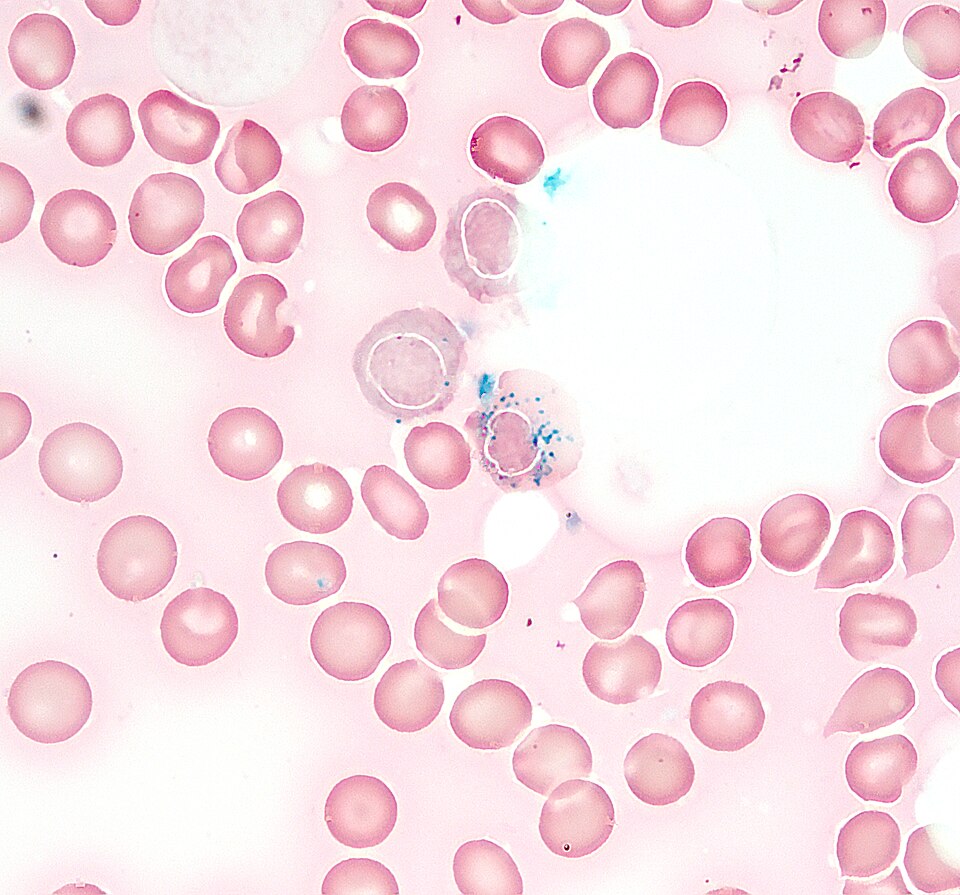
Sideroblastic Anemia - Iron Trapped in the Wrong Place
What's happening here?
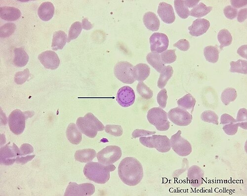
The body has plenty of iron. The heme factory is broken. Iron enters the developing red cell, hits a wall, and piles up inside the mitochondria around the nucleus instead of getting built into hemoglobin.
What does that look like on biopsy?
The iron-loaded mitochondria form a ring around the nucleus. That's a ringed sideroblast. The name is literally what it looks like.
Iron stores are full (high ferritin, high serum iron), but the cells are still small and pale. The warehouse is stuffed. The factory can't use it.

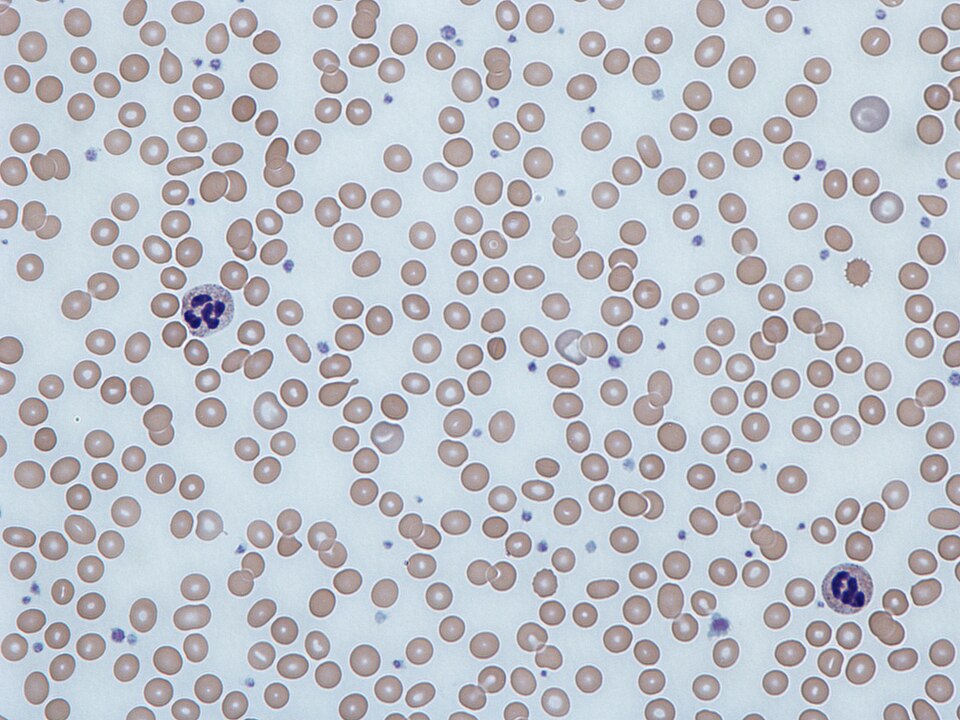
Causes: lead, alcohol, B6 deficiency, isoniazid, MDS
Smear: microcytosis + basophilic stippling
Iron labs: HIGH ferritin, HIGH serum iron
Lead Poisoning - Two Enzymes, One Disaster
Lead hits two specific points in the heme synthesis pipeline. Everything else in lead poisoning follows from that.
Succinyl-CoA + Glycine
↓ALA synthase
ALA (aminolevulinic acid)
↓
LEAD BLOCKS
ALA dehydratase
→ ALA builds up in urine
Porphobilinogen (PBG)
↓...several steps...
Protoporphyrin IX
↓
LEAD BLOCKS
Ferrochelatase (Fe²⁺ insertion)
→ FEP rises in red cells
HEME(never made)
What even is ALA in urine?
ALA (aminolevulinic acid) is the precursor that would become heme. ALA dehydratase is supposed to push it forward. Lead blocks the enzyme. ALA has nowhere to go → it spills into urine. That's your screening marker for lead exposure.
What even is FEP?
FEP = Free Erythrocyte Protoporphyrin. Ferrochelatase was supposed to stick iron into protoporphyrin to finish building heme. Lead blocks it. Naked protoporphyrin piles up inside the red cell with no iron attached. That's FEP. When lead hits, FEP skyrockets.
Lead's Other Damage (Beyond the Blood)

Burton's Lines
Blue-black band at the gumline. Lead deposits in gingival tissue as lead sulfide. The dark stripe appears exactly where the teeth meet the gum. Lead also deposits at growth plate lines on X-ray (dense white bands at the ends of long bones in kids). Wellcome Collection, public domain.
Wrist / Foot Drop
Lead demyelinates the radial nerve (wrist drop, can't extend wrist) and peroneal nerve (foot drop, can't dorsiflex foot). Peripheral motor neuropathy, not central. Classic in adults with occupational lead exposure. Commons, public domain.
Abdominal Colic
Lead causes smooth muscle spasm in the GI tract. Severe crampy abdominal pain with constipation. Often misdiagnosed as appendicitis or obstruction. No peritoneal signs - the abdomen is soft.
Encephalopathy (Kids)
Lead crosses the blood-brain barrier easily in children. It swaps for calcium in neural signaling. Result: cerebral edema, seizures, vomiting, papilledema. Even low levels cause permanent IQ drop. No safe level exists in kids. Classic trigger: old paint chips.