Start with the peripheral wire and tube, then carry the same injury logic into the brain: Wallerian degeneration, red neurons, gliosis, and the UMN versus LMN exam split.
Before you scroll: A 24-year-old man wakes after a deep sleep with his arm draped over a hard chair rail and cannot extend his wrist. There is no wound and no bleeding. On examination his wrist and finger extensors are weak, and vital signs are within normal limits with a nerve conduction study showing a focal block. Two months later his wrist works perfectly again, with full recovery. Which of the following best describes the layer of the nerve that was actually damaged?
The axon was cut but the connective-tissue tubes were spared
Only the myelin was bruised. The axon and all the tubes stayed intact
The whole nerve lost continuity and needed surgery to heal
Good instinct if you reached for a cut axon: a dead arm feels dramatic. But look at the ending. It came all the way back, fast, on its own. You know how a garden hose can get pinched under a car tire, and the water stops, but the moment you move the tire the flow returns, because the hose itself was never punctured? That is neurapraxia: the pressure bruised the insulation and blocked conduction, while the axon inside and its guide-tubes never broke. A truly cut axon would drag out over months and might not fully recover. Full recovery with no wound means demyelination only. That is neurapraxia, every time.
scroll to build the ladder ↓
The Seddon ladder
Three Rungs, One Nerve
Same nerve, three depths of damage. Tap each rung and watch two things change: what got cut, and whether it comes back.
Rung 1 · Neurapraxia
Rung 2 · Axonotmesis
Rung 3 · Neurotmesis
Neurapraxia
Mild · the insulation is bruised, the wire is fine
RecoveryPoor. Often needs surgical repair (neurorrhaphy)
Board trap: Axonotmesis and neurotmesis both cut the axon, so both trigger Wallerian degeneration. The split is the tube: axonotmesis keeps the connective-tissue sheath, neurotmesis loses it. Intact tube means guided regrowth and a good outcome. No tube means the sprouts wander and you reach for the operating room.
Two questions, done
The Discriminator
Commit to each branch before the answer shows. The whole classification is just two yes-or-no gates.
1
Gate one: is the axon still in one piece? A patient has a dead limb after firm compression, then makes a full recovery in a few weeks with no surgery. What does that ending tell you about the axon?
The axon was never interrupted, only the myelin was blocked
The axon was interrupted, so now I need to ask about the tube
2
Gate two: the axon is cut, so Wallerian degeneration is coming. Now, are the connective-tissue tubes (endoneurium and perineurium) still intact to guide the regrowth?
Tubes intact. A crush or stretch bruised the sheath but did not tear it
Tubes gone. A clean laceration cut the whole nerve across
Board trap: Students try to grade nerve injury by how bad the symptoms look. Do not. A completely paralyzed limb can be pure neurapraxia and recover fully. Grade it by the two structural gates, axon and tube, not by the drama of the exam.
Predict, then reveal
Classify the Injury
Meet Axel, one axon inside its guide-tube. Set the two switches for a case, predict the rung, then light it up and read the fate.
Axel the Axon presents his fiber
Set the two switches
Switch A. Is the axon intact?
Switch B. Are the connective-tissue tubes intact?
Set both switches to light up the rung.
The pattern that falls out
InjuryWhat is cutFate
NeurapraxiaMyelin onlyFull recovery
AxonotmesisAxon, tube sparedGood, guided regrowth
NeurotmesisAxon and tubePoor, needs neurorrhaphy
Board trap: There is no rung for "axon intact but tubes torn." If the tube is severed, the axon running through it is severed too. The two switches that matter as a pair are: myelin-only (neurapraxia), axon-cut-tube-kept (axonotmesis), and both-cut (neurotmesis).
What happens after the cut
Degeneration and Regrowth
Three predictions. Commit to each one before you reveal the answer. Each one hides a classic board trap.
A healthy myelinated fiber: the axon is the wire, the myelin is the insulation. Neurapraxia bruises only the insulation.
An axon is cut in the mid-forearm. Which part of the fiber undergoes Wallerian degeneration?
Correct: the distal segment. Cut an axon and the piece past the cut loses its lifeline from the cell body, so it and its myelin fall apart while macrophages clear the debris. In the peripheral nerve, Schwann cells then line up into a cleared road for regrowth. Trap: it does not happen in neurapraxia at all, because there the axon is never interrupted, and Wallerian means an axon was cut, not that the cell died.
Same cut, two locations. Which recovers better on its own, a peripheral nerve or a central spinal cord tract?
Correct: the peripheral nerve. Schwann cells build a guiding tract, and if the endoneurial tubes are intact the regrowth is organized. The central nervous system has no Schwann-cell guidance, and astrocytes form a glial scar whose inhibitory molecules block the axon. Trap: same injury logic, opposite neighborhood, which is why a cut nerve can come back but a severed cord tract does not.
Which single injury type is the one that actually needs neurorrhaphy, the surgical repair of a severed nerve?
Correct: neurotmesis. When the whole nerve loses continuity, the cut ends cannot find each other, so the surgeon stitches them so the endoneurial tubes line up again. Trap: a crush with intact tubes (axonotmesis) usually does not need surgery because the tubes already guide, and a demyelinating block (neurapraxia) never does.
Board trap: Do not let a stem convince you the central nervous system recovers with time and rehab like a peripheral nerve. The missing Schwann-cell tract and the astrocytic glial scar are the reason a spinal cord tract stays cut.
Central injury clock
When Hypoxia Hits Brain Tissue
Irreversible injury starts fast, but the microscope lags behind. Pick the time window before the reveal.
Red neurons on histology
The patient had global hypoxia. The neuron is already doomed within minutes, but when does the classic eosinophilic red neuron show up?
Correct window: 12 to 24 hours. Minutes tells you irreversibility has begun. Red cytoplasm and pyknotic nuclei are the visible light under the microscope later.
Neutrophils enter the necrotic field
The infarct is now visibly necrotic and acute inflammation is walking in first. Choose the clock slot.
Correct window: 24 to 72 hours. Early necrosis brings neutrophils first. The cleanup crew changes later.
Microglia and macrophages clean up
The dead zone is being eaten out. Which window fits microglial and macrophage cleanup?
Correct window: 3 to 5 days. Microglia are the CNS macrophages, so they dominate when the brain starts clearing liquefactive necrosis.
MinutesIrreversible neuronal injury begins after severe hypoxia.
12 to 24 hRed neurons: eosinophilic cytoplasm, pyknotic nuclei.
24 to 72 hNecrosis with neutrophils.
3 to 5 dMacrophages and microglia clear debris.
1 to 2 wkReactive astrocytes, vessels, then a gliotic cyst wall.
Board trap: Do not answer "red neurons at 5 minutes." Five minutes is when irreversible injury begins. The red neuron is the delayed tissue clue, classically 12 to 24 hours after ischemic injury.
UMN, LMN, bulbar
Sort the Motor Pattern
The exam signs are not random. Decide whether inhibition was removed from above or the final motor wire was damaged below.
UMN patternDescending inhibition is gone, so reflex arcs run hot: spastic tone, hyperreflexia, Babinski, clasp-knife.
LMN patternThe final motor pathway is broken, so the muscle loses drive: flaccid weakness, low reflexes, atrophy, fasciculations.
Bulbar splitBulbar is LMN cranial nerve damage. Pseudobulbar is bilateral UMN corticobulbar damage.
Spastic leg, hyperreflexia, Babinski
A corticospinal tract lesion below the cortex but above the anterior horn removes the brake from spinal reflexes.
Correct: UMN. The reflex circuit is intact below, but the descending brake is gone, so tone and reflexes rise.
Flaccid weakness, atrophy, fasciculations
The anterior horn cell, root, plexus, peripheral nerve, or motor axon is damaged.
Correct: LMN. The muscle lost its final motor input, so it goes limp, wastes, and twitches.
Clasp-knife tone after stroke
The limb resists movement at first, then suddenly gives way as the reflex adapts.
Correct: UMN. Spasticity and clasp-knife tone are overactive reflex signs from loss of descending inhibition.
Tongue wasting, fasciculations, absent gag
The cranial nerve nuclei or their LMN fibers are damaged.
Correct: bulbar palsy. It is the LMN pattern of cranial motor injury: weak, wasted, fasciculating, and under-reflexive.
The cranial motor nuclei are alive, but bilateral corticobulbar control from above is lost.
Correct: pseudobulbar palsy. It is an UMN cranial pattern, so reflexes are brisk or exaggerated rather than absent.
Foot drop after fibular head compression
A peripheral nerve compression hits the final pathway to ankle dorsiflexors.
Correct: LMN. A named peripheral nerve lesion is below the anterior horn cell and gives a lower motor neuron pattern.
Board trap: "Bulbar" does not just mean throat symptoms. Bulbar palsy is LMN cranial motor damage. Pseudobulbar palsy is the UMN lookalike from bilateral corticobulbar injury.
Make it stick
Hooks and Real Tissue
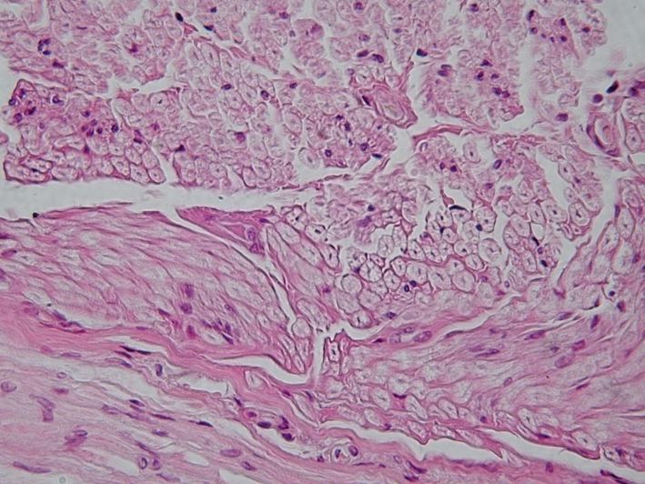
Tap a hook to unblur. Then see the actual histology the words describe.
🧩
The suffix is the depth
Praxia = just a practice run, nothing cut. Tmesis (Greek for "cutting") = something is cut. Axon-o-tmesis cuts the axon, neuro-tmesis cuts the whole nerve. Bigger word, deeper cut.
Select to reveal
🔌
Two switches, three rungs
Myelin only equals neurapraxia. Axon cut, tube kept, equals axonotmesis. Axon and tube both cut equals neurotmesis. There is no rung for tube-cut-but-axon-spared.
Select to reveal
⬇️
Wallerian points downstream
Wallerian degeneration is the DISTAL stump dying. Think of the cord unplugged from the wall: the far end goes dark. And a plugged-in wire (neurapraxia, axon intact) never goes dark at all.
Select to reveal
🌑
Schwann builds the road
Peripheral nerves have Schwann cells that pave a regrowth road. The central nervous system paves a wall instead: the astrocyte glial scar. Road versus wall is why one comes back and one does not.
Select to reveal
NERVE CROSS SECTION · tap to expand
WALLERIAN DEGENERATION · tap to expand
NERVE BUNDLE IN TISSUE · tap to expand
Board trap: The three sheaths nest inside out: endoneurium hugs each fiber, perineurium wraps a fascicle, epineurium binds the whole nerve. Axonotmesis keeps the inner tubes so regrowth stays on track. Neurotmesis blows through all of them.
Board walkthrough
Prove It
Board-style vignettes, one at a time, shuffled and never repeated until the bank is exhausted.
Vignette 1Never-repeat tracking ready
Exam tools: right-click (or long-press on mobile) to cross out a choice. Double-click (or double-tap) to highlight one. Tools switch off after you answer.
Medically reviewed by Kaitlyn Cocuzzo, MD and Fatima Ali, DO
Sources: Gray's Anatomy for Students; standard peripheral nerve pathology, ischemic brain injury pathology, and motor neuron localization references. Histology images courtesy of Wikimedia Commons contributors under their respective open licenses.
Bone Wizardry · Board Review
Bone Wizardry is an independent educational resource for visual learning in the medical sciences. It is not affiliated with, endorsed by, or sponsored by any licensing or examination board, contains no real or recalled examination questions, and does not guarantee any educational or examination outcome.
 Bone WizardryMEDICAL EDUCATION