Brachial Plexus and Upper Extremity Nerve Injuries
Localize upper-limb weakness from plexus roots to hand nerves, distinguish shoulder and finger patterns, and apply careful radial-head naming without overreading tests.
A weak hand or immobile shoulder becomes localizable when you ask which motions and sensory territories fail together. By the end, you can trace a lesion from roots through branches, distinguish proximal from distal nerve injuries, and explain why an apparently smaller injury may make a more conspicuous claw.
Trace the plexus before naming a palsy
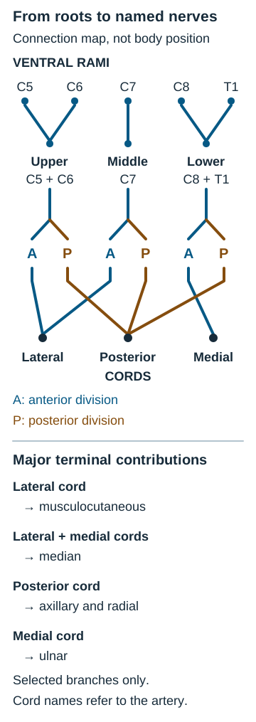
Question: Does weakness in both shoulder abduction and elbow flexion point to one terminal nerve? The brachial plexus carries ventral rami C5 through T1 through roots, upper/middle/lower trunks, anterior/posterior divisions, lateral/posterior/medial cords, and terminal branches. Remember the sequence as roots, trunks, divisions, cords, branches; the mnemonic “Randy Travis Drinks Cold Beer” is optional, not a substitute for the map.
The cords are named around the axillary artery. The median nerve receives lateral- and medial-cord contributions; musculocutaneous arises from lateral, axillary and radial from posterior, and ulnar from medial. Medial cutaneous branches carry sensation rather than being a sixth major motor terminal nerve. [1]
Trace C5-C6 into the upper trunk, then C8-T1 into the lower trunk. Musculocutaneous is chiefly C5-C7, axillary C5-C6, radial C5-T1, median C5-T1, and ulnar chiefly C8-T1; overlapping root contributions vary. The same root can contribute to more than one terminal nerve; diagram prepared for this lesson from standard plexus anatomy. [1]
Trace the deltoid and biceps backward with a finger, then trace an interosseous muscle. The first two travel in different terminal nerves but share upper-plexus roots; an interosseous depends largely on lower-plexus supply. Thus one root/trunk lesion can cross named nerve boundaries, whereas an isolated axillary lesion cannot weaken biceps. Transfer this to a patient with weak elbow flexion but normal deltoid: assess musculocutaneous territory and other C5-C6 muscles before declaring an upper-trunk lesion.
Use sensory findings as supporting evidence, not an exact coloring book. C5 commonly reaches lateral upper arm, C6 the thumb, C7 the middle finger, C8 the little finger, and T1 medial forearm; adjacent dermatomes overlap, and the medial upper arm also receives T2 through the intercostobrachial nerve. Compare a root pattern with a peripheral nerve's autonomous sensory zone and reflexes: biceps predominantly C5-C6, brachioradialis C6, triceps C7. An absent reflex does not identify a root by itself. [1]
The axillary nerve (C5-C6) passes through the quadrangular space beside the posterior circumflex humeral artery, at risk with anterior shoulder dislocation or surgical-neck fracture. Deltoid and teres minor weakness with altered lateral-shoulder sensation are useful findings. Before and after reduction of a dislocated shoulder, document axillary-region sensation, deltoid action and distal neurovascular status. [7] Supraspinatus initiates abduction and the deltoid contributes through a broad range, so there is no absolute “cannot abduct beyond 15 degrees” cutoff; compare resisted abduction and inspect the shoulder, while excluding painful structural injury. [1]
Upper trunk, lower trunk, or outlet?
Question: A newborn has weak shoulder abduction and elbow flexion but preserved hand motion: which level connects these losses? Lateral separation of head and shoulder during difficult delivery, or analogous adult traction, can injure C5-C6 upper trunk. Deltoid, supraspinatus, biceps, brachialis, brachioradialis and external rotators may weaken, with diminished elbow flexion and forearm supination. In newborns, an asymmetric Moro response with preserved finger grasp supports an upper-plexus pattern; compare this infant reflex with voluntary adult grip, not with an adult “grasp reflex.” The arm may rest adducted and internally rotated with an extended elbow and pronated forearm, the classic Erb-Duchenne “waiter's tip”; a diminished biceps reflex supports the pattern.
This posture reflects relative muscle imbalance, gravity and variable injury, not proof that every internal rotator or pronator is wholly spared. [1]
Upper plexus C5-C6
Shoulder and elbow deficits dominate; distal hand action may remain. Check biceps reflex and several upper-root muscles, not posture alone.
Lower plexus C8-T1
Intrinsic-hand weakness and medial forearm/hand sensory changes dominate; proximal shoulder motion may remain. Hyperabduction traction can injure the lower trunk.
Cover the labels and predict which examination is most discriminating: intrinsic finger abduction or elbow flexion? Reveal by reading the comparison. Weak ulnar interosseous finger abduction together with median thenar weakness favors lower plexus over a single distal nerve; elbow flexion loss with preserved hand motion favors upper plexus. Apply the same logic to an adult traction injury, but assess pain, fractures and multi-level deficits rather than assigning a named syndrome from appearance alone.
The ocular sympathetic pathway descends from the hypothalamus to the ciliospinal spinal-cord region around C8-T2; preganglionic fibers then leave the cord and ascend to the superior cervical ganglion, and postganglionic ocular fibers travel along the internal carotid artery. Severe proximal injury along this pathway may produce ipsilateral ptosis, miosis and sometimes anhidrosis. [16] Horner syndrome raises concern for a proximal sympathetic lesion, including root avulsion, but does not prove Klumpke palsy or identify its exact level. A dramatic all-finger claw is not inevitable with lower-trunk injury: patterns depend on which intrinsic and extrinsic muscles remain functional. [1]
Scalene triangle: plexus plus artery; vein travels anterior to anterior scalene.
Costoclavicular: clavicle over first rib.
Subcoracoid: beneath pectoralis minor.
Could positional symptoms instead arise at the thoracic outlet? The interscalene triangle is bounded by anterior scalene, middle scalene and first rib and contains the brachial plexus and subclavian artery, not subclavian vein. The costoclavicular interval lies between clavicle and first rib; the subcoracoid space lies beneath pectoralis minor. Neurogenic TOS is the most common category and may affect lower plexus fibers, causing activity-related pain, medial forearm symptoms and weakness of both ulnar interossei and median thenar muscles.
A cool pale hand or ischemic pain raises urgent arterial concern; a swollen cyanotic arm suggests venous obstruction and also requires prompt assessment. [5][18] Adson combines neck rotation toward the examined side with a deep breath, military brace draws the shoulders back and down, and Wright raises the arm in abduction. These positions may provoke symptoms but cannot diagnose TOS alone; history, examination and appropriate exclusion or vascular testing matter.
Ulnar neuropathy at the cubital tunnel behind the medial epicondyle can also be positional, especially with prolonged elbow flexion or pressure. [4]
Compare medial-hand tingling during overhead work with tingling when sleeping with the elbow flexed. Ask whether symptoms accompany shoulder elevation or elbow pressure, then check medial forearm sensation and intrinsic muscles. The consequence of changing the provoking posture is a better localization hypothesis, not a positive-test diagnosis. Transfer this approach to a patient with hand swelling: evaluate vascular causes promptly rather than calling all positional symptoms neurogenic TOS.
Try it here · Checkpoint 1 of 3
Make your prediction before reading the choices. A first attempt is just a starting point.
Case 2
Show answer and explanations for case 2
A. Forearm supination by supinator (Why this does not fit)
Supinator is supplied by radial C5-C6 fibers; its deficit points toward an upper-root distribution, not the medial forearm and hand pattern.
Reasoning steps for option A
Which roots predominantly supply supinator?
C5-C6.
Are upper-root actions preserved here?
Yes, shoulder abduction and elbow flexion are preserved.
B. Thumb MCP extension by extensor pollicis brevis (Why this does not fit)
This radial-innervated extensor is more strongly associated with C7-C8 and does not discriminate the lower pattern as well as median thenar weakness.
Reasoning steps for option B
Which nerve supplies extensor pollicis brevis?
The radial nerve.
Which accompanying deficit favors a broader lower-plexus injury here?
Medial forearm hypesthesia.
C. Palmar thumb abduction by abductor pollicis brevis (Best answer)
A lower C8-T1 plexus pattern extends beyond ulnar hand muscles to median-innervated thenar abductor pollicis brevis.
Reasoning steps for option C
Which nerve supplies abductor pollicis brevis?
The median nerve.
Would a C8-T1 plexus lesion also affect this thenar muscle?
Yes, its lower-root fibers can be involved.
D. Shoulder external rotation by infraspinatus (Why this does not fit)
Suprascapular C5-C6 function should remain intact given the preserved upper-root actions.
Reasoning steps for option D
Which roots predominate in infraspinatus?
C5-C6.
What upper-root function is preserved in this patient?
Shoulder abduction.
Takeaway: Infer lower-plexus involvement from medial forearm, intrinsic hand and sympathetic findings; predict median thenar abduction weakness.
Question: Why can a wrist-level ulnar lesion show a stronger claw than an elbow-level lesion? Ulnar motor fibers supply interossei, hypothenar muscles, adductor pollicis and usually lumbricals three and four; the nerve is exposed at the cubital tunnel behind the medial epicondyle and at Guyon canal near the wrist, where prolonged handlebar pressure may affect cyclists.
The dorsal ulnar cutaneous branch leaves before Guyon canal, so its dorsal-hand territory can remain normal in a canal lesion. Proximal branches also supply flexor carpi ulnaris and the ulnar half of flexor digitorum profundus (FDP) to ring and little fingers. Loss of intrinsic MCP flexion and IP extension permits extensor digitorum to hyperextend the MCP joints; retained long flexors bend the IP joints.
The little finger and ulnar half of ring finger are characteristic palmar ulnar territories; dorsal ulnar hand is supplied by the dorsal cutaneous branch. The dorsal and palmar ulnar sensory distribution and branch pattern help distinguish elbow from Guyon's canal lesions. Froment's sign is thumb IP flexion by median-innervated flexor pollicis longus compensating for weak ulnar adductor pollicis during paper pinch. [15]
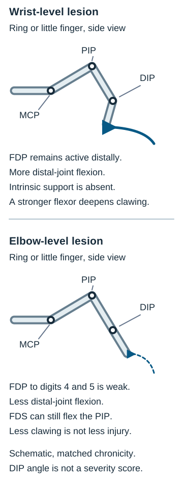
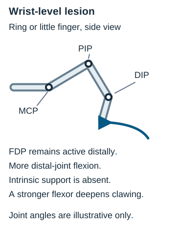
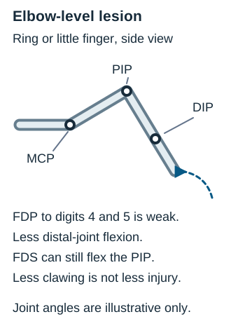
Compare the stacked views while holding injury completeness and chronicity equal. A wrist lesion leaves ring/little FDP working; an elbow lesion interrupts its ulnar motor supply. Anatomical force comparison created for this lesson. [15]
Predict the DIP before opening either view. At the wrist, FDP branches have already left the nerve: ring and little finger DIP flexion retains force, and intrinsic loss makes the MCP-hyperextended/IP-flexed claw conspicuous. At the elbow, the same intrinsic deficit is accompanied by weak ulnar FDP; DIP flexion is reduced and the visible claw can be less pronounced. This is the ulnar paradox, not evidence that the more proximal injury is milder overall. Flexor digitorum superficialis is median-innervated and is not lost in an isolated proximal ulnar lesion; PIP flexion may remain. [15]
Inspect a wrist-level lesionDistal to FDP branching: stronger DIP pull despite intrinsic loss.Inspect an elbow-level lesionProximal to FDP branching: less DIP pull, not less neural injury.
Compare the two DIP angles, then predict a patient with preserved ring DIP flexion but lost finger abduction: a distal ulnar site is more plausible. Check the lesion location, time since injury, contracture and completeness before comparing real hands. Chronic fixed deformity can obscure this dynamic prediction.
Median entrapment near the pronator teres can produce proximal volar forearm aching and median-distribution paresthesia, potentially including the palm. This differs from an isolated motor AIN pattern. [2] Supracondylar humeral fractures can injure median or AIN fibers and require neurovascular assessment. Neither mechanism automatically implies the complete proximal median pattern described below. [19]
Intrinsic muscles also extend the IP joints through the extensor expansions while flexing the MCP joints. That action can remain available in PIN injury: absent extrinsic MCP extension does not imply loss of every route to IP extension. [20]
Median lesions require a different task. A proximal median injury can weaken pronation, flexor pollicis longus, lateral FDP, flexor digitorum superficialis, lateral lumbricals and thenar muscles including abductor pollicis brevis, opponens pollicis and the superficial head of flexor pollicis brevis (variable dual innervation); asking the patient to make a fist exposes failure to flex index and middle fingers, traditionally called a “hand of benediction” on attempted fist.
Median compression within the carpal tunnel more often begins with nocturnal paresthesia of thumb, index, middle and radial ring fingers, sometimes relieved by shaking; chronic severe disease may cause abductor pollicis brevis weakness and resting thenar wasting, sometimes called “ape hand.” These are different tasks and lesion levels, not interchangeable resting postures. The palmar cutaneous median branch leaves before the tunnel, so central thenar-palm sensation can be spared in isolated CTS.
The anterior interosseous nerve (AIN) supplies flexor pollicis longus, index FDP, variably middle FDP, and pronator quadratus; its lesion impairs tip-to-tip pinch or an “OK” circle without cutaneous sensory loss. [2]
Ask a patient to flex the index DIP against resistance, oppose the thumb, and identify whether numbness reaches the central palm. Preserved palm sensation with nocturnal digital paresthesias favors tunnel-level median compression; weak thumb IP and index DIP flexion without numbness suggests AIN. Transfer to a proximal forearm injury: pronation or long-finger flexion deficits broaden localization beyond the tunnel.
Separate extension deficits from tunnel symptoms
Question: Is inability to straighten a finger the same as wrist drop? A radial injury in the axilla (including prolonged crutch pressure) may involve triceps as well as distal extensors; a spiral-groove injury after humeral shaft fracture or sustained compression commonly weakens wrist and MCP finger extension with variable brachioradialis involvement while often preserving triceps.
Test dorsal first web-space sensation as a relatively useful radial sensory site. A posterior interosseous nerve (PIN) lesion is principally motor: MCP finger and thumb extension weaken without cutaneous loss. ECRL is usually supplied before PIN, while ECRB branch origin varies, whereas ECU is supplied through PIN; wrist extension may persist with radial deviation, not necessarily normal force.
Intrinsic hand muscles extend interphalangeal joints via the extensor hood, so preserved IP extension does not exclude radial/PIN loss of MCP extension. [2]
Axilla: triceps may weaken → spiral groove: wrist and MCP extension plus possible first-web sensation → PIN: MCP extension weak, no skin loss, ECRL-driven wrist extension may radially deviate.
Hold the wrist supported and ask for MCP extension rather than merely “straight fingers.” Next ask for resisted wrist extension and note its direction, then test triceps and sensation. Weak MCP extension plus preserved radially deviating wrist extension and normal skin sensation points toward PIN; wrist drop with first-web sensory loss points more proximally. Transfer this pattern to a midshaft fracture, where associated structural injury requires assessment rather than reliance on a posture label.
Why does CTS produce night symptoms? Nine flexor tendons, four superficial, four deep and flexor pollicis longus, share the bounded carpal tunnel with the median nerve under the flexor retinaculum. Pregnancy-related fluid shifts, inflammatory tenosynovitis, rheumatoid arthritis, diabetes, hypothyroidism and acromegaly can accompany CTS; repetitive exposure alone does not establish a diagnosis. Phalen's sustained wrist flexion, Tinel percussion and Durkan compression may reproduce symptoms but each has limited standalone accuracy: do not rank them as universally most sensitive or specific.
The 2024 AAOS guideline supports the CTS-6 clinical prediction tool for diagnosis and does not require routine electrodiagnostic testing or ultrasound for every typical presentation; tests can be useful for diagnostic uncertainty or selected management decisions. [3]
For nocturnal thumb-through-radial-ring paresthesia with spared central palm, collect symptom pattern, thenar strength and relevant examination findings before considering a CTS-6 estimate. The consequence is a reasoned clinical probability, not a diagnosis from a positive Phalen test. Transfer to numbness involving the little finger or forearm: revisit ulnar, cervical or broader neuropathic causes.
Try it here · Checkpoint 2 of 3
Make your prediction before reading the choices. A first attempt is just a starting point.
Case 5
Show answer and explanations for case 5
A. Active IP extension remains possible only in fingers 4-5 (Why this does not fit)
Isolated PIN injury spares both ulnar and median intrinsic pathways, so IP extension is not selectively confined to ulnar digits.
Reasoning steps for option A
Which intrinsics extend ring/little IP joints?
Ulnar lumbricals and interossei.
Are median-innervated index/middle intrinsics also lost in this PIN lesion?
No, this forearm motor lesion spares them.
B. Active IP extension remains possible in all four fingers (Best answer)
PIN motor loss weakens extrinsic MCP extension, while median- and ulnar-innervated lumbricals and interossei act through the extensor expansions to extend IP joints when MCPs are supported.
Reasoning steps for option B
How do lumbricals and interossei extend IP joints?
Through extensor expansions.
Which fingers have their MCP joints supported here?
Fingers 2-5.
Is the intrinsic extension mechanism denervated by the inferred lesion?
No. The spared median and ulnar pathways permit IP extension in these fingers.
C. Active IP extension remains possible only in fingers 2-3 (Why this does not fit)
Isolated PIN injury spares both median and ulnar intrinsic pathways, so IP extension is not selectively confined to median digits.
Reasoning steps for option C
Which nerve supplies index/middle lumbricals?
The median nerve.
Are ulnar-side intrinsics lost in this patient?
No, the lesion implicates radial PIN output.
D. Active IP extension is absent in all four fingers (Why this does not fit)
Radial PIN fibers supply extrinsic extensors, but median and ulnar nerves supply the hand intrinsics that can extend IP joints.
Reasoning steps for option D
Does PIN supply hand lumbricals?
No, median and ulnar nerves do.
Can this patient use those spared intrinsics with MCPs supported?
Yes, in all four tested fingers.
Takeaway: With full passive motion, PIN loss of extrinsic MCP extension can coexist with retained median- and ulnar-intrinsic IP extension when MCPs are supported.
Use motion and shape to distinguish joint from tendon
Question: Does painful abduction prove a supraspinatus tear? The suprascapular nerve supplies both supraspinatus and infraspinatus, linking their actions in a proximal neuropathy. [2] Supraspinatus is the most commonly torn cuff tendon and helps initiate and sustain abduction alongside deltoid; infraspinatus and teres minor externally rotate, while subscapularis internally rotates and attaches at the lesser tuberosity (the others attach to the greater tuberosity).
A painful arc around 60-120 degrees, empty-can/Jobe resisted elevation in scapular plane with thumb down, drop-arm inability to lower an abducted arm smoothly, Neer passive forward elevation, and Hawkins-Kennedy internal rotation with the shoulder and elbow flexed each contribute imperfectly to assessment of cuff disease or subacromial pain. Combine history, range, strength and multiple findings; no maneuver is an anatomical verdict.
Speed tests resisted shoulder flexion with palm up; Yergason tests resisted supination with the elbow bent. Pain during either can suggest biceps involvement but is nonspecific. Anterior apprehension in abducted external rotation, relieved by posteriorly directed relocation, supports symptomatic instability in context; downward traction producing a sulcus below the acromion shows inferior laxity, which alone does not establish instability. [6]
Compare active with passive abduction, then examine resisted external rotation and weakness versus pain. Preserved passive range with weak active elevation suggests muscle/tendon or nerve dysfunction rather than a fixed joint block. After a seizure or electric injury, an adducted internally rotated arm with marked loss of passive external rotation raises concern for locked posterior dislocation. AP “lightbulb” appearance can be subtle or misleading; obtain orthogonal imaging, preferably an axillary or modified Velpeau view when standard positioning is too painful. Transfer: do not dismiss a locked shoulder because an AP image appears nearly normal. [7]
Distinguish finger postures and radial wrist pain
Question: When a PIP bends while a DIP straightens, is the flexor tendon necessarily shortened? Compare the joint positions before naming a structure. In the table, trace a finger from palm to tip, then identify the joint whose direction changes between swan-neck and boutonnière patterns. The consequence is a different extensor-balance hypothesis, which transfers to a painful injured finger requiring fracture or central-slip assessment.
Finger posture is an output of different structures
Pattern
PIP
DIP
Underlying finding
PatternSwan neck
PIPHyperextended
DIPFlexed
Underlying findingVolar plate laxity or extensor imbalance, often with inflammatory disease such as RA
PatternBoutonnière
PIPFlexed
DIPHyperextended
Underlying findingCentral slip rupture after trauma or inflammatory disease such as RA
PatternDupuytren contracture
PIPFlexion contracture, often ring/little
DIPVariable
Underlying findingPalmar fascial cord, often MCP/PIP rather than DIP; not a flexor tendon lesion
Swan-neck and boutonnière patterns can both occur in rheumatoid disease, but opposite PIP positions reflect different soft-tissue failures. [8][9]
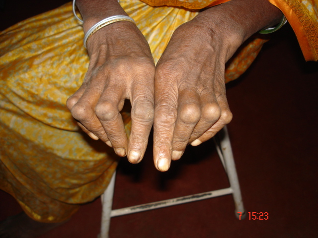
Dupuytren disease is fibroproliferation of palmar fascia, associated with heredity, diabetes, alcohol use and seizure disorders. These associations do not establish phenytoin as a cause. Examine the cord and passive finger extension rather than assuming tendon shortening. [10] Radial-styloid pain with thumb motion suggests first dorsal extensor compartment disease involving abductor pollicis longus and extensor pollicis brevis, often termed de Quervain tenosynovitis, including during the postpartum period.
Radial styloid pain is distinct from the median-digit paresthesia of CTS. Finkelstein's examiner-held thumb traction and ulnar deviation differs from the patient's thumb-in-fist ulnar-deviation maneuver commonly called Eichhoff; the latter may provoke pain nonspecifically. Neither test replaces consideration of other radial wrist pathology. [11][12]
Sketch PIP and DIP angles before assigning a deformity name, then palpate whether a palmar cord restricts passive extension or radial-styloid motion provokes focal pain. The consequence is a structural hypothesis that selects the next examination, not a diagnosis from one photograph. Transfer to a painful post-traumatic finger: exclude fracture and tendon rupture before labeling an inflammatory deformity.
Translate forearm restriction into OMM naming
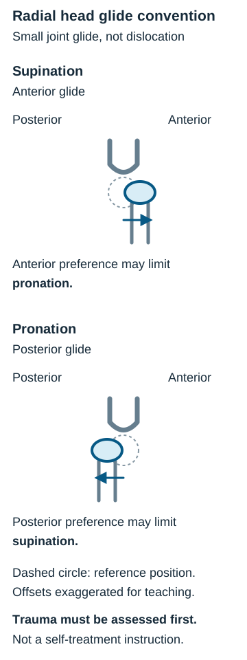
Question: If pronation is limited, is the radial head named for the missing motion or the position it prefers? In osteopathic convention an anterior radial-head dysfunction is described as preferentially supinated with restricted pronation; a posterior dysfunction preferentially pronated with restricted supination. Palpate the radial head just distal to the lateral epicondyle during gentle pronation-supination, without assuming every tender or blocked elbow has somatic dysfunction.
During forearm rotation the radial head's relative anterior-posterior relationship at the proximal radioulnar joint is the teaching model. The term describes a palpatory and motion finding, not proof of a discrete fixed dislocation or of treatment effectiveness. [13]
Radial head naming follows its preferred position; explanatory diagram created for this lesson from osteopathic convention, not an outcome claim. [13]
Anterior radial head
Supination relatively free; pronation restricted. In the conventional direct-technique description, pronation is paired with a posteriorly directed force on the radial head. [14]
Posterior radial head
Pronation relatively free; supination restricted. In the conventional direct-technique description, supination is paired with an anteriorly directed force on the radial head. [14]
Cover each name and predict the restricted motion, then check the adjacent column. Naming records the preferred position, not the restriction; that reversal is the visible consequence of the rotation model. Transfer to a fall on an outstretched hand: trauma, focal bony tenderness, deformity or inability to rotate requires fracture/dislocation assessment before any manual technique. [17] Hands-on OMM belongs only with a trained clinician and appropriate examination; these conventions are not evidence of improved outcomes. [13]
In the elbow-level ulnar convention, an abducted forearm has a relatively medial olecranon and lateral distal ulna; the associated wrist preference is adduction (ulnar deviation), with radial deviation relatively restricted. An adducted forearm reverses these relationships: lateral olecranon, medial distal ulna, and wrist abduction (radial deviation) preference with ulnar deviation relatively restricted. These are coupled examination descriptions, not a diagnosis made solely at the distal radioulnar joint. Specify the elbow finding, wrist motion and trauma assessment rather than naming the ulna from wrist restriction alone. [14]
Try it here · Checkpoint 3 of 3
Make your prediction before reading the choices. A first attempt is just a starting point.
Case 23
Show answer and explanations for case 23
A. Arrange delayed review without initial protection because no fracture line is visible (Why this does not fit)
Review is useful, but a traumatic effusion with focal radial-head tenderness warrants initial protection while occult fracture is assessed. A negative visible fracture line does not justify waiting without protection.
Reasoning steps for option A
Can a radial-head fracture lack a visible line initially?
Yes.
Which radiographic sign still requires initial protection?
A posterior fat-pad sign.
B. Use the palpatory anterior preference to select an immediate direct technique (Why this does not fit)
Restricted pronation may resemble a named somatic dysfunction, but traumatic bony tenderness and an effusion can cause the same restriction. Technique selection must wait until structural injury is appropriately assessed.
Reasoning steps for option B
What could anterior radial-head palpatory preference suggest?
A somatic dysfunction label.
Which finding makes immediate direct treatment unsafe to prioritize?
Focal traumatic radial-head tenderness.
C. Obtain electrodiagnostic testing before deciding whether the elbow needs protection (Why this does not fit)
Electrodiagnostics can assess suspected neuropathy, but this stem supplies a traumatic joint pattern rather than a focal motor/sensory nerve deficit. It would not resolve the immediate occult-fracture concern.
Reasoning steps for option C
What do electrodiagnostic tests investigate?
Neuropathy.
What objective injury finding instead needs attention?
A traumatic elbow effusion.
D. Manage the injury as a possible occult fracture and arrange appropriate reassessment (Best answer)
A negative visible fracture line does not exclude an occult radial-head injury. Trauma, focal bony tenderness and an effusion make fracture assessment and appropriate protection/reassessment the priority; palpatory preference cannot clear the elbow for manual care.
Reasoning steps for option D
What does a posterior fat pad after trauma imply?
Possible occult intra-articular fracture.
What immediate approach follows?
Protection with appropriate reassessment.
Takeaway: Traumatic effusion and focal bony tenderness take precedence over a convenient somatic-dysfunction label.
A. Weak finger abduction with preserved supination (Why this does not fit)
Intrinsic hand weakness with preserved biceps action instead suggests lower-plexus predominance.
Reasoning steps for option A
Which nerve supplies the interossei?
The ulnar nerve.
Is grasp preserved in this infant?
Yes, distal grasp is preserved.
B. Weak elbow extension with diminished triceps reflex (Why this does not fit)
A predominantly C7 radial pattern does not predict the characteristic biceps and deltoid deficits.
Reasoning steps for option B
Which root predominates in the triceps reflex?
C7.
Which reflex is reduced in this infant?
The biceps reflex.
C. Weak thumb IP flexion with a normal triceps reflex (Why this does not fit)
Thumb IP flexion depends on FPL through the anterior interosseous branch of the median nerve, predominantly C8-T1. That distal flexor prediction does not fit the supplied C5-C6 shoulder/elbow pattern with preserved grasp.
Reasoning steps for option C
Which nerve supplies thumb IP flexion?
The median anterior interosseous nerve.
Which upper-plexus findings are present in this infant?
Weak shoulder abduction and a reduced biceps reflex.
D. Weak forearm supination with preserved finger flexion (Best answer)
Biceps-mediated supination depends substantially on C5-C6, while distal finger flexion can remain intact with upper-plexus injury.
Reasoning steps for option D
Which muscle contributes strongly to supination?
The biceps.
Does this infant have evidence of upper-plexus involvement?
Yes, elbow flexion and the biceps reflex are impaired.
A. Elbow flexion by brachialis (Why this does not fit)
Brachialis is supplied mainly by musculocutaneous nerve. The distal motor and sensory pattern identifies radial nerve, so shifting that lesion proximally does not primarily target brachialis.
Reasoning steps for option A
Which nerve primarily supplies brachialis?
The musculocutaneous nerve.
Which nerve is identified by wrist drop and first-web loss here?
The radial nerve.
B. Shoulder abduction by deltoid (Why this does not fit)
Deltoid is an axillary target. Radial nerve injury in the axilla is not synonymous with injury to every nerve in that region; the hypothetical change concerns the same radial nerve.
Reasoning steps for option B
Which nerve supplies deltoid?
The axillary nerve.
Is the hypothetical lesion of that nerve?
No, it remains a radial-nerve lesion.
C. Thumb opposition by opponens pollicis (Why this does not fit)
Opponens pollicis is a median thenar target. Radial motor and sensory deficits do not predict new median weakness simply because their lesion is more proximal.
Reasoning steps for option C
Which nerve supplies opponens pollicis?
The median nerve.
Does moving this radial lesion proximally imply median injury?
No.
D. Elbow extension by triceps (Best answer)
The findings initially favor radial injury distal to the proximal triceps branches. An axillary radial lesion can involve those branches, adding elbow-extension weakness to the distal radial deficits.
Reasoning steps for option D
Which nerve supplies triceps?
The radial nerve.
What current finding shows its proximal branches are spared?
Strong elbow extension.
Takeaway: Triceps sparing alongside distal radial motor and sensory deficits supports groove-level injury.
A. Loss over both palmar and dorsal ulnar hand, with medial forearm spared (Best answer)
A proximal ulnar lesion can affect both ulnar cutaneous branches and forearm motor branches; medial forearm skin belongs to the medial antebrachial cutaneous nerve.
Reasoning steps for option A
Where does the dorsal ulnar cutaneous branch leave the nerve?
Proximal to the wrist.
Does forearm FCU/FDP weakness place this lesion above that branch?
Yes.
B. Loss over palmar ulnar hand with dorsal ulnar hand and medial forearm spared (Why this does not fit)
A wrist canal lesion may spare the proximal dorsal cutaneous branch, but would not explain forearm ulnar muscle weakness.
Reasoning steps for option B
What site can spare dorsal ulnar skin while affecting the palmar hand?
Guyon canal.
Does this patient have motor loss proximal to Guyon canal?
Yes, FCU and ulnar FDP are weak.
C. Loss limited to dorsal ulnar hand, with palmar ulnar hand and medial forearm spared (Why this does not fit)
Isolated dorsal ulnar cutaneous branch injury would not explain the broad proximal ulnar motor loss.
Reasoning steps for option C
Does an isolated dorsal cutaneous branch supply FCU?
No, it is sensory.
Is FCU weak in this patient?
Yes, ulnar-deviating wrist flexion is weak.
D. Loss over palmar and dorsal ulnar hand and medial forearm (Why this does not fit)
Medial forearm loss adds lower plexus or medial cord sensory involvement beyond an isolated ulnar nerve lesion.
Reasoning steps for option D
Which nerve supplies medial forearm skin?
The medial antebrachial cutaneous nerve.
Does isolated ulnar nerve injury require medial forearm sensory loss?
No.
Takeaway: Forearm and hand ulnar motor loss predicts palmar plus dorsal ulnar sensory loss, not medial forearm loss.
A. Loss of ulnar medial FDP with preserved median FDS and lateral FDP (Why this does not fit)
This reverses the expected medial-versus-lateral finger weakness and does not match median sensory/pronation findings.
Reasoning steps for option A
Which nerve supplies medial FDP?
The ulnar nerve.
Which fingers still flex relatively well here?
Ring and little fingers.
B. Loss of median lateral FDP with preserved FDS and ulnar medial FDP (Why this does not fit)
Preserved FDS would still support PIP flexion of index/middle fingers; a proximal median wound can impair both median flexor pathways.
Reasoning steps for option B
Which median muscle flexes index/middle PIP joints?
Flexor digitorum superficialis.
Are isolated PIP movements weak in these fingers?
Yes, both index and middle PIP flexion are weak.
C. Loss of median FDS with preserved lateral FDP and ulnar medial FDP (Why this does not fit)
If lateral FDP were fully preserved, index/middle DIP flexion would be less impaired than described; this understates proximal median involvement.
Reasoning steps for option C
Which median contribution flexes index/middle DIPs?
Lateral FDP.
Are isolated DIP movements weak in these fingers?
Yes, both index and middle DIP flexion are weak.
D. Loss of median FDS and lateral FDP contribution with preserved ulnar medial FDP (Best answer)
A proximal median injury can weaken FDS and lateral FDP while ulnar medial FDP still flexes ring/little DIPs, supporting relative medial finger closure.
Reasoning steps for option D
Which nerve supplies FDS and lateral FDP?
The median nerve.
Which neighboring flexor contribution remains in this proximal median injury?
Ulnar medial FDP.
Takeaway: Proximal median damage weakens median flexor contributions to the lateral digits while ulnar medial FDP retains medial DIP flexion.
A. Obtain cervical spine imaging now to investigate a possible root lesion (Why this does not fit)
Cervical assessment becomes more compelling with neck/radicular symptoms or proximal findings, absent here; a clinical score better integrates this typical wrist distribution first.
Reasoning steps for option A
What finding would favor a cervical root assessment?
Neck or proximal radicular signs.
Are neck pain or proximal weakness present here?
No.
B. Apply a validated multi-feature clinical score such as CTS-6 to the history and examination (Best answer)
A composite validated score integrates characteristic symptoms and signs despite one negative provocation; further tests may be appropriate if uncertainty or alternatives arise.
Reasoning steps for option B
Can a multi-feature score integrate symptoms despite one negative maneuver?
Yes.
Which characteristic symptoms are present here?
Nocturnal median-digit tingling relieved by shaking.
C. Order nerve conduction studies now to distinguish diffuse neuropathy from focal entrapment (Why this does not fit)
Electrodiagnostics can resolve diagnostic uncertainty or atypical distributions, but the supplied focal typical history and exam support clinical scoring first.
Reasoning steps for option C
When are nerve conduction studies especially helpful?
When localization remains uncertain.
Is a diffuse or nonmedian sensory pattern described here?
No.
D. Order wrist ultrasonography now to assess for a space-occupying lesion (Why this does not fit)
Ultrasound can be helpful for suspected structural pathology, but no mass or atypical focal sign is provided to prioritize it over a validated clinical assessment.
Reasoning steps for option D
What finding would prioritize imaging for a wrist mass?
A palpable mass.
Is a thenar mass present here?
No.
Takeaway: A validated composite clinical score integrates typical carpal-tunnel features without overinterpreting one negative provocative maneuver.
A. Reduced thenar-palm sensation with preserved dorsal ulnar-hand sensation (Why this does not fit)
Thenar-palm skin is a median palmar cutaneous territory. This would require a different nerve rather than the ulnar nerve at the elbow pressure site.
Reasoning steps for option A
Which nerve supplies thenar-palm skin?
The median palmar cutaneous branch.
Where does this musician experience tingling?
The little finger after elbow pressure.
B. Reduced dorsal ulnar-hand sensation with preserved medial forearm sensation (Best answer)
Elbow-level ulnar injury may involve the dorsal ulnar cutaneous fibers. Medial forearm sensation travels in a separate medial antebrachial cutaneous nerve, so its preservation supports a mononeuropathy rather than a broad lower-plexus process.
Reasoning steps for option B
Which nerve carries dorsal ulnar-hand sensation?
The dorsal ulnar cutaneous branch.
Would elbow-level ulnar compression affect fibers destined for it?
Yes.
C. Reduced dorsal ulnar-hand and medial forearm sensation (Why this does not fit)
Concurrent medial forearm loss extends beyond the ulnar nerve sensory territory and is more consistent with lower-plexus or medial-cord involvement than focal elbow compression.
Reasoning steps for option C
Which nerve carries medial forearm sensation?
The medial antebrachial cutaneous nerve.
Is that territory symptomatic in this musician?
No.
D. Reduced lateral forearm sensation with preserved little-finger sensation (Why this does not fit)
Lateral forearm sensation travels with the musculocutaneous terminal cutaneous branch. This does not match the pressure site or symptomatic ulnar digits.
Reasoning steps for option D
Which nerve carries lateral forearm sensation?
The musculocutaneous terminal sensory branch.
Does the elbow-pressure symptom involve its territory?
No, it involves the little finger.
Takeaway: Medial forearm sensation is not an ulnar cutaneous territory; compare it with the ulnar hand when distinguishing nerve from lower plexus.
A. Obtain cervical spine imaging alone to investigate a lower cervical root lesion (Why this does not fit)
A lower cervical root lesion is a genuine proximal alternative. Imaging the spine alone does not distinguish root disease from lower-plexus involvement or adequately localize progressive mixed-nerve wasting; timely specialist evaluation with selected electrodiagnostic and anatomical studies is preferable.
Reasoning steps for option A
What proximal alternative could affect hand muscles?
A lower cervical root lesion.
Why is spine imaging alone insufficient here?
Wasting spans median and ulnar intrinsic territories.
B. Use arterial imaging to establish the cause of the intrinsic muscle wasting (Why this does not fit)
Arterial imaging is appropriate when ischemia is suspected. Bilateral positional pulse reduction without ischemic symptoms does not establish an arterial explanation for this motor and sensory pattern.
Reasoning steps for option B
When is arterial imaging compelling?
When limb ischemia is suspected.
What undermines the pulse finding here?
It also occurs in the asymptomatic arm.
C. Arrange specialist plexus assessment with selected electrodiagnostic and anatomical studies (Best answer)
Weakness across median and ulnar intrinsic territories plus medial forearm sensory loss suggests a proximal process. Progressive atrophy warrants timely specialist assessment. Selected studies help localize and exclude alternatives; the bilateral pulse response does not confirm a neurogenic outlet diagnosis.
Reasoning steps for option C
Which sensory territory extends beyond isolated ulnar neuropathy?
The medial forearm.
What demands timely localization?
Progressive interosseous and thenar wasting.
D. Confirm a single ulnar entrapment with ultrasound limited to the elbow (Why this does not fit)
Ulnar entrapment can cause interosseous weakness, so elbow assessment may contribute. It does not account well for median thenar wasting or medial forearm sensory loss, and restricting the study to the elbow would miss the broader pattern.
Reasoning steps for option D
Which wasted muscles make elbow ulnar entrapment tempting?
The interossei.
Which additional muscle group defeats elbow-only localization?
The thenar muscles.
Takeaway: Progressive multi-nerve weakness requires localization and assessment, not a diagnosis from a positional pulse maneuver.
A. Median and ulnar nerve conduction studies (Why this does not fit)
These studies can help localize neuropathy, but they do not assess the vascular obstruction suggested by swelling and cyanosis. Normal arterial pulses should not redirect this presentation into a routine nerve study.
Reasoning steps for option A
What do median and ulnar conduction studies assess?
Peripheral nerve function.
Which sign instead points to venous congestion?
Prominent superficial veins.
B. Cervical spine MRI (Why this does not fit)
A cervical root lesion may cause arm pain or paresthesia, but it does not explain unilateral venous distension and diffuse swelling. Imaging the spine first would not evaluate the urgent vascular question.
Reasoning steps for option B
What can cervical root disease cause?
Arm pain or paresthesia.
What feature is not explained by a root lesion?
Diffuse unilateral arm swelling.
C. Arterial CT angiography of the upper limb (Why this does not fit)
Acute arterial compromise can follow outlet compression, but it more often produces a cool pale hand and impaired perfusion. This swollen, dusky, warm arm with venous prominence favors venous outflow obstruction.
Reasoning steps for option C
What finding would support arterial insufficiency?
A cool poorly perfused hand.
How is this patient's hand perfused?
It is warm with palpable pulses.
D. Upper-extremity venous duplex ultrasonography (Best answer)
Effort-related venous obstruction or thrombosis can produce swelling, cyanosis and venous prominence while arterial pulses remain present. Urgent venous assessment is appropriate; further imaging may be needed if the central subclavian segment is inadequately seen.
Reasoning steps for option D
Which imaging modality first evaluates upper-limb venous flow?
Venous duplex ultrasonography.
Which symptom makes venous obstruction urgent here?
Persistent dusky arm swelling.
Takeaway: A warm swollen cyanotic arm can have serious venous obstruction despite palpable arterial pulses.
A. Weak scapular retraction from rhomboid dysfunction (Why this does not fit)
Rhomboids are supplied by the dorsal scapular nerve. Their weakness would suggest another or more proximal process, not the deltoid and lateral-shoulder sensory combination supplied here.
Reasoning steps for option A
Which nerve supplies rhomboids?
The dorsal scapular nerve.
Which sensory loss instead localizes this injury?
Lateral shoulder pinprick loss.
B. Weak external rotation from infraspinatus dysfunction (Why this does not fit)
Infraspinatus is an external rotator, but its nerve is suprascapular. It can fail with upper plexus or cuff pathology; it is not the second motor target of an isolated axillary injury.
Reasoning steps for option B
Which muscle externally rotates besides teres minor?
Infraspinatus.
Which nerve supplies that alternative?
The suprascapular nerve.
Which injured muscle identifies the axillary nerve here?
Deltoid.
C. Weak elbow flexion from brachialis dysfunction (Why this does not fit)
Brachialis receives its main supply from the musculocutaneous nerve, not the axillary nerve. A proximal plexus process could affect both, but the preserved elbow flexion and focused lateral-shoulder deficit favor the isolated nerve at risk in dislocation.
Reasoning steps for option C
Which nerve principally supplies brachialis?
The musculocutaneous nerve.
Which preserved action disfavors its involvement?
Elbow flexion.
D. Weak external rotation from teres minor dysfunction (Best answer)
The axillary nerve supplies deltoid and teres minor. The combination of deltoid weakness and lateral-shoulder sensory loss identifies that nerve; teres minor weakness may accompany it even though other external rotators still function.
Reasoning steps for option D
Which muscle shares axillary innervation with deltoid?
Teres minor.
Which action might therefore weaken?
Shoulder external rotation.
Takeaway: After localizing an axillary injury, predict teres minor involvement without assuming loss of every external rotator.
A. Infraspinatus - external rotation (Best answer)
The active-passive mismatch and isolated ultrasound defect support a supraspinatus tendon lesion, while preserved external rotation argues against current infraspinatus dysfunction. In contrast, a hypothetical proximal suprascapular nerve injury before its branches affects both supraspinatus and infraspinatus and adds external-rotation weakness despite an intact infraspinatus tendon.
Reasoning steps for option A
Which cuff muscle chiefly supports scapular-plane elevation?
Supraspinatus.
Which second muscle shares its suprascapular nerve?
Infraspinatus.
What additional action would a prebranch nerve lesion weaken?
External rotation.
B. Teres minor - external rotation (Why this does not fit)
Teres minor also externally rotates the shoulder but receives the axillary nerve, not the nerve supplying supraspinatus.
Reasoning steps for option B
Which muscle is another external rotator?
Teres minor.
Which nerve separates it from supraspinatus?
The axillary nerve.
C. Biceps brachii - elbow flexion (Why this does not fit)
Biceps flexes the elbow but receives the musculocutaneous nerve rather than the suprascapular nerve.
Reasoning steps for option C
What is biceps brachii's relevant action?
Elbow flexion.
Which nerve supplies biceps instead?
Musculocutaneous nerve.
D. Subscapularis - internal rotation (Why this does not fit)
Subscapularis is a cuff muscle but receives subscapular nerves rather than suprascapular innervation.
Reasoning steps for option D
Which cuff muscle internally rotates the shoulder?
Subscapularis.
Which nerves supply it instead?
Subscapular nerves.
Takeaway: An isolated supraspinatus tendon tear need not weaken external rotation; a proximal suprascapular nerve injury can additionally weaken infraspinatus external rotation.
A. Anteriorly directed support (Why this does not fit)
Anterior pressure can be relevant to a posterior instability assessment, but here it would favor the translation threatened in the abducted, externally rotated position. The symptom is apprehension in an anterior-instability position.
Reasoning steps for option A
Which instability direction can anterior support counter?
Posterior translation.
What direction is threatened by abducted external rotation here?
Anterior translation.
B. Posteriorly directed support (Best answer)
The position and apprehension suggest anterior glenohumeral instability. Posteriorly directed relocation support opposes anterior translation and may reduce apprehension. Symmetric inferior laxity alone does not identify the symptomatic direction.
Reasoning steps for option B
What direction of support counters anterior translation?
Posteriorly directed support.
What symptom is provoked in this position?
Fear of dislocation.
C. Superiorly directed humeral-head support (Why this does not fit)
Superior support would oppose inferior translation. Inferior laxity is similar in the asymptomatic shoulder, whereas apprehension is specifically provoked by abducted external rotation; posterior support addresses the suspected anterior translation more directly.
Reasoning steps for option C
Which translation would superior support oppose?
Inferior translation.
Why does the inferior laxity not determine support direction?
It is symmetric in the asymptomatic shoulder.
D. Inferiorly directed traction (Why this does not fit)
Inferior traction is used to elicit a sulcus, not to counter anterior translation in this position. The bilateral laxity finding may be asymptomatic and does not override the reproducible apprehension pattern.
Reasoning steps for option D
What does inferior traction help elicit?
An inferior sulcus.
Which movement provokes this patient's apprehension instead?
Abduction with external rotation.
Takeaway: Apprehension and its relieving direction help identify symptomatic instability; painless laxity alone does not.
A. A Velpeau modified axillary projection (Best answer)
Seizure plus blocked passive external rotation raises concern for posterior dislocation rather than isolated weakness. A modified axillary view can show glenohumeral alignment without requiring standard painful abduction; maintain support and obtain appropriate urgent assessment.
Reasoning steps for option A
What does a Velpeau modified axillary view show?
Orthogonal glenohumeral alignment.
Which motion can it avoid?
Forced shoulder abduction.
B. Dedicated AP imaging of both acromioclavicular joints (Why this does not fit)
AC views address separation and side-to-side AC alignment. They do not adequately answer whether the humeral head is posterior to the glenoid in a patient with a locked internally rotated shoulder.
Reasoning steps for option B
Which joint do dedicated bilateral AC views compare?
The acromioclavicular joints.
Which relationship is unresolved here?
Humeral head relative to glenoid.
C. A standard axillary projection requiring shoulder abduction (Why this does not fit)
The standard axillary view can demonstrate the desired glenohumeral alignment when tolerated. In a locked painful shoulder, however, the abduction it requires should not be forced; the modified Velpeau projection supplies the needed orthogonal assessment without it.
Reasoning steps for option C
What is useful about a standard axillary projection?
It depicts glenohumeral alignment.
Which positioning constraint rules it out here?
Passive external rotation is blocked and abduction should not be forced.
D. A repeat AP image with the arm in the same position (Why this does not fit)
Repeat AP imaging may improve technical quality, but it does not provide the missing orthogonal relationship of the humeral head and glenoid. A posterior dislocation can be missed on an AP view.
Reasoning steps for option D
What plane would another AP image repeat?
The anteroposterior plane.
Which dislocation can remain occult on AP alone?
Posterior glenohumeral dislocation.
Takeaway: A locked internally rotated shoulder after seizure needs orthogonal glenohumeral assessment, not reassurance from one AP image.
A. PIP flexion with DIP flexion (Why this does not fit)
Combined flexion may occur with other flexor or fixed-contracture problems. Here the specified lateral-band imbalance predicts distal hyperextension, not merely a loss of all extensor action.
Reasoning steps for option A
What does central-slip injury weaken?
Active PIP extension.
What happens distally after palmward lateral-band shift?
The DIP can hyperextend.
B. PIP hyperextension with DIP flexion (Why this does not fit)
This is a swan-neck pattern, often related to a different imbalance such as inadequate volar restraint. A disrupted central slip with palmward lateral-band displacement instead permits PIP flexion and can increase terminal extension.
Reasoning steps for option B
What named pattern has PIP hyperextension and DIP flexion?
Swan neck.
Which PIP direction follows this central-slip deficit?
Flexion.
C. PIP flexion with DIP hyperextension (Best answer)
Weak active PIP extension and dorsal tenderness suggest central slip disruption. Palmward displacement of the lateral bands converts their PIP effect toward flexion while their distal pull can hyperextend the DIP, producing a boutonniere pattern.
Reasoning steps for option C
Which extensor structure is suggested by dorsal PIP tenderness?
The central slip.
Which DIP direction can displaced lateral bands produce?
Hyperextension.
D. PIP hyperextension with DIP hyperextension (Why this does not fit)
Both joints can be held extended in other states, but the injured central PIP extensor and palmward lateral bands cannot sustain PIP extension in the predicted deformity. The PIP should flex.
Reasoning steps for option D
What happens to lateral bands at the PIP in the stated progression?
They shift palmward.
What PIP posture results?
Flexion.
Takeaway: An early central-slip deficit can precede the recognizable boutonniere posture.
A. Shortening of a palmar fascial cord across the MCP and PIP (Why this does not fit)
A fascial cord can restrict finger extension, but no cord is present and the PIP is hyperextended, not fixed in flexion. Preventing hyperextension does not release a shortened palmar cord.
Reasoning steps for option A
What can a palmar fascial cord restrict?
Finger extension.
Is a palmar cord palpable in this patient?
No.
B. Inadequate volar restraint against PIP hyperextension (Best answer)
The posture is swan neck rather than boutonniere or a palmar fascial contracture. Limiting PIP hyperextension substitutes for inadequate volar restraint and reduces the imbalance driving that joint beyond extension. This is a mechanical explanation, not an individualized prescription.
Reasoning steps for option B
Which joint direction does the orthosis block?
PIP hyperextension.
What restraint does it replace?
Volar PIP restraint.
C. Fixed shortening of the dorsal PIP capsule that prevents flexion (Why this does not fit)
A shortened dorsal capsule can limit flexion, but the stem describes a passively correctable posture. The proposed orthosis allows flexion and prevents hyperextension; it supplies restraint rather than stretching a fixed flexion block.
Reasoning steps for option C
What would fixed dorsal capsular shortening restrict?
Passive PIP flexion.
Is the observed posture fixed?
No, it is passively correctable.
D. Loss of the central extensor slip that normally extends the PIP (Why this does not fit)
Central slip disruption is tempting because it is another rheumatoid extensor problem. Its characteristic PIP consequence is flexion, however, whereas this orthosis is designed to prevent the opposite direction.
Reasoning steps for option D
Which PIP direction follows central-slip loss?
Flexion.
Which opposite direction is present here?
Hyperextension.
Takeaway: Describe the PIP direction first, then identify which mechanical restraint would oppose it.
A. Ring-finger extension stops as a palpable palmar cord becomes taut (Best answer)
A cord continuous with a palmar nodule that physically limits passive extension supports Dupuytren disease. A neural lesion may change active posture, but without secondary contracture it does not itself create the characteristic taut fascial cord.
Reasoning steps for option A
What structure can connect a palmar nodule to a finger contracture?
A palmar fascial cord.
What happens to it as extension stops?
It becomes taut.
B. The MCP extends while the PIP and DIP flex, with passive correction possible (Why this does not fit)
A correctable intrinsic-minus posture can reflect intrinsic muscle weakness but does not demonstrate a fixed palmar tether.
Reasoning steps for option B
Which posture can intrinsic weakness produce?
MCP extension with IP flexion.
Which feature shows this option lacks fixed tethering?
Passive correction remains possible.
C. The finger catches during active flexion and then releases with a snap (Why this does not fit)
Dynamic catching with release favors a trigger digit at the flexor pulley, rather than a palmar cord limiting passive extension.
Reasoning steps for option C
What mechanism produces catching followed by a snap?
Trigger digit at a flexor pulley.
What passive finding would instead link the palmar lesion?
A taut cord stopping extension.
D. Passive extension stops at an enlarged finger joint while the palmar tissue remains slack (Why this does not fit)
Articular enlargement can mechanically restrict extension, but it does not link the painless palmar nodule to a longitudinal tightening cord. Palmar tethering is the expected finding in this presentation.
Reasoning steps for option D
What may an enlarged joint itself prevent?
Passive extension.
What does slack palmar tissue indicate?
The nodule is not the mechanical tether.
Takeaway: A palpable cord that tightens during passive extension distinguishes a fascial tether from primary motor imbalance.
A. Flexor pollicis longus and flexor digitorum superficialis (Why this does not fit)
These flexors pass through the carpal tunnel with other flexor tendons. They do not occupy the radial styloid first dorsal compartment; a tunnel-level median syndrome would more typically feature digital paresthesia.
Reasoning steps for option A
Where do FPL and FDS run at the wrist?
Through the carpal tunnel.
Where is this patient's focal tenderness?
At the radial styloid.
B. Abductor pollicis longus and extensor pollicis brevis (Best answer)
The clinical pattern favors de Quervain disease rather than a median sensory syndrome. APL and EPB share the first dorsal compartment at the radial wrist and are stressed during thumb use.
Reasoning steps for option B
Which compartment contains APL and EPB?
The first dorsal compartment.
What activity loads the symptomatic thumb tendons?
Resisted thumb extension.
C. Extensor carpi radialis longus and brevis (Why this does not fit)
These tendons share the second dorsal compartment, more relevant to a different dorsal wrist location. The radial styloid and thumb-linked loading in this presentation favor the first compartment.
Reasoning steps for option C
Which compartment holds ECRL and ECRB?
The second dorsal compartment.
What location favors the first instead?
Radial-styloid tenderness.
D. Extensor digitorum and extensor indicis (Why this does not fit)
These tendons share the fourth dorsal compartment and extend the fingers. The supplied radial-styloid tenderness and thumb-loading pain instead indicate the first compartment.
Reasoning steps for option D
Which compartment contains ED and EI?
The fourth dorsal compartment.
Which digit's use reproduces this patient's pain?
The thumb.
Takeaway: Localize radial wrist pain to a tendon compartment without assuming a new parent is postpartum.
A. Anterior radial head; posteriorly directed force applied at the radial head with pronation (Best answer)
Restricted posterior glide and pronation imply an anterior positional preference in this convention. A conventional direct approach applies a posteriorly directed force at the radial head with guided pronation. This is educational terminology, not proof of efficacy or self-treatment instruction.
Reasoning steps for option A
Which glide is restricted with limited pronation?
Posterior radial-head glide.
What is the preferred radial-head position called?
Anterior.
Which force is applied at the radial head in this direct approach?
A posteriorly directed force with pronation.
B. Posterior radial head; posteriorly directed force applied at the radial head with pronation (Why this does not fit)
Posteriorly directed force with pronation addresses the supplied restriction, but calling the preferred position posterior reverses the naming convention. The combination is therefore internally inconsistent.
Reasoning steps for option B
Which part of this option addresses the restricted glide?
Posteriorly directed force at the radial head.
Why is its positional name incorrect?
The preference is anterior.
C. Posterior radial head; anteriorly directed force applied at the radial head with supination (Why this does not fit)
That pairing belongs to the opposite convention: a posterior preference restricts anterior glide and supination. Here the restricted posterior glide accompanies limited pronation.
Reasoning steps for option C
Which positional preference pairs with limited supination?
Posterior radial head.
Which rotation is actually restricted here?
Pronation.
D. Anterior radial head; anteriorly directed force applied at the radial head with supination (Why this does not fit)
The name fits the inferred anterior preference, but the direction of applied force and rotation favor the freer direction. A conventional direct approach addresses the restricted posterior glide with pronation.
Reasoning steps for option D
Which part of this option names the preference correctly?
Anterior radial head.
Which applied force at the radial head would address reduced posterior glide?
Posteriorly directed force.
Takeaway: Separate naming the preferred position from the direction of force applied to address the restricted glide.
A. Adducted ulna with wrist radial-deviation preference (Best answer)
A lateral olecranon and medial distal ulna describe an adducted forearm at the elbow. The associated wrist preference is abduction, meaning radial deviation. This predicts a coupled examination pattern, not a diagnosis made from wrist restriction alone.
Reasoning steps for option A
What elbow geometry names an adducted ulna?
Lateral olecranon with medial distal ulna.
Which wrist deviation is its coupled preference?
Radial deviation.
B. Abducted ulna with wrist ulnar-deviation preference (Why this does not fit)
This is a coherent pairing for the opposite elbow geometry: medial olecranon and lateral distal ulna. The supplied landmarks reverse that relationship.
Reasoning steps for option B
What elbow geometry would indicate abducted ulna?
Medial olecranon with lateral distal ulna.
What geometry is actually supplied?
Lateral olecranon with medial distal ulna.
C. Abducted ulna with wrist radial-deviation preference (Why this does not fit)
Radial wrist preference fits one part of the expected coupling, but a lateral olecranon with medial distal ulna corresponds to an adducted forearm, not abducted.
Reasoning steps for option C
Which wrist deviation matches the expected coupling?
Radial deviation.
What name matches the observed elbow landmarks?
Adducted ulna.
D. Adducted ulna with wrist ulnar-deviation preference (Why this does not fit)
The elbow-level name fits, but the conventional associated wrist preference is abduction, or radial deviation. Ulnar-deviation preference is paired with the opposite forearm pattern.
Reasoning steps for option D
Which elbow name matches the landmarks?
Adducted ulna.
Which wrist preference contradicts that coupling?
Ulnar deviation.
Takeaway: Use elbow geometry for ulnar naming, then assess the associated wrist pattern instead of inferring the elbow from wrist motion alone.