Trace venous drainage, interpret posture and Doppler, and decide when a right varicocele needs abdominal evaluation or treatment for pain, growth or infertility.
Does a right-sided varicocele mean a retroperitoneal tumor? Not by itself. Trace the venous drainage, test what changes with posture, and decide whether the problem calls for reassurance, abdominal investigation, or immediate scrotal care.
By the end, you should be able to localize impaired drainage, interpret examination and Doppler findings together, and separate a reason to investigate from a reason to repair. The clinical cases are original educational scenarios.
Follow the vein before naming the cause
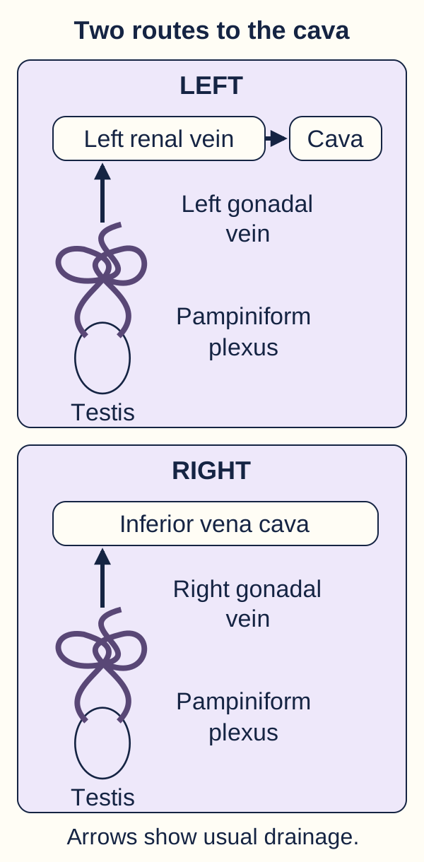
Why is a left-sided varicocele common, yet an isolated right-sided finding deserves particular attention? A varicocele is dilation of the pampiniform venous plexus, the network draining the testis, associated with venous reflux. It is a vascular structure above and around the testis, not a solid lump within it. The left gonadal vein usually enters the left renal vein; the right gonadal vein usually enters the inferior vena cava directly. [1][7]
The longer left venous column and near-perpendicular renal-vein junction favor higher hydrostatic pressure. Incompetent valves and reflux contribute to the common primary pattern. Geometry helps explain laterality but does not establish a patient's diagnosis. Right-sided reflux can occur without a tumor, and venous variants can alter the usual routes.
Trace the drainage diagram from each testis toward the heart. Now imagine narrowing the left renal vein before its junction with the cava. Predict which gonadal route experiences back pressure first.
Trace each route toward the cava. A left renal-vein obstruction can affect the left gonadal tributary; the usual right gonadal outlet is separate. [1][7]Check the side predicted by the route
The left route encounters that narrowing. The usual right route enters the cava independently.
The visible routes explain the consequence: left renal venous hypertension can transmit pressure to the left gonadal vein, as in symptomatic left renal-vein compression. A right renal-vein lesion alone does not usually obstruct the right gonadal vein. A right renal tumor can nevertheless produce a right varicocele through caval extension, external compression, or variant drainage. [1]
Apply this to a different distribution: right scrotal venous fullness plus bilateral leg swelling and abdominal-wall collaterals suggests a central outflow problem, not merely an isolated scrotal valve. Consider the cava and retroperitoneum. The distribution localizes the investigation; it does not identify whether the obstruction is tumor, thrombus, or compression.
Use posture as a venous pressure experiment
Does a soft scrotal mass behave like a pressure-dependent vein? Examine in a warm room with the patient standing. Inspect and palpate both testes and cords, then ask for Valsalva. The irregular compressible plexus is often described as a bag of worms. Straining can expose reflux and make small varicoceles palpable. Repeat the examination supine, noting whether the plexus becomes substantially less prominent. [1][7]
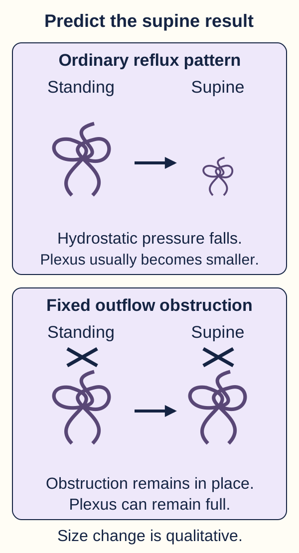
The posture diagram compares a freely draining system with impaired proximal drainage. Use it as a tracing exercise rather than a cancer test. For each system, follow standing, straining, then lying flat. Predict which component of pressure decreases when the patient lies down.
Compare the size change, not just the side. Lying flat reduces the hydrostatic contribution but does not correct a fixed outflow obstruction. This is a qualitative model, not a diagnostic test for cancer. [1]Check the effect of lying flat
The vertical hydrostatic contribution decreases. A fixed proximal obstruction is not corrected by posture.
A typical primary varicocele usually decompresses supine. Persistence, especially when newly developed, large, or tense, warrants investigation for a secondary cause. Incomplete collapse alone does not prove malignancy. Examination technique, severity and alternate scrotal masses also affect what is felt.
Example A: Years of mild fullness after standing, larger with Valsalva, nearly absent supine. This behavior supports ordinary reflux.
Example B: New fullness over several weeks, still tense supine, with flank discomfort. Lowering the hydrostatic component has not relieved filling, so proximal obstruction becomes more plausible.
Apply the comparison to an uncertain examination in a patient with a thick scrotal wall: failure to feel a plexus is not proof that reflux is absent. Scrotal color and spectral Doppler can document venous structures and flow, assess both testes, and distinguish a vascular plexus from another mass. Imaging answers a specific uncertainty; it is not obligatory screening for every adult with an otherwise clear examination. [2]
Let time course determine the first priority
Can a known varicocele explain every new episode of scrotal pain? Chronic venous aching usually worsens with prolonged standing and improves with rest or support. Abrupt severe pain, nausea, a high or horizontal testis, or an absent cremasteric reflex instead raises concern for torsion. Suspected torsion requires immediate surgical assessment, not an abdominal tumor search first. [5]
Patterns that require different first responses
Pattern
First clinical question
PatternSudden severe pain with abnormal testicular lie
First clinical questionIs the spermatic cord twisted?
PatternFever, dysuria and epididymal tenderness
First clinical questionIs inflammation or infection present?
PatternSoft cord veins filling while standing
First clinical questionDoes the plexus decompress supine?
PatternPersistent solid testicular or groin mass
First clinical questionIs this a tumor, hernia or another nonvenous lesion?
A teenager has sudden pain on the right and a longstanding small left varicocele. Select the finding that determines the first response: the old venous dilation or the new painful testis with an abnormal lie.
Check the priority
The new painful testis determines the emergency response. The contralateral venous finding does not explain it.
Doppler is useful when the diagnosis is uncertain and it can be obtained without delaying necessary intervention. Preserved arterial flow does not exclude early, partial, or intermittent torsion. Likewise, urinary findings cannot independently exclude torsion. Hyperemia and epididymal tenderness favor epididymitis in the appropriate context, but no single sign replaces the whole assessment. [5]
Transfer the rule to another mass: a smooth fluid collection around the testis is different from serpiginous refluxing veins; a groin-connected swelling may be a hernia. A painful irreducible hernia is also urgent. Evaluate the structure causing the current symptoms rather than assigning every scrotal abnormality to a pre-existing varicocele.
Describe the lesion without prescribing from its size
Does a larger vein mean that surgery is required? Clinical grade describes detection, not fertility and not cancer risk. Grade I is palpable only during Valsalva; grade II is palpable standing without Valsalva; grade III is visible. A subclinical varicocele is detected only by imaging, not by inspection or palpation even with Valsalva. [3]
When ultrasound is indicated, evaluate both sides, standing and supine, at rest and during Valsalva. Record the largest vein's diameter, location, patient position and reflux duration. ESUR-SPIWG uses a diameter of at least 3 mm while upright during Valsalva and considers reflux lasting more than 2 seconds abnormal. EAU describes diameter greater than 3 mm with reflux greater than 2 seconds. These are technique-dependent diagnostic supports, not treatment thresholds. [1][3]
Compare two reports: a 3.4 mm vein with 3.1 seconds of reflux measured standing during Valsalva, versus a 3.4 mm vein measured supine without a reflux assessment. Decide which report better characterizes venous dysfunction.
Check what the second report is missing
It lacks the provoked flow assessment. Diameter alone does not establish equivalent reflux.
The first report documents both dilation and abnormal flow under stated conditions. Neither report says whether the lesion is palpable or whether the patient has pain, testicular growth impairment, or infertility. Obtain those facts before discussing intervention. A tiny right-sided Doppler finding accompanying a palpable left varicocele is not the same presentation as an isolated clinical right varicocele.
Apply this distinction after repair: veins can remain enlarged even when reflux has stopped. Recurrent symptoms or unsatisfactory semen results can justify Doppler to assess persistent reflux, but enlarged veins alone do not establish procedural failure. For fertility treatment, follow semen findings and the reproductive plan rather than routinely scanning an improving patient. [1]
Investigate the abdomen for a clinical reason
Which right-sided presentation needs abdominal investigation? Compare a small stable lesion that empties supine with a newly developed large lesion that remains full and accompanies hematuria, flank symptoms, weight loss, a palpable abdominal mass, leg edema, or thrombosis risk. The latter combination raises concern for renal, caval, gonadal-vein or retroperitoneal disease. Neither right-sidedness nor a single examination sign proves cancer. [1][2]
Adult recommendations differ in how broadly they use abdominal ultrasound. AUA/ASRM's 2020 statement advises against routine abdominal imaging solely for an isolated small or moderate right varicocele. ESUR allows extending ultrasound to the abdomen for an isolated clinical right lesion, and EAU recommends further assessment for abdominal, retroperitoneal and congenital abnormalities. These positions do not require automatic CT for every asymptomatic adult. [1][2][3]
Renal and abdominal ultrasound can assess a suspected secondary cause without radiation. Cross-sectional CT or MRI is appropriate when the clinical concern, a detected mass, suspected venous extension, or an incomplete ultrasound requires more definition. A normal study that did not adequately visualize the relevant cava does not exclude caval disease. Choose the test to answer the unresolved question rather than requiring every patient to pass through the same sequence.
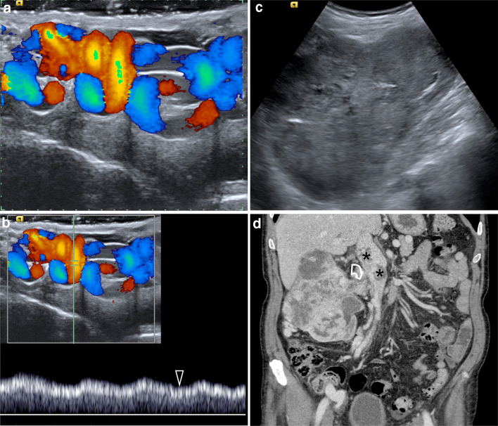
In the clinical figure, compare the refluxing scrotal vessels with the right renal mass and venous extension. Trace panel d from the renal mass through the renal vein toward the cava. Decide why the cava matters to the usual right gonadal drainage route.
Bertolotto et al., Figure 7: secondary right varicocele in an 85-year-old patient. Compare scrotal reflux (a, b) with the renal mass (c) and renal-vein/caval extension (d). The cava receives the usual right gonadal vein. Reproduced unchanged under CC BY 4.0; this selected example does not estimate malignancy risk. Image: Michele Bertolotto et al. and ESUR-SPIWG; original source; CC BY 4.0. [1].Check the structure connecting these findings
The cava receives the right gonadal vein. Tumor extending into it can compromise that outflow route.
The publication identifies this as an 85-year-old patient's secondary right varicocele. Panel a shows dilated vessels; panel b shows basal reflux that does not change with Valsalva; panel c shows a renal mass; panel d demonstrates renal-vein and caval extension. This is a selected teaching example, not evidence that most right-sided varicoceles have this cause. [1]
In a retrospective cohort selected for isolated right varicoceles without other evidence of malignancy, no causative cancer was found among 210 patients; the 95% confidence interval was 0.0% to 1.4%. That supports avoiding indiscriminate screening, not a zero-risk claim and not dismissal of a patient with systemic symptoms. [6]
Transfer the decision carefully across ages. Pediatric EAU guidance recommends abdominal ultrasound for all prepubertal boys with varicocele and for isolated right varicoceles in children and adolescents. Do not apply adult selective-imaging language to those groups. Similarly, a new tense left varicocele with systemic symptoms can be secondary: the concerning behavior matters on either side. [4][1]
Repair a clinical problem, not an isolated number
What outcome would treatment be intended to improve? In some patients, reflux impairs testicular temperature regulation and is associated with hypoxia and oxidative stress, potentially affecting spermatogenesis. Many patients remain fertile. The presence or grade of a varicocele therefore cannot substitute for functional assessment. [7]
For an infertile couple, discuss repair when a varicocele is palpable, semen parameters are abnormal, and the couple's overall evaluation supports benefit. Repeat an initially abnormal semen analysis because values vary, and assess both partners concurrently. Reference limits describe a population, not a rigid boundary between fertile and infertile. Routine repair of an imaging-only varicocele is not recommended. Non-obstructive azoospermia requires separate specialist counseling because evidence that repair helps before assisted reproduction is uncertain. [2][3][7]
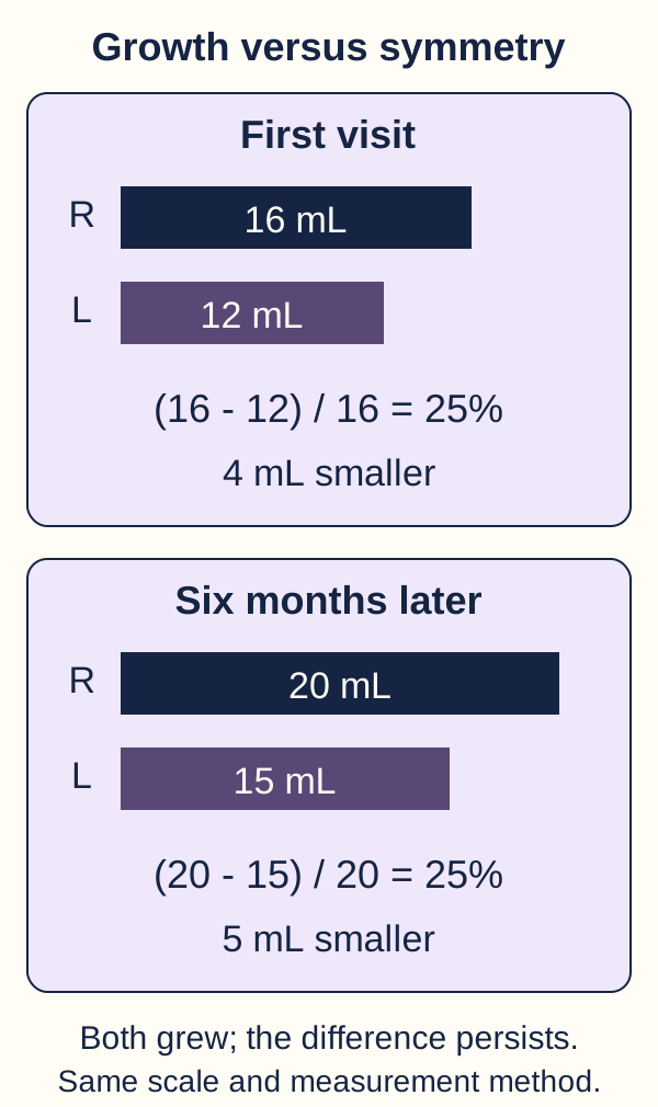
For adolescents, serial testicular growth is more informative than one measurement. Pediatric guidance supports offering surgery for a persistently smaller testis, using a difference greater than 2 mL or 20%, confirmed on two visits six months apart. The diagram compares absolute growth with persistent relative asymmetry. Use the larger testis as the denominator: percentage difference = (larger volume minus smaller volume) / larger volume × 100. [4]
Calculate the difference for testes measuring 16 and 12 mL. Then repeat for 20 and 15 mL six months later. Has growth of the smaller testis resolved the asymmetry?
Calculate the relative difference at both visits. Both testes grew, yet the left remains 25% smaller. Consistent measurements and persistent asymmetry inform adolescent counseling. [4]Check the two comparisons
Both differences are 25%. Absolute growth occurred, but relative asymmetry persisted.
The consequence is a specialist discussion of repair, not a promise of future paternity. In contrast, one small difference with no symptoms merits follow-up rather than immediate prophylactic surgery. Use consistent measurement methods; asynchronous pubertal growth and measurement variation can create temporary differences. Selected older adolescents may also be assessed with semen analysis. [4]
For chronic dull pain, first exclude torsion, infection, hernia and a mass. Begin scrotal support, activity adjustment and appropriate analgesia. Persistent function-limiting pain despite conservative care can justify intervention, but pain can persist afterward. A varicocele coincident with pain does not establish causation. [7]
Microsurgical inguinal or subinguinal ligation aims to interrupt refluxing veins while preserving arteries and lymphatics. Microsurgical approaches generally have lower recurrence and hydrocele rates than nonmicrosurgical surgery. Laparoscopic approaches are alternatives; lymphatic preservation reduces hydrocele risk. Injury to the testicular artery can rarely cause atrophy, so arterial preservation matters. Percutaneous embolization avoids a surgical incision at the cord but requires venous access and brings fluoroscopic radiation, contrast, technical failure and rare coil-related risks. Method selection depends on anatomy, expertise, prior repair and patient priorities. [3][4]
Apply the fertility decision to a couple with limited ovarian reserve: waiting several months for semen improvement may conflict with the reproductive timeline. Coordinate repair and assisted reproduction rather than assuming surgery must always precede fertility treatment. Improvement in semen parameters is possible; pregnancy is not guaranteed. [3][7]
Independent clinical practice
Case 1
Show answer and explanations for case 1
A. Cava between the iliac confluence and the gonadal outlet (Why this does not fit)
An obstruction below the gonadal outlet can impair venous return from both legs. The right gonadal vein enters above this segment, so this location does not directly obstruct both supplied drainage territories.
Reasoning steps for option A
Can a caval lesion below the right gonadal outlet impede both iliac veins?
An obstruction below the gonadal outlet can impair venous return from both legs.
Why does the right gonadal outlet escape that lower caval lesion?
The right gonadal vein enters above this segment, so this location does not directly obstruct both supplied drainage territories.
B. Iliac confluence with a patent cava above it (Why this does not fit)
The iliac confluence is shared lower-limb venous outflow and can explain bilateral leg congestion. The right gonadal vein joins the cava farther upstream and would not have its usual outlet directly obstructed by an isolated confluence lesion.
Reasoning steps for option B
Which drainage territory meets at the iliac confluence?
The iliac confluence is shared lower-limb venous outflow and can explain bilateral leg congestion.
Why would an isolated iliac lesion spare the right gonadal outlet?
The right gonadal vein joins the cava farther upstream and would not have its usual outlet directly obstructed by an isolated confluence lesion.
C. Cava above the right gonadal-vein outlet (Best answer)
Bilateral leg edema and abdominal collaterals localize a central lower-body venous outflow problem. The persistent right varicocele then places the shared obstruction above the usual right gonadal outlet rather than below it.
Reasoning steps for option C
What do bilateral edema and abdominal-wall collaterals imply about venous outflow?
Bilateral leg edema and abdominal collaterals localize a central lower-body venous outflow problem.
Where must one lesion sit relative to the right gonadal outlet to also sustain the right varicocele?
The persistent right varicocele then places the shared obstruction above the usual right gonadal outlet rather than below it.
D. Right gonadal vein below its caval outlet (Why this does not fit)
A gonadal lesion can explain unilateral scrotal venous pressure and failure to decompress. It does not lie in the shared drainage route of both legs and therefore cannot explain the full distribution as one lesion.
Reasoning steps for option D
What scrotal finding could an isolated right gonadal-vein lesion explain?
A gonadal lesion can explain unilateral scrotal venous pressure and failure to decompress.
Why can that unilateral lesion not account for bilateral ankle edema?
It does not lie in the shared drainage route of both legs and therefore cannot explain the full distribution as one lesion.
Takeaway: Use leg drainage to identify a central vessel, then gonadal drainage to determine the relevant level.
A. Left gonadal reflux without bilateral iliac congestion (Best answer)
The narrowed vein connecting the left renal hilum to the cava is the left renal vein. Its gonadal tributary can transmit pressure to the left scrotal plexus, while patent iliac and caval drainage argues against bilateral leg congestion.
Reasoning steps for option A
Which vein is compressed between the aorta and superior mesenteric artery near the left renal hilum?
The narrowed vein connecting the left renal hilum to the cava is the left renal vein.
How does its gonadal tributary produce scrotal reflux without obstructing the patent iliacs?
Its gonadal tributary can transmit pressure to the left scrotal plexus, while patent iliac and caval drainage argues against bilateral leg congestion.
B. Right gonadal reflux without left gonadal involvement (Why this does not fit)
The right gonadal vein usually enters the cava directly. A lesion localized to the vein from the left renal hilum instead predicts pressure transmission through the left gonadal tributary.
Reasoning steps for option B
Where does the usual right gonadal vein empty relative to the compressed left renal vein?
The right gonadal vein usually enters the cava directly.
Which side should receive pressure from this left hilar narrowing instead?
A lesion localized to the vein from the left renal hilum instead predicts pressure transmission through the left gonadal tributary.
C. Bilateral iliac congestion without gonadal reflux (Why this does not fit)
Bilateral iliac congestion suggests a problem in common lower-body venous drainage. The described renal-hilar route receives the left gonadal vein rather than both iliac veins.
Reasoning steps for option C
What common outflow lesion would be needed for bilateral iliac congestion?
An obstruction in shared lower-body outflow, such as the cava, could congest both iliac territories. The described lesion is instead confined to the left renal vein, with patent cava and iliacs.
Does the compressed left renal vein receive iliac drainage or the left gonadal tributary?
The described renal-hilar route receives the left gonadal vein rather than both iliac veins.
D. Right renal congestion with right gonadal reflux (Why this does not fit)
A shared caval obstruction could affect right renal and gonadal drainage. The patent cava and localized left hilar venous narrowing do not support that right-sided distribution.
Reasoning steps for option D
What shared obstruction could congest the right kidney and right gonadal route?
A shared caval obstruction could affect right renal and gonadal drainage.
How do the patent cava and left-sided location contradict this predicted right pattern?
The patent cava and localized left hilar venous narrowing do not support that right-sided distribution.
Takeaway: First identify the compressed vessel, then trace which tributaries can transmit its elevated pressure.
A. Obtain contrast abdominal CT at this visit (Why this does not fit)
CT can evaluate suspected renal or retroperitoneal disease. Long duration, complete decompression and absence of concerning findings do not justify CT solely for laterality.
Reasoning steps for option A
What suspicion would make contrast CT useful for an isolated right varicocele?
CT can evaluate suspected renal or retroperitoneal disease.
Which stable and decompressive features argue against CT for laterality alone?
Long duration, complete decompression and absence of concerning findings do not justify CT solely for laterality.
B. Obtain abdominal MRI within the next week (Why this does not fit)
MRI can characterize an abdominal mass or venous extension. No such abnormality is suggested here, and substituting MRI does not create an indication for routine screening.
Reasoning steps for option B
What lesion or extension is abdominal MRI ordinarily intended to characterize here?
MRI can characterize an abdominal mass or venous extension.
Why does choosing MRI rather than CT not justify screening this asymptomatic adult?
No such abnormality is suggested here, and substituting MRI does not create an indication for routine screening.
C. Obtain one-time renal and abdominal ultrasonography (Why this does not fit)
ESUR and EAU support broader abdominal assessment in isolated clinical right varicocele. The question specifically asks for the AUA/ASRM adult statement, which does not mandate imaging solely for this stable small lesion.
Reasoning steps for option C
Which societies allow broader ultrasound assessment of an isolated clinical right lesion?
ESUR and EAU support broader abdominal assessment in isolated clinical right varicocele.
Why does the specified AUA/ASRM recommendation not require that ultrasound here?
The question specifically asks for the AUA/ASRM adult statement, which does not mandate imaging solely for this stable small lesion.
D. Reserve abdominal imaging for additional concern (Best answer)
AUA/ASRM advises against routine abdominal imaging solely for a small or moderate isolated right varicocele. The stable, decompressive adult presentation fits that recommendation, while a new concerning feature would change the decision.
Reasoning steps for option D
What does AUA/ASRM say about routine abdominal imaging for a small isolated right varicocele?
AUA/ASRM advises against routine abdominal imaging solely for a small or moderate isolated right varicocele.
Which new feature would reverse the selective-imaging decision in this patient?
New onset or enlargement, persistence supine, hematuria, weight loss or edema would raise concern for a secondary cause. None is present in this stable, small, decompressive adult lesion.
Takeaway: State the guideline and the adult clinical context when applying selective abdominal imaging.
A. Repeat scrotal Doppler after six months (Why this does not fit)
Follow-up Doppler can reassess a stable scrotal lesion. It would not resolve the current systemic symptoms and incompletely evaluated abdominal drainage.
Reasoning steps for option A
What clinical problem would repeat scrotal Doppler actually reassess?
Follow-up Doppler can reassess a stable scrotal lesion.
Why cannot six-month scrotal follow-up settle his hematuria and poorly seen cava?
It would not resolve the current systemic symptoms and incompletely evaluated abdominal drainage.
B. Contrast-enhanced abdominal and pelvic CT (Best answer)
Cross-sectional imaging can define renal or retroperitoneal disease and venous involvement. Persistent new reflux with hematuria, weight loss and an incomplete abdominal ultrasound warrants resolving that proximal question.
Reasoning steps for option B
What proximal structures can contrast abdominal and pelvic CT define?
Cross-sectional imaging can define renal or retroperitoneal disease and venous involvement.
Which red flags and ultrasound limitation justify CT now?
Persistent new reflux with hematuria, weight loss and an incomplete abdominal ultrasound warrants resolving that proximal question.
C. Repeat targeted renal ultrasound in six months (Why this does not fit)
Ultrasound can evaluate renal and selected proximal venous pathology. Deferring reassessment does not resolve current hematuria, weight loss and an incompletely seen retroperitoneum in a suspicious new presentation.
Reasoning steps for option C
What can a targeted renal ultrasound assess when visualization is adequate?
Ultrasound can evaluate renal and selected proximal venous pathology.
Why is six-month deferral unsafe with weight loss and incomplete retroperitoneal views?
Deferring reassessment does not resolve current hematuria, weight loss and an incompletely seen retroperitoneum in a suspicious new presentation.
D. Obtain noncontrast CT limited to urinary stones (Why this does not fit)
A noncontrast stone study can investigate selected causes of hematuria. It is not the best tailored examination when the unresolved concern is renal or retroperitoneal disease and venous involvement.
Reasoning steps for option D
Which limited differential does noncontrast urinary-stone CT address?
A noncontrast stone protocol targets urinary calculi as a cause of hematuria, rather than defining the suspected renal or retroperitoneal disease and venous involvement.
Why is a stone-only study insufficient to characterize possible venous extension?
It is not the best tailored examination when the unresolved concern is renal or retroperitoneal disease and venous involvement.
Takeaway: An incomplete negative ultrasound cannot dismiss a clinically suspicious proximal obstruction.
A. Arrange outpatient reassessment after analgesia (Why this does not fit)
Observation may be appropriate for a benign chronic pain pattern. Sudden pain, vomiting and abnormal testicular lie demand urgent assessment despite detectable arterial flow.
Reasoning steps for option A
For which tempo of scrotal discomfort could outpatient observation be reasonable?
Observation may be appropriate for a benign chronic pain pattern.
Which acute findings outweigh detectable Doppler arterial flow here?
B. Begin antibiotics and review after 48 hours (Why this does not fit)
Epididymitis can cause painful scrotal inflammation. The abrupt onset and abnormal lie strongly suggest torsion, and an antibiotic trial would delay necessary assessment.
Reasoning steps for option B
Which competing inflammatory condition might warrant antibiotics?
Epididymitis can cause painful scrotal inflammation.
What features instead make a 48-hour antibiotic trial hazardous?
The abrupt onset and abnormal lie strongly suggest torsion, and an antibiotic trial would delay necessary assessment.
C. Obtain immediate urologic surgical assessment (Best answer)
Partial or early torsion may retain detectable arterial flow. The acute symptoms and abnormal examination warrant immediate surgical evaluation rather than reassurance from flow alone.
Reasoning steps for option C
Can partial or early torsion preserve some arterial perfusion?
Partial or early torsion may retain detectable arterial flow.
What examination findings mandate immediate urologic assessment despite that flow?
The horizontal testicular lie and absent cremasteric reflex, with sudden pain and vomiting, strongly suggest torsion and require immediate surgical assessment despite detectable arterial flow.
D. Repeat expert Doppler before contacting urology (Why this does not fit)
Expert Doppler can help resolve an equivocal acute scrotal presentation. This strongly suspicious examination warrants immediate surgical assessment without waiting for a repeat imaging study.
Reasoning steps for option D
When can expert Doppler clarify an acute scrotal presentation?
Expert Doppler can help resolve an equivocal acute scrotal presentation.
Why must repeat imaging not precede calling urology in this teenager?
This strongly suspicious examination warrants immediate surgical assessment without waiting for a repeat imaging study.
A. Right primary varicocele causing referred inflammatory pain (Why this does not fit)
A varicocele can cause aching associated with standing. The reflux is contralateral and does not explain the febrile urinary syndrome or focal left epididymal abnormality.
Reasoning steps for option A
What usual pain pattern can a right primary varicocele produce?
A varicocele can cause aching associated with standing.
Why does right-only provoked reflux fail to explain fever and left epididymal hyperemia?
The reflux is contralateral and does not explain the febrile urinary syndrome or focal left epididymal abnormality.
B. Left epididymal inflammation causing the painful swelling (Best answer)
Epididymal tenderness, urinary symptoms and hyperemia support epididymitis. The contralateral provoked venous finding is incidental to the symptomatic inflammatory pattern.
Reasoning steps for option B
Which combination localizes inflammation to the left epididymis?
Epididymal tenderness, urinary symptoms and hyperemia support epididymitis.
Why is the contralateral Valsalva-only plexus not the current pain source?
The contralateral provoked venous finding is incidental to the symptomatic inflammatory pattern.
C. Left gonadal-vein obstruction causing venous congestion (Why this does not fit)
Proximal obstruction can cause persistent gonadal venous distention. The described abnormality is epididymal rather than evidence of gonadal obstruction, and the urinary-inflammatory pattern favors epididymitis.
Reasoning steps for option C
What venous behavior would favor left proximal gonadal obstruction?
Proximal obstruction can cause persistent gonadal venous distention.
Which observed tissue and urinary features instead point to epididymitis?
Left epididymal tenderness, enlargement and hyperemia accompany dysuria and fever. That localized inflammatory pattern supports epididymitis rather than left gonadal-vein obstruction.
D. Left appendix-testis torsion causing the whole syndrome (Why this does not fit)
Appendix-testis torsion can cause focal upper-pole pain and secondary inflammation. The combination of dysuria, fever and diffuse epididymal enlargement is more consistent with epididymitis than isolated appendage torsion.
Reasoning steps for option D
What focal pain pattern can appendix-testis torsion cause?
Appendix-testis torsion can cause focal upper-pole pain and secondary inflammation.
Why do dysuria and diffuse hyperemic epididymal enlargement favor another cause?
The combination of dysuria, fever and diffuse epididymal enlargement is more consistent with epididymitis than isolated appendage torsion.
Takeaway: Match symptoms to the affected structure; preserved arterial flow alone never excludes torsion.
A. Grade I; offer a fertility-directed repair discussion (Why this does not fit)
Grade I requires Valsalva for palpation. Repair counseling may fit the reproductive findings, but this lesion is palpable without straining and is therefore grade II.
Reasoning steps for option A
What maneuver distinguishes grade I from this standing-palpable lesion?
Grade I requires Valsalva for palpation.
Why can fertility counseling still be reasonable despite the incorrect grade label?
Repeated concentration and motility below the supplied limits, 18 months of infertility and a favorable partner evaluation support repair counseling. Palpability without Valsalva makes the lesion grade II, not grade I.
B. Grade II; defer repair discussion until visible (Why this does not fit)
Palpability at rest without visibility defines grade II. Visibility is not required when infertility and repeat semen abnormalities support a clinical-varicocele treatment discussion.
Reasoning steps for option B
How is a nonvisible vein palpable without Valsalva graded?
Palpability at rest without visibility defines grade II.
Why need this infertile couple not wait for visible grade III disease?
Visibility is not required when infertility and repeat semen abnormalities support a clinical-varicocele treatment discussion.
C. Grade I; defer repair discussion until visible (Why this does not fit)
An imaging or examination descriptor alone is not an operative indication. The lesion is grade II rather than grade I, and the supplied functional and couple findings justify discussion without waiting for visibility.
Reasoning steps for option C
Can grade alone decide whether fertility-directed repair merits discussion?
An imaging or examination descriptor alone is not an operative indication.
Which grade and functional findings contradict postponing until visibility?
Palpability without Valsalva establishes grade II. Repeated low concentration and motility with infertility and a favorable partner evaluation support discussing repair without waiting for visibility.
D. Grade II; offer a fertility-directed repair discussion (Best answer)
Palpability without Valsalva and absence of visibility define grade II. Repeated low concentration and motility in an infertile couple with a favorable partner evaluation support discussing repair.
Reasoning steps for option D
What examination findings establish grade II rather than grade I?
Palpability without Valsalva and absence of visibility define grade II.
Which repeated semen and partner findings support repair counseling now?
Concentrations of 10 and 11 million/mL are below 16, and progressive motilities of 22% and 24% are below 30%. These repeated abnormalities, infertility and a favorable partner evaluation support repair counseling.
Takeaway: Classify the examination and then apply functional criteria; neither grade nor semen findings alone completes the decision.
A. Continue the couple's infertility evaluation (Best answer)
Ultrasound can identify reflux that is not clinically palpable. An imaging-only varicocele does not meet the conventional repair indication even when semen findings are abnormal.
Reasoning steps for option A
How can Doppler detect a varicocele despite repeated negative standing examinations?
Ultrasound can identify reflux that is not clinically palpable.
Why is this imaging-only lesion not a conventional repair indication despite low concentration?
An imaging-only varicocele does not meet the conventional repair indication even when semen findings are abnormal.
B. Offer microsurgical repair based on the vein diameter (Why this does not fit)
A dilated refluxing vein supports an ultrasound diagnosis. Diameter does not replace clinical palpability when selecting conventional fertility-directed repair.
Reasoning steps for option B
What does a 3.2 mm refluxing vein demonstrate on ultrasound?
A dilated refluxing vein supports an ultrasound diagnosis.
Why does diameter not substitute for clinical palpability in fertility repair decisions?
The 3.2 mm vein supports an imaging diagnosis, but repeated examinations find no palpable varicocele. Diameter cannot supply the clinical criterion needed for conventional fertility-directed repair.
C. Offer embolization based on the reflux duration (Why this does not fit)
Prolonged reflux demonstrates venous dysfunction under the measured conditions. Changing the treatment method does not overcome the lack of a clinical repair indication.
Reasoning steps for option C
What does 2.8 seconds of standing Valsalva reflux document?
Prolonged reflux demonstrates venous dysfunction under the measured conditions.
Why does embolization not bypass the missing clinical indication?
Embolization still treats a varicocele; changing the procedure does not make this repeatedly nonpalpable, imaging-only lesion a conventional fertility-treatment indication.
D. Repeat Doppler monthly until repair is indicated (Why this does not fit)
Doppler describes caliber and reflux under specified conditions. Serial imaging does not replace the clinical palpability criterion or advance the remaining couple-level infertility evaluation.
Reasoning steps for option D
What information can repeated scrotal Doppler measure?
Doppler describes caliber and reflux under specified conditions.
Why would monthly imaging not resolve the missing palpable finding or couple assessment?
Serial imaging does not replace the clinical palpability criterion or advance the remaining couple-level infertility evaluation.
Takeaway: Abnormal semen plus imaging-only reflux is not the same indication as a palpable varicocele.
A. Resolved asymmetry; observe because both testes grew (Why this does not fit)
Growth in both testes indicates pubertal development. The left testis remains 25% smaller at both visits, so absolute growth does not resolve asymmetry.
Reasoning steps for option A
What pubertal change is evident from 16 to 20 mL and 12 to 15 mL?
Growth in both testes indicates pubertal development.
Why does growth of both testes not mean their relative size difference resolved?
The left testis remains 25% smaller at both visits, so absolute growth does not resolve asymmetry.
B. New 25% asymmetry; wait six months to confirm it (Why this does not fit)
An isolated finding of asymmetry should be confirmed across visits. Both supplied visits already demonstrate 25% asymmetry six months apart, so this is not a newly unconfirmed difference.
Reasoning steps for option B
When does a single observation of adolescent testicular asymmetry need confirmation?
An isolated finding of asymmetry should be confirmed across visits.
Why are these two 25% readings not a newly unconfirmed asymmetry?
Both supplied visits already demonstrate 25% asymmetry six months apart, so this is not a newly unconfirmed difference.
C. Persistent 25% asymmetry; offer specialist repair counseling (Best answer)
Both (16 minus 12)/16 and (20 minus 15)/20 equal 25%. Persistence across visits six months apart meets the guideline pattern for discussing surgery for a small ipsilateral testis.
Reasoning steps for option C
What percentage results from each visit using the larger testis as denominator?
Both (16 minus 12)/16 and (20 minus 15)/20 equal 25%.
Why does six-month persistence above 20% justify specialist repair counseling?
The left testis is 25% smaller at both visits six months apart. This persistent difference exceeds 20% and supports specialist counseling about repair for a small ipsilateral testis.
D. Persistent 15% asymmetry; continue routine observation (Why this does not fit)
A smaller difference without other concerns may justify continued surveillance. Using the larger testis as denominator gives 25%, not 15%, and this persistent difference meets the threshold for offering repair counseling.
Reasoning steps for option D
What management might follow a genuinely small relative difference?
A smaller difference without other concerns may justify continued surveillance.
Why is 15% inconsistent with the supplied 16:12 and 20:15 volumes?
Using the larger testis as denominator gives 25%, not 15%, and this persistent difference meets the threshold for offering repair counseling.
Takeaway: Serial relative size matters even when both testes increase in absolute volume.
A. True bilateral volume loss with persistent asymmetry; discuss repair (Why this does not fit)
The final reported volumes fall when the multiplier changes from 0.71 to 0.52. Unchanged dimensions do not establish true volume loss, even though the persistent left-right difference warrants specialist discussion.
Reasoning steps for option A
How does changing the volume multiplier affect reported absolute volumes?
The final reported volumes fall when the multiplier changes from 0.71 to 0.52.
Why do identical dimensions fail to show true bilateral tissue loss?
Unchanged dimensions do not establish true volume loss, even though the persistent left-right difference warrants specialist discussion.
B. Formula-related change with persistent asymmetry; discuss repair (Best answer)
The raw products are 28 and 21 cm cubed at both visits, so either shared multiplier leaves the left testis 25% smaller. The apparent overall decline is methodological, but persistent 25% asymmetry six months apart supports a specialist repair discussion.
Reasoning steps for option B
What are the unchanged right and left dimension products and their relative difference?
The raw products are 28 and 21 cm cubed at both visits, so either shared multiplier leaves the left testis 25% smaller.
How should persistent 25% asymmetry be interpreted despite changed multipliers?
The apparent overall decline is methodological, but persistent 25% asymmetry six months apart supports a specialist repair discussion.
C. Formula-related change with resolved asymmetry; continue observation (Why this does not fit)
Changing the multiplier can account for lower absolute volume estimates without tissue loss. It does not resolve the left-right ratio: (28 minus 21)/28 remains 25% at both visits.
Reasoning steps for option C
Can switching from 0.71 to 0.52 make absolute volumes appear lower?
Changing the multiplier can account for lower absolute volume estimates without tissue loss.
Why does that switch leave the left-right percentage asymmetry unresolved?
It does not resolve the left-right ratio: (28 minus 21)/28 remains 25% at both visits.
D. True bilateral volume loss with resolved asymmetry; continue observation (Why this does not fit)
Smaller reported numbers can suggest tissue loss if measurement methods are comparable. Here the methods differ and the unchanged dimension products preserve the same 25% asymmetry, so neither part of this interpretation fits.
Reasoning steps for option D
When would falling reported bilateral volumes suggest true tissue loss?
Smaller reported numbers can suggest tissue loss if measurement methods are comparable.
Which unchanged dimensions and ratio refute both loss and resolved asymmetry?
The unchanged dimension products remain 28 on the right and 21 on the left, preserving 25% asymmetry. Only the shared multiplier changed, so the reports show neither demonstrated tissue loss nor resolved asymmetry.
Takeaway: Correct the method artifact without overlooking a persistent clinically relevant size difference.
A. Pain indication without a semen indication; discuss intervention (Best answer)
Both concentration and progressive motility are above the supplied reference limits, so the data do not support a semen-based repair indication. The postural pain pattern with persistent limitation after conservative care supports a separate intervention discussion, with no guarantee of relief.
Reasoning steps for option A
How do the two concentration and motility results compare with supplied limits?
Concentrations of 32 and 35 million/mL exceed 16, and progressive motilities of 42% and 44% exceed 30%. Neither sample supplies the abnormal semen findings needed for a semen-based repair indication.
What separate pain pattern and failed treatment support discussion of intervention?
The postural pain pattern with persistent limitation after conservative care supports a separate intervention discussion, with no guarantee of relief.
B. Pain indication without a semen indication; wait for abnormal semen (Why this does not fit)
The symptoms and reproductive data support a pain concern rather than a semen-based indication. Abnormal semen is not required before discussing intervention for persistent appropriately evaluated pain.
Reasoning steps for option B
Which indication is supported by refractory postural pain rather than these semen samples?
The symptoms and reproductive data support a pain concern rather than a semen-based indication.
Why is an abnormal semen result not a prerequisite for pain-directed counseling?
Abnormal semen is not required before discussing intervention for persistent appropriately evaluated pain.
C. Semen indication without a pain indication; discuss intervention (Why this does not fit)
A clinical varicocele with abnormal semen can support fertility-directed repair in the appropriate couple. These repeated parameters exceed the supplied limits, while the refractory postural pain is the relevant treatment concern.
Reasoning steps for option C
What fertility criterion would abnormal semen add to a palpable varicocele?
A clinical varicocele with abnormal semen can support fertility-directed repair in the appropriate couple.
Why do these two above-limit samples fail to establish that indication?
These repeated parameters exceed the supplied limits, while the refractory postural pain is the relevant treatment concern.
D. Both pain and semen indications; discuss intervention (Why this does not fit)
Persistent postural pain after conservative care can support treatment discussion. The two semen samples do not show the abnormalities needed to add a semen-based indication to this assessment.
Reasoning steps for option D
What does three months of failed support and analgesia imply for his chronic pain?
Persistent postural pain after conservative care can support treatment discussion.
Why cannot normal-range semen values be counted as a second indication?
The two semen samples do not show the abnormalities needed to add a semen-based indication to this assessment.
Takeaway: Interpret reproductive data separately from the pain pattern before naming the reason for intervention.
A. Schedule embolization based on this abnormal sample (Why this does not fit)
Both concentration and motility are below the supplied references in this sample. One sample and an unassessed partner do not complete the evaluation needed for a reproductive treatment plan.
Reasoning steps for option A
Which two semen measures fall below reference limits in the first sample?
Concentration is 13 million/mL versus a lower reference limit of 16, and progressive motility is 27% versus 30%. Both measures are low, but this is only the first sample.
Why is immediate embolization premature without repeat testing and partner evaluation?
One sample and an unassessed partner do not complete the evaluation needed for a reproductive treatment plan.
B. Repeat male testing before starting any partner evaluation (Why this does not fit)
Repeating an initially abnormal semen analysis helps characterize a variable result. Deferring partner assessment makes the evaluation sequential when concurrent assessment is recommended for an infertile couple.
Reasoning steps for option B
Why repeat an initially abnormal semen sample?
Repeating an initially abnormal semen analysis helps characterize a variable result.
Why should the partner not wait until male retesting finishes?
Deferring partner assessment makes the evaluation sequential when concurrent assessment is recommended for an infertile couple.
C. Defer further evaluation until two years of infertility (Why this does not fit)
Infertility evaluation is appropriate after 12 months of regular unprotected intercourse. Fourteen months with an abnormal semen sample is sufficient reason to proceed now.
Reasoning steps for option C
After how many months of infertility is evaluation generally appropriate?
Infertility evaluation is appropriate after 12 months of regular unprotected intercourse.
Why does this couple's 14-month history not require waiting to two years?
Fourteen months with an abnormal semen sample is sufficient reason to proceed now.
D. Repeat semen testing and assess both partners concurrently (Best answer)
Semen parameters vary between ejaculates and partner factors affect treatment timing and benefit. Repeat testing and concurrent couple assessment clarify whether repair fits the full reproductive plan.
Reasoning steps for option D
Why do variable semen results and partner factors matter before repair?
Semen parameters vary between ejaculates and partner factors affect treatment timing and benefit.
Which parallel investigations complete the next step for this couple?
Repeat testing and concurrent couple assessment clarify whether repair fits the full reproductive plan.
Takeaway: Do not convert one abnormal semen result into a completed couple-level treatment decision.
A. Repair candidate; require a six-month postoperative interval before assisted reproduction (Why this does not fit)
A palpable varicocele with repeated semen abnormalities supports considering repair. A mandatory interval to await uncertain semen improvement can conflict with the partner's limited ovarian reserve.
Reasoning steps for option A
What male findings make repair a reasonable discussion?
A palpable varicocele with repeated semen abnormalities supports considering repair.
Why can a compulsory six-month postoperative wait harm this couple's timeline?
A mandatory interval to await uncertain semen improvement can conflict with the partner's limited ovarian reserve.
B. Not a repair candidate; proceed with assisted reproduction alone (Why this does not fit)
The partner's ovarian reserve can make assisted reproduction time-sensitive. It does not erase the man's clinical-varicocele repair criteria, so repair can still be discussed within the shared plan.
Reasoning steps for option B
Why can diminished ovarian reserve make assisted reproduction urgent?
At age 39 with diminished ovarian reserve, the partner has a time-sensitive reproductive concern. Waiting months for an uncertain response to male repair may conflict with her treatment timeline.
Why does that urgency not negate the man's clinical repair candidacy?
It does not erase the man's clinical-varicocele repair criteria, so repair can still be discussed within the shared plan.
C. Repair candidate; coordinate care without a mandatory repair-first delay (Best answer)
The examination, repeat semen abnormalities and infertility support a repair discussion. Possible semen improvement takes months, so limited ovarian reserve favors coordinated planning rather than automatically delaying assisted reproduction.
Reasoning steps for option C
Which exam, semen and infertility data support considering repair?
A palpable varicocele, two years of infertility, concentrations of 9 and 10 million/mL and progressive motilities of 21% and 23% support repair discussion. Both semen measures are repeatedly below the supplied limits.
How should months-long potential semen improvement affect sequencing with low ovarian reserve?
Possible semen improvement takes months, so limited ovarian reserve favors coordinated planning rather than automatically delaying assisted reproduction.
D. Not a repair candidate; defer partner treatment pending semen normalization (Why this does not fit)
A male assessment and semen results contribute to a couple-level plan. This man has findings supporting consideration of repair, and waiting for normalization does not address the partner's time-sensitive reproductive concern.
Reasoning steps for option D
What information should the male assessment contribute to a shared fertility plan?
The palpable varicocele and repeated low concentration and motility support male repair candidacy. Those findings must be considered alongside the partner's diminished ovarian reserve when choosing treatment sequence.
Why are both denial of repair candidacy and waiting for semen normalization inappropriate?
This man has findings supporting consideration of repair, and waiting for normalization does not address the partner's time-sensitive reproductive concern.
Takeaway: Eligibility for a repair discussion does not determine that repair must precede assisted reproduction.
Arterial injury can threaten testicular perfusion and cause atrophy. Preserved perfusion and a simple fluid collection favor impaired lymphatic drainage rather than arterial failure.
Reasoning steps for option A
Would testicular artery damage produce normal perfusion and isolated simple fluid?
Arterial damage can compromise testicular perfusion and cause atrophy. A normally perfused testis with simple surrounding fluid instead favors a drainage complication rather than arterial failure.
Which postoperative finding argues against ischemic atrophy?
Preserved perfusion and a simple fluid collection favor impaired lymphatic drainage rather than arterial failure.
B. Testicular lymphatic vessels (Best answer)
Lymphatics return interstitial fluid from the testicular region. Disruption during varicocele surgery can cause a postoperative hydrocele despite normal arterial perfusion and absent reflux.
Reasoning steps for option B
What drainage pathway fails when a hydrocele follows varicocele repair?
Disrupted testicular lymphatic vessels fail to return interstitial fluid normally, allowing a postoperative hydrocele to develop around the testis.
Why can lymphatic injury cause swelling despite intact arterial flow and no reflux?
Lymphatic disruption impairs fluid drainage, so a hydrocele can develop even when arterial perfusion is preserved and Doppler shows no recurrent venous reflux.
C. Internal spermatic veins (Why this does not fit)
Persistent or recurrent reflux through gonadal venous branches can cause a recurrent varicocele. The supplied finding is simple fluid without reflux, favoring lymphatic disruption rather than recurrent venous disease.
Reasoning steps for option C
What would recurrent internal spermatic venous reflux produce on Doppler?
Recurrent internal spermatic venous disease would produce venous reflux in a recurrent varicocele, not merely the simple fluid collection seen here.
Why does simple peritesticular fluid argue against recurrent venous disease?
The supplied finding is simple fluid without reflux, favoring lymphatic disruption rather than recurrent venous disease.
D. Vas deferens (Why this does not fit)
Vasal injury can impair sperm transport. It does not directly explain a new simple peritesticular fluid collection after venous surgery.
Reasoning steps for option D
What function is threatened by vas deferens injury?
Vasal injury can impair sperm transport.
Does impaired sperm transport explain a painless fluid collection around the testis?
It does not directly explain a new simple peritesticular fluid collection after venous surgery.
Takeaway: A postoperative hydrocele reflects fluid drainage, not proof of recurrent venous reflux.
A. No proven recurrence; continue couple-level fertility care (Best answer)
Residual dilation without reflux does not establish recurrent varicocele. Semen concentration has improved but remains below the reference limit and pregnancy has not occurred, so reproductive follow-up remains appropriate.
Reasoning steps for option A
Does a residual 3.3 mm cord vein prove recurrent reflux when standing Valsalva is negative?
Residual dilation without reflux does not establish recurrent varicocele.
Why is fertility follow-up still warranted after concentrations rise from 7 to 14 million/mL?
Semen concentration has improved but remains below the reference limit and pregnancy has not occurred, so reproductive follow-up remains appropriate.
B. No proven recurrence; end fertility follow-up after improvement (Why this does not fit)
The absence of reflux argues against recurrence despite residual dilation. Improvement alone is not a completed reproductive outcome, especially with continuing infertility and concentrations below the supplied reference limit.
Reasoning steps for option B
What does absent reflux imply about the residual dilated vein?
The absence of reflux argues against recurrence despite residual dilation.
Does improvement below 16 million/mL resolve the couple’s persistent infertility?
Improvement alone is not a completed reproductive outcome, especially with continuing infertility and concentrations below the supplied reference limit.
C. Recurrent varicocele; arrange repeat venous intervention (Why this does not fit)
Clinically relevant recurrent reflux can justify considering another venous procedure. The study shows no reflux, so residual diameter and incomplete semen normalization do not establish recurrence.
Reasoning steps for option C
What functional Doppler finding would support another venous intervention?
Clinically relevant recurrent reflux can justify considering another venous procedure.
Do diameter and incomplete semen normalization supply that finding here?
The study shows no reflux, so residual diameter and incomplete semen normalization do not establish recurrence.
D. Recurrent varicocele; defer fertility care until veins shrink (Why this does not fit)
Persistent enlarged veins can prompt an appropriate Doppler assessment. Their size does not prove recurrence without reflux, and waiting for anatomical shrinkage would not address ongoing couple infertility.
Reasoning steps for option D
Can enlarged veins alone establish recurrence after repair?
No. Enlarged veins can remain after repair and prompt Doppler assessment, but the absence of reflux at rest and during standing Valsalva does not establish recurrence.
Why should the couple not wait for visible venous shrinkage before further fertility planning?
Their size does not prove recurrence without reflux, and waiting for anatomical shrinkage would not address ongoing couple infertility.
Takeaway: Separate the evidence for recurrent reflux from the evidence that reproductive care is still needed.
A. Residual dilation without recurrence; continue routine observation (Why this does not fit)
Residual venous caliber can persist after successful repair. Here prolonged provoked reflux, recurrent palpable disease and symptoms provide evidence beyond diameter alone.
Reasoning steps for option A
When is residual diameter alone compatible with a successful repair?
Residual caliber can persist without recurrent reflux after successful repair. Here the 3.4-second reflux signal and recurrent palpable, painful plexus provide evidence beyond caliber alone.
Which additional findings here refute simple residual dilation?
Here prolonged provoked reflux, recurrent palpable disease and symptoms provide evidence beyond diameter alone.
B. Postoperative hydrocele; discuss fluid-directed treatment (Why this does not fit)
Hydrocele causes peritesticular fluid rather than a refluxing cord plexus. No fluid collection is present, and the measured prolonged venous reflux supports recurrent venous disease.
Reasoning steps for option B
Where would ultrasound locate fluid from a postoperative hydrocele?
Hydrocele causes peritesticular fluid rather than a refluxing cord plexus.
What observed flow and fluid findings exclude a hydrocele explanation?
No fluid collection is present, and the measured prolonged venous reflux supports recurrent venous disease.
C. Recurrent reflux; use diameter alone to select immediate embolization (Why this does not fit)
The combination of clinical recurrence and prolonged reflux supports persistent or recurrent venous disease. Diameter alone does not select an intervention; symptom evaluation, prior anatomy and procedural suitability must inform treatment counseling.
Reasoning steps for option C
Which observations support recurrent reflux after this repair?
The combination of clinical recurrence and prolonged reflux supports persistent or recurrent venous disease.
Why does a 3.6 mm diameter not dictate immediate embolization?
Diameter alone does not select an intervention; symptom evaluation, prior anatomy and procedural suitability must inform treatment counseling.
D. Recurrent reflux; reassess symptoms and discuss treatment options (Best answer)
A 3.4-second reflux signal during standing Valsalva exceeds the usual abnormal duration criterion. Together with recurrent palpable disease and postural symptoms, it supports reassessment for clinically relevant recurrence without predetermining the procedure.
Reasoning steps for option D
How does 3.4 seconds of standing Valsalva reflux compare with the abnormal duration criterion?
A 3.4-second reflux signal during standing Valsalva exceeds the usual abnormal duration criterion.
Why do palpable recurrence and postural aching call for individualized treatment discussion?
Together with recurrent palpable disease and postural symptoms, it supports reassessment for clinically relevant recurrence without predetermining the procedure.
Takeaway: Interpret standardized flow data first, then decide whether clinical findings support a treatment discussion.
A. Epididymal cystic lesion; repeat scrotal imaging alone (Why this does not fit)
An epididymal cystic lesion can resemble a lobulated extratesticular mass. Venous filling and provoked retrograde flow establish vascular channels rather than nonvascular cysts.
Reasoning steps for option A
Can a nonvascular epididymal cyst show positional venous filling and Valsalva reflux?
No. A cystic lesion may look like a lobulated extratesticular mass, but positional venous filling and provoked retrograde flow identify vascular channels rather than nonvascular cysts.
Which dynamic findings disprove the proposed cystic diagnosis?
Venous filling and provoked retrograde flow establish vascular channels rather than nonvascular cysts.
B. Varicocele; repeat scrotal imaging after puberty (Why this does not fit)
The dynamic Doppler findings support a varicocele. Its prepubertal occurrence calls for abdominal assessment now rather than deferral to puberty.
Reasoning steps for option B
What diagnosis follows from refluxing extratesticular tubular channels?
The dynamic Doppler findings support a varicocele.
Why is waiting until puberty inappropriate for this seven-year-old?
Its prepubertal occurrence calls for abdominal assessment now rather than deferral to puberty.
C. Varicocele; obtain renal and abdominal ultrasound (Best answer)
Dilated extratesticular venous channels with positional filling and reflux identify a varicocele. Pediatric guidance recommends abdominal ultrasound for a prepubertal varicocele even when it is left-sided and decreases supine.
Reasoning steps for option C
Which flow behavior identifies these spaces as a varicocele?
Dilated extratesticular venous channels with positional filling and reflux identify a varicocele.
Does prepubertal left-sided location still trigger renal and abdominal ultrasound?
Pediatric guidance recommends abdominal ultrasound for a prepubertal varicocele even when it is left-sided and decreases supine.
D. Epididymal cystic lesion; obtain renal and abdominal ultrasound (Why this does not fit)
Abdominal ultrasound would be appropriate for a confirmed prepubertal varicocele. The vascular findings do not support the proposed cystic diagnosis, so this paired assessment misclassifies the local lesion.
Reasoning steps for option D
Would abdominal ultrasound be appropriate if this prepubertal lesion is a varicocele?
Abdominal ultrasound would be appropriate for a confirmed prepubertal varicocele.
Why does the paired cystic diagnosis remain wrong despite the appropriate investigation?
The vascular findings do not support the proposed cystic diagnosis, so this paired assessment misclassifies the local lesion.
Takeaway: Identify the vascular lesion from its dynamic behavior before applying the age-specific abdominal evaluation.
A. Accept the renal study; observe without repair (Why this does not fit)
Observation rather than immediate repair fits the absence of pain or persistent asymmetry. The limited study has not completed the abdominal assessment recommended for an isolated pediatric right varicocele.
Reasoning steps for option A
Is observation reasonable with no pain and symmetric serial testicular volumes?
Observation rather than immediate repair fits the absence of pain or persistent asymmetry.
Does normal renal parenchyma complete evaluation when the cava was not seen?
The limited study has not completed the abdominal assessment recommended for an isolated pediatric right varicocele.
B. Complete abdominal assessment; observe without repair (Best answer)
Pediatric guidance calls for abdominal evaluation of an isolated right varicocele, and the current study omits relevant proximal drainage. Symmetric volumes and absence of symptoms do not establish a repair indication, so complete the evaluation while continuing observation.
Reasoning steps for option B
What abdominal structures remain unassessed in this isolated pediatric right varicocele?
The cava and much of the retroperitoneum were not visualized. Normal renal parenchyma therefore does not complete the abdominal assessment recommended for an isolated pediatric right varicocele.
What findings permit observation without immediate repair after completing imaging?
Symmetric volumes and absence of symptoms do not establish a repair indication, so complete the evaluation while continuing observation.
C. Complete abdominal assessment; schedule repair now (Why this does not fit)
The inadequately assessed cava and retroperitoneum warrant completion of the abdominal evaluation. Right-sided location alone, without pain or persistent growth impairment, does not establish a pediatric repair indication.
Reasoning steps for option C
Why should incomplete visualization of the cava prompt further assessment?
The inadequately assessed cava and retroperitoneum warrant completion of the abdominal evaluation.
Does right-sided location alone justify surgery in a symptom-free adolescent?
Right-sided location alone, without pain or persistent growth impairment, does not establish a pediatric repair indication.
D. Accept the renal study; schedule repair now (Why this does not fit)
A normal renal parenchymal study is reassuring about visible renal lesions. It neither establishes caval patency nor supplies a treatment indication for this asymptomatic boy with symmetric testes.
Reasoning steps for option D
What does a normal renal parenchymal report actually establish?
A normal renal parenchymal study is reassuring about visible renal lesions.
Why does it neither exclude proximal venous disease nor mandate repair?
It neither establishes caval patency nor supplies a treatment indication for this asymptomatic boy with symmetric testes.
Takeaway: Assess completeness of the abdominal investigation separately from whether the adolescent needs repair.
A. Investigate because this presentation differs from the cohort (Best answer)
Zero observed cases in a selected cohort does not establish zero risk; only 118 of 210 underwent CT or MRI, with the remainder followed clinically. Hematuria and weight loss distinguish this patient from a reassuring isolated-varicocele screening presentation and support targeted investigation.
Reasoning steps for option A
Can zero causative cancers among 210 selected cases establish zero secondary-cancer risk?
Zero observed cases in a selected cohort does not establish zero risk; only 118 of 210 underwent CT or MRI, with the remainder followed clinically.
Which current symptoms make targeted investigation appropriate despite the cohort result?
Hematuria and weight loss distinguish this patient from a reassuring isolated-varicocele screening presentation and support targeted investigation.
B. The absence of study events makes abdominal investigation unnecessary (Why this does not fit)
A cohort with no observed causative cancers provides evidence against indiscriminate screening. It cannot be generalized to ignore systemic features suggesting a secondary process in this patient.
Reasoning steps for option B
What screening inference is supported by no observed cancers in isolated right varicoceles?
A cohort with no observed causative cancers provides evidence against indiscriminate screening.
Why can that inference not dismiss hematuria and weight loss?
It cannot be generalized to ignore systemic features suggesting a secondary process in this patient.
C. The upper confidence limit gives this patient a 1.4% cancer probability (Why this does not fit)
A confidence interval describes uncertainty around the study population estimate. It is not an individual risk calculator for a symptomatic patient who differs from the selected cohort.
Reasoning steps for option C
What does a 0.0% to 1.4% confidence interval quantify?
A confidence interval describes uncertainty around the study population estimate.
Why is 1.4% not this symptomatic patient’s personal cancer probability?
It is not an individual risk calculator for a symptomatic patient who differs from the selected cohort.
D. The symptomatic presentation proves that the cohort conclusion is false (Why this does not fit)
A study supporting selective screening does not assert that secondary malignant varicoceles never occur. A concerning individual presentation can coexist with a low observed screening yield in a different population.
Reasoning steps for option D
Did the cohort claim malignant secondary varicoceles never occur?
A study supporting selective screening does not assert that secondary malignant varicoceles never occur.
How can selective screening remain justified while this patient needs workup?
A concerning individual presentation can coexist with a low observed screening yield in a different population.
Takeaway: Use the study population and uncertainty interval before transferring a reassuring result to an individual patient.
A. Right renal vein alone; direct pressure transmission into the right gonadal vein (Why this does not fit)
The right renal vein drains the kidney into the cava. The usual right gonadal vein has a separate caval outlet, so renal-vein involvement alone does not directly transmit pressure through a shared tributary.
Reasoning steps for option A
Where does the usual right renal vein empty?
The right renal vein drains the kidney into the cava.
Why does right renal vein involvement alone not directly obstruct the usual gonadal outlet?
The usual right gonadal vein has a separate caval outlet, so renal-vein involvement alone does not directly transmit pressure through a shared tributary.
B. Right external iliac vein; direct pressure transmission into the right gonadal vein (Why this does not fit)
External iliac obstruction can cause ipsilateral leg congestion. The vessel adjacent to the aorta receiving renal venous drainage is the cava, and the right gonadal vein does not usually empty into the external iliac vein.
Reasoning steps for option B
What congestion can external iliac venous obstruction produce?
External iliac obstruction can cause ipsilateral leg congestion.
Why does a vessel receiving renal outflow beside the aorta not identify the external iliac vein?
The vessel adjacent to the aorta receiving renal venous drainage is the cava, and the right gonadal vein does not usually empty into the external iliac vein.
C. Inferior vena cava; pressure transmission through the left renal vein only (Why this does not fit)
The larger vessel receiving right renal venous outflow is the cava. Narrowing above the right gonadal outlet can affect that direct tributary as well, so limiting the consequence to the left renal route does not explain the right varicocele.
Reasoning steps for option C
Which large vessel receives the right renal vein?
The larger vessel receiving right renal venous outflow is the cava.
Why can its narrowing affect the right gonadal outlet rather than only the left renal route?
Narrowing above the right gonadal outlet can affect that direct tributary as well, so limiting the consequence to the left renal route does not explain the right varicocele.
D. Inferior vena cava; pressure transmission into the right gonadal vein (Best answer)
Tracing right renal venous outflow to the large right-sided vessel identifies the cava. Narrowing above its gonadal outlet can raise right gonadal venous pressure and maintain scrotal distention supine.
Reasoning steps for option D
Which vessel is narrowed above the right gonadal outlet?
Tracing right renal venous outflow to the large right-sided vessel identifies the cava.
How does upstream caval pressure keep the right pampiniform plexus distended supine?
Narrowing above its gonadal outlet can raise right gonadal venous pressure and maintain scrotal distention supine.
Takeaway: Trace the involved renal outflow vessel before predicting which gonadal route is obstructed.
A. Normal kidneys make an abdominal cause unlikely enough to stop testing (Why this does not fit)
Normal renal imaging lowers concern for a visible renal mass. It does not evaluate the cava, which could account for the combined gonadal and bilateral leg congestion.
Reasoning steps for option A
What lesion does normal kidney imaging make less likely?
Normal renal imaging lowers concern for a visible renal mass.
Which unexamined vessel could still explain leg edema and right gonadal congestion?
It does not evaluate the cava, which could account for the combined gonadal and bilateral leg congestion.
B. An isolated right gonadal valve defect explains the entire pattern (Why this does not fit)
A gonadal valve defect can explain local reflux. It does not account for bilateral leg swelling and abdominal-wall collaterals.
Reasoning steps for option B
What local phenomenon can faulty right gonadal valves explain?
A gonadal valve defect can explain local reflux.
Why can local valve reflux not explain bilateral swelling and abdominal-wall collaterals?
An isolated gonadal valve defect affects the local scrotal drainage route, not the shared outflow of both legs. Bilateral swelling and abdominal-wall collaterals require assessment for a central venous problem.
C. Caval patency still requires targeted evaluation (Best answer)
The cava is a shared outflow route for the right gonadal vein and both legs. Thrombosis history plus widespread venous congestion warrants evaluating the unexamined cava despite normal kidneys.
Reasoning steps for option C
Which common venous outflow route connects the right gonadal system and both legs?
The cava is a shared outflow route for the right gonadal vein and both legs.
Why does thrombosis history make targeted caval patency evaluation important despite normal kidneys?
Thrombosis history plus widespread venous congestion warrants evaluating the unexamined cava despite normal kidneys.
D. The scrotal lesion should be repaired before further venous testing (Why this does not fit)
Repair addresses refluxing gonadal veins in selected patients. A potentially central obstructive cause should be characterized before treating the scrotal consequence in isolation.
Reasoning steps for option D
What problem does isolated scrotal vein repair address?
Repair addresses refluxing gonadal veins in selected patients.
Why must possible central obstruction be assessed before local repair?
A potentially central obstructive cause should be characterized before treating the scrotal consequence in isolation.
Takeaway: A normal kidney study is not a normal caval study.
A. A refluxing pampiniform plexus (Why this does not fit)
A pampiniform varicocele consists of venous channels whose flow can be characterized with Doppler. Epididymal cystic spaces without venous flow or provoked enlargement favor a nonvenous mimic.
Reasoning steps for option A
What Doppler behavior would identify a refluxing pampiniform plexus?
Venous Doppler flow with reflux would identify a refluxing pampiniform plexus. These epididymal spaces instead lack flow under optimized standing and straining assessment and do not enlarge with Valsalva.
Why do epididymal spaces without flow or Valsalva enlargement oppose varicocele?
Epididymal cystic spaces without venous flow or provoked enlargement favor a nonvenous mimic.
B. An epididymal cystic lesion (Best answer)
Spermatoceles and epididymal cyst clusters can resemble a lobulated cord mass. The epididymal location and absence of venous flow under appropriate testing support a cystic lesion rather than varicocele.
Reasoning steps for option B
How can an epididymal cyst cluster imitate a palpable cord lesion?
Spermatoceles and epididymal cyst clusters can resemble a lobulated cord mass.
What combination of location and optimized Doppler results favors a cystic lesion?
The epididymal location and absence of venous flow under appropriate testing support a cystic lesion rather than varicocele.
C. Tubular ectasia within the rete testis (Why this does not fit)
Tubular ectasia can produce a cluster of nonvascular cystic or tubular spaces. Its intratesticular rete location differs from the explicitly epididymal-head lesion described here.
Reasoning steps for option C
Where is tubular ectasia of the rete testis located?
Rete testis tubular ectasia is intratesticular and can form nonvascular cystic or tubular spaces. This lesion is explicitly centered outside the testis in the epididymal head.
Why does the stated epididymal-head location exclude that diagnosis?
Its intratesticular rete location differs from the explicitly epididymal-head lesion described here.
D. A loculated peritesticular hydrocele (Why this does not fit)
Loculated fluid around the testis can create cystic scrotal findings. A cluster centered within the epididymal head is better explained by an epididymal cystic lesion than fluid around the testis.
Reasoning steps for option D
Where is fluid located in a peritesticular hydrocele?
Loculated fluid around the testis can create cystic scrotal findings.
Why is a cluster centered within the epididymal head a different structure?
A cluster centered within the epididymal head is better explained by an epididymal cystic lesion than fluid around the testis.
Takeaway: A lobulated structure above the testis is not a varicocele until its venous nature is supported.
A. Repair the varicocele before treating the tubal disease (Why this does not fit)
A palpable varicocele with abnormal semen may justify fertility-directed repair. Normal semen and a defined partner factor do not support making repair a prerequisite to her treatment.
Reasoning steps for option A
Which semen finding ordinarily supports fertility-directed repair of a palpable varicocele?
A palpable varicocele with abnormal semen may justify fertility-directed repair.
Why do two normal semen analyses and bilateral tubal occlusion undermine mandatory male repair first?
Normal semen and a defined partner factor do not support making repair a prerequisite to her treatment.
B. Embolize because visible disease predicts a treatment response (Why this does not fit)
Visibility defines grade III disease rather than functional impairment. Clinical grade alone does not establish a reproductive benefit from embolization.
Reasoning steps for option B
What does visibility establish about clinical varicocele grade?
Visibility defines grade III disease rather than functional impairment.
Does grade III visibility itself predict an embolization benefit despite normal semen?
Clinical grade alone does not establish a reproductive benefit from embolization.
C. Repair because couple infertility satisfies the indication (Why this does not fit)
Infertility is one part of a conventional varicocele repair assessment. The semen abnormalities required for that conventional indication are absent, and another reproductive factor is present.
Reasoning steps for option C
Besides couple infertility and palpable varicocele, what conventional repair criterion is missing?
Abnormal semen parameters are missing: both samples are normal. Couple infertility and a palpable varicocele alone do not complete the conventional fertility-directed repair indication.
How does the established tubal factor change the proposed indication?
Bilateral tubal occlusion supplies an identified partner factor, while the man lacks the semen abnormalities required for the conventional indication. Infertility alone does not justify his repair.
D. Address the couple's infertility without routine varicocele repair (Best answer)
Routine fertility-directed repair is not recommended when semen parameters are normal. There is also no supplied pain or growth indication, so management should address the couple's identified reproductive problem.
Reasoning steps for option D
What does conventional guidance say about routine repair when semen parameters are normal?
Routine fertility-directed repair is not recommended when semen parameters are normal.
Which identified reproductive problem should the couple address instead?
The partner's bilateral tubal occlusion is the identified reproductive problem. With normal semen and no pain or testicular asymmetry, routine male varicocele repair is not supported.
Takeaway: A visible varicocele does not outweigh normal semen and a different established cause of infertility.
A. Impaired sperm production; discuss uncertain benefit of repair (Best answer)
Repeated azoospermia with small testes and high FSH supports impaired sperm production rather than isolated outflow obstruction. Definitive evidence that varicocele repair benefits this setting before assisted reproduction is lacking, so it should not be a required first step.
Reasoning steps for option A
What do repeated azoospermia, small testes and FSH 21 IU/L suggest about sperm production?
Repeated azoospermia with small testes and high FSH supports impaired sperm production rather than isolated outflow obstruction.
Why should repair not be required before assisted reproduction in this pattern?
Definitive evidence that varicocele repair benefits this setting before assisted reproduction is lacking, so it should not be a required first step.
B. Impaired sperm production; require repair before assisted reproduction (Why this does not fit)
The hormonal and testicular findings support non-obstructive azoospermia. The conventional repair evidence for clinical varicocele with other semen abnormalities cannot be assumed to establish mandatory pre-ART benefit here.
Reasoning steps for option B
Which findings favor non-obstructive rather than obstructive azoospermia?
Small testes and FSH of 21 IU/L, above the 12.4 upper reference limit, accompany repeated azoospermia. Together they favor impaired sperm production rather than isolated ductal obstruction.
Can evidence for repair in other abnormal semen patterns require repair in non-obstructive azoospermia?
The conventional repair evidence for clinical varicocele with other semen abnormalities cannot be assumed to establish mandatory pre-ART benefit here.
C. Ductal obstruction; discuss uncertain benefit of varicocele repair (Why this does not fit)
Normal semen volume can occur with some obstructive conditions. It does not outweigh small testes and elevated FSH supporting impaired production, so the proposed diagnosis does not fit the combined findings.
Reasoning steps for option C
Can normal semen volume alone distinguish obstruction from production failure?
No. Normal semen volume can occur with some obstructive conditions, but it does not outweigh the small testes and elevated FSH that favor impaired sperm production here.
Why do small testes and elevated FSH weigh against the proposed ductal obstruction?
It does not outweigh small testes and elevated FSH supporting impaired production, so the proposed diagnosis does not fit the combined findings.
D. Ductal obstruction; use varicocele repair to restore duct patency (Why this does not fit)
Obstructive azoospermia requires evaluation of sperm transport anatomy. The findings instead favor impaired production, and gonadal venous repair is not a procedure to reopen an obstructed reproductive duct.
Reasoning steps for option D
What anatomical problem defines ductal obstructive azoospermia?
Ductal obstruction impairs sperm transport and requires assessment of that anatomy. The small testes and high FSH in this case instead favor impaired production.
Can gonadal vein ligation reopen a blocked sperm transport duct?
The findings instead favor impaired production, and gonadal venous repair is not a procedure to reopen an obstructed reproductive duct.
Takeaway: Interpret the azoospermia pattern before deciding which repair evidence can be applied.
A. Accept the study as excluding a pressure-dependent venous lesion (Why this does not fit)
A well-performed negative study may lower the probability of varicocele. A supine study without provocation may miss reflux that appears mainly while standing or straining.
Reasoning steps for option A
Which physiological conditions were absent from the negative 2.4 mm Doppler study?
Standing and Valsalva provocation were omitted. A negative supine-only examination therefore did not reproduce the pressure-dependent conditions associated with his symptoms.
Why may a supine unprovoked study miss symptoms that occur after standing?
A supine study without provocation may miss reflux that appears mainly while standing or straining.
B. Obtain CT to search for a retroperitoneal mass (Why this does not fit)
CT can investigate concerning secondary obstruction. The unresolved issue is a technically limited scrotal evaluation of a decompressive postural finding, not evidence of an abdominal mass.
Reasoning steps for option B
When would abdominal CT answer a suspected secondary-obstruction question?
CT can investigate concerning secondary obstruction.
What feature of this postural, overnight-resolving complaint favors repeat scrotal testing first?
The unresolved issue is a technically limited scrotal evaluation of a decompressive postural finding, not evidence of an abdominal mass.
C. Repeat bilateral positional color and spectral Doppler (Best answer)
Venous caliber and reflux depend on position and provocation. Color and spectral Doppler at rest and during Valsalva in both standing and supine positions directly test the conditions omitted by the first study.
Reasoning steps for option C
How can upright posture and Valsalva alter venous caliber and reflux?
Upright posture and straining can reveal venous dilation and reflux that decompress or disappear supine. Testing those conditions addresses fullness after standing that resolves overnight.
Which bilateral color and spectral Doppler positions and maneuvers resolve the initial study’s gap?
Color and spectral Doppler at rest and during Valsalva in both standing and supine positions directly test the conditions omitted by the first study.
D. Repeat supine Doppler with optimized low-flow settings (Why this does not fit)
Optimizing Doppler settings improves detection of slow venous flow. Repeating only the supine examination still fails to reproduce the standing symptoms and assess Valsalva-provoked reflux.
Reasoning steps for option D
What advantage do optimized low-flow Doppler settings offer?
Optimizing Doppler settings improves detection of slow venous flow.
Why is optimized supine imaging still insufficient without standing and provocation?
Repeating only the supine examination still fails to reproduce the standing symptoms and assess Valsalva-provoked reflux.
Takeaway: A negative study is meaningful only for the physiological conditions actually tested.