Galactose and fructose. Follow the sugar, predict the consequence
Trace galactose and fructose through their enzyme reactions, compare lens and hepatic consequences, and practice testing, treatment and transfer decisions.
Two feeds can taste sweet and reach different enzymes. Before naming a disorder, follow the actual ingredient, locate the blocked reaction, then ask which organ consequence follows.
Try a prediction before each model. The drawings show qualitative relationships, not concentrations, time to injury, or an individual patient's response. All six sections remain readable in any order.
1. Start with the ingredient
A formula change followed by vomiting does not identify a sugar. Lactose supplies glucose plus galactose. Sucrose supplies glucose plus fructose. Sorbitol can become fructose. Read the formulation rather than treating a food category as a biochemical result.
Hydrolysis is not removal
Lactase splits lactose. It does not destroy the galactose half. Milk treated with lactase can therefore still deliver galactose even when its label says lactose-free. Compare that with a formulation whose stated carbohydrate is glucose, with no lactose, galactose, sucrose, fructose or sorbitol listed.
A soy protein label describes protein, not every carbohydrate or excipient. A sucrose-containing formulation can expose an infant with hereditary fructose intolerance before solid foods begin. Age at presentation follows exposure as well as enzyme function. [2]
Read the sugar, trace the destination
Lactose↓ hydrolysisGlucose + galactose
Reference. Lactose yields glucose plus galactose; sucrose yields glucose plus fructose; sorbitol can yield fructose. Glucose alone does not supply either substrate through these direct routes.
Glucose does not need the GALT exchange or the fructose-to-F1P reaction to enter its own metabolism. That distinction will matter when an acutely hypoglycemic patient needs dextrose. It does not make every glucose-containing commercial product suitable; the complete ingredient list still matters.
Transfer question. If lactase-treated dairy causes concern in a galactose disorder, which released monosaccharide explains why? Keep that molecule in mind as you enter the Leloir pathway.
Case 11
Show answer and explanations for case 11
A. Intact lactose decreases; substrate for Gal-1-P formation is eliminated (Why this does not fit)
Lactase hydrolyzes lactose rather than removing its carbon. Released galactose remains available for phosphorylation to Gal-1-P.
What does lactase do to the disaccharide?
Lactase hydrolyzes lactose rather than removing its carbon.
Which released sugar remains available to GALK?
Released galactose remains available for phosphorylation to Gal-1-P.
B. Intact lactose is unchanged; substrate for Gal-1-P formation is eliminated (Why this does not fit)
Lactase splits lactose into glucose and galactose. The stipulated processing removes no sugar, so galactose substrate remains.
Does lactase leave the disaccharide intact?
Lactase splits lactose into glucose and galactose.
Does splitting remove the galactose carbon?
The stipulated processing removes no sugar, so galactose substrate remains.
C. Intact lactose decreases; substrate for Gal-1-P formation remains available (Best answer)
Lactase reduces intact lactose by hydrolysis. Glucose and galactose remain; hydrolysis does not remove the galactose moiety. GALK can still form Gal-1-P from galactose despite deficient downstream GALT.
What changes during the processing step?
Lactase reduces intact lactose by hydrolysis.
What does the process leave for intestinal absorption?
Glucose and galactose remain; hydrolysis does not remove the galactose moiety.
What can intact GALK do after absorption?
GALK can still form Gal-1-P from galactose despite deficient downstream GALT.
D. Intact lactose is unchanged; substrate for Gal-1-P formation remains available (Why this does not fit)
Galactose remains available for GALK. Active lactase reduces intact lactose; the disaccharide is not unchanged.
Is the retained intracellular substrate prediction reasonable?
Galactose remains available for GALK.
Which processing prediction fails?
Active lactase reduces intact lactose; the disaccharide is not unchanged.
Takeaway: Assess substrate persistence rather than product naming.
The tempting shortcut is to draw galactose becoming glucose in a straight vertical line. That loses the UDP-sugar partner. First, galactokinase, GALK, uses ATP to phosphorylate galactose. Then GALT exchanges a uridylyl group between two sugar phosphates.
Read that reaction from both sides. The glucose-1-P released in one exchange comes from the UDP-glucose already present. The incoming galactose carbon skeleton is now in UDP-galactose. UDP-glucose is a reacting substrate, not merely a spectator cofactor. [5]
GALE interconverts UDP-galactose ⇌ UDP-glucose. It changes the configuration at carbon 4 while the sugar remains attached to UDP. Recycling replenishes the UDP-glucose partner. A later exchange can release glucose-1-P containing carbon that originally entered as galactose. Phosphoglucomutase interconverts glucose-1-P and glucose-6-P, connecting this pathway with central glucose metabolism. [6]
Keep both sides of the exchange
Qualitative educational model. No patient values or dose predictions.
Step the drawing to compare substrate, pool or geometry changes. Original mechanism diagram.
Read the worked reference
Worked comparison. Intact GALK forms Gal-1-P. Intact GALT uses Gal-1-P plus UDP-glucose to release glucose-1-P plus UDP-galactose. GALE interconverts the UDP sugars. A GALK block retains free galactose; a GALT block retains Gal-1-P. Pool sizes are conceptual, not measured concentrations.
With a GALK block, the teaching model retains more free galactose upstream. With a GALT block, Gal-1-P formation continues but its main exchange route is impaired. These are different biochemical starting points. The model omits other disposal routes and cannot establish a patient's residual activity or severity.
GALE deficiency also has more than one clinical pattern. Peripheral deficiency is largely detected in circulating blood cells; generalized deficiency can cause serious systemic disease. A low red-cell result alone cannot distinguish them. Specialist tissue-specific testing and molecular interpretation matter, and availability of expanded enzyme testing is limited. [6]
Case 2
Show answer and explanations for case 2
A. Labeled glucose-1-P with unlabeled UDP-galactose (Why this does not fit)
The starting UDP-glucose contains unlabeled glucose. GALT transfers a uridylyl group rather than converting galactose into glucose.
Which sugar starts attached to UDP?
The starting UDP-glucose contains unlabeled glucose.
Can GALT epimerize the incoming carbon skeleton?
GALT transfers a uridylyl group rather than converting galactose into glucose.
B. Labeled UDP-galactose with unlabeled glucose-1-P (Best answer)
The incoming Gal-1-P carbon becomes the sugar in UDP-galactose. Released glucose-1-P comes from the supplied UDP-glucose. GALE would allow the labeled UDP-sugar to be epimerized later.
Where does the incoming Gal-1-P carbon go?
The incoming Gal-1-P carbon becomes the sugar in UDP-galactose.
Where does released glucose originate?
Released glucose-1-P comes from the supplied UDP-glucose.
Why stop before GALE?
GALE would allow the labeled UDP-sugar to be epimerized later.
C. Labeled UDP-glucose with unlabeled galactose-1-P (Why this does not fit)
GALE performs that epimerization. The stated preparation lacks GALE.
Which enzyme would change UDP-galactose into UDP-glucose?
GALE performs that epimerization.
Is that enzyme in the preparation?
The stated preparation lacks GALE.
D. Labeled glucose-6-P with unlabeled UDP-galactose (Why this does not fit)
GALT releases glucose-1-P. Phosphoglucomutase is needed for the positional conversion.
Which phosphate position does GALT release?
GALT releases glucose-1-P.
What else would glucose-6-P require?
Phosphoglucomutase is needed for the positional conversion.
Takeaway: Separate the first exchange from later epimerization.
A cataract tells you that the lens has lost transparency. It does not identify the enzyme or the cause. In galactose disorders, excess galactose can be reduced to galactitol by aldose reductase. Galactitol accumulation increases osmotic stress, drawing water into lens tissue and disrupting its optical organization. [4]
What happens to the light path?
Qualitative educational model. No patient values or dose predictions.
Step the drawing to compare substrate, pool or geometry changes. Original mechanism diagram.
Read the worked reference
Worked comparison. Excess galactose supplies aldose reductase. Galactitol accumulates and draws water into lens tissue. Swelling and disturbed optical organization scatter light. This is a qualitative mechanism, not a forecast of cataract timing, severity or reversibility. It does not describe rubella cataract.
Glucose reduction produces sorbitol, whereas galactose reduction produces galactitol. Do not swap those names. Neither a cloudy lens nor this osmotic model explains every organ problem in classic galactosemia. GALT disease involves multiple biochemical disturbances; describing the liver and brain as simply bursting from Gal-1-P is inadequate.
Predominantly cataracts still needs treatment
GALK deficiency is strongly associated with cataracts and galactitol. It warrants specialist galactose restriction and ophthalmic assessment. Early treatment can improve some cataracts, but visual recovery is not guaranteed. Established visual impairment may require surgery. The GalNet series included 53 patients and reported findings beyond cataracts, so an absolute promise of lens-only disease is not justified. [4]
Classic GALT deficiency can instead cause a sick milk-fed newborn with liver dysfunction, poor feeding, hypoglycemia or infection. Cataracts may be absent at presentation. A clear lens must not delay investigation or treatment of that systemic pattern. [1]
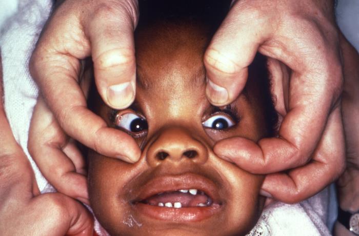
A historical CDC photograph documents congenital rubella cataract, not galactosemia. The visible opacity can teach recognition, but its cause cannot be reassigned to a metabolic disorder. Rubella cataract does not establish a galactitol mechanism. [8]
Find the left lens opacity and distinguish the visible cataract from its cause. Ask about metabolic and infectious history before attributing an infant cataract to galactosemia. Image: CDC / Dr. Andre J. Lebrum, 1969; CDC PHIL 17455, public domain.
Transfer question. What additional evidence would make you treat a cloudy lens as part of a systemic galactose emergency? Look for feeding exposure, liver involvement, glucose status and confirmatory biochemical testing.
4. A phosphate trap can limit glucose production
Fructose can enter a major hepatic route through ketohexokinase, also called fructokinase or KHK. Fructose + ATP → fructose-1-P + ADP. The phosphate is transferred from ATP. There is no additional free-phosphate reactant in that kinase reaction. [14]
Aldolase B cleaves F1P into DHAP plus glyceraldehyde. Glyceraldehyde still needs phosphorylation by triose kinase to become glyceraldehyde-3-P. [9] These triose products connect below phosphofructokinase-1 in glycolysis. Bypassing that particular control point does not mean all fructose metabolism is unregulated or that alternative routes are impossible. [2]
Same input, different phosphate burden
Qualitative educational model. No patient values or dose predictions.
Step the drawing to compare substrate, pool or geometry changes. Original mechanism diagram.
Read the worked reference
Worked comparison. In the intact pathway, KHK forms F1P and aldolase B cleaves it into DHAP plus glyceraldehyde. In aldolase B deficiency, F1P accumulates; available Pi and ATP decline, impairing hepatic glucose output. KHK deficiency limits entry before F1P formation. The bars express relative relationships only.
In hereditary fructose intolerance, impaired aldolase B activity allows F1P to accumulate after relevant exposure. Phosphate becomes sequestered in F1P. Available inorganic phosphate, Pi, and ATP decline. Phosphate is trapped in a metabolite; ATP itself is not trapped.
Glycogen phosphorylase uses Pi to release glucose-1-P from glycogen. [10] Less available Pi can impair glycogenolysis even when glycogen is present. Gluconeogenesis also becomes impaired through energy and metabolite disturbances. This explains why stored carbohydrate does not guarantee adequate hepatic glucose delivery during an HFI crisis. It is not a universal rule that low energy switches glycogenolysis off. [2]
KHK deficiency interrupts the route before the F1P burden forms. Proven essential fructosuria is generally benign, with intermittent urinary fructose. Urinary sugar alone does not prove that diagnosis, however. Identify the sugar and interpret the clinical and molecular evidence. [7]
ATP depletion can increase adenine nucleotide degradation, contributing to hyperuricemia. Lactic acidemia and proximal renal tubular dysfunction can accompany the hepatic crisis. These findings support a systemic metabolic assessment rather than a diagnosis from urine sugar alone. [2]
Human KHK mutation studies support the benign upstream-defect contrast. A historical human phosphorus-spectroscopy report found no change in F1P, ATP or Pi in its essential-fructosuria participant. These small historical observations are not a safe diagnostic loading protocol. [12][13]
These models deliberately show relative pool sizes. Their bars are neither laboratory values nor a dose-response calculator. Compare mechanisms without predicting how much fructose a patient could tolerate.
Case 12
Show answer and explanations for case 12
A. Stored glycogen ensures hepatic output; dextrose directly supplies circulating glucose (Why this does not fit)
Glycogen breakdown still requires the reaction substrate Pi. Dextrose directly supplies glucose, but glycogen presence does not ensure normal hepatic output.
Is a stored substrate enough for product release?
Glycogen breakdown still requires the reaction substrate Pi.
Which half of this statement is supported?
Dextrose directly supplies glucose, but glycogen presence does not ensure normal hepatic output.
B. Stored glycogen ensures hepatic output; dextrose must first become liver glycogen (Why this does not fit)
Limited Pi can impair glycogenolysis despite stored glycogen. Intravenous dextrose supplies circulating glucose directly.
Can a phosphate-limited store guarantee adequate glucose release?
Limited Pi can impair glycogenolysis despite stored glycogen.
C. Pi depletion can impair hepatic output; dextrose must first become liver glycogen (Why this does not fit)
Pi depletion can impair glycogen breakdown. Administered dextrose does not need prior conversion to liver glycogen.
Does the first half correctly identify a supply problem?
Pi depletion can impair glycogen breakdown.
Is hepatic storage needed for the immediate rescue?
Administered dextrose does not need prior conversion to liver glycogen.
D. Pi depletion can impair hepatic output; dextrose directly supplies circulating glucose (Best answer)
Glycogen phosphorylase requires available Pi. The F1P burden can reduce available Pi. Dextrose supplies circulating glucose without first requiring hepatic glucose production.
What does glycogen phosphorylase require besides glycogen?
Glycogen phosphorylase requires available Pi.
What can the F1P burden do to that supply?
The F1P burden can reduce available Pi.
What does intravenous dextrose bypass immediately?
Dextrose supplies circulating glucose without first requiring hepatic glucose production.
Takeaway: Explain exogenous glucose rescue without claiming enzyme correction.
5. Use the result without waiting to protect the patient
A milk-fed newborn with suspected classic galactosemia needs dietary intervention while confirmation proceeds. Manage hypoglycemia and suspected sepsis concurrently. Stopping a substrate does not treat an established infection. E. coli sepsis is an important association, not a prerequisite for diagnosis. [1][3]
Confirmation can combine erythrocyte GALT activity, erythrocyte Gal-1-P and molecular testing. Recent red-cell transfusion can make a red-cell enzyme result misleading because donor cells contribute enzyme activity. Tell the laboratory about transfusion and choose an appropriate confirmatory strategy. [11] A newborn screen is a screening result, not a substitute for assessing a symptomatic infant.
For suspected HFI, stop relevant fructose, sucrose and sorbitol exposures and stabilize the patient, including intravenous dextrose when indicated. ALDOB molecular testing is preferred for confirmation. A provocative fructose challenge is dangerous and should not be used. Review medicines and nutritional formulations as carefully as foods. [2]
Location of the problem changes the pattern
Intestinal fructose malabsorption can cause bloating and osmotic diarrhea after a large fructose load without the systemic phosphate-trap pattern. Fasting hypoglycemia broadens the differential to hepatic glucose-production disorders. Fructose can also precipitate illness in fructose-1,6-bisphosphatase deficiency, so exposure alone is not uniquely diagnostic of HFI.
Use decisive findings, not a remembered age cutoff. Severe hypoglycemia, liver or proximal tubular involvement and confirmatory enzyme or genetic findings carry more weight than whether the first illness happened before or after weaning. Galactosemia can cause hypoglycemia too.
6. Protect the acute phase, then keep following development
Early dietary treatment of classic galactosemia prevents major acute harm. It does not eliminate later speech, motor or ovarian risks. A complication despite adherence is not proof of dietary failure. Chronic mechanisms are multifactorial and incompletely resolved; endogenous metabolite production is relevant but is not a complete proven explanation. [1]
Follow speech and development, neurologic function, growth, nutrition, bone health and reproductive concerns with the appropriate team. Long-term galactose guidance is not a ban on every fruit and vegetable. Consensus recommendations allow non-dairy plant sources while restricting major dairy lactose and galactose exposures, with individualized nutritional support. [3]
HFI management includes continued attention to fructose, sucrose and sorbitol, including medicine excipients. Learned avoidance of sweets may hide earlier symptoms; it does not confirm a diagnosis or replace testing. A lower sugar exposure can reduce cariogenic substrate, but lack of dental caries is not diagnostic and does not imply immunity to decay. Nutrition, growth, liver and renal follow-up still matter. [2]
Classic GALT disease, clinical variants and biochemical variants are not interchangeable prognostic categories. Residual activity, metabolites and genotype need integrated interpretation. A confirmed Duarte biochemical variant should not automatically receive the same lifelong treatment plan as classic disease; consensus guidance does not recommend treating Duarte galactosemia. [1][3]
For an autosomal recessive disorder in a family with two confirmed carrier parents, each pregnancy has its own 25% affected probability. Known familial variants permit targeted assessment of a newborn sibling before symptoms develop. Do not wait for a feeding-triggered crisis to establish a protective plan. [1]
Use this sequence in a new case. Name the ingredient, write the reaction, locate the accumulated substrate, connect it to the observed organ problem, then decide what must happen before confirmation returns. Pause here if you like. The remaining cases are independent opportunities to test transfer.
Practice across mechanisms and decisions
Predict the answer before opening the choices. After responding, explain the decisive finding and test why the closest alternative falls short. All worked explanations remain available without JavaScript.
Case 1
Show answer and explanations for case 1
A. Replace the feed with a specialist-selected galactose-restricted formulation (Best answer)
Lactose digestion supplies galactose. Cataracts are not required for an acute GALT presentation. Stop the dietary galactose load while confirming the diagnosis.
What does lactose supply?
Lactose digestion supplies galactose.
Can clear lenses exclude systemic GALT disease?
Cataracts are not required for an acute GALT presentation.
What must change before confirmation returns?
Stop the dietary galactose load while confirming the diagnosis.
B. Replace the feed with lactase-treated cow milk (Why this does not fit)
Lactase splits lactose into its monosaccharides. Released galactose remains available for Gal-1-P formation.
What does lactase change?
Lactase splits lactose into its monosaccharides.
Which harmful substrate remains?
Released galactose remains available for Gal-1-P formation.
C. Continue the feed until erythrocyte testing confirms the screen (Why this does not fit)
A screen alone is not a definitive diagnosis. The compatible acute illness warrants dietary treatment during confirmation.
Why seek confirmation?
A screen alone is not a definitive diagnosis.
Does that justify continued exposure?
The compatible acute illness warrants dietary treatment during confirmation.
D. Replace only sucrose-containing medicines and retain the feed (Why this does not fit)
Sucrose supplies fructose in HFI. The current lactose feed supplies galactose to the suspected GALT block.
Which disorder makes sucrose especially concerning?
Sucrose supplies fructose in HFI.
What exposure is demonstrated here?
The current lactose feed supplies galactose to the suspected GALT block.
Takeaway: Combine systemic illness with the screen to act before confirmation.
A. Observe the lenses while retaining unrestricted galactose intake (Why this does not fit)
The infant currently lacks systemic illness. Galactose can continue supplying lens galactitol.
Why might observation seem reasonable?
The infant currently lacks systemic illness.
What process continues during exposure?
Galactose can continue supplying lens galactitol.
B. Use lactase-treated dairy and review vision only if growth falters (Why this does not fit)
Hydrolysis leaves galactose available. Growth does not establish that lens injury has stopped.
Does lactose hydrolysis eliminate galactose?
Hydrolysis leaves galactose available.
Does normal growth measure visual risk?
Growth does not establish that lens injury has stopped.
C. Restrict galactose with metabolic guidance and arrange ophthalmic care (Best answer)
Aldose reductase reduces galactose to galactitol. Restriction reduces the substrate burden driving lens injury. Established cataracts need direct ophthalmic evaluation.
Which substrate feeds galactitol production?
Aldose reductase reduces galactose to galactitol.
What does restriction aim to reduce?
Restriction reduces the substrate burden driving lens injury.
Why also assess vision?
Established cataracts need direct ophthalmic evaluation.
D. Restrict fructose and sucrose while retaining the existing milk intake (Why this does not fit)
A. Conversion to fructose followed by F1P accumulation (Best answer)
Sorbitol can supply fructose. KHK forms F1P from fructose. Deficient aldolase B impairs F1P disposal.
Which sugar can sorbitol supply?
Sorbitol can supply fructose.
Which hepatic phosphorylation follows?
KHK forms F1P from fructose.
Why does the inherited defect matter?
Deficient aldolase B impairs F1P disposal.
B. Conversion to fructose followed by F6P accumulation (Why this does not fit)
That route can form F6P. The characteristic blocked pool is F1P rather than F6P.
Can fructose enter an alternative hexokinase route?
That route can form F6P.
Which phosphorylated pool explains the ALDOB defect?
The characteristic blocked pool is F1P rather than F6P.
C. Luminal retention followed by osmotic intestinal water loss (Why this does not fit)
Poorly absorbed sorbitol can contribute to osmotic symptoms. The hypoglycemic episode in established HFI requires consideration of hepatic fructose metabolism.
Can sorbitol cause gastrointestinal symptoms through luminal effects?
Poorly absorbed sorbitol can contribute to osmotic symptoms.
What needs an additional explanation here?
The hypoglycemic episode in established HFI requires consideration of hepatic fructose metabolism.
D. Conversion to fructose followed by enhanced hepatic glucose release (Why this does not fit)
Fructose carbon can participate in downstream hepatic metabolism. The F1P burden impairs rather than assures hepatic glucose output.
What would intact handling permit?
Fructose carbon can participate in downstream hepatic metabolism.
How does ALDOB deficiency change the prediction?
The F1P burden impairs rather than assures hepatic glucose output.
Takeaway: Locate F1P production upstream of ALDOB deficiency.
A. More F1P accumulation; less available phosphate (Why this does not fit)
KHK forms F1P. Inhibition reduces rather than increases F1P formation.
What reaction forms F1P?
KHK forms F1P.
What direction follows its inhibition?
Inhibition reduces rather than increases F1P formation.
B. Less F1P accumulation; more available phosphate (Best answer)
KHK acts upstream of the ALDOB block. KHK inhibition reduces F1P formation and limits the accumulating phosphorylated burden. Available phosphate is better preserved than in untreated ALDOB-deficient cells.
Where does the inhibited reaction occur?
KHK acts upstream of the ALDOB block.
What does reduced F1P formation limit?
KHK inhibition reduces F1P formation and limits the accumulating phosphorylated burden.
What comparison follows?
Available phosphate is better preserved than in untreated ALDOB-deficient cells.
C. Less F1P accumulation; less available phosphate (Why this does not fit)
Blocking the kinase does not consume an extra phosphate substrate. Less F1P sequestration favors phosphate preservation rather than further depletion.
Does blocking KHK itself use an extra phosphate substrate?
Blocking the kinase does not consume an extra phosphate substrate.
How does reduced burden affect the comparison?
Less F1P sequestration favors phosphate preservation rather than further depletion.
D. More F1P accumulation; more available phosphate (Why this does not fit)
Yes, more fructose can remain unphosphorylated when KHK is inhibited. Free fructose must be phosphorylated before it contributes to the F1P pool.
Can more substrate remain as free fructose?
Yes, more fructose can remain unphosphorylated when KHK is inhibited.
Does free fructose equal F1P?
Free fructose must be phosphorylated before it contributes to the F1P pool.
Takeaway: Predict less phosphate sequestration without claiming clinical efficacy.
A normal mixed-cell result can mask the infant deficiency.
B. Perform a galactose feeding challenge to reproduce the abnormal result (Why this does not fit)
Molecular testing does not require a dietary challenge. Renewed galactose loading could worsen suspected disease.
Is renewed exposure necessary for molecular confirmation?
Molecular testing does not require a dietary challenge.
What risk does the challenge create?
Renewed galactose loading could worsen suspected disease.
C. Use a urine glucose-oxidase strip as the definitive enzyme test (Why this does not fit)
Glucose oxidase detects glucose. A urine glucose result does not measure GALT.
What does the strip detect?
Glucose oxidase detects glucose.
Can it establish GALT activity?
A urine glucose result does not measure GALT.
D. Document transfusion and pursue appropriate GALT molecular testing (Best answer)
Transfused cells can supply enzyme activity. Appropriately collected molecular testing can identify GALT variants. Continue the indicated dietary protection while resolving the diagnosis.
Why can the repeat assay differ from the screen?
Transfused cells can supply enzyme activity.
What testing is not a donor-cell enzyme measurement?
Appropriately collected molecular testing can identify GALT variants.
What happens to protection during clarification?
Continue the indicated dietary protection while resolving the diagnosis.
Takeaway: Choose confirmation that addresses the assay confound.
A. Arrange targeted familial testing and protective feeding from birth (Best answer)
Presymptomatic newborns can appear well. Known familial variants permit targeted testing for the two disease-associated alleles. Protection can begin before a substrate-triggered illness.
Does a normal examination before exposure exclude the disorder?
Presymptomatic newborns can appear well.
How do known familial variants help?
Known familial variants permit targeted testing for the two disease-associated alleles.
Why plan feeding now?
Protection can begin before a substrate-triggered illness.
B. Wait for cataracts before ordering targeted testing (Why this does not fit)
Family risk exists before lens findings. Systemic illness can precede recognized cataracts.
Are cataracts required to identify an at-risk sibling?
Family risk exists before lens findings.
What would waiting miss?
Systemic illness can precede recognized cataracts.
C. Wait for the routine screen before changing the first feed (Why this does not fit)
It can identify unsuspected risk. Known familial disease supports protective action before screening returns.
Why is routine screening useful?
It can identify unsuspected risk.
What extra information is already available?
Known familial disease supports protective action before screening returns.
D. Treat the infant as a carrier because both parents are carriers (Why this does not fit)
Each parent can transmit a pathogenic allele. The infant may inherit both pathogenic alleles.
Must two carrier parents have only carrier children?
Each parent can transmit a pathogenic allele.
What outcome remains possible?
The infant may inherit both pathogenic alleles.
Takeaway: Use presymptomatic testing plus early protection.
A. Both have the same prognosis because erythrocyte GALE activity is similar (Why this does not fit)
Erythrocyte activity does not establish nonblood tissue activity. The nonblood assay demonstrates a different distribution of deficiency.
Does a blood-cell result describe all tissues?
Erythrocyte activity does not establish nonblood tissue activity.
What evidence differs between the infants?
The nonblood assay demonstrates a different distribution of deficiency.
B. Infant B has a generalized GALE pattern requiring systemic assessment (Best answer)
It suggests generalized rather than peripheral deficiency. Generalized GALE deficiency can resemble classic galactosemia. Expanded tissue testing is not universally available clinically.
What does reduced activity across tissues suggest?
It suggests generalized rather than peripheral deficiency.
Does systemic illness fit that distribution?
Generalized GALE deficiency can resemble classic galactosemia.
What limits routine use of this assay?
Expanded tissue testing is not universally available clinically.
C. Infant A has classic GALT disease because GALE activity is low in blood (Why this does not fit)
They catalyze different Leloir reactions. A GALE result cannot substitute for a deficient GALT result.
Are GALT and GALE the same reaction?
They catalyze different Leloir reactions.
Does low blood GALE diagnose GALT deficiency?
A GALE result cannot substitute for a deficient GALT result.
D. Infant B has isolated GALK disease because cataracts can occur (Why this does not fit)
Cataracts are not specific to GALK deficiency. The measured defect is widespread GALE deficiency.
Can several galactose disorders cause cataracts?
Cataracts are not specific to GALK deficiency.
Which enzyme defect is directly demonstrated?
The measured defect is widespread GALE deficiency.
Takeaway: Connect generalized deficiency to systemic phenotype.
A. Pi is a reaction substrate, so sequestration can limit glycogen breakdown (Best answer)
Inorganic phosphate participates in phosphorolysis. ATP concentration was held unchanged. Less available Pi can reduce glucose-1-P release independently of an ATP fall.
What molecule is incorporated when phosphorylase cleaves glycogen?
Inorganic phosphate participates in phosphorolysis.
What did the experiment hold constant?
ATP concentration was held unchanged.
What explanation remains sufficient?
Less available Pi can reduce glucose-1-P release independently of an ATP fall.
B. ATP directly supplies the phosphate used by glycogen phosphorylase (Why this does not fit)
ATP was held constant. The reduced reagent was inorganic phosphate.
Was ATP reduced in the experiment?
ATP was held constant.
Which reagent actually changed?
The reduced reagent was inorganic phosphate.
C. Low energy necessarily suppresses every glycogenolytic reaction (Why this does not fit)
It does not lower ATP. The experiment identifies substrate limitation rather than a universal low-energy switch.
Does this experiment demonstrate lower ATP?
It does not lower ATP.
What narrower conclusion fits?
The experiment identifies substrate limitation rather than a universal low-energy switch.
D. F1P must competitively occupy the glucose-binding site of phosphorylase (Why this does not fit)
The stated manipulation reduces Pi without adding F1P. Pi substrate limitation already explains reduced product formation.
Was F1P added to this preparation?
The stated manipulation reduces Pi without adding F1P.
Is direct competition required to explain the result?
Pi substrate limitation already explains reduced product formation.
Takeaway: Separate Pi dependence from ATP-dependent explanations.
A. Reduced lactate-derived glucose; reduced early glycogen-derived output (Why this does not fit)
Lactate-derived glucose requires gluconeogenesis through FBP1. With adequate Pi and stores, the separate early glycogen route can remain functional.
Which route does lactate require to become glucose?
Lactate-derived glucose requires gluconeogenesis through FBP1.
Does the isolated FBP1 block also interrupt glycogen phosphorolysis?
With adequate Pi and stores, the separate early glycogen route can remain functional.
B. Reduced lactate-derived glucose; preserved early glycogen-derived output (Best answer)
Gluconeogenesis from lactate depends on fructose-1,6-bisphosphatase. Stored glycogen can supply glucose without passing through FBP1. As stored glycogen support declines, impaired gluconeogenesis becomes more consequential.
Which route is blocked when lactate carbon must reach glucose?
Gluconeogenesis from lactate depends on fructose-1,6-bisphosphatase.
Which early route bypasses that reaction?
Stored glycogen can supply glucose without passing through FBP1.
Why does fasting expose the defect?
As stored glycogen support declines, impaired gluconeogenesis becomes more consequential.
C. Preserved lactate-derived glucose; reduced early glycogen-derived output (Why this does not fit)
A glycogen-breakdown defect would preferentially lower that route. FBP1 deficiency instead impairs gluconeogenesis from lactate.
Which defect would preferentially lower the glycogen route?
A glycogen-breakdown defect would preferentially lower that route.
Does that match the established defect?
FBP1 deficiency instead impairs gluconeogenesis from lactate.
D. Preserved lactate-derived glucose; preserved early glycogen-derived output (Why this does not fit)
Adequate energy does not replace fructose-1,6-bisphosphatase. Lactate-derived glucose production remains impaired.
Can adequate ATP bypass a missing gluconeogenic enzyme?
Adequate energy does not replace fructose-1,6-bisphosphatase.
Which measured output must therefore be affected?
Lactate-derived glucose production remains impaired.
Takeaway: Connect it to fasting glucose production rather than dietary F1P disposal.
A. Hepatic F1P retention with phosphate depletion and impaired glucose output (Why this does not fit)
Hypoglycemia or hepatic injury would raise concern. The current evidence favors an intestinal absorption problem.
What systemic consequence would raise concern for HFI?
Hypoglycemia or hepatic injury would raise concern.
What compartment best fits the described symptoms and normal studies?
The current evidence favors an intestinal absorption problem.
B. Gal-1-P retention after lactose digestion with acute hepatocellular injury (Why this does not fit)
The beverage supplies fructose. Neither that exposure nor hepatic injury is described.
Which exposure is actually specified?
The beverage supplies fructose.
Is milk-associated hepatic injury demonstrated?
Neither that exposure nor hepatic injury is described.
C. Incomplete intestinal fructose absorption with osmotic retention of water (Best answer)
It can retain water within the intestinal lumen. Bacterial fermentation can contribute to bloating. It supports a luminal explanation rather than an acute hepatic crisis.
Where can unabsorbed fructose retain water?
It can retain water within the intestinal lumen.
What else can unabsorbed carbohydrate produce?
Bacterial fermentation can contribute to bloating.
Why does the normal systemic assessment matter?
It supports a luminal explanation rather than an acute hepatic crisis.
D. Absent hepatic KHK with urinary fructose causing intestinal water retention (Why this does not fit)
Some fructose appears in urine. Renal excretion is not the demonstrated intestinal mechanism.
Where does fructose accumulate in essential fructosuria?
Some fructose appears in urine.
Does urinary fructose explain luminal osmotic diarrhea?
Renal excretion is not the demonstrated intestinal mechanism.
Takeaway: Use systemic negatives to favor malabsorption rather than a phosphate crisis.
A. F1P formation remains absent; a sustained F1P phosphate trap develops (Why this does not fit)
KHK-dependent phosphorylation of fructose to F1P is restored. A sustained F1P trap requires accumulating F1P, not its absence.
Which reaction has been restored?
KHK-dependent phosphorylation of fructose to F1P is restored.
Can an absent F1P pool itself create the proposed trap?
A sustained F1P trap requires accumulating F1P, not its absence.
B. F1P formation returns; a sustained F1P phosphate trap develops (Why this does not fit)
Restored KHK can form F1P. Intact ALDOB permits downstream F1P disposal, so the characteristic sustained trap is not expected.
Does entry through KHK return?
Restored KHK can form F1P.
What distinguishes these cells from ALDOB-deficient cells?
Intact ALDOB permits downstream F1P disposal, so the characteristic sustained trap is not expected.
C. F1P formation remains absent; the sustained F1P phosphate trap is not expected (Why this does not fit)
Normal ALDOB supports disposal rather than the characteristic sustained trap. The missing KHK reaction has been restored with ATP and uptake controlled.
What does the second half correctly predict?
Normal ALDOB supports disposal rather than the characteristic sustained trap.
Why should F1P formation no longer remain absent?
The missing KHK reaction has been restored with ATP and uptake controlled.
D. F1P formation returns; the sustained F1P phosphate trap is not expected (Best answer)
Fructose can again be phosphorylated to F1P through KHK. ALDOB can cleave F1P into DHAP and glyceraldehyde. F1P formation alone does not establish a sustained phosphate trap when downstream disposal is functional.
What changes when KHK is restored?
Fructose can again be phosphorylated to F1P through KHK.
What happens to that product with functional ALDOB?
ALDOB can cleave F1P into DHAP and glyceraldehyde.
Does transient formation imply a sustained trap?
F1P formation alone does not establish a sustained phosphate trap when downstream disposal is functional.
Takeaway: Locate the proven defect upstream of the phosphate trap.
A. Recognize residual chronic risk and arrange targeted developmental and endocrine care (Best answer)
Early restriction reduces severe neonatal complications. Speech and ovarian problems can still occur. Assess the complications without assuming dietary blame.
Did early treatment have an acute benefit?
Early restriction reduces severe neonatal complications.
Does that guarantee absence of later complications?
Speech and ovarian problems can still occur.
What follows from documented adherence?
Assess the complications without assuming dietary blame.
B. Assume hidden dairy exposure is the sole cause and defer specialist assessment (Why this does not fit)
Chronic complications can occur despite adherence. The child needs evaluation of current speech and endocrine concerns.
Does a complication prove an exposure error?
Chronic complications can occur despite adherence.
What would delaying assessment miss?
The child needs evaluation of current speech and endocrine concerns.
C. Explain that endogenous Gal-1-P is the fully proven sole cause of both problems (Why this does not fit)
Endogenous galactose-related metabolism persists. The chronic mechanisms remain multifactorial and incompletely resolved.
Can endogenous metabolism remain relevant?
Endogenous galactose-related metabolism persists.
Does that establish one complete causal explanation?
The chronic mechanisms remain multifactorial and incompletely resolved.
D. Stop dietary management because early treatment did not prevent every complication (Why this does not fit)
Failure to prevent every outcome does not negate dietary protection. Continue appropriate dietary care alongside surveillance and treatment.
Does an incomplete chronic benefit erase acute benefit?
Failure to prevent every outcome does not negate dietary protection.
What management conclusion follows?
Continue appropriate dietary care alongside surveillance and treatment.
Takeaway: Choose targeted care without unsupported causal certainty.
A. Continue excluding plant foods; correct the nutritional deficits despite normal serum calcium (Why this does not fit)
Consensus guidance permits fruits, vegetables and legumes. The documented nutritional deficits need correction, but broad plant-food exclusion is unnecessary.
Does classic galactosemia require a universal plant-food ban?
Consensus guidance permits fruits, vegetables and legumes.
Which half of this plan is appropriate?
The documented nutritional deficits need correction, but broad plant-food exclusion is unnecessary.
B. Allow appropriate plant foods; correct the nutritional deficits despite normal serum calcium (Best answer)
Major dairy lactose and galactose restriction remains appropriate while permitted plant foods can return. Normal serum calcium does not negate the low 25-hydroxyvitamin D or documented low calcium intake. Individualized dietary review and calcium/vitamin D support should address the demonstrated deficits.
Which major dietary restriction remains appropriate?
Major dairy lactose and galactose restriction remains appropriate while permitted plant foods can return.
Does normal serum calcium demonstrate adequate vitamin D or intake?
Normal serum calcium does not negate the low 25-hydroxyvitamin D or documented low calcium intake.
What follow-up is needed?
Individualized dietary review and calcium/vitamin D support should address the demonstrated deficits.
C. Allow appropriate plant foods; defer nutritional correction because serum calcium is normal (Why this does not fit)
Permitted non-dairy plant foods can be included. The low vitamin D measurement and inadequate intake remain actionable findings.
Which dietary change follows consensus guidance?
Permitted non-dairy plant foods can be included.
Why is reassurance from serum calcium insufficient?
The low vitamin D measurement and inadequate intake remain actionable findings.
D. Continue excluding plant foods; defer nutritional correction because serum calcium is normal (Why this does not fit)
A universal plant-food exclusion is not recommended. Normal serum calcium does not establish adequate intake or vitamin D status.
Does eliminating every plant source establish better protection?
A universal plant-food exclusion is not recommended.
Can normal serum calcium override both measured deficits?
Normal serum calcium does not establish adequate intake or vitamin D status.
Takeaway: Protect nutritional adequacy without promising prevention of all chronic outcomes.
A. Hyperglycemic overflow has exceeded an otherwise normal glucose threshold (Why this does not fit)
The blood glucose is low rather than high. Hyperglycemic overflow alone does not explain generalized aminoaciduria and phosphate wasting.
What is the simultaneous blood glucose?
The blood glucose is low rather than high.
Does overflow explain the other solute losses?
Hyperglycemic overflow alone does not explain generalized aminoaciduria and phosphate wasting.
B. Proximal tubular dysfunction is impairing reclamation of several solutes (Best answer)
They are reclaimed substantially in the proximal tubule. Low simultaneous plasma glucose argues against hyperglycemic overflow as the reason for glycosuria. HFI can involve proximal renal tubular dysfunction.
What links glucose, amino acids and phosphate reabsorption?
They are reclaimed substantially in the proximal tubule.
Why does low plasma glucose matter?
Low simultaneous plasma glucose argues against hyperglycemic overflow as the reason for glycosuria.
What complication can HFI produce?
HFI can involve proximal renal tubular dysfunction.
C. Essential fructosuria is producing isolated urinary fructose (Why this does not fit)
Glucose and multiple other solutes are reported. It does not explain generalized proximal solute losses.
Which urinary analytes are reported?
Glucose and multiple other solutes are reported.
Does isolated fructose excretion explain this pattern?
It does not explain generalized proximal solute losses.
D. Intestinal fructose malabsorption is directly producing renal glucose leakage (Why this does not fit)
It is in intestinal absorption. The combined urinary solute losses require impaired renal handling.
Where is the primary defect in malabsorption?
It is in intestinal absorption.
What observation needs a renal explanation?
The combined urinary solute losses require impaired renal handling.
Takeaway: Localize multiple losses to proximal tubular handling.
A. Galactose is identified, establishing classic GALT deficiency (Why this does not fit)
Copper reduction detects reducing substances without specifying galactose. The glucose-specific strip does not identify GALT deficiency.
Does copper reduction identify a particular sugar?
Copper reduction detects reducing substances without specifying galactose.
Does the strip supply enzyme identity?
The glucose-specific strip does not identify GALT deficiency.
B. Fructose is identified, establishing benign essential fructosuria (Why this does not fit)
Several non-glucose reducing sugars can produce the pattern. The urine pair alone does not establish a benign diagnosis.
Can non-glucose sugars share this test pattern?
Several non-glucose reducing sugars can produce the pattern.
Can benignity be inferred from it?
The urine pair alone does not establish a benign diagnosis.
C. A non-glucose reducing substance is suggested, requiring further identification (Best answer)
A reducing substance is detected. The glucose-oxidase strip does not detect glucose in that sample. Sugar identity and the cause require further assessment.
What does the positive broad test establish?
A reducing substance is detected.
What does the negative specific strip add?
The glucose-oxidase strip does not detect glucose in that sample.
What remains unresolved?
Sugar identity and the cause require further assessment.
D. Glucose is identified, establishing hyperglycemic overflow (Why this does not fit)
The glucose-oxidase strip is glucose-specific. That strip was negative, so the pair does not identify glucose overflow.
Which assay is glucose-specific?
The glucose-oxidase strip is glucose-specific.
What result was obtained?
That strip was negative, so the pair does not identify glucose overflow.
Takeaway: Recognize the limit of diagnostic inference.
A. Administer fructose under observation and measure the glucose nadir (Why this does not fit)
It would reintroduce a potentially dangerous substrate. Molecular confirmation does not require a fructose challenge.
What would the challenge expose the infant to?
It would reintroduce a potentially dangerous substrate.
Is that required for HFI confirmation?
Molecular confirmation does not require a fructose challenge.
B. Use fructose hydrogen breath testing as definitive evidence of ALDOB deficiency (Why this does not fit)
It assesses intestinal handling and fermentation of a carbohydrate load. It does not establish ALDOB deficiency.
What does breath testing primarily assess?
It assesses intestinal handling and fermentation of a carbohydrate load.
Does it directly establish hepatic ALDOB genotype?
It does not establish ALDOB deficiency.
C. Use a negative newborn screen to exclude HFI without further testing (Why this does not fit)
It does not establish ALDOB function. The exposure-associated systemic illness remains unexplained.
Does a routine galactosemia screen establish ALDOB function?
It does not establish ALDOB function.
What evidence still needs explanation?
The exposure-associated systemic illness remains unexplained.
D. Obtain appropriate molecular testing while avoiding renewed fructose exposure (Best answer)
Biallelic pathogenic ALDOB variants can establish HFI in the compatible setting. A phenotype-appropriate molecular panel can assess competing disorders. A provocative fructose challenge should not be used.
What evidence can establish HFI?
Biallelic pathogenic ALDOB variants can establish HFI in the compatible setting.
What if the differential remains broad?
A phenotype-appropriate molecular panel can assess competing disorders.
What should not be used to settle it?
A provocative fructose challenge should not be used.
Takeaway: Select molecular confirmation without recreating a crisis.
A. Galactose-pathway dysfunction with evidence of excess galactitol (Best answer)
It shows a lens opacity in its documented clinical context. Similar opacity can arise from different causes. Galactose-pathway dysfunction with excess galactitol supports that route.
What does the photograph itself establish?
It shows a lens opacity in its documented clinical context.
Can appearance alone identify the biochemical cause?
Similar opacity can arise from different causes.
What evidence supports the proposed metabolic route?
Galactose-pathway dysfunction with excess galactitol supports that route.
B. Cloudiness in the same location without any biochemical testing (Why this does not fit)
Anatomic resemblance alone cannot establish etiology. Evidence linking the opacity to galactose metabolism is missing.