Leber hereditary optic neuropathy: central vision, maternal inheritance, uncertain expression
Trace central visual loss to retinal ganglion cells, distinguish key mimics, interpret maternal inheritance and testing, and plan realistic LHON care.
A young adult can lose the ability to read while still seeing someone approach from the side. Why can a disorder carried in mitochondrial DNA selectively damage central vision, and how can a visually normal mother transmit it? This lesson connects retinal localization, clinical course and maternal inheritance so you can choose an investigation and counsel a family without promising a fixed outcome.
Localize the loss before naming the disease
Does failure to recognize a face mean the whole retina has stopped working? In Leber hereditary optic neuropathy (LHON), the main injury is to retinal ganglion cells and their axons, particularly the papillomacular bundle carrying central visual information toward the optic disc. These are output neurons, not the rods and cones that first detect light. Their small, unmyelinated axons near the nerve head have demanding energy requirements. Dysfunction of mitochondrial complex I makes this population especially vulnerable. Energy failure and oxidative injury both contribute; the biology is not explained by ATP depletion alone. [1]
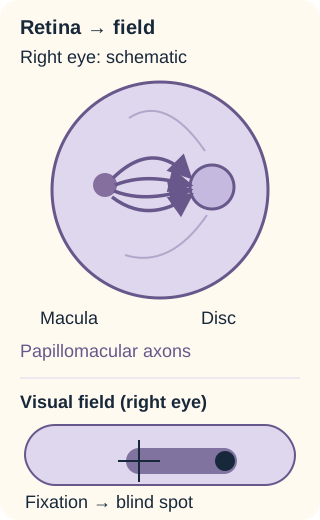
The papillomacular bundle links central retinal function to the disc. A cecocentral defect spans fixation toward the blind spot. [1]
A central scotoma is an area of impaired vision around fixation. A cecocentral scotoma connects fixation to the physiological blind spot, which corresponds to the optic disc. Loss of central acuity, reduced color discrimination and impaired contrast can coexist with relatively preserved peripheral vision. A person may navigate a room but be unable to read its signs. Preserved peripheral vision does not mean mild disability. [1]
Trace the central retinal fibers to the disc in the diagram. Now cover the shaded field region with a finger or describe its boundary aloud. Predict whether reading small print or detecting someone approaching from the side will be more impaired.
Compare your prediction
Reading depends on the central field and is disproportionately impaired. Side detection can remain possible because the peripheral field is relatively preserved. A central defect does not require loss of the entire field.
The consequence is practical: test acuity, color vision and formal fields separately. Apply the same reasoning to a patient who reports that red objects appear faded despite recognizing large peripheral objects. This combination supports optic-nerve dysfunction, but it does not identify its cause; inflammatory, toxic and nutritional injury can affect the same fibers.
Read the two eyes as a time series
Does an apparently swollen disc prove inflammation? Not in LHON. The typical presentation is painless, subacute central blurring in one eye, with the other eye affected weeks to months later. Simultaneous bilateral onset also occurs. Young men are commonly affected, but women, children and older adults can develop disease. Demographics adjust suspicion rather than determine eligibility for testing. [1][2]
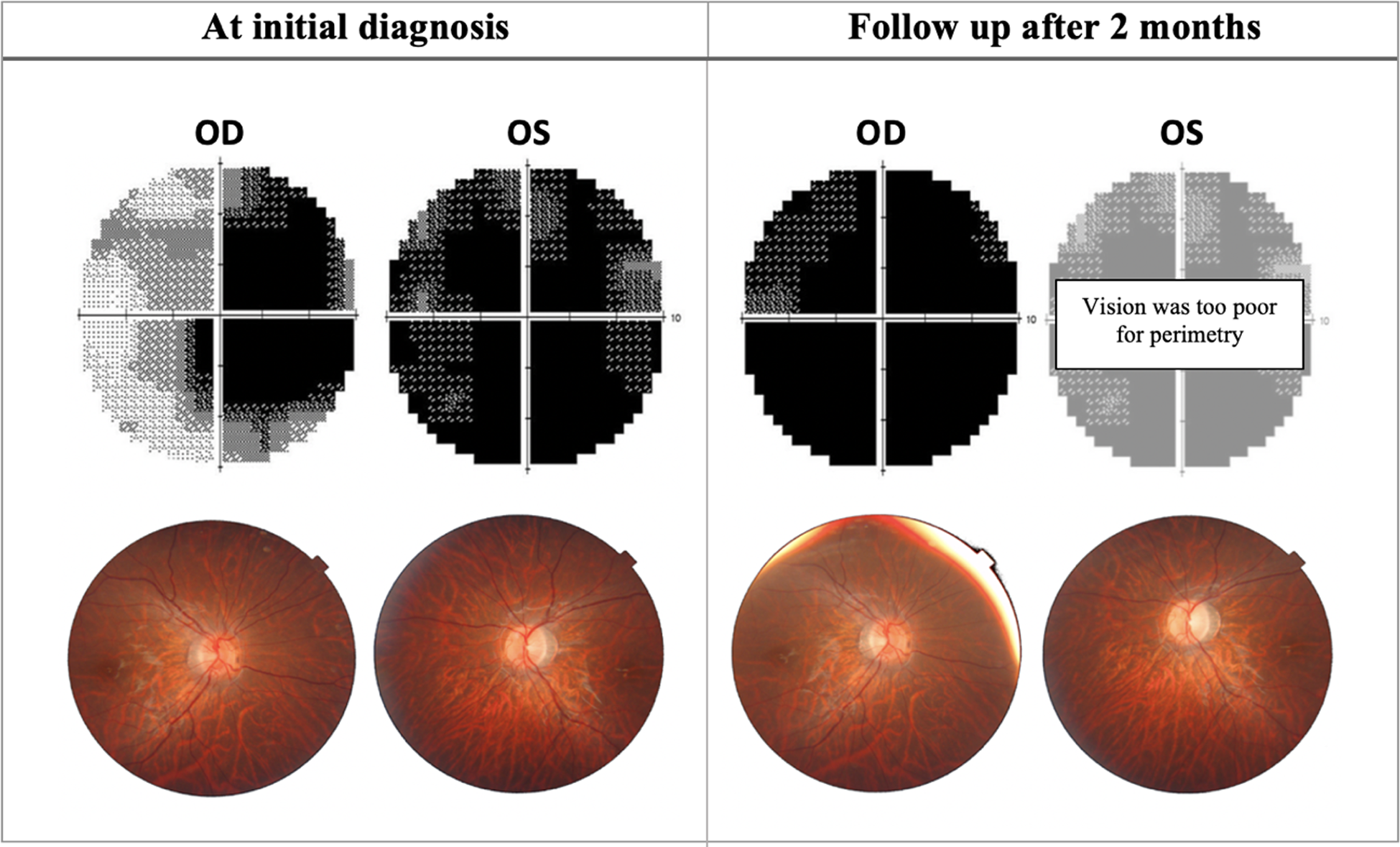
Figure 2 from Esmaeil, Ali and Behbehani: an 18-year-old patient had 20/400 right-eye acuity and counting-fingers left-eye acuity at diagnosis; the right eye progressed to counting fingers after two months. Image: Esmaeil, Ali and Behbehani, Figure 2 (2023), CC BY 4.0. [1].
Acute fundus findings can include peripapillary telangiectatic small vessels, vascular tortuosity, disc hyperemia and retinal nerve fiber layer (RNFL) swelling. Fluorescein angiography typically shows no leakage from the apparent swelling, hence the term pseudoedema. Some affected eyes have an initially normal-appearing fundus. Later, optic-disc pallor and RNFL thinning reflect axonal loss. The sequence is useful, but a photograph alone cannot establish a molecular diagnosis. [1]
Optical coherence tomography (OCT) measures tissue structure. Early RNFL thickening can coexist with macular ganglion cell loss. Later RNFL thickness may decline through the normal range before becoming abnormally thin. A less swollen nerve therefore does not necessarily mean recovery. Compare structure with acuity, color testing and fields rather than reading a green OCT classification as proof of intact function.
Compare the initial and follow-up panels in the clinical figure. Predict what a later pale disc means if central vision has worsened rather than recovered.
Interpret the change
Loss of viable axons can replace swelling with pallor. Less swelling plus worse vision supports degeneration, not resolution with restored function. The published patient illustrates one course, not a required timetable.
For transfer, imagine the first eye is already pale while the newly symptomatic second eye looks hyperemic. The different appearances can reflect different disease stages in the same person. Do not require both discs to match. A relative afferent pupillary defect may be absent with bilateral disease, and pupillary light responses can be relatively preserved despite profound central loss. Neither finding cancels the patient's measured deficit. [1]
Use the competing explanation, not a single sign
Is painless central loss specific to LHON? No. First localize an optic neuropathy, then compare tempo, symmetry, exposure, examination and imaging. A maternal relative with visual loss matters, but a vague family history cannot override a structural lesion or a treatable toxic exposure.
Similar visual deficits, different supporting evidence
Pattern
Evidence to seek
Consequence
PatternLHON
Evidence to seekPainless sequential central loss; maternal-line disease; characteristic fundus or OCT course
ConsequenceMolecular testing with neuro-ophthalmic assessment
PatternInflammatory optic neuritis
Evidence to seekPain on eye rotation; optic-nerve enhancement; other demyelinating findings
ConsequenceBrain and orbital MRI and directed inflammatory evaluation
PatternToxic or nutritional injury
Evidence to seekOften symmetric bilateral central loss; medication, diet or malabsorption history
ConsequenceAssess the exposure or deficiency and treat it promptly
PatternCompression
Evidence to seekProgressive asymmetry; field loss suggesting chiasmal involvement; a corresponding mass
ConsequenceStructural imaging and cause-specific care
Typical demyelinating optic neuritis often causes pain with eye rotation and an afferent pupillary defect when one eye is less affected. Gadolinium-enhanced MRI can demonstrate optic-nerve inflammation. However, central scotoma and dyschromatopsia are shared findings. Rare optic-nerve enhancement and an associated multiple-sclerosis-like illness have been reported in LHON, so enhancement is not an absolute genetic exclusion. Interpret the whole presentation. [9][1]
Ethambutol exposure and nutritional deficiency after bariatric surgery are examples of competing causes of bilateral cecocentral loss. Establish medication timing, nutritional intake and malabsorption risk; test suspected deficiencies rather than assuming every central field defect is hereditary. Progressive temporal field loss in both eyes suggests the chiasm rather than an isolated papillomacular process and warrants imaging. [10][1]
Compare two patients with the same central field defect: one has severe pain on eye rotation and optic-nerve enhancement; the other has symmetric decline after several months of ethambutol. State which new fact changes your next investigation in each.
Compare the investigations
The first needs an inflammatory optic-neuropathy evaluation. The second needs urgent medication review and ocular assessment for toxicity. Their shared field pattern localizes injury; the differing context directs the cause-specific workup.
Apply this separation when family history is absent: LHON remains possible because relatives may carry the variant without symptoms, but common and reversible mimics still need assessment. Sudden severe visual loss, severe headache, painful red eye or new focal neurologic deficits require urgent evaluation rather than a wait for genetic results.
Separate mitochondrial transmission from visual outcome
If a mother sees normally, can she transmit a pathogenic variant? Yes. Transmission asks who receives mitochondrial DNA; penetrance asks who develops disease. Mitochondrial DNA is maternally inherited. A man with an LHON-associated mtDNA variant does not transmit it to his children. A homoplasmic woman transmits that variant to her sons and daughters, whether or not she has visual loss. Affected men and unaffected women in the same pedigree do not make the disorder X-linked. [2][8]
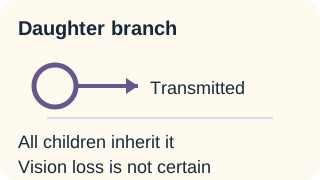
In this homoplasmic pedigree, a maternal variant can reach all children without causing vision loss in all. The grandchildren’s other parents are noncarriers. Fathers do not transmit mtDNA; heteroplasmy can modify risk. [1]
Homoplasmy means the mtDNA copies at the tested site carry the same sequence; in a homoplasmic affected family they all carry the pathogenic variant. Heteroplasmy means variant and nonvariant mtDNA coexist. Most classic LHON families are homoplasmic. In heteroplasmic mothers, the proportion transmitted can differ between children, and blood measurements need not represent retinal tissue. Do not use a mother's blood percentage as a precise probability of blindness in a child. Neither heteroplasmy nor its absence makes clinical outcome deterministic. [1][8]
Three primary variants account for most classic mtDNA-associated LHON: m.11778G>A in MT-ND4, m.3460G>A in MT-ND1, and m.14484T>C in MT-ND6. These genes encode complex I subunits. Complex I normally accepts electrons from NADH and contributes to the proton gradient across the inner mitochondrial membrane. ATP synthase uses that gradient. Impaired electron transfer and increased oxidative stress can injure ganglion cells even though the variant is present in many tissues. [1]
Complex I helps build the proton gradient used for ATP synthesis. LHON-associated complex I dysfunction can impair energy supply and increase oxidative stress. [1]
Male sex and age influence penetrance; mitochondrial background, nuclear modifiers and environmental exposures also contribute. Published family estimates vary, so a single lifetime percentage is not an individual forecast. The m.14484T>C variant generally has a better chance of spontaneous visual recovery than m.11778G>A, but neither result promises a particular outcome. [1]
Trace the family route
In this model, a homoplasmic grandmother has a son and daughter who both carry the variant. Their partners do not carry it. Before opening either branch, trace whose mitochondria each grandchild receives. Predict variant transmission, not visual ability.
Trace the daughter’s descendants
Maternal transmission continues through the daughter. Both sexes inherit her homoplasmic variant.
The daughter supplies her children's mitochondria. Her own normal vision would not interrupt this route.
Trace the son’s descendants
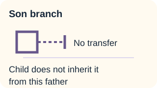
Paternal transmission stops. His affected status would not change mitochondrial parentage.
The son does not supply the children's mtDNA. Their mother supplies it and is specified as a noncarrier in this model.
The worked result remains simple: the daughter's children inherit her variant, while the son's children do not inherit his. Visual loss remains uncertain in those who inherit it. To reset the comparison, close both branch answers. For a new situation, replace the grandmother with a heteroplasmic woman: maternal inheritance still applies, but variant proportion is no longer fixed across descendants. The diagram is a transmission model, not a penetrance calculator.
Confirm the cause without making the test do too much
Does a negative three-variant panel end the evaluation? It reduces support for the common molecular causes but does not exclude rarer LHON variants or a different inherited optic neuropathy. In a patient with a convincing phenotype, specialist assessment can extend testing to broader mtDNA sequencing or an appropriate optic-neuropathy panel, while revisiting acquired causes. A variant of uncertain significance is not equivalent to a confirmed pathogenic explanation. [1]
Begin by documenting acuity, color function, fields, fundus appearance and OCT. Obtain a three-generation history that distinguishes maternal from paternal relatives and actual diagnoses from unspecified poor eyesight. Targeted testing for the three common mtDNA variants is a usual initial molecular approach. When a familial pathogenic variant is already known, targeted testing for that specific variant can answer whether an at-risk relative carries it. MRI addresses compression or inflammation; it is not a substitute for DNA analysis. [1]
Example result A: typical sequential optic neuropathy plus a known pathogenic m.11778G>A variant supports a molecular diagnosis.
Example result B: normal vision plus the same variant establishes carrier status, not current visual disease.
Example result C: persistent optic neuropathy plus a negative limited panel leaves the cause unresolved.
Choose which result above requires broader etiologic investigation rather than reassurance. Then ask whether result B predicts an age of onset.
Interpret the laboratory result
Result C requires reassessment of the phenotype, test coverage and alternatives. Result B cannot predict onset, severity or progression. A genetic result must be interpreted alongside what the patient currently experiences.
For transfer, a maternal cousin requests predictive testing after a relative's diagnosis. Offer informed genetic counseling before testing and explain the difference between variant detection and disease prediction. Prenatal or preimplantation approaches require specialist reproductive counseling; a measured fetal variant proportion cannot reliably specify future visual function. Do not imply that testing eliminates uncertainty. [8]
Act early while being honest about recovery
Should rehabilitation wait until a treatment succeeds or fails? No. New visual symptoms need prompt ophthalmic or neuro-ophthalmic assessment. Low-vision aids, accessible study materials, workplace adaptations, mobility support and psychological support can begin while the diagnosis and treatment plan are refined. Loss of central vision affects education, employment and independence even when peripheral navigation remains possible. [8]
Strongly advise carriers not to smoke, and offer practical cessation support. Avoid heavy alcohol intake and binge drinking. The smoking association is more consistent than the alcohol association; in a large observational study, adjusted alcohol estimates were not statistically significant. These are risk-reduction recommendations, not proof that an individual caused their illness or that abstinence guarantees prevention. Review potentially mitochondrial-toxic drugs with the treating clinician rather than stopping essential medication indiscriminately. [3][8]
Idebenone is not a guaranteed reversal. In the EU, Raxone is authorized for visual impairment in adults and adolescents aged 12 years and older with LHON. Specialist-supervised dosing is 300 mg three times daily with food. Access and prescribing rules differ by region. Diarrhea and other adverse effects require discussion. Early assessment is appropriate because evidence has favored treatment in the earlier disease period, but later presentation should prompt individualized discussion rather than automatic dismissal. [4]
What it supportsSignals of benefit in selected analyses
What it cannot promiseThe primary endpoint was not statistically significant in the full trial population
EvidenceLEROS, open-label with external natural-history controls
What it supportsAdditional evidence in patients up to five years after onset
What it cannot promiseExternal controls do not supply the protection against bias of concurrent randomization
Read these findings together: treatment may help some patients, effects vary by disease stage and variant, and normal sight is not assured. Gene therapy remains a specialist, variant-specific discussion. The EMA records withdrawal of the Lumevoq application in April 2023; trial participation or an early-access pathway is not the same as marketing authorization. Confirm current local availability rather than treating a research report as an approved prescription. [5][6][7]
A newly affected student can still navigate the campus but cannot read projected slides. Compare waiting for visual recovery with arranging accessible materials now. Which choice addresses the current disability without assuming the eventual outcome?
Compare the care plans
Arrange accessible materials and low-vision support now, alongside specialist treatment discussion. Rehabilitation does not imply that recovery is impossible; it meets present needs while the prognosis remains uncertain.
Apply the same principle to family care: offer counseling and informed testing to appropriate maternal relatives, not a prediction of inevitable blindness. Ask about neurologic symptoms and cardiac symptoms; LHON can occasionally coexist with neurologic disease or cardiac conduction abnormalities. A pre-excitation pattern warrants cardiology referral, and new neurologic deficits need their own evaluation. A molecular diagnosis should improve care, not close the differential. [1][8]
Apply the lesson
Case 1
Show answer and explanations for case 1
A. Peripheral rod photoreceptors; complex I (Why this does not fit)
The variant affects complex I, but peripheral rod dysfunction would favor night and peripheral visual deficits. The cecocentral and color pattern instead implicates papillomacular retinal ganglion cells.
Reasoning steps for option A
Would peripheral rods explain preserved peripheral fields in this 22-year-old?
No. Rod-predominant injury tends to disturb night and peripheral vision, not selectively produce bilateral cecocentral loss and dyschromatopsia.
Does m.11778G>A support the complex I half of this choice?
Yes. This MT-ND4 variant disrupts a complex I subunit; the respiratory assignment is right even though the cellular target is wrong.
Why reject this rod and complex I pairing overall?
The central field and color deficits point to papillomacular retinal ganglion cells, so a correct complex cannot rescue the rod localization.
B. Papillomacular retinal ganglion cells; complex I (Best answer)
The field and color pattern localizes to central retinal ganglion-cell axons. The m.11778G>A variant affects MT-ND4, a complex I subunit, linking that vulnerable population to impaired mitochondrial function.
Reasoning steps for option B
Which retinal output cells explain cecocentral scotomas with peripheral sparing?
Papillomacular retinal ganglion cells and their axons carry central visual information; injury there fits the visual fields and color loss.
What respiratory complex contains the protein encoded by variant-bearing MT-ND4?
MT-ND4 encodes a complex I subunit, linking m.11778G>A to impaired mitochondrial electron transport in susceptible ganglion cells.
Does this pairing account for both the field pattern and molecular result?
Yes. Papillomacular ganglion-cell injury explains central dysfunction, while the identified MT-ND4 change identifies complex I.
C. Papillomacular retinal ganglion cells; complex IV (Why this does not fit)
The cellular localization fits the central field and color deficits. MT-ND4 is a complex I subunit, not a complex IV subunit.
Reasoning steps for option C
Does papillomacular ganglion-cell injury fit the cecocentral and color deficits?
Yes. These central retinal ganglion-cell projections are selectively vulnerable in LHON, unlike peripheral rod pathways.
Is m.11778G>A a defect in cytochrome c oxidase, complex IV?
No. It is in MT-ND4, whose protein contributes to complex I rather than complex IV.
Why does the complex IV assignment invalidate the otherwise fitting papillomacular localization?
The complex IV designation contradicts the specified genotype; the cellular target alone is not enough.
D. Peripheral rod photoreceptors; complex IV (Why this does not fit)
Peripheral rod disease can impair vision, but it does not fit the selective cecocentral pattern. The identified MT-ND4 variant also implicates complex I rather than IV.
Reasoning steps for option D
What visual complaint would peripheral rod damage predict instead of this pattern?
Rod dysfunction more readily predicts night blindness and peripheral field loss than preserved periphery with cecocentral scotomas.
Does MT-ND4 m.11778G>A encode a complex IV component?
No. MT-ND4 is a complex I subunit, so the stated respiratory defect is also misplaced.
How many parts of the rod and complex IV proposal conflict with the vignette?
Both: the field localization favors papillomacular ganglion cells and the genotype identifies complex I.
Takeaway: Use the phenotype to localize and the genotype to identify the molecular defect.
A. Resolution of axonal swelling with restored nerve function (Why this does not fit)
Decreasing thickness can accompany resolution of swelling. Worsening acuity and ganglion cell loss show that restored function is not the explanation here.
Reasoning steps for option A
What does an RNFL change from thick to reference range establish by itself?
It establishes reduced measured thickness, not restored axonal function; early swelling can resolve while axons are lost.
Do 20/80 to 20/400 acuity and falling ganglion-cell thickness support recovery?
No. Both worsening vision and macular ganglion-cell loss oppose the claim of restored nerve function.
Why is resolution with restored function untenable despite a normal-range RNFL?
A reference-range RNFL can be a transient point on a trajectory from edema toward atrophy; the independent functional and structural trends are adverse.
B. Primary photoreceptor degeneration with an unaffected optic nerve (Why this does not fit)
Photoreceptor degeneration can reduce acuity. The documented ganglion cell loss and preceding RNFL swelling instead implicate the inner retinal output pathway.
Reasoning steps for option B
Which tissue is directly changing besides the peripapillary RNFL?
The macular ganglion-cell layer is thinning, pointing to inner retinal neuronal loss rather than isolated photoreceptor degeneration.
Can an unaffected optic nerve explain the earlier thick RNFL and later decline?
No. Serial RNFL change in an LHON carrier indicates involvement of the ganglion-cell axons composing the optic nerve.
Why does primary photoreceptor disease fail as the chief explanation?
The option ignores concordant ganglion-cell thinning and RNFL evolution, both characteristic of the retinal ganglion-cell output pathway.
C. Transition from swelling toward axonal loss (Best answer)
An early thick RNFL can pass through a normal measurement as tissue is lost. Concurrent ganglion cell thinning and poorer acuity make a degenerative transition more likely than recovery.
Reasoning steps for option C
How can an initially thick RNFL enter the reference interval during decline?
Early axonal swelling can subside while damaged axons disappear, causing measured thickness to cross the normal interval on its way downward.
Which simultaneous findings distinguish degeneration from recovery?
Macular ganglion-cell thinning and acuity worsening from 20/80 to 20/400 both favor loss of neural tissue and function.
What does the combined four-month OCT and acuity trajectory indicate?
Transition from swelling toward axonal loss is more coherent than restored function despite the nominally normal RNFL reading.
D. Stable structure with measurement variation alone (Why this does not fit)
Device variation can affect OCT classifications. Parallel deterioration in acuity and macular ganglion cell thickness makes variation alone an inadequate explanation.
Reasoning steps for option D
Could OCT measurement variability alone create a reference-range RNFL value?
An individual reading may vary, but variability by itself cannot explain a coherent serial change together with declining visual performance.
What independent observation contradicts stable retinal structure?
Macular ganglion-cell thickness has fallen, documenting structural change outside the RNFL classification.
Why not call the whole course stable?
Both objective inner retinal thinning and a drop from 20/80 to 20/400 argue for progressive injury, not noise alone.
Takeaway: An OCT reference-range result is not proof of functional recovery.
A. Initial pseudoedema; later axonal degeneration (Best answer)
Nonleaking apparent swelling in this setting supports pseudoedema. Concurrent ganglion-cell thinning and worsening acuity indicate that declining RNFL thickness reflects degeneration rather than functional recovery.
Reasoning steps for option A
What does apparent disc swelling without fluorescein leakage favor here?
In this painless sequential presentation, tortuous peripapillary vessels and nonleaking apparent swelling favor LHON-associated pseudoedema.
What do subsequent RNFL and ganglion-cell thinning with worse acuity signify?
Loss of ganglion-cell axons and falling visual function support subsequent degeneration rather than simple disappearance of swelling.
How do the early angiogram and later measurements fit together?
The initial nonleaking pseudoedema is followed by axonal degeneration; neither leaking inflammation nor structural recovery is required.
B. Initial leaking inflammatory edema; later axonal degeneration (Why this does not fit)
The later structural and functional decline supports degeneration. However, the initial angiogram explicitly lacks the leakage invoked by the first part of this interpretation.
Reasoning steps for option B
Which observation challenges the proposed initial inflammatory edema?
Fluorescein angiography showed no disc leakage despite the swollen appearance, contrary to the explicitly leaking edema in this choice.
Does the later part, axonal degeneration, match the follow-up?
Yes. RNFL and macular ganglion-cell thinning alongside worsening acuity support degeneration at two months.
Why is this mixed interpretation still wrong?
It describes the late decline correctly but inserts an initial leakage finding that the angiogram explicitly excludes.
C. Initial pseudoedema; later structural recovery (Why this does not fit)
The initial nonleaking appearance supports pseudoedema. Falling ganglion-cell thickness with worsening acuity contradicts the proposed recovery interpretation.
Reasoning steps for option C
Is initial pseudoedema compatible with tortuous vessels and a nonleaking disc?
Yes. Apparent swelling without angiographic leakage in this LHON-like presentation supports pseudoedema.
Would structural recovery predict falling ganglion-cell thickness?
No. Loss of the macular ganglion-cell layer is tissue loss, especially when acuity also deteriorates from 20/80 to 20/400.
Which half of the pseudoedema and recovery pairing fails?
The later recovery claim fails: a thinner RNFL accompanied by cell loss and visual decline is not regenerative improvement.
D. Initial leaking inflammatory edema; later structural recovery (Why this does not fit)
Inflammatory edema can later resolve, but the supplied angiogram did not show leakage. The later decline in both ganglion-cell structure and visual function also argues against recovery.
Reasoning steps for option D
Does the fluorescein study show the leakage required by inflammatory edema?
No. The early angiogram explicitly lacks leakage even though the discs looked swollen.
Does the two-month course suggest recovery from edema?
No. RNFL thinning occurs with ganglion-cell thinning and worsening acuity, a pattern favoring axonal degeneration.
Why reject this two-stage inflammatory recovery story?
It contradicts both time points: early leakage was absent and later structure and function deteriorated.
Takeaway: The meaning of a thinner RNFL depends on the other structural and functional findings.
A. A: inherited optic neuropathy; B: inherited optic neuropathy (Why this does not fit)
The inherited direction fits B’s time course, fundus findings and family history. A’s painful enhancing neuropathy with characteristic cerebral lesions instead supports an inflammatory evaluation.
Reasoning steps for option A
Can A’s central scotoma alone identify an inherited neuropathy?
No. Central loss is shared by etiologies; A’s pain on eye movement, acute unilateral course and enhancement favor inflammatory optic neuritis.
Does inherited neuropathy make sense as B’s initial priority?
Yes. B has painless sequential loss, nonleaking pseudoedema and a maternal relative with a confirmed primary LHON variant.
What prevents assigning inherited disease to both patients?
A’s periventricular demyelinating lesions and painful enhancing optic nerve demand inflammatory evaluation despite the common central field symptom.
B. A: inherited optic neuropathy; B: inflammatory demyelination (Why this does not fit)
Inherited disease can cause central loss, but A has strong inflammatory evidence. B’s painless sequential course and molecular family history favor inherited optic neuropathy rather than the proposed reversed priorities.
Reasoning steps for option B
Do A’s three-day painful symptoms and MRI favor inherited disease?
No. Pain with eye movement, optic-nerve enhancement and demyelinating cerebral lesions prioritize inflammatory demyelination.
Does B’s maternal molecular family history favor inflammatory demyelination?
No. Coupled with painless sequential loss and nonleaking pseudoedema, it prioritizes inherited mitochondrial optic neuropathy.
Why are the priorities in this option reversed?
It assigns the inherited workup to the patient with inflammatory signs and inflammatory workup to the one with characteristic LHON evidence.
C. A: inflammatory demyelination; B: inflammatory demyelination (Why this does not fit)
The inflammatory direction fits A. B requires a mitochondrial evaluation because the temporal pattern, pseudoedema and family evidence point elsewhere despite a shared field pattern.
Reasoning steps for option C
Which features make inflammatory evaluation appropriate for A?
Acute unilateral loss over three days with eye-movement pain, optic-nerve enhancement and periventricular lesions all support it.
Do B’s ten-week painless sequential course and nonleaking pseudoedema fit the same priority?
Not best. Together with a confirmed maternal-family LHON variant, they point toward inherited optic neuropathy testing.
Why not let the shared scotomas force the same diagnosis?
The shared visual symptom is nonspecific; the divergent tempo, pain, imaging, fundus and family evidence distinguish A from B.
D. A: inflammatory demyelination; B: inherited optic neuropathy (Best answer)
A’s pain, enhancement and central nervous system lesions support inflammatory evaluation. B’s painless sequential loss, nonleaking pseudoedema and defined maternal family disease support inherited optic-neuropathy testing.
Reasoning steps for option D
What diagnosis should A’s painful enhancing optic nerve and brain lesions prioritize?
Inflammatory demyelination is the initial priority given eye-movement pain, acute unilateral onset and typical periventricular lesions.
What diagnosis should B’s painless sequential nonleaking presentation prioritize?
Inherited optic neuropathy merits initial testing because the course and pseudoedema align with a confirmed maternal relative carrying LHON.
Why is this split priority stronger than a shared-cause answer?
It incorporates each patient’s independent discriminators rather than treating their common central scotomas as etiologically specific.
Takeaway: A shared visual-field pattern does not establish a shared cause.
A. Continue ethambutol pending genetic attribution; children do not inherit his mtDNA (Why this does not fit)
The paternal transmission statement is correct. The temporally plausible toxic exposure still requires urgent ocular assessment and coordinated drug cessation or substitution rather than waiting to assign all symptoms to the known variant.
Reasoning steps for option A
Does prior asymptomatic homoplasmy explain away new loss after ethambutol?
No. The recent exposure and symmetric central decline raise a potentially toxic optic neuropathy even in a known LHON carrier.
Should ethambutol continue until genetic causation is settled?
No. Prompt ocular assessment and prescriber-coordinated cessation or substitution are indicated while competing causes remain under evaluation.
Is the prediction about this father’s mtDNA transmission correct?
Yes. His children ordinarily inherit mitochondrial DNA from their noncarrier mother, not from him; only the drug plan is wrong.
B. Coordinate ethambutol substitution and ocular assessment; children do not inherit his mtDNA (Best answer)
A plausible toxic optic neuropathy warrants prompt assessment and prescriber-coordinated drug action even in a genetic carrier. Independently, a father does not transmit his mtDNA to his children.
Reasoning steps for option B
What immediate action follows months of ethambutol and new symmetric central loss?
Arrange urgent ocular evaluation and coordinate ethambutol cessation or substitution with the treating prescriber; do not wait for definitive attribution.
Does his homoplasmic LHON variant negate possible medication toxicity?
No. Genetic susceptibility and toxic exposure can coexist, and normal nutritional studies do not eliminate ethambutol toxicity.
Will his son or daughter inherit his variant through paternal mtDNA?
Ordinarily no. With a noncarrier partner, neither child receives his mitochondrial genome; both parts of this plan fit.
C. Coordinate ethambutol substitution and ocular assessment; all children inherit his mtDNA (Why this does not fit)
The exposure response is appropriate. Homoplasmy does not change mitochondrial parentage: the children receive mtDNA from their mother, not this father.
Reasoning steps for option C
Is coordination of ethambutol substitution and ocular assessment justified?
Yes. Recent ethambutol exposure plus new visual loss warrants prompt drug review and assessment even if LHON also contributes.
Does paternal homoplasmy mean all children inherit this man’s mtDNA?
No. Homoplasmy describes his own mitochondrial population, not the route of inheritance; children receive their mother’s mtDNA.
Which assertion spoils an otherwise appropriate management plan?
The claim that all children inherit his variant wrongly applies maternal transmission to a father with a noncarrier partner.
D. Continue ethambutol pending genetic attribution; all children inherit his mtDNA (Why this does not fit)
The established variant does not remove the need to address a plausible toxic cause. The transmission statement also applies the maternal homoplasmic rule to a father, who does not transmit his mtDNA.
Reasoning steps for option D
Can the known LHON variant justify deferring a response to ethambutol?
No. A treatable drug-associated cause remains plausible after several months of exposure and merits urgent coordinated assessment.
Would a homoplasmic father pass the variant to all offspring?
No. Neither sons nor daughters ordinarily inherit paternal mtDNA, irrespective of the father’s homoplasmy.
Why reject both continued ethambutol pending attribution and transmission to all of this man's children?
It delays action on possible ethambutol toxicity and incorrectly predicts paternal mitochondrial transmission.
Takeaway: A genetic finding neither dismisses a treatable exposure nor changes mitochondrial parentage.
A. Optic chiasm; vitamin B12 deficiency (Why this does not fit)
The nutritional history, low B12 and paresthesias support deficiency. The cecocentral pattern localizes to papillomacular fibers, whereas a chiasmal lesion typically produces a different, often bitemporal field pattern.
Reasoning steps for option A
Does a chiasmal lesion explain bilateral cecocentral rather than bitemporal defects?
No. Papillomacular fibers account for central-to-blind-spot loss; chiasmal compression typically disrupts crossing fibers and temporal hemifields.
Is B12 deficiency supported despite the wrong proposed site?
Yes. B12 of 110 pg/mL after bariatric surgery with restricted intake and paresthesias warrants prompt nutritional attention.
Why reject the chiasm and B12 combination?
Its etiologic priority is justified, but its localization conflicts with the symmetric cecocentral field pattern and absent compressive MRI lesion.
B. Optic chiasm; inherited mitochondrial disease (Why this does not fit)
The family history makes inherited disease worth recording but is nonspecific. Neither chiasmal localization nor prioritizing it over documented symptomatic B12 deficiency fits the supplied field and laboratory data.
Reasoning steps for option B
What field geometry would strengthen a chiasmal localization?
Bitemporal defects respecting the vertical meridian, not this patient’s symmetric cecocentral scotomas and color loss.
Does a grandmother’s unspecified late-life poor vision outweigh measured B12 of 110 pg/mL?
No. That family clue is nonspecific, while postoperative restricted intake, biochemical deficiency and paresthesias converge on B12 deficiency.
Why is inherited disease at the chiasm doubly unsupported as the priority?
The field does not localize to the chiasm and a documented actionable deficiency has stronger immediate support than vague family history.
C. Papillomacular fibers; vitamin B12 deficiency (Best answer)
The symmetric central-to-blind-spot defects localize to papillomacular fibers. Documented low B12 with malabsorption risk and paresthesias makes nutritional correction the immediate etiologic priority.
Reasoning steps for option C
Which fibers link central vision to the blind spot in these bilateral defects?
Papillomacular fibers carry the central retinal ganglion-cell signal to each optic disc, matching symmetric cecocentral loss.
What makes B12 deficiency the actionable etiologic priority?
The 110 pg/mL result is below the stated 200-900 range, with bariatric malabsorption risk, restricted intake and paresthesias.
How does this pairing address localization and cause without overclaiming?
It fits the field anatomy and prioritizes treatment of documented symptomatic deficiency without asserting that no other cause can coexist.
D. Papillomacular fibers; inherited mitochondrial disease (Why this does not fit)
The anatomic localization is appropriate. A vague late-life family history is less persuasive than the directly documented deficiency and associated neurologic symptoms, which require prompt treatment.
Reasoning steps for option D
Does papillomacular localization fit reduced color vision and cecocentral fields?
Yes. Inner retinal fibers subserving central vision can produce this combination when injured.
Is inherited mitochondrial disease the best immediate priority from this pedigree?
No. Unspecified late-life poor vision in a grandmother is weaker evidence than severe measured B12 deficiency with neurologic symptoms.
Which part of this pairing is outweighed by the laboratory and history?
The etiologic priority: address documented nutritional deficiency promptly even though the proposed papillomacular localization is sound.
Takeaway: Localize the visual deficit and separately identify the most actionable cause.
A. The papillomacular bundle of each optic nerve (Why this does not fit)
Papillomacular injury can cause bilateral central deficits. It does not account for temporal hemifield loss respecting the vertical meridian with a corresponding sellar lesion.
Reasoning steps for option A
Would bilateral papillomacular bundle injury produce temporal hemifield loss respecting the vertical meridian?
It chiefly produces central or cecocentral scotomas, not loss of each eye’s temporal half respecting the vertical meridian.
Which fibers must be interrupted to lose both temporal visual fields?
Crossing nasal retinal fibers at the optic chiasm carry temporal-field information from both eyes.
How does the sellar mass discriminate against this optic-nerve choice?
A mass compressing the structure above the sella matches the chiasm, whereas the distant LHON relative does not explain this geometry.
B. The left optic tract (Why this does not fit)
An optic tract lesion causes contralateral homonymous field loss. Both temporal halves are affected here, indicating a different site.
Reasoning steps for option B
What deficit is expected from a left optic tract lesion?
A right homonymous field defect affecting corresponding right hemifields, not temporal halves in both eyes.
Would a sellar mass just above the sella ordinarily target the left tract first?
The structure directly above the sella is the optic chiasm; its crossing fibers explain this mass-associated bitemporal pattern.
Why does the tract choice fail even though it lies in the visual pathway?
Its postchiasmal fibers encode the opposite visual hemifield from both eyes, creating homonymous rather than heteronymous loss.
C. The right occipital cortex (Why this does not fit)
Occipital injury produces a homonymous defect. The supplied bitemporal pattern and sellar anatomy point anterior to the optic tracts.
Reasoning steps for option C
What kind of field deficit would right occipital injury produce?
A left homonymous defect, because right visual cortex processes the left visual hemifield from both eyes.
Does the MRI mass anatomically implicate the right occipital cortex?
No. A sellar mass compressing the immediately superior structure implicates the chiasm far anterior to the cortex.
Which finding most directly rules out this cortical localization?
Bitemporal loss respecting the vertical meridian is heteronymous, not the homonymous pattern expected from a unilateral occipital lesion.
D. Crossing fibers in the optic chiasm (Best answer)
Nasal retinal fibers cross in the chiasm and carry temporal visual fields. Their compression by the sellar mass unifies the bilateral field geometry and progressive course.
Reasoning steps for option D
Which retinal fibers carry each eye’s temporal visual field?
Nasal retinal fibers receive temporal-field images and cross at the optic chiasm.
Where would a sellar mass compress those fibers?
Immediately above the sella at the chiasm, matching the MRI and progressive bilateral field change.
Does a distant maternal LHON relative change the best localization?
No. The bitemporal vertical-meridian pattern and directly corresponding sellar compression identify crossing chiasmal fibers.
Takeaway: A matching structural lesion outweighs a distant family association.
A. Both inherit the variant; visual loss is not certain in either (Best answer)
A homoplasmic mother supplies the variant to children of both sexes. Incomplete penetrance means inheritance does not guarantee visual loss, although sex and other factors influence risk.
Reasoning steps for option A
Will an unaffected homoplasmic mother transmit the variant to both a son and daughter?
Yes. Both children receive maternal mtDNA; the noncarrier father and sex of each child do not restrict transmission.
Does her affected brother imply both children must lose vision?
No. Her own lack of visual symptoms despite homoplasmy illustrates incomplete penetrance, though their individual risks may differ.
What separate predictions make this option sound?
Variant inheritance is expected in both offspring, but clinical visual loss is not certain in either child.
B. Both inherit the variant; both will develop visual loss (Why this does not fit)
The transmission prediction is correct for a homoplasmic mother. It incorrectly treats variant inheritance as complete clinical penetrance.
Reasoning steps for option B
Does homoplasmy support the option’s inheritance claim?
Yes. A mother homoplasmic for the primary variant is expected to pass it to both her son and daughter.
Can variant-positive status guarantee loss of central vision?
No. LHON penetrance is incomplete; the visually unaffected homoplasmic mother is an immediate counterexample to inevitability.
What specifically makes the all-affected forecast unsound?
It conflates transmission of mitochondrial genotype with expression of optic neuropathy in both children.
C. Only the son inherits the variant; his visual loss is not certain (Why this does not fit)
The uncertainty about expression is appropriate for a carrier. The sex-limited transmission pattern is not: maternal mtDNA goes to daughters as well as sons.
Reasoning steps for option C
Does LHON’s male predominance mean only sons receive maternal mtDNA?
No. Male-biased clinical penetrance does not mean male-only transmission; daughters inherit maternal mtDNA as well.
Is the option right that visual loss in a carrier son is uncertain?
Yes. Even an inherited primary variant does not guarantee symptomatic optic neuropathy.
Which missing descendant invalidates this otherwise cautious prediction?
The daughter also receives the homoplasmic mother’s variant, despite the option predicting inheritance only for the son.
D. Only the son inherits the variant; he will develop visual loss (Why this does not fit)
Male predominance can resemble sex-linked inheritance. It does not restrict maternal mtDNA transmission to sons or establish inevitable visual loss.
Reasoning steps for option D
Would this woman’s daughter be spared inheritance solely because she is female?
No. Both sexes inherit mtDNA from their mother, regardless of the affected brother’s sex.
Does male sex guarantee that a variant-positive son develops LHON?
No. Male risk is higher, but penetrance remains incomplete; an affected male relative cannot establish certainty for this son.
Why is this son-only, certain-disease prediction wrong twice?
It excludes maternal transmission to the daughter and treats increased male penetrance as a guarantee of disease.
Takeaway: Transmission and clinical expression require separate predictions.
A. Both siblings transmit to their children; visual loss remains uncertain (Why this does not fit)
The uncertainty about phenotype is appropriate for carriers. The father does not transmit his mtDNA, so the two siblings do not have the same transmission pattern.
Reasoning steps for option A
Does a homoplasmic brother transmit his mtDNA to his son or daughter?
Ordinarily no. His noncarrier partner supplies each child’s mitochondrial genome, so paternal homoplasmy does not imply transmission.
What is different about his homoplasmic sister’s children?
Her son and daughter are expected to inherit her primary variant through maternal mtDNA.
Can uncertainty about visual loss remain valid when the prediction of transmission by both siblings is wrong?
Visual disease is uncertain among variant-positive descendants, but claiming the father also transmits his variant is false.
B. Both siblings transmit to their children; all variant-positive children become affected (Why this does not fit)
This applies the maternal rule to a father. It also treats variant-positive status as inevitable visual loss despite incomplete penetrance.
Reasoning steps for option B
Can both carrier siblings pass this variant to their offspring?
No. Only the sister passes her mtDNA; her brother’s children inherit mtDNA from their noncarrier mother.
Do all variant-positive descendants necessarily become visually affected?
No. Homoplasmy does not eliminate LHON’s incomplete penetrance, as the visually unaffected sister demonstrates.
Why do neither both-sibling transmission nor inevitable visual disease follow from these homoplasmic results?
The carrier brother does not transmit his mtDNA, and incomplete penetrance means the sister's variant-positive children are not certain to develop visual disease.
C. Only the sister transmits to her children; visual loss remains uncertain (Best answer)
The sister supplies her children’s mtDNA, whereas the man does not supply his children’s mtDNA. Clinical penetrance remains incomplete among those who inherit the variant.
Reasoning steps for option C
Which sibling supplies mtDNA to their own son and daughter?
The homoplasmic sister supplies mtDNA to both children; the man does not pass his variant to either child with a noncarrier partner.
What does the visually unaffected sister reveal about phenotype prediction?
She carries the same homoplasmic variant without visual disease, showing genotype does not ensure clinical optic neuropathy.
How do maternal transmission and incomplete penetrance distinguish the sister's descendants from the brother's?
Only the sister’s children are expected to inherit this variant, and visual loss in those variant-positive children remains uncertain.
D. Only the sister transmits to her children; all her children become affected (Why this does not fit)
The maternal-versus-paternal transmission pattern is correct. The clinical forecast is not: a homoplasmic variant does not make disease expression inevitable.
Reasoning steps for option D
Does only-sister transmission match mitochondrial parentage?
Yes. Her son and daughter receive her mtDNA, while the carrier brother transmits none of his mtDNA to his children.
Must both of the sister’s variant-positive children develop optic neuropathy?
No. Incomplete penetrance persists even for a homoplasmic primary variant; their mother is unaffected herself.
Which part of this option overstates certainty?
The predicted disease in all her children, not the prediction of maternal transmission, exceeds what the evidence supports.
Takeaway: Trace mitochondrial parentage first, then consider penetrance.
A. Independent new variants; blood percentage specifies visual-loss probability (Why this does not fit)
All three assays identify the same familial variant, so independent new variants are not required. Blood proportion also cannot be used as a direct percentage probability of future visual loss.
Reasoning steps for option A
Do the 35%, 12%, and 61% assays imply three independent mutations?
No. Mother and children carry the same identified familial variant; differing fractions can arise through heteroplasmic maternal transmission and mitochondrial segregation.
Does a 12% or 61% blood fraction translate into that child’s probability of visual loss?
No. Blood is not the retinal ganglion-cell compartment, and penetrance depends on factors beyond the sampled variant fraction.
Why does the independent-variant, direct-risk interpretation fail twice here?
It invents separate variants despite a shared familial sequence and treats a molecular fraction as a personal lifetime disease probability despite normal current visual tests.
B. Heteroplasmic transmission; blood percentage does not specify visual-loss probability (Best answer)
Different proportions of the same familial variant are compatible with heteroplasmy and variable transmission. Blood measurements may not reflect retinal tissue, and penetrance is not a one-to-one percentage calculation.
Reasoning steps for option B
What mechanism accommodates one maternal variant at 35% and offspring fractions of 12% and 61%?
Heteroplasmic transmission and segregation can change the proportion of mutant mtDNA between mother and children without changing the variant’s identity.
What do the children’s normal visual tests establish despite their positive blood assays?
Neither has demonstrable visual disease now; carrier status and measured blood fraction do not establish present optic neuropathy.
Can either blood percentage specify future LHON penetrance?
No. Retinal tissue heteroplasmy and other penetrance modifiers are not measured by these blood values, so heteroplasmic transmission with limited individual prediction fits.
C. Independent new variants; blood percentage does not specify visual-loss probability (Why this does not fit)
The limitation on visual prediction is correct. Different proportions of the same inherited variant do not establish independent new mutations.
Reasoning steps for option C
Is refusing a percentage-based visual forecast justified for these two unaffected children?
Yes. The 12% and 61% blood fractions cannot be read as 12% and 61% probabilities of future visual loss.
Does a change in measured fraction from the mother’s 35% identify a new variant in each child?
No. All carry the same familial sequence; mitochondrial segregation can produce distinct fractions of that sequence.
Which half of the independent-variants, uncertain-risk option remains unsupported?
Its new-variant claim. The prognosis caution is valid, but distinct blood proportions do not demonstrate independent mutations.
D. Heteroplasmic transmission; blood percentage specifies visual-loss probability (Why this does not fit)
The differing proportions fit heteroplasmic transmission. That explanation does not validate converting a blood variant fraction directly into lifetime visual-loss probability.
Reasoning steps for option D
Does the shared variant with 35%, 12%, and 61% fractions support heteroplasmic transmission?
Yes. Different mutant mtDNA fractions among maternal relatives are compatible with heteroplasmy and segregation.
Would 61% blood heteroplasmy mean a 61% lifetime chance of blindness?
No. A fraction of molecules in blood is not a calibrated penetrance estimate for retinal ganglion-cell disease.
Why does normal vision in both children matter when judging the direct-risk claim?
The positive assays document carrier status without current disease; neither normal testing nor variant fraction can supply an exact future visual-loss probability.
Takeaway: Variant fraction is a molecular measurement, not a personal visual-risk percentage.
A. Complex II electron entry; reduced contribution to the proton gradient (Why this does not fit)
Reduced proton-gradient support can limit energy production. However, the relatively preserved succinate route argues against complex II as the impaired electron-entry step.
Reasoning steps for option A
Which entry pathway is spared when succinate-supported respiration is relatively preserved?
Succinate feeds electrons through complex II, so its preserved route argues against complex II entry as the primary defect.
Could defective complex II explain selectively reduced NADH-supported respiration?
No. NADH supplies complex I, while succinate bypasses complex I; the substrate contrast localizes the deficiency upstream at complex I.
Does the proposed reduced proton-gradient contribution rescue the complex II option?
Reduced electron transfer can diminish gradient formation, but naming complex II conflicts with the intact succinate route and functional downstream machinery.
B. Complex II electron entry; increased contribution to the proton gradient (Why this does not fit)
The substrate pattern points away from complex II. Impaired entry would not increase energy available for proton pumping during the deficient respiratory condition.
Reasoning steps for option B
What does preserved succinate-driven respiration say about complex II electron entry?
It argues that complex II entry is relatively intact, not the cause of the NADH-selective deficit.
Would impaired electron entry increase proton-gradient support during NADH respiration?
No. Less respiratory electron flow cannot increase the proton-pumping contribution of the deficient pathway.
Which two claims contradict the substrate experiment in this option?
It assigns failure to the spared complex II route and predicts increased rather than reduced energetic support during impaired NADH respiration.
C. Complex I electron entry; increased contribution to the proton gradient (Why this does not fit)
The NADH-specific impairment localizes to complex I. Reducing its electron transfer does not increase its proton-pumping contribution; that would reverse the coupling relationship.
Reasoning steps for option C
Why does reduced NADH respiration with preserved succinate respiration implicate complex I?
NADH enters at complex I, whereas succinate enters through complex II and bypasses complex I.
What happens to complex I proton pumping if its NADH-linked electron transfer is impaired?
Its proton-pumping contribution falls; an impaired complex I does not generate an increased gradient contribution.
Why is complex I with increased gradient contribution still incorrect despite the correct localization?
The entry-site inference is sound, but its energetic consequence reverses the coupling between complex I electron transport and proton translocation.
D. Complex I electron entry; reduced contribution to the proton gradient (Best answer)
NADH enters through complex I, whereas succinate bypasses that entry step through complex II. Impaired complex I transfer reduces its contribution to the gradient supporting ATP synthesis when NADH is the input.
Reasoning steps for option D
Which complex is singled out by deficient NADH respiration and preserved succinate respiration?
Complex I is the NADH-linked entry point; succinate can bypass it via complex II.
Why do functional downstream complexes and ATP synthase sharpen that localization?
They make a downstream electron-transfer or ATP-synthase defect less consistent with a deficit confined to NADH-supported respiration.
What is the gradient consequence of impaired complex I transfer under NADH input?
Complex I contributes less proton pumping to the electrochemical gradient supporting ATP synthesis, matching the proposed reduced contribution.
Takeaway: Electron-entry localization and energetic consequences are related but distinct deductions.
A. Mixed mtDNA populations are not demonstrated; a positive result alone does not establish current visual disease (Best answer)
Within the tested tissues and assay limits, the results are consistent with homoplasmy rather than demonstrated heteroplasmy. The clinically unaffected brother shows that variant detection alone does not establish current optic neuropathy; this does not predict his lifetime outcome.
Reasoning steps for option A
Do blood and buccal assays detect both variant and nonvariant mtDNA in either brother?
No. Only the variant sequence was detected above each assay’s stated limit; a mixture is not demonstrated in those sampled tissues.
Does identical variant detection imply identical current optic-nerve disease?
No. One brother has characteristic optic neuropathy, while the other has normal acuity, color, fields and OCT.
What do assay detection limits and normal current visual testing leave unresolved for the unaffected brother?
The assays cannot exclude low-level nonvariant mtDNA below detection or in other tissues, and normal testing now cannot predict the unaffected brother’s lifetime outcome.
B. Mixed mtDNA populations are demonstrated; a positive result alone does not establish current visual disease (Why this does not fit)
The clinical distinction is correct. The assays did not detect a mixture of variant and nonvariant sequence, so they do not demonstrate heteroplasmy in the tested samples.
Reasoning steps for option B
What laboratory evidence would demonstrate mixed mtDNA populations?
Detection of both variant and nonvariant sequences above the assay limit in the sampled tissue would demonstrate a mixture; these assays detected only the variant.
Is the option right that a positive variant result alone does not diagnose current visual disease?
Yes. The second carrier’s normal acuity, color, fields and OCT provide direct evidence against current optic neuropathy.
Why reject this option despite its sound clinical distinction?
Its assertion of demonstrated heteroplasmy contradicts the blood and buccal results; below-limit or unsampled mixtures remain possible, not demonstrated.
C. Mixed mtDNA populations are not demonstrated; a positive result alone establishes current visual disease (Why this does not fit)
The molecular interpretation respects the assay results. The unaffected brother’s normal examination and testing show why variant positivity alone is not a diagnosis of current visual disease.
Reasoning steps for option C
What does detection of only variant sequence permit one to say about heteroplasmy?
Mixed populations were not demonstrated within the blood and buccal assay limits; this is not proof about every tissue or subthreshold molecules.
Which findings directly challenge a claim of current disease in every positive carrier?
The second brother has normal visual acuity, color vision, fields and OCT despite carrying the established variant.
Why does detecting only variant mtDNA still not establish current visual disease in the normally sighted brother?
The brothers share the molecular finding but have divergent visual examinations. Variant positivity therefore cannot replace clinical evidence of current optic neuropathy.
D. Mixed mtDNA populations are demonstrated; a positive result alone establishes current visual disease (Why this does not fit)
Neither interpretation fits the evidence. The assays do not demonstrate a sequence mixture, and one variant-positive brother has no current visual phenotype.
Reasoning steps for option D
Did the reported assays demonstrate a variant/nonvariant mixture?
No. They found only variant sequence in both sampled tissues above the stated detection threshold.
Does the unaffected brother have evidence of present optic neuropathy?
No. His acuity, color testing, fields and OCT are normal despite his positive molecular result.
Why are both assertions in this option unsupported?
Neither heteroplasmy in the tested samples nor current visual disease in every variant-positive brother is established by these data.
Takeaway: A molecular result describes its sample; clinical testing determines the current phenotype.
A. A: repeat the same two-variant assay; B: broader mtDNA or optic-neuropathy testing (Why this does not fit)
Broader evaluation is reasonable for B’s unresolved phenotype after the common panel. Repeating A’s unchanged assay still omits the known familial m.14484T>C variant.
Reasoning steps for option A
Which specific familial target is absent from A’s negative report?
The report covered m.11778G>A and m.3460G>A, not the mother’s m.14484T>C variant.
Would repeating A’s unchanged two-variant assay resolve that omission?
No. A second assay with the same coverage still cannot answer whether A carries m.14484T>C.
Why is B’s proposed broader evaluation nevertheless appropriate?
B has convincing optic neuropathy despite a negative panel for all three common variants; rarer mtDNA and other inherited optic-neuropathy causes remain possible.
B. A: repeat the same two-variant assay; B: no further genetic evaluation (Why this does not fit)
A’s assay coverage gap remains unresolved by repetition. B’s negative common panel also does not exclude rarer mitochondrial or other inherited causes.
Reasoning steps for option B
Can an identical two-variant retest address A’s maternal m.14484T>C?
No. The target is outside the assay, so repetition preserves the same blind spot.
Does B’s negative three-common-variant panel exclude inherited optic neuropathy?
No. It excludes only the assayed common variants at that assay’s limits, not rarer mitochondrial or other genetic causes.
Why is stopping genetic evaluation for B particularly unsupported?
B still has convincing painless sequential optic neuropathy after acquired causes were assessed, leaving an unresolved phenotype that warrants phenotype-guided broader testing.
C. A: test the familial m.14484T>C variant; B: broader mtDNA or optic-neuropathy testing (Best answer)
A has a specific omitted familial target, so that target should be tested directly. B has already had the common variants assessed and needs phenotype-guided broader evaluation rather than treating that limited panel as exhaustive.
Reasoning steps for option C
What should A be tested for after a negative panel omitting the maternal variant?
Test the known familial m.14484T>C directly; the prior negative result did not interrogate it.
What remains unresolved for B after all three common primary variants test negative?
A convincing optic-neuropathy phenotype remains; the common panel is not comprehensive for rarer mtDNA or other inherited causes.
Why are targeted testing for A and broader testing for B complementary rather than contradictory?
A has an identified missed familial target, while B lacks one and has already exhausted the common panel, so each next test addresses a different coverage gap.
D. A: test the familial m.14484T>C variant; B: no further genetic evaluation (Why this does not fit)
The targeted approach correctly resolves A’s known coverage gap. B’s convincing phenotype remains unresolved because the three-variant assay does not assess every inherited cause.
Reasoning steps for option D
Does direct m.14484T>C testing address A’s precise assay gap?
Yes. His mother carries this variant, and his earlier two-variant report did not include it.
Can B’s full common-variant panel settle every genetic explanation of the optic neuropathy?
No. Testing three common variants leaves rarer mitochondrial and other inherited optic-neuropathy genes unassessed.
What is the defect in the proposed no-further-testing decision for B?
It mistakes a negative limited panel for exclusion of genetic disease despite B’s convincing phenotype after acquired causes were assessed.
Takeaway: A negative result must be interpreted against both assay coverage and the clinical question.
A. Assign the new syndrome to isolated LHON; prioritize maternal-line counseling (Why this does not fit)
The maternal family direction fits mitochondrial inheritance. The new pain, enhancement and central nervous system lesions nevertheless require investigation for accompanying demyelinating disease.
Reasoning steps for option A
Which features make isolated LHON an insufficient explanation for the new episode?
Painful unilateral loss, leg weakness, optic-nerve enhancement and demyelinating brain lesions call for evaluation of inflammatory demyelination.
Which side of the family is relevant to the established primary mtDNA variant?
Maternal-line relatives are the initial counseling priority because primary mtDNA variants are maternally inherited.
Why does correct family routing not validate this clinical label?
A carrier can develop another or accompanying disorder; the new inflammatory features should not be attributed automatically to isolated LHON.
B. Assign the new syndrome to isolated LHON; prioritize paternal-line counseling (Why this does not fit)
This dismisses evidence of an inflammatory syndrome. It also directs variant-related counseling toward the wrong inheritance route for a primary mtDNA variant.
Reasoning steps for option B
Do pain, leg weakness, enhancement and demyelinating lesions support an isolated LHON explanation?
No. This combination requires investigation for a demyelinating disorder even in a known LHON carrier.
Would paternal-line relatives be the first priority for transmission of her mtDNA variant?
No. Primary mitochondrial DNA inheritance follows the maternal line, not paternal transmission.
Why do isolated LHON and paternal-line counseling each conflict with this woman's clinical and molecular evidence?
The new pain, enhancement and demyelinating lesions require inflammatory evaluation, while the established primary mtDNA variant directs family counseling toward maternal relatives.
C. Investigate a demyelinating disorder; prioritize paternal-line counseling (Why this does not fit)
The clinical evaluation is appropriate. Primary mtDNA variants are maternally inherited, so maternal relatives are the relevant initial family-counseling direction.
Reasoning steps for option C
What new evidence supports investigating demyelination in this carrier?
Painful optic loss with leg weakness, enhancing optic nerve and characteristic brain lesions suggests inflammatory central nervous system disease.
Does the correct clinical investigation imply paternal transmission of her primary LHON variant?
No. Inheritance of primary mtDNA variants is maternal regardless of what caused this new symptomatic episode.
Which part of this combined plan needs correction?
Retain the demyelinating workup but prioritize informed variant-related counseling for maternal-line relatives, without assuming any relative is symptomatic.
D. Investigate a demyelinating disorder; prioritize maternal-line counseling (Best answer)
The neurologic and imaging findings warrant inflammatory evaluation even in a genetic carrier. Independently, maternal-line relatives are the relevant initial counseling group for the primary mtDNA variant; relatives require informed assessment rather than an assumption that all are affected.
Reasoning steps for option D
Why should the new painful enhancing optic neuropathy be investigated rather than assigned to LHON alone?
Concurrent leg weakness and demyelinating brain lesions raise a separate inflammatory possibility that carrier status does not rule out.
Who should be considered first for counseling about the primary mtDNA variant?
Maternal-line relatives, because the mitochondrial variant follows maternal inheritance; counseling does not assume all carriers have disease.
Why can these two actions proceed independently?
The neurologic workup addresses current atypical symptoms, whereas family counseling addresses transmission of an established variant, not the cause of every new symptom.
Takeaway: The evaluation of new symptoms and the family inheritance assessment remain separate tasks.
A. Relatively better recovery with m.14484T>C; defer accommodations for that patient (Why this does not fit)
The comparative prognosis is correct. It does not guarantee recovery during the semester or justify leaving that student’s present reading disability unsupported.
Reasoning steps for option A
Which genotype has the relatively more favorable spontaneous-recovery pattern?
m.14484T>C generally has a better spontaneous-recovery pattern than m.11778G>A; this is a group comparison, not a semester guarantee.
Does the m.14484T>C student currently need help reading course materials?
Yes. That student has 20/400 central acuity and cannot read required materials now, just like the other student.
Why is deferring accommodations for the better-prognosis genotype inappropriate?
Possible later improvement does not remove present disability or establish that reading will recover in time for this semester.
B. Relatively better recovery with m.14484T>C; provide accommodations for both (Best answer)
The m.14484T>C variant generally has a more favorable spontaneous-recovery pattern. Both students nevertheless have a current central visual disability, so accommodations should not depend on whether either eventually recovers.
Reasoning steps for option B
What prognosis comparison is supported for m.14484T>C versus m.11778G>A?
Spontaneous recovery is generally more favorable with m.14484T>C, though an individual outcome and timing remain uncertain.
What common functional finding governs the coming-semester decision?
Both students have bilateral 20/400 central acuity and cannot read the required course materials.
Why provide accommodations to both despite different genotypes?
Accessible course support addresses their shared current reading impairment; it need not wait on uncertain genotype-associated recovery.
C. Relatively better recovery with m.11778G>A; provide accommodations for both (Why this does not fit)
The current-function response is appropriate. The genotype comparison is reversed: spontaneous recovery is generally more favorable with m.14484T>C.
Reasoning steps for option C
Do these two common variants have the recovery ranking this option proposes?
No. The usual spontaneous-recovery comparison favors m.14484T>C over m.11778G>A.
Are accommodations for both justified by the current findings?
Yes. Both have bilateral 20/400 central acuity and cannot read course materials, regardless of genotype.
What makes this option wrong even though its support plan is right?
It reverses the comparative prognosis while correctly recognizing that both students need immediate functional accommodations.
D. Relatively better recovery with m.11778G>A; defer accommodations for that patient (Why this does not fit)
This reverses the usual genotype-associated recovery comparison. It also uses an uncertain prognosis to defer support for an established disability.
Reasoning steps for option D
Is m.11778G>A usually the more favorable recovery genotype here?
No. m.14484T>C generally has a better spontaneous-recovery pattern, with no guaranteed individual timeline.
Can the m.11778G>A student’s present reading barrier be deferred on prognostic grounds?
No. That student also has bilateral 20/400 central acuity and cannot read required materials this semester.
Why does this proposed plan fail on both prognosis and support?
It reverses the genotype comparison and withholds accommodations for a documented present disability on the basis of uncertain future recovery.
Takeaway: Prognostic differences do not erase current support needs.
A. Smoking association supported; heavy-alcohol association statistically uncertain (Best answer)
The smoking interval excludes an odds ratio of 1. The alcohol interval includes 1, so this estimate is compatible with no association as well as increased risk; neither result is an individual risk forecast or proof of causation.
Reasoning steps for option A
Does the smoking 95% confidence interval of 1.5 to 5.1 include the null odds ratio?
No. It excludes 1, supporting an adjusted smoking association in this hypothetical observational dataset.
Does the heavy-alcohol interval of 0.8 to 2.1 exclude no association?
No. It spans 1, so an odds ratio of 1 remains compatible with the reported estimate despite the point estimate of 1.3.
What cannot be inferred for this asymptomatic carrier from either odds ratio?
Neither estimate supplies an individual visual-loss probability or proves that the exposure causes disease; they are observational association estimates.
B. Smoking association supported; heavy-alcohol association also established (Why this does not fit)
The smoking interpretation fits its confidence interval. The alcohol interval includes the null value, so these data do not establish that association at the stated confidence level.
Reasoning steps for option B
Why is a smoking association supported at the stated confidence level?
The adjusted smoking odds ratio is 2.8 with a 95% interval of 1.5 to 5.1, entirely above 1.
Can heavy alcohol be called established from an odds ratio of 1.3 alone?
No. Its 95% interval of 0.8 to 2.1 contains the null odds ratio of 1.
Which part of this option overstates the hypothetical data?
It treats the alcohol point estimate as conclusive despite an interval compatible with no association; smoking evidence does not change that interval.
C. Smoking association statistically uncertain; heavy-alcohol association established (Why this does not fit)
This reverses the interval interpretation. Smoking excludes the null value, whereas the alcohol interval includes it.
Reasoning steps for option C
Is the smoking estimate statistically uncertain in the null-inclusion sense?
No. The 95% interval from 1.5 to 5.1 excludes the null value of 1.
Does the heavy-alcohol interval establish a non-null association?
No. Its 0.8 to 2.1 range includes 1, even though its point estimate is above 1.
Why does this option invert both interval interpretations?
It discounts the smoking association supported by its interval and asserts an alcohol association that its interval does not establish.
D. Smoking association statistically uncertain; heavy-alcohol association also uncertain (Why this does not fit)
The uncertainty characterization fits the alcohol estimate. The smoking interval excludes 1 and therefore supports an adjusted association in this hypothetical dataset.
Reasoning steps for option D
What uncertainty is warranted for the heavy-alcohol estimate of 1.3?
Its 95% interval spans 0.8 to 2.1 and includes 1, so the data do not establish a non-null association at that level.
Does the smoking interval similarly straddle an odds ratio of 1?
No. Its 1.5 to 5.1 interval lies entirely above 1 and supports an adjusted association in the teaching dataset.
Why is labeling both associations uncertain not justified by these estimates?
The alcohol finding is statistically uncertain, but smoking excludes the null; neither result should be recast as a causal or personal-risk prediction.
Takeaway: Read each estimate and interval separately; association is not an individual probability.
A. Use serial OCT to decide when accommodations are needed (Why this does not fit)
Serial OCT helps characterize disease progression. It does not justify postponing accommodations for a current measured reading disability.
Reasoning steps for option A
What can serial OCT contribute for this student with confirmed LHON?
It can characterize retinal structural change over time as part of clinical monitoring.
Is OCT progression needed to establish a current reading barrier?
No. Bilateral central acuity of 20/400 and inability to read examination materials already document a functional barrier.
Why should accommodations not wait for an OCT-based decision?
Structural monitoring does not replace accessible materials now, and treatment response remains uncertain while the student has immediate coursework needs.
B. Use preserved mobility to infer adequate reading access (Why this does not fit)
Mobility assessment can identify useful retained function. It does not replace evaluation of the central vision needed for reading and coursework.
Reasoning steps for option B
What does navigating familiar corridors with peripheral vision demonstrate?
It demonstrates useful retained peripheral function for mobility, not intact central acuity.
Does that mobility establish access to printed examination materials?
No. Reading depends on central vision, which is severely impaired at 20/400 bilaterally.
Why is preserved mobility an inadequate reason to withhold academic support?
The student can navigate but cannot read exam materials; these functions test different parts of vision and require separate accommodations.
C. Provide accessible materials alongside treatment assessment (Best answer)
The central deficit explains reading difficulty despite peripheral navigation. Rehabilitation and academic adaptations address current needs without predicting whether disease-specific treatment will help.
Reasoning steps for option C
Which deficit explains inability to read despite navigating corridors?
Bilateral central acuity of 20/400 impairs fine-detail reading while usable peripheral vision supports navigation.
Should accessible materials await a disease-specific treatment response?
No. The reading disability exists now and treatment benefit has not been established for this individual.
How should treatment assessment and academic support be timed?
Proceed concurrently: specialist treatment discussion addresses the disease, while accessible materials and rehabilitation address current functional barriers.
D. Arrange rehabilitation and defer disease-specific assessment (Why this does not fit)
Rehabilitation addresses present disability. It can proceed concurrently with specialist assessment rather than delaying discussion of potentially time-sensitive disease-specific treatment.
Reasoning steps for option D
What part of this option addresses the student’s present disability?
Rehabilitation is appropriate for current 20/400 central vision and difficulty reading examinations.
What is lost by deferring the specialist’s disease-specific assessment?
A potentially time-sensitive treatment discussion is postponed without any need to delay it for rehabilitation.
Why is a sequential rehabilitation-then-treatment plan inferior?
Academic adaptations and specialist treatment assessment can occur together; neither the preserved peripheral mobility nor uncertain response justifies postponing either.
Takeaway: Rehabilitation and disease-specific treatment are complementary.
A. The subgroup analysis establishes confirmatory efficacy in the selected population (Why this does not fit)
The subgroup finding may identify patients worth studying further. Because the selection was post hoc, it does not have the confirmatory status of a successful prespecified primary comparison.
Reasoning steps for option A
What did the prespecified 24-week visual-acuity analysis in 85 RHODOS participants show?
The full randomized comparison did not meet statistical significance on its primary endpoint.
Were the apparently favorable patients selected before or after examining the RHODOS results?
They were selected post hoc, so the subgroup signal is vulnerable to multiple testing and data-driven selection.
Can that signal establish efficacy specifically in those patients?
No. It motivates a prospectively specified test, rather than confirming benefit in the selected subgroup.
B. The primary analysis establishes equivalence between idebenone and placebo (Why this does not fit)
The primary comparison did not demonstrate a statistically significant benefit. Failure to show superiority is not an equivalence result and does not exclude a clinically relevant effect.
Reasoning steps for option B
What result would be needed to claim idebenone and placebo are equivalent?
An appropriately designed equivalence comparison with prespecified margins and sufficient precision, not merely a failed superiority test.
What did the RHODOS primary comparison actually establish?
Its visual-acuity endpoint was nonsignificant in the full 85-person analysis over 24 weeks.
Does failure to reject no benefit prove no clinically important difference?
No; a nonsignificant superiority result leaves benefit uncertain and cannot establish equivalence.
C. Randomization protects the post hoc subgroup estimate from selection and multiplicity concerns (Why this does not fit)
Randomization strengthens the original group comparison. It does not eliminate the interpretive problems created by choosing subgroups after examining the data.
Reasoning steps for option C
Which RHODOS comparison inherits protection from random allocation?
The original idebenone-versus-placebo assignment supports the prespecified overall comparison.
What changes when favorable subgroups are identified only after seeing the data?
Searching among patient subsets increases chance findings and creates selection and multiplicity concerns.
Does initial randomization make the post hoc subgroup efficacy estimate confirmatory?
No. Randomization does not undo the analytic choice made after outcomes were known.
D. The primary result is inconclusive for benefit, and subgroup signals remain exploratory (Best answer)
The full primary comparison did not demonstrate a statistically significant benefit. Favorable post hoc findings may inform further study and broader counseling but do not establish a guaranteed response.
Reasoning steps for option D
What is the evidentiary status of the nonsignificant primary visual-acuity endpoint?
It does not demonstrate idebenone benefit in the overall randomized RHODOS population, nor does it prove equivalence.
How should favorable post hoc subgroup results be classified?
Exploratory signals because the selected comparisons were not prespecified confirmatory tests.
What can the patient reasonably take from both results together?
Benefit remains uncertain; subgroup observations can guide future study and nuanced counseling but cannot promise a response.
Takeaway: A nonsignificant primary comparison and a favorable post hoc subgroup answer different questions.
A. The treated group has a more favorable spontaneous-recovery variant distribution (Best answer)
The m.14484T>C variant generally has a more favorable recovery pattern. Its greater frequency in the treated group can bias an externally controlled comparison toward apparent benefit, despite matching on age and disease duration.
Reasoning steps for option A
Which cohort contains more m.14484T>C carriers in this comparison?
Thirty percent of treated patients versus ten percent of external controls, a 20-percentage-point excess.
How does m.14484T>C affect prognosis independently of idebenone?
This LHON variant has relatively favorable spontaneous visual recovery.
In what direction could that genotype imbalance distort observed recovery?
It can inflate apparent treatment benefit because the treated cohort starts with a higher proportion predisposed to recover.
B. The control group has a more favorable spontaneous-recovery variant distribution (Why this does not fit)
Genotype imbalance can confound the comparison. The supplied percentages favor the treated group, not the controls, because m.14484T>C is more frequent among treated patients.
Reasoning steps for option B
Do the reported genotype proportions favor untreated controls?
No; controls have 10% m.14484T>C versus 30% in the treated cohort.
Which group should have more spontaneous recovery attributable to this variant mix?
The treated group, because m.14484T>C is relatively favorable for recovery.
Why is a control-favored variant distribution the wrong concern here?
It reverses the direction of the specified imbalance and thus the likely prognostic confounding.
C. Matching on age and disease duration also balances the variant-related recovery difference (Why this does not fit)
Those matching variables reduce selected differences. They do not correct the explicit genotype imbalance supplied here.
Reasoning steps for option C
What did the investigators match between treated and external-control patients?
Age and time since visual-loss onset, not mitochondrial genotype.
Does matching those two variables force m.14484T>C prevalence to match?
No; the given cohorts still differ substantially, 30% versus 10%.
What remains unaccounted for when attributing the treated cohort’s recovery to idebenone?
Variant-related spontaneous recovery can explain part of the difference despite age and duration matching.
D. The genotype imbalance would tend to conceal a treatment benefit (Why this does not fit)
An imbalance toward a less favorable prognostic variant in the treated group could conceal benefit. The supplied imbalance is in the opposite direction: the treated group has more of the relatively favorable m.14484T>C variant.
Reasoning steps for option D
Which cohort is enriched for the favorable spontaneous-recovery variant?
The idebenone-treated cohort: 30% carry m.14484T>C versus 10% of controls.
Would enrichment of a favorable prognosis in treated patients hide or exaggerate recovery attributed to treatment?
It tends to exaggerate apparent benefit; an unfavorable treated-case mix would be the direction that could hide benefit.
Can the greater recovery be cleanly credited to idebenone under this imbalance?
No; the genotype distribution is an alternative explanation for the observed difference.
Takeaway: External comparisons must account for prognostic genotype, not only time since onset.
A. His variant affects ND4; the supplied record does not establish routine authorization (Why this does not fit)
The regulatory interpretation is appropriate. However, m.3460G>A affects MT-ND1, not MT-ND4, so the variant assignment is incorrect.
Reasoning steps for option A
Which mitochondrial gene contains this patient’s m.3460G>A change?
MT-ND1, not MT-ND4, despite both encoding complex-I subunits.
What genetic lesion does the proposed normal-ND4 transfer address?
It supplies ND4 for an ND4-directed strategy, not the patient’s ND1 variant.
Does the orphan designation plus April 2023 application withdrawal prove routine EU authorization?
No; the regulatory half is cautious, but the option fails because it misidentifies the patient’s gene.
B. His variant affects ND1; the supplied record does not establish routine authorization (Best answer)
The common m.3460G>A variant affects MT-ND1, so an ND4-directed intervention cannot be assumed applicable. Orphan designation is separate from marketing authorization, and the supplied record documents withdrawal.
Reasoning steps for option B
Does m.3460G>A map to the proposed ND4 target?
No. The patient’s primary LHON variant is in MT-ND1, so ND4 transfer cannot simply be assumed variant-matched.
What does orphan designation establish about Lumevoq?
It confers a development-related regulatory status, not marketing authorization.
What follows from the cited EMA withdrawal in April 2023?
That record does not establish routine EU authorization; current local status still requires checking.
C. His variant affects ND4; orphan designation establishes routine authorization (Why this does not fit)
This pairs two superficially relevant facts about mitochondrial disease and drug development. The variant affects MT-ND1, and orphan designation does not establish marketing authorization.
Reasoning steps for option C
Does m.3460G>A identify an ND4 mutation?
No; m.3460G>A affects MT-ND1, whereas the transfer approach supplies normal ND4.
Is an EU orphan designation equivalent to permission for routine marketing?
No; orphan designation and marketing authorization are separate regulatory decisions.
How does the withdrawn application bear on the clinic’s claim?
The April 2023 withdrawal supplies no basis for claiming routine authorization, so both halves of this option fail.
D. His variant affects ND1; orphan designation establishes routine authorization (Why this does not fit)
The gene assignment is correct. The authorization inference is not: an orphan designation supports development but does not replace a marketing authorization decision.
Reasoning steps for option D
Which half of the molecular claim fits the reported sequence variant?
m.3460G>A is an MT-ND1 variant, not an ND4 variant.
What is the scope of an orphan designation?
It recognizes a rare-disease development pathway rather than granting routine access through marketing authorization.
Does the EMA withdrawal rescue the authorization claim?
No. Withdrawal of the application in April 2023 undercuts that inference; molecular identification alone cannot validate the clinic’s regulatory claim.
Takeaway: Confirm molecular applicability; a historical regulatory record does not replace current local verification.
A. Low-vision rehabilitation to assess whether visual strain explains the episodes (Why this does not fit)
Low-vision care remains appropriate for visual disability. It does not evaluate the documented cardiac conduction pattern associated with the palpitations.
Reasoning steps for option A
What objective finding accompanies the 26-year-old’s palpitations?
A short PR interval with a slurred initial QRS upstroke on ECG.
Which process does that ECG pattern suggest rather than visual strain?
Ventricular pre-excitation via an accessory conduction pathway, potentially associated with tachyarrhythmia.
Would low-vision rehabilitation assess this symptomatic electrical risk?
No; it can support visual disability but cannot evaluate the documented cardiac conduction abnormality.
B. Clinical genetics to repeat the familial variant assay before cardiac assessment (Why this does not fit)
Genetic counseling can address familial and reproductive questions. Repeating the known variant assay does not determine the clinical significance of ventricular pre-excitation.
Reasoning steps for option B
Is this patient’s LHON molecular diagnosis currently in doubt?
No; he has genetically confirmed LHON and unchanged visual findings.
Would repeating a familial variant assay characterize the short PR and slurred QRS?
No; that assay establishes mitochondrial variant status, not the mechanism or risk of palpitations.
What assessment should not be postponed for repeat genetic testing?
Cardiac evaluation of symptomatic ventricular pre-excitation, even though he is stable and has no syncope.
C. Cardiology to assess the pre-excitation pattern and arrhythmia risk (Best answer)
The ECG identifies a cardiac electrical abnormality in a patient with palpitations. Conduction abnormalities can occur with LHON, but cause-specific cardiac evaluation is needed independently of stable visual function.
Reasoning steps for option C
How do the short PR and slurred QRS relate to his episodic palpitations?
Together they suggest ventricular pre-excitation with a possible arrhythmic substrate.
Does being currently stable without syncope remove the need to evaluate the ECG?
No; absence of current instability does not make a symptomatic pre-excitation pattern benign by default.
Which referral directly addresses the new objective abnormality?
Cardiology can assess the conduction pathway and arrhythmia risk independently of LHON visual follow-up.
D. Neuro-ophthalmology to determine whether optic atrophy explains the episodes (Why this does not fit)
Neuro-ophthalmology remains important for the established visual disorder. Stable optic neuropathy does not explain away a pre-excitation pattern requiring cardiac evaluation.
Reasoning steps for option D
What changed while the patient’s visual findings remained stable?
He developed episodic palpitations and an ECG pre-excitation pattern.
Can optic atrophy produce a short PR interval and slurred QRS upstroke?
No; those are cardiac conduction findings, not measures of optic nerve injury.
Should neuro-ophthalmology replace evaluation of these episodes?
No; ongoing eye care remains relevant, but cardiology addresses the new symptomatic electrical finding.
Takeaway: An established visual diagnosis does not explain every new symptom.
A. Repeat m.11778G>A testing; his children would not inherit his mtDNA (Why this does not fit)
The paternal transmission statement is correct. Repeating an assay that omits the known maternal m.3460G>A variant does not establish whether he carries that familial variant.
Reasoning steps for option A
Which maternal LHON variant is already known in this family?
The mother carries m.3460G>A; the son’s negative report assessed only m.11778G>A.
Would repeating the same m.11778G>A-only assay determine whether he carries m.3460G>A?
No. It would again leave the actual familial site untested.
Is the option’s statement about his children nevertheless correct?
Yes. Even if he carries the variant, paternal mtDNA is not ordinarily transmitted to children; the proposed test is the faulty half.
B. Repeat m.11778G>A testing; his children would inherit his mtDNA (Why this does not fit)
The proposed assay again misses the known familial variant. The offspring prediction also incorrectly applies maternal mtDNA transmission to a father.
Reasoning steps for option B
Does the negative m.11778G>A result rule out his mother’s m.3460G>A variant?
No; the laboratory explicitly tested no other mtDNA site.
Would repeating that assay close the coverage gap?
No. Carrier testing must target m.3460G>A rather than repeat m.11778G>A.
Would a positive result mean this man transmits his mtDNA to his children?
No. Both the testing choice and the assertion of paternal mitochondrial transmission are incorrect.
C. Test m.3460G>A specifically; his children would inherit his mtDNA (Why this does not fit)
Targeting the familial variant resolves the assay gap. A positive result in a man does not mean he transmits his mtDNA to his children.
Reasoning steps for option C
Which assay resolves the son’s unknown familial carrier status?
Specific testing for his mother’s m.3460G>A variant, omitted from the initial report.
What would a positive targeted result mean for him personally?
It would identify his mtDNA carrier status, without by itself predicting whether he develops visual loss.
Would that positive result make his children inherit the variant from him?
No; human mtDNA follows maternal rather than paternal transmission, so the reproductive half fails.
D. Test m.3460G>A specifically; his children would not inherit his mtDNA (Best answer)
The report did not test the known familial sequence, so targeted m.3460G>A testing answers the carrier question. Even if he is positive, a father does not transmit his mtDNA; his own visual prognosis remains a separate counseling issue.
Reasoning steps for option D
Why is his negative m.11778G>A test insufficient?
It tested a different LHON site and says nothing about the maternal m.3460G>A variant.
What test answers his actual carrier question?
Target m.3460G>A specifically, with interpretation of the result in the familial context.
If he carries it, what is the implication for his children?
He does not ordinarily transmit mtDNA as a father; his own penetrance and visual risk are separate from offspring transmission.
Takeaway: Carrier testing and the consequences of a positive result are distinct questions.
A. Compatible with tissue heteroplasmy; neither sample directly specifies later retinal burden (Best answer)
Different cell populations can contain different variant proportions. Neither measurement is a direct determination of later retinal distribution, and clinical penetrance adds further uncertainty to visual prognosis.
Reasoning steps for option A
What do discordant proportions of one familial variant in two fetal cell populations indicate?
Different populations can carry different mutant-to-normal mtDNA fractions under heteroplasmy.
Must a laboratory error explain the discrepancy?
No; biological tissue or cell-population variation is compatible with the difference, and technical review excluded mix-up and assay failure.
Can either sampled fraction specify the child’s future retinal fraction?
No. Neither sampled population is a direct later-retina measurement, and retinal burden and visual expression remain uncertain.
B. Biologically incompatible without assay error; neither sample specifies later retinal burden (Why this does not fit)
The limitation on retinal prediction is appropriate. Different proportions across cell populations are nevertheless compatible with heteroplasmy and do not require the excluded technical error.
Reasoning steps for option B
Does a difference between fetal-cell variant fractions require assay failure?
No; heteroplasmic mtDNA can segregate unevenly between cell populations.
What does the technical review contribute?
It finds no mix-up or assay failure, supporting a real biological difference rather than an obligatory technical explanation.
Is the option wholly sound because it declines a retinal prediction?
No. Its caution about later retina is sound, but claiming biological incompatibility contradicts tissue heteroplasmy.
C. Compatible with tissue heteroplasmy; the higher proportion specifies later retinal burden (Why this does not fit)
The molecular difference is biologically compatible with heteroplasmy. Choosing the higher measured proportion does not turn that sampled population into a direct measure of the later retina.
Reasoning steps for option C
Is the difference across fetal cell populations consistent with heteroplasmy?
Yes; variant proportions can vary with cellular lineage and sampling.
What does selecting the higher measured fraction actually measure?
Only the higher sampled fetal-cell population, not a retinal ganglion-cell population later in life.
Can that higher fraction determine eventual retinal burden?
No; fetal sampled fractions cannot directly specify later retinal distribution or clinical visual outcome.
D. Biologically incompatible without assay error; the higher proportion specifies later retinal burden (Why this does not fit)
Neither conclusion follows. Tissue proportions can differ without assay failure, and the higher sampled value cannot be treated as a retinal measurement.
Reasoning steps for option D
What is wrong with calling the two measured fractions biologically incompatible?
Tissue heteroplasmy allows genuine variation across fetal cell populations without a failed assay.
Does the absence of technical error force one fraction to be the definitive value?
No; both samples can be valid representations of different cell populations.
Can the higher of those values nevertheless specify future retina?
No. Neither measured population directly maps to later retinal mtDNA burden, so both claims fail.
Takeaway: Different sampled fractions can both be real without either defining future retinal burden.
A. Wait for second-eye involvement before seeking assessment (Why this does not fit)
Sequential involvement is common in LHON. Requiring a second affected eye delays assessment of the current deficit and potential alternative causes.
Reasoning steps for option A
What visual change has occurred in this previously asymptomatic carrier?
Persistent central blur in one eye for five days, despite a normal examination two months ago.
Does his brother’s sequential second-eye disease define a safe trigger for first assessment?
No; bilateral sequential involvement is possible but not required before the first eye is evaluated.
What is lost by waiting for the fellow eye?
A chance to document and investigate the present deficit promptly, including alternative treatable causes and time-sensitive care.
B. Arrange prompt ophthalmic assessment of the new symptom (Best answer)
A normal examination two months earlier does not exclude a new optic neuropathy. Prompt assessment documents current function, evaluates alternatives and permits timely discussion of disease-specific care.
Reasoning steps for option B
Does the normal annual examination exclude new LHON-related visual dysfunction two months later?
No; it describes a prior baseline, not the current five-day central blur.
Which examination answers whether the present symptom is optic neuropathy or another cause?
Prompt ophthalmic, preferably neuro-ophthalmic, assessment of current acuity, fields and optic nerve findings.
Why arrange assessment now rather than after bilateral involvement?
Early evaluation can document onset and guide differential diagnosis and timely discussion of disease-specific management.
C. Repeat the familial genetic test before arranging eye evaluation (Why this does not fit)
The familial variant is already known. Repeating it does not characterize or safely triage the new visual deficit.
Reasoning steps for option C
Has this patient’s familial LHON carrier status already been established?
Yes; he is a known carrier, with new symptoms rather than an unresolved familial variant test.
Would repeating the same genetic test explain five days of central blur?
No; variant confirmation does not measure current visual function or rule out another ocular cause.
What must come before redundant carrier testing?
Prompt eye assessment of the symptomatic eye instead of delaying clinical triage for a repeated assay.
D. Keep the next annual appointment because carrier status was previously asymptomatic (Why this does not fit)
Annual prior normal testing can provide a baseline. It does not justify deferring assessment of a new persistent central visual symptom.
Reasoning steps for option D
What was the clinical state at the examination two months ago?
He was an asymptomatic carrier with a normal eye examination then.
Is he still asymptomatic when central blur persists for five days?
No; the new central symptom changes the risk assessment irrespective of the previous annual schedule.
Why is waiting until the next annual visit inappropriate?
It delays evaluation of possible active LHON visual loss or an alternative cause in the currently affected eye.
Takeaway: A new visual symptom changes the plan even after a recent normal examination.
A. Autosomal dominant inheritance; incomplete penetrance (Why this does not fit)
Incomplete penetrance can conceal disease in a clinical pedigree. The identified primary variant is in mtDNA, however, so autosomal dominant inheritance is the wrong molecular framework.
Reasoning steps for option A
Where is the patient’s established m.11778G>A pathogenic variant located?
In mitochondrial DNA, not in an autosomal nuclear gene.
What does normal vision in his mother despite her carrying that same variant indicate?
A carrier may remain visually unaffected, consistent with incomplete penetrance.
Why does this option still fail despite correctly invoking incomplete penetrance?
Autosomal dominant inheritance misclassifies the measured mtDNA variant and its maternal transmission.
B. Autosomal dominant inheritance; complete penetrance (Why this does not fit)
This neither matches the mitochondrial variant nor explains the clinically unaffected carrier through incomplete expression. A nuclear autosomal pattern should not replace the measured mtDNA finding.
Reasoning steps for option B
Can an autosomal-dominant model replace the measured mtDNA finding?
No. Patient and mother share pathogenic mitochondrial m.11778G>A.
Does complete penetrance fit a visually normal mother who carries the variant?
No; she is a molecular carrier without the expected visual phenotype.
Why do shared mitochondrial m.11778G>A and an unaffected carrier mother undermine autosomal dominance with complete penetrance?
The measured variant is in mtDNA rather than an autosomal gene, and the mother's normal vision despite carrying it is consistent with incomplete rather than complete penetrance.
C. Maternal mitochondrial inheritance; incomplete penetrance (Best answer)
The defined mtDNA variant and positive mother support maternal transmission. Incomplete penetrance explains how that transmission can occur without a prior history of visually affected relatives.
Reasoning steps for option C
What transmission route fits m.11778G>A in both patient and mother?
Maternal mitochondrial inheritance, because the established pathogenic sequence is in mtDNA.
Does the visually unaffected carrier mother refute transmission?
No; she can carry and transmit the variant without developing central visual loss.
What explains the previously negative clinical pedigree?
Incomplete penetrance permits affected offspring despite a mother and other known relatives with normal vision.
D. Maternal mitochondrial inheritance; complete penetrance (Why this does not fit)
The inheritance assignment fits the molecular findings. Complete penetrance is not the appropriate expression model for LHON and would obscure why clinically unaffected carriers occur.
Reasoning steps for option D
Does maternal inheritance match the mtDNA variant shared by the patient and his mother?
Yes; the molecular finding supports transmission through the maternal line.
What would complete penetrance predict for the variant-positive mother?
It would predict visual disease in every carrier, contrary to her normal vision.
Which expression model resolves that mismatch?
Incomplete, not complete, penetrance explains her unaffected carrier state and the apparently negative pedigree.
Takeaway: An apparently negative clinical pedigree can conceal maternal variant transmission.