Locate gut territories, trace gastric arteries and autonomic pathways, and explain why collateral connections do not guarantee protection from low flow.
If a bowel segment loses blood flow or nerve input, what else should change? Locate the tissue first, trace its supply, then predict the consequence.
Locate the bowel before naming its supply
Why can an arterial boundary leave the parasympathetic source unchanged? By the end, you should be able to identify an injured vessel from its neighbors, distinguish autonomic from somatic loss, and explain why a visible collateral vessel does not prove adequate perfusion. Start with one relationship: a gut territory has a characteristic arterial source and parasympathetic source, but those two systems do different jobs. [1][3][7]
Three territories, two parasympathetic sources
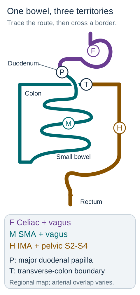
Abdominal foregut: the abdominal esophagus, stomach, and duodenum proximal to the major papilla belong with the celiac arterial territory and vagal parasympathetic input. The liver, biliary apparatus, and pancreas are foregut derivatives. This is an abdominal supply map, not a claim that the celiac trunk supplies the entire embryologic foregut. The pancreatic head has overlapping celiac and superior mesenteric supply. [1][3][4][6]
Midgut: begin just distal to the major duodenal papilla and continue through the jejunum, ileum, cecum, ascending colon, and proximal two thirds of the transverse colon. Match this territory to the superior mesenteric artery (SMA) and vagus. Crossing the foregut-midgut boundary changes the principal arterial root without changing the parasympathetic source. [1][3][7]
Hindgut: the distal third of the transverse colon, descending and sigmoid colon, rectum, and upper anal canal belong with inferior mesenteric artery (IMA) territory and pelvic splanchnic parasympathetics from S2-S4. The rectum also receives internal-iliac arterial contributions, so the developmental map does not exclude additional suppliers. The lower anal canal below the pectinate line is a separate somatic sensory territory. [1][5][7][11]
Trace it: use the territory diagram to follow the bowel from the duodenum to the descending colon. Identify the first boundary where only the arterial root changes, then the boundary where both the root and parasympathetic source change. The labeled routes remain visible while you compare them.
Follow the route from stomach to rectum. At P the principal arterial root changes but vagal input continues; at T the broad artery and parasympathetic pairing both change. The diagram stops at the rectum and does not depict the anal-canal sensory boundary. [1][3][5][7]Check the two boundary predictions
At the major papilla, celiac-dominant supply gives way to SMA-dominant supply while vagal input continues. At the developmental transverse-colon boundary, the broad pairing changes from SMA-vagus to IMA-pelvic splanchnics. Actual arterial overlap is not a sharp painted line. [1][3][7]
Use the distinction: injury limited to pelvic splanchnic input can affect hindgut function while leaving jejunal vagal input intact. Conversely, naming the vagus alone cannot distinguish a stomach target from an ascending-colon target. Identify the organ as well as the nerve. [7][11]
Trace the celiac branches from parent to destination
Does every artery with a foregut destination arise directly from the celiac trunk? No. The usual first branches are left gastric, splenic, and common hepatic. Several clinically important arteries are one or more branch points farther downstream. Confusing a destination with a direct origin leads to the wrong vessel during an angiographic or operative problem. [6]
The left gastric artery reaches the lesser curvature and gives branches toward the distal esophagus. The splenic artery travels toward the spleen, giving pancreatic branches, short gastric vessels, and the left gastroepiploic artery. The common hepatic artery gives the gastroduodenal artery (GDA) and continues as the proper hepatic artery. The proper hepatic artery proceeds toward the liver. The gallbladder is supplied by the cystic artery, most often from the right hepatic artery; this is a hepatic-region route, not an IMA branch. [14] These are the standard relationships; individual branching patterns can differ. [6]
Build a visible route rather than a disconnected name list: celiac trunk → common hepatic → gastroduodenal → right gastroepiploic. The GDA also contributes superior pancreaticoduodenal branches. Therefore, loss of the GDA can affect structures near the proximal duodenum and the right side of the greater-curvature arcade, not merely one organ whose name appears in the artery. [3][6]
Predict: a catheter is in the usual common hepatic artery, just before the GDA origin. Which two downstream directions are available: toward the liver and toward the duodenum, or toward the spleen and the descending colon?
Check the branch-point prediction
The liver and duodenum are the relevant downstream directions. Proper hepatic flow continues toward the liver; the GDA descends behind the proximal duodenum. The splenic artery is a separate first celiac branch, and the descending colon is principally an IMA target. [3][6]
Transfer: when an unfamiliar angiogram shows a branch behind the first part of the duodenum, trace backward to the common hepatic artery before deciding that it is a direct celiac branch. Position identifies the likely GDA; its parent identifies the next upstream branch point. [6]
Follow the stomach's borders, then its neighbors
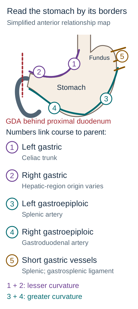
Could two branches from the splenic artery serve different gastric regions? Two arterial pairs follow the stomach's curvatures. Use the stomach silhouette to distinguish the inner, shorter border from the outer, longer border before naming a vessel. An artery's parent and its course should agree with the same answer. [6]
Trace each numbered artery along the border it supplies, then read its parent in the key. Compare an interrupted greater-curvature feeder with interrupted short gastric branches. The dashed GDA marks a posterior relationship; vessel caliber and organ proportions are schematic, and individual branching varies. [6][8]
Lesser curvature: left and right gastric arteries meet along this border within the lesser omentum. The left gastric artery usually comes directly from the celiac trunk. The right gastric artery often arises from the proper hepatic circulation, but other hepatic or gastroduodenal origins occur. A lesser-curvature course is more informative than insisting on one universal right-gastric origin. Catheterization through the left gastric artery can reach the right gastric artery across their connection. [6][8]
Greater curvature: the left gastroepiploic artery comes from splenic circulation, while the right gastroepiploic comes from the GDA. These paired vessels communicate along the greater curvature in the greater omentum. Gastroepiploic and gastroomental are alternative names for the same vessels. A branch from the splenic artery is not automatically a short gastric artery: follow its destination. [6]
Fundus: short gastric vessels travel through the gastrosplenic ligament from splenic circulation to the upper stomach. Their interruption is a different injury from interrupting a curvature arcade. The stomach has a rich vascular network; neither division of a single artery nor preservation of one visible arcade, by itself, proves the final viability of every gastric region. Assess the specific branches and the tissue supplied. [6]
Neighbor that explains bleeding: the GDA passes posterior to the first part of the duodenum. A posterior duodenal ulcer can erode this artery. The lesser-curvature gastric arteries fit a different surface, even though all these vessels ultimately communicate with the celiac circulation. A location-based explanation is more useful than memorizing that any upper gastrointestinal bleed must be from a celiac branch. [6]
Trace and compare: follow both greater-curvature suppliers on the diagram, then mentally interrupt the right-hand feeder before it reaches the stomach. Identify the vessel on the opposite side that could still deliver blood along the arcade. Next, compare this with interruption of small vessels entering the fundus.
Check the alternate-route prediction
Left gastroepiploic flow from the splenic artery can reach the greater-curvature arcade after loss of the right gastroepiploic feeder, provided that connection is adequate. Short gastric vessels enter the fundus by a different route; intact curvature vessels do not establish that every fundal branch is intact. [6]
Transfer: a splenic branch that travels a long distance along the greater curvature is left gastroepiploic; several short branches crossing the gastrosplenic ligament toward the fundus are short gastric. Parent alone is insufficient because both originate in splenic circulation. [6]
Locate the synapse before labeling the nerve
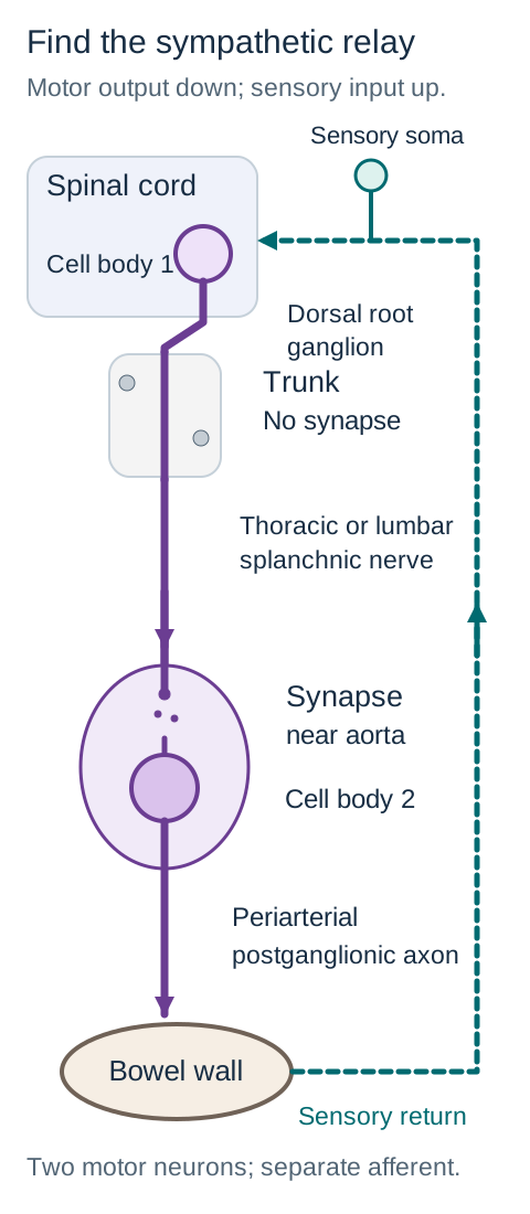
Does passing through a sympathetic ganglion mean that an axon has already synapsed? Not necessarily. For the abdominal route shown in the neural diagram, a preganglionic sympathetic axon begins in the thoracolumbar intermediolateral region, exits through a ventral root, enters a white ramus, and passes through the sympathetic trunk without synapsing there. A thoracic or lumbar splanchnic nerve then carries it toward a prevertebral relay. [7][12]
Follow the solid motor route and identify the gap where neuron 1 signals to neuron 2. Then follow the dashed route in the opposite direction: shared travel does not turn an afferent into sympathetic motor output. This is a functional route diagram, not a scaled spinal section; only one representative pathway is drawn. [4][7][12][13]
Celiac and mesenteric ganglia and plexuses organize the abdominal distribution. After the prevertebral synapse, a different neuron sends a postganglionic axon along the arterial routes to the viscera. A ganglion contains neuronal cell bodies; a plexus is a network of nerve fibers and may include ganglia. They are not interchangeable labels for the same structure. [7][12]
The greater thoracic splanchnic route emphasizes foregut targets through the celiac region; lesser thoracic and lumbar routes contribute to mesenteric distribution. These are teaching associations, not rigidly isolated cables. The least thoracic splanchnic nerve mainly contributes to renal innervation and should not be presented as another equivalent gut supplier. [7]
Parasympathetics use a different relay location. Vagal and pelvic splanchnic preganglionic axons reach ganglia near or within the target organ. Pelvic splanchnic nerves are parasympathetic despite the shared word splanchnic. Both autonomic divisions use acetylcholine at the preganglionic synapse on nicotinic receptors; parasympathetic postganglionic signaling to effectors is cholinergic and muscarinic. [11][12]
Sympathetic activity generally reduces digestive motility and secretion, increases sphincter tone, and constricts splanchnic vessels. Parasympathetic input generally promotes digestive activity. The gut also contains local enteric circuits: the myenteric plexus coordinates muscular activity, while the submucosal plexus regulates secretion and local vascular responses. Losing an extrinsic nerve does not erase the entire enteric nervous system. [4][5][7]
Trace in opposite directions: follow the efferent arrow from spinal cord to bowel, stopping at the prevertebral synapse. Then follow the separate afferent arrow from bowel toward the spinal cord. Pain signals from the small bowel travel centrally along visceral afferents that accompany sympathetic routes; the sensory axon is not a sympathetic motor neuron and does not acquire a postganglionic identity there. Its primary spinal sensory cell body is in a dorsal root ganglion, outside the spinal cord. [4][7][12][13]
Check the interruption prediction
A selective interruption before the prevertebral sympathetic synapse removes that preganglionic input while leaving the downstream neuron's cell body anatomically separate. An interruption after the synapse affects the postganglionic route. A peripheral nerve can also contain sensory axons, so an actual nonselective lesion may affect both motor modulation and visceral sensation. [7][12]
Transfer to the anal canal: painful pinprick below the pectinate line and voluntary external-sphincter contraction depend on somatic pudendal pathways, not on vagal or pelvic parasympathetic fibers. Above the line, visceral stretch sensation and autonomic smooth-muscle regulation are the relevant comparison. Shared sacral roots do not make pudendal and pelvic splanchnic nerves functionally interchangeable. [5][7][11]
Use vertebral levels as a starting coordinate
Could a vessel be identified from one axial level alone? A level narrows the search, but its direction, branches, and target organs should confirm the identification. The abdominal aorta gives anterior unpaired visceral branches, paired lateral branches such as the renal arteries, and terminal common iliac branches. A paired vessel at roughly the same height as a gut artery is not necessarily another gut supplier. [2]
The familiar approximate sequence is celiac near T12, SMA near L1, and IMA near L3. The usual celiac pattern reaches the foregut organs; the SMA reaches small bowel and right colon; the IMA gives left-colic and sigmoid supply and continues toward the superior rectal circulation. Anatomical studies demonstrate variation in origin level and branching, so the coordinate is not an absolute identification rule. [3][6][10]
Predict from two observations: a branch begins a little higher than the usual IMA level, but its distal course gives a left colic branch and then superior rectal supply. Decide whether its course or the memorized coordinate should determine its identity.
Check the imaging interpretation
The distribution identifies the IMA despite an atypical origin height. Calling it the SMA solely because it arises higher would conflict with the traced left-colic and superior-rectal branches. Use the level to search, then verify the branch tree. [3][10]
Transfer: an anterior root near L1 that supplies jejunum and ascending colon is consistent with the SMA. A branch at a similar height that runs laterally into a kidney fits renal circulation instead. The target territory prevents an origin-level mnemonic from overruling anatomy. [2][3]
Ask whether a connection can deliver enough flow
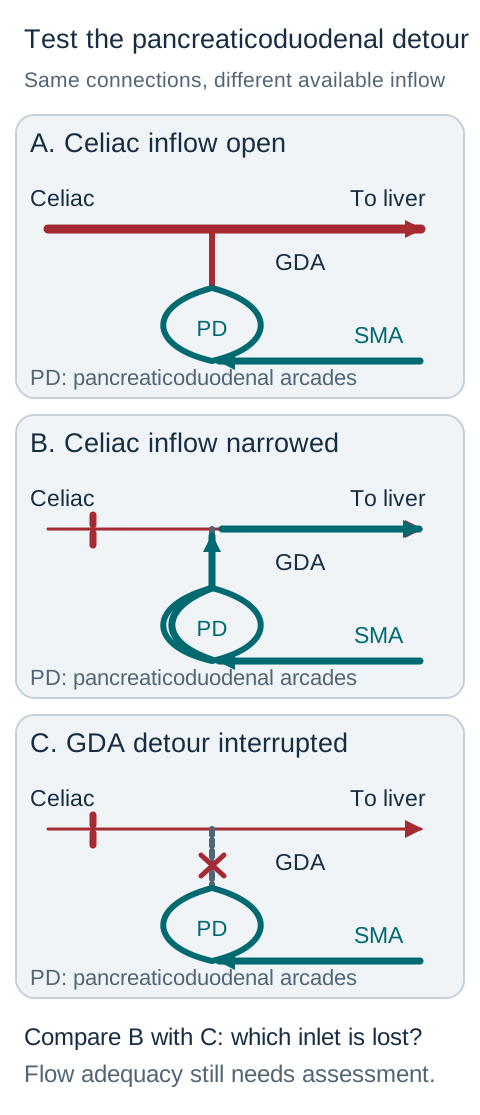
A collateral is an alternate route, not a guarantee of survival. It needs an open connection and an adequate driving pressure. Trace its inlet and outlet before deciding what an interruption would do. The collateral diagram shows an arterial detour around the pancreatic head and a separate colonic border-zone connection. [3][9]
Celiac to SMA: superior pancreaticoduodenal branches from the GDA meet inferior pancreaticoduodenal branches from the SMA around the duodenum and pancreatic head. With important celiac narrowing, SMA blood can pass through these arcades into the GDA and toward hepatic arterial circulation. Thus, a vessel usually described as a downstream branch can become part of a retrograde supply route. [3][9]
Change one condition: compare the visible routes with the celiac origin open, with it narrowed and the pancreaticoduodenal detour open, and with the detour interrupted as well. Trace from a patent source to the liver in each state. The direction of the detour depends on pressure, not on whether a vessel is named superior or inferior.
Trace from each patent source toward the liver, then compare state B with state C. The teal arrows in B show how an SMA-fed detour can support hepatic arterial inflow; losing that route can matter despite preserved portal flow. Arrow size is not a measured flow rate, and the lower panel does not claim that every GDA interruption causes hepatic ischemia. [3][6][9]Check the three-state flow prediction
With a patent celiac root, hepatic arterial inflow normally reaches the common and proper hepatic arteries directly. With celiac narrowing, the SMA-fed pancreaticoduodenal route can contribute retrograde GDA inflow. Interrupting the GDA in a patient dependent on that route can reduce hepatic arterial inflow; the actual response must be assessed rather than presumed from the presence of a portal vein. [3][6][9]
SMA to IMA: colic branches communicate through the marginal artery along the mesenteric edge of the colon. Near the splenic flexure, middle-colic and left-colic contributions link the two territories. Their caliber and continuity vary. A low-pressure state can injure a border zone even when contrast demonstrates an anatomically patent connection. Conversely, an occluded main root does not prove immediate infarction if collateral delivery remains sufficient. [3][10]
Do not mix the rectal maps. Arterial inflow can come from the IMA through the superior rectal artery and from the internal iliac system through middle rectal branches and the internal pudendal-inferior rectal route. These are oxygen-delivery routes. Venous return follows a different question: superior rectal blood reaches the inferior mesenteric vein and the portal system, usually through the splenic vein; middle and inferior rectal pathways reach the systemic internal iliac circulation. These veins provide a portosystemic connection, not an arterial bridge from the IMA to a vein. [5][6]
Transfer: diminished rectal arterial inflow calls for an arterial supply explanation. Portal hypertension with dilated anorectal collateral veins calls for a venous-pressure explanation. An arterial anastomosis does not bypass the liver, and a venous collateral does not replace lost arterial oxygen delivery. In a real patient, severe abdominal pain or bleeding requires urgent clinical assessment; a territory diagram cannot establish viability or choose treatment by itself. [3][5][9]
Apply the anatomy to a new patient
Identify the affected tissue, establish the relevant route, and predict the consequence before comparing the options. Each case has one best answer.
Case 1
Show answer and explanations for case 1
A. Reduced jejunal stimulation with preserved descending-colon stimulation (Why this does not fit)
Jejunal parasympathetic input is vagal, whereas descending-colon input is pelvic splanchnic. The specified injury affects left-colon input, so this reverses the affected and spared targets. [1][7][11]
Reasoning steps for option A
What anatomical or functional relationship does Reduced jejunal stimulation with preserved descending-colon stimulation imply?
Jejunal parasympathetic input is vagal, whereas descending-colon input is pelvic splanchnic.
Using the stated finding "During resection of a lesion in the distal third of the transverse colon, extrinsic parasympathetic fibers traveling toward the left colon are injured", which discriminator determines whether "Reduced jejunal stimulation with preserved descending-colon stimulation" matches this case?
The specified injury affects left-colon input, so this reverses the affected and spared targets.
B. Reduced ascending-colon stimulation with preserved sigmoid stimulation (Why this does not fit)
Ascending-colon input is predominantly vagal, whereas sigmoid input is pelvic splanchnic. Intact vagal trunks do not explain a selective ascending-colon deficit from the stated left-colon injury. [1][7][11]
Reasoning steps for option B
What anatomical or functional relationship does Reduced ascending-colon stimulation with preserved sigmoid stimulation imply?
Ascending-colon input is predominantly vagal, whereas sigmoid input is pelvic splanchnic.
Using the stated finding "During resection of a lesion in the distal third of the transverse colon, extrinsic parasympathetic fibers traveling toward the left colon are injured", which discriminator determines whether "Reduced ascending-colon stimulation with preserved sigmoid stimulation" matches this case?
Intact vagal trunks do not explain a selective ascending-colon deficit from the stated left-colon injury.
C. Reduced descending-colon stimulation with preserved jejunal stimulation (Best answer)
The distal transverse and descending colon belong to the pelvic-splanchnic hindgut territory. The injury reduces that extrinsic input while the separate vagal route to the jejunum remains intact. [1][7][11]
Reasoning steps for option C
What anatomical or functional relationship does Reduced descending-colon stimulation with preserved jejunal stimulation imply?
The distal transverse and descending colon belong to the pelvic-splanchnic hindgut territory.
Using the stated finding "During resection of a lesion in the distal third of the transverse colon, extrinsic parasympathetic fibers traveling toward the left colon are injured", which discriminator determines whether "Reduced descending-colon stimulation with preserved jejunal stimulation" matches this case?
The injury reduces that extrinsic input while the separate vagal route to the jejunum remains intact.
D. Reduced gastric stimulation with preserved rectal stimulation (Why this does not fit)
The stomach receives vagal parasympathetics and the rectum receives pelvic parasympathetics. A lesion confined to fibers destined for the left colon does not directly interrupt gastric vagal input. [1][7][11]
Reasoning steps for option D
What anatomical or functional relationship does Reduced gastric stimulation with preserved rectal stimulation imply?
The stomach receives vagal parasympathetics and the rectum receives pelvic parasympathetics.
Using the stated finding "During resection of a lesion in the distal third of the transverse colon, extrinsic parasympathetic fibers traveling toward the left colon are injured", which discriminator determines whether "Reduced gastric stimulation with preserved rectal stimulation" matches this case?
A lesion confined to fibers destined for the left colon does not directly interrupt gastric vagal input.
A. Vagal fibers terminating in enteric ganglia (Best answer)
Terminal ileum and ascending colon are midgut targets supplied by the SMA and vagal parasympathetics. The root location plus the two distal territories identifies the SMA distribution and therefore the vagal pathway. [3][7][11]
Reasoning steps for option A
What anatomical or functional relationship does Vagal fibers terminating in enteric ganglia imply?
Terminal ileum and ascending colon are midgut targets supplied by the SMA and vagal parasympathetics.
Using the stated finding "CT angiography in a patient with abrupt abdominal pain shows an occluded anterior aortic branch arising near L1", which discriminator determines whether "Vagal fibers terminating in enteric ganglia" matches this case?
The root location plus the two distal territories identifies the SMA distribution and therefore the vagal pathway.
B. Pelvic splanchnic fibers terminating in enteric ganglia (Why this does not fit)
Pelvic splanchnic parasympathetics primarily serve hindgut and pelvic organs. They do not provide the usual shared parasympathetic supply to terminal ileum and ascending colon. [3][7][11]
Reasoning steps for option B
What anatomical or functional relationship does Pelvic splanchnic fibers terminating in enteric ganglia imply?
Pelvic splanchnic parasympathetics primarily serve hindgut and pelvic organs.
Using the stated finding "CT angiography in a patient with abrupt abdominal pain shows an occluded anterior aortic branch arising near L1", which discriminator determines whether "Pelvic splanchnic fibers terminating in enteric ganglia" matches this case?
They do not provide the usual shared parasympathetic supply to terminal ileum and ascending colon.
C. Thoracic splanchnic fibers terminating in prevertebral ganglia (Why this does not fit)
Thoracic splanchnic efferents provide sympathetic rather than parasympathetic input. They can accompany the relevant abdominal distribution but have the wrong autonomic identity for the question. [3][7][11]
Reasoning steps for option C
What anatomical or functional relationship does Thoracic splanchnic fibers terminating in prevertebral ganglia imply?
Thoracic splanchnic efferents provide sympathetic rather than parasympathetic input.
Using the stated finding "CT angiography in a patient with abrupt abdominal pain shows an occluded anterior aortic branch arising near L1", which discriminator determines whether "Thoracic splanchnic fibers terminating in prevertebral ganglia" matches this case?
They can accompany the relevant abdominal distribution but have the wrong autonomic identity for the question.
D. Lumbar splanchnic fibers terminating in prevertebral ganglia (Why this does not fit)
Lumbar splanchnic efferents are sympathetic contributors to lower abdominal distribution. Their prevertebral relay does not match the requested parasympathetic route to the two midgut segments. [3][7][11]
Reasoning steps for option D
What anatomical or functional relationship does Lumbar splanchnic fibers terminating in prevertebral ganglia imply?
Lumbar splanchnic efferents are sympathetic contributors to lower abdominal distribution.
Using the stated finding "CT angiography in a patient with abrupt abdominal pain shows an occluded anterior aortic branch arising near L1", which discriminator determines whether "Lumbar splanchnic fibers terminating in prevertebral ganglia" matches this case?
Their prevertebral relay does not match the requested parasympathetic route to the two midgut segments.
Takeaway: Select vagal preganglionic input with an enteric relay. [3][7][11]
Jejunal arteries arise from the SMA rather than from the left-colic arterial tree. A clamp on the mapped left-colic and sigmoid root does not directly interrupt jejunal SMA inflow. [3][5][10]
Reasoning steps for option A
What anatomical or functional relationship does Proximal jejunal wall imply?
Jejunal arteries arise from the SMA rather than from the left-colic arterial tree.
Using the stated finding "During aortic repair, a temporary clamp interrupts an anterior visceral branch near L3 before its first division", which discriminator determines whether "Proximal jejunal wall" matches this case?
A clamp on the mapped left-colic and sigmoid root does not directly interrupt jejunal SMA inflow.
B. Gastric lesser curvature (Why this does not fit)
The gastric arteries supplying the lesser curvature belong to celiac circulation. The described branch pattern identifies a different root and does not directly clamp the gastric feeders. [3][5][10]
Reasoning steps for option B
What anatomical or functional relationship does Gastric lesser curvature imply?
The gastric arteries supplying the lesser curvature belong to celiac circulation.
Using the stated finding "During aortic repair, a temporary clamp interrupts an anterior visceral branch near L3 before its first division", which discriminator determines whether "Gastric lesser curvature" matches this case?
The described branch pattern identifies a different root and does not directly clamp the gastric feeders.
C. Ascending colonic wall (Why this does not fit)
Ascending-colon branches arise in the SMA distribution. The left colic and sigmoid branches identify IMA rather than right-colon inflow. [3][5][10]
Reasoning steps for option C
What anatomical or functional relationship does Ascending colonic wall imply?
Ascending-colon branches arise in the SMA distribution.
Using the stated finding "During aortic repair, a temporary clamp interrupts an anterior visceral branch near L3 before its first division", which discriminator determines whether "Ascending colonic wall" matches this case?
The left colic and sigmoid branches identify IMA rather than right-colon inflow.
D. Upper rectal wall (Best answer)
The IMA gives left colic and sigmoid branches and continues as the superior rectal artery. Clamping that root interrupts its direct superior rectal contribution, although other rectal arterial routes may remain. [3][5][10]
Reasoning steps for option D
What anatomical or functional relationship does Upper rectal wall imply?
The IMA gives left colic and sigmoid branches and continues as the superior rectal artery.
Using the stated finding "During aortic repair, a temporary clamp interrupts an anterior visceral branch near L3 before its first division", which discriminator determines whether "Upper rectal wall" matches this case?
Clamping that root interrupts its direct superior rectal contribution, although other rectal arterial routes may remain.
Takeaway: Predict loss of direct upper-rectal inflow without presuming total rectal ischemia. [3][5][10]
A. Within a ganglion of the thoracic sympathetic trunk (Why this does not fit)
The thoracic sympathetic trunk is part of sympathetic routing. The identified jejunal segment receives vagal parasympathetics whose peripheral relay is not in that trunk. [3][4][11]
Reasoning steps for option A
What anatomical or functional relationship does Within a ganglion of the thoracic sympathetic trunk imply?
The thoracic sympathetic trunk is part of sympathetic routing.
Using the stated finding "A bowel segment resected after a focal mesenteric injury has prominent folds, relatively few arterial arcades, and long straight vessels entering its wall", which discriminator determines whether "Within a ganglion of the thoracic sympathetic trunk" matches this case?
The identified jejunal segment receives vagal parasympathetics whose peripheral relay is not in that trunk.
B. Within an enteric ganglion of the bowel wall (Best answer)
Vagal preganglionic axons reach enteric ganglia near or within the bowel wall. The proximal small-bowel features identify jejunum, so this is the relevant parasympathetic relay location. [3][4][11]
Reasoning steps for option B
What anatomical or functional relationship does Within an enteric ganglion of the bowel wall imply?
Vagal preganglionic axons reach enteric ganglia near or within the bowel wall.
Using the stated finding "A bowel segment resected after a focal mesenteric injury has prominent folds, relatively few arterial arcades, and long straight vessels entering its wall", which discriminator determines whether "Within an enteric ganglion of the bowel wall" matches this case?
The proximal small-bowel features identify jejunum, so this is the relevant parasympathetic relay location.
C. Within the superior mesenteric prevertebral ganglion (Why this does not fit)
The superior mesenteric prevertebral region organizes sympathetic output to midgut. Sharing the SMA territory does not make this the final parasympathetic synapse. [3][4][11]
Reasoning steps for option C
What anatomical or functional relationship does Within the superior mesenteric prevertebral ganglion imply?
The superior mesenteric prevertebral region organizes sympathetic output to midgut.
Using the stated finding "A bowel segment resected after a focal mesenteric injury has prominent folds, relatively few arterial arcades, and long straight vessels entering its wall", which discriminator determines whether "Within the superior mesenteric prevertebral ganglion" matches this case?
Sharing the SMA territory does not make this the final parasympathetic synapse.
D. Within the celiac prevertebral ganglion (Why this does not fit)
Celiac ganglia contain sympathetic relay neurons for abdominal targets. The question concerns a vagal-enteric parasympathetic relay rather than a celiac sympathetic relay. [3][4][11]
Reasoning steps for option D
What anatomical or functional relationship does Within the celiac prevertebral ganglion imply?
Celiac ganglia contain sympathetic relay neurons for abdominal targets.
Using the stated finding "A bowel segment resected after a focal mesenteric injury has prominent folds, relatively few arterial arcades, and long straight vessels entering its wall", which discriminator determines whether "Within the celiac prevertebral ganglion" matches this case?
The question concerns a vagal-enteric parasympathetic relay rather than a celiac sympathetic relay.
Takeaway: Place its peripheral relay in or near the bowel wall. [3][4][11]
A. Reduced gastric and descending-colon input, with cecal input spared (Why this does not fit)
Gastric and cecal extrinsic parasympathetics are vagal, but descending-colon input is pelvic. This pattern incorrectly spares a vagal target and includes a pelvic target. [1][7][11]
Reasoning steps for option A
What anatomical or functional relationship does Reduced gastric and descending-colon input, with cecal input spared imply?
Gastric and cecal extrinsic parasympathetics are vagal, but descending-colon input is pelvic.
Using the stated finding "After truncal vagotomy for refractory ulcer disease, a researcher compares extrinsic parasympathetic signaling to the stomach, cecum, and descending colon", which discriminator determines whether "Reduced gastric and descending-colon input, with cecal input spared" matches this case?
This pattern incorrectly spares a vagal target and includes a pelvic target.
B. Reduced gastric and cecal input, with descending-colon input spared (Best answer)
The stomach and cecum lie in foregut and midgut territories that share vagal input. Truncal vagotomy interrupts this shared input while intact pelvic pathways still reach the descending colon. [1][7][11]
Reasoning steps for option B
What anatomical or functional relationship does Reduced gastric and cecal input, with descending-colon input spared imply?
The stomach and cecum lie in foregut and midgut territories that share vagal input.
Using the stated finding "After truncal vagotomy for refractory ulcer disease, a researcher compares extrinsic parasympathetic signaling to the stomach, cecum, and descending colon", which discriminator determines whether "Reduced gastric and cecal input, with descending-colon input spared" matches this case?
Truncal vagotomy interrupts this shared input while intact pelvic pathways still reach the descending colon.
C. Reduced cecal and descending-colon input, with gastric input spared (Why this does not fit)
The cecum is vagally supplied, while the descending colon has pelvic input. The gastric target should not be spared by interruption of both vagal trunks. [1][7][11]
Reasoning steps for option C
What anatomical or functional relationship does Reduced cecal and descending-colon input, with gastric input spared imply?
The cecum is vagally supplied, while the descending colon has pelvic input.
Using the stated finding "After truncal vagotomy for refractory ulcer disease, a researcher compares extrinsic parasympathetic signaling to the stomach, cecum, and descending colon", which discriminator determines whether "Reduced cecal and descending-colon input, with gastric input spared" matches this case?
The gastric target should not be spared by interruption of both vagal trunks.
D. Reduced descending-colon input, with gastric and cecal input spared (Why this does not fit)
Pelvic parasympathetics serve the descending colon independently of vagal trunks. This describes pelvic rather than vagal interruption and reverses the specified pattern. [1][7][11]
Reasoning steps for option D
What anatomical or functional relationship does Reduced descending-colon input, with gastric and cecal input spared imply?
Pelvic parasympathetics serve the descending colon independently of vagal trunks.
Using the stated finding "After truncal vagotomy for refractory ulcer disease, a researcher compares extrinsic parasympathetic signaling to the stomach, cecum, and descending colon", which discriminator determines whether "Reduced descending-colon input, with gastric and cecal input spared" matches this case?
This describes pelvic rather than vagal interruption and reverses the specified pattern.
Takeaway: Predict selective extrinsic deficits without claiming loss of all local motility. [1][7][11]
A. Pelvic splanchnic parasympathetic fibers (Why this does not fit)
Pelvic splanchnic fibers regulate hindgut and pelvic smooth-muscle function. They do not explain the paired loss of somatic pinprick sensation and voluntary external-sphincter contraction. [5][7][11]
Reasoning steps for option A
What anatomical or functional relationship does Pelvic splanchnic parasympathetic fibers imply?
Pelvic splanchnic fibers regulate hindgut and pelvic smooth-muscle function.
Using the stated finding "After pelvic surgery, a patient has reduced pinprick sensation below the pectinate line and weak voluntary anal squeeze", which discriminator determines whether "Pelvic splanchnic parasympathetic fibers" matches this case?
They do not explain the paired loss of somatic pinprick sensation and voluntary external-sphincter contraction.
B. Lumbar splanchnic sympathetic fibers (Why this does not fit)
Lumbar sympathetic pathways modulate visceral smooth muscle and vascular tone. The observed cutaneous sensation and voluntary squeeze deficits localize outside this visceral motor pathway. [5][7][11]
Reasoning steps for option B
What anatomical or functional relationship does Lumbar splanchnic sympathetic fibers imply?
Lumbar sympathetic pathways modulate visceral smooth muscle and vascular tone.
Using the stated finding "After pelvic surgery, a patient has reduced pinprick sensation below the pectinate line and weak voluntary anal squeeze", which discriminator determines whether "Lumbar splanchnic sympathetic fibers" matches this case?
The observed cutaneous sensation and voluntary squeeze deficits localize outside this visceral motor pathway.
C. Vagal parasympathetic fibers (Why this does not fit)
The vagus serves foregut and midgut rather than the somatic lower anal canal. Intact rectal smooth-muscle responses do not turn the lower anal sensory deficit into a vagal injury. [5][7][11]
Reasoning steps for option C
What anatomical or functional relationship does Vagal parasympathetic fibers imply?
The vagus serves foregut and midgut rather than the somatic lower anal canal.
Using the stated finding "After pelvic surgery, a patient has reduced pinprick sensation below the pectinate line and weak voluntary anal squeeze", which discriminator determines whether "Vagal parasympathetic fibers" matches this case?
Intact rectal smooth-muscle responses do not turn the lower anal sensory deficit into a vagal injury.
D. Pudendal inferior rectal branches (Best answer)
Pudendal inferior rectal branches carry somatic input from the lower canal and motor input to the external sphincter. Their injury accounts for both reduced pinprick and weak voluntary squeeze while autonomic internal-sphincter function can persist. [5][7][11]
Reasoning steps for option D
What anatomical or functional relationship does Pudendal inferior rectal branches imply?
Pudendal inferior rectal branches carry somatic input from the lower canal and motor input to the external sphincter.
Using the stated finding "After pelvic surgery, a patient has reduced pinprick sensation below the pectinate line and weak voluntary anal squeeze", which discriminator determines whether "Pudendal inferior rectal branches" matches this case?
Their injury accounts for both reduced pinprick and weak voluntary squeeze while autonomic internal-sphincter function can persist.
Takeaway: Localize the combined findings to inferior rectal pudendal branches. [5][7][11]
The GDA usually branches from the common hepatic artery and gives the right gastroepiploic artery. Its posterior duodenal course and downstream greater-curvature distribution account for both observations. [6][8]
Reasoning steps for option A
What anatomical or functional relationship does Gastroduodenal artery imply?
The GDA usually branches from the common hepatic artery and gives the right gastroepiploic artery.
Using the stated finding "Before hepatic arterial infusion, angiography shows a catheter tip in the common hepatic artery proximal to its usual major branch point", which discriminator determines whether "Gastroduodenal artery" matches this case?
Its posterior duodenal course and downstream greater-curvature distribution account for both observations.
B. Proper hepatic artery (Why this does not fit)
The proper hepatic artery continues toward the liver after the GDA branch point. Its direct hepatic course does not explain the vessel behind the duodenum that supplies the right gastroepiploic route. [6][8]
Reasoning steps for option B
What anatomical or functional relationship does Proper hepatic artery imply?
The proper hepatic artery continues toward the liver after the GDA branch point.
Using the stated finding "Before hepatic arterial infusion, angiography shows a catheter tip in the common hepatic artery proximal to its usual major branch point", which discriminator determines whether "Proper hepatic artery" matches this case?
Its direct hepatic course does not explain the vessel behind the duodenum that supplies the right gastroepiploic route.
C. Left gastric artery (Why this does not fit)
The left gastric artery usually arises directly from the celiac trunk and follows the lesser curvature. Both its parent and its curvature differ from the observed common-hepatic diversion. [6][8]
Reasoning steps for option C
For the finding "Before hepatic arterial infusion, angiography shows a catheter tip in the common hepatic artery proximal to its usual major branch point", what specifically makes "Left gastric artery" anatomically or physiologically fit or fail?
The left gastric artery usually arises directly from the celiac trunk and follows the lesser curvature.
Using the stated finding "Before hepatic arterial infusion, angiography shows a catheter tip in the common hepatic artery proximal to its usual major branch point", which discriminator determines whether "Left gastric artery" matches this case?
Both its parent and its curvature differ from the observed common-hepatic diversion.
D. Splenic artery (Why this does not fit)
The splenic artery gives the left gastroepiploic artery and short gastric vessels. It is a separate celiac branch, not the usual common-hepatic branch passing behind the first duodenal segment. [6][8]
Reasoning steps for option D
What anatomical or functional relationship does Splenic artery imply?
The splenic artery gives the left gastroepiploic artery and short gastric vessels.
Using the stated finding "Before hepatic arterial infusion, angiography shows a catheter tip in the common hepatic artery proximal to its usual major branch point", which discriminator determines whether "Splenic artery" matches this case?
It is a separate celiac branch, not the usual common-hepatic branch passing behind the first duodenal segment.
Takeaway: Select the common-hepatic branch explaining the entire observed route. [6][8]
A. Splenic artery through the short gastric vessels (Why this does not fit)
Short gastric vessels enter the fundus from splenic circulation. They are not the demonstrated continuous lesser-curvature arcade leading to the right gastric artery. [6][8]
Reasoning steps for option A
What anatomical or functional relationship does Splenic artery through the short gastric vessels imply?
Short gastric vessels enter the fundus from splenic circulation.
Using the stated finding "An interventional radiologist cannot catheterize a sharply angled right gastric origin from the hepatic circulation", which discriminator determines whether "Splenic artery through the short gastric vessels" matches this case?
They are not the demonstrated continuous lesser-curvature arcade leading to the right gastric artery.
B. Gastroduodenal artery through the right gastroepiploic artery (Why this does not fit)
The right gastroepiploic artery follows the greater curvature. That route approaches a different gastric border than the lesser-curvature connection shown. [6][8]
Reasoning steps for option B
What anatomical or functional relationship does Gastroduodenal artery through the right gastroepiploic artery imply?
The right gastroepiploic artery follows the greater curvature.
Using the stated finding "An interventional radiologist cannot catheterize a sharply angled right gastric origin from the hepatic circulation", which discriminator determines whether "Gastroduodenal artery through the right gastroepiploic artery" matches this case?
That route approaches a different gastric border than the lesser-curvature connection shown.
C. Celiac trunk through the left gastric artery (Best answer)
The left gastric artery reaches the lesser curvature and communicates with the right gastric artery. This route approaches the distal right gastric vessel from the opposite end of the demonstrated arcade. [6][8]
Reasoning steps for option C
What anatomical or functional relationship does Celiac trunk through the left gastric artery imply?
The left gastric artery reaches the lesser curvature and communicates with the right gastric artery.
Using the stated finding "An interventional radiologist cannot catheterize a sharply angled right gastric origin from the hepatic circulation", which discriminator determines whether "Celiac trunk through the left gastric artery" matches this case?
This route approaches the distal right gastric vessel from the opposite end of the demonstrated arcade.
D. Splenic artery through the left gastroepiploic artery (Why this does not fit)
The left gastroepiploic artery communicates with the right gastroepiploic artery. This is a greater-curvature connection rather than the specified right-gastric connection. [6][8]
Reasoning steps for option D
What anatomical or functional relationship does Splenic artery through the left gastroepiploic artery imply?
The left gastroepiploic artery communicates with the right gastroepiploic artery.
Using the stated finding "An interventional radiologist cannot catheterize a sharply angled right gastric origin from the hepatic circulation", which discriminator determines whether "Splenic artery through the left gastroepiploic artery" matches this case?
This is a greater-curvature connection rather than the specified right-gastric connection.
Takeaway: Approach the right-gastric distal bed through the demonstrated lesser-curvature connection. [6][8]
The left gastric artery primarily follows the lesser curvature. It does not match the remaining GDA-derived feeder entering the greater-curvature arcade. [6]
Reasoning steps for option A
For the finding "During surgery near the splenic hilum, a vessel traveling toward the greater curvature is divided", what specifically makes "Left gastric artery" anatomically or physiologically fit or fail?
The left gastric artery primarily follows the lesser curvature.
Using the stated finding "During surgery near the splenic hilum, a vessel traveling toward the greater curvature is divided", which discriminator determines whether "Left gastric artery" matches this case?
It does not match the remaining GDA-derived feeder entering the greater-curvature arcade.
B. Right gastric artery (Why this does not fit)
The right gastric artery follows the lesser curvature from a variable hepatic-region origin. A greater-curvature course from the GDA identifies a different named artery. [6]
Reasoning steps for option B
For the finding "During surgery near the splenic hilum, a vessel traveling toward the greater curvature is divided", what specifically makes "Right gastric artery" anatomically or physiologically fit or fail?
The right gastric artery follows the lesser curvature from a variable hepatic-region origin.
Using the stated finding "During surgery near the splenic hilum, a vessel traveling toward the greater curvature is divided", which discriminator determines whether "Right gastric artery" matches this case?
A greater-curvature course from the GDA identifies a different named artery.
C. Left gastroepiploic artery (Why this does not fit)
The left gastroepiploic artery comes from splenic circulation and follows the greater curvature. Its course identifies the divided vessel, not the opposite GDA-derived feeder that remains. [6]
Reasoning steps for option C
For the finding "During surgery near the splenic hilum, a vessel traveling toward the greater curvature is divided", what specifically makes "Left gastroepiploic artery" anatomically or physiologically fit or fail?
The left gastroepiploic artery comes from splenic circulation and follows the greater curvature.
Using the stated finding "During surgery near the splenic hilum, a vessel traveling toward the greater curvature is divided", which discriminator determines whether "Left gastroepiploic artery" matches this case?
Its course identifies the divided vessel, not the opposite GDA-derived feeder that remains.
D. Right gastroepiploic artery (Best answer)
The right gastroepiploic artery arises from the GDA and joins the left gastroepiploic arcade. It accounts for preserved filling from the opposite end after the splenic-side feeder was divided. [6]
Reasoning steps for option D
For the finding "During surgery near the splenic hilum, a vessel traveling toward the greater curvature is divided", what specifically makes "Right gastroepiploic artery" anatomically or physiologically fit or fail?
The right gastroepiploic artery arises from the GDA and joins the left gastroepiploic arcade.
Using the stated finding "During surgery near the splenic hilum, a vessel traveling toward the greater curvature is divided", which discriminator determines whether "Right gastroepiploic artery" matches this case?
It accounts for preserved filling from the opposite end after the splenic-side feeder was divided.
Takeaway: Assign the remaining direct feeder to the right gastroepiploic artery. [6]
Short gastric vessels cross the gastrosplenic ligament to supply the fundus. Their local interruption fits the fundal perfusion change despite preserved greater-curvature feeders. [6]
Reasoning steps for option A
What anatomical or functional relationship does Short gastric branches imply?
Short gastric vessels cross the gastrosplenic ligament to supply the fundus.
Using the stated finding "Following dissection of the gastrosplenic ligament, several small arterial stumps are seen near the upper stomach", which discriminator determines whether "Short gastric branches" matches this case?
Their local interruption fits the fundal perfusion change despite preserved greater-curvature feeders.
B. Left gastroepiploic branches (Why this does not fit)
The left gastroepiploic artery follows the greater curvature from splenic circulation. The stem documents that feeder as patent and instead identifies small interrupted fundal vessels. [6]
Reasoning steps for option B
What anatomical or functional relationship does Left gastroepiploic branches imply?
The left gastroepiploic artery follows the greater curvature from splenic circulation.
Using the stated finding "Following dissection of the gastrosplenic ligament, several small arterial stumps are seen near the upper stomach", which discriminator determines whether "Left gastroepiploic branches" matches this case?
The stem documents that feeder as patent and instead identifies small interrupted fundal vessels.
C. Right gastroepiploic branches (Why this does not fit)
The right gastroepiploic artery enters the greater-curvature arcade from the GDA. Its patent status and nonsplenic route conflict with the described gastrosplenic injury. [6]
Reasoning steps for option C
What anatomical or functional relationship does Right gastroepiploic branches imply?
The right gastroepiploic artery enters the greater-curvature arcade from the GDA.
Using the stated finding "Following dissection of the gastrosplenic ligament, several small arterial stumps are seen near the upper stomach", which discriminator determines whether "Right gastroepiploic branches" matches this case?
Its patent status and nonsplenic route conflict with the described gastrosplenic injury.
D. Right gastric branches (Why this does not fit)
The right gastric artery reaches the lesser curvature from hepatic-region circulation. That course does not explain several small stumps in the gastrosplenic ligament adjacent to the fundus. [6]
Reasoning steps for option D
What anatomical or functional relationship does Right gastric branches imply?
The right gastric artery reaches the lesser curvature from hepatic-region circulation.
Using the stated finding "Following dissection of the gastrosplenic ligament, several small arterial stumps are seen near the upper stomach", which discriminator determines whether "Right gastric branches" matches this case?
That course does not explain several small stumps in the gastrosplenic ligament adjacent to the fundus.
Takeaway: Localize the interrupted fundal inflow without assuming inevitable total gastric infarction. [6]
The left gastric artery normally branches directly from the celiac trunk. The posterior duodenal artery is the GDA, whose interruption does not directly divide left-gastric inflow. [6]
Reasoning steps for option A
For the finding "A patient has brisk bleeding from a posterior ulcer in the first part of the duodenum", what specifically makes "Left gastric artery" anatomically or physiologically fit or fail?
The left gastric artery normally branches directly from the celiac trunk.
Using the stated finding "A patient has brisk bleeding from a posterior ulcer in the first part of the duodenum", which discriminator determines whether "Left gastric artery" matches this case?
The posterior duodenal artery is the GDA, whose interruption does not directly divide left-gastric inflow.
B. Right gastric artery (Why this does not fit)
The right gastric artery commonly arises from hepatic circulation and follows the lesser curvature. In the usual branching pattern requested, it is not the distal greater-curvature branch of the GDA. [6]
Reasoning steps for option B
For the finding "A patient has brisk bleeding from a posterior ulcer in the first part of the duodenum", what specifically makes "Right gastric artery" anatomically or physiologically fit or fail?
The right gastric artery commonly arises from hepatic circulation and follows the lesser curvature.
Using the stated finding "A patient has brisk bleeding from a posterior ulcer in the first part of the duodenum", which discriminator determines whether "Right gastric artery" matches this case?
In the usual branching pattern requested, it is not the distal greater-curvature branch of the GDA.
C. Right gastroepiploic artery (Best answer)
The right gastroepiploic artery arises from the GDA. Posterior D1 erosion identifies the GDA, so proximal interruption directly reduces inflow to its right gastroepiploic branch. [6]
Reasoning steps for option C
For the finding "A patient has brisk bleeding from a posterior ulcer in the first part of the duodenum", what specifically makes "Right gastroepiploic artery" anatomically or physiologically fit or fail?
The right gastroepiploic artery arises from the GDA.
Using the stated finding "A patient has brisk bleeding from a posterior ulcer in the first part of the duodenum", which discriminator determines whether "Right gastroepiploic artery" matches this case?
Posterior D1 erosion identifies the GDA, so proximal interruption directly reduces inflow to its right gastroepiploic branch.
D. Left gastroepiploic artery (Why this does not fit)
The left gastroepiploic artery arises from splenic circulation. It may provide collateral support to the right side, but its own direct parent inflow is not the injured GDA. [6]
Reasoning steps for option D
For the finding "A patient has brisk bleeding from a posterior ulcer in the first part of the duodenum", what specifically makes "Left gastroepiploic artery" anatomically or physiologically fit or fail?
The left gastroepiploic artery arises from splenic circulation.
Using the stated finding "A patient has brisk bleeding from a posterior ulcer in the first part of the duodenum", which discriminator determines whether "Left gastroepiploic artery" matches this case?
It may provide collateral support to the right side, but its own direct parent inflow is not the injured GDA.
Takeaway: Predict loss of direct right-gastroepiploic inflow with collateral adequacy assessed separately. [6]
A. Right gastroepiploic artery (Why this does not fit)
The right gastroepiploic artery usually arises from the GDA and follows the greater curvature. Its expected course does not match the observed lesser-curvature route to the left-gastric territory. [6][8]
Reasoning steps for option A
For the finding "Angiography before gastric surgery shows a small artery arising from the left hepatic artery", what specifically makes "Right gastroepiploic artery" anatomically or physiologically fit or fail?
The right gastroepiploic artery usually arises from the GDA and follows the greater curvature.
Using the stated finding "Angiography before gastric surgery shows a small artery arising from the left hepatic artery", which discriminator determines whether "Right gastroepiploic artery" matches this case?
Its expected course does not match the observed lesser-curvature route to the left-gastric territory.
B. Right gastric artery (Best answer)
The right gastric artery has variable hepatic-region origins and follows the lesser curvature toward the left gastric artery. The course and anastomotic partner identify it despite the documented left-hepatic origin. [6][8]
Reasoning steps for option B
For the finding "Angiography before gastric surgery shows a small artery arising from the left hepatic artery", what specifically makes "Right gastric artery" anatomically or physiologically fit or fail?
The right gastric artery has variable hepatic-region origins and follows the lesser curvature toward the left gastric artery.
Using the stated finding "Angiography before gastric surgery shows a small artery arising from the left hepatic artery", which discriminator determines whether "Right gastric artery" matches this case?
The course and anastomotic partner identify it despite the documented left-hepatic origin.
C. Left gastroepiploic artery (Why this does not fit)
The left gastroepiploic artery follows the greater curvature from splenic circulation. The observed pyloric-to-lesser-curvature course conflicts with that arterial identity. [6][8]
Reasoning steps for option C
For the finding "Angiography before gastric surgery shows a small artery arising from the left hepatic artery", what specifically makes "Left gastroepiploic artery" anatomically or physiologically fit or fail?
The left gastroepiploic artery follows the greater curvature from splenic circulation.
Using the stated finding "Angiography before gastric surgery shows a small artery arising from the left hepatic artery", which discriminator determines whether "Left gastroepiploic artery" matches this case?
The observed pyloric-to-lesser-curvature course conflicts with that arterial identity.
D. Short gastric artery (Why this does not fit)
Short gastric arteries cross the gastrosplenic ligament toward the fundus. A long lesser-curvature route beginning in hepatic circulation does not fit these fundal branches. [6][8]
Reasoning steps for option D
What anatomical or functional relationship does Short gastric artery imply?
Short gastric arteries cross the gastrosplenic ligament toward the fundus.
Using the stated finding "Angiography before gastric surgery shows a small artery arising from the left hepatic artery", which discriminator determines whether "Short gastric artery" matches this case?
A long lesser-curvature route beginning in hepatic circulation does not fit these fundal branches.
Takeaway: Accept the imaged origin variant rather than rejecting the vessel identity. [6][8]
The myenteric plexus lies between the circular and longitudinal muscle layers and coordinates intestinal motor activity. Its intact local circuitry can support distension-related contractions despite loss of extrinsic vagal input. [4][6][12]
Reasoning steps for option A
What anatomical or functional relationship does Myenteric plexus imply?
The myenteric plexus lies between the circular and longitudinal muscle layers and coordinates intestinal motor activity.
Using the stated finding "After bilateral vagal interruption, an isolated intestinal segment still produces coordinated local contractions when distended", which discriminator determines whether "Myenteric plexus" matches this case?
Its intact local circuitry can support distension-related contractions despite loss of extrinsic vagal input.
B. Submucosal plexus (Why this does not fit)
The submucosal plexus mainly regulates secretion and local vascular responses from within the submucosa. That location differs from the observed intermuscular neurons responsible for the coordinated motor response. [4][6][12]
Reasoning steps for option B
What anatomical or functional relationship does Submucosal plexus imply?
The submucosal plexus mainly regulates secretion and local vascular responses from within the submucosa.
Using the stated finding "After bilateral vagal interruption, an isolated intestinal segment still produces coordinated local contractions when distended", which discriminator determines whether "Submucosal plexus" matches this case?
That location differs from the observed intermuscular neurons responsible for the coordinated motor response.
C. Superior mesenteric ganglion (Why this does not fit)
The superior mesenteric prevertebral ganglion organizes extrinsic sympathetic input. It is outside the bowel wall and does not match the preserved intermuscular neuronal network. [4][6][12]
Reasoning steps for option C
What anatomical or functional relationship does Superior mesenteric ganglion imply?
The superior mesenteric prevertebral ganglion organizes extrinsic sympathetic input.
Using the stated finding "After bilateral vagal interruption, an isolated intestinal segment still produces coordinated local contractions when distended", which discriminator determines whether "Superior mesenteric ganglion" matches this case?
It is outside the bowel wall and does not match the preserved intermuscular neuronal network.
D. Dorsal root ganglion (Why this does not fit)
A dorsal root ganglion contains primary spinal sensory neuron cell bodies. It cannot account for neuronal cell bodies observed between the bowel muscle layers. [4][6][12]
Reasoning steps for option D
What anatomical or functional relationship does Dorsal root ganglion imply?
Using the stated finding "After bilateral vagal interruption, an isolated intestinal segment still produces coordinated local contractions when distended", which discriminator determines whether "Dorsal root ganglion" matches this case?
It cannot account for neuronal cell bodies observed between the bowel muscle layers.
Takeaway: Explain preserved local motor activity without claiming normal overall digestion. [4][6][12]
A. Postganglionic sympathetic axon in a gray ramus (Why this does not fit)
Gray rami return postganglionic sympathetic fibers toward spinal nerves. The traced axon has not yet synapsed and is traveling toward a prevertebral rather than a spinal-nerve destination. [7][12]
Reasoning steps for option A
What anatomical or functional relationship does Postganglionic sympathetic axon in a gray ramus imply?
Using the stated finding "In a neural tracing preparation, an axon leaves a T8 intermediolateral neuron, traverses a white ramus, and passes through the sympathetic trunk without a synapse", which discriminator determines whether "Postganglionic sympathetic axon in a gray ramus" matches this case?
The traced axon has not yet synapsed and is traveling toward a prevertebral rather than a spinal-nerve destination.
B. Preganglionic parasympathetic axon in the vagus (Why this does not fit)
Vagal preganglionic axons originate in the brainstem and reach enteric ganglia. The stated T8 intermediolateral origin and celiac relay identify a sympathetic route instead. [7][12]
Reasoning steps for option B
What anatomical or functional relationship does Preganglionic parasympathetic axon in the vagus imply?
Vagal preganglionic axons originate in the brainstem and reach enteric ganglia.
Using the stated finding "In a neural tracing preparation, an axon leaves a T8 intermediolateral neuron, traverses a white ramus, and passes through the sympathetic trunk without a synapse", which discriminator determines whether "Preganglionic parasympathetic axon in the vagus" matches this case?
The stated T8 intermediolateral origin and celiac relay identify a sympathetic route instead.
C. Preganglionic sympathetic thoracic splanchnic fiber (Best answer)
A thoracic splanchnic nerve carries sympathetic preganglionic axons past the trunk toward a prevertebral synapse. The uninterrupted T8 axon remains preganglionic until it contacts the separate celiac ganglion neuron. [7][12]
Reasoning steps for option C
What anatomical or functional relationship does Preganglionic sympathetic thoracic splanchnic fiber imply?
A thoracic splanchnic nerve carries sympathetic preganglionic axons past the trunk toward a prevertebral synapse.
Using the stated finding "In a neural tracing preparation, an axon leaves a T8 intermediolateral neuron, traverses a white ramus, and passes through the sympathetic trunk without a synapse", which discriminator determines whether "Preganglionic sympathetic thoracic splanchnic fiber" matches this case?
The uninterrupted T8 axon remains preganglionic until it contacts the separate celiac ganglion neuron.
D. Postganglionic parasympathetic axon in an enteric plexus (Why this does not fit)
Parasympathetic postganglionic axons begin in ganglia near or within their target organ. The traced segment is still outside the organ and before its first sympathetic synapse. [7][12]
Reasoning steps for option D
What anatomical or functional relationship does Postganglionic parasympathetic axon in an enteric plexus imply?
Parasympathetic postganglionic axons begin in ganglia near or within their target organ.
Using the stated finding "In a neural tracing preparation, an axon leaves a T8 intermediolateral neuron, traverses a white ramus, and passes through the sympathetic trunk without a synapse", which discriminator determines whether "Postganglionic parasympathetic axon in an enteric plexus" matches this case?
The traced segment is still outside the organ and before its first sympathetic synapse.
Takeaway: Place the traced segment in a thoracic splanchnic route. [7][12]
A. Smooth muscle of the intestinal arterioles (Why this does not fit)
Arteriolar smooth muscle must function for direct distal nerve stimulation to cause the response. Its preserved response argues against the effector as the interrupted site. [7][12]
Reasoning steps for option A
What anatomical or functional relationship does Smooth muscle of the intestinal arterioles imply?
Arteriolar smooth muscle must function for direct distal nerve stimulation to cause the response.
Using the stated finding "In a preparation of midgut sympathetic innervation, stimulating an incoming splanchnic nerve normally produces a vascular response", which discriminator determines whether "Smooth muscle of the intestinal arterioles" matches this case?
Its preserved response argues against the effector as the interrupted site.
B. The prevertebral ganglionic relay (Best answer)
The incoming preganglionic signal must cross the prevertebral synapse to recruit the distal neuron. Failure of incoming stimulation with preserved distal stimulation localizes interruption to this relay or its immediately adjacent input. [7][12]
Reasoning steps for option B
What anatomical or functional relationship does The prevertebral ganglionic relay imply?
The incoming preganglionic signal must cross the prevertebral synapse to recruit the distal neuron.
Using the stated finding "In a preparation of midgut sympathetic innervation, stimulating an incoming splanchnic nerve normally produces a vascular response", which discriminator determines whether "The prevertebral ganglionic relay" matches this case?
Failure of incoming stimulation with preserved distal stimulation localizes interruption to this relay or its immediately adjacent input.
C. The distal periarterial axons (Why this does not fit)
Periarterial postganglionic axons transmit the ganglion output to intestinal vessels. Direct stimulation still works through those axons, making a complete distal axonal interruption inconsistent with the finding. [7][12]
Reasoning steps for option C
What anatomical or functional relationship does The distal periarterial axons imply?
Periarterial postganglionic axons transmit the ganglion output to intestinal vessels.
Using the stated finding "In a preparation of midgut sympathetic innervation, stimulating an incoming splanchnic nerve normally produces a vascular response", which discriminator determines whether "The distal periarterial axons" matches this case?
Direct stimulation still works through those axons, making a complete distal axonal interruption inconsistent with the finding.
D. The intestinal parasympathetic terminals (Why this does not fit)
Parasympathetic terminals are part of a separate vagal-enteric route. They do not explain selective failure across the experimentally isolated sympathetic relay. [7][12]
Reasoning steps for option D
What anatomical or functional relationship does The intestinal parasympathetic terminals imply?
Parasympathetic terminals are part of a separate vagal-enteric route.
Using the stated finding "In a preparation of midgut sympathetic innervation, stimulating an incoming splanchnic nerve normally produces a vascular response", which discriminator determines whether "The intestinal parasympathetic terminals" matches this case?
They do not explain selective failure across the experimentally isolated sympathetic relay.
Takeaway: Identify the prevertebral relay rather than an effector or parasympathetic defect. [7][12]
A. Kidney-directed preganglionic sympathetic input (Why this does not fit)
The least thoracic splanchnic route from the lower thoracic region contributes mainly to renal innervation. The stem preserves that separate route and interrupts the greater thoracic route toward the celiac region. [7][11]
Reasoning steps for option A
What anatomical or functional relationship does Kidney-directed preganglionic sympathetic input imply?
The least thoracic splanchnic route from the lower thoracic region contributes mainly to renal innervation.
Using the stated finding "A mass interrupts a thoracic splanchnic pathway formed mainly from T5-T9 fibers before those fibers reach ganglia around the celiac origin", which discriminator determines whether "Kidney-directed preganglionic sympathetic input" matches this case?
The stem preserves that separate route and interrupts the greater thoracic route toward the celiac region.
B. Stomach-directed preganglionic parasympathetic input (Why this does not fit)
Preganglionic parasympathetic input to the stomach is vagal. T5-T9 fibers proceeding to the celiac region identify a sympathetic rather than vagal pathway. [7][11]
Reasoning steps for option B
What anatomical or functional relationship does Stomach-directed preganglionic parasympathetic input imply?
Preganglionic parasympathetic input to the stomach is vagal.
Using the stated finding "A mass interrupts a thoracic splanchnic pathway formed mainly from T5-T9 fibers before those fibers reach ganglia around the celiac origin", which discriminator determines whether "Stomach-directed preganglionic parasympathetic input" matches this case?
T5-T9 fibers proceeding to the celiac region identify a sympathetic rather than vagal pathway.
C. Descending-colon preganglionic parasympathetic input (Why this does not fit)
Descending-colon parasympathetics arise through pelvic splanchnics from S2-S4. That sacral route does not pass through the interrupted upper thoracic splanchnic pathway. [7][11]
Reasoning steps for option C
What anatomical or functional relationship does Descending-colon preganglionic parasympathetic input imply?
Descending-colon parasympathetics arise through pelvic splanchnics from S2-S4.
Using the stated finding "A mass interrupts a thoracic splanchnic pathway formed mainly from T5-T9 fibers before those fibers reach ganglia around the celiac origin", which discriminator determines whether "Descending-colon preganglionic parasympathetic input" matches this case?
That sacral route does not pass through the interrupted upper thoracic splanchnic pathway.
D. Stomach-directed preganglionic sympathetic input (Best answer)
Greater thoracic splanchnic fibers reach celiac-region sympathetic relays serving foregut targets. The T5-T9 origin and celiac destination identify this route while the separate renal-directed pathway is spared. [7][11]
Reasoning steps for option D
What anatomical or functional relationship does Stomach-directed preganglionic sympathetic input imply?
Using the stated finding "A mass interrupts a thoracic splanchnic pathway formed mainly from T5-T9 fibers before those fibers reach ganglia around the celiac origin", which discriminator determines whether "Stomach-directed preganglionic sympathetic input" matches this case?
The T5-T9 origin and celiac destination identify this route while the separate renal-directed pathway is spared.
Takeaway: Distinguish foregut input from the separately preserved renal route. [7][11]
A. Increased secretion from sympathetic activation (Why this does not fit)
Sympathetic activation can explain the vascular narrowing and reduced propulsion. Its general digestive effect is reduced, rather than increased, secretion under the stated conditions. [4][7][12]
Reasoning steps for option A
What anatomical or functional relationship does Increased secretion from sympathetic activation imply?
Sympathetic activation can explain the vascular narrowing and reduced propulsion.
Using the stated finding "In a controlled intestinal preparation, stimulation of periarterial axons from a prevertebral ganglion reduces propulsive contractions and narrows intestinal arterioles", which discriminator determines whether "Increased secretion from sympathetic activation" matches this case?
Its general digestive effect is reduced, rather than increased, secretion under the stated conditions.
B. Reduced secretion from parasympathetic activation (Why this does not fit)
Reduced secretion belongs to the general sympathetic inhibitory pattern. Parasympathetic activity normally promotes digestion and does not fit the prevertebral-periarterial route with vascular narrowing. [4][7][12]
Reasoning steps for option B
What anatomical or functional relationship does Reduced secretion from parasympathetic activation imply?
Reduced secretion belongs to the general sympathetic inhibitory pattern.
Using the stated finding "In a controlled intestinal preparation, stimulation of periarterial axons from a prevertebral ganglion reduces propulsive contractions and narrows intestinal arterioles", which discriminator determines whether "Reduced secretion from parasympathetic activation" matches this case?
Parasympathetic activity normally promotes digestion and does not fit the prevertebral-periarterial route with vascular narrowing.
C. Reduced secretion from sympathetic activation (Best answer)
Prevertebral sympathetic output can reduce propulsion and secretion while constricting intestinal vessels. The route and the two observed responses converge on the sympathetic pattern, predicting reduced secretion. [4][7][12]
Reasoning steps for option C
What anatomical or functional relationship does Reduced secretion from sympathetic activation imply?
Prevertebral sympathetic output can reduce propulsion and secretion while constricting intestinal vessels.
Using the stated finding "In a controlled intestinal preparation, stimulation of periarterial axons from a prevertebral ganglion reduces propulsive contractions and narrows intestinal arterioles", which discriminator determines whether "Reduced secretion from sympathetic activation" matches this case?
The route and the two observed responses converge on the sympathetic pattern, predicting reduced secretion.
D. Increased secretion from parasympathetic activation (Why this does not fit)
Parasympathetic activity generally supports secretion and digestive propulsion. That pattern conflicts with the observed reduced propulsion and vascular narrowing after prevertebral stimulation. [4][7][12]
Reasoning steps for option D
What anatomical or functional relationship does Increased secretion from parasympathetic activation imply?
Parasympathetic activity generally supports secretion and digestive propulsion.
Using the stated finding "In a controlled intestinal preparation, stimulation of periarterial axons from a prevertebral ganglion reduces propulsive contractions and narrows intestinal arterioles", which discriminator determines whether "Increased secretion from parasympathetic activation" matches this case?
That pattern conflicts with the observed reduced propulsion and vascular narrowing after prevertebral stimulation.
Takeaway: Predict reduced secretion as part of the same general response. [4][7][12]
A. Pelvic splanchnic nerve; near or within the rectal wall (Best answer)
Pelvic splanchnic nerves carry S2-S4 preganglionic parasympathetics toward hindgut and pelvic targets. Their rectal destination and organ-associated relay distinguish them from the neighboring somatic pudendal route. [5][7][11]
Reasoning steps for option A
What anatomical or functional relationship does Pelvic splanchnic nerve; near or within the rectal wall imply?
Using the stated finding "During pelvic surgery, a small nerve is traced from anterior rami of S2-S4 toward the inferior hypogastric plexus and then the rectum", which discriminator determines whether "Pelvic splanchnic nerve; near or within the rectal wall" matches this case?
Their rectal destination and organ-associated relay distinguish them from the neighboring somatic pudendal route.
B. Pudendal nerve; near or within the rectal wall (Why this does not fit)
The pudendal nerve has sacral roots but supplies somatic perineal targets, including the external sphincter. Shared roots do not explain the autonomic route through the pelvic plexus to rectal smooth muscle. [5][7][11]
Reasoning steps for option B
What anatomical or functional relationship does Pudendal nerve; near or within the rectal wall imply?
The pudendal nerve has sacral roots but supplies somatic perineal targets, including the external sphincter.
Using the stated finding "During pelvic surgery, a small nerve is traced from anterior rami of S2-S4 toward the inferior hypogastric plexus and then the rectum", which discriminator determines whether "Pudendal nerve; near or within the rectal wall" matches this case?
Shared roots do not explain the autonomic route through the pelvic plexus to rectal smooth muscle.
C. Pelvic splanchnic nerve; in a prevertebral mesenteric ganglion (Why this does not fit)
Pelvic splanchnics are parasympathetic rather than abdominal sympathetic efferents. The name fits, but the proposed prevertebral sympathetic relay does not fit their usual terminal parasympathetic connection. [5][7][11]
Reasoning steps for option C
What anatomical or functional relationship does Pelvic splanchnic nerve; in a prevertebral mesenteric ganglion imply?
Pelvic splanchnics are parasympathetic rather than abdominal sympathetic efferents.
Using the stated finding "During pelvic surgery, a small nerve is traced from anterior rami of S2-S4 toward the inferior hypogastric plexus and then the rectum", which discriminator determines whether "Pelvic splanchnic nerve; in a prevertebral mesenteric ganglion" matches this case?
The name fits, but the proposed prevertebral sympathetic relay does not fit their usual terminal parasympathetic connection.
D. Lumbar splanchnic nerve; in a prevertebral mesenteric ganglion (Why this does not fit)
Lumbar splanchnics carry sympathetic input from thoracolumbar pathways. The S2-S4 anterior-ramus origin and parasympathetic rectal route identify a different nerve. [5][7][11]
Reasoning steps for option D
What anatomical or functional relationship does Lumbar splanchnic nerve; in a prevertebral mesenteric ganglion imply?
Lumbar splanchnics carry sympathetic input from thoracolumbar pathways.
Using the stated finding "During pelvic surgery, a small nerve is traced from anterior rami of S2-S4 toward the inferior hypogastric plexus and then the rectum", which discriminator determines whether "Lumbar splanchnic nerve; in a prevertebral mesenteric ganglion" matches this case?
The S2-S4 anterior-ramus origin and parasympathetic rectal route identify a different nerve.
Takeaway: Place their peripheral relay near or within the target organ. [5][7][11]
A. In the superior mesenteric ganglion (Why this does not fit)
The superior mesenteric ganglion contains sympathetic postganglionic motor neurons. The traced sensory axons pass through that region rather than arising as postganglionic motor output. [4][7][12][13]
Reasoning steps for option A
What anatomical or functional relationship does In the superior mesenteric ganglion imply?
The superior mesenteric ganglion contains sympathetic postganglionic motor neurons.
Using the stated finding "A patient with ischemic jejunal distension has diffuse abdominal pain before localized peritoneal tenderness develops", which discriminator determines whether "In the superior mesenteric ganglion" matches this case?
The traced sensory axons pass through that region rather than arising as postganglionic motor output.
B. In the intermediolateral spinal column (Why this does not fit)
The intermediolateral column contains preganglionic sympathetic motor neuron cell bodies. The signal travels centrally through dorsal roots from painful bowel distension, not outward through a ventral-root motor route. [4][7][12][13]
Reasoning steps for option B
What anatomical or functional relationship does In the intermediolateral spinal column imply?
The intermediolateral column contains preganglionic sympathetic motor neuron cell bodies.
Using the stated finding "A patient with ischemic jejunal distension has diffuse abdominal pain before localized peritoneal tenderness develops", which discriminator determines whether "In the intermediolateral spinal column" matches this case?
The signal travels centrally through dorsal roots from painful bowel distension, not outward through a ventral-root motor route.
C. In the intestinal myenteric plexus (Why this does not fit)
The myenteric plexus contains local enteric neurons coordinating bowel activity. It is not the location of the primary spinal sensory cell bodies associated with the traced dorsal-root entry. [4][7][12][13]
Reasoning steps for option C
What anatomical or functional relationship does In the intestinal myenteric plexus imply?
The myenteric plexus contains local enteric neurons coordinating bowel activity.
Using the stated finding "A patient with ischemic jejunal distension has diffuse abdominal pain before localized peritoneal tenderness develops", which discriminator determines whether "In the intestinal myenteric plexus" matches this case?
It is not the location of the primary spinal sensory cell bodies associated with the traced dorsal-root entry.
D. In thoracolumbar dorsal root ganglia (Best answer)
Primary spinal visceral afferent neuron cell bodies lie in dorsal root ganglia. The central direction and dorsal-root entry identify sensory neurons even though their peripheral axons accompany sympathetic routes. [4][7][12][13]
Reasoning steps for option D
What anatomical or functional relationship does In thoracolumbar dorsal root ganglia imply?
Using the stated finding "A patient with ischemic jejunal distension has diffuse abdominal pain before localized peritoneal tenderness develops", which discriminator determines whether "In thoracolumbar dorsal root ganglia" matches this case?
The central direction and dorsal-root entry identify sensory neurons even though their peripheral axons accompany sympathetic routes.
Takeaway: Keep sensory identity separate from accompanying sympathetic efferents. [4][7][12][13]
A. Celiac trunk through splenic and short gastric arteries (Why this does not fit)
Splenic and short gastric arteries mainly provide splenic, pancreatic, and gastric branches. That route does not explain why interrupting the GDA reversibly reduces hepatic arterial inflow from an SMA-fed arcade. [3][6][9]
Reasoning steps for option A
What anatomical or functional relationship does Celiac trunk through splenic and short gastric arteries imply?
Splenic and short gastric arteries mainly provide splenic, pancreatic, and gastric branches.
Using the stated finding "Before pancreatic resection, imaging shows severe celiac-origin narrowing and an enlarged pancreaticoduodenal arcade supplied by the SMA", which discriminator determines whether "Celiac trunk through splenic and short gastric arteries" matches this case?
That route does not explain why interrupting the GDA reversibly reduces hepatic arterial inflow from an SMA-fed arcade.
B. SMA through the pancreaticoduodenal arcade and GDA (Best answer)
Pancreaticoduodenal connections can carry SMA blood toward the GDA and hepatic arterial circulation when celiac inflow is restricted. The reversible Doppler change during GDA occlusion demonstrates functional dependence on this alternate arterial route in the supplied case. [3][6][9]
Reasoning steps for option B
What anatomical or functional relationship does SMA through the pancreaticoduodenal arcade and GDA imply?
Pancreaticoduodenal connections can carry SMA blood toward the GDA and hepatic arterial circulation when celiac inflow is restricted.
Using the stated finding "Before pancreatic resection, imaging shows severe celiac-origin narrowing and an enlarged pancreaticoduodenal arcade supplied by the SMA", which discriminator determines whether "SMA through the pancreaticoduodenal arcade and GDA" matches this case?
The reversible Doppler change during GDA occlusion demonstrates functional dependence on this alternate arterial route in the supplied case.
C. IMA through marginal and middle colic arteries (Why this does not fit)
The marginal artery can connect IMA and SMA colic territories. The observed dependency is on a pancreaticoduodenal-GDA route to the liver, not a demonstrated colonic detour. [3][6][9]
Reasoning steps for option C
What anatomical or functional relationship does IMA through marginal and middle colic arteries imply?
The marginal artery can connect IMA and SMA colic territories.
Using the stated finding "Before pancreatic resection, imaging shows severe celiac-origin narrowing and an enlarged pancreaticoduodenal arcade supplied by the SMA", which discriminator determines whether "IMA through marginal and middle colic arteries" matches this case?
The observed dependency is on a pancreaticoduodenal-GDA route to the liver, not a demonstrated colonic detour.
D. Portal vein through the inferior mesenteric venous system (Why this does not fit)
Portal venous delivery and hepatic arterial delivery are distinct inflows. Preserved portal flow does not explain the GDA-dependent hepatic arterial Doppler response. [3][6][9]
Reasoning steps for option D
What anatomical or functional relationship does Portal vein through the inferior mesenteric venous system imply?
Portal venous delivery and hepatic arterial delivery are distinct inflows.
Using the stated finding "Before pancreatic resection, imaging shows severe celiac-origin narrowing and an enlarged pancreaticoduodenal arcade supplied by the SMA", which discriminator determines whether "Portal vein through the inferior mesenteric venous system" matches this case?
Preserved portal flow does not explain the GDA-dependent hepatic arterial Doppler response.
Takeaway: Use the reversible interruption to infer retrograde GDA contribution to hepatic arterial flow. [3][6][9]
A. Inferior pancreaticoduodenal artery to GDA to left colic artery (Why this does not fit)
The pancreaticoduodenal arcade links SMA and celiac circulation near the pancreatic head. It does not directly connect the transverse-colon margin with left-colic branches as observed. [3][10]
Reasoning steps for option A
What anatomical or functional relationship does Inferior pancreaticoduodenal artery to GDA to left colic artery imply?
The pancreaticoduodenal arcade links SMA and celiac circulation near the pancreatic head.
Using the stated finding "After interruption of proximal IMA inflow, the descending colon still enhances during selective SMA angiography", which discriminator determines whether "Inferior pancreaticoduodenal artery to GDA to left colic artery" matches this case?
It does not directly connect the transverse-colon margin with left-colic branches as observed.
B. Middle colic artery to marginal artery to left colic artery (Best answer)
Middle-colic and left-colic branches can communicate through the marginal artery near the splenic flexure. Selective SMA filling of the descending territory follows this demonstrated connection despite interrupted direct IMA inflow. [3][10]
Reasoning steps for option B
What anatomical or functional relationship does Middle colic artery to marginal artery to left colic artery imply?
Middle-colic and left-colic branches can communicate through the marginal artery near the splenic flexure.
Using the stated finding "After interruption of proximal IMA inflow, the descending colon still enhances during selective SMA angiography", which discriminator determines whether "Middle colic artery to marginal artery to left colic artery" matches this case?
Selective SMA filling of the descending territory follows this demonstrated connection despite interrupted direct IMA inflow.
C. Ileocolic artery to appendicular artery to left colic artery (Why this does not fit)
The ileocolic artery supplies terminal ileum and right-colonic structures, including appendicular branches. The appendicular artery is not the observed transverse-to-left-colic marginal connection. [3][10]
Reasoning steps for option C
What anatomical or functional relationship does Ileocolic artery to appendicular artery to left colic artery imply?
The ileocolic artery supplies terminal ileum and right-colonic structures, including appendicular branches.
Using the stated finding "After interruption of proximal IMA inflow, the descending colon still enhances during selective SMA angiography", which discriminator determines whether "Ileocolic artery to appendicular artery to left colic artery" matches this case?
The appendicular artery is not the observed transverse-to-left-colic marginal connection.
D. Left gastric artery to splenic artery to left colic artery (Why this does not fit)
Left gastric and splenic arteries belong to celiac circulation and supply upper abdominal targets. Their names do not establish a direct colonic marginal connection from the injected SMA. [3][10]
Reasoning steps for option D
What anatomical or functional relationship does Left gastric artery to splenic artery to left colic artery imply?
Left gastric and splenic arteries belong to celiac circulation and supply upper abdominal targets.
Using the stated finding "After interruption of proximal IMA inflow, the descending colon still enhances during selective SMA angiography", which discriminator determines whether "Left gastric artery to splenic artery to left colic artery" matches this case?
Their names do not establish a direct colonic marginal connection from the injected SMA.
Takeaway: Trace demonstrated SMA-to-left-colic delivery rather than assume infarction from root interruption alone. [3][10]
A. Increased venous backpressure across the colonic circulation (Why this does not fit)
Raised venous pressure can reduce the arteriovenous perfusion gradient. The supplied venous pressure is unchanged, whereas the observed major change is a severe fall in arterial pressure. [3][10]
Reasoning steps for option A
What anatomical or functional relationship does Increased venous backpressure across the colonic circulation imply?
Raised venous pressure can reduce the arteriovenous perfusion gradient.
Using the stated finding "During profound hypotension, mean arterial pressure falls from 92 to 46 mmHg", which discriminator determines whether "Increased venous backpressure across the colonic circulation" matches this case?
The supplied venous pressure is unchanged, whereas the observed major change is a severe fall in arterial pressure.
B. Complete occlusion of the left-colic arterial origin (Why this does not fit)
A focal arterial occlusion can reduce perfusion in its downstream territory. The described patent roots and continuous marginal connection do not establish such an occlusion; systemic hypotension directly explains impaired delivery. [3][10]
Reasoning steps for option B
What anatomical or functional relationship does Complete occlusion of the left-colic arterial origin imply?
A focal arterial occlusion can reduce perfusion in its downstream territory.
Using the stated finding "During profound hypotension, mean arterial pressure falls from 92 to 46 mmHg", which discriminator determines whether "Complete occlusion of the left-colic arterial origin" matches this case?
The described patent roots and continuous marginal connection do not establish such an occlusion; systemic hypotension directly explains impaired delivery.
C. Loss of vagal innervation to the descending colon (Why this does not fit)
Extrinsic nerve loss can alter digestive function independently of vessel patency. The descending colon normally has pelvic parasympathetic input, and the abrupt pressure fall better explains this border-zone perfusion injury. [3][10]
Reasoning steps for option C
What anatomical or functional relationship does Loss of vagal innervation to the descending colon imply?
Extrinsic nerve loss can alter digestive function independently of vessel patency.
Using the stated finding "During profound hypotension, mean arterial pressure falls from 92 to 46 mmHg", which discriminator determines whether "Loss of vagal innervation to the descending colon" matches this case?
The descending colon normally has pelvic parasympathetic input, and the abrupt pressure fall better explains this border-zone perfusion injury.
D. Reduced driving pressure through limited collateral vessels (Best answer)
An open collateral needs sufficient pressure and capacity to deliver blood. Profound hypotension can make a narrow marginal route inadequate at a border zone despite anatomical continuity. [3][10]
Reasoning steps for option D
What anatomical or functional relationship does Reduced driving pressure through limited collateral vessels imply?
An open collateral needs sufficient pressure and capacity to deliver blood.
Using the stated finding "During profound hypotension, mean arterial pressure falls from 92 to 46 mmHg", which discriminator determines whether "Reduced driving pressure through limited collateral vessels" matches this case?
Profound hypotension can make a narrow marginal route inadequate at a border zone despite anatomical continuity.
Takeaway: Explain ischemic injury without requiring complete arterial occlusion. [3][10]
A. Internal iliac vein, common iliac vein, inferior vena cava (Best answer)
Middle rectal venous drainage reaches internal iliac and then common iliac systemic veins. This route explains how blood crossing from the superior rectal portal territory can return systemically without first entering the liver. [5][6]
Reasoning steps for option A
What anatomical or functional relationship does Internal iliac vein, common iliac vein, inferior vena cava imply?
Middle rectal venous drainage reaches internal iliac and then common iliac systemic veins.
Using the stated finding "A patient with portal hypertension has dilated submucosal rectal collateral veins", which discriminator determines whether "Internal iliac vein, common iliac vein, inferior vena cava" matches this case?
This route explains how blood crossing from the superior rectal portal territory can return systemically without first entering the liver.
B. Inferior mesenteric vein, splenic vein, portal vein (Why this does not fit)
Superior rectal drainage ordinarily reaches the portal system through the inferior mesenteric and usually splenic veins. This is the portal-side route, not the onward systemic route after the demonstrated middle-rectal communication. [5][6]
Reasoning steps for option B
What anatomical or functional relationship does Inferior mesenteric vein, splenic vein, portal vein imply?
Superior rectal drainage ordinarily reaches the portal system through the inferior mesenteric and usually splenic veins.
Using the stated finding "A patient with portal hypertension has dilated submucosal rectal collateral veins", which discriminator determines whether "Inferior mesenteric vein, splenic vein, portal vein" matches this case?
This is the portal-side route, not the onward systemic route after the demonstrated middle-rectal communication.
C. Superior mesenteric vein, portal vein, hepatic sinusoids (Why this does not fit)
Superior mesenteric venous blood contributes to portal inflow to the liver. The stated middle rectal vein drains toward iliac systemic veins rather than through this hepatic route. [5][6]
Reasoning steps for option C
What anatomical or functional relationship does Superior mesenteric vein, portal vein, hepatic sinusoids imply?
Superior mesenteric venous blood contributes to portal inflow to the liver.
Using the stated finding "A patient with portal hypertension has dilated submucosal rectal collateral veins", which discriminator determines whether "Superior mesenteric vein, portal vein, hepatic sinusoids" matches this case?
The stated middle rectal vein drains toward iliac systemic veins rather than through this hepatic route.
D. Left gastric vein, portal vein, hepatic sinusoids (Why this does not fit)
Left gastric venous drainage is a portal tributary associated with the stomach and distal esophagus. It neither follows the observed rectal communication nor bypasses the liver in the stated direction. [5][6]
Reasoning steps for option D
What anatomical or functional relationship does Left gastric vein, portal vein, hepatic sinusoids imply?
Left gastric venous drainage is a portal tributary associated with the stomach and distal esophagus.
Using the stated finding "A patient with portal hypertension has dilated submucosal rectal collateral veins", which discriminator determines whether "Left gastric vein, portal vein, hepatic sinusoids" matches this case?
It neither follows the observed rectal communication nor bypasses the liver in the stated direction.
Takeaway: Follow the iliac route to the inferior vena cava without first crossing hepatic sinusoids. [5][6]
The superior rectal artery continues from IMA circulation. Its diminished filling is already documented, and it does not arise from the imaged internal pudendal route. [5]
Reasoning steps for option A
What anatomical or functional relationship does Superior rectal artery imply?
The superior rectal artery continues from IMA circulation.
Using the stated finding "Following loss of IMA inflow, imaging shows reduced superior rectal arterial filling but preserved perfusion of the lower anal canal", which discriminator determines whether "Superior rectal artery" matches this case?
Its diminished filling is already documented, and it does not arise from the imaged internal pudendal route.
B. Middle rectal artery (Why this does not fit)
Middle rectal branches usually arise from internal iliac circulation and supply rectal tissue. The specific branch traced onward from the internal pudendal artery toward the lower anal canal is the inferior rectal artery. [5]
Reasoning steps for option B
What anatomical or functional relationship does Middle rectal artery imply?
Middle rectal branches usually arise from internal iliac circulation and supply rectal tissue.
Using the stated finding "Following loss of IMA inflow, imaging shows reduced superior rectal arterial filling but preserved perfusion of the lower anal canal", which discriminator determines whether "Middle rectal artery" matches this case?
The specific branch traced onward from the internal pudendal artery toward the lower anal canal is the inferior rectal artery.
C. Inferior rectal artery (Best answer)
The inferior rectal artery arises from internal pudendal circulation and supplies the lower anal canal. The observed internal iliac-to-internal pudendal sequence identifies the preserved feeder despite reduced superior rectal inflow. [5]
Reasoning steps for option C
What anatomical or functional relationship does Inferior rectal artery imply?
The inferior rectal artery arises from internal pudendal circulation and supplies the lower anal canal.
Using the stated finding "Following loss of IMA inflow, imaging shows reduced superior rectal arterial filling but preserved perfusion of the lower anal canal", which discriminator determines whether "Inferior rectal artery" matches this case?
The observed internal iliac-to-internal pudendal sequence identifies the preserved feeder despite reduced superior rectal inflow.
D. Left colic artery (Why this does not fit)
The left colic artery branches from the IMA to supply left-colonic tissue. It does not follow the imaged internal pudendal pathway into the lower anal canal. [5]
Reasoning steps for option D
What anatomical or functional relationship does Left colic artery imply?
The left colic artery branches from the IMA to supply left-colonic tissue.
Using the stated finding "Following loss of IMA inflow, imaging shows reduced superior rectal arterial filling but preserved perfusion of the lower anal canal", which discriminator determines whether "Left colic artery" matches this case?
It does not follow the imaged internal pudendal pathway into the lower anal canal.
Left-colic, sigmoid, and superior-rectal branches identify the IMA distribution despite variation in origin height. Its principal colonic territory is hindgut, whose parasympathetic input is pelvic splanchnic rather than vagal. [1][3][7][10]
Reasoning steps for option A
What anatomical or functional relationship does IMA with pelvic splanchnic input imply?
Left-colic, sigmoid, and superior-rectal branches identify the IMA distribution despite variation in origin height.
Using the stated finding "Preoperative CT shows an anterior unpaired aortic branch arising at L2, higher than its usual textbook level", which discriminator determines whether "IMA with pelvic splanchnic input" matches this case?
Its principal colonic territory is hindgut, whose parasympathetic input is pelvic splanchnic rather than vagal.
B. SMA with vagal parasympathetic input (Why this does not fit)
The SMA supplies midgut and is commonly identified near L1. The actual left-colic and superior-rectal branching pattern overrides a guess based only on the higher aortic coordinate. [1][3][7][10]
Reasoning steps for option B
What anatomical or functional relationship does SMA with vagal parasympathetic input imply?
The SMA supplies midgut and is commonly identified near L1.
Using the stated finding "Preoperative CT shows an anterior unpaired aortic branch arising at L2, higher than its usual textbook level", which discriminator determines whether "SMA with vagal parasympathetic input" matches this case?
The actual left-colic and superior-rectal branching pattern overrides a guess based only on the higher aortic coordinate.
C. IMA with vagal parasympathetic input (Why this does not fit)
The branch pattern correctly identifies the IMA. The vagus supplies foregut and midgut; hindgut parasympathetic input comes from pelvic splanchnics. [1][3][7][10]
Reasoning steps for option C
What anatomical or functional relationship does IMA with vagal parasympathetic input imply?
The branch pattern correctly identifies the IMA.
Using the stated finding "Preoperative CT shows an anterior unpaired aortic branch arising at L2, higher than its usual textbook level", which discriminator determines whether "IMA with vagal parasympathetic input" matches this case?
The vagus supplies foregut and midgut; hindgut parasympathetic input comes from pelvic splanchnics.
D. SMA with pelvic splanchnic input (Why this does not fit)
Pelvic splanchnics fit hindgut parasympathetic input, whereas the SMA fits midgut arterial supply. The proposed pairing combines different territories and fails the demonstrated IMA branch pattern. [1][3][7][10]
Reasoning steps for option D
What anatomical or functional relationship does SMA with pelvic splanchnic input imply?
Pelvic splanchnics fit hindgut parasympathetic input, whereas the SMA fits midgut arterial supply.
Using the stated finding "Preoperative CT shows an anterior unpaired aortic branch arising at L2, higher than its usual textbook level", which discriminator determines whether "SMA with pelvic splanchnic input" matches this case?
The proposed pairing combines different territories and fails the demonstrated IMA branch pattern.
Takeaway: Match hindgut to pelvic splanchnic parasympathetic input. [1][3][7][10]