Trace foregut arteries, portal drainage, and surgical landmarks to explain bleeding, ischemia, vascular compression, and safe operative decisions.
A posterior duodenal ulcer and bleeding gastric varices can both produce hematemesis, but they involve different vessels and different pressure systems. Start by locating the lesion, then decide whether the problem is arterial supply, venous drainage, or compression. The celiac trunk becomes useful when you can explain why a vessel lies beside an organ and what happens when that connection is interrupted.
Build the arterial tree from the aorta outward
The celiac trunk is a short anterior branch of the abdominal aorta, usually near T12, just below the aortic hiatus. The superior mesenteric artery, or SMA, arises lower, approximately at L1; the inferior mesenteric artery, or IMA, usually arises near L3. These are orientation levels, not coordinates that replace a patient's imaging. The classic celiac branches are the left gastric, splenic, and common hepatic arteries.
Their territories include the abdominal esophagus, stomach, proximal duodenum, liver, biliary apparatus, spleen, and much of the pancreas. The spleen receives celiac blood but develops from mesenchyme in the dorsal mesogastrium, not foregut endoderm. [28][1][2]
Follow the usual arterial routes. Indentation means a branch relationship, not physical distance or guaranteed anatomy.
Celiac trunk
Left gastric artery supplies the proximal lesser curvature and gives esophageal branches.
Splenic artery follows the superior pancreatic border. Pancreatic branches, short gastric arteries, and the left gastroepiploic artery arise along its course or near its termination.
Common hepatic artery
Gastroduodenal artery passes behind the first duodenal part and supplies superior pancreaticoduodenal branches and the right gastroepiploic artery.
Proper hepatic artery continues toward the liver and divides into right and left hepatic branches. The cystic artery usually comes from the right hepatic branch.
SMA supplies inferior pancreaticoduodenal branches and the midgut.
IMA supplies the hindgut through left colic, sigmoid, and superior rectal branches.
The right gastric artery joins the left gastric along the lesser curvature; its origin varies, often from the proper hepatic artery. The greater curvature has a right gastroepiploic artery from the gastroduodenal and a left gastroepiploic artery from the splenic.
The foregut to midgut transition lies around the major duodenal papilla in the second duodenal part. The pancreatic head and adjacent duodenum therefore have paired celiac and SMA routes. The pancreatic body and tail mainly receive splenic arterial branches, including variably arising dorsal, great, and caudal pancreatic vessels. Do not assign the entire pancreas to one artery. [1][2]
Variant hepatic arteries matter before clipping, embolization, transplantation, or pancreatic surgery. A replaced right hepatic artery from the SMA substitutes for the usual branch; an accessory right hepatic artery supplements one that is still present. Left hepatic supply can arise from the left gastric artery. Trace what supplies the lobe instead of treating an unusual origin as expendable. A 1,000-case surgical series documented substantial variation without supporting one universal percentage for every population. [5]
Try it here · Checkpoint 1 of 3
Make your prediction before reading the choices. A first attempt is just a starting point.
Case 28
Show answer and explanations for case 28
A. Replaced right hepatic artery from the SMA (Best answer)
The usual right hepatic branch is absent, and the SMA-origin vessel substitutes for it. That makes it replaced rather than merely additional.
Reasoning steps for option A
Is there a right hepatic branch from the proper hepatic artery?
No. The CTA shows none.
What term fits an SMA vessel that substitutes for the missing branch?
Replaced, because it is the only arterial supply to the right lobe.
B. Accessory right hepatic artery from the SMA (Why this does not fit)
An accessory vessel supplements a usual right hepatic artery that remains present. The stem explicitly states that the usual branch is absent.
Reasoning steps for option B
Why might accessory seem right for an SMA-origin right hepatic artery?
Accessory right hepatic arteries from the SMA are common.
What distinguishes accessory from replaced?
An accessory vessel coexists with the usual branch; here the usual branch is absent.
C. Replaced left hepatic artery from the left gastric artery (Why this does not fit)
This is another recognized variant, but it involves the other lobe and a different parent artery than the CTA findings.
Reasoning steps for option C
Why might a replaced left hepatic artery be considered?
It is another common hepatic variant with a non-hepatic parent.
Which lobe and parent does this CTA show?
The right lobe from the SMA, not the left lobe from the left gastric artery.
D. Normal cystic artery arising from the SMA (Why this does not fit)
The imaged artery supplies the right hepatic territory, not just the gallbladder. Calling it cystic would underestimate the tissue dependent on it.
Reasoning steps for option D
Why might an SMA branch toward the liver be called cystic?
The cystic artery arises in the right upper quadrant from right-sided vessels.
What territory does this vessel supply?
The right hepatic lobe, so calling it cystic would understate the liver depending on it.
Takeaway: Replaced means substitution; accessory means an additional route alongside the usual artery.
Predict bleeding from the wall and its neighboring vessel
The stomach has communicating vessels along both curvatures and multiple fundal branches. This redundancy helps explain why gastric ischemia is less common than intestinal ischemia, but it does not make the stomach immune to poor perfusion. Shock, extensive vascular interruption, or altered surgical anatomy can defeat collateral supply. A proximal lesser-curvature ulcer points toward left gastric branches. A posterior first-part duodenal ulcer can erode the gastroduodenal artery; an anterior ulcer is more likely to open into the peritoneal cavity and produce free air and peritonitis. These are anatomical tendencies, not proof that every ulcer follows that pattern. [1][27]
Arterial localization does not automatically select open surgery. For substantial peptic ulcer hemorrhage, resuscitation and endoscopic hemostasis lead management. Recurrent bleeding commonly warrants another endoscopic attempt; transcatheter arterial embolization is considered when endoscopic control fails, with surgery when embolization is unavailable or unsuccessful. The 2026 ESGE guideline also allows consideration of prophylactic embolization in selected high-risk situations. The patient's stability, lesion, local expertise, and durability of hemostasis determine the sequence. [6]
A Dieulafoy lesion illustrates why a tiny mucosal defect can bleed briskly. An unusually large submucosal artery retains its caliber near the surface rather than tapering normally. The defect can look small despite serious arterial hemorrhage. It is not a portal varix and does not require a large peptic crater to explain the blood loss. [26]
The short gastric vessels cross the gastrosplenic ligament to the fundus. Their division during splenic or gastric surgery reduces one route of supply, but fundal necrosis is not inevitable. During fundoplication, short gastric division may help achieve suitable fundal positioning in selected anatomy; preservation is also acceptable. The gastrocolic ligament contains the gastroepiploic vessels, while the splenorenal ligament carries the splenic vessels and pancreatic tail near the splenic hilum.
Protecting the tail helps prevent a pancreatic leak after splenic hilar dissection. The gastrohepatic portion of the lesser omentum connects the liver to the lesser curvature and contains gastric vascular branches. [3][22]
Keep the splenic artery separate from the vein. The artery is usually tortuous along the superior pancreatic border; the vein runs posterior to the gland. Pancreatitis can damage an arterial wall and produce a pseudoaneurysm, obstruct the splenic vein and redirect venous drainage, or produce a pancreatic fluid collection. Those are three different mechanisms. Sudden left upper abdominal pain and shock during pregnancy should include ruptured splenic artery aneurysm in the emergency differential. Pregnancy changes assessment even for a small unruptured aneurysm; a nonpregnant size threshold cannot be applied automatically. [7][11][23]
Gastric operations also change neural and emptying anatomy. Left and right vagal contributions predominantly form the anterior and posterior trunks after foregut rotation. Both surfaces have nerves of Latarjet along the lesser curvature. Highly selective vagotomy aims to interrupt acid-secretory territory while preserving terminal antral and pyloric innervation. The posterior fundal branch called the criminal nerve of Grassi can sustain acid secretion if unintentionally spared.
Preservation of antral branches does not guarantee freedom from postoperative symptoms. Billroth I connects stomach to duodenum; Billroth II connects stomach to jejunum. Early dumping produces gastrointestinal and vasomotor symptoms soon after eating, while late dumping reflects postprandial hypoglycemia. [15][21]
After splenectomy, susceptibility to severe infection persists. Prevention includes risk-based pneumococcal, meningococcal, and Hib vaccination, routine recommended vaccines, and prompt assessment of fever. Current pneumococcal options depend on prior doses and include PCV15 followed by PPSV23 or PCV20 or PCV21 pathways. When indicated, Hib vaccination is preferably given at least 14 days before elective splenectomy. Howell-Jolly bodies are nuclear remnants that persist when splenic filtering is absent or impaired. Reactive thrombocytosis can follow splenectomy; platelet findings and the patient's overall thrombotic risk require assessment rather than an automatic identical treatment plan. [29][30][13][14]
Use three-dimensional boundaries before dividing anything
In the free edge of the lesser omentum, the hepatoduodenal ligament contains the portal triad. In the usual arrangement, the bile duct is anterior and to the patient's right, the proper hepatic artery anterior and left, and the portal vein posterior. A Pringle maneuver compresses this inflow bundle. Persistent hepatic bleeding despite adequate inflow control raises concern for hepatic venous or retrohepatic caval injury, although incomplete compression or variant inflow must also be considered. [2][23]
Orient the portal triad from the patient's anatomy, then locate the opening immediately behind it.
Anterior layer of the hepatoduodenal ligament
Bile duct on the patient's right; hepatic artery on the patient's left.
Posterior layer of the ligament
Portal vein behind the duct and artery.
Omental foramen, also called the foramen of Winslow
Hepatoduodenal ligament anteriorly; inferior vena cava posteriorly; caudate lobe superiorly; first duodenal part inferiorly.
Lesser sac beyond the foramen
Behind the stomach and in front of the pancreas. A posterior gastric lesion can communicate with this space.
The modern hepatocystic triangle is bounded by the cystic duct, common hepatic duct, and inferior liver surface. The historical Calot triangle used the cystic artery as a boundary, so state which definition you mean. Seeing a triangle is insufficient for the critical view of safety. Clear fat and fibrous tissue from the hepatocystic triangle, separate the lower third of the gallbladder from the liver to expose the cystic plate, and establish that only two structures enter the gallbladder.
These requirements work together. The common hepatic and common bile ducts need not be exposed. An unexpected duct is a reason to reassess anatomy, not proof that it is another cystic duct. [4]
The duodenum provides a second spatial framework. The first part has the gastroduodenal artery, bile duct, and portal vein behind it. The descending second part receives the major papilla on its medial wall. The horizontal third part passes behind the SMA and mesenteric vessels but in front of the aorta. The ascending fourth part reaches the duodenojejunal flexure on the left.
The suspensory muscle of the duodenum, commonly called the ligament of Treitz, has diaphragmatic and connective-tissue attachments supporting this flexure; it is not simply a horizontal band across the bowel. It is also the traditional landmark separating upper from lower gastrointestinal bleeding; current ACG terminology instead calls bleeding between the ampulla of Vater and the ileocecal valve small-bowel bleeding. [27][33]
Fluid behind the stomach after pancreatitis belongs in the lesser-sac differential. A pancreatic pseudocyst is an encapsulated collection with essentially fluid contents and no epithelial lining, usually after more than four weeks. A mature collection containing necrotic material is walled-off necrosis. Timing alone cannot distinguish them; contents and the type of pancreatitis matter. [20]
Refractory or multiple ulcers with diarrhea raise a separate physiological question about excessive acid secretion. Gastrinomas may occur in the duodenum or pancreas and can accompany MEN1. The historical gastrinoma triangle extends between the cystic duct and common bile duct junction, the second and third duodenal junction, and the pancreatic neck-body junction. It guides anatomical attention, not proof that every tumor lies inside it. Proton pump inhibitors affect gastrin interpretation, so testing requires a supervised diagnostic plan rather than interpreting one high result in isolation. [18][19]
Trace where venous blood can go when its route is blocked
The superior mesenteric and splenic veins unite behind the pancreatic neck to form the portal vein. The inferior mesenteric vein commonly joins the splenic vein, with variation. Portal blood enters the liver; hepatic veins then drain toward the inferior vena cava. A vessel can be patent yet exposed to high pressure elsewhere in that circuit. Arterial perfusion and portal drainage should therefore be mapped separately. [23]
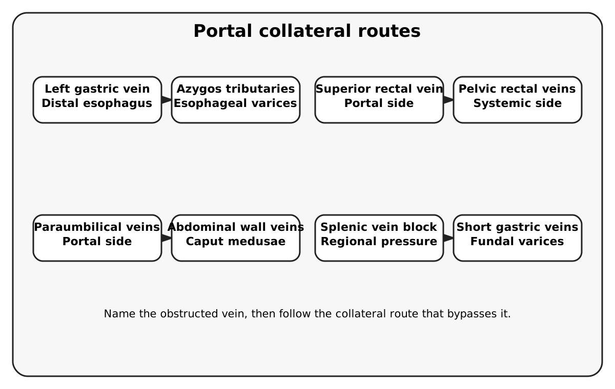
Distal esophagus connects the left gastric portal territory to esophageal veins of the azygos system. Raised portal pressure can enlarge submucosal veins into varices.
Rectum connects superior rectal drainage through the IMV with systemic middle and inferior rectal routes toward the internal iliac circulation. Rectal varices are distinct from hemorrhoidal cushions and hemorrhoidal disease.
Umbilicus connects paraumbilical veins with superficial abdominal wall veins. Enlarged outward pathways can produce caput medusae. The round ligament is the remnant of the fetal umbilical vein, not an artery.
Three portosystemic pathways (esophagus, rectum, umbilicus) plus the regional splenic-to-short-gastric route that produces fundal varices when the splenic vein is blocked. Name the obstructed vein, then follow the collateral route that bypasses it. [9][10][11][23][31]
Do not diagnose portal hypertension from hemorrhoids alone. A primary prospective study demonstrated that hemorrhoids and rectal varices can coexist but are separate findings. Conversely, suspected acute variceal hemorrhage is not managed as an isolated arterial ulcer. In cirrhotic patients with upper gastrointestinal bleeding, guideline-based care includes resuscitation, early vasoactive therapy when variceal bleeding is suspected, antibiotic prophylaxis, and appropriate endoscopic treatment. Selected patients benefit from TIPS. [10][9]
Splenic vein thrombosis can cause left-sided, or sinistral, portal hypertension while the main portal vein and liver remain relatively normal. Splenic venous blood is redirected through short gastric and other collateral veins, producing fundal varices. Pancreatitis is a useful setting for this mechanism. Isolated gastric varices support investigation of splenic outflow; they are not by themselves pathognomonic. Observational evidence does not support automatic prophylactic splenectomy for every asymptomatic pancreatitis-associated splenic vein thrombosis. [11]
A distal splenorenal, or Warren, shunt connects the splenic end of the splenic vein to the left renal vein to decompress gastroesophageal venous territory selectively. [31] TIPS creates an intrahepatic connection between the portal and hepatic venous systems. Both can alter the route by which gut-derived substances encounter the liver; encephalopathy is a relevant concern. Spontaneous portosystemic shunts can also form, so a splenorenal connection on imaging does not always imply previous surgery. [9][23]
Try it here · Checkpoint 2 of 3
Make your prediction before reading the choices. A first attempt is just a starting point.
Case 8
Show answer and explanations for case 8
A. The findings establish diffuse cirrhosis despite normal hepatic imaging (Why this does not fit)
Cirrhosis can cause gastric varices, but the demonstrated isolated splenic obstruction provides a direct regional mechanism. These findings do not establish diffuse liver disease.
Reasoning steps for option A
Why would cirrhosis be the first thought for gastric varices?
Cirrhosis is the most common cause of portal hypertension and gastric varices.
Which findings argue against diffuse liver disease here?
Liver morphology is normal, the main portal vein is patent, and a local splenic vein thrombosis is shown.
B. Splenic venous flow diverts to short gastric and other collateral veins (Best answer)
Obstruction of splenic outflow can create localized venous hypertension with gastric collateral enlargement even when the main portal vein remains patent.
Reasoning steps for option B
What happens to splenic venous blood when the splenic vein thromboses?
It is diverted through short gastric and other collateral veins toward the stomach.
Why can fundal varices form while the main portal vein is patent?
The pressure rise is confined to the splenic bed, producing sinistral portal hypertension.
C. Splenic arterial pressure is transmitted directly into gastric mucosal arteries (Why this does not fit)
The imaging identifies venous outflow obstruction. Gastric varices are enlarged veins, not an arterial-pressure lesion.
Reasoning steps for option C
Why might arterial pressure seem to explain enlarged gastric vessels?
Pancreatitis can damage the splenic artery, and splenomegaly suggests increased splenic flow.
What kind of vessels are varices, and what did CT show?
Varices are dilated veins, and CT shows venous outflow obstruction, not an arterial lesion.
D. The patent main portal vein excludes a portal-pressure mechanism (Why this does not fit)
Regional splenic venous hypertension can exist without main portal vein thrombosis or cirrhosis. Patency of the main vessel does not exclude this localized process.
Reasoning steps for option D
Why does a patent main portal vein seem to exclude a portal-pressure cause?
Most causes of portal hypertension involve the main portal vein or the liver, both normal here.
Which obstruction still raises pressure upstream of a patent portal vein?
Splenic vein thrombosis raises pressure in the splenic territory alone, so main portal patency does not exclude it.
Takeaway: Separate sinistral portal hypertension from generalized portal hypertension; treatment is individualized rather than automatic prophylactic splenectomy.
The pancreaticoduodenal arcades connect superior branches from the gastroduodenal artery to inferior branches from the SMA. With celiac stenosis, blood may travel backward through this route to support hepatic inflow. Dividing the gastroduodenal artery during pancreaticoduodenectomy can then interrupt a crucial collateral. The arcades are a major connection, not the only possible one; a persistent arc of Bühler is a direct variant connection between celiac and SMA territories. Operative planning must establish direction and adequacy of flow in the actual anatomy. [1][7]
Along the colon, the marginal artery of Drummond links neighboring colic branches near the bowel. The more central arc of Riolan describes a variable connection between middle colic and left colic territories. Griffith's point at the splenic flexure is an SMA-IMA watershed. Sudeck's point is the junction of the last sigmoid and superior rectal arterial territories, both ordinarily IMA branches.
Pelvic rectal collaterals are real, but they do not redefine Sudeck's point as a direct IMA to internal iliac boundary. Collateral caliber and continuity vary, so neither the splenic flexure nor any other segment is guaranteed to be affected first. [1][12]
Postprandial pain, reduced eating, and weight loss may indicate chronic mesenteric ischemia when supported by vascular findings and exclusion of competing diagnoses. Extensive disease increases suspicion, but severe single-vessel disease can be symptomatic. The SMA is a major revascularization target because of its intestinal territory; that statement does not mean it is always the most diseased artery on a scan.
In contrast, abrupt severe pain with little early tenderness in a patient with atrial fibrillation raises concern for embolic acute mesenteric ischemia. Obtain urgent CTA and emergency assessment; a reassuring early examination or laboratory result cannot safely exclude it. Jejunal and ileal arcades exist, but an acute occlusion can outstrip their capacity. The embolus location determines which branches remain perfused. [7][8]
Distal branch reasoning is equally useful. The ileocolic artery supplies terminal ileum and cecal territory; the appendicular artery usually arises through its branches and travels in the mesoappendix. Limited alternative supply contributes to appendiceal ischemia with obstruction and inflammation, but no fixed interval predicts perforation in every patient. Normal midgut development includes about 270 degrees of counterclockwise rotation around the SMA, viewed from the front.
Malrotation can leave a narrow mesenteric attachment that permits volvulus and vascular compromise. [28] Bilious vomiting in a neonate requires urgent evaluation. Surgical treatment of malrotation addresses any volvulus and obstructing Ladd bands; counterclockwise detorsion and division of constricting mesenteric bands address the abnormal anatomy rather than recreating embryonic rotation. [32] In a premature infant, abdominal distension, bloody stool, and pneumatosis instead suggest necrotizing enterocolitis, a multifactorial illness rather than a simple named watershed infarct; portal venous gas alone does not establish modified Bell stage III. [16][17]
For an aortic clamp or reconstruction, specify which branch origins lie above and below the clamp, which arteries are patent, and which collateral routes remain. A clamp below the SMA but above both renal arteries compromises direct renal and IMA inflow while preserving direct celiac and SMA inflow. Colonic viability then depends on actual collateral and pelvic perfusion. IMA reimplantation is not mandated or excluded by one memorized vessel name, and there is no universal minute at which all warm ischemia becomes irreversible. [1][7]
Collateral direction exercise
Start with severe celiac origin stenosis and imagine that SMA pressure is preserved. Follow one continuous arterial route from the SMA to the proper hepatic artery.
Name the SMA branch that reaches the pancreatic head.
Continue through the communicating pancreaticoduodenal vessels.
Decide which familiar artery now carries blood toward the liver rather than away from it.
Compare your route
The expected route is SMA to inferior pancreaticoduodenal to superior pancreaticoduodenal to gastroduodenal to common hepatic. The crucial reversal is retrograde flow through the gastroduodenal artery.
The visible consequence is testable: temporary gastroduodenal occlusion will reduce hepatic arterial flow when that collateral is carrying the liver's arterial inflow. This is why a routine division can become hazardous in celiac stenosis. [7]
Try it here · Checkpoint 3 of 3
Make your prediction before reading the choices. A first attempt is just a starting point.
Case 14
Show answer and explanations for case 14
A. Wait for peritoneal rigidity before ordering vascular imaging (Why this does not fit)
Peritonitis can reflect advanced bowel injury. Waiting for it discards the opportunity to identify ischemia before irreversible damage.
Reasoning steps for option A
Why might a clinician wait for peritoneal signs?
The abdomen is only mildly tender, so the examination seems too soft to justify urgent imaging.
What does peritoneal rigidity usually mean in mesenteric ischemia?
Transmural bowel injury; pain out of proportion to the examination is the earlier warning.
B. Exclude mesenteric ischemia because jejunal and ileal arcades are present (Why this does not fit)
Arcades do exist, but their capacity can be overwhelmed by acute embolic occlusion. Their presence does not establish adequate perfusion.
Reasoning steps for option B
Why might intestinal arcades seem protective?
Jejunal and ileal arcades are real collateral networks.
Can arcades compensate for an acute embolus?
Often not. A sudden embolus, typical with atrial fibrillation, can overwhelm their capacity.
C. Diagnose isolated gastric ulcer disease from the normal lactate (Why this does not fit)
A normal lactate does not localize the pain to the stomach. The abrupt presentation and embolic risk require mesenteric vascular assessment.
Reasoning steps for option C
What makes a normal lactate reassuring to some clinicians?
Lactate rises with bowel ischemia, so a normal value seems to lower the likelihood.
What does a normal early lactate establish in this patient?
Very little. Early values can be normal, and it neither localizes pain to the stomach nor excludes an embolus.
D. Urgent CT angiography with emergency surgical and vascular assessment (Best answer)
The history and pain-examination mismatch warrant prompt evaluation for acute mesenteric ischemia. A normal early lactate cannot safely exclude threatened bowel.
Reasoning steps for option D
Which features make acute embolic mesenteric ischemia likely?
Atrial fibrillation, abrupt severe pain and only mild tenderness, pain out of proportion to the examination.
Which test and team should follow immediately?
Urgent CT angiography with emergency surgical and vascular assessment, without waiting for lactate or peritonitis.
Takeaway: Acute mesenteric ischemia is a time-sensitive clinical suspicion supported by urgent imaging, not excluded by an early reassuring laboratory result.
Nearby vessels participate in different compression problems. Read each relationship in front-to-back order.
Median arcuate ligament syndrome
The diaphragmatic ligament indents the proximal celiac artery. Compression is commonly greater during expiration. Compatible symptoms and exclusion of other causes are necessary because imaging compression can be incidental.
Superior mesenteric artery syndrome
SMA in front, third duodenal part in the middle, aorta behind. Loss of the intervening fat cushion can narrow this space and obstruct the duodenum, causing early satiety and vomiting after substantial weight loss.
Anterior nutcracker syndrome
SMA in front, left renal vein in the middle, aorta behind. Symptomatic renal venous hypertension can produce hematuria and gonadal venous congestion. Compression on imaging without relevant symptoms is not the same diagnosis.
May-Thurner anatomy
Right common iliac artery in front of the left common iliac vein, against the spine. The expected venous consequences involve the left leg rather than the stomach or renal collecting system.
The diaphragm ascends during expiration; it does not descend to explain expiratory celiac narrowing. Respiratory-phase duplex assessment helps evaluate suspected median arcuate ligament syndrome, but neither a bruit nor a hooked arterial contour independently establishes the cause of abdominal pain. Likewise, duodenal obstruction and renal venous hypertension may involve the same aortomesenteric angle but affect different structures. Name the compressed organ or vessel before naming the syndrome. [7][24][25][27]
Apply the vascular and operative relationships
Case 1
Show answer and explanations for case 1
A. Gastroduodenal artery (Best answer)
The gastroduodenal artery descends behind the first duodenal part. A posterior ulcer can erode it and cause substantial arterial hemorrhage.
Reasoning steps for option A
Which vessel lies directly behind the posterior wall of the first duodenal part?
The gastroduodenal artery descends behind the first part, so a deep posterior bulb ulcer can erode into it.
What does naming the gastroduodenal artery change about this patient's initial management?
It localizes the bleeding vessel, but resuscitation and endoscopic hemostasis still come first, with embolization if endoscopy fails.
B. Left gastric artery (Why this does not fit)
Left gastric branches supply the lesser curvature and distal esophagus. They are a stronger localization for a proximal lesser-curvature gastric lesion than for this posterior duodenal ulcer.
Reasoning steps for option B
Why does the left gastric artery come to mind in an NSAID-related ulcer bleed?
It is the classic source when a high lesser-curvature gastric ulcer bleeds, a common NSAID lesion.
What about this ulcer's location rules the left gastric artery out?
The ulcer is in the posterior duodenal bulb, well beyond the lesser-curvature territory the left gastric artery supplies.
C. Splenic artery (Why this does not fit)
The splenic artery follows the superior pancreatic border toward the spleen. It is not the characteristic artery immediately posterior to the duodenal bulb.
Reasoning steps for option C
What makes the splenic artery tempting for a posterior ulcer?
Posterior gastric ulcers can erode the splenic artery, which runs behind the stomach along the pancreas.
Why is the splenic artery the wrong vessel for a duodenal bulb ulcer?
It runs leftward along the superior pancreatic border toward the spleen, away from the first duodenal part.
D. Inferior pancreaticoduodenal artery (Why this does not fit)
This SMA branch supplies the lower pancreatic head and adjacent duodenum through arcades. The vessel directly behind the first duodenal part is the gastroduodenal artery.
Reasoning steps for option D
Why might a pancreaticoduodenal branch seem relevant to duodenal bleeding?
Pancreaticoduodenal arcades supply the duodenal wall, so duodenal lesions can bleed from them.
Which part of the duodenum does the inferior pancreaticoduodenal artery serve, compared with this ulcer?
It comes from the SMA and serves the lower head and distal duodenum; the posterior first part is related to the gastroduodenal artery.
Takeaway: Localize the arterial threat from the ulcer wall; identifying the gastroduodenal artery does not bypass resuscitation and endoscopic treatment.
A. Left gastric artery to inferior phrenic artery (Why this does not fit)
These are arterial connections. They do not explain dilated submucosal veins caused by portal hypertension.
Reasoning steps for option A
Why could a left gastric connection sound right for distal esophageal bleeding?
The left gastric artery does send branches to the distal esophagus, where these vessels bled.
What kind of vessel did endoscopy show, and why does that exclude an arterial pairing?
It showed dilated submucosal veins from portal pressure; an arterial link to inferior phrenic branches cannot form varices.
B. Superior rectal vein to internal iliac tributaries (Why this does not fit)
This is a rectal portosystemic connection, not the distal esophageal route seen at endoscopy.
Reasoning steps for option B
Why is the superior rectal to internal iliac route a reasonable guess in cirrhosis?
It is a genuine portosystemic anastomosis that enlarges with portal hypertension.
Where would that connection produce varices, compared with this endoscopy?
In the rectum; the bleeding here is from the distal esophagus.
C. Paraumbilical veins to superficial abdominal wall veins (Why this does not fit)
This connection explains periumbilical collaterals such as caput medusae, not the esophageal varices in this patient.
Reasoning steps for option C
Why might paraumbilical collaterals be considered in this patient?
They are a classic portosystemic route that enlarges in cirrhosis and can coexist with varices.
What would the paraumbilical route produce instead of this bleed?
Caput medusae on the abdominal wall, not submucosal varices in the esophagus.
D. Left gastric vein to azygos esophageal tributaries (Best answer)
The left gastric vein belongs to portal drainage, while esophageal veins communicate with the azygos systemic route. Raised portal pressure expands this connection.
Reasoning steps for option D
Which portal tributary drains the region where these veins bled?
The left gastric, or coronary, vein drains the lesser curvature and distal esophagus into the portal system.
Where does the systemic side of this esophageal connection drain?
Into azygos tributaries, so raised portal pressure dilates the submucosal channels between the two systems.
Takeaway: Match each variceal location to its portal tributary and systemic outlet.
The proper hepatic artery ascends in the hepatoduodenal ligament toward the liver. The imaged course toward the left upper abdomen favors the splenic artery.
Reasoning steps for option A
Why could a hepatic arterial aneurysm be considered when a visceral aneurysm ruptures?
Hepatic artery aneurysms are among the more common visceral aneurysms and can bleed into the peritoneum.
Which features of this bleed point away from the proper hepatic artery?
The pain is left-sided with shoulder radiation, and the aneurysm lies along the superior pancreatic border, not in the hepatoduodenal ligament.
B. Inferior mesenteric artery (Why this does not fit)
The IMA arises lower on the aorta and supplies hindgut structures. It does not run along the superior border of the pancreas.
Reasoning steps for option B
Why might a mesenteric artery be suspected in a pregnant patient with intraperitoneal bleeding?
Any visceral aneurysm can rupture into the peritoneum, and the IMA is a named unpaired visceral branch.
Where does the IMA run relative to the pancreas?
It arises near L3 and runs to the hindgut, far below the superior pancreatic border.
C. Splenic artery (Best answer)
Its course along the superior pancreatic border and the left upper abdominal hemorrhage fit splenic artery aneurysm rupture, an especially consequential diagnosis during pregnancy.
Reasoning steps for option C
Which celiac branch follows the superior border of the pancreas?
The tortuous splenic artery runs along that border toward the splenic hilum, matching the imaged aneurysm.
Why does pregnancy make this diagnosis urgent even for a small aneurysm?
Rupture in pregnancy threatens mother and fetus, and ESVS advises considering treatment in pregnancy regardless of size.
D. Left gastric artery (Why this does not fit)
This celiac branch approaches the lesser curvature. It does not follow the long superior pancreatic course described by the imaging.
Reasoning steps for option D
What makes the left gastric artery a plausible left upper abdominal bleeding source?
It is a celiac branch near the lesser curvature that can bleed massively.
How does the imaged course separate the left gastric artery from the implicated vessel?
The left gastric artery ascends to the cardia and lesser curvature; it does not follow the long superior pancreatic border.
Takeaway: Pregnancy and shock make a ruptured splenic artery aneurysm an emergency; small size would not exclude danger.
A. Recognition of a triangular space bounded by the cystic artery (Why this does not fit)
That describes a historical Calot boundary. The shape of a triangle does not establish safe identification of the structures entering this gallbladder.
Reasoning steps for option A
Why might recognizing a triangle bounded by the cystic artery seem sufficient?
Calot's original triangle used the cystic artery as a boundary, so seeing it sounds like the classic safety landmark.
What does the critical view demand beyond seeing a triangle?
Three findings together; the lower gallbladder is still attached, so the view is incomplete however clear the triangle looks.
B. Lower-third separation exposing the cystic plate, with only two structures entering the gallbladder (Best answer)
The critical view requires all three components. Clearing the triangle and seeing two apparent structures is insufficient without exposing the cystic plate by separating the lower third.
Reasoning steps for option B
Which critical-view component has this surgeon already met?
The hepatocystic triangle has been cleared of fat and fibrous tissue.
What must still be shown before the view is complete?
The lower third of the gallbladder must be separated to expose the cystic plate, and only two structures may be seen entering the gallbladder.
C. Circumferential exposure of the common bile duct down to the duodenum (Why this does not fit)
The common bile duct does not need to be exposed for the critical view. Extending dissection this way does not substitute for separating the lower gallbladder.
Reasoning steps for option C
Why could exposing the common bile duct sound like extra safety?
Bile duct injury is the feared complication, so seeing the duct directly seems protective.
Is common bile duct exposure part of the critical view?
No. It is not required, it adds dissection, and it leaves the unfinished lower-third separation untouched.
D. Identification of the right hepatic artery at its origin (Why this does not fit)
The cystic artery often comes from the right hepatic artery, but tracing the parent origin is not one of the three required critical-view components.
Reasoning steps for option D
Why is the right hepatic artery relevant during this dissection?
The cystic artery usually arises from it, and it can loop close to the triangle and be injured.
Does tracing the right hepatic origin complete the critical view?
No. Its origin is not one of the three components, and the lower gallbladder remains attached.
Takeaway: The critical view is a set of three anatomical demonstrations, not a quick view of a duct and artery.
A. Fundus via short gastric arteries in the gastrosplenic ligament (Best answer)
The short gastric arteries pass from splenic branches through the gastrosplenic ligament to the fundus. Other gastric connections may still preserve perfusion.
Reasoning steps for option A
Which peritoneal fold connects the splenic hilum directly to the fundus?
The gastrosplenic ligament, which carries the short gastric arteries from splenic branches to the fundus.
Does dividing these vessels make fundal necrosis inevitable?
No. Left gastric, left gastroepiploic and other gastric routes usually keep the fundus perfused.
B. Pyloric lesser curvature through the right gastric artery (Why this does not fit)
The right gastric artery follows the lesser curvature, not the fold directly connecting the splenic hilum to the fundus.
Reasoning steps for option B
Why might the right gastric artery be considered when gastric vessels are divided?
It is one of the named arteries of the stomach and is often ligated in gastric surgery.
Where does the right gastric artery run relative to the divided fold?
Along the distal lesser curvature near the pylorus, nowhere near the spleen-to-fundus fold.
C. Right greater curvature through the right gastroepiploic artery (Why this does not fit)
The right gastroepiploic artery comes from the gastroduodenal artery and travels in the gastrocolic region. It is not a short splenic-to-fundal vessel.
Reasoning steps for option C
What makes a gastroepiploic vessel tempting during splenic dissection?
The gastroepiploic arcade runs along the greater curvature, which extends toward the spleen.
Which side and parent separate the right gastroepiploic artery from the divided vessels?
It arises from the gastroduodenal artery and supplies the right greater curvature through the gastrocolic region, not the fundus from the spleen.
D. Distal esophagus through left gastric esophageal branches (Why this does not fit)
These branches ascend from the left gastric artery near the cardia. They do not describe the vessels divided at the splenic-fundal fold.
Reasoning steps for option D
Why might esophageal branches be confused with vessels to the fundus?
The cardia and fundus are adjacent, and left gastric esophageal branches supply that proximal region.
What parent and route do those esophageal branches have?
They ascend from the left gastric artery on the lesser-curvature side, not through a fold from the splenic hilum.
Takeaway: Short gastric division reduces fundal supply but does not make fundal necrosis inevitable.
A. Superior and inferior pancreaticoduodenal territories (Why this does not fit)
These connect celiac and SMA supply around the pancreatic head and duodenum, not at the splenic flexure.
Reasoning steps for option A
Why could the pancreaticoduodenal arcades come up in a watershed question?
They are a classic boundary between two arterial territories, celiac and SMA.
Where is the pancreaticoduodenal boundary relative to the injured segment?
Around the pancreatic head and duodenum, not at the splenic flexure seen on CT.
B. Last sigmoid and superior rectal territories (Why this does not fit)
That junction defines Sudeck's point near the rectosigmoid region. The CT abnormality here is much more proximal.
Reasoning steps for option B
What makes the sigmoid and superior rectal junction a tempting watershed answer?
Sudeck's point is the other classic colonic watershed vulnerable to low-flow ischemia.
How does the CT location separate Sudeck's point from this injury?
Sudeck's point is at the rectosigmoid; this injury is centered at the splenic flexure.
C. Right gastric and left gastric territories (Why this does not fit)
These meet along the lesser curvature of the stomach. They cannot explain a colonic lesion at the splenic flexure.
Reasoning steps for option C
Why might gastric arterial borders be considered after hypotension?
The right and left gastric arteries form a named arterial meeting point along the lesser curvature.
Can a gastric boundary explain this colonic injury?
No. The lesion is at the splenic flexure, and the stomach's rich collaterals make gastric ischemia uncommon.
D. Middle colic (SMA) and left colic (IMA) territories (Best answer)
The splenic flexure lies near Griffith's watershed between these territories. Low perfusion and variable marginal connections can expose it to ischemia.
Reasoning steps for option D
Which arterial systems meet at the splenic flexure?
Middle colic branches of the SMA and left colic branches of the IMA, at Griffith's point.
Why does prolonged hypotension expose this border?
Low perfusion pressure and a variably thin marginal artery leave the watershed least protected, though it is not always injured first.
Takeaway: A watershed identifies a vulnerable border; it does not guarantee that the same segment is always injured first.
A. SMA, left renal vein, aorta (Why this does not fit)
This describes the structure involved in anterior nutcracker compression. It can produce renal venous symptoms but does not identify the obstructed bowel.
Reasoning steps for option A
Why is an SMA, left renal vein and aorta sequence tempting after weight loss?
Loss of retroperitoneal fat narrows the aortomesenteric angle, which can also compress the left renal vein.
Which structure is obstructed in this vignette?
The dilated stomach and proximal duodenum with a narrowed horizontal segment show that bowel, not renal vein, is compressed.
B. Right common iliac artery, third duodenal part, left common iliac vein (Why this does not fit)
The iliac arterial-venous crossing is lower in the pelvis and is associated with left iliac venous compression, not this duodenal obstruction.
Reasoning steps for option B
What makes an artery-over-structure crossing attractive here?
May-Thurner anatomy is another front-to-back compression with an artery in front.
Why can the iliac crossing not obstruct the duodenum?
It lies at the pelvic inlet and compresses the left common iliac vein; the duodenum does not pass there.
C. SMA, third duodenal part, aorta (Best answer)
The third duodenal part passes behind the SMA and in front of the aorta. Loss of intervening fat can narrow this interval and obstruct the bowel.
Reasoning steps for option C
Which duodenal segment crosses between the SMA and aorta?
The horizontal third part passes behind the SMA and in front of the aorta.
How does major weight loss narrow that interval?
Losing the fat cushion around the SMA closes the aortomesenteric angle, pinching the third part and causing proximal dilation with bilious vomiting.
D. Celiac trunk, second duodenal part, portal vein (Why this does not fit)
The second part descends beside the pancreatic head. It is not the horizontal bowel segment in the aortomesenteric interval.
Reasoning steps for option D
Why might the second duodenal part be considered for this obstruction?
It is part of the dilated segment and lies next to major vessels at the pancreatic head.
Which duodenal part is narrowed between two vessels in this stem?
The horizontal third part; the second part descends and is not squeezed between celiac trunk and portal vein.
Takeaway: SMA syndrome compresses the duodenum; nutcracker syndrome involves the left renal vein in a nearby interval.
A. Celiac compression may be incidental; it alone does not establish median arcuate ligament syndrome (Best answer)
The syndrome requires compatible clinical findings and careful exclusion of competing causes. The scan supplies anatomy, while the characteristic symptoms are absent.
Reasoning steps for option A
What does this scan show, and what does the history lack?
Celiac compression that increases on expiration, but no postprandial pain, weight loss or altered eating.
Why does that combination argue for calling the finding incidental?
Median arcuate compression is common on imaging in people without symptoms; the syndrome needs compatible symptoms and exclusion of other causes.
B. The scan alone establishes symptomatic chronic mesenteric ischemia requiring revascularization (Why this does not fit)
Stenosis or compression on imaging can be asymptomatic. The absence of a compatible illness prevents this automatic diagnostic and treatment conclusion.
Reasoning steps for option B
Why might a hooked celiac narrowing suggest mesenteric ischemia?
It looks like a significant narrowing of a major mesenteric artery.
What is missing that chronic mesenteric ischemia would require?
Symptoms. Imaging narrowing alone neither diagnoses ischemia nor justifies revascularization.
C. The expiratory change is caused by the diaphragm descending during expiration (Why this does not fit)
The diaphragm ascends during expiration. Greater expiratory compression is recognized, but the proposed direction of diaphragmatic motion is reversed.
Reasoning steps for option C
Why does a diaphragm-motion explanation sound plausible?
Expiratory narrowing is real, and the median arcuate ligament is part of the diaphragm, so diaphragmatic movement seems to explain it.
Which way does the diaphragm actually move during expiration?
It ascends; the direction in this explanation is reversed.
D. The finding proves third-part duodenal obstruction from SMA syndrome (Why this does not fit)
This scan describes compression of the celiac artery, not dilation and obstruction of the duodenum in the aortomesenteric interval.
Reasoning steps for option D
Why could an aortomesenteric compression syndrome come to mind here?
Both involve compression of a structure beside the proximal visceral aorta.
Which structure does SMA syndrome affect, compared with this scan?
The third duodenal part below the SMA; this scan shows celiac compression and no duodenal dilation.
Takeaway: Call the anatomical finding compression; reserve the clinical syndrome for an appropriately evaluated symptomatic patient.
A. Marginal artery of Drummond (Why this does not fit)
The marginal artery connects colic territories near the bowel wall. It does not pass around the pancreatic head to the gastroduodenal artery.
Reasoning steps for option A
Why is the marginal artery a natural first thought for a collateral route?
It is the best-known collateral linking two mesenteric territories.
Where does the marginal artery run relative to the pancreatic head?
Along the colon wall, linking colic branches, not around the pancreatic head to the gastroduodenal artery.
B. Arc of Riolan (Why this does not fit)
This is a variable central connection between middle colic and left colic territories, linking SMA and IMA supply rather than this celiac-SMA route.
Reasoning steps for option B
Why might the arc of Riolan be suspected when a visceral origin is stenosed?
It is a central collateral that enlarges when a mesenteric origin narrows.
Which territories does Riolan connect, compared with the imaged route?
SMA and IMA through middle and left colic branches, not SMA to gastroduodenal around the pancreas.
C. Short gastric arterial branches (Why this does not fit)
These travel from splenic branches to the fundus. They do not originate from the SMA or form the arcades around the pancreatic head.
Reasoning steps for option C
What makes short gastric branches a tempting celiac-territory collateral?
They are small celiac-territory vessels that can carry collateral flow.
What parent and destination do the short gastric arteries have?
They leave splenic branches for the fundus; they neither arise from the SMA nor surround the pancreatic head.
D. Inferior to superior pancreaticoduodenal arcades (Best answer)
Inferior pancreaticoduodenal branches originate from the SMA and communicate with superior branches supplied through the gastroduodenal artery. This provides the imaged route around the head.
Reasoning steps for option D
Which SMA branch reaches the pancreatic head?
The inferior pancreaticoduodenal artery.
How does that flow reach the gastroduodenal artery when the celiac origin is stenosed?
Its arcades join the superior pancreaticoduodenal branches, so blood runs retrograde into the gastroduodenal artery.
Takeaway: The pancreaticoduodenal arcades are a major celiac-SMA connection, while other variant connections may also exist.
A. Portal venous patency guarantees that hepatic arterial interruption is harmless (Why this does not fit)
The liver has dual inflow, but portal patency does not make loss of an important arterial route harmless, particularly for the biliary arterial supply.
Reasoning steps for option A
Why does portal patency seem reassuring about losing an arterial route?
The portal vein supplies most hepatic blood flow, so losing one artery sounds tolerable.
What does portal flow fail to protect?
The bile ducts depend on arterial supply, and the test occlusion shows hepatic arterial inflow depends on this route.
B. The flow change proves that the splenic vein is thrombosed (Why this does not fit)
A change in hepatic arterial flow during arterial occlusion assesses an arterial collateral. It cannot establish a diagnosis of splenic venous thrombosis.
Reasoning steps for option B
Why might a venous diagnosis come up in pancreatic surgery planning?
Hepatic arterial dependence on the gastroduodenal route; it says nothing about splenic vein patency.
C. Hepatic arterial flow relies on retrograde SMA flow via the gastroduodenal artery (Best answer)
The flow reduction during test occlusion demonstrates dependence on the pathway being divided. Celiac disease makes routine division potentially consequential unless adequate hepatic inflow is secured.
Reasoning steps for option C
What does the drop in hepatic flow during gastroduodenal occlusion show?
The liver's arterial inflow is currently running through the gastroduodenal artery.
Where does that flow come from when the celiac origin is severely stenosed?
From the SMA through the pancreaticoduodenal arcades, retrograde in the gastroduodenal artery, so dividing it could devascularize the liver unless inflow is restored.
D. The gastroduodenal artery must be a direct IMA branch in this patient (Why this does not fit)
An enlarged pancreaticoduodenal network links the SMA and celiac territories. It does not imply an IMA origin for the gastroduodenal artery.
Reasoning steps for option D
Why might a distant arterial origin be proposed for an enlarged collateral?
The IMA can enlarge and carry collateral flow when other mesenteric origins are diseased.
Which territories does an enlarged pancreaticoduodenal network connect?
The SMA and celiac territories; the gastroduodenal artery keeps its usual common hepatic origin.
Takeaway: Before dividing a familiar branch, determine whether disease has made it an essential collateral.
A. The inferior rectal veins normally drain directly into the splenic vein (Why this does not fit)
Inferior rectal drainage runs toward systemic pelvic veins, commonly through the internal pudendal route. The splenic vein is on the portal side of the circuit.
Reasoning steps for option A
Why could the splenic vein be linked with rectal drainage?
The IMV, which carries upper rectal blood, commonly joins the splenic vein.
Which rectal veins reach the splenic side, and which do not?
Only superior rectal drainage reaches it through the IMV; inferior rectal veins drain to internal pudendal and internal iliac veins.
B. Superior rectal veins drain via the IMV to portal veins; middle and inferior rectal veins drain to systemic pelvic veins (Best answer)
These pathways explain rectal portosystemic varices. The simultaneous hemorrhoids are a distinct finding and should not be treated as the same lesion.
Reasoning steps for option B
Which rectal veins drain to the portal side?
The superior rectal veins, through the IMV.
Why does this anastomosis explain the documented rectal varices?
Middle and inferior rectal veins drain systemically, so raised portal pressure dilates the connecting submucosal channels, separate from the hemorrhoidal cushions.
C. The superior rectal vein normally empties directly into the inferior vena cava (Why this does not fit)
Its usual route is through the IMV and portal system. Direct caval drainage would not describe the portal side of this connection.
Reasoning steps for option C
Why might caval drainage of the superior rectal vein sound reasonable?
Part of the rectum does drain systemically toward the inferior vena cava.
Which rectal vein is the portal-side partner in this anastomosis?
The superior rectal vein, which drains through the IMV; the systemic side is the middle and inferior rectal veins.
D. Hemorrhoids and rectal varices are interchangeable names for one portal lesion (Why this does not fit)
They are anatomically and clinically distinct and may occur independently. Their coexistence does not make hemorrhoids proof of portal hypertension.
Reasoning steps for option D
Why do hemorrhoids and rectal varices get confused?
Both are dilated anorectal vascular structures, and both can bleed in portal hypertension.
Does their coexistence here make them one lesion?
No. Prospective data show they are distinct and occur independently, so hemorrhoids do not prove portal hypertension.
Takeaway: Identify rectal varices separately from hemorrhoidal disease before assigning the bleeding source.
A. Both have celiac supply; splenic origin is mesenchymal, not foregut endodermal (Best answer)
Shared arterial supply does not require shared embryonic tissue origin. The spleen develops within the dorsal mesogastrium while receiving splenic arterial supply from the celiac system.
Reasoning steps for option A
Which arterial system supplies both the proximal duodenum and the spleen?
The celiac system: the gastroduodenal branch of the common hepatic artery and the splenic artery.
Does shared arterial supply mean a shared germ layer?
No. The spleen develops from dorsal mesogastric mesenchyme, while duodenal epithelium is foregut endoderm.
B. The spleen is a foregut endodermal organ because its artery comes from the celiac trunk (Why this does not fit)
The conclusion incorrectly turns an arterial territory into a germ-layer assignment. Splenic development is mesenchymal.
Reasoning steps for option B
Why does calling the spleen a foregut organ sound logical?
It receives celiac blood, the foregut artery, and sits among foregut organs.
What is the spleen's actual embryonic origin?
Mesenchyme of the dorsal mesogastrium; arterial territory does not assign a germ layer.
C. The celiac-to-SMA boundary lies at the ileocecal junction (Why this does not fit)
The boundary is around the major duodenal papilla. The ileocecal region lies well within the SMA midgut territory.
Reasoning steps for option C
Why might the ileocecal junction be taken as a vascular boundary?
It is a major junction between small bowel and colon with its own named ileocolic supply.
Where is the real celiac-SMA transition?
At the major duodenal papilla in the second part; the ileocecal region is deep within SMA territory.
D. The celiac trunk normally supplies the descending and sigmoid colon (Why this does not fit)
Those colonic regions are predominantly IMA territory. The celiac branches supply upper abdominal organs such as the stomach and proximal duodenum.
Reasoning steps for option D
What makes celiac supply to the left colon superficially plausible?
The splenic flexure lies next to the spleen, a celiac-supplied organ.
Which artery supplies the descending and sigmoid colon?
The IMA, through left colic and sigmoid branches; celiac branches serve upper abdominal organs.
Takeaway: Distinguish an organ's arterial territory from its embryonic origin.
A. Portal vein within the compressed ligament (Why this does not fit)
Portal inflow has been adequately occluded in this scenario. Persistent posterior hemorrhage therefore raises concern beyond that controlled vessel.
Reasoning steps for option A
Why is portal vein injury a reasonable worry in a deep liver wound?
The portal vein is a large, high-flow inflow vessel to the injured liver.
What has the clamp already done to portal flow?
It has occluded portal inflow in the ligament, so persistent bleeding points beyond it.
B. Cystic artery confined to the gallbladder wall (Why this does not fit)
The described deep posterior liver injury near the cava is anatomically different from a gallbladder arterial source. It also would not explain the identified outflow-region bleeding.
Reasoning steps for option B
Why might a gallbladder vessel be suspected in right upper quadrant bleeding?
The cystic artery is a common operative bleeding source near the liver.
Where is the bleeding coming from in this stem?
From the posterior liver near the cava, far from the gallbladder wall.
C. Hepatic vein or retrohepatic inferior vena cava (Best answer)
These are hepatic outflow structures outside the compressed portal triad. Their injury can continue bleeding despite adequate inflow control.
Reasoning steps for option C
Which vessels does a Pringle maneuver leave uncontrolled?
The hepatic veins and retrohepatic inferior vena cava, which carry outflow outside the portal triad.
How does the bleeding location support an outflow source?
Posterior bleeding near the cava despite adequate inflow occlusion fits hepatic venous or caval injury.
D. Proper hepatic artery within the compressed ligament (Why this does not fit)
The stem establishes adequate arterial inflow occlusion. An injury to that controlled inflow alone would not best explain persistent posterior bleeding.
Reasoning steps for option D
Why could the hepatic artery still be blamed for brisk bleeding?
Arterial bleeding is brisk, and the proper hepatic artery feeds the liver.
What does the Pringle maneuver do to hepatic arterial flow?
It occludes it in the hepatoduodenal ligament, as the stem confirms.
Takeaway: A Pringle maneuver controls the inflow bundle; it does not directly compress hepatic venous or caval outflow.
It runs near the colonic border and links neighboring colic branches, potentially allowing SMA-to-IMA collateral perfusion when continuity and pressure are adequate.
Reasoning steps for option A
Where does the collateral on this angiogram run?
Close to the mesenteric border of the colon, linking adjacent colic branches.
Which named vessel has that course, and what limits its value?
The marginal artery of Drummond; its continuity and caliber vary, so perfusion still needs assessment.
B. Arc of Riolan (Why this does not fit)
Riolan describes a more central, variable middle colic to left colic connection. The stem specifically describes the peripheral course along the colon.
Reasoning steps for option B
Why is the arc of Riolan a strong competitor after IMA interruption?
It also carries SMA-to-IMA collateral flow when the IMA is lost.
What separates Riolan's course from the imaged vessel?
Riolan runs centrally in the mesentery; the stem describes a peripheral vessel along the bowel border.
C. Arc of Bühler (Why this does not fit)
This is a variant direct celiac-SMA connection in the upper abdomen, not the peripheral colonic arterial route.
Reasoning steps for option C
Why might the arc of Bühler come to mind for an unusual collateral?
It is a named direct connection between two visceral arterial systems.
Which territories does the arc of Bühler connect?
The celiac and SMA systems in the upper abdomen, not colic branches along the colon.
D. Gastroduodenal artery (Why this does not fit)
The gastroduodenal artery lies near the proximal duodenum and pancreatic head. It does not run along the colonic mesenteric border.
Reasoning steps for option D
Why might the gastroduodenal artery be mentioned in a collateral question?
It is the classic celiac-SMA collateral through the pancreaticoduodenal arcades.
Does the gastroduodenal artery reach the colon?
No. It stays near the pylorus, proximal duodenum and pancreatic head.
Takeaway: Distinguish the marginal artery's peripheral course from a central mesenteric collateral; anatomy must still be assessed for adequate perfusion.
A. Gastroduodenal artery, through the right gastroepiploic branch (Why this does not fit)
That branch approaches the greater curvature from the right. The stem specifies a vessel entering from the splenic hilar side.
Reasoning steps for option A
Why might a gastroduodenal origin fit a greater-curvature vessel?
The right gastroepiploic artery from the gastroduodenal artery also supplies the greater curvature.
From which side does this vessel approach the curvature?
From the splenic hilum on the left, the opposite end from the right gastroepiploic artery.
B. Proper hepatic artery, through the right gastric branch (Why this does not fit)
The right gastric artery typically travels along the lesser curvature. It does not explain the imaged left greater-curvature course.
Reasoning steps for option B
What makes the proper hepatic artery a candidate parent for a gastric vessel?
It often gives the right gastric artery to the stomach.
Which curvature does the right gastric artery supply?
The lesser curvature, not the left greater curvature being resected.
C. Left gastric artery, through an esophageal branch (Why this does not fit)
Esophageal branches extend toward the distal esophagus and cardia. They do not form this splenic-to-greater-curvature route.
Reasoning steps for option C
Why might the left gastric artery seem a likely source for left-sided gastric supply?
It is the major artery to the proximal stomach.
Where do left gastric branches run, compared with this vessel?
To the esophagus and lesser curvature, not from the splenic hilum along the greater curvature.
D. Splenic artery, through the left gastroepiploic branch (Best answer)
The left gastroepiploic artery arises from the splenic arterial system and supplies the greater curvature from the left, where it communicates with the right-sided counterpart.
Reasoning steps for option D
Which artery leaves the splenic hilum to run along the greater curvature?
The left gastroepiploic artery.
What is its usual parent, and which vessel does it meet?
The splenic artery; it anastomoses with the right gastroepiploic artery along the greater curvature.
Takeaway: Both gastroepiploic arteries supply the greater curvature; side and parent artery distinguish them.
A. Proper hepatic artery superiorly and inferior vena cava immediately behind the entire gland (Why this does not fit)
The proper hepatic artery ascends toward the liver in the hepatoduodenal ligament. It does not follow the length of the pancreatic body toward the splenic hilum.
Reasoning steps for option A
Why might the proper hepatic artery and cava be considered near the pancreas?
Both lie close to the pancreatic head region.
Where do those two vessels run relative to the pancreatic body?
The proper hepatic artery rises in the hepatoduodenal ligament and the cava lies to the right; neither follows the body.
B. SMA along the superior body border and SMV behind the entire pancreatic body (Why this does not fit)
The SMA and SMV have important relationships to the pancreatic neck and uncinate process, but they do not form the long leftward vessel pair along the pancreatic body described here.
Reasoning steps for option B
What makes the SMA and SMV attractive as a pancreatic vessel pair?
They are closely related to the pancreatic neck and uncinate process.
Which direction do the SMA and SMV run relative to the pancreatic body?
Vertically behind the neck; they do not follow the body's superior border and posterior surface to the left.
C. Splenic artery superiorly and splenic vein posteriorly (Best answer)
These are the characteristic splenic vessel relationships to the pancreatic body. Preserving the distinction helps interpret both surgical anatomy and pancreatitis complications.
Reasoning steps for option C
Which vessel runs tortuously along the superior pancreatic border?
The splenic artery.
Which vessel lies behind the pancreatic body?
The splenic vein, which runs posteriorly to join the SMV behind the neck.
D. Splenic vein superiorly and splenic artery posteriorly (Why this does not fit)
This reverses the usual relationship described in the stem. The prominent superior tortuous vessel is arterial.
Reasoning steps for option D
Why might the splenic vessel planes be reversed?
The splenic artery and vein run together toward the spleen, so their positions are easy to swap.
Which vessel is tortuous and superior in this stem?
The artery; the broad vein lies behind the gland.
Takeaway: The splenic artery and vein are adjacent to the pancreas in different planes.
A. Reassess and clarify the ductal anatomy before dividing the unexpected structure (Best answer)
An accessory or aberrant duct may be present. The required critical view has not been established, so an assumed identity is insufficient grounds for division.
Reasoning steps for option A
What makes this duct unexpected, and what is still missing?
It crosses toward the liver, and the cystic plate and two-structure criteria are not yet shown.
What is the safe response to that uncertainty?
Stop and clarify the anatomy, since it may be an accessory or aberrant duct, before dividing anything.
B. Clip it because every duct in the hepatocystic triangle is a cystic duct (Why this does not fit)
The triangle can contain variable structures. Location alone does not make an unexpected duct safe to divide.
Reasoning steps for option B
Why might clipping sound reasonable?
Most ducts reaching the gallbladder through the triangle are cystic ducts.
Why is location insufficient grounds for division?
Aberrant hepatic ducts can cross the triangle, and the critical view has not been established.
C. Ignore the lower gallbladder attachment because identifying one artery establishes the critical view (Why this does not fit)
The critical view requires three findings, including lower-third separation and confirmation of only two entering structures. Seeing an artery is insufficient.
Reasoning steps for option C
What partial finding might tempt a surgeon to proceed?
An artery has been identified, which can feel like adequate orientation.
How many findings does the critical view need?
Three: a cleared triangle, lower-third separation exposing the cystic plate, and only two entering structures.
D. Dissect the common bile duct circumferentially as the mandatory definition of the critical view (Why this does not fit)
The SAGES definition does not require common bile duct exposure. This would not itself resolve the unexpected duct's identity or complete the missing lower-third criterion.
Reasoning steps for option D
Why might extensive common bile duct dissection seem protective?
Seeing the bile duct seems to guard against injuring it.
Does the critical view require bile duct dissection, and would it help here?
No. It is not required, adds risk, and does not identify the unexpected duct.
Takeaway: Unexpected structures should interrupt anatomical assumptions, not be forced into the expected two-structure pattern.
A. The anterior bulb directly overlies the splenic vein along the pancreatic tail (Why this does not fit)
The splenic vein lies behind the pancreas. This remote relationship does not explain the anterior duodenal hole.
Reasoning steps for option A
Why might a splenic relationship be proposed for a duodenal ulcer?
Deep ulcers can penetrate neighboring retroperitoneal structures.
Where is the splenic vein relative to the anterior bulb?
Behind the pancreas and to the left, nowhere near the anterior duodenal wall.
B. Every duodenal perforation remains sealed inside the lesser sac (Why this does not fit)
Perforation location and adjacent tissues determine the space involved. An anterior bulb perforation can communicate with the greater peritoneal cavity as it has here.
Reasoning steps for option B
Why might an ulcer perforation seem likely to stay contained?
Posterior gastric perforations can be contained in the lesser sac.
What shows this perforation is not contained?
Free intraperitoneal air and a rigid abdomen show spill into the greater peritoneal cavity.
C. Anterior duodenal perforation can spill contents into the peritoneal cavity (Best answer)
The anterior bulb faces the peritoneal compartment. Perforation explains free air and peritonitis, contrasting with posterior erosion into the gastroduodenal artery.
Reasoning steps for option C
Which space does the anterior duodenal bulb face?
The greater peritoneal cavity.
How does that explain the presentation, compared with a posterior ulcer?
Perforation releases air and contents, causing peritonitis; a posterior ulcer instead tends to erode the gastroduodenal artery.
D. An anterior bulb ulcer primarily opens into the portal vein and creates varices (Why this does not fit)
Varices arise from venous pressure and collateral enlargement. The identified luminal hole and free air are explained by perforation, not varix formation.
Reasoning steps for option D
Why might a portal vein injury be considered in a duodenal ulcer?
The portal vein lies behind the first duodenal part.
What do the findings show instead of a venous injury?
A hole in the anterior wall with free air; varices come from portal pressure, not ulcers.
Takeaway: The wall involved predicts whether an ulcer is likely to bleed into a vessel, penetrate an adjacent structure, or perforate into the peritoneum.
A. Superior rectal artery from the IMA (Why this does not fit)
The superior rectal artery is the distal continuation of the IMA toward the rectum, far from the ileocecal operative segment.
Reasoning steps for option A
Why might a distal IMA branch come to mind for a bowel resection?
It is a named terminal branch that supplies bowel.
Which region does the superior rectal artery supply?
The upper rectum, at the opposite end of the colon from the ileocecal region.
B. Ileocolic artery from the SMA (Best answer)
The ileocolic artery supplies the terminal ileal and cecal region and contributes the appendicular arterial route. The specified resection lies in its territory.
Reasoning steps for option B
Which SMA branch reaches the terminal ileum and cecum?
The ileocolic artery.
What else does the ileocolic system usually supply?
The appendix through the appendicular artery, all within the resected field.
C. Left colic artery from the IMA (Why this does not fit)
The left colic artery supplies descending and adjacent transverse colonic territory. It does not primarily supply the terminal ileum or cecum.
Reasoning steps for option C
What makes the left colic artery a plausible colonic answer?
It is a major named colic branch.
Which part of the colon does the left colic artery supply?
The descending and distal transverse colon, not the right-sided ileocecal region.
D. Superior pancreaticoduodenal artery from the gastroduodenal artery (Why this does not fit)
This supplies the pancreatic head and proximal duodenum. It does not reach the ileocecal segment.
Reasoning steps for option D
Why might a pancreaticoduodenal branch be considered for small-bowel supply?
It supplies the duodenum, which is small bowel.
How far does the superior pancreaticoduodenal artery reach?
Only the pancreatic head and proximal duodenum, far from the terminal ileum.
Takeaway: Terminal ileum and cecum localize to the ileocolic SMA territory regardless of the cause of disease.
The midgut rotates around the SMA during development. Malrotation can leave a narrow mesenteric attachment that permits volvulus and compromises this arterial supply.
Reasoning steps for option A
Around which vessel does the midgut rotate?
The SMA.
Why does malrotation endanger that vessel?
A narrow mesenteric base lets the midgut twist around the SMA and cut off its flow, so bilious vomiting demands urgent evaluation.
B. Celiac trunk (Why this does not fit)
The celiac trunk supplies foregut territory. It is not the central axis of the midgut mesentery described in the stem.
Reasoning steps for option B
Why might the celiac trunk be suspected in an infant with vomiting?
Vomiting suggests proximal obstruction, near foregut territory.
Which gut does the celiac trunk supply?
The foregut; the twisting mesentery here belongs to the midgut.
C. Inferior mesenteric artery (Why this does not fit)
The IMA supplies hindgut territory. It does not form the developmental axis around which this midgut volvulus occurs.
Reasoning steps for option C
What makes the IMA plausible in an intestinal twist?
Volvulus is common in the sigmoid, which is IMA territory.
Is the sigmoid involved in midgut volvulus?
No. Midgut volvulus twists around the SMA; sigmoid volvulus is a separate, usually adult, condition.
D. Ileocolic artery (Why this does not fit)
The ileocolic artery is a branch within the SMA territory. It supplies the ileocecal region but is not the central arterial axis of the entire midgut rotation.
Reasoning steps for option D
Why might the ileocolic artery be chosen?
The cecum is often malpositioned in malrotation, and the ileocolic artery supplies it.
Is the ileocolic artery the axis of rotation?
No. It is one SMA branch; the twist threatens the whole SMA territory.
Takeaway: Neonatal bilious vomiting requires urgent evaluation; volvulus threatens bowel perfusion around the SMA axis.
The splenic artery is the usual parent of the left gastroepiploic artery, not the right-sided vessel specified in the report.
Reasoning steps for option A
Why might the splenic artery seem to feed a gastroepiploic graft?
It gives the left gastroepiploic artery.
Which gastroepiploic artery is used for this graft?
The right one, which arises on the other side from the gastroduodenal artery.
B. Left gastric artery (Why this does not fit)
The left gastric artery follows the lesser curvature and supplies esophageal branches. It is not the usual direct origin of the right gastroepiploic artery.
Reasoning steps for option B
What makes the left gastric artery a plausible gastric graft source?
It is the largest artery to the stomach.
Which curvature does the left gastric artery follow?
The lesser curvature; it does not give the greater-curvature gastroepiploic vessels.
C. Superior mesenteric artery (Why this does not fit)
The SMA can communicate through pancreaticoduodenal collaterals, but it is not the usual direct parent of the right gastroepiploic artery.
Reasoning steps for option C
Why might the SMA be proposed as the graft's inflow?
SMA flow can reach the gastroduodenal region through pancreaticoduodenal collaterals.
Is the SMA the direct parent of the right gastroepiploic artery?
No. That is a collateral route, not the usual origin.
D. Gastroduodenal artery (Best answer)
The right gastroepiploic artery usually arises from the gastroduodenal artery. Preserving that attachment retains inflow through the common hepatic and celiac route.
Reasoning steps for option D
Which artery gives the right gastroepiploic artery?
The gastroduodenal artery, near the lower border of the first duodenal part.
What inflow chain does the preserved attachment retain?
Celiac trunk to common hepatic to gastroduodenal to right gastroepiploic.
Takeaway: A graft's retained parent artery follows the branch anatomy; this question does not prescribe a preferred modern bypass conduit.
A. The scan establishes median arcuate ligament syndrome (Why this does not fit)
That syndrome involves extrinsic compression of the proximal celiac artery. The demonstrated lesion is isolated SMA stenosis.
Reasoning steps for option A
Why might a compression syndrome be considered for postprandial pain?
Median arcuate ligament syndrome also causes postprandial pain and weight loss.
Which vessel is diseased in this patient?
The SMA, with intrinsic stenosis; the celiac origin is patent without extrinsic compression.
B. Postprandial pain proves ischemia even if all imaging and alternative evaluations are normal (Why this does not fit)
The symptom pattern is suggestive, not specific. In this patient it gains significance from the demonstrated severe lesion and exclusion of other causes.
Reasoning steps for option B
Why does this symptom triad feel diagnostic?
Postprandial pain, food avoidance and weight loss are the classic chronic mesenteric ischemia pattern.
Can symptoms alone establish ischemia?
No. They need a supporting vascular lesion and exclusion of other causes, both present here.
C. Chronic mesenteric ischemia remains possible despite disease in only one major vessel (Best answer)
The compatible symptoms, severe SMA lesion, and limited collaterals support further specialist assessment. Multivessel disease is not an absolute diagnostic requirement.
Reasoning steps for option C
Which findings support chronic mesenteric ischemia here?
Reproducible postprandial pain, food avoidance and weight loss with a severe SMA stenosis, limited collaterals and other causes excluded.
Is multivessel disease required for the diagnosis?
No. Severe single-vessel disease can be symptomatic, especially with poor collaterals.
D. Patent celiac and IMA origins categorically exclude intestinal ischemia (Why this does not fit)
Their patency does not guarantee sufficient collateral flow to the affected SMA territory. Anatomy and symptoms must be assessed together.
Reasoning steps for option D
Why do patent celiac and IMA origins seem reassuring?
Collaterals from those vessels often compensate for SMA disease.
What does the CTA show about collaterals in this patient?
Collateral development is limited, so the open origins are not supplying enough flow.
Takeaway: Single-vessel disease needs careful clinical correlation, but it cannot be dismissed solely because two other origins are open.
A. Pneumatosis excludes NEC because it is specific to malrotation (Why this does not fit)
Pneumatosis is an important finding in NEC in this clinical setting. It does not specifically establish rotational malformation.
Reasoning steps for option A
Why might pneumatosis be linked to a rotational problem?
Both NEC and malrotation are neonatal surgical emergencies with feeding intolerance.
What does pneumatosis indicate in a premature infant?
It is the hallmark radiographic finding of NEC, not of malrotation.
B. NEC is strongly suspected; portal venous gas alone does not establish modified Bell stage III (Best answer)
The clinical and radiographic pattern supports NEC. Staging incorporates the overall systemic and intestinal findings; portal venous gas can occur in stage IIB.
Reasoning steps for option B
Which findings support NEC in this infant?
Prematurity, feeding intolerance, distension, bloody stool and pneumatosis.
How do the absence of shock and free air bear on the stage?
Portal venous gas can occur in stage IIB; stage III needs advanced systemic illness or perforation, not present here.
C. Portal venous gas alone proves perforation and stage IIIB disease (Why this does not fit)
Free intraperitoneal air is the imaging finding associated with perforation in this staging distinction. Portal venous gas is not equivalent to free air.
Reasoning steps for option C
Why does portal venous gas sound like advanced disease?
It shows gas tracking from the bowel wall into the portal system and signals severity.
What finding separates stage IIIB from this picture?
Pneumoperitoneum from perforation; there is no free air here.
D. The infant has an isolated IMA watershed infarct by definition (Why this does not fit)
NEC is multifactorial and commonly involves small bowel and proximal colon. It cannot be reduced to a mandatory IMA watershed mechanism.
Reasoning steps for option D
Why might a watershed explanation be attractive for NEC?
NEC involves ischemic bowel injury.
Is NEC confined to IMA territory?
No. It is multifactorial and commonly affects the terminal ileum and proximal colon.
Takeaway: Use the full clinical and imaging picture for NEC; do not turn portal venous gas into an automatic stage III label.
A. Griffith's point between middle colic and left colic arteries (Why this does not fit)
That watershed lies at the splenic flexure, not at the rectosigmoid junction being examined.
Reasoning steps for option A
Why might Griffith's point come to mind for a colonic watershed?
It is the other classic colonic watershed.
Where is Griffith's point relative to this anastomosis?
At the splenic flexure, not the rectosigmoid.
B. The direct junction of the SMA with the common iliac artery (Why this does not fit)
Neither named branch in the stem has that origin, and there is no such direct junction defining Sudeck's point.
Reasoning steps for option B
What makes a direct iliac connection sound plausible for the rectum?
The rectum does receive internal iliac supply through the middle rectal arteries.
Which system provides the two branches named in the stem?
Both the last sigmoid and superior rectal arteries come from the IMA, not the SMA or iliac arteries.
C. A connection that is always complete and therefore guarantees rectal perfusion (Why this does not fit)
Anatomical studies demonstrate variation in this connection. Its name does not guarantee adequate collateral caliber or perfusion.
Reasoning steps for option C
Why might a named anastomosis seem reliable?
An anatomical name suggests a consistent connection.
What do anatomical studies show about the sigmoid-rectal connection?
It varies in presence and caliber, so rectal perfusion must be assessed directly.
D. Sudeck's point within the usual IMA arterial system (Best answer)
The last sigmoid and superior rectal territories are both ordinarily IMA branches. Their variable connection is the anatomical basis of this named point.
Reasoning steps for option D
Which arterial system gives both the last sigmoid and superior rectal arteries?
The inferior mesenteric artery.
What is their junction called?
Sudeck's point, a variable watershed within the IMA system.
Takeaway: Sudeck's point is a last-sigmoid to superior-rectal relationship, not a redefinition of the pelvic arterial boundary.
A. Hepatic veins and inferior vena cava (Why this does not fit)
This is the liver's systemic outflow relationship. It is downstream of the liver and is not the portal confluence behind the pancreatic neck.
Reasoning steps for option A
Why might hepatic veins and cava be linked with TIPS?
TIPS drains portal flow into a hepatic vein and on to the cava.
Which side of the liver do the hepatic veins lie on?
The outflow side; the trunk behind the pancreatic neck is the portal inflow.
B. Inferior mesenteric vein and left renal vein (Why this does not fit)
The IMV usually reaches the portal system through the splenic vein, whereas the left renal vein drains systemically. This pair does not normally form the portal trunk.
Reasoning steps for option B
Why might the IMV appear in portal trunk anatomy?
It is a portal tributary.
Where does the IMV usually drain, and what is the left renal vein?
The IMV usually joins the splenic vein; the left renal vein is systemic.
C. Superior mesenteric vein and splenic vein (Best answer)
These veins unite behind the pancreatic neck to form the portal vein. TIPS then connects the intrahepatic portal system with hepatic venous outflow.
Reasoning steps for option C
Which veins meet behind the pancreatic neck?
The superior mesenteric vein and splenic vein.
What does TIPS then connect?
An intrahepatic portal branch to a hepatic vein.
D. Superior mesenteric artery and splenic artery (Why this does not fit)
These are arterial inflow vessels and do not merge into the portal venous trunk.
Reasoning steps for option D
What makes the superior mesenteric and splenic arteries tempting here?
They share names with the correct veins.
Can arteries form the portal trunk?
No. The portal vein is venous; these arteries only run near the same-named veins.
Takeaway: The portal confluence and hepatic venous outflow are distinct sides of the liver's circulation.
A. Residual fundal vagal stimulation may persist via the criminal nerve of Grassi (Best answer)
This posterior fundal branch is a recognized cause of incomplete acid-reducing vagotomy. It is distinct from terminal antral branches intended for preservation in highly selective vagotomy.
Reasoning steps for option A
Which branch lies behind the esophagus and supplies the fundus?
The criminal nerve of Grassi, from the posterior vagal trunk.
What happens if that branch is left intact?
Fundal acid secretion can persist, causing incomplete vagotomy and recurrent ulceration.
B. Its preservation proves that both nerves of Latarjet have been divided (Why this does not fit)
The fundal branch is a separate anatomical issue. Its presence does not establish division of either anterior or posterior Latarjet pathway.
Reasoning steps for option B
Why might the nerves of Latarjet be linked to this branch?
Both are vagal branches handled during selective vagotomy.
Does sparing Grassi's branch reveal anything about Latarjet?
No. It is a separate fundal branch, independent of the Latarjet nerves.
C. The branch is the only vagal supply to the pylorus and must always be preserved for emptying (Why this does not fit)
The stem locates it in the posterior fundal territory. Terminal antral and pyloric branches, not this fundal branch alone, are the intended preservation target.
Reasoning steps for option C
Why might this branch be mistaken for pyloric innervation?
Posterior vagal branches do supply distal gastric structures.
Which fibers does highly selective vagotomy aim to preserve?
The terminal antral and pyloric branches of Latarjet, not this fundal branch.
D. The finding means the posterior stomach has no other vagal innervation (Why this does not fit)
Both anterior and posterior vagal pathways supply the stomach. Identifying one branch does not eliminate the rest of that innervation.
Reasoning steps for option D
Why might one branch seem to be the posterior stomach's only supply?
Only one posterior branch was noticed during dissection.
Does the posterior stomach have other vagal innervation?
Yes. The posterior trunk and its Latarjet branches also supply it.
Takeaway: In highly selective vagotomy, distinguish acid-secretory fundal innervation from the antral branches intended to remain functional.
A. Portocaval shunt connecting the portal trunk directly to the inferior vena cava (Why this does not fit)
That connection involves the portal trunk and cava, not the splenic and left renal veins identified here.
Reasoning steps for option A
Why might a portocaval shunt be considered after variceal bleeding surgery?
It is the classic operation for decompressing portal hypertension.
Which veins does a portocaval shunt join?
The portal trunk and inferior vena cava, not the splenic and left renal veins.
B. Splenic artery bypass to the left renal artery (Why this does not fit)
The operative anatomy is venous and designed to decompress variceal territory. An arterial bypass would not create this portosystemic drainage route.
Reasoning steps for option B
Why might an arterial bypass be confused with this operation?
The splenic and renal arteries lie beside the same-named veins.
Can an arterial bypass decompress varices?
No. Only a venous connection can divert portal pressure.
C. Distal splenorenal, or Warren, shunt (Best answer)
This operation selectively redirects splenic and gastroesophageal venous drainage toward the left renal vein while aiming to preserve mesenteric portal inflow.
Reasoning steps for option C
Which vessels does this anastomosis join?
The distal splenic vein and the left renal vein.
What is the operation called, and what does it aim to preserve?
The distal splenorenal, or Warren, shunt, which aims to preserve mesenteric portal flow to the liver.
D. Transjugular intrahepatic portosystemic shunt (Why this does not fit)
TIPS is an intrahepatic portal-to-hepatic venous connection. It does not use a surgically divided splenic vein joined to the left renal vein.
Reasoning steps for option D
Why might TIPS be mentioned for variceal bleeding?
It is the most common current shunt for variceal bleeding.
Where is the TIPS connection made?
Inside the liver, between portal and hepatic veins, not at the left renal vein.
Takeaway: Name a shunt from the actual vessels joined, and distinguish surgical connections from spontaneous collateral pathways.
A. Celiac artery compression by the median arcuate ligament (Why this does not fit)
That affects arterial flow near the celiac origin and is assessed in an abdominal symptom context. It does not account for the demonstrated renal venous pressure pattern.
Reasoning steps for option A
Why might median arcuate ligament syndrome be considered with the SMA and aorta?
It is another compression syndrome at the visceral aorta.
What does the median arcuate ligament compress, compared with this imaging?
The celiac artery, not the left renal vein.
B. Left renal venous hypertension from anterior nutcracker compression (Best answer)
Compression of left renal outflow can raise renal and left gonadal venous pressures, fitting the hematuria and varicocele when alternative causes have been evaluated.
Reasoning steps for option B
Which structure is compressed between the SMA and aorta here?
The left renal vein.
How does that explain hematuria and a varicocele?
Raised renal venous pressure causes hematuria, and back-pressure in the left gonadal vein dilates the pampiniform plexus.
C. Third-part duodenal obstruction from SMA syndrome (Why this does not fit)
This involves bowel in the aortomesenteric interval and primarily causes obstructive gastrointestinal symptoms. It does not directly explain renal venous dilation.
Reasoning steps for option C
Why might SMA syndrome come to mind with an aortomesenteric narrowing?
The same interval can compress the third duodenal part.
What structure is compressed in this patient?
The left renal vein, with hematuria and a varicocele, not bowel.
D. Left common iliac vein compression by the right common iliac artery (Why this does not fit)
This is the usual May-Thurner relationship, associated with left leg venous symptoms. It is lower than the imaged left renal vein obstruction.
Reasoning steps for option D
Why might a left-sided venous compression suggest May-Thurner?
It is also a left-sided vein compressed by an artery.
Where does May-Thurner occur, and what does it affect?
At the iliac level, affecting left leg venous drainage, not the renal vein.
Takeaway: At the aortomesenteric angle, identify whether the compressed structure is bowel or renal vein before assigning the syndrome.
A. It is the ligament of Treitz region, the traditional boundary between upper and lower gastrointestinal bleeding (Best answer)
Treitz supports the duodenojejunal flexure, the traditional landmark separating upper from lower gastrointestinal bleeding. A proximal jejunal lesion lies distal to it; current ACG terminology classes bleeding between the ampulla of Vater and the ileocecal valve as small-bowel bleeding.
Reasoning steps for option A
What supports the duodenojejunal flexure?
The suspensory muscle of the duodenum, the ligament of Treitz.
How is this landmark used in gastrointestinal bleeding?
It is the traditional boundary between upper and lower bleeding; a proximal jejunal lesion lies distal to it and is now classed as small-bowel bleeding.
B. It marks the junction of the first and second duodenal parts at the major papilla (Why this does not fit)
The major papilla lies in the second part. Treitz is farther distal at the duodenojejunal flexure.
Reasoning steps for option B
Why might a duodenal junction near the papilla be proposed?
The papilla is the other major duodenal landmark in surgery.
Where is the papilla relative to Treitz?
In the second part, well proximal to the duodenojejunal flexure.
C. It is the free edge of the lesser omentum containing the portal triad (Why this does not fit)
That is the hepatoduodenal ligament, a different structure near the first duodenal part and liver.
Reasoning steps for option C
Why might the lesser omentum's free edge be proposed?
It is a well-known ligament beside the duodenum.
What does the hepatoduodenal ligament contain, and where is it?
The portal triad, at the first duodenal part near the liver.
D. It is the gastrosplenic ligament containing the short gastric vessels (Why this does not fit)
The gastrosplenic ligament connects stomach to spleen. It does not support the duodenojejunal junction.
Reasoning steps for option D
Why might the gastrosplenic ligament come to mind for a left-sided suspensory structure?
The duodenojejunal flexure lies on the left, near the spleen's peritoneal attachments.
What does the gastrosplenic ligament connect?
The stomach to the spleen; it does not support the duodenojejunal flexure.
Takeaway: Treitz, the major papilla, and the portal-triad ligament are three distinct landmarks.
A. Isolated splenic vein thrombosis (Why this does not fit)
Venous thrombosis produces an outflow obstruction and may cause fundal varices. It does not itself form an arterial-phase sac directly communicating with the splenic artery.
Reasoning steps for option A
Why is splenic vein thrombosis a common consideration after pancreatitis?
It is a frequent vascular complication of pancreatic inflammation.
What does this CT show that venous thrombosis would not?
An arterial-phase sac filling from the splenic artery, with a falling hemoglobin.
B. Uncomplicated pancreatic pseudocyst containing only fluid (Why this does not fit)
A pseudocyst is a pancreatic fluid collection. Demonstrated arterial communication and falling hemoglobin require recognition of a vascular lesion rather than assuming a simple cyst.
Reasoning steps for option B
Why might a pseudocyst be suspected for a sac near the pancreas?
Pseudocysts are common round collections after pancreatitis.
What distinguishes this sac from a simple pseudocyst?
It fills with arterial-phase contrast and communicates with the splenic artery.
C. Walled-off pancreatic necrosis without vascular involvement (Why this does not fit)
Necrotic contents characterize walled-off necrosis. The specific contrast communication in the stem establishes an arterial complication that cannot be explained by necrotic material alone.
Reasoning steps for option C
Why might walled-off necrosis be considered during recovery?
It is a common mature collection after necrotizing pancreatitis.
What finding points to a vascular lesion instead of necrosis?
Arterial contrast filling the sac and a falling hemoglobin indicate bleeding, not necrotic debris.
D. Splenic artery pseudoaneurysm from arterial wall disruption (Best answer)
A contrast-filled sac communicating with an artery indicates a contained arterial leak. Pancreatitis can injure adjacent arterial walls and create this dangerous complication.
Reasoning steps for option D
What does a contrast-filled sac connected to an artery indicate?
A pseudoaneurysm, a contained leak through a disrupted arterial wall.
Why does pancreatitis cause this lesion?
Pancreatic enzymes and inflammation can erode adjacent arterial walls, especially the splenic artery.
Takeaway: After pancreatitis, distinguish arterial pseudoaneurysm, venous thrombosis, and pancreatic collections by what the imaging actually contains and connects to.
A. Only the celiac trunk because it is the lowest unpaired visceral branch (Why this does not fit)
The celiac trunk is the highest of these major unpaired branches. The clamp described is below the SMA, not above the celiac origin.
Reasoning steps for option A
Why might the celiac trunk be singled out?
It is the best-known unpaired visceral branch.
Where is the celiac trunk relative to this clamp?
It is the highest unpaired branch, above the SMA and therefore above the clamp.
B. No renal inflow because renal arteries are paired and therefore bypass any aortic clamp (Why this does not fit)
Being paired does not provide an independent upstream source. Both renal origins are below the clamp in this explicit anatomy.
Reasoning steps for option B
Why might paired renal arteries seem protected?
Two renal arteries sound like built-in redundancy.
Where do the renal origins lie relative to the clamp?
Both are below it, so both lose direct inflow.
C. Both renal arteries and IMA; direct celiac and SMA inflow stays above the clamp (Best answer)
The named renal and IMA origins lie below the occlusion. The celiac and SMA origins lie above it, although downstream collateral adequacy still requires assessment.
Reasoning steps for option C
Which origins lie below this clamp?
Both renal arteries and the IMA.
Why must colonic perfusion still be assessed?
With the IMA below the clamp, the left colon depends on collaterals from the SMA and pelvis.
D. Celiac and SMA inflow, while renal and IMA inflow is preserved (Why this does not fit)
This reverses the relationship of the branch origins to the clamp. The upper branches remain connected directly to the upstream aorta.
Reasoning steps for option D
Why might the upper visceral branches be thought interrupted?
Aortic clamping is commonly associated with visceral ischemia.
Where are the celiac and SMA origins relative to this clamp?
Above it, so they keep direct aortic inflow.
Takeaway: Aortic clamp effects follow branch origins and collateral routes, not a universal safe ischemia duration.
A. Recurrent posterior duodenal ulcer eroding the gastroduodenal artery (Why this does not fit)
An arterial ulcer bleed would be assessed through bleeding and hemodynamic findings. This reproducible immediate post-meal syndrome without evidence of hemorrhage has a different mechanism.
Reasoning steps for option A
Why might an ulcer come to mind after gastric surgery?
Recurrent and marginal ulcers are known complications of gastric surgery.
Which findings argue against ulcer bleeding here?
The symptoms follow each meal with flushing and lightheadedness, without hematemesis, melena or signs of hemorrhage.
B. Early dumping: rapid delivery of hyperosmolar contents after gastrojejunostomy, with vasomotor responses (Best answer)
Billroth II connects the stomach to jejunum. Symptoms within the first hour with normal glucose fit early dumping more closely than delayed hypoglycemia.
Reasoning steps for option B
What happens to a carbohydrate-rich meal after gastrojejunostomy?
Hyperosmolar contents empty rapidly into the jejunum and draw fluid into the bowel.
How do the timing and glucose fit early dumping?
Cramping, flushing and lightheadedness within the first hour with normal glucose fit early dumping.
C. Late dumping from hypoglycemia several hours after eating (Why this does not fit)
Late dumping is linked to postprandial hypoglycemia and usually occurs later. The short interval and normal measured glucose favor early dumping here.
Reasoning steps for option C
Why might late dumping seem to fit post-meal lightheadedness?
Late dumping also causes lightheadedness after meals following gastric surgery.
What timing and glucose finding separate late dumping from this episode?
Late dumping occurs one to three hours after eating with hypoglycemia; this began at 20 minutes with normal glucose.
D. Biliary obstruction at a Billroth I gastroduodenal anastomosis (Why this does not fit)
The stem specifies Billroth II, which is a gastrojejunostomy. Meal-triggered vasomotor symptoms also do not specifically indicate biliary obstruction.
Reasoning steps for option D
Why might a biliary problem be considered after a Billroth reconstruction?
Biliary complications can follow gastric and duodenal reconstruction.
Which reconstruction does this patient have?
Billroth II, a gastrojejunostomy, not a Billroth I gastroduodenal anastomosis.
Takeaway: Early and late dumping differ in timing and physiology; also identify whether the reconstruction joins stomach to duodenum or jejunum.
The origin level and left colic, sigmoid, and superior rectal branches identify the IMA and its hindgut territory.
Reasoning steps for option A
Which unpaired anterior branch arises near L3?
The IMA.
Which branch pattern confirms the IMA?
After giving the left colic and sigmoid arteries, its trunk ends as the superior rectal artery, together supplying the distal transverse colon through the upper rectum.
B. Superior mesenteric artery (Why this does not fit)
The SMA usually arises higher near L1 and gives midgut branches such as ileocolic and middle colic arteries, rather than this complete hindgut pattern.
Reasoning steps for option B
Why might the SMA be proposed for colic branches?
The SMA also gives several colic branches.
Which colic branches does the SMA give, and at what level does it arise?
Middle colic, right colic and ileocolic, from an origin near L1 rather than L3.
C. Celiac trunk (Why this does not fit)
The celiac trunk usually arises near T12 and divides into left gastric, splenic, and common hepatic branches. None of those define the pattern in the stem.
Reasoning steps for option C
Why might the celiac trunk be proposed for an anterior aortic branch?
It is also an unpaired anterior visceral branch.
What branch pattern and level does the celiac trunk have?
Left gastric, splenic and common hepatic branches, arising near T12.
D. Internal iliac artery (Why this does not fit)
Internal iliac branches contribute to pelvic structures and rectal supply, but the left colic and sigmoid branches do not ordinarily arise from it.
Reasoning steps for option D
Why might an internal iliac artery be proposed for rectal supply?
It supplies the rectum through middle rectal branches.
Where does the internal iliac artery originate?
From the common iliac artery, not the anterior aorta, and it gives no colic or sigmoid branches.
Takeaway: Confirm a vessel by both its origin and its branch pattern; level alone is less reliable.
A. Celiac artery as an automatic substitute regardless of SMA anatomy (Why this does not fit)
The celiac artery can be an appropriate treatment target in selected anatomy, but the SMA is generally the principal intestinal target when choosing one vessel in this stated atherosclerotic setting. Celiac treatment is not an automatic substitute.
Reasoning steps for option A
Why might the celiac artery be preferred for revascularization?
It supplies the stomach, liver and spleen and is often diseased in this setting.
Why is the SMA generally prioritized when one vessel is treated?
It supplies most of the small bowel and proximal colon, the territory at risk in intestinal ischemia.
B. Inferior mesenteric artery solely because it is the most distal visceral origin (Why this does not fit)
The IMA contributes hindgut supply and can provide collaterals, but its distal origin does not make it the principal target for this celiac-SMA disease pattern.
Reasoning steps for option B
What makes the IMA a tempting collateral source?
The IMA can feed SMA territory through the arc of Riolan.
Does a distal origin make the IMA the principal target?
No. Its own territory is the hindgut.
C. Gastroduodenal artery without addressing either diseased major origin (Why this does not fit)
The gastroduodenal artery participates in celiac-SMA collateral circulation. Treating it alone does not generally replace restoration of major mesenteric inflow in this setting.
Reasoning steps for option C
Why might gastroduodenal treatment be proposed?
It is a key celiac-SMA collateral.
Does treating a collateral restore the principal inflow?
No. Treating the diseased major origin, usually the SMA, restores intestinal inflow.
D. Superior mesenteric artery (Best answer)
The SMA supplies most small bowel and proximal colon and is generally prioritized when one vessel is selected in this setting. The actual plan still depends on anatomy and patient factors.
Reasoning steps for option D
Which vessel supplies the largest intestinal territory?
The SMA, supplying most small bowel and the proximal colon.
Does this priority mean the SMA always has the worst stenosis?
No. It reflects territory; the final plan depends on anatomy and patient factors.
Takeaway: Prioritizing the SMA reflects the threatened intestinal territory, not a claim that it must always contain the worst stenosis.
A. All short gastric vessels must always be divided for any successful fundoplication (Why this does not fit)
Current guidance does not impose universal division. The need is individualized rather than established by the operation's name alone.
Reasoning steps for option A
Why might routine short gastric division seem necessary?
Division is often done to mobilize the fundus for a tension-free wrap.
What does current SAGES guidance say about short gastric division?
Either division or preservation can be appropriate; it is not mandatory.
B. Preserving short gastric vessels proves that the pancreatic tail cannot be injured during nearby dissection (Why this does not fit)
The tail lies near the splenic hilum in the splenorenal ligament. Preserving one set of gastric vessels does not eliminate risk to this adjacent structure.
Reasoning steps for option B
Why might preserving the short gastrics seem to protect the pancreas?
Less dissection near the spleen seems to mean fewer nearby injuries.
Where is the pancreatic tail relative to the short gastric vessels?
At the splenic hilum in the splenorenal ligament, so preserving gastric vessels does not guarantee its safety.
C. The vessels lie in the gastrosplenic ligament, and either division or preservation may be appropriate (Best answer)
Short gastric vessels connect the splenic arterial system to the fundus. SAGES guidance permits either strategy according to anatomy and the operative situation.
Reasoning steps for option C
Where do the short gastric vessels run?
In the gastrosplenic ligament, from splenic branches to the fundus.
How should the surgeon decide whether to divide them?
By fundal mobility and the patient's anatomy, since guidance supports either approach.
D. The vessels lie in the hepatoduodenal ligament and must be divided to expose the portal vein (Why this does not fit)
The hepatoduodenal ligament contains the portal triad. Short gastric vessels are in the gastrosplenic connection, so this proposed dissection confuses two regions.
Reasoning steps for option D
Why might the hepatoduodenal ligament be confused with this dissection?
It is another vessel-bearing peritoneal ligament near the stomach.
What does the hepatoduodenal ligament contain?
The portal triad; short gastric vessels lie in the gastrosplenic ligament.
Takeaway: Use the correct ligament and individual anatomy when deciding whether short gastric division is needed.
A. Right gastroepiploic artery arising from the gastroduodenal artery (Why this does not fit)
That artery follows the gastric greater curvature. It is not the vessel traversing the mesoappendix.
Reasoning steps for option A
Why might a gastroepiploic artery be mentioned for a vessel in a peritoneal fold?
It is a named artery traveling within a fold alongside bowel.
Where does the right gastroepiploic artery run?
Along the gastric greater curvature, far from the mesoappendix.
B. Appendicular artery arising through the ileocolic arterial system (Best answer)
The appendicular artery usually derives from the ileocolic system and travels through the mesoappendix. Its relatively limited collateral support matters during appendiceal inflammation.
Reasoning steps for option B
Which vessel runs in the mesoappendix?
The appendicular artery.
What is the appendicular artery's usual parent?
The ileocolic system of the SMA, with limited collateral backup.
C. Superior rectal artery arising from the IMA (Why this does not fit)
This vessel runs toward the rectum. It does not ordinarily enter the mesoappendix to supply the appendix.
Reasoning steps for option C
Why might the superior rectal artery be considered for the appendix?
It is a named terminal intestinal artery.
Where does the superior rectal artery go?
To the rectum, at the opposite end of the colon.
D. Left colic artery arising from the IMA (Why this does not fit)
The left colic artery serves left colonic territory, not the cecal appendage on the right.
Reasoning steps for option D
Why might the left colic artery be proposed for the appendix?
It is a colic branch.
Which side of the colon does the left colic artery serve?
The left; the appendix hangs from the cecum on the right.
Takeaway: Appendicular supply is usually ileocolic and relatively limited, but anatomy varies and perforation has no universal clock.
A. Pancreatic pseudocyst in the lesser-sac region (Best answer)
The mature fluid-only collection after interstitial pancreatitis fits a pseudocyst. Its position behind the stomach and anterior to the pancreas is the lesser-sac region.
Reasoning steps for option A
Which features define a pancreatic pseudocyst?
An encapsulated fluid-only collection without necrosis, usually more than four weeks after interstitial pancreatitis.
Where is this collection located?
Behind the stomach and in front of the pancreas, the lesser-sac region.
B. Walled-off necrosis solely because more than four weeks have passed (Why this does not fit)
Walled-off necrosis requires necrotic contents and follows necrotizing pancreatitis. Timing alone does not override the fluid-only description.
Reasoning steps for option B
Why might walled-off necrosis be proposed at six weeks?
Both pseudocysts and walled-off necrosis mature after about four weeks.
What do the contents and preceding illness show?
Homogeneous fluid after interstitial edematous pancreatitis, not necrotic material.
C. Free perforation into the omental foramen by definition (Why this does not fit)
A contained mature collection is not synonymous with free perforation. The omental foramen is an opening bounded by the portal ligament, cava, caudate lobe, and first duodenal part.
Reasoning steps for option C
Why might the omental foramen come up with a lesser-sac collection?
It is the opening into the lesser sac.
Is a well-encapsulated collection a free perforation?
No. Encapsulation means containment.
D. An epithelial-lined gastric duplication cyst established by pancreatitis timing (Why this does not fit)
A pancreatic pseudocyst lacks an epithelial lining. The clinical course and contents support a pancreatic collection, not a congenital epithelial duplication.
Reasoning steps for option D
Why might a duplication cyst be considered for a cyst beside the stomach?
Duplication cysts are round fluid lesions adjacent to the stomach.
Does a pseudocyst have an epithelial lining?
No. Pseudocysts lack epithelium, and the pancreatitis history points to a pancreatic collection.
Takeaway: Classify pancreatic collections by contents and disease context as well as elapsed time.
A. Cystic duct, common hepatic duct, and inferior liver surface (Why this does not fit)
These bound the modern hepatocystic triangle used in cholecystectomy, not the gastrinoma triangle.
Reasoning steps for option A
Why might the hepatocystic boundaries be confused with the gastrinoma triangle?
Both are named triangles near the biliary tree.
Which operation uses the cystic duct, common hepatic duct and liver edge?
Cholecystectomy; those bound the hepatocystic triangle.
B. Hepatoduodenal ligament, inferior vena cava, and caudate lobe (Why this does not fit)
These are three boundaries of the omental foramen. They do not define the gastrinoma localization triangle.
Reasoning steps for option B
Why might the omental foramen boundaries be proposed?
They are a well-known set of landmarks around the duodenum.
What do those boundaries define?
The omental foramen, not a tumor localization region.
C. Splenic hilum, gastric fundus, and left renal hilum (Why this does not fit)
These are left upper abdominal landmarks related to splenic ligaments and vessels. They are not the historical gastrinoma triangle around the duodenum and pancreatic neck.
Reasoning steps for option C
Why might left upper abdominal landmarks seem relevant?
Pancreatic neuroendocrine tumors can arise in the tail near the splenic hilum.
Where is the gastrinoma triangle?
Around the duodenum and pancreatic head, on the right.
D. Cystic-common bile duct junction, second-third duodenal junction, and pancreatic neck-body junction (Best answer)
These three landmarks define the original gastrinoma triangle. The region can guide localization but does not establish that every gastrinoma must lie within it.
Reasoning steps for option D
Which three points bound the gastrinoma triangle?
The cystic-common bile duct junction, the second-third duodenal junction and the pancreatic neck-body junction.
Does the triangle contain every gastrinoma?
No. It is a localization aid; tumors can lie outside it.
Takeaway: Keep the gastrinoma triangle separate from the hepatocystic triangle and omental foramen, and interpret gastrin within a supervised medication-aware evaluation.
A. A fixed population percentage proves that this patient's anastomosis will fail (Why this does not fit)
A population frequency cannot determine individual perfusion or outcome. The patient's anatomy and operative circumstances require direct assessment.
Reasoning steps for option A
Why might a population statistic seem useful here?
Published frequencies describe how often the marginal artery is deficient.
Can a frequency predict this patient's outcome?
No. It describes groups, not this patient's perfusion.
B. The presence of a splenic artery guarantees left colonic perfusion after any colic division (Why this does not fit)
The splenic artery primarily supplies upper abdominal structures and is not a substitute for the colonic arterial connections being assessed.
Reasoning steps for option B
Why might the splenic artery seem to protect the left colon?
The splenic flexure lies next to the spleen.
Does the splenic artery supply the left colon?
No. Left colonic supply comes from colic branches and the marginal artery.
C. Base the decision on actual perfusion and variable marginal continuity and caliber (Best answer)
A named marginal artery does not guarantee a robust connection. The small vessel seen on this patient's imaging is relevant to the planned division and anastomotic perfusion.
Reasoning steps for option C
What makes the marginal connection a concern in this patient?
It is very small at the splenic flexure, the watershed that vessel division may leave dependent on it.
How should the division decision be made?
By assessing actual perfusion and continuity rather than relying on the vessel's name.
D. The marginal artery is uniformly complete in every adult, so the finding can be ignored (Why this does not fit)
Anatomical variation is clinically relevant. A textbook diagram cannot invalidate a small or inadequate connection observed in this patient.
Reasoning steps for option D
Why might a textbook marginal artery be assumed?
Diagrams show the marginal artery as continuous.
What does this patient's imaging show?
A very small connection at the splenic flexure, which cannot be ignored.
Takeaway: Collateral names describe potential routes; individual caliber, continuity, and pressure determine their usefulness.
A. Diffuse ischemic gastric necrosis caused solely by absence of all gastric collaterals (Why this does not fit)
The focal lesion does not demonstrate diffuse necrosis, and the stomach normally has multiple communicating arterial routes.
Reasoning steps for option A
Why might gastric ischemia be proposed for proximal gastric bleeding?
Ischemic mucosa can bleed.
Is this lesion diffuse?
No. It is a tiny focal defect, and the stomach has rich collateral supply.
B. A caliber-persistent submucosal artery producing a Dieulafoy lesion (Best answer)
The artery remains unusually large near the mucosa and can bleed through a small defect. This fits the mismatch between defect size and hemorrhage.
Reasoning steps for option B
What mismatch does the endoscopy show?
A tiny mucosal defect with a disproportionately large bleeding vessel.
What explains that mismatch?
A caliber-persistent submucosal artery, a Dieulafoy lesion, which fails to taper near the mucosa.
C. Dilated portal-systemic submucosal veins producing esophageal varices (Why this does not fit)
Varices are venous lesions, often appearing as enlarged columns. The stem describes a focal arterial vessel through a tiny gastric defect.
Reasoning steps for option C
Why might varices be considered in recurrent hematemesis?
Varices cause brisk, recurrent upper bleeding.
What does endoscopy show instead of varices?
A focal gastric vessel through a tiny defect, without variceal columns.
D. A large posterior duodenal ulcer eroding the gastroduodenal artery (Why this does not fit)
That is a plausible cause of major upper gastrointestinal bleeding, but the visualized lesion is a tiny proximal gastric defect without a large duodenal crater.
Reasoning steps for option D
Why might a posterior duodenal ulcer come to mind for brisk hematemesis?
It is a classic source of major arterial upper bleeding.
Where is this lesion, and how large is it?
A tiny proximal gastric defect without a large crater.
Takeaway: A small visible mucosal lesion can conceal a large arterial bleeding source.
A. Review prior doses; give indicated pneumococcal, meningococcal and Hib vaccines with preoperative timing when possible (Best answer)
Asplenia increases susceptibility to serious infections. Vaccine choice and scheduling depend on prior doses and current guidance; indicated Hib is preferably given at least 14 days before elective splenectomy.
Reasoning steps for option A
Which infections threaten a person without a spleen?
Encapsulated bacteria such as pneumococcus, meningococcus and Hib.
How should this patient's vaccine plan be built?
From prior doses and current CDC guidance, with indicated Hib preferably at least 14 days before elective splenectomy.
B. Give only an influenza vaccine because loss of the spleen does not alter bacterial infection risk (Why this does not fit)
Routine influenza vaccination remains useful, but it does not address the additional pneumococcal, meningococcal, and Hib considerations associated with asplenia.
Reasoning steps for option B
Why might an influenza vaccine alone seem adequate?
Influenza vaccination is recommended for most adults.
Does asplenia change bacterial infection risk?
Yes. It raises the risk of overwhelming encapsulated bacterial infection.
C. Use the same historical pneumococcal product sequence for every adult regardless of previous doses (Why this does not fit)
Current pneumococcal options and intervals depend on vaccination history and include newer conjugate products. A fixed outdated sequence can be inappropriate.
Reasoning steps for option C
Why might one fixed pneumococcal sequence seem simpler?
Older schedules used a standard pneumococcal sequence for everyone.
What do current pneumococcal recommendations depend on?
Prior doses, with PCV15 followed by PPSV23, or PCV20 or PCV21.
D. Consider a high postoperative platelet count proof that all infection prevention is unnecessary (Why this does not fit)
Thrombocytosis does not restore splenic immune or filtering function. Platelet changes cannot substitute for vaccination and prompt fever assessment.
Reasoning steps for option D
Why might thrombocytosis seem to signal recovery?
Platelet counts often rise after splenectomy.
Does thrombocytosis restore splenic function?
No. It replaces neither filtering nor immune function, so vaccination and prompt fever assessment remain necessary.
Takeaway: Splenectomy changes lifelong prevention needs; apply current risk-based vaccine guidance to the actual immunization history.