Use cord anatomy, covering and associated findings to distinguish ventral wall defects, predict complications and choose neonatal care or childhood follow-up.
Bowel outside the abdomen is a finding, not a complete diagnosis. Use the cord attachment, covering and neighboring organs to distinguish the defect, predict the next risk and choose safe initial care. By the end, you should be able to explain normal wall formation, recognize the major patterns and separate urgent neonatal protection from observation of a skin-covered childhood hernia.
How can bowel outside the abdomen be normal, then abnormal?
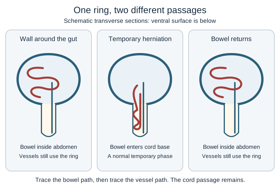
Start with two different events: formation of the body wall and temporary passage of the growing midgut into the cord. Cranial and caudal folding bring the cardiogenic and cloacal regions toward the ventral surface. Lateral folding brings the body sides around the gut while a connection remains at the umbilical ring. The ring is a necessary passage, not evidence that the whole wall failed to form. [13][17]
The somatopleure contains ectoderm and somatic lateral plate mesoderm and contributes to the ventral wall. Do not assign every wall tissue to that mesoderm: abdominal skeletal muscle precursors come from the somites. Human development also involves unequal regional growth, so the familiar folded-sheet drawing is a useful spatial model rather than a complete explanation of every defect. [13]
Trace the first diagram: follow the bowel from the abdominal cavity into the base of the cord and back again. Keep the cord vessels at the ring after the bowel returns. The usual embryology shorthand places physiologic midgut herniation around developmental week 6 and return around week 10. Developmental age is counted after fertilization; obstetric gestational age is approximately two weeks greater. Clinical descriptions use varying age conventions, so these approximate labels are not stand-alone ultrasound diagnostic cutoffs. [2][13][17]
Trace bowel return separately from persistent umbilical vascular passage. [13][17]
The important consequence is persistence, not simply seeing an early protrusion. A first-trimester scan must be interpreted with dating, size, contents and follow-up. Persistent cord-base herniation later in pregnancy is abnormal. Failure of normal return helps explain an omphalocele, but large liver-containing defects also involve abnormal wall development and cannot be reduced to delayed bowel return alone. [2][8]
Try a different setting: a toddler has a soft, skin-covered bulge that appears during crying. That is not the normal fetal herniation still waiting to return. Bowel returned before birth, but the umbilical fascial ring has not yet closed completely. [4]
Which neighboring organs should the defect make you examine?
Localize the opening before naming an embryologic failure. A defect extending toward the lower sternum points to a different anatomic neighborhood from exposed urinary tissue below the umbilicus. Use cranial, lateral and caudal as a map for associated structures, not as proof of a single causal event. [14][17]
Use the level of the defect to choose the next anatomic survey
Region
Visible pattern
Structures to assess
RegionCranial or supraumbilical
Visible patternLower sternal defect, ectopia cordis, central upper abdominal defect
Structures to assessAnterior diaphragm, diaphragmatic pericardium and intracardiac anatomy
RegionAbdominal
Visible patternBowel through or beside the umbilical ring
Structures to assessCord insertion, covering, contents and bowel perfusion
RegionCaudal or infraumbilical
Visible patternExposed urinary plate, abnormal genitalia or bowel between bladder halves
Structures to assessBladder, anorectum, genitalia, pubic bones and spine
Point to the neighboring structure: when the heart lies partly outside an incompletely formed lower sternum, predict an abnormal diaphragm or pericardium before checking the rest of the case. These findings can belong to the Cantrell spectrum. The complete pattern includes a supraumbilical wall defect, lower sternal defect, anterior diaphragmatic defect, diaphragmatic pericardial defect and intracardiac abnormality; incomplete presentations occur. [14]
At the opposite end, bladder exstrophy exposes the bladder mucosa through the lower wall; urine can drain rather than collect normally. Cloacal exstrophy is more extensive: exposed intestinal tissue lies between two bladder halves and may coexist with omphalocele, an imperforate anus and spinal abnormalities. Those four components form the OEIS pattern. An isolated anorectal partitioning problem does not explain all of these external findings. [9][10]
Apply the map: a fetus with an upper midline defect and an abnormal sternum needs a cardiac and thoracic survey, not just measurement of the bowel opening. A fetus with lower urinary and anorectal abnormalities needs a pelvic and spinal survey. In contrast, a typical right-sided gastroschisis does not establish a proven lateral-fold mechanism. Competing developmental hypotheses remain under discussion. [11][12]
Read the cord, the covering and the contents together
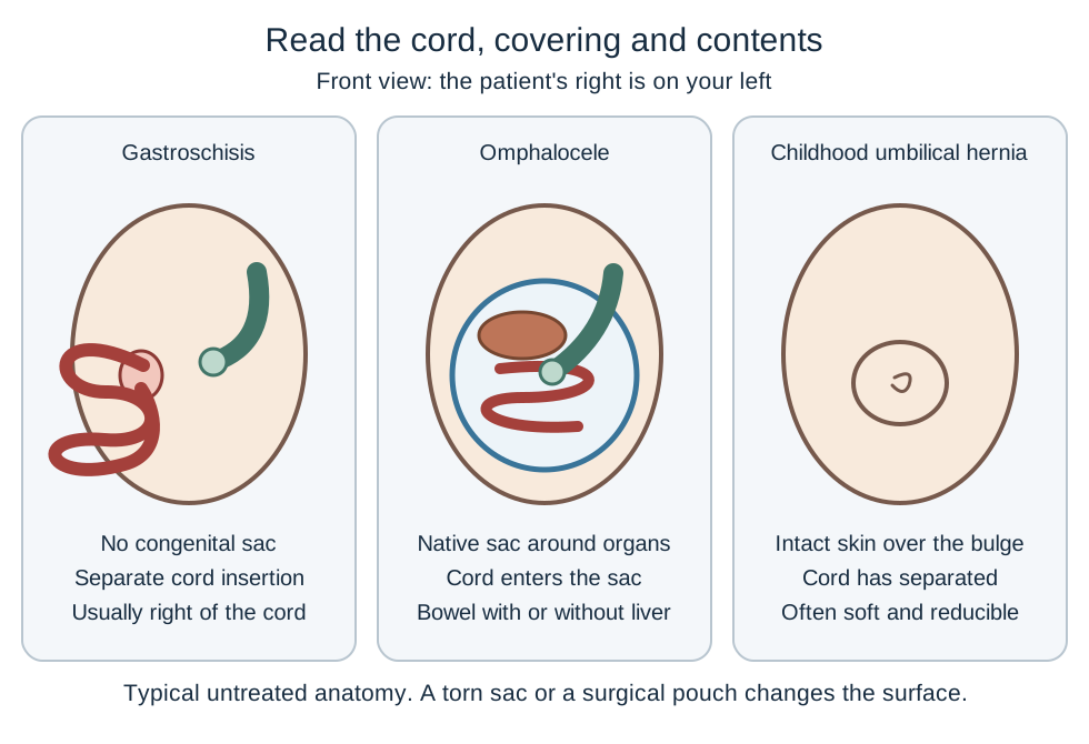
Does every exposed loop mean gastroschisis? No. The three-part comparison below is more reliable than the presence or absence of one membrane. Gastroschisis is a full-thickness wall defect. First locate the cord attachment, then identify the covering, then identify the organs. The diagram shows typical untreated appearances; treatment and sac rupture can change the surface. [1][2]
Compare the cord attachment and type of covering before assigning a diagnosis. [1][2][3][4]
Three different relationships to the umbilical ring
Feature
Gastroschisis
Omphalocele
Childhood umbilical hernia
FeatureOpening
GastroschisisUsually right of a separate cord insertion
OmphaloceleCentral, at the cord base
Childhood umbilical herniaPersistent umbilical fascial ring
FeatureCovering
GastroschisisNo congenital sac
OmphaloceleSac unless ruptured
Childhood umbilical herniaIntact skin
FeatureCord relationship
GastroschisisCord enters the abdomen separately
OmphaloceleCord inserts into the sac
Childhood umbilical herniaUmbilicus overlies the bulge after the cord separates
FeatureContents and behavior
GastroschisisCommonly exposed bowel, sometimes thickened or matted
OmphaloceleBowel with or without liver
Childhood umbilical herniaOften reducible bowel or fat; bulge increases with abdominal pressure
Compare two sketches without their diagnosis labels: trace the cord to the abdomen in one and to the apex of a central sac in the other. In the first, the cord and defect are separate; in the second, the herniation occupies the cord base. That relationship explains why a liver-containing central sac supports omphalocele even when its size is small. [2][8]
An omphalocele membrane contains an inner peritoneal layer, an outer amniotic layer and intervening tissue including Wharton's jelly. It is not ordinary skin. In gastroschisis the bowel has no such congenital barrier and can contact amniotic fluid directly. A skin-covered childhood hernia has a different natural history from either neonatal open-wall defect. [3][4]
Transfer the comparison: the sac has torn during delivery and liver and bowel are now exposed. Review the prenatal images and cord attachment. A formerly central cord-containing sac still supports a ruptured omphalocele; the new exposure changes urgent protection needs, not the developmental diagnosis. [2][8]
Change one feature and predict what changes next
Imagine the same bowel in three states: an intact congenital sac, a torn sac and a surgical silo. A sac can limit direct exposure, but its presence does not establish that the bowel is perfused or that the infant is stable. A silo is a pouch placed by the surgical team after birth, not a congenital membrane. [3][7]
Use the comparison as a trace-and-predict exercise: keep the cord attachment fixed, then change only the covering. For an intact central sac that tears, predict increased heat and fluid losses and a need for urgent organ protection. Do not relabel the defect as gastroschisis. Next keep the congenital diagnosis fixed and add a surgical pouch: the surface now looks covered, but the original defect has not become an omphalocele. [2][3]
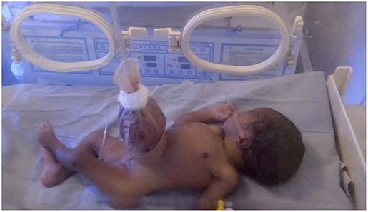
The clinical photograph shows a neonate with a postnatal silo described by Oyinloye and colleagues. Identify the suspended manufactured pouch rather than trying to diagnose the untreated opening from this photograph. The small image does not establish bowel viability or the original cord relationship. It illustrates how a treatment changes appearance, not instructions for constructing an improvised device. [16]
Gastroschisis after placement of a postnatal silo. The manufactured pouch is not an omphalocele membrane. Figure 1, Oyinloye AO, Abubakar AM, Wabada S and Oyebanji LO, Frontiers in Surgery (2020), CC BY 4.0. This historical improvised device illustrates treatment-related appearance, not a recommendation for device construction. Image: Adewale O. Oyinloye; Auwal M. Abubakar; Samuel Wabada; Lateef O. Oyebanji; original source; CC BY 4.0. [16].
Now change perfusion instead of covering. In gastroschisis, prolonged amniotic exposure can contribute to swollen, inflamed bowel. Compression at the opening or twisting of the mesentery can additionally compromise circulation. Atresia, necrosis or perforation makes the condition complex and predicts a more difficult feeding course than an intact, continuous, well-perfused intestine. Stenosis can narrow the intestinal lumen, and volvulus can obstruct the bowel and threaten mesenteric blood flow. Extensive loss of functioning small bowel can produce short-bowel syndrome. [1][7][15]
Why is a darker loop a different problem from a missing membrane?
A missing membrane describes exposure. A new dusky color may indicate impaired blood supply and requires urgent clinical assessment of tension, twisting and systemic perfusion. Protective wrapping must not compress the bowel. Neither a diagnosis label nor a photograph proves that circulation is adequate. [3]
Apply this to a new observation: both infants have gastroschisis, but one has continuous viable bowel and the other has an atretic segment and necrosis. The second infant's longer nutritional recovery is explained by intestinal injury and function, not by a greater chance of a chromosomal syndrome merely because the bowel looks worse. [7][15]
What determines prognosis beyond the size of the opening?
A small wall defect can accompany a serious cardiac or genetic condition; a large one can create a major respiratory problem even with a normal chromosome result. The defect is the start of the assessment, not the entire prognosis. Omphalocele warrants detailed fetal anatomy, cardiac assessment including fetal echocardiography, and genetic counseling with testing selected for the presentation. Brain, spine, gastrointestinal and genitourinary abnormalities also matter. [2][8]
Compare two imagined fetal assessments: in one, a small central sac accompanies an abnormal four-chamber view. In the other, a large liver-containing sac accompanies small lung volumes. Predict the immediate postnatal concern before naming a repair technique. The first finding directs attention to congenital heart disease; the second suggests pulmonary hypoplasia. A normal karyotype would not reverse either structural finding. [8]
Omphalocele with macroglossia, lateralized overgrowth or neonatal hypoglycemia raises concern for Beckwith-Wiedemann spectrum. Abnormal regulation of imprinted genes at 11p15.5 can underlie the condition. A normal chromosome count does not exclude an imprinting disorder. Persistent hypoglycemia may reflect excessive insulin action and needs prompt treatment while a specialist team directs molecular testing and subsequent surveillance. Not every infant has the full phenotype. [6]
Gastroschisis is more often isolated and has a much weaker association with aneuploidy than omphalocele. Isolated cases often have a favorable long-term outcome after neonatal surgical care, but major bowel injury changes that outlook. The genetic association is a relative comparison, not a guarantee of normal genetics or anatomy. Growth, bowel condition, associated intestinal atresia and the infant's overall physiology remain important. Complex gastroschisis is associated with longer parenteral nutrition and time to full feeds in cohort evidence. [3][7][15]
Apply the distinction: a normal chromosome study in a fetus with a large omphalocele does not justify canceling cardiac or respiratory planning. Conversely, intestinal atresia in an infant with otherwise typical gastroschisis changes the bowel and nutrition plan without by itself establishing an aneuploidy syndrome. [7][8]
Protect physiology before deciding how to close the wall
Should exposed bowel be pushed back immediately? Not as a routine bedside response. At birth, coordinate neonatal resuscitation and urgent pediatric surgical contact. Organize transfer to an appropriate neonatal surgical unit in parallel with stabilization rather than waiting for every assessment to finish. Airway, breathing and circulation remain the first priorities. [3]
Support and protect exposed organs with an appropriate transparent noncompressive barrier, prevent heat loss, establish intravenous access, monitor perfusion and glucose, and decompress the stomach with an orogastric or nasogastric tube. Fluids and replacement of losses require repeated assessment. Antibiotics follow the local neonatal surgical protocol. Keep the infant without enteral feeds during initial stabilization. Avoid pressure, twisting, unnecessary handling and a tight dressing around the exposed bowel or sac. An intact omphalocele sac also needs support and protection; a ruptured one requires urgent exposed-bowel precautions. [3][7]
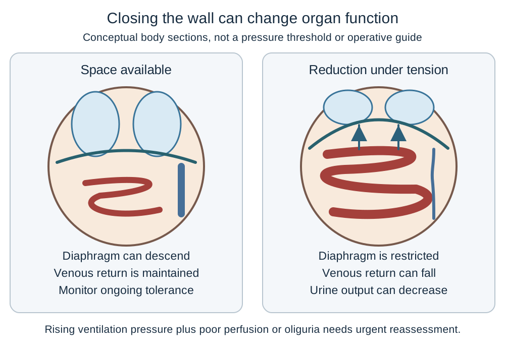
Trace the pressure diagram: place a large volume of swollen bowel into a small abdominal cavity, then predict effects on the diaphragm and blood flow. Excessive intra-abdominal pressure can impair ventilation, venous return and organ perfusion. Worsening respiratory support needs with oliguria or circulatory deterioration after reduction calls for immediate reassessment, not a tighter closure. [3]
Predict combined respiratory, circulatory and urinary changes when reduction produces excessive abdominal pressure. [3][7][8]
Primary closure is suitable for selected infants whose organs can be reduced safely. A silo permits staged reduction when immediate closure would create excessive pressure or the infant needs a different approach. A giant omphalocele may require staged or delayed treatment, especially with pulmonary hypoplasia. Repair choice depends on anatomy, bowel condition and physiology, not a universal size threshold. Nutritional support continues until intestinal function supports advancing feeds; closing the skin does not mean feeding readiness. [7][8]
Prenatal planning should involve fetal medicine, neonatology and pediatric surgery. Follow growth and bowel findings and plan birth where appropriate neonatal care is available. Gastroschisis alone does not require cesarean delivery; obstetric indications and fetal status matter. A large liver-containing omphalocele requires individualized delivery planning because of rupture and organ-injury risks. Exact timing is not determined by the diagnosis word alone. [3][7][8]
A skin-covered childhood hernia follows a different pathway
A well child with a painless, easily reducible umbilical bulge usually needs observation and routine follow-up, not neonatal exposed-bowel care. Many close spontaneously; clinicians commonly observe uncomplicated defects until about age 4 to 5 years. Persistent, large or symptomatic defects merit pediatric surgical discussion. Additional malformations or an unusual growth pattern also warrant a broader assessment rather than automatic reassurance that the hernia is isolated. Pain, vomiting, skin discoloration or inability to reduce a previously reducible bulge requires urgent assessment for incarceration or strangulation. Taping a coin over the bulge does not repair the fascial ring. [4]
Use the numbers correctly: a cohort found that 88.6% of all diagnosed umbilical hernias had closed by age 5. Among hernias still present at age 3, the chance of closure with observation to age 5 was 34.8%. Those are different starting groups; the first percentage is not the remaining chance for a three-year-old. The study was retrospective and does not predict an individual child's outcome with certainty. [5]
Transfer the plan: a comfortable three-year-old with a reducible defect can still benefit from observation. The same child with a newly tender irreducible bulge and vomiting needs urgent assessment today. Age-based observation applies only while the hernia remains uncomplicated. [4][5]
Apply the anatomy to clinical decisions
Use the findings in each independent case to select one best answer. All essential teaching above remains readable without answering a question.
Case 1
Show answer and explanations for case 1
A. Loss of intestinal absorptive surface (Why this does not fit)
Extensive intestinal loss can cause malabsorption and nutritional failure. No resection or loss of bowel length is described; this does not explain rapid cooling before feeding.
Reasoning steps for option A
When does loss of absorptive surface become clinically important?
Extensive intestinal loss can cause malabsorption and nutritional failure.
Why does loss of absorptive surface fail to explain cooling and delayed refill before this newborn has fed?
No resection or loss of bowel length is described; this does not explain rapid cooling before feeding.
B. Evaporative heat and fluid losses (Best answer)
Exposed moist viscera permit substantial evaporative heat and fluid losses. A separate right-sided opening without a sac indicates gastroschisis, making cooling and impaired perfusion coherent consequences.
Reasoning steps for option B
What is lost across an exposed moist bowel surface?
Exposed moist viscera permit substantial evaporative heat and fluid losses.
How does the right-sided opening without a sac connect the falling temperature to impaired perfusion?
A separate right-sided opening without a sac indicates gastroschisis, making cooling and impaired perfusion coherent consequences.
C. Reduced renal excretion of sodium (Why this does not fit)
Sodium retention tends to expand extracellular volume. That direction does not explain acute volume depletion with an unprotected abdominal defect.
Reasoning steps for option C
What happens to extracellular volume when sodium is retained?
Sodium retention tends to expand extracellular volume.
Why is sodium retention inconsistent with acute volume depletion from this unprotected defect?
Retaining sodium expands extracellular volume, whereas the unprotected bowel and delayed refill suggest acute fluid loss. Sodium retention also does not explain cooling.
D. Reduced pulmonary vascular resistance (Why this does not fit)
Pulmonary vascular resistance normally falls during transition after birth. This does not explain heat loss and deteriorating perfusion specifically associated with exposed bowel.
Reasoning steps for option D
How does normal pulmonary transition differ from surface fluid loss?
Pulmonary vascular resistance normally falls during transition after birth.
Why does a normal fall in pulmonary vascular resistance not explain deterioration beside exposed bowel?
The normal postnatal fall in pulmonary vascular resistance does not explain the evaporative cooling and fluid loss associated with this exposed bowel.
Takeaway: Identify the defect, then predict the physiologic cost of exposure.
A. Await the chromosome result before requesting fetal echocardiography (Why this does not fit)
Chromosomal findings can inform counseling and the broader anomaly assessment. A pending result does not justify postponing targeted cardiac assessment for a fetus with omphalocele.
Reasoning steps for option A
Does a pending chromosome result remove the present indication for cardiac assessment?
Chromosomal findings can inform counseling and the broader anomaly assessment.
Why should cardiac assessment of this central liver-containing sac proceed before the chromosome result?
The central liver-containing sac with cord insertion at its apex indicates omphalocele, whose cardiac association warrants focused assessment without waiting for chromosome results.
B. Use routine four-chamber views instead of specialist echocardiography (Why this does not fit)
Routine ultrasound provides an important initial cardiac screen. A normal screening view does not replace focused fetal echocardiography for this associated-anomaly pattern.
Reasoning steps for option B
Is a screening four-chamber view equivalent to a fetal echocardiogram?
Routine ultrasound provides an important initial cardiac screen.
Why is the normal four-chamber screening view insufficient in this fetus with a cord-apex sac?
The normal four-chamber view is a screening result, not a substitute for focused fetal echocardiography in this fetus with omphalocele.
C. Reserve echocardiography for an abnormal newborn oxygen screen (Why this does not fit)
Newborn oxygen screening can identify some clinically important heart disease. It is not a substitute for indicated prenatal evaluation when the abdominal defect is already recognized.
Reasoning steps for option C
Can newborn screening replace an indicated prenatal cardiac assessment?
Newborn oxygen screening can identify some clinically important heart disease.
Why would waiting for an abnormal newborn oxygen screen miss the indicated prenatal assessment?
The omphalocele is already recognized prenatally, so its indicated cardiac evaluation should not wait for a newborn oxygen screen that detects only some heart disease.
D. Arrange fetal echocardiography despite the reassuring screening view (Best answer)
Omphalocele has an important association with congenital heart disease. The central liver-containing sac and cord insertion establish that risk despite normal screening images and absent family history.
Reasoning steps for option D
Why does this cord-base anatomy justify a focused cardiac study?
Omphalocele has an important association with congenital heart disease.
Which sac and cord findings justify fetal echocardiography despite normal screening and no family history?
The central liver-containing sac and cord insertion establish that risk despite normal screening images and absent family history.
Takeaway: Reassuring growth or family history does not cancel the cardiac evaluation associated with omphalocele.
A. Observation with routine follow-up (Best answer)
Small uncomplicated childhood umbilical hernias frequently close spontaneously. A painless, skin-covered, reducible bulge in a thriving infant supports observation rather than neonatal defect repair.
Reasoning steps for option A
What is the expected natural history of an uncomplicated infant hernia?
Small uncomplicated childhood umbilical hernias frequently close spontaneously.
Which features of this 8-month-old's bulge support observation instead of repair?
A painless, skin-covered, reducible bulge in a thriving infant supports observation rather than neonatal defect repair.
B. Elective repair during this month (Why this does not fit)
Early elective repair can be appropriate for selected symptomatic or otherwise concerning hernias. This infant has none of those features, and age alone does not justify routine repair now.
Reasoning steps for option B
Which hernia features would justify earlier elective intervention?
Early elective repair can be appropriate for selected symptomatic or otherwise concerning hernias.
What indication for repair this month is missing in this thriving infant with a painless reducible bulge?
The infant is thriving and the hernia is painless, reducible and normally skin-covered. No symptomatic or concerning feature supports routine elective repair this month.
C. Compression with an adhesive dressing (Why this does not fit)
External compression can flatten the visible bulge without closing the fascial defect. Taping is not effective treatment and is unnecessary in this uncomplicated presentation.
Reasoning steps for option C
Does flattening the skin bulge repair the ring?
External compression can flatten the visible bulge without closing the fascial defect.
Why does adhesive taping offer no therapeutic benefit for this uncomplicated umbilical hernia?
Taping can flatten the visible bulge but does not close the fascial defect. This painless reducible infant hernia needs observation, not compression.
D. Admission for intravenous antibiotics (Why this does not fit)
Antibiotics treat suspected infection or are used in selected surgical settings. There is no evidence of infection, exposed bowel or an acute surgical complication.
Reasoning steps for option D
What would justify antibiotics in a child with an abdominal wall finding?
Antibiotics treat suspected infection or are used in selected surgical settings.
Which infectious or acute surgical findings needed to justify admission for antibiotics are absent here?
There is no evidence of infection, exposed bowel or an acute surgical complication.
Takeaway: Intact skin, reducibility and absence of symptoms support observation of a childhood umbilical hernia.
A. Gastroschisis with reduced syndromic association (Why this does not fit)
Gastroschisis usually has uncovered bowel beside a separate cord insertion. A previously documented central liver-containing sac remains evidence of omphalocele after rupture.
Reasoning steps for option A
What anatomy would support gastroschisis before any tear?
Gastroschisis usually has uncovered bowel beside a separate cord insertion.
Why does the prenatal liver-containing central sac outweigh the gastroschisis-like appearance after tearing?
A previously documented central liver-containing sac remains evidence of omphalocele after rupture.
B. Umbilical hernia with spontaneous closure potential (Why this does not fit)
A childhood umbilical hernia is covered by skin and often closes with growth. An amniotic sac containing liver before birth is not the skin-covered childhood lesion.
Reasoning steps for option B
How does the covering of a childhood umbilical hernia differ?
A childhood umbilical hernia is covered by skin and often closes with growth.
Why is the prenatal sac containing liver incompatible with a skin-covered childhood umbilical hernia?
An amniotic sac containing liver before birth is not the skin-covered childhood lesion.
C. Ruptured omphalocele with retained associated-anomaly risks (Best answer)
Rupture changes protection needs but does not change the underlying cord-base defect. The documented omphalocele still warrants its cardiac and genetic assessment while the organs receive urgent protection.
Reasoning steps for option C
Does sac rupture alter the original developmental anatomy?
Rupture changes protection needs but does not change the underlying cord-base defect.
Why must cardiac and genetic assessment continue after this documented omphalocele ruptures?
The documented omphalocele still warrants its cardiac and genetic assessment while the organs receive urgent protection.
D. Acquired abdominal trauma without congenital malformation (Why this does not fit)
Trauma can expose organs through an acquired abdominal injury. The abnormal sac and cord attachment were present prenatally, so an isolated traumatic explanation is insufficient.
Reasoning steps for option D
What timing would support a purely acquired injury?
Trauma can expose organs through an acquired abdominal injury.
Which prenatal observations rule out an exclusively acquired traumatic explanation for the exposed organs?
The abnormal sac and cord attachment were present prenatally, so an isolated traumatic explanation is insufficient.
Takeaway: Use pre-rupture anatomy to preserve the diagnosis and its associated workup.
A. Posterolateral diaphragmatic discontinuity (Why this does not fit)
A posterolateral diaphragmatic hernia can impair lung development. Its location does not match the anterior midline thoracoabdominal pattern established by the sternum and heart.
Reasoning steps for option A
Where is the defect in a typical posterolateral diaphragmatic hernia?
A posterolateral diaphragmatic hernia can impair lung development.
Why does a posterolateral diaphragmatic defect mismatch the location of the sternal and cardiac abnormalities?
A posterolateral defect is not in the anterior midline region implicated by the incomplete lower sternum and partly external heart.
B. Anterior diaphragmatic and pericardial discontinuity (Best answer)
The Cantrell spectrum links upper midline wall and lower sternal defects with anterior diaphragm and diaphragmatic pericardial defects. Ectopia cordis with the supplied upper midline abnormalities fits that anatomic cluster.
Reasoning steps for option B
Which diaphragm and pericardial regions belong to the Cantrell pattern?
The Cantrell spectrum links upper midline wall and lower sternal defects with anterior diaphragm and diaphragmatic pericardial defects.
How do ectopia cordis and the lower sternal defect support anterior diaphragmatic and pericardial involvement?
Ectopia cordis with the supplied upper midline abnormalities fits that anatomic cluster.
C. Sacral dysraphism with bladder plate exposure (Why this does not fit)
Spinal defects and bladder exposure occur in the lower exstrophy spectrum. Normal lower urinary anatomy and an upper thoracoabdominal defect favor a different regional association.
Reasoning steps for option C
Which wall region is associated with bladder exposure and spinal defects?
Spinal defects and bladder exposure occur in the lower exstrophy spectrum.
Why do normal lower urinary anatomy and an upper wall defect argue against a sacral-bladder association?
Normal lower urinary anatomy and an upper thoracoabdominal defect favor a different regional association.
D. Esophageal discontinuity with distal fistula (Why this does not fit)
Esophageal atresia with fistula affects separation of foregut structures. It does not specifically account for the linked lower sternal and anterior body-wall abnormalities.
Reasoning steps for option D
Which developmental region is involved in esophageal atresia with fistula?
Esophageal atresia with fistula affects separation of foregut structures.
Why does an esophageal fistula not specifically fit the linked lower sternal and anterior wall defects?
It does not specifically account for the linked lower sternal and anterior body-wall abnormalities.
Takeaway: Use the vertical level and neighboring structures to predict the associated anomaly.
A. Absent normal bladder filling with preserved amniotic fluid (Best answer)
An open bladder plate cannot accumulate urine normally even when both kidneys produce it. Urine drainage from infraumbilical mucosa and pubic separation support bladder exstrophy, so normal fluid does not require a visible filling bladder.
Reasoning steps for option A
How does an exposed bladder plate alter storage of fetal urine?
An open bladder plate cannot accumulate urine normally even when both kidneys produce it.
How can this urine-draining infraumbilical plate coexist with preserved fluid but no normal bladder filling?
Urine drainage from infraumbilical mucosa and pubic separation support bladder exstrophy, so normal fluid does not require a visible filling bladder.
B. A persistently enlarged bladder with bilateral hydroureter (Why this does not fit)
A large retained bladder with upstream dilation suggests lower urinary tract obstruction. The described bladder is open and drains externally rather than retaining urine behind an obstruction.
Reasoning steps for option B
What produces a persistently distended fetal bladder?
A large retained bladder with upstream dilation suggests lower urinary tract obstruction.
Why would a distended bladder with hydroureter conflict with the externally draining plate?
The described bladder is open and drains externally rather than retaining urine behind an obstruction.
C. Absent kidneys with severe reduction of amniotic fluid (Why this does not fit)
Bilateral renal agenesis markedly reduces fetal urine production. Both kidneys are present and the plate produces urine, opposing failure of urine production as the explanation.
Reasoning steps for option C
What connects bilateral renal agenesis to amniotic fluid volume?
Which findings contradict renal agenesis as the cause of the abnormal prenatal bladder appearance?
Both kidneys are present and the plate produces urine, opposing failure of urine production as the explanation.
D. A normally cycling bladder with free-floating bowel loops (Why this does not fit)
Normal bladder cycling with uncovered paraumbilical bowel can occur in gastroschisis. The exposed surface here drains urine below the umbilicus and is accompanied by pubic separation rather than isolated bowel extrusion.
Reasoning steps for option D
Which defect can expose bowel while preserving bladder cycling?
Normal bladder cycling with uncovered paraumbilical bowel can occur in gastroschisis.
Why do urine drainage and pubic separation favor bladder exstrophy over free-floating gastroschisis bowel?
The exposed surface here drains urine below the umbilicus and is accompanied by pubic separation rather than isolated bowel extrusion.
Takeaway: Normal fetal urine production can coexist with absent bladder filling when the bladder is open.
A. Renal artery Doppler for renovascular disease (Why this does not fit)
Renal vascular imaging evaluates an arterial perfusion disorder. It does not address the spinal association or the asymmetric leg activity in this extensive lower midline defect.
Reasoning steps for option A
What clinical problem is renal artery Doppler designed to assess?
Renal vascular imaging evaluates an arterial perfusion disorder.
Why would renal artery Doppler leave the asymmetric leg activity in this lower midline defect unexplained?
Renal artery Doppler examines arterial perfusion, not the spinal abnormalities associated with cloacal exstrophy or the asymmetric leg activity that makes them a concern here.
B. Cranial imaging for isolated holoprosencephaly (Why this does not fit)
Holoprosencephaly can accompany some syndromic malformations. The bladder halves, intestinal plate and absent anus point specifically to cloacal exstrophy, with spinal assessment a priority given the leg findings.
Reasoning steps for option B
Which observations would make a primary forebrain malformation the leading concern?
Holoprosencephaly can accompany some syndromic malformations.
Why do the bladder halves and absent anus prioritize spinal assessment over imaging for isolated holoprosencephaly?
The bladder halves, intestinal plate and absent anus point specifically to cloacal exstrophy, with spinal assessment a priority given the leg findings.
C. Diaphragmatic imaging for the Cantrell pattern (Why this does not fit)
Cantrell-pattern defects involve the upper midline wall and thoracic structures. This infant instead has a lower urinary and anorectal pattern with abnormal leg activity.
Reasoning steps for option C
Which anatomic level characterizes the Cantrell pattern?
Cantrell-pattern defects involve the upper midline wall and thoracic structures.
Why is Cantrell-focused diaphragm imaging a regional mismatch for this urinary and anorectal pattern?
This infant instead has a lower urinary and anorectal pattern with abnormal leg activity.
D. Spinal imaging for associated dysraphism (Best answer)
Cloacal exstrophy belongs to the OEIS pattern, which includes spinal defects. The exposed intestinal plate between bladder halves, absent anus and asymmetric leg activity directly support evaluating the spine.
Reasoning steps for option D
Which associated structure can explain abnormal leg activity in cloacal exstrophy?
Cloacal exstrophy belongs to the OEIS pattern, which includes spinal defects.
Which combined exstrophy and leg findings directly support imaging for spinal dysraphism?
The exposed intestinal plate between bladder halves, absent anus and asymmetric leg activity directly support evaluating the spine.
Takeaway: Recognize the combined cloacal pattern rather than treating each opening as an isolated defect.
A. Classify persistent omphalocele from the cord-base position (Why this does not fit)
Persistent central cord-base herniation later in pregnancy supports omphalocele. At this early gestational age, location alone cannot distinguish physiologic herniation from persistence.
Reasoning steps for option A
When does persistence become essential to interpreting a cord-base prominence?
Persistent central cord-base herniation later in pregnancy supports omphalocele.
Why cannot cord-base position alone establish persistent omphalocele on a 9-week scan?
At this early gestational age, location alone cannot distinguish physiologic herniation from persistence.
B. Classify gastroschisis from the bowel-only sac contents (Why this does not fit)
Gastroschisis is identified mainly by an uncovered opening beside a separate cord insertion. Bowel-only contents do not establish that anatomy, and physiologic herniation may be bowel-containing.
Reasoning steps for option B
Which relationship, rather than absence of liver, supports gastroschisis?
Gastroschisis is identified mainly by an uncovered opening beside a separate cord insertion.
Why do bowel-only contents at 9 weeks fail to establish the separate uncovered opening of gastroschisis?
Bowel-only contents do not establish that anatomy, and physiologic herniation may be bowel-containing.
C. Reassess anatomy after the physiologic herniation window (Best answer)
Normal first-trimester midgut herniation can resemble a small cord-base prominence. The early timing and bowel-only appearance require interval reassessment rather than a definitive permanent-defect label.
Reasoning steps for option C
Why does an early cord-base finding require a time dimension?
Normal first-trimester midgut herniation can resemble a small cord-base prominence.
Why should this early bowel-only prominence be reassessed after the physiologic herniation window?
The early timing and bowel-only appearance require interval reassessment rather than a definitive permanent-defect label.
D. Classify the anatomy as normal and end reassessment (Why this does not fit)
An early examination has limits and does not guarantee that all structures will prove normal. The observation needs follow-up; neither a permanent defect nor its absence is established by this scan alone.
Reasoning steps for option D
Can an early study definitively exclude all later structural abnormalities?
An early examination has limits and does not guarantee that all structures will prove normal.
Why is ending follow-up unjustified after this single early cord-base examination?
The observation needs follow-up; neither a permanent defect nor its absence is established by this scan alone.
Takeaway: Interpret an early cord-base prominence with dating and interval anatomy, not location alone.
A. Somatic lateral plate mesoderm (Why this does not fit)
Somatic lateral plate mesoderm contributes to connective components of the body wall. It is not the direct source of the hypaxial skeletal muscle precursors singled out in the specimen.
Reasoning steps for option A
Which body-wall contribution comes from somatic lateral plate mesoderm?
Somatic lateral plate mesoderm contributes to connective components of the body wall.
Why does a body-wall connective-tissue contribution not make somatic lateral plate the missing muscle lineage?
Somatic lateral plate contributes body-wall connective components, not the hypaxial skeletal muscle precursors specifically deficient in this specimen.
B. Hypaxial somitic myotomes (Best answer)
Hypaxial myotomal precursors give rise to abdominal skeletal muscles. A specific deficit in those muscle precursors implicates somites despite otherwise enclosed gut and a normal cord passage.
Reasoning steps for option B
Which lineage supplies abdominal skeletal muscle?
Hypaxial myotomal precursors give rise to abdominal skeletal muscles.
Why does the muscle-precursor deficit implicate hypaxial somites despite normal gut enclosure?
A specific deficit in those muscle precursors implicates somites despite otherwise enclosed gut and a normal cord passage.
C. Splanchnic lateral plate mesoderm (Why this does not fit)
Splanchnic mesoderm contributes to tissues associated with the gut. The observed defect concerns skeletal muscle of the external abdominal wall rather than gut-wall tissues.
Reasoning steps for option C
How do splanchnic derivatives differ from abdominal skeletal muscle?
Splanchnic mesoderm contributes to tissues associated with the gut.
Why are gut-associated splanchnic derivatives not the tissue missing from this abdominal-wall specimen?
The observed defect concerns skeletal muscle of the external abdominal wall rather than gut-wall tissues.
D. Surface ectoderm at the umbilicus (Why this does not fit)
Surface ectoderm contributes to the external epithelial covering. An epithelial lineage does not directly supply the missing abdominal skeletal muscle precursors.
Reasoning steps for option D
What external tissue is derived from surface ectoderm?
Surface ectoderm contributes to the external epithelial covering.
Why cannot surface ectoderm directly account for the absent skeletal muscle precursors?
An epithelial lineage does not directly supply the missing abdominal skeletal muscle precursors.
Takeaway: The wall is composite: its skeletal muscle derives from somites, not from every tissue associated with the somatopleure.
A. Direct amniotic contact with uncovered bowel (Best answer)
The absence of a congenital sac permits direct amniotic exposure and inflammatory bowel changes. The first infant has exposed matted bowel, while the second has bowel protected by a native membrane.
Reasoning steps for option A
Which prenatal exposure differs when the sac is absent?
The absence of a congenital sac permits direct amniotic exposure and inflammatory bowel changes.
How does the contrast between uncovered matted loops and smooth sac-enclosed bowel support amniotic exposure?
The first infant has exposed matted bowel, while the second has bowel protected by a native membrane.
B. Meconium leakage through a prenatal perforation (Why this does not fit)
Prenatal perforation can produce meconium peritonitis with inflammatory changes and calcification. The supplied intact bowel and absence of perforation or calcifications favor surface exposure rather than leakage.
Reasoning steps for option B
What additional evidence supports meconium peritonitis?
Prenatal perforation can produce meconium peritonitis with inflammatory changes and calcification.
Which operative and imaging findings weaken prenatal meconium leakage as the cause of the matted bowel?
The supplied intact bowel and absence of perforation or calcifications favor surface exposure rather than leakage.
C. Sustained mesenteric vascular occlusion before birth (Why this does not fit)
Prolonged vascular occlusion can cause ischemic or necrotic bowel injury. Both intestines are viable and perfused at assessment; the differing congenital coverings provide the more direct explanation for surface inflammation.
Reasoning steps for option C
How does sustained occlusion differ from inflammatory surface exposure?
Prolonged vascular occlusion can cause ischemic or necrotic bowel injury.
Why is sustained mesenteric occlusion less persuasive when both infants have viable perfused bowel?
Both intestines are viable and perfused at assessment; the differing congenital coverings provide the more direct explanation for surface inflammation.
D. Intrauterine bacterial infection involving the intestine (Why this does not fit)
Infection can inflame bowel and cause systemic or focal illness. No infection evidence is present, and infection is not required to explain inflammatory bowel exposed to amniotic fluid.
Reasoning steps for option D
Must bacteria be present for exposed fetal bowel to become inflamed?
Infection can inflame bowel and cause systemic or focal illness.
Why is bacterial infection unnecessary to explain inflammation confined to the congenitally uncovered bowel?
No infection evidence is present, and infection is not required to explain inflammatory bowel exposed to amniotic fluid.
Takeaway: Bowel inflammation at birth can reflect prenatal exposure without proving infection.
A. Earlier full feeds because the damaged segment was excised (Why this does not fit)
Resection eliminates nonviable tissue but does not guarantee rapid restoration of intestinal function. Atresia and loss of bowel identify complex gastroschisis, which is associated with delayed feeding recovery.
Reasoning steps for option A
Does excision of damaged bowel guarantee normal remaining function?
Resection eliminates nonviable tissue but does not guarantee rapid restoration of intestinal function.
Why do atresia and resection argue against earlier full feeds in infant B?
Atresia and loss of bowel identify complex gastroschisis, which is associated with delayed feeding recovery.
B. Shorter admission because extraintestinal anatomy is normal (Why this does not fit)
Normal cardiac anatomy avoids one source of morbidity. It does not cancel the intestinal injury and resection that distinguish these two infants.
Reasoning steps for option B
Can a normal cardiac evaluation erase the consequences of bowel injury?
Normal cardiac anatomy avoids one source of morbidity.
Why does normal cardiac anatomy not justify a shorter admission for the infant with necrotic bowel?
Normal cardiac anatomy removes one possible source of morbidity but does not offset infant B's ileal atresia, necrosis and resection, which delay intestinal recovery.
C. Equivalent nutrition needs because the wall location matches (Why this does not fit)
The same external defect location can occur with very different bowel integrity. Atresia and necrosis make infant B clinically different despite the shared paraumbilical opening.
Reasoning steps for option C
Does the position of the opening specify intestinal continuity and viability?
The same external defect location can occur with very different bowel integrity.
Why does a matching paraumbilical opening not imply equivalent nutritional needs in these two infants?
Atresia and necrosis make infant B clinically different despite the shared paraumbilical opening.
D. Longer parenteral nutrition because intestinal function is impaired (Best answer)
Complex gastroschisis includes atresia, necrosis or perforation and is associated with longer nutritional recovery. Infant B has both loss of continuity and tissue injury, so prolonged dependence on intravenous nutrition is more likely.
Reasoning steps for option D
How does complex bowel injury affect the expected feeding course?
Complex gastroschisis includes atresia, necrosis or perforation and is associated with longer nutritional recovery.
Which intestinal injuries make longer intravenous nutrition more likely in infant B than in infant A?
Infant B has both loss of continuity and tissue injury, so prolonged dependence on intravenous nutrition is more likely.
Takeaway: Intestinal continuity and viability predict recovery better than matching external defect locations.
A. Give fluid replacement before changing the external support (Why this does not fit)
Systemic volume depletion can reduce bowel perfusion and may require fluid treatment. Stable systemic findings with duskiness directly beneath a tight edge make correction of local pressure the immediate priority.
Reasoning steps for option A
Would systemic fluid treatment directly eliminate pressure from the barrier?
Systemic volume depletion can reduce bowel perfusion and may require fluid treatment.
Why should stable blood pressure and focal duskiness beneath the barrier shift priority away from fluids alone?
Stable systemic findings with duskiness directly beneath a tight edge make correction of local pressure the immediate priority.
B. Reassess the support and relieve external compression urgently (Best answer)
A tight barrier or distorted pedicle can compromise local blood flow. The newly dusky loop beneath a constricting edge requires immediate assessment and correction while surgical help is engaged.
Reasoning steps for option B
How can local pressure impair bowel circulation despite stable systemic status?
A tight barrier or distorted pedicle can compromise local blood flow.
Why does a newly dusky loop under a tight edge require immediate relief of external compression?
The newly dusky loop beneath a constricting edge requires immediate assessment and correction while surgical help is engaged.
C. Obtain abdominal radiographs before altering the protective barrier (Why this does not fit)
Imaging can help evaluate some neonatal bowel complications. It should not delay correction of an obvious constricting edge associated with an acute focal color change.
Reasoning steps for option C
Does a visible external constriction require imaging before initial correction?
Imaging can help evaluate some neonatal bowel complications.
Why should radiographs not precede correction of the visibly constricting barrier edge?
It should not delay correction of an obvious constricting edge associated with an acute focal color change.
D. Continue transfer and reassess the color after arrival (Why this does not fit)
Timely transfer is important for definitive neonatal surgical care. A new possible perfusion threat should be assessed immediately rather than left untreated during transport.
Reasoning steps for option D
Can a time-critical focal perfusion concern safely wait for arrival?
Timely transfer is important for definitive neonatal surgical care.
Why is waiting until arrival inappropriate for a loop that became dusky just after wrapping?
A new possible perfusion threat should be assessed immediately rather than left untreated during transport.
Takeaway: A protective barrier must protect without constricting; new focal color change requires urgent assessment.
A. Omphalocele treated with a postnatal surgical silo (Why this does not fit)
A silo can be part of staged management for a congenital wall defect. The pretreatment separate cord and lateral uncovered opening support gastroschisis rather than a central cord-base sac.
Reasoning steps for option A
Which pretreatment cord relationship distinguishes omphalocele?
A silo can be part of staged management for a congenital wall defect.
Which pretreatment cord and opening findings contradict an omphalocele diagnosis despite later silo placement?
The pretreatment separate cord and lateral uncovered opening support gastroschisis rather than a central cord-base sac.
B. Umbilical hernia protected by its native skin covering (Why this does not fit)
A childhood umbilical hernia is covered by skin and can protrude during straining. Free bowel at birth and a later manufactured pouch are not a skin-covered childhood hernia.
Reasoning steps for option B
What covering is present over an uncomplicated childhood umbilical hernia?
A childhood umbilical hernia is covered by skin and can protrude during straining.
Why do free bowel at birth and a later manufactured pouch rule out native skin over an umbilical hernia?
Free bowel at birth and a later manufactured pouch are not a skin-covered childhood hernia.
C. Gastroschisis protected by a postnatal surgical silo (Best answer)
A silo covers exposed bowel after birth and permits staged reduction. The initial lateral opening and separate cord identify gastroschisis; the later manufactured pouch explains the new coverage.
Reasoning steps for option C
How does a surgical silo change the appearance of gastroschisis?
A silo covers exposed bowel after birth and permits staged reduction.
How do the initial lateral opening and subsequent manufactured pouch establish gastroschisis treated with a silo?
The initial lateral opening and separate cord identify gastroschisis; the later manufactured pouch explains the new coverage.
D. Omphalocele retaining its original congenital sac (Why this does not fit)
An intact omphalocele is covered by a native sac associated with the cord base. The initial note describes no congenital covering and a separate cord, while the later covering is manufactured.
Reasoning steps for option D
What evidence would establish that the visible covering is a native sac?
An intact omphalocele is covered by a native sac associated with the cord base.
Which timing and material clues contradict the claim that the later covering is an original omphalocele sac?
The initial note describes no congenital covering and a separate cord, while the later covering is manufactured.
Takeaway: Treatment-created coverage must not be mistaken for a congenital sac.
A. Excess intra-abdominal pressure impairing ventilation and perfusion (Best answer)
High abdominal pressure restricts diaphragmatic excursion and can impair venous return and renal perfusion. The simultaneous respiratory and circulatory deterioration immediately after a tense reduction supports pressure-related organ dysfunction.
Reasoning steps for option A
How can high abdominal pressure affect both breathing and urine output?
High abdominal pressure restricts diaphragmatic excursion and can impair venous return and renal perfusion.
Why do rising inflation pressure, oliguria and poor perfusion immediately after tense closure implicate abdominal pressure?
The simultaneous respiratory and circulatory deterioration immediately after a tense reduction supports pressure-related organ dysfunction.
B. Persistent pulmonary hypertension causing isolated oxygenation failure (Why this does not fit)
Pulmonary hypertension can cause severe neonatal hypoxemia. It does not best explain the abrupt tense abdomen with reduced urine output and greater inflation pressure after reduction.
Reasoning steps for option B
Which primary problem is expected with persistent pulmonary hypertension?
Pulmonary hypertension can cause severe neonatal hypoxemia.
Why does isolated pulmonary hypertension not unify the tense abdomen, oliguria and increased inflation pressure?
It does not best explain the abrupt tense abdomen with reduced urine output and greater inflation pressure after reduction.
C. Primary renal obstruction causing isolated urinary retention (Why this does not fit)
Urinary obstruction can reduce measured urine output. It does not account for the simultaneous mechanical ventilation change and impaired systemic perfusion after tight closure.
Reasoning steps for option C
Would urinary obstruction explain the increased ventilation pressure?
Urinary obstruction can reduce measured urine output.
Which simultaneous respiratory and circulatory changes are unexplained by isolated urinary obstruction?
It does not account for the simultaneous mechanical ventilation change and impaired systemic perfusion after tight closure.
D. Resolution of bowel edema increasing effective circulating volume (Why this does not fit)
Improvement in edema would not be expected to create a suddenly tense abdomen with impaired organ function. The time course and direction of the changes instead indicate intolerance of the reduction.
Reasoning steps for option D
What physiologic direction would be expected as edema resolves?
Improvement in edema would not be expected to create a suddenly tense abdomen with impaired organ function.
Why does deterioration immediately after reduction contradict improvement from resolving bowel edema?
Resolution of edema should not cause a suddenly tense abdomen with rising ventilation pressure, oliguria and worsening perfusion. Their onset after reduction instead supports pressure intolerance.
Takeaway: A technically closed wall can be physiologically intolerable; worsening organ function demands immediate reassessment.
A. Respiratory limitation from bowel herniated into the thorax (Why this does not fit)
Intrathoracic bowel in a diaphragmatic hernia can compress developing lungs. The organs here occupy a central external sac rather than a described thoracic hernia, so that is not the supported mechanism.
Reasoning steps for option A
What location of abdominal organs would support diaphragmatic herniation?
Intrathoracic bowel in a diaphragmatic hernia can compress developing lungs.
Why does the external liver-containing sac not support intrathoracic bowel as the cause of small lungs?
The organs here occupy a central external sac rather than a described thoracic hernia, so that is not the supported mechanism.
B. Respiratory limitation from structural cardiac outflow obstruction (Why this does not fit)
Major cardiac defects can produce serious neonatal respiratory or circulatory compromise. Focused echocardiography is reassuring; the positive unresolved finding is substantially reduced lung volume.
Reasoning steps for option B
Which study addresses a major structural cardiac explanation?
Major cardiac defects can produce serious neonatal respiratory or circulatory compromise.
Why is structural cardiac outflow obstruction less supported than a lung-volume problem after echocardiography?
Focused echocardiography is reassuring; the positive unresolved finding is substantially reduced lung volume.
C. Respiratory limitation from persistent upper-airway obstruction (Why this does not fit)
Airway obstruction can impair ventilation despite adequate lung tissue. No obstructive airway anatomy is described, whereas the lungs are specifically small for gestational age.
Reasoning steps for option C
What evidence would support an airway rather than lung-volume problem?
Airway obstruction can impair ventilation despite adequate lung tissue.
Why is upper-airway obstruction unsupported when the identified abnormality is small lungs for gestational age?
No obstructive airway anatomy is described, whereas the lungs are specifically small for gestational age.
D. Respiratory limitation from reduced functional lung mass (Best answer)
Giant omphalocele can be associated with pulmonary hypoplasia. Reduced prenatal lung volumes remain relevant despite normal chromosome and structural cardiac results.
Reasoning steps for option D
What does markedly reduced fetal lung volume imply about neonatal reserve?
Giant omphalocele can be associated with pulmonary hypoplasia.
Why do reduced fetal lung volumes remain important after normal chromosome and structural heart studies?
Reduced prenatal lung volumes remain relevant despite normal chromosome and structural cardiac results.
Takeaway: Normal chromosome and cardiac results do not exclude pulmonary hypoplasia in a large omphalocele.
A. Failure of cortisol synthesis with increased ketogenesis (Why this does not fit)
Cortisol deficiency can impair fasting glucose maintenance. Detectable insulin with suppressed ketones and an overgrowth phenotype instead supports excessive insulin action.
Reasoning steps for option A
How does cortisol deficiency differ from an insulin-mediated fuel pattern?
Cortisol deficiency can impair fasting glucose maintenance.
Why do detectable insulin and suppressed ketones favor excessive insulin action over cortisol deficiency here?
Detectable insulin with suppressed ketones and an overgrowth phenotype instead supports excessive insulin action.
B. Excess insulin action associated with an overgrowth disorder (Best answer)
Beckwith-Wiedemann spectrum can include neonatal hyperinsulinemic hypoglycemia. Omphalocele, macroglossia and asymmetry support that phenotype, while suppressed ketones during hypoglycemia support inappropriate insulin action.
Reasoning steps for option B
Which metabolic abnormality can accompany this overgrowth phenotype?
Beckwith-Wiedemann spectrum can include neonatal hyperinsulinemic hypoglycemia.
How do omphalocele, macroglossia and asymmetry connect the hypoketotic hypoglycemia to an overgrowth disorder?
Omphalocele, macroglossia and asymmetry support that phenotype, while suppressed ketones during hypoglycemia support inappropriate insulin action.
C. Reduced glycogen stores from fetal growth restriction (Why this does not fit)
Limited glycogen reserve can contribute to neonatal hypoglycemia. This infant is large rather than growth restricted, and the insulin and ketone findings indicate a different mechanism.
Reasoning steps for option C
Which growth pattern supports a simple glycogen-reserve explanation?
Limited glycogen reserve can contribute to neonatal hypoglycemia.
Why does this infant's size and insulin-ketone pattern argue against depleted stores from growth restriction?
This infant is large rather than growth restricted, and the insulin and ketone findings indicate a different mechanism.
D. Increased renal glucose loss with appropriate insulin suppression (Why this does not fit)
Renal glucose wasting can lower circulating glucose without requiring excess insulin. The supplied insulin response and suppressed ketones fit insulin-mediated hypoglycemia rather than isolated urinary loss.
Reasoning steps for option D
Would isolated renal glucose wasting require detectable insulin during hypoglycemia?
Renal glucose wasting can lower circulating glucose without requiring excess insulin.
Why is the observed insulin response inconsistent with renal glucose loss accompanied by appropriate insulin suppression?
The supplied insulin response and suppressed ketones fit insulin-mediated hypoglycemia rather than isolated urinary loss.
Takeaway: Combine the overgrowth phenotype with the critical-sample pattern; treat hypoglycemia promptly rather than awaiting genetic confirmation.
A. Repeat conventional chromosome counting (Why this does not fit)
A conventional karyotype identifies numerical and large structural chromosome abnormalities. Repeating a normal chromosome count does not directly assess the imprinting mechanism suggested by this phenotype.
Reasoning steps for option A
Which abnormalities are assessed by a chromosome count?
A conventional karyotype identifies numerical and large structural chromosome abnormalities.
Why would repeating a normal chromosome count fail to assess the suspected overgrowth-related imprinting defect?
Repeating a normal chromosome count does not directly assess the imprinting mechanism suggested by this phenotype.
B. Screening for common CFTR variants (Why this does not fit)
CFTR testing addresses cystic fibrosis and selected obstructive bowel presentations. It does not target the imprinting-associated combination of omphalocele, macroglossia and asymmetric overgrowth.
Reasoning steps for option B
Which disease mechanism is assessed by common CFTR variant screening?
CFTR testing addresses cystic fibrosis and selected obstructive bowel presentations.
Why does CFTR screening not target the mechanism linking omphalocele, macroglossia and asymmetric overgrowth?
It does not target the imprinting-associated combination of omphalocele, macroglossia and asymmetric overgrowth.
C. 11p15 imprinting-region methylation analysis (Best answer)
Beckwith-Wiedemann spectrum can result from altered imprinting regulation at 11p15.5. A normal karyotype and nondiagnostic copy-number study do not exclude methylation abnormalities; testing is directed by a genetics team.
Reasoning steps for option C
What molecular change can escape chromosome counting and copy-number assessment?
Beckwith-Wiedemann spectrum can result from altered imprinting regulation at 11p15.5.
Why can 11p15 methylation testing remain informative after normal karyotype and nondiagnostic copy-number testing?
A normal karyotype and nondiagnostic copy-number study do not exclude methylation abnormalities; testing is directed by a genetics team.
D. Testing for a mitochondrial DNA deletion (Why this does not fit)
Mitochondrial deletions cause a different group of multisystem disorders. The specified overgrowth and abdominal-wall phenotype more directly implicates an imprinting region than mitochondrial DNA.
Reasoning steps for option D
Which genomic compartment does a mitochondrial deletion test examine?
Mitochondrial deletions cause a different group of multisystem disorders.
Why does this overgrowth and abdominal-wall phenotype favor imprinting evaluation over mitochondrial deletion testing?
The specified overgrowth and abdominal-wall phenotype more directly implicates an imprinting region than mitochondrial DNA.
Takeaway: Normal chromosome number and copy number do not exclude an imprinting disorder.
A. The smaller opening does not establish the better overall prognosis (Best answer)
Associated cardiac disease can dominate risk independently of the external opening size. The first fetus has a significant cardiac abnormality despite a smaller sac and normal chromosomes, so size alone cannot rank the overall outcomes.
Reasoning steps for option A
Why can associated anatomy matter more than the visible opening size?
Associated cardiac disease can dominate risk independently of the external opening size.
Why can the fetus with the smaller sac still have an unfavorable overall risk profile in this comparison?
The first fetus has a significant cardiac abnormality despite a smaller sac and normal chromosomes, so size alone cannot rank the overall outcomes.
B. The normal chromosome study excludes a syndromic or structural risk (Why this does not fit)
A chromosome result addresses the abnormalities detectable by that test, not every structural or molecular condition. A significant cardiac defect is already documented and remains clinically relevant after a normal chromosome result.
Reasoning steps for option B
What does a normal chromosome study leave unresolved?
A chromosome result addresses the abnormalities detectable by that test, not every structural or molecular condition.
Which documented structural abnormality remains relevant despite the normal chromosome result?
A significant cardiac defect is already documented and remains clinically relevant after a normal chromosome result.
C. The larger sac establishes the greater risk in every organ system (Why this does not fit)
Large defects can increase closure and respiratory challenges. The comparison includes significant heart disease in the smaller-defect fetus, so a universal size-based ordering is unsupported.
Reasoning steps for option C
Does larger defect size determine all cardiac and genetic risks?
Large defects can increase closure and respiratory challenges.
Why does the larger sac not establish greater risk in every organ system when the other fetus has heart disease?
The comparison includes significant heart disease in the smaller-defect fetus, so a universal size-based ordering is unsupported.
D. The smaller sac makes the cardiac finding unlikely to affect care (Why this does not fit)
The severity of an intracardiac defect is assessed on its own physiologic consequences. A small abdominal opening does not make a significant heart defect clinically unimportant.
Reasoning steps for option D
Does wall size reduce the physiologic effect of a cardiac abnormality?
The severity of an intracardiac defect is assessed on its own physiologic consequences.
Why does the small abdominal opening not diminish the clinical significance of the intracardiac defect?
A small abdominal opening does not make a significant heart defect clinically unimportant.
Takeaway: Counsel from the entire anatomy and physiology rather than a single size or chromosome result.
A. Complete stabilization and genetic testing before arranging transfer (Why this does not fit)
Stabilization is necessary, but neonatal surgical consultation and retrieval can be arranged simultaneously. Waiting for completion of genetic testing delays time-critical specialist care without improving immediate perfusion or temperature.
Reasoning steps for option A
Must transfer arrangements wait until every evaluation is complete?
Stabilization is necessary, but neonatal surgical consultation and retrieval can be arranged simultaneously.
Why would waiting for genetic testing before arranging transfer fail this hypothermic, poorly perfused neonate?
Waiting for completion of genetic testing delays time-critical specialist care without improving immediate perfusion or temperature.
B. Begin oral feeds and transfer after documenting feeding tolerance (Why this does not fit)
Enteral feeding is not the initial test of readiness in an unstable infant with exposed bowel. Hypothermia and poor perfusion require resuscitation, organ protection and decompression rather than a feeding trial.
Reasoning steps for option B
Is a feeding trial appropriate during exposed-bowel stabilization?
Enteral feeding is not the initial test of readiness in an unstable infant with exposed bowel.
Why do a temperature of 35.4 C and 4-second refill call for resuscitation rather than an oral feeding trial?
Hypothermia and poor perfusion require resuscitation, organ protection and decompression rather than a feeding trial.
C. Perform forceful bedside reduction before calling the surgical unit (Why this does not fit)
Reduction can create dangerous pressure or compromise bowel perfusion if forced. The infant needs coordinated resuscitation and expert assessment rather than routine immediate manual reduction.
Reasoning steps for option C
What risk does forced reduction add before surgical assessment?
Reduction can create dangerous pressure or compromise bowel perfusion if forced.
Why is forceful reduction before contacting the surgical unit inappropriate for this unstable exposed-bowel infant?
The infant needs coordinated resuscitation and expert assessment rather than routine immediate manual reduction.
D. Protect and resuscitate while arranging urgent surgical transfer (Best answer)
Thermal care, noncompressive organ protection, vascular access and gastric decompression address the acute risks. The surgical and transfer teams should be contacted in parallel with stabilization; definitive testing must not delay that coordination.
Reasoning steps for option D
How can immediate stabilization and specialist access proceed together?
Thermal care, noncompressive organ protection, vascular access and gastric decompression address the acute risks.
How should contact with the surgical and transfer teams be timed relative to thermal care and resuscitation?
The surgical and transfer teams should be contacted in parallel with stabilization; definitive testing must not delay that coordination.
Takeaway: Stabilization and transfer coordination proceed in parallel; neither forceful reduction nor delayed referral is the default.
A. A fixed congenital jejunal obstruction (Why this does not fit)
Jejunal atresia would interrupt intestinal continuity and can cause bilious drainage. Operative documentation of continuous intestine and contrast reaching the colon argue against that fixed obstruction.
Reasoning steps for option A
What would an atresia do to intestinal continuity?
Jejunal atresia would interrupt intestinal continuity and can cause bilious drainage.
Which operative and contrast findings argue against fixed jejunal atresia as the cause of bilious drainage?
Operative documentation of continuous intestine and contrast reaching the colon argue against that fixed obstruction.
B. Persistent postoperative intestinal dysmotility (Best answer)
Inflamed bowel can recover motility later than the abdominal wall is closed. A patent intestinal route with persistent distention and drainage supports delayed functional recovery rather than feeding readiness based on the wound.
Reasoning steps for option B
Can anatomically continuous bowel remain functionally slow after repair?
Inflamed bowel can recover motility later than the abdominal wall is closed.
Why does a patent route to the colon with continued distention support delayed motility recovery after closure?
A patent intestinal route with persistent distention and drainage supports delayed functional recovery rather than feeding readiness based on the wound.
C. Acute pressure-related intestinal ischemia (Why this does not fit)
Intolerance of tight closure can impair perfusion and cause acute organ dysfunction. The soft abdomen, stable perfusion and absence of new ischemic signs argue against this as the best explanation for the current isolated feeding delay.
Reasoning steps for option C
Which changes would increase concern for pressure-related ischemia?
Intolerance of tight closure can impair perfusion and cause acute organ dysfunction.
Which abdominal and perfusion findings weaken acute pressure-related ischemia as the explanation for feeding delay?
The soft abdomen, stable perfusion and absence of new ischemic signs argue against this as the best explanation for the current isolated feeding delay.
D. Short-bowel malabsorption after extensive resection (Why this does not fit)
Loss of substantial functioning small intestine can cause short-bowel syndrome. No bowel was resected, and delayed transit before full feeds is not evidence of a resection-related loss of absorptive surface.
Reasoning steps for option D
What anatomic history would support short-bowel syndrome?
Loss of substantial functioning small intestine can cause short-bowel syndrome.
Why does the absence of resection argue against short-bowel malabsorption in this postoperative neonate?
No bowel was resected, and delayed transit before full feeds is not evidence of a resection-related loss of absorptive surface.
Takeaway: A closed wall and stable circulation are not substitutes for evidence of intestinal recovery.
A. About 89 closures, because the overall age-five proportion applies (Why this does not fit)
The 88.6% proportion includes children whose hernias closed before age three. Applying it to hernias that have already persisted to age three uses the wrong starting group.
Reasoning steps for option A
Which children are included in the overall age-five closure proportion?
The 88.6% proportion includes children whose hernias closed before age three.
Why does applying 88.6% to hernias still present at three use the wrong starting population?
The 88.6% includes hernias that closed before age three. These 100 children all still have hernias at three, so that overall percentage is not their conditional chance of subsequent closure.
B. About 65 closures, because persistence is the complement of closure (Why this does not fit)
The complement of 34.8% is 65.2%, which represents continued persistence in this conditional comparison. Using that complement as the number of closures reverses the outcome being asked about.
Reasoning steps for option B
What outcome is represented by one minus the conditional closure probability?
The complement of 34.8% is 65.2%, which represents continued persistence in this conditional comparison.
Why does the 65.2% complement count persistent hernias rather than closures among these 100 children?
Subtracting 34.8% from 100% gives 65.2% still persistent at five among hernias present at three. Calling those approximately 65 children closures reverses the requested outcome.
C. About 35 closures among hernias still present at three (Best answer)
The relevant estimate conditions on a hernia still being present at the current age. Multiplying 100 by 0.348 gives about 35 further closures, a group estimate rather than a promise for this child.
Reasoning steps for option C
Which denominator matches a child whose hernia remains at age three?
The relevant estimate conditions on a hernia still being present at the current age.
How does applying 34.8% to 100 hernias still present at three yield a group estimate rather than a guarantee?
Multiplying 100 by 0.348 gives about 35 further closures, a group estimate rather than a promise for this child.
D. About 11 closures, using the complement of the overall proportion (Why this does not fit)
The complement of 88.6% describes overall persistence by age five, not future closure after age three. It uses both the wrong outcome and the wrong starting group for the question.
Reasoning steps for option D
Why does the complement of overall closure answer a different question?
The complement of 88.6% describes overall persistence by age five, not future closure after age three.
Why does the 11.4% complement of overall closure use both the wrong population and the wrong outcome?
The 11.4% complement describes persistence by five among all diagnosed hernias. The question instead asks for closure by five among hernias still present at three, so both outcome and starting group differ.
Takeaway: Match the denominator to the child at the decision point before interpreting a natural-history percentage.
A. Urgent surgical assessment for trapped and compromised bowel (Best answer)
A newly irreducible tender hernia with vomiting suggests incarceration, and discoloration raises concern for strangulation. The change from a stable reducible bulge requires emergency assessment rather than continuation of routine observation.
Reasoning steps for option A
Which findings suggest threatened bowel in a childhood hernia?
A newly irreducible tender hernia with vomiting suggests incarceration, and discoloration raises concern for strangulation.
Why does the new tender, irreducible, dusky bulge with vomiting require emergency surgical assessment?
The change from a stable reducible bulge requires emergency assessment rather than continuation of routine observation.
B. Routine observation until the fifth birthday is reached (Why this does not fit)
Age-based observation is used for uncomplicated reducible umbilical hernias. Tenderness, vomiting, irreducibility and discoloration remove this child from that uncomplicated group.
Reasoning steps for option B
To which clinical group does observation until early childhood apply?
Age-based observation is used for uncomplicated reducible umbilical hernias.
Which new findings invalidate routine observation until age five in this previously reducible hernia?
Tenderness, vomiting, irreducibility and discoloration remove this child from that uncomplicated group.
C. External compression to keep the protrusion below the skin (Why this does not fit)
Pressure over a hernia does not repair the fascial ring and can worsen discomfort or compromise. A dusky irreducible bulge must not be treated as a cosmetic protrusion to flatten.
Reasoning steps for option C
Would compressing the surface correct trapped or ischemic bowel?
Pressure over a hernia does not repair the fascial ring and can worsen discomfort or compromise.
Why must a dusky irreducible hernia not be managed by compressing the protrusion beneath the skin?
A dusky irreducible bulge must not be treated as a cosmetic protrusion to flatten.
D. Elective genetic evaluation before deciding on surgical referral (Why this does not fit)
Genetic evaluation can be appropriate for a broader syndrome but is not the immediate response to acute incarceration. This child has a new bowel-threat pattern that cannot wait for etiologic testing.
Reasoning steps for option D
Can genetic testing resolve the acute risk signaled by these symptoms?
Genetic evaluation can be appropriate for a broader syndrome but is not the immediate response to acute incarceration.
Why cannot genetic evaluation precede surgical referral when vomiting and discoloration suggest threatened bowel?
This child has a new bowel-threat pattern that cannot wait for etiologic testing.
Takeaway: New pain, vomiting, irreducibility or discoloration overrides the routine observation plan.
A. Arrange emergency repair for presumed strangulation (Why this does not fit)
Strangulation requires urgent assessment when a hernia is painful, irreducible or associated with obstruction or discoloration. None of those findings is present, so persistence alone does not create an emergency.
Reasoning steps for option A
Which findings distinguish strangulation from uncomplicated persistence?
Strangulation requires urgent assessment when a hernia is painful, irreducible or associated with obstruction or discoloration.
Which absent acute findings make emergency repair inappropriate for this painless reducible hernia?
The bulge remains painless and easily reducible, without obstruction or discoloration. Persistence at 5 years 8 months alone is not evidence of strangulation.
B. Continue observation without referral until late adolescence (Why this does not fit)
Observation allows spontaneous closure during early childhood. A persistent defect beyond the usual age-four-to-five observation window merits elective discussion rather than routine deferral to adolescence.
Reasoning steps for option B
When does continued persistence justify a surgical discussion?
Observation allows spontaneous closure during early childhood.
Why is deferral until adolescence inappropriate after persistence beyond the usual age-four-to-five observation period?
A persistent defect beyond the usual age-four-to-five observation window merits elective discussion rather than routine deferral to adolescence.
C. Start a course of external compression therapy (Why this does not fit)
External compression can conceal the bulge without closing the fascial ring. Pressure therapy is not an effective alternative to discussing a persistent defect with pediatric surgery.
Reasoning steps for option C
Can external compression repair the fascial defect?
External compression can conceal the bulge without closing the fascial ring.
Why is compression not an effective substitute for surgical discussion of this persistent fascial defect?
Pressure therapy is not an effective alternative to discussing a persistent defect with pediatric surgery.
D. Discuss elective repair with pediatric surgery (Best answer)
Persistent defects beyond about age five commonly prompt elective repair consideration. This child is beyond the usual observation window but remains clinically uncomplicated, so elective consultation matches the urgency.
Reasoning steps for option D
How should age and absence of acute symptoms shape referral urgency?
Persistent defects beyond about age five commonly prompt elective repair consideration.
How do age 5 years 8 months and continued painless reducibility support elective rather than emergency consultation?
This child is beyond the usual observation window but remains clinically uncomplicated, so elective consultation matches the urgency.
Takeaway: Persistence beyond the usual observation window supports elective surgical discussion, not an emergency label.
A. Plan cesarean birth at the local routine maternity unit (Why this does not fit)
A cesarean is not universally required for isolated gastroschisis. The infant still needs a coordinated neonatal surgical plan, so this reverses both relevant principles.
Reasoning steps for option A
Does the diagnosis alone determine cesarean delivery and remove the need for specialist access?
A cesarean is not universally required for isolated gastroschisis.
Why does routine local cesarean delivery fail both the delivery-mode and neonatal-resource requirements here?
Isolated gastroschisis does not itself mandate cesarean delivery, and exposed bowel still requires coordinated neonatal surgical resources. Routine local cesarean delivery does not correct either mistaken premise.
B. Plan vaginal birth with coordinated neonatal surgical care (Best answer)
Gastroschisis alone does not mandate cesarean delivery; fetal and obstetric factors guide the plan. Reassuring surveillance permits consideration of vaginal birth, while the exposed-bowel risk still requires appropriate neonatal resources.
Reasoning steps for option B
Which factors determine mode of delivery, and which determine the birth setting?
Gastroschisis alone does not mandate cesarean delivery; fetal and obstetric factors guide the plan.
How do reassuring surveillance and exposed-bowel care needs support vaginal birth with coordinated surgical care?
Reassuring surveillance permits consideration of vaginal birth, while the exposed-bowel risk still requires appropriate neonatal resources.
C. Plan vaginal birth locally, referring after the first feeds (Why this does not fit)
The mode of delivery remains responsive to fetal and obstetric indications. Postnatal feeding is not the prerequisite for referral of an infant with exposed bowel.
Reasoning steps for option C
Can a universal vaginal-delivery rule or feeding-based referral delay be justified?
The mode of delivery remains responsive to fetal and obstetric indications.
Why should referral for this infant's exposed bowel not wait until after the first feeds?
Postnatal feeding is not the prerequisite for referral of an infant with exposed bowel.
D. Plan cesarean birth, referring if postnatal complications appear (Why this does not fit)
Operative delivery does not eliminate the need for immediate specialized neonatal assessment. Neither an automatic cesarean rule nor routine delayed referral follows from the supplied stable isolated gastroschisis.
Reasoning steps for option D
Does operative delivery resolve the infant's exposed-bowel care needs?
Operative delivery does not eliminate the need for immediate specialized neonatal assessment.
Why do stable isolated gastroschisis and no obstetric indication support neither automatic cesarean nor complication-triggered referral?
Neither an automatic cesarean rule nor routine delayed referral follows from the supplied stable isolated gastroschisis.
Takeaway: Separate delivery mode from neonatal care capability; one does not replace the other.
A. The formed surrounding wall favors a torn central sac (Why this does not fit)
A ruptured omphalocele is evaluated using the original central sac and cord relationship. A formed surrounding wall with a separate cord and right-sided opening remains compatible with gastroschisis.
Reasoning steps for option A
Does formation of the surrounding wall exclude a localized paraumbilical defect?
A ruptured omphalocele is evaluated using the original central sac and cord relationship.
Why does a formed surrounding wall not establish a torn central sac when the cord is separate from a right-sided opening?
A formed surrounding wall with a separate cord and right-sided opening remains compatible with gastroschisis.
B. The missing sac favors a prenatal vascular interruption (Why this does not fit)
Vascular explanations are among proposed mechanisms of gastroschisis. The visible opening and lack of a sac do not identify a unique preceding vascular event.
Reasoning steps for option B
Can an observed surface phenotype establish one specific prenatal vascular event?
Vascular explanations are among proposed mechanisms of gastroschisis.
Why cannot the absent sac and visible opening identify a unique prenatal vascular event?
The visible opening and lack of a sac do not identify a unique preceding vascular event.
C. The cord relationship identifies the defect, not its cause (Best answer)
Gastroschisis is recognized clinically from the characteristic opening and cord relationship. Competing developmental hypotheses remain; an accurate anatomic diagnosis does not establish a single proven pathogenesis.
Reasoning steps for option C
How does recognizing a phenotype differ from proving its embryologic cause?
Gastroschisis is recognized clinically from the characteristic opening and cord relationship.
What causal uncertainty remains after the right paraumbilical opening and separate cord identify gastroschisis?
Competing developmental hypotheses remain; an accurate anatomic diagnosis does not establish a single proven pathogenesis.
D. The lateral opening favors failed return of a central sac (Why this does not fit)
Abnormal persistence of central cord-base herniation is associated with omphalocele. The separate cord and lateral opening do not demonstrate that a central sac failed to return.
Reasoning steps for option D
Which cord relationship supports persistent central herniation rather than a separate opening?
Abnormal persistence of central cord-base herniation is associated with omphalocele.
Why does the lateral opening beside a separate cord not demonstrate failed return of central cord-base herniation?
The separate cord and lateral opening do not demonstrate that a central sac failed to return.
Takeaway: An anatomic diagnosis can be secure while its precise developmental mechanism remains uncertain.