Connect ATP7B dysfunction to copper retention, interpret discordant tests, and choose safe treatment using organ findings, drug exposure and clinical trajectory.
Can a patient have too much tissue copper when the blood copper result is low? Yes. Wilson disease disrupts hepatic copper disposal and loading onto ceruloplasmin. The central decision is to recognize a compatible multisystem pattern, verify copper handling with complementary tests, and distinguish a stable treatment problem from liver failure that needs urgent transplant-center care. A reassuring isolated result must not override the rest of the presentation. [1][2]
Which combination deserves a copper evaluation?
Start with two patients: a child with persistent hepatitis and an adult with new dysarthria, dystonia and personality change. They may have the same underlying disorder even if neither has the other's dominant symptom. Wilson disease is autosomal recessive and results from disease-causing variants affecting both copies of ATP7B. Most presentations occur earlier in life, but older age does not exclude it. [1][2]
Hepatic disease can resemble steatotic liver disease, viral hepatitis or autoimmune hepatitis. It can progress to cirrhosis or present with acute liver failure. Neurologic findings include tremor, dystonia, parkinsonism, dysarthria, impaired gait and swallowing difficulty. A proximal postural tremor with the arms abducted is a recognizable pattern, not a required finding. Depression, altered behavior or psychosis can precede recognition of the liver or neurologic disorder. Psychiatric illness also has other causes; it is the accompanying organ findings that make this evaluation important. [1]
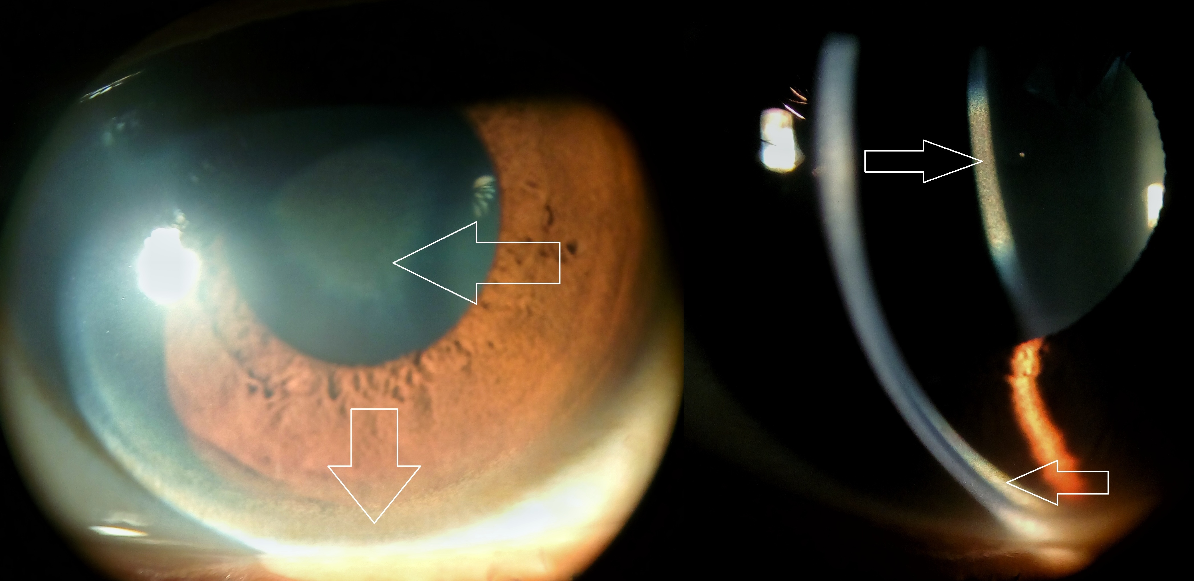
Look at the clinical photograph: identify the peripheral corneal band separately from the central lens opacity. The peripheral deposit is a Kayser-Fleischer ring in Descemet membrane. The sunflower cataract is a lens finding, not the ring itself. The photograph contains the original photographer's arrows; no additional markings have been applied. [1][8]
Locate the peripheral Kayser-Fleischer ring in the cornea, then distinguish it from the central sunflower-pattern cataract in the lens. Image: [1][8] Imrankabirhossain; original source; CC BY-SA 4.0.
Slit-lamp examination is more informative than an unaided inspection. Rings are frequent in neurologic Wilson disease but may be absent in a hepatic presentation, especially earlier in the course. Rare chronic cholestatic disorders can also produce similar corneal deposition, so even a ring belongs in an integrated assessment. [1]
Try the comparison: a 13-year-old has persistent unexplained hepatitis and no corneal ring. A second patient has a new motor syndrome plus a peripheral ring. Should either evaluation end at the eye examination?
Visible result: neither should. The child still needs copper studies because absence of a ring is not a reliable exclusion in hepatic disease. The second patient needs biochemical and genetic correlation because the eye finding is strong supporting evidence, not a complete diagnosis. [1][2]
Apply it elsewhere: add a falling hemoglobin, reticulocytosis and a negative direct antiglobulin test to the child's hepatitis. That combination raises concern for copper-associated nonimmune hemolysis and increases urgency. A negative antibody test alone does not prove copper injury; the full liver, blood and copper pattern matters. [1][3]
What fails when hepatic copper cannot leave normally?
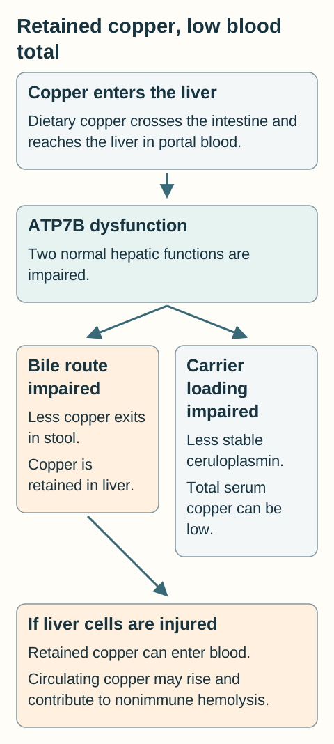
Follow copper, not bilirubin. Dietary copper crosses the intestine, travels through portal blood and enters the liver. ATP7B normally enables copper incorporation into newly made ceruloplasmin and delivery of excess copper into bile. Biliary loss into stool is the principal normal elimination route. Urinary copper becomes useful as a disease measurement or treatment output; it is not the dominant normal disposal route. [1]
The copper-routing diagram separates the two ATP7B-dependent functions. In Wilson disease, impaired biliary export favors hepatic retention. Impaired loading of apoceruloplasmin produces less stable circulating copper-carrying protein. Because much of total serum copper is normally carried by ceruloplasmin, total serum copper can be low despite excess copper in tissues. During severe hepatocyte injury, copper released into the circulation can instead make total serum copper high. Do not equate either single blood concentration with the entire body burden. [1][3]
Use the two hepatic functions to explain why retained tissue copper can coexist with low total serum copper. The arrows depict qualitative routes, not measured flow rates. [1][1]
Predict the direction before reading the result
Use this copper-routing exercise. Keep intestinal input unchanged. Compare intact ATP7B function with impaired biliary export. Then predict what changes when zinc reduces intestinal uptake or a chelator increases urinary copper excretion. These are qualitative directions, not a dosing model or a simulation of a patient's copper balance. [1][6]
Copper-routing experiment: the intervention determines which route changes
Condition
Direct route affected
Consequence to predict
ConditionATP7B dysfunction
Direct route affectedLess copper reaches bile; less enters stable ceruloplasmin
Consequence to predictHepatic retention can coexist with low total serum copper
ConditionZinc treatment
Direct route affectedMore copper remains bound inside intestinal cells
Consequence to predictLess enters portal blood; copper exits with shed cells in stool
ConditionCopper chelator
Direct route affectedDrug binds copper for urinary excretion
Consequence to predictUrinary copper can initially rise while copper is being cleared
Check the prediction: does a higher urine result after starting chelation prove deterioration?
No. The drug deliberately increases urinary copper excretion. Compare treatment phase, actual doses taken, clinical state, liver tests and subsequent measurements before interpreting the number. A diagnostic untreated threshold is not a treatment target. [1][5]
Transfer task: explain a pretreatment patient with low total serum copper, low ceruloplasmin and high urinary copper. The circulating carrier pool is reduced while abnormal copper handling permits tissue injury and increased urinary loss. The laboratory results describe different compartments, so they need not rise and fall together. [1]
What exactly did the laboratory measure?
A copper result has four parts: specimen, units, collection quality and treatment status. A basal 24-hour urine collection is obtained before copper-directed treatment when feasible. Use a trace-element collection container and the laboratory's instructions; missed voids can make an apparently reassuring result misleading. Urine creatinine and the collection history help assess completeness. Do not substitute a random concentration for the total copper excreted over 24 hours. [1]
Diagnostic measurements: useful reference points, not independent verdicts
Measurement
Interpretation
Important limit
MeasurementSerum ceruloplasmin
InterpretationOften low; a very low value increases suspicion
Important limitNormal values do not exclude disease; low values have other causes
MeasurementBasal urinary copper
InterpretationOften above 100 micrograms/24 h in symptomatic disease; above 40 warrants attention in children or asymptomatic people
Important limitOverlap with other liver disease; collection and treatment status matter
MeasurementHepatic copper, dry weight
InterpretationAbove 250 micrograms/g strongly supports excess hepatic copper; normal is usually below 50
Important limitCholestasis can raise it; an inadequate or patchy specimen can underrepresent burden
Use the laboratory's own reference interval alongside these guideline-based decision points. A value of 70 micrograms/24 h in an untreated child is not reassuring merely because it is below 100. Conversely, a value above 100 does not uniquely identify Wilson disease, because inflammatory and cholestatic liver disorders can overlap. [1]
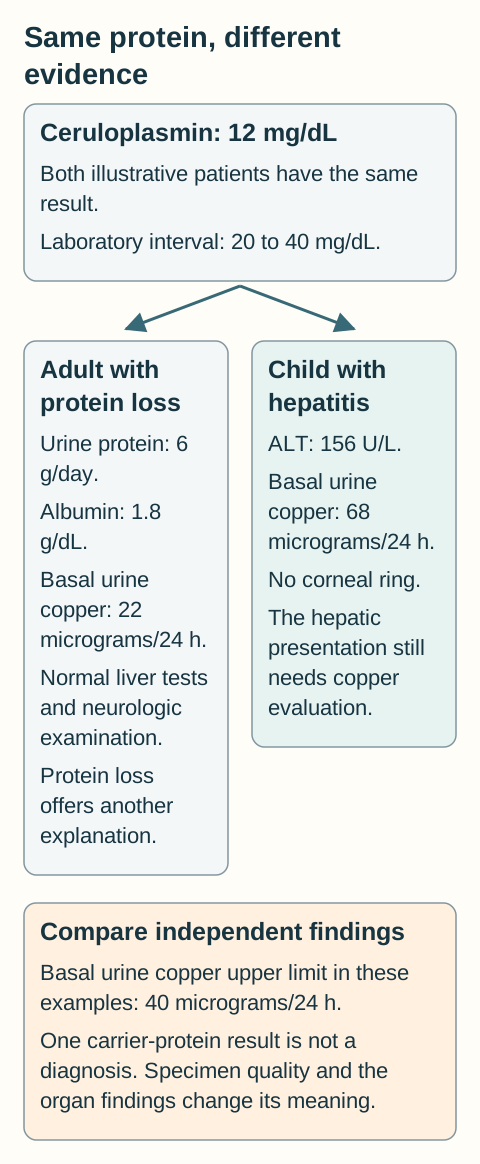
The evidence-comparison diagram contrasts two patients with the same low ceruloplasmin but different accompanying findings. Protein loss, malnutrition, infancy, severe hepatic synthetic failure and heterozygosity can lower ceruloplasmin without Wilson disease. Inflammation or estrogen exposure can raise it, including in an affected patient. Immunologic concentration assays also do not directly measure functional copper incorporation. [1]
Compare independent findings before interpreting a low carrier-protein concentration. Values are constructed teaching examples, not patient records. [1][1]
Test the sampling claim: a patient with persistent unexplained liver disease has hepatic copper of 180 micrograms/g from a very small specimen. Is the result diagnostic, normal, or unresolved?
Visible result: unresolved. It is above the usual normal range but below the commonly used strong-support threshold. Review adequacy, cholestasis and the other tests rather than declaring either confirmation or exclusion. A prospective study supports using an adequate core and interpreting copper in the correct disease context. [1][4]
New application: before comparing a later urine result with the first, ask whether a chelator was started between collections. A change in the route of excretion can explain the difference without a matching change in total tissue burden. [1]
How do discordant tests change the next action?
Build the diagnosis from the clinical phenotype, liver tests, blood count, ceruloplasmin, basal urinary copper and expert eye examination. Add ATP7B testing and, when needed, hepatic copper measurement. Brain MRI is appropriate for neurologic abnormalities, but it does not replace biochemical assessment. A Leipzig score organizes several findings; a score of at least 4 supports a highly likely diagnosis. Its components still require valid specimens and attention to alternative explanations. [1][2]
Work through a discordance: a 24-year-old with dystonia and unexplained hepatitis has a ceruloplasmin concentration within the laboratory interval during an inflammatory illness. Do not discard the phenotype. Complete urinary copper testing and the eye examination, interpret the current result in its acute-phase context, and pursue genetic or tissue evaluation if the integrated picture remains uncertain. Repeating only the same carrier-protein test leaves the principal question unanswered. [1]
Two pathogenic or likely pathogenic variants must affect the two separate ATP7B copies to establish the recessive molecular explanation. A variant of uncertain significance is not equivalent to a pathogenic variant. Two variants on the same parental copy do not demonstrate disease on both copies. When a family has known pathogenic variants, targeted testing can resolve an asymptomatic relative's status more directly than waiting for organ injury. First-degree relatives need appropriate evaluation; a normal examination does not exclude presymptomatic disease. [1]
Relative exchangeable copper is an additional diagnostic tool recommended in the 2025 EASL-ERN guideline. It expresses measured exchangeable copper as a percentage of total copper. It is not the same as subtracting estimated ceruloplasmin-bound copper from total serum copper. Analytical methods and availability matter, so use an experienced laboratory rather than applying an unvalidated universal cutoff. Small diagnostic studies showing excellent discrimination do not make any test infallible in every setting. [2][7]
Choose the next question: an asymptomatic sibling has both known familial pathogenic variants, confirmed on opposite copies, but no current biochemical organ injury. Is the remaining question whether to wait for symptoms or how to prevent injury?
Visible result: prevention. Confirm the full baseline assessment and start specialist-directed copper treatment; presymptomatic disease still needs therapy. A sibling carrying only one familial pathogenic variant is a different genetic category and should not automatically receive the affected sibling's prescription. [1]
Transfer task: if a person instead has only a low ceruloplasmin during substantial urinary protein loss, investigate that explanation and seek independent copper evidence. Recessive disease is not established by one nonspecific biochemical result. [1]
Which tissue explains the extrahepatic finding?
The same retained metal can affect different structures, but the clinical consequence depends on location. A peripheral corneal ring reflects deposition in Descemet membrane. A sunflower lens opacity is anatomically separate. Tremor, dystonia, dysarthria and parkinsonism implicate motor networks, often including the basal ganglia. MRI may show abnormalities in basal ganglia, thalami, brainstem or white matter; neither a dramatic named MRI sign nor its absence settles the diagnosis. [1][8]
Localize before labeling: urine glucose with a normal plasma glucose, aminoaciduria and phosphate loss suggest proximal tubular dysfunction rather than diabetes mellitus. Copper-related renal disease can include these tubular losses and nephrolithiasis. Later, after penicillamine starts, new heavy albuminuria deserves a different assessment: drug-associated glomerular injury is not the same compartment as proximal tubular solute loss. [1][5]
Hepatocyte injury can release copper that contributes to oxidative red-cell damage. Reticulocytosis, increased lactate dehydrogenase, reduced haptoglobin and a negative direct antiglobulin test support a nonimmune hemolytic pattern when interpreted together. No single one of these proves the mechanism: hepatic failure itself can lower haptoglobin, and a negative antiglobulin result alone cannot fully exclude immune hemolysis. Combine the blood findings with the liver presentation and copper evaluation. [1][3]
Compare two compartments: patient A has glucose and amino acids in urine before treatment. Patient B develops 4 g/day of urinary protein with edema months after penicillamine begins. Which one points toward tubular dysfunction and which toward a glomerular drug complication?
Visible result: A fits proximal tubular solute loss. B requires prompt assessment for penicillamine-associated glomerulopathy and a safe alternative copper-control plan. The drug exposure and protein pattern prevent attributing every kidney finding to untreated copper toxicity. [1][5]
New application: behavioral change plus progressive dysarthria should prompt an examination for other neurologic and hepatic signs. Do not assign all symptoms to a primary psychiatric diagnosis without evaluating the acquired multisystem pattern. Swallowing difficulty also creates a practical aspiration and nutrition concern even while the diagnostic workup proceeds. [1]
Does this patient need copper control or emergency liver support?
First separate acute liver failure from stable disease. New hepatic encephalopathy together with significant coagulopathy in an acute liver presentation requires urgent hospital and transplant-center assessment. Encephalopathy by itself does not define liver failure. In Wilson disease, marked jaundice, nonimmune hemolysis, relatively modest aminotransferase increases, low alkaline phosphatase and worsening kidney function are an especially concerning combination. Evaluation and supportive treatment proceed in parallel; genetic results must not delay referral. [1][2][3]
An alkaline phosphatase to total bilirubin ratio below 4, using alkaline phosphatase in U/L and total bilirubin in mg/dL, and an AST to ALT ratio above 2.2 can support suspicion in acute liver failure. For example, 48 U/L divided by 24 mg/dL gives 2. These ratios came from a limited cohort and are not standalone confirmation or exclusion rules. Changing bilirubin units without conversion invalidates the comparison. [3]
Wilson-associated acute liver failure has a high risk of death without transplantation. Transplant specialists assess trajectory and prognostic measures, such as the revised Wilson prognostic index, rather than waiting for one diagnostic assay. Plasma exchange or other extracorporeal support can reduce circulating copper and hemolysis while a definitive plan proceeds. Improvement after exchange is not proof that the underlying hepatic defect has resolved. [1][2]
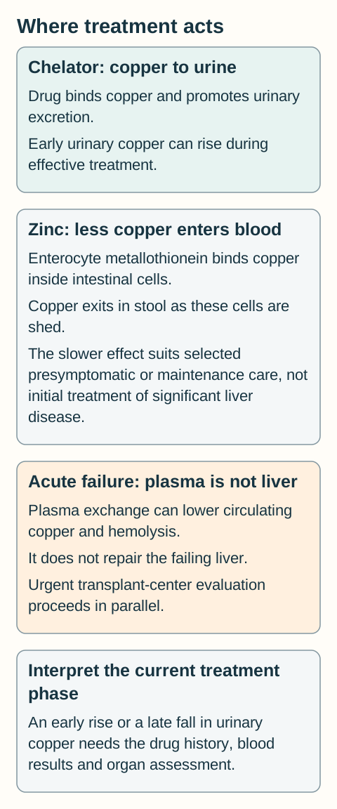
For significant symptomatic hepatic disease outside that emergency, trientine or D-penicillamine provides active copper chelation. Zinc increases enterocyte metallothionein, which retains copper in intestinal cells until they are shed into stool. Its slower absorption-blocking action makes it an option for selected presymptomatic patients or maintenance, not a substitute for initial chelation in significant liver disease. The treatment-routes diagram shows why these therapies affect different measurements. Product indications, formulation and dosing require an individualized prescription. [1][2][5][6]
Predict which copper measurement changes when a treatment acts. These are parallel mechanisms rather than a required treatment sequence; acute liver failure needs urgent transplant-center assessment. [1][2][5][6][1][2][5][6]
Follow clinical and neurologic status, liver tests, blood count, renal function, urinalysis and copper measurements. Penicillamine can cause cytopenias or glomerular injury; early neurologic deterioration can occur with initiation of copper treatment. Reassess promptly rather than simply increasing a dose. Stable follow-up is usually at least twice yearly and more frequent when starting, changing or troubleshooting therapy. Food or mineral interactions, affordability and missed doses can undermine an otherwise appropriate plan. [1][5][6]
Interpret a low urine result: a patient taking a chelator has very low urinary copper, low serum copper and new anemia with neutropenia. A second patient has low urinary copper after stopping the chelator, with worsening liver tests and increasing serum copper. Should both receive a larger dose?
Visible result: no. The first pattern suggests copper depletion from overtreatment and needs supervised reassessment, often dose reduction or a brief monitored interruption. The second suggests loss of effective treatment; address the interruption and reassess severity. A low urine number alone cannot choose between them. [1]
Apply the long-term rule: anti-copper medication generally continues for life, including an individualized plan during pregnancy. Do not abruptly stop because symptoms improve or pregnancy begins. Pregnancy care may require a lower chelator dose and closer monitoring, while balancing fetal and maternal risks. After successful liver transplantation, the graft corrects hepatic copper handling and anti-copper medication is generally no longer required, but transplant medications and follow-up continue. [1][5]
Dietary advice reduces avoidable copper exposure, especially from very high-copper foods and supplements, while preserving adequate nutrition. It does not replace medication. A practical plan must also address swallowing safety, mental health, rehabilitation and family screening. The best laboratory target is not useful if the patient cannot safely and consistently carry out the treatment plan. [1]
Apply the copper model to new patients
Commit to one answer using the findings that distinguish it from the closest alternative. Then compare the option-specific reasoning with your explanation.
Case 1
Show answer and explanations for case 1
A. Reduced biliary export with increased ceruloplasmin loading (Why this does not fit)
It can explain hepatic copper retention. No. The same transporter is needed for copper incorporation into ceruloplasmin, so loading is impaired too. [1][2]
Reasoning steps for option A
What does reduced bile export explain?
It can explain hepatic copper retention.
Does increased loading fit ATP7B dysfunction?
No. The same transporter is needed for copper incorporation into ceruloplasmin, so loading is impaired too.
B. Increased biliary export with reduced ceruloplasmin loading (Why this does not fit)
It can lower stable circulating ceruloplasmin. No. Enhanced disposal would oppose the hepatic accumulation supported by the organ pattern. [1][2]
Reasoning steps for option B
What can impaired carrier loading explain?
It can lower stable circulating ceruloplasmin.
Would increased bile export explain progressive retention?
No. Enhanced disposal would oppose the hepatic accumulation supported by the organ pattern.
C. Reduced biliary export with reduced ceruloplasmin loading (Best answer)
Abnormal copper retention and distribution link these compartments. Excess copper delivery into bile and copper loading onto ceruloplasmin both require ATP7B function. [1][2]
Reasoning steps for option C
Which process links the cornea, liver and motor findings?
Abnormal copper retention and distribution link these compartments.
Which two hepatic functions require ATP7B?
Excess copper delivery into bile and copper loading onto ceruloplasmin both require ATP7B function.
D. Increased biliary export with increased ceruloplasmin loading (Why this does not fit)
They would favor copper disposal and stable carrier production. No. The affected hepatic transporter has impaired, not increased, function. [1][2]
Reasoning steps for option D
What would these two increases tend to support?
They would favor copper disposal and stable carrier production.
Do they match this retention phenotype?
No. The affected hepatic transporter has impaired, not increased, function.
A. Reduced carrier-bound copper with abnormal tissue copper retention (Best answer)
The ceruloplasmin-bound component of total serum copper can fall. No. Those independent findings support abnormal copper handling despite a low total serum concentration. [1]
Reasoning steps for option A
Which circulating pool falls when ceruloplasmin is low?
The ceruloplasmin-bound component of total serum copper can fall.
Does that negate the high urine copper and hepatitis?
No. Those independent findings support abnormal copper handling despite a low total serum concentration.
B. Whole-body copper depletion demonstrated by the serum concentration (Why this does not fit)
A low serum result can occur in copper deficiency. Untreated hepatitis and markedly increased basal urinary copper require a different integrated explanation. [1]
Reasoning steps for option B
Why is depletion a tempting interpretation?
A low serum result can occur in copper deficiency.
Which findings resist that conclusion here?
Untreated hepatitis and markedly increased basal urinary copper require a different integrated explanation.
C. Intact biliary disposal demonstrated by the increased urine copper (Why this does not fit)
It measures renal copper excretion over 24 hours. No. Increased renal loss can accompany defective hepatic disposal rather than demonstrate normal bile export. [1]
Reasoning steps for option C
Which route does the urine collection measure?
It measures renal copper excretion over 24 hours.
Does it directly assess the biliary route?
No. Increased renal loss can accompany defective hepatic disposal rather than demonstrate normal bile export.
D. An invalid urine assay caused by the low serum ceruloplasmin (Why this does not fit)
Collection completeness, contamination control and assay performance determine validity. No. These are complementary measurements of different copper compartments. [1]
Reasoning steps for option D
What determines validity of a timed urine collection?
Collection completeness, contamination control and assay performance determine validity.
Does low blood ceruloplasmin invalidate a complete urine sample?
No. These are complementary measurements of different copper compartments.
Takeaway: Low total serum copper does not exclude copper excess when the carrier pool is reduced.
A. New drug-associated renal injury demonstrated by the urine change (Why this does not fit)
Solute wasting or new renal abnormalities would support tubular injury. No. The medication is intended to increase urinary copper, and no renal injury findings are provided. [1][5]
Reasoning steps for option A
What findings would support tubular injury?
Solute wasting or new renal abnormalities would support tubular injury.
Does copper output alone establish that injury?
No. The medication is intended to increase urinary copper, and no renal injury findings are provided.
B. Worsening hepatic copper retention demonstrated by the urine change (Why this does not fit)
Persistently high output can accompany ineffective treatment in some maintenance settings. No. Initial chelation intentionally changes the excretion route, while the supplied organ findings are stable. [1][5]
Reasoning steps for option B
Why might a high urine value prompt concern later?
Persistently high output can accompany ineffective treatment in some maintenance settings.
Can the same interpretation be imposed during the first week?
No. Initial chelation intentionally changes the excretion route, while the supplied organ findings are stable.
C. Completed tissue decoppering demonstrated by the urine change (Why this does not fit)
Copper is being excreted during treatment. No. Tissue burden and clinical control cannot be inferred from one early collection. [1][5]
Reasoning steps for option C
What does increased output demonstrate?
Copper is being excreted during treatment.
Does a single output measurement prove tissue stores are corrected?
No. Tissue burden and clinical control cannot be inferred from one early collection.
D. An expected early pharmacologic increase in urinary copper (Best answer)
It binds copper and increases urinary excretion. They give no evidence of current clinical deterioration; continue supervised monitoring instead of diagnosing failure from this number alone. [1][5]
Reasoning steps for option D
How does a chelator change copper disposal?
It binds copper and increases urinary excretion.
What do the unchanged organ findings add?
They give no evidence of current clinical deterioration; continue supervised monitoring instead of diagnosing failure from this number alone.
Takeaway: Initial chelation can raise urinary copper while promoting net copper clearance.
A. Repeat ceruloplasmin after recovery before obtaining any other copper tests (Why this does not fit)
It can show whether the acute-phase context altered ceruloplasmin. Not by itself. Complementary studies should not be postponed solely to repeat the same nonspecific protein. [1][2]
Reasoning steps for option A
What can repeat testing clarify?
It can show whether the acute-phase context altered ceruloplasmin.
Would it resolve the present independent neurologic and hepatic concern?
Not by itself. Complementary studies should not be postponed solely to repeat the same nonspecific protein.
B. Obtain basal urinary copper and an expert slit-lamp examination (Best answer)
No. Inflammation and estrogen exposure can raise the measured concentration. Urinary copper and expert ocular examination assess independent aspects of copper handling, with genetics or tissue studies as indicated. [1][2]
Reasoning steps for option B
Can a normal ceruloplasmin exclude Wilson disease here?
No. Inflammation and estrogen exposure can raise the measured concentration.
What addresses the residual clinical suspicion?
Urinary copper and expert ocular examination assess independent aspects of copper handling, with genetics or tissue studies as indicated.
C. Classify the motor syndrome as primary after the normal protein result (Why this does not fit)
Dystonia and dysarthria can have nonhepatic causes. The unexplained liver abnormalities and confounded carrier measurement leave a treatable multisystem disorder unresolved. [1][2]
Reasoning steps for option C
Why might a primary motor disorder enter the differential?
Dystonia and dysarthria can have nonhepatic causes.
What prevents using this result to settle the differential?
The unexplained liver abnormalities and confounded carrier measurement leave a treatable multisystem disorder unresolved.
D. Measure serum ferritin as the sole next test for metal accumulation (Why this does not fit)
It contributes to assessment of iron status and possible iron overload. No. The phenotype still warrants complementary copper studies; ferritin cannot substitute for them. [1][2]
Reasoning steps for option D
What disorder can ferritin help evaluate?
It contributes to assessment of iron status and possible iron overload.
Does it answer the specific unresolved copper question?
No. The phenotype still warrants complementary copper studies; ferritin cannot substitute for them.
Takeaway: Normal ceruloplasmin during inflammation or estrogen exposure does not end a compatible Wilson evaluation.
A. Normal ATP7B function inferred from normal basal copper excretion (Why this does not fit)
It provides no biochemical evidence of increased basal copper excretion in this sample. No. A urine test does not establish the sequence or function of both gene copies. [1]
Reasoning steps for option A
What does this urine result contribute?
It provides no biochemical evidence of increased basal copper excretion in this sample.
Can it determine the genotype?
No. A urine test does not establish the sequence or function of both gene copies.
B. A specific Wilson marker inferred from the two low circulating proteins (Why this does not fit)
It documents a substantial protein abnormality. No. Shared protein loss creates another explanation for the low concentration. [1]
Reasoning steps for option B
Why might low albumin matter?
It documents a substantial protein abnormality.
Does that increase ceruloplasmin specificity for Wilson disease?
No. Shared protein loss creates another explanation for the low concentration.
C. Protein loss lowering the circulating ceruloplasmin concentration (Best answer)
Nephrotic-range proteinuria with hypoalbuminemia identifies a strong alternative mechanism. Normal liver and neurologic assessments with normal basal urine copper do not independently support that diagnosis. [1]
Reasoning steps for option C
Which findings identify major protein loss?
Nephrotic-range proteinuria with hypoalbuminemia identifies a strong alternative mechanism.
How do the other findings affect a Wilson conclusion?
Normal liver and neurologic assessments with normal basal urine copper do not independently support that diagnosis.
D. Absent renal copper excretion until neurologic disease becomes evident (Why this does not fit)
It can be abnormal in hepatic or presymptomatic disease, before neurologic manifestations. No. The result is useful in context, but it neither specifies the genotype nor establishes a diagnosis. [1]
Reasoning steps for option D
When can urinary copper be abnormal in Wilson disease?
It can be abnormal in hepatic or presymptomatic disease, before neurologic manifestations.
Does the absence of neurologic disease invalidate this collection?
No. The result is useful in context, but it neither specifies the genotype nor establishes a diagnosis.
Takeaway: Protein loss can lower ceruloplasmin without demonstrating an ATP7B disorder.
A. An abnormal pediatric screen requiring further copper assessment (Best answer)
It is above 40 micrograms/24 h and warrants attention in an untreated child. No. Rings may be absent in hepatic childhood disease; further biochemical and genetic correlation is appropriate. [1][2]
Reasoning steps for option A
How does 68 compare with the supplied upper limit?
It is above 40 micrograms/24 h and warrants attention in an untreated child.
Do absent rings override hepatitis and the copper results?
No. Rings may be absent in hepatic childhood disease; further biochemical and genetic correlation is appropriate.
B. A normal pediatric screen because urine copper remains below 100 (Why this does not fit)
It is a common reference point in symptomatic patients, not a universal exclusion cutoff. The collection is above the laboratory limit and the child has persistent hepatic abnormalities. [1][2]
Reasoning steps for option B
What is the usual role of the 100 threshold?
It is a common reference point in symptomatic patients, not a universal exclusion cutoff.
Why is using it to dismiss this child unsafe?
The collection is above the laboratory limit and the child has persistent hepatic abnormalities.
C. A definitive diagnosis from the two modest biochemical abnormalities (Why this does not fit)
Both ceruloplasmin and basal urine copper are abnormal. No. They overlap with other conditions and require an integrated assessment. [1][2]
Reasoning steps for option C
Why do these results increase suspicion?
Both ceruloplasmin and basal urine copper are abnormal.
Are these modest abnormalities uniquely diagnostic?
No. They overlap with other conditions and require an integrated assessment.
D. A negative hepatic copper assessment because corneal rings are absent (Why this does not fit)
They reveal peripheral corneal copper deposition when present. Yes. Absence of corneal deposition does not establish normal hepatic copper handling. [1][2]
Reasoning steps for option D
What do rings assess?
They reveal peripheral corneal copper deposition when present.
Can hepatic disease precede this eye finding?
Yes. Absence of corneal deposition does not establish normal hepatic copper handling.
Takeaway: Basal urine copper above 40 micrograms/24 h can matter in a child even without corneal rings.
A. Multiply the reported result by the number of omitted voids (Why this does not fit)
It would require knowing copper content and volume in the missing urine. No. Arbitrary multiplication cannot reconstruct the actual 24-hour excretion. [1]
Reasoning steps for option A
What would a numerical correction require?
It would require knowing copper content and volume in the missing urine.
Are those missing quantities supplied?
No. Arbitrary multiplication cannot reconstruct the actual 24-hour excretion.
B. Repeat a complete timed collection using trace-element instructions (Best answer)
The sample excludes known portions of the 24-hour output. A properly collected repeat sample permits comparison with a basal excretion threshold while the broader evaluation continues. [1]
Reasoning steps for option B
Which defect compromises the result?
The sample excludes known portions of the 24-hour output.
What directly resolves that defect?
A properly collected repeat sample permits comparison with a basal excretion threshold while the broader evaluation continues.
C. Obtain a random urine concentration to replace the timed excretion (Why this does not fit)
Copper per unit urine volume at a particular time. No. Dilution and timing prevent direct substitution for the timed excretion used here. [1]
Reasoning steps for option C
What does a random concentration measure?
Copper per unit urine volume at a particular time.
Is that interchangeable with total copper over 24 hours?
No. Dilution and timing prevent direct substitution for the timed excretion used here.
D. Use the submitted result to discontinue copper-focused investigation (Why this does not fit)
It is below the laboratory upper limit for a complete basal collection. No. Known missing voids prevent applying that reassurance to this sample. [1]
Reasoning steps for option D
Why might 19 initially appear reassuring?
It is below the laboratory upper limit for a complete basal collection.
Was the required complete collection actually obtained?
No. Known missing voids prevent applying that reassurance to this sample.
Takeaway: A known incomplete urine collection cannot reliably exclude increased 24-hour copper excretion.
A. Primary genetic copper overload established by tissue concentration (Why this does not fit)
It supports increased hepatic copper content. No. Chronic cholestasis can also retain copper, and this patient has direct biliary evidence. [1][4]
Reasoning steps for option A
What does a value above 250 strongly support?
It supports increased hepatic copper content.
Does it uniquely establish a recessive transporter defect?
No. Chronic cholestasis can also retain copper, and this patient has direct biliary evidence.
B. Absence of copper-related hepatic disease established by ceruloplasmin (Why this does not fit)
Ceruloplasmin is often low in Wilson disease. No. It neither excludes Wilson disease nor erases the documented hepatic copper accumulation. [1][4]
Reasoning steps for option B
Why might a normal result appear reassuring?
Ceruloplasmin is often low in Wilson disease.
Can normal ceruloplasmin settle this tissue abnormality?
No. It neither excludes Wilson disease nor erases the documented hepatic copper accumulation.
C. A separate copper disease without relation to the biliary abnormality (Why this does not fit)
A single disorder sometimes explains several abnormal tests. Yes. Copper normally leaves through bile, so the biliary findings can explain the tissue result. [1][4]
Reasoning steps for option C
Why can a unifying diagnosis be appealing?
A single disorder sometimes explains several abnormal tests.
Is there a direct link between bile obstruction and copper retention?
Yes. Copper normally leaves through bile, so the biliary findings can explain the tissue result.
D. Secondary retention in the setting of chronic cholestasis (Best answer)
Biliary copper disposal can be reduced. It requires correlation with the biliary disorder and other copper or genetic findings rather than automatic Wilson confirmation. [1][4]
Reasoning steps for option D
Which normal elimination route is impaired in cholestasis?
Biliary copper disposal can be reduced.
How does that change interpretation of 310 micrograms/g?
It requires correlation with the biliary disorder and other copper or genetic findings rather than automatic Wilson confirmation.
Takeaway: High hepatic copper is not specific for Wilson disease when chronic cholestasis is present.
A. Intermediate copper with sampling uncertainty requiring integrated assessment (Best answer)
It is above normal but below the conventional strong-support threshold of 250 micrograms/g dry weight. Patchy distribution and limited tissue can misrepresent hepatic burden, so the complementary abnormalities remain relevant. [1][4]
Reasoning steps for option A
Where does 180 lie relative to the usual tissue reference points?
It is above normal but below the conventional strong-support threshold of 250 micrograms/g dry weight.
Why does the small specimen matter?
Patchy distribution and limited tissue can misrepresent hepatic burden, so the complementary abnormalities remain relevant.
B. A normal tissue result that outweighs the serum and urine abnormalities (Why this does not fit)
Normal hepatic copper is generally below 50 micrograms/g dry weight. No. Treating it as normal ignores both the numerical result and the specimen limitation. [1][4]
Reasoning steps for option B
What is the usual normal hepatic copper range?
Normal hepatic copper is generally below 50 micrograms/g dry weight.
Is 180 within that range?
No. Treating it as normal ignores both the numerical result and the specimen limitation.
C. Definitive Wilson confirmation independent of the specimen limitation (Why this does not fit)
The patient has hepatic disease and multiple abnormal copper-related measurements. No. The result is intermediate, and its limited sampling adds uncertainty rather than definitive specificity. [1][4]
Reasoning steps for option C
Why might the combined findings increase suspicion?
The patient has hepatic disease and multiple abnormal copper-related measurements.
Does the tissue result alone settle the diagnosis?
No. The result is intermediate, and its limited sampling adds uncertainty rather than definitive specificity.
D. A contaminated urine sample demonstrated by the tissue disagreement (Why this does not fit)
Contamination or collection problems can invalidate a urine sample. No. The provided limitation concerns the tissue specimen, not documented urine contamination. [1][4]
Reasoning steps for option D
What could invalidate urine copper?
Contamination or collection problems can invalidate a urine sample.
Does an intermediate small-biopsy result demonstrate either problem?
No. The provided limitation concerns the tissue specimen, not documented urine contamination.
Takeaway: Intermediate hepatic copper from limited tissue requires correlation, not automatic confirmation or exclusion.
A. 4%, representing the exchangeable fraction of circulating copper (Why this does not fit)
The fraction must be exchangeable copper divided by total copper, multiplied by 100. No. It yields 0.25, which is 25%. [2][7]
Reasoning steps for option A
What calculation gives the stated percentage?
The fraction must be exchangeable copper divided by total copper, multiplied by 100.
Does 2 divided by 8 yield 4%?
No. It yields 0.25, which is 25%.
B. 25%, representing the fraction of all body copper in serum (Why this does not fit)
The calculation gives 25%. No. The denominator is serum copper, not all copper in the body. [2][7]
Reasoning steps for option B
What numerical result follows from the supplied formula?
The calculation gives 25%.
Does total serum copper include all tissue stores?
No. The denominator is serum copper, not all copper in the body.
C. 25%, representing the exchangeable fraction of circulating copper (Best answer)
It is 25%. It describes the measured exchangeable fraction within serum copper and requires assay-specific clinical interpretation. [2][7]
Reasoning steps for option C
What is 100 times 2.0 divided by 8.0?
It is 25%.
What does that percentage describe?
It describes the measured exchangeable fraction within serum copper and requires assay-specific clinical interpretation.
D. 400%, representing the fraction of all body copper in serum (Why this does not fit)
Reversing the ratio gives 100 times 8 divided by 2. It reverses the defined numerator and denominator and incorrectly substitutes whole-body copper for serum copper. [2][7]
Reasoning steps for option D
What produces 400% from these two values?
Reversing the ratio gives 100 times 8 divided by 2.
Why is that interpretation doubly incorrect?
It reverses the defined numerator and denominator and incorrectly substitutes whole-body copper for serum copper.
Takeaway: REC is a measured serum fraction, not a calculated total-body copper burden.
A. Routine examination alone until a hepatic or neurologic symptom appears (Why this does not fit)
It suggests that overt clinical injury has not yet developed. No. Waiting would allow avoidable accumulation before symptoms identify damage. [1]
Reasoning steps for option A
Why does the normal examination initially appear reassuring?
It suggests that overt clinical injury has not yet developed.
Does it correct the inherited copper-handling defect?
No. Waiting would allow avoidable accumulation before symptoms identify damage.
B. Carrier counseling without treatment because liver tests are normal (Why this does not fit)
One pathogenic copy with no pathogenic variant on the other copy would support carrier status. No. The two familial pathogenic variants are on opposite copies. [1]
Reasoning steps for option B
What genotype would support a simple carrier interpretation?
One pathogenic copy with no pathogenic variant on the other copy would support carrier status.
Is that the genotype in this child?
No. The two familial pathogenic variants are on opposite copies.
C. Specialist-directed anti-copper therapy with baseline and ongoing monitoring (Best answer)
They support the molecular diagnosis of Wilson disease. Copper-directed therapy can prevent progression while the child is presymptomatic. [1]
Reasoning steps for option C
What do pathogenic variants on opposite copies establish?
They support the molecular diagnosis of Wilson disease.
Why begin care before organ injury appears?
Copper-directed therapy can prevent progression while the child is presymptomatic.
D. Dietary copper restriction alone with medication reserved for jaundice (Why this does not fit)
They can reduce avoidable copper exposure. No. Dietary advice complements, rather than replaces, the prescribed anti-copper plan. [1]
Reasoning steps for option D
What can dietary changes contribute?
They can reduce avoidable copper exposure.
Are they adequate sole therapy for established disease?
No. Dietary advice complements, rather than replaces, the prescribed anti-copper plan.
Takeaway: Biallelic pathogenic disease requires treatment even before symptoms or structural injury.
A. Biallelic disease is established by the two pathogenic variant names (Why this does not fit)
Disease-causing variants must affect both copies. No. Counting names without phase information would misclassify the result. [1]
Reasoning steps for option A
What is required for the usual recessive molecular diagnosis?
Disease-causing variants must affect both copies.
Do two variants inherited on one copy meet that requirement?
No. Counting names without phase information would misclassify the result.
B. Biallelic disease is not established by the reported in-cis variants (Best answer)
Both reported variants affect the same paternal copy. The initial test has not demonstrated a pathogenic maternal-copy variant, so the clinical and genetic evaluation must be reconciled. [1]
Reasoning steps for option B
What does paternal cosegregation reveal?
Both reported variants affect the same paternal copy.
What remains unresolved in this symptomatic patient?
The initial test has not demonstrated a pathogenic maternal-copy variant, so the clinical and genetic evaluation must be reconciled.
C. The normal maternal report excludes a copper disorder despite the phenotype (Why this does not fit)
The initial assay has not identified the required second pathogenic copy. No. Limitations of the test and complementary biochemical evidence still require review. [1]
Reasoning steps for option C
Why might the maternal result lower molecular confidence?
The initial assay has not identified the required second pathogenic copy.
Does that assay result alone erase the hepatic presentation?
No. Limitations of the test and complementary biochemical evidence still require review.
D. The paternal result establishes dominant inheritance in this family (Why this does not fit)
It is autosomal recessive. No. Phase information changes interpretation of the genotype, not the disorder into a dominant trait. [1]
Reasoning steps for option D
What inheritance pattern is associated with ATP7B Wilson disease?
It is autosomal recessive.
Does finding two variants on one paternal copy change that inheritance pattern?
No. Phase information changes interpretation of the genotype, not the disorder into a dominant trait.
Takeaway: Two pathogenic variants on one chromosome do not establish the usual biallelic Wilson genotype.
A. 50% affected risk; normal aminotransferases exclude affected status (Why this does not fit)
It is the proportion expected to inherit one pathogenic copy, not both. No. A presymptomatic affected child can have a normal current assessment. [1]
Reasoning steps for option A
What does the 50% proportion represent in this mating?
It is the proportion expected to inherit one pathogenic copy, not both.
Do normal aminotransferases determine an untested child's genotype?
No. A presymptomatic affected child can have a normal current assessment.
B. 25% affected risk; a normal examination establishes carrier status (Why this does not fit)
Each parent transmits the pathogenic copy with probability one-half, giving one-quarter together. No. Familial testing and appropriate biochemical evaluation are needed. [1]
Reasoning steps for option B
What is the risk of inheriting both pathogenic copies?
Each parent transmits the pathogenic copy with probability one-half, giving one-quarter together.
Can a normal examination distinguish a carrier from an affected presymptomatic child?
No. Familial testing and appropriate biochemical evaluation are needed.
C. 75% affected risk; normal aminotransferases require no family testing (Why this does not fit)
It is the chance of inheriting at least one pathogenic copy. No. A single-copy carrier is distinct from an affected biallelic individual. [1]
Reasoning steps for option C
What does 75% represent in the simple carrier-parent model?
It is the chance of inheriting at least one pathogenic copy.
Is inheriting one copy equivalent to this recessive disease?
No. A single-copy carrier is distinct from an affected biallelic individual.
D. 25% affected risk; a normal examination does not determine genotype (Best answer)
One-half times one-half gives one-quarter, or 25%, for each pregnancy. The clinical examination may be normal before copper-related injury becomes apparent. [1]
Reasoning steps for option D
How do two independent one-half transmission probabilities combine?
One-half times one-half gives one-quarter, or 25%, for each pregnancy.
Why is family evaluation still relevant at age eight?
The clinical examination may be normal before copper-related injury becomes apparent.
Takeaway: For two carrier parents, each pregnancy has a 25% affected risk; a normal young sibling still needs evaluation.
A. Generalized proximal tubular reabsorption impairment (Best answer)
Glucose is being lost despite no excessive filtered glucose load. The proximal tubule reabsorbs all these solutes, so generalized dysfunction unifies the pattern. [1]
Reasoning steps for option A
What does glucosuria at normal plasma glucose suggest?
Glucose is being lost despite no excessive filtered glucose load.
Which site also reclaims amino acids, phosphate and bicarbonate?
The proximal tubule reabsorbs all these solutes, so generalized dysfunction unifies the pattern.
B. Selective distal tubular hydrogen secretion impairment (Why this does not fit)
It can produce metabolic acidosis. No. Those reabsorptive functions localize to the proximal tubule. [1]
Reasoning steps for option B
What can impaired distal hydrogen secretion cause?
It can produce metabolic acidosis.
Does it explain glucose and amino-acid wasting together?
No. Those reabsorptive functions localize to the proximal tubule.
C. Selective collecting-duct aldosterone resistance (Why this does not fit)
Impaired potassium and acid excretion can produce hyperkalemic acidosis. No. Glucose, amino acids and phosphate point to proximal reabsorptive dysfunction. [1]
Reasoning steps for option C
What abnormalities commonly accompany impaired aldosterone action?
Impaired potassium and acid excretion can produce hyperkalemic acidosis.
Does that account for the stated multi-solute urinary losses?
No. Glucose, amino acids and phosphate point to proximal reabsorptive dysfunction.
D. Glomerular albumin filtration barrier impairment (Why this does not fit)
Substantial albumin-predominant proteinuria would support that compartment. No. The supplied abnormalities concern tubular solute reclamation rather than heavy albumin loss. [1]
Reasoning steps for option D
What finding would most directly support a glomerular protein barrier lesion?
Substantial albumin-predominant proteinuria would support that compartment.
Is that the combination supplied?
No. The supplied abnormalities concern tubular solute reclamation rather than heavy albumin loss.
Takeaway: Normoglycemic glucosuria with aminoaciduria and phosphate loss localizes to proximal tubular dysfunction.
A. Impaired marrow production from isolated folate deficiency (Why this does not fit)
An inadequate reticulocyte response would support underproduction. It supports marrow compensation for peripheral red-cell loss rather than isolated production failure. [1][3]
Reasoning steps for option A
What pattern is expected when production is inadequate?
An inadequate reticulocyte response would support underproduction.
How does the 9% reticulocyte response change that interpretation?
It supports marrow compensation for peripheral red-cell loss rather than isolated production failure.
B. Oxidative erythrocyte injury associated with circulating copper (Best answer)
The hemoglobin fall, reticulocytosis and increased LDH support hemolysis together. Hepatocyte injury can release copper that contributes to oxidative red-cell damage without antibody-mediated destruction. [1][3]
Reasoning steps for option B
Which measurements support active red-cell destruction?
The hemoglobin fall, reticulocytosis and increased LDH support hemolysis together.
How does the liver and copper context explain the nonimmune pattern?
Hepatocyte injury can release copper that contributes to oxidative red-cell damage without antibody-mediated destruction.
C. Antibody-mediated erythrocyte destruction from a warm autoantibody (Why this does not fit)
It can cause anemia, reticulocytosis and increased LDH. The negative antiglobulin result plus established hepatic copper disease favors copper-associated nonimmune hemolysis, although no single test is absolute. [1][3]
Reasoning steps for option C
Why could warm immune hemolysis enter the differential?
It can cause anemia, reticulocytosis and increased LDH.
Which supplied findings favor the alternative mechanism?
The negative antiglobulin result plus established hepatic copper disease favors copper-associated nonimmune hemolysis, although no single test is absolute.
D. Reduced erythropoietin production from chronic renal disease (Why this does not fit)
It reduces red-cell production. No. The supplied pattern is acute peripheral destruction with a compensatory marrow response. [1][3]
Reasoning steps for option D
What does deficient erythropoietin primarily impair?
It reduces red-cell production.
Does that explain rapid hemoglobin loss with this reticulocytosis?
No. The supplied pattern is acute peripheral destruction with a compensatory marrow response.
Takeaway: Acute liver injury with supported nonimmune hemolysis can reflect copper-associated oxidative erythrocyte injury.
A. Acute autoimmune hepatitis (Why this does not fit)
Autoimmune hepatitis can cause acute liver failure in a young patient. Marked jaundice with low relative ALP, AST predominance and nonimmune hemolysis is a characteristic Wilson-associated pattern. [1][3]
Reasoning steps for option A
Why is this a reasonable competing cause?
Autoimmune hepatitis can cause acute liver failure in a young patient.
Which combined findings particularly favor the copper disorder instead?
Marked jaundice with low relative ALP, AST predominance and nonimmune hemolysis is a characteristic Wilson-associated pattern.
B. Acetaminophen-associated hepatic necrosis (Why this does not fit)
Acetaminophen toxicity is a major cause of acute liver failure and must be assessed. The comparatively modest aminotransferases and supported nonimmune hemolysis with low ALP relative to bilirubin favor another cause. [1][3]
Reasoning steps for option B
Why is this an important emergency differential?
Acetaminophen toxicity is a major cause of acute liver failure and must be assessed.
What makes it less fitting as the leading explanation here?
The comparatively modest aminotransferases and supported nonimmune hemolysis with low ALP relative to bilirubin favor another cause.
C. Acute hepatitis A infection (Why this does not fit)
Hepatitis A can produce jaundice and occasionally acute liver failure. The combination of copper-associated ratio patterns and nonimmune hemolysis raises Wilson disease above an isolated viral explanation. [1][3]
Reasoning steps for option C
Why can an acute viral cause be considered?
Hepatitis A can produce jaundice and occasionally acute liver failure.
Which additional pattern is less well explained?
The combination of copper-associated ratio patterns and nonimmune hemolysis raises Wilson disease above an isolated viral explanation.
D. Wilson-associated acute liver failure (Best answer)
ALP divided by bilirubin is 36/24 = 1.5; AST divided by ALT is 360/120 = 3. Supported nonimmune hemolysis strengthens suspicion for copper-related acute failure and warrants urgent transplant-center assessment while confirmation proceeds. [1][3]
Reasoning steps for option D
What are the two calculated ratios?
ALP divided by bilirubin is 36/24 = 1.5; AST divided by ALT is 360/120 = 3.
How does the anemia change the interpretation?
Supported nonimmune hemolysis strengthens suspicion for copper-related acute failure and warrants urgent transplant-center assessment while confirmation proceeds.
Takeaway: Unit-correct ratio patterns plus nonimmune hemolysis should urgently raise Wilson-associated acute liver failure.
A. Lower circulating copper with persistent hepatic functional failure (Best answer)
It can reduce the circulating copper burden and associated hemolysis. Hepatic functional failure remains, so the transplant assessment must continue despite the improved copper measurement. [1][2]
Reasoning steps for option A
Which compartment does exchange directly reduce?
It can reduce the circulating copper burden and associated hemolysis.
What do persistent encephalopathy and INR 4.1 indicate?
Hepatic functional failure remains, so the transplant assessment must continue despite the improved copper measurement.
B. Restored biliary copper export with delayed laboratory normalization (Why this does not fit)
Functional hepatic copper handling is required for sustained normal biliary disposal. No. It changes circulating constituents without replacing the damaged liver or its ATP7B defect. [1][2]
Reasoning steps for option B
What would restore normal ATP7B-dependent export?
Functional hepatic copper handling is required for sustained normal biliary disposal.
Does plasma exchange repair that intracellular function?
No. It changes circulating constituents without replacing the damaged liver or its ATP7B defect.
C. Corrected hepatic synthesis with persistent isolated neurologic injury (Why this does not fit)
Improving coagulation function would support recovery of synthetic capacity. No. It is evidence that the hepatic failure has not resolved. [1][2]
Reasoning steps for option C
What would improvement in hepatic synthesis tend to support?
Improving coagulation function would support recovery of synthetic capacity.
Does the persistent severe INR abnormality establish such recovery?
No. It is evidence that the hepatic failure has not resolved.
D. Completed tissue decoppering with an unrelated coagulation abnormality (Why this does not fit)
It describes circulating copper after an extracorporeal intervention. No. Neither claim follows, and ongoing encephalopathy and coagulopathy remain clinically central. [1][2]
Reasoning steps for option D
What can a post-exchange blood value describe?
It describes circulating copper after an extracorporeal intervention.
Can it establish that tissue stores are corrected or the INR is unrelated?
No. Neither claim follows, and ongoing encephalopathy and coagulopathy remain clinically central.
Takeaway: Plasma exchange can reduce circulating toxicity while hepatic failure still requires urgent transplant assessment.
A. Transjugular biopsy before requesting transplant-center acceptance (Why this does not fit)
It can provide histology and tissue copper when clinically indicated. No. It must not delay assessment of time-critical liver failure. [1][2][3]
Reasoning steps for option A
What can transjugular biopsy contribute in selected patients?
It can provide histology and tissue copper when clinically indicated.
Should tissue confirmation precede referral in this unstable syndrome?
No. It must not delay assessment of time-critical liver failure.
B. Urgent transplant-center consultation and transfer (Best answer)
Acute hepatic encephalopathy, severe coagulopathy and renal injury indicate high-risk organ failure. No. Stabilization, diagnostic evaluation and urgent transplant assessment should proceed in parallel. [1][2][3]
Reasoning steps for option B
What determines the immediate setting of care?
Acute hepatic encephalopathy, severe coagulopathy and renal injury indicate high-risk organ failure.
Does pending sequencing justify waiting?
No. Stabilization, diagnostic evaluation and urgent transplant assessment should proceed in parallel.
C. An inpatient chelator trial before requesting transplant consultation (Why this does not fit)
Copper-directed medication may form part of specialist management. No. Oral therapy may not reverse acute failure quickly enough, and the trajectory already warrants referral. [1][2][3]
Reasoning steps for option C
What role can medication have?
Copper-directed medication may form part of specialist management.
Is a trial response required before contacting a transplant center here?
No. Oral therapy may not reverse acute failure quickly enough, and the trajectory already warrants referral.
D. Expedited genetic processing before discussing transplant eligibility (Why this does not fit)
It can establish the molecular diagnosis and assist family evaluation. No. The clinical emergency already requires specialist assessment regardless of the laboratory turnaround time. [1][2][3]
Reasoning steps for option D
What can sequencing clarify?
It can establish the molecular diagnosis and assist family evaluation.
Does it measure or resolve the immediate functional failure?
No. The clinical emergency already requires specialist assessment regardless of the laboratory turnaround time.
Takeaway: Suspected Wilson-associated acute liver failure requires urgent transplant-center involvement without waiting for genetics.
A. Zinc acetate monotherapy (Why this does not fit)
It limits intestinal copper absorption by increasing enterocyte metallothionein. Its slower absorption blockade is not the recommended initial approach to significant hepatic disease requiring active chelation. [1][2][5][6]
Reasoning steps for option A
Where does zinc act first?
It limits intestinal copper absorption by increasing enterocyte metallothionein.
Why is it less suitable as the initial sole strategy here?
Its slower absorption blockade is not the recommended initial approach to significant hepatic disease requiring active chelation.
B. Trientine-based chelation (Best answer)
It binds copper and promotes urinary excretion. There is significant symptomatic hepatic disease with accumulated copper requiring active treatment. [1][2][5][6]
Reasoning steps for option B
What immediate copper-directed action does trientine provide?
It binds copper and promotes urinary excretion.
Why does that fit this patient better than absorption blockade alone?
There is significant symptomatic hepatic disease with accumulated copper requiring active treatment.
C. Ursodeoxycholic acid monotherapy (Why this does not fit)
It is used in selected cholestatic disorders. No. It does not correct the stated need to clear retained copper. [1][2][5][6]
Reasoning steps for option C
What type of liver problem can this drug address?
It is used in selected cholestatic disorders.
Does it replace copper chelation for this confirmed disorder?
No. It does not correct the stated need to clear retained copper.
D. Prednisone-based immunosuppression (Why this does not fit)
Autoimmune hepatitis can produce inflammatory liver injury. Wilson disease is confirmed, so the primary metabolic treatment is copper-directed rather than empiric immune suppression. [1][2][5][6]
Reasoning steps for option D
Why might immunosuppression be considered in another hepatic presentation?
Autoimmune hepatitis can produce inflammatory liver injury.
What changes the disease-directed choice here?
Wilson disease is confirmed, so the primary metabolic treatment is copper-directed rather than empiric immune suppression.
Takeaway: Initial treatment of significant hepatic Wilson disease should include an appropriate copper chelator.
A. Direct binding of plasma copper into renally excreted complexes (Why this does not fit)
Copper chelators bind copper for increased urinary excretion. No. Zinc primarily acts through metallothionein in enterocytes. [6]
Reasoning steps for option A
Which treatment mechanism primarily matches this description?
Copper chelators bind copper for increased urinary excretion.
Is this zinc's main absorption-blocking mechanism?
No. Zinc primarily acts through metallothionein in enterocytes.
B. Restoration of ATP7B-mediated canalicular copper transport (Why this does not fit)
It would correct the inherited hepatic disposal defect. No. Its benefit does not require repairing the hepatic gene or transporter. [6]
Reasoning steps for option B
Which defect would this directly correct?
It would correct the inherited hepatic disposal defect.
Does zinc replace functional ATP7B in hepatocytes?
No. Its benefit does not require repairing the hepatic gene or transporter.
C. Increased ceruloplasmin synthesis that sequesters intestinal copper (Why this does not fit)
The liver synthesizes the circulating carrier protein. No. Enterocyte metallothionein, not circulating ceruloplasmin, explains the direct intestinal action. [6]
Reasoning steps for option C
Which organ normally synthesizes ceruloplasmin?
The liver synthesizes the circulating carrier protein.
Does this explain copper retained inside intestinal cells?
No. Enterocyte metallothionein, not circulating ceruloplasmin, explains the direct intestinal action.
D. Increased enterocyte metallothionein retaining copper for fecal loss (Best answer)
It binds copper and limits transfer into portal blood. It leaves in stool as intestinal cells are shed. [6]
Reasoning steps for option D
What does metallothionein do in the intestinal cell?
It binds copper and limits transfer into portal blood.
How is the retained copper ultimately lost?
It leaves in stool as intestinal cells are shed.
Takeaway: Zinc limits copper absorption through enterocyte metallothionein and fecal loss.
A. Copper-associated generalized proximal tubular dysfunction (Why this does not fit)
Glucose, amino-acid, phosphate or bicarbonate wasting would favor that site. Heavy albumin-predominant loss with normal glucose and phosphate points toward a glomerular process. [1][5]
Reasoning steps for option A
What urinary pattern would favor a generalized proximal lesion?
Glucose, amino-acid, phosphate or bicarbonate wasting would favor that site.
Which supplied findings instead identify another compartment?
Heavy albumin-predominant loss with normal glucose and phosphate points toward a glomerular process.
B. Renal phosphate wasting from isolated tubular transporter dysfunction (Why this does not fit)
Hypophosphatemia with inappropriate urinary phosphate loss would support it. No. Serum phosphate is normal, and substantial albumin loss is the dominant lesion. [1][5]
Reasoning steps for option B
What abnormality would support an isolated phosphate transport problem?
Hypophosphatemia with inappropriate urinary phosphate loss would support it.
Does that explain the present edema and albuminuria?
No. Serum phosphate is normal, and substantial albumin loss is the dominant lesion.
C. Penicillamine-associated membranous glomerulopathy (Best answer)
Penicillamine can cause membranous glomerular injury and nephrotic syndrome. Proteinuria appeared after exposure, is albumin-predominant, and lacks the supplied tubular solute losses. [1][5]
Reasoning steps for option C
Which treatment is associated with new proteinuria and glomerulopathy?
Penicillamine can cause membranous glomerular injury and nephrotic syndrome.
What makes that concern more fitting than untreated tubular disease?
Proteinuria appeared after exposure, is albumin-predominant, and lacks the supplied tubular solute losses.
D. Copper-associated nephrolithiasis with postrenal obstruction (Why this does not fit)
Stones can occur in patients with Wilson disease. They do not best explain new nephrotic-range albuminuria after penicillamine exposure. [1][5]
Reasoning steps for option D
Why is nephrolithiasis in the wider differential?
Stones can occur in patients with Wilson disease.
Would stones explain this protein pattern without colic or obstruction?
They do not best explain new nephrotic-range albuminuria after penicillamine exposure.
Takeaway: New nephrotic-range albuminuria on penicillamine requires prompt assessment for a glomerular drug complication.
A. Increase chelator intensity to restore the previous urinary copper output (Why this does not fit)
Maintenance values help assess treatment over time. It could worsen depletion; urinary output is not a target to increase regardless of the clinical context. [1]
Reasoning steps for option A
Why might a prior urine value be a tempting target?
Maintenance values help assess treatment over time.
Would increasing chelation address these cytopenias and very low serum copper?
It could worsen depletion; urinary output is not a target to increase regardless of the clinical context.
B. Add zinc to suppress absorption while maintaining the current chelator dose (Why this does not fit)
It would further limit copper entry from the intestine. No. Very low copper measurements with cytopenias raise concern for excessive treatment. [1]
Reasoning steps for option B
What additional effect would zinc produce?
It would further limit copper entry from the intestine.
Does the current pattern suggest a need for more copper restriction?
No. Very low copper measurements with cytopenias raise concern for excessive treatment.
C. Substitute a higher-dose chelator because cytopenias indicate disease progression (Why this does not fit)
Yes. They can accompany copper depletion or treatment adverse effects. Both serum and urinary copper are very low while hepatic tests remain stable. [1]
Reasoning steps for option C
Can cytopenias occur for reasons other than progressive Wilson disease?
Yes. They can accompany copper depletion or treatment adverse effects.
What argues against treating these findings as uncontrolled copper accumulation?
Both serum and urinary copper are very low while hepatic tests remain stable.
D. Reassess copper depletion and reduce treatment with close specialist follow-up (Best answer)
New cytopenias accompany very low serum copper and urinary output despite verified treatment. Dose reduction or a brief monitored interruption, with reassessment, rather than permanent abandonment of copper care. [1]
Reasoning steps for option D
Which findings support possible overtreatment?
New cytopenias accompany very low serum copper and urinary output despite verified treatment.
What supervised change may be needed after confirmation?
Dose reduction or a brief monitored interruption, with reassessment, rather than permanent abandonment of copper care.
Takeaway: Very low copper measurements with new cytopenias on verified treatment suggest copper depletion and need supervised reassessment.
A. Successful tissue depletion despite the worsening blood measurements (Why this does not fit)
Less available copper may lower excretion in an appropriately assessed stable patient. Serum copper and ALT are rising immediately after a known treatment interruption. [1]
Reasoning steps for option A
Why can low urine copper occur after successful treatment?
Less available copper may lower excretion in an appropriately assessed stable patient.
Which trends contradict that explanation here?
Serum copper and ALT are rising immediately after a known treatment interruption.
B. Reduced drug-driven excretion during loss of effective copper control (Best answer)
The direct pharmacologic contribution to urinary copper output falls. They argue against depletion and support prompt review of the interruption, access and safe treatment resumption. [1]
Reasoning steps for option B
What happens to chelator-stimulated excretion when doses stop?
The direct pharmacologic contribution to urinary copper output falls.
What do the rising serum copper and ALT add?
They argue against depletion and support prompt review of the interruption, access and safe treatment resumption.
C. Progressive copper deficiency caused by continued chelator exposure (Why this does not fit)
Very low copper with ongoing effective removal would support depletion. No. The drug was stopped and serum copper is increasing. [1]
Reasoning steps for option C
What does a depletion explanation usually require here?
Very low copper with ongoing effective removal would support depletion.
Are ongoing doses and falling serum copper present?
No. The drug was stopped and serum copper is increasing.
D. Recovery of biliary copper transport after the brief medication interruption (Why this does not fit)
No. ATP7B function does not recover because the chelator is stopped. ALT and serum copper are increasing, which does not support restored control. [1]
Reasoning steps for option D
Would a brief interruption correct the inherited defect?
No. ATP7B function does not recover because the chelator is stopped.
Do the hepatic trends support recovery?
ALT and serum copper are increasing, which does not support restored control.
Takeaway: Recent chelator interruption can lower urine copper while circulating copper and liver injury worsen.
A. Stop anti-copper medication through the first trimester and then reassess (Why this does not fit)
It would reduce fetal drug exposure. Interruption can lead to loss of copper control and serious hepatic deterioration, so it is not the routine plan. [1][5]
Interruption can lead to loss of copper control and serious hepatic deterioration, so it is not the routine plan.
B. Maintain the previous induction dose without pregnancy-specific review (Why this does not fit)
It prevents loss of copper control. No. Chelator doses and monitoring often need individualized adjustment to balance maternal stability and fetal risk. [1][5]
Reasoning steps for option B
Why is maintaining treatment important?
It prevents loss of copper control.
Does that eliminate the need to review the regimen during pregnancy?
No. Chelator doses and monitoring often need individualized adjustment to balance maternal stability and fetal risk.
C. Continue specialist-adjusted anti-copper therapy during pregnancy (Best answer)
Stopping it can allow dangerous disease deterioration. Hepatology and obstetric care individualize the agent, dose and monitoring while preserving control. [1][5]
Reasoning steps for option C
Why should copper therapy continue?
Stopping it can allow dangerous disease deterioration.
How are medication risks addressed instead?
Hepatology and obstetric care individualize the agent, dose and monitoring while preserving control.
D. Replace medication with dietary copper restriction until delivery (Why this does not fit)
It can reduce avoidable copper exposure while maintaining nutrition. No. It does not adequately compensate for the inherited disposal defect. [1][5]
Reasoning steps for option D
What can diet contribute during pregnancy?
It can reduce avoidable copper exposure while maintaining nutrition.
Can diet alone replace treatment of established Wilson disease?
No. It does not adequately compensate for the inherited disposal defect.
Takeaway: Pregnancy requires a coordinated anti-copper plan, not abrupt cessation of effective treatment.
A. Reassess the regimen and swallowing safety urgently with the specialist team (Best answer)
Yes. Early neurologic deterioration can occur during initiation of copper therapy. Coughing with liquids raises concern for aspiration, requiring prompt assessment while the regimen and alternative causes are reviewed. [1][6]
Reasoning steps for option A
Can neurologic status worsen while liver measurements improve?
Yes. Early neurologic deterioration can occur during initiation of copper therapy.
What makes the new swallowing symptom actionable now?
Coughing with liquids raises concern for aspiration, requiring prompt assessment while the regimen and alternative causes are reviewed.
B. Increase the chelator dose immediately to match the worsening motor symptoms (Why this does not fit)
They can suggest inadequate disease control in some contexts. The timing raises concern for treatment-associated worsening, and the new swallowing risk requires reassessment rather than an unexamined dose increase. [1][6]
Reasoning steps for option B
Why might worsening symptoms tempt escalation?
They can suggest inadequate disease control in some contexts.
Why is automatic escalation inappropriate here?
The timing raises concern for treatment-associated worsening, and the new swallowing risk requires reassessment rather than an unexamined dose increase.
C. Continue the same plan until the next routine visit because bilirubin improved (Why this does not fit)
A hepatic laboratory improvement has occurred. No. The neurologic trajectory is worsening, and aspiration risk cannot wait for routine follow-up. [1][6]
Reasoning steps for option C
What improvement has actually been demonstrated?
A hepatic laboratory improvement has occurred.
Does that establish neurologic or swallowing safety?
No. The neurologic trajectory is worsening, and aspiration risk cannot wait for routine follow-up.
D. Stop all disease-directed therapy permanently and use speech exercises alone (Why this does not fit)
It can address important functional problems and rehabilitation needs. No. A revised specialist plan is needed rather than permanent abandonment of copper control. [1][6]
Reasoning steps for option D
What role can speech and swallowing therapy have?
It can address important functional problems and rehabilitation needs.
Does it eliminate the inherited copper-handling problem?
No. A revised specialist plan is needed rather than permanent abandonment of copper control.
Takeaway: Early neurologic worsening requires prompt reassessment; improving liver tests do not establish swallowing safety.
A. Increase the dose while retaining the breakfast and iron schedule (Why this does not fit)
Low effective drug exposure can contribute to inadequate control. Food and iron can reduce absorption, so the schedule itself needs correction and reassessment. [1][5]
Reasoning steps for option A
Why might a larger dose seem reasonable with poor control?
Low effective drug exposure can contribute to inadequate control.
What identified cause should be addressed before simply increasing the dose?
Food and iron can reduce absorption, so the schedule itself needs correction and reassessment.
B. Replace iron with a mineral combination taken with the same chelator dose (Why this does not fit)
The review identified a supplement-drug interaction. No. It can preserve mineral-binding interactions and does not address the food timing. [1][5]
Reasoning steps for option B
Why might changing a supplement appear to solve the problem?
The review identified a supplement-drug interaction.
Does taking another mineral combination with the chelator reliably resolve it?
No. It can preserve mineral-binding interactions and does not address the food timing.
C. Separate penicillamine from food and iron according to the prescribed schedule (Best answer)
A dose can be swallowed consistently yet poorly absorbed. Taking penicillamine with food and iron can reduce absorption; an appropriate separated schedule addresses the identified problem. [1][5]
Reasoning steps for option C
What distinguishes adherence from adequate exposure?
A dose can be swallowed consistently yet poorly absorbed.
Which reported behaviors directly impair absorption?
Taking penicillamine with food and iron can reduce absorption; an appropriate separated schedule addresses the identified problem.
D. Declare chelator resistance and proceed directly to a transplant listing request (Why this does not fit)
Advanced or failing liver disease despite appropriate management can require it. No. A correctable administration problem is documented, without the supplied findings of acute failure. [1][5]
Reasoning steps for option D
When might transplant assessment become necessary?
Advanced or failing liver disease despite appropriate management can require it.
Has intrinsic medication failure been established here?
No. A correctable administration problem is documented, without the supplied findings of acute failure.
Takeaway: A swallowed dose may still be poorly absorbed when penicillamine is taken with food and iron.
A. The procedure corrects ATP7B variants in all recipient tissues (Why this does not fit)
The recipient receives donor liver tissue with donor genes. No. Recipient genetic variants remain despite correction of hepatic copper handling. [1][2]
Reasoning steps for option A
What genetic change occurs when a liver is transplanted?
The recipient receives donor liver tissue with donor genes.
Does this rewrite the recipient's germline and all extrahepatic cells?
No. Recipient genetic variants remain despite correction of hepatic copper handling.
B. The donor liver supplies functional hepatic copper transport (Best answer)
The liver directs excess copper into bile. The donor liver restores that hepatic function, although transplant medications and monitoring remain necessary. [1][2]
Reasoning steps for option B
Which organ normally provides the dominant copper disposal route?
The liver directs excess copper into bile.
Why can copper-specific treatment usually end after a successful graft?
The donor liver restores that hepatic function, although transplant medications and monitoring remain necessary.
C. The procedure blocks intestinal uptake of dietary copper (Why this does not fit)
Zinc reduces uptake through enterocyte metallothionein. No. The transplanted organ restores hepatic handling rather than blocking intestinal uptake. [1][2]
Reasoning steps for option C
What treatment chiefly reduces intestinal copper uptake?
Zinc reduces uptake through enterocyte metallothionein.
Is replacement of intestinal absorption the mechanism of liver transplantation?
No. The transplanted organ restores hepatic handling rather than blocking intestinal uptake.
D. The donor liver continuously chelates copper for urinary excretion (Why this does not fit)
A copper chelator binds copper for renal elimination. No. Its restored physiologic function is mainly hepatic processing and biliary disposal. [1][2]
Reasoning steps for option D
What mechanism produces drug-copper urinary excretion?
A copper chelator binds copper for renal elimination.
Does a liver graft act as a continuously administered chelator?
No. Its restored physiologic function is mainly hepatic processing and biliary disposal.
Takeaway: Successful liver transplantation restores hepatic copper handling, while long-term graft care remains essential.