Localize nutrient deficits along the bowel, trace the B12 handoffs, and predict how pancreatic failure, ileal disease, and surgery disrupt absorption.
Iron deficiency and vitamin B12 deficiency can follow different intestinal operations. Which part of absorption failed: preparation in the gut, entry through its lining, or delivery in blood? Use the nutrient pattern to locate a vulnerable function, then test that explanation against the patient's anatomy and laboratory findings.
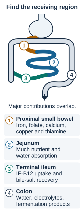
A principal absorption site is not an exclusive address
Does an intact bowel segment guarantee adequate nutrient uptake? No. Nutrients must reach the segment in a usable form. A bypass can leave healthy mucosa out of contact with food; pancreatic failure can leave food inadequately digested before it reaches healthy mucosa. Regional specialization makes the absorption map useful, but many nutrients have overlapping sites and reserve capacity. [15][19][22]
Duodenum and proximal jejunum
Iron uptake is concentrated here. Proximal small bowel also makes important contributions to folate, calcium, copper and thiamine absorption. These nutrients do not all use the same transporter or have identical distributions. [1][2][4][20][21]
Jejunum
A large share of carbohydrate, protein, lipid and water absorption occurs here. Folate uptake favors upper small bowel; fat-soluble vitamin uptake depends on digestion and bile as well as the lining. [2][19][22]
Terminal ileum
Specialized uptake of the intrinsic-factor-B12 complex and active bile-salt reclamation occur distally. The colon does not provide an equivalent downstream intrinsic-factor-dependent B12 pathway. [5][7][14]
Colon
Water, electrolytes and energy from bacterial fermentation can be salvaged here. This contribution is valuable after small-bowel loss, but is not a substitute for the small intestine's usual micronutrient absorption. [15][17][22]
Use the regional diagram to trace iron and B12 separately. Put an imaginary barrier across the proximal receiving surface, not across the whole intestinal lumen. Iron has less access to its major uptake region, while prepared B12 may still reach its distal receiving site. Now place the barrier at terminal ileum: the characteristic concern shifts toward B12 and bile salts.
Trace iron and B12 to different receiving regions, then compare a proximal bypass with terminal-ileal loss. The numbered schematic shows major contributions, not exclusive territories or anatomical scale. [1][2][4][5][7][17][20][21][22]Predict the effect of bypassing healthy proximal mucosa
Less food contacts the principal iron-absorbing region. Normal-looking tissue cannot contribute fully without access to the nutrient. This predicts impaired iron uptake; it does not prove that a particular patient's anemia has only one cause. [1]
Separate preparation, membrane transport and blood carriage
Nonheme iron commonly arrives as ferric iron, Fe3+. Reduction supplies ferrous iron, Fe2+, for entry through the intestinal divalent-metal transporter. Ferroportin exports iron from the enterocyte; oxidation permits binding to plasma transferrin. Ferritin stores iron, whereas transferrin carries it in blood. Reduced gastric acidity can decrease nonheme iron availability, and vitamin C can enhance its absorption. Neither changes the anatomical meaning of a proximal bypass. [1][24]
Folate supports DNA synthesis. Dietary folates are processed before uptake through intestinal transport systems, including the proton-coupled folate transporter in upper small bowel. Poor intake, increased requirements, proximal mucosal disease and medications such as sulfasalazine can each reduce effective folate supply. Finding low folate is not enough to select one of those causes. [2][3]
Vitamin D supports active calcium absorption, prominently in proximal small bowel. Passive absorption also contributes, especially with greater calcium intake. Normal serum calcium does not establish adequate absorption because hormonal responses can preserve blood calcium at the expense of other reserves. Fat-soluble vitamins A, D, E and K share dependence on adequate fat handling; bile supports micellar delivery and pancreatic enzymes support digestion. After enterocyte processing, much absorbed dietary lipid and associated fat-soluble vitamin cargo enters lymph in chylomicrons. [4][19][22]
Apply the map to a different problem: a patient has both low ferritin and low folate despite adequate intake. Identify their shared vulnerable region. Then ask whether that region is diseased or simply bypassed before calling the pattern a diagnosis.
B12 must arrive with the appropriate binding partner
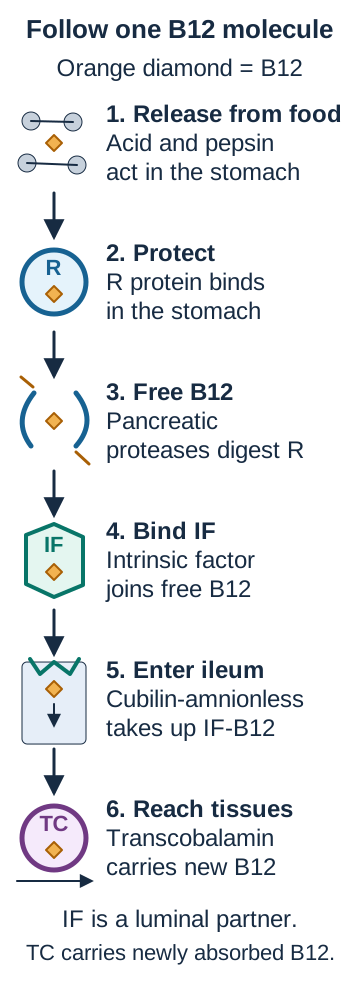
A patient has pancreatic failure, functioning gastric parietal cells, and a functioning terminal ileum. Could B12 handling still be impaired? Yes. A working receiving surface does not establish that enough B12 reaches it with the appropriate binding partner. Follow the usual food-bound pathway before deciding where the defect lies. [5][6]
Gastric acid and pepsin release B12, also called cobalamin, from dietary protein.
Salivary and gastric haptocorrin, also called R protein, binds the released vitamin in the acidic stomach.
In the duodenum, pancreatic proteases digest R protein and free B12.
Freed B12 binds intrinsic factor, a protein made by gastric parietal cells.
The intrinsic-factor-B12 complex reaches terminal ileum and enters absorptive cells through cubilin and amnionless uptake machinery.
After absorption, transcobalamin carries newly absorbed B12 onward to tissues. This carrier is also called transcobalamin II. [5][6][7][8]
The B12 diagram follows the vitamin rather than treating each protein as the same vehicle. Trace the small vitamin symbol through stomach, duodenum and ileum. At each boundary, name what stays behind and what must accompany the vitamin next. Gastric release from food protein and duodenal release from R protein are different digestive events. Chief cells supply pepsinogen, which becomes active pepsin in the acidic stomach; parietal cells supply acid and intrinsic factor. [28]
Trace the orange vitamin symbol, then predict which handoff fails with inadequate pancreatic proteolysis or unavailable intrinsic factor. The final stage depicts newly absorbed B12 delivery, not the distribution of all circulating B12. [5][6][7][8][9]
Change one dependency at a time. Pancreatic proteolysis makes B12 available for intrinsic factor; it does not manufacture intrinsic factor. Loss of parietal-cell function can leave the binding partner unavailable even when pancreatic digestion works. Ileal disease can impair entry after normal preparation. These defects converge on inadequate B12 supply through different steps. Pancreatic insufficiency can impair B12 handling without causing clinical B12 deficiency in every affected patient. [5][6][7]
Follow the vitamin when pancreatic proteolysis is inadequate
R-bound B12 reaches the duodenum, but transfer to intrinsic factor is impaired. Intrinsic factor can be present and the ileal receptor can function, yet less appropriately prepared vitamin reaches that receptor. The interrupted step lies before epithelial uptake. [6]
Follow the vitamin when intrinsic factor is absent instead
Pancreatic enzymes can free the vitamin from R protein, but cannot supply the missing intrinsic factor. Normal digestion therefore does not restore the usual receptor-dependent ileal pathway. [5][7]
Preparation for absorption must also be distinguished from carriage after absorption. Intrinsic factor is not the plasma delivery protein. Transcobalamin carries newly absorbed B12 to tissues, but most total circulating B12 is bound to haptocorrin. A measurement of all circulating B12 and a measurement of B12 on transcobalamin answer related but different questions. [8][9]
Fortified foods and supplements contain B12 that does not require release from food protein. A small intrinsic-factor-independent passive fraction can be absorbed, which helps explain selected high-dose oral replacement strategies. This does not make the colon a replacement for the normal ileal receptor pathway or justify assuming every oral regimen works after surgery. Replacement route depends on the cause, severity and clinical plan. [5][10]
Now consider bacterial competition in a stagnant small-bowel segment: host transport machinery may be intact while less B12 remains available to it. [25] Contrast this with complete terminal-ileal resection, where appropriate luminal preparation cannot replace the missing specialized uptake surface. Large body stores can delay overt B12 deficiency for years; recent surgery plus immediate symptoms also warrants assessment of pre-existing deficiency and other causes. [5]
A blood pattern identifies a problem before it identifies a site
Can a normal mean corpuscular volume exclude nutritional anemia? No. MCV averages red-cell size. Coexisting iron deficiency and a megaloblastic process can pull that average in opposite directions. Read the blood count with the smear, nutrient measurements, symptoms and clinical setting rather than interpreting one number as a complete explanation. [1][5][10]
Iron deficiency often produces microcytosis, and low ferritin supports depleted iron stores. Ferritin can increase during inflammation, so an apparently reassuring value needs context. Both folate and B12 deficiency can impair DNA synthesis and produce macro-ovalocytes and hypersegmented neutrophils. B12 deficiency can also cause paresthesias, impaired vibration or position sense, and gait difficulty, sometimes without anemia or macrocytosis. Those neurologic findings favor assessment of B12 but do not establish that the lesion is ileal. [1][3][5][10]
Consider an original example: after gastric surgery, a patient has hemoglobin 9.2 g/dL, MCV 89 fL (80 to 100), ferritin 7 ng/mL (15 to 150), and both small and large red cells on the smear. Foot numbness is progressing. The normal average size does not negate the low iron stores or the need to assess B12. The combination asks for more than one explanation.
Predict what iron replacement alone might reveal
As the iron-limited small-cell population is corrected, an untreated megaloblastic process may become more apparent in the average cell size. Neurologic symptoms still require their own assessment. A change in MCV does not demonstrate that treatment created the second deficiency. [1][5][10]
Methylmalonic acid, or MMA, can support B12 deficiency when serum B12 is borderline. Homocysteine can increase with B12 or folate deficiency, while MMA is not expected to increase from isolated folate deficiency. Kidney impairment can raise MMA and homocysteine independently, so supplied renal function and the laboratory's reference range matter. Neither biomarker identifies a unique anatomical cause. [3][5]
For example, macrocytosis with low folate, normal MMA and normal renal function favors a folate problem. Replace normal MMA with clearly raised MMA and add impaired vibration sense: the assessment must address B12 even if folate intake is also poor. Suspected megaloblastic anemia with neurologic features warrants prompt clinical assessment and B12 replacement without waiting for delayed confirmatory results. Folate alone is not an adequate response to suspected B12-related neurologic injury. [5][10]
After bariatric surgery, anemia and neurologic symptoms are not specific to B12. Copper deficiency can produce anemia, neutropenia and neurologic abnormalities. Vitamin E is another consideration in otherwise unexplained malabsorptive neuropathy; accompanying cytopenias make copper assessment particularly important. Persistent vomiting creates a different urgent risk: thiamine deficiency can present with confusion, eye-movement abnormalities or ataxia. Not every feature must be present before treatment is considered. These distinctions keep a nutrient map from becoming a one-diagnosis shortcut. [18][20]
Apply this to a patient with neurologic symptoms, normal MMA, normal renal function and neutropenia after proximal bypass. Which additional nutrient deserves assessment? Copper becomes important; a normal B12-related marker does not establish normal nutritional status. [18][20]
Incoming antigen and outgoing IgA use different routes
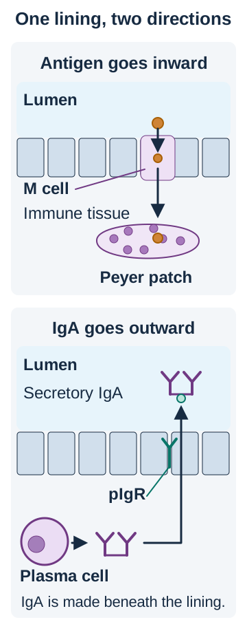
A labeled particle crosses ileal epithelium and enters underlying immune tissue. Is this ordinary nutrient uptake? Identify its destination before naming a transporter. Nutrient uptake supplies the body; antigen sampling delivers material to immune cells. Antigen is material the immune system can recognize. Sharing a location does not make these processes interchangeable. [11]
Peyer patches are unencapsulated lymphoid aggregates concentrated on the antimesenteric side of distal ileum, opposite its mesenteric attachment. The aggregates occupy lamina propria and submucosa beneath the surface epithelium. B-cell follicles and germinal centers are organized with neighboring immune-cell regions. Antigen-presenting cells receive sampled material and communicate with lymphocytes. A Peyer patch is the underlying tissue, not the name of every cell in its covering epithelium. [23]
Specialized microfold cells, or M cells, overlie the follicles. They transport sampled luminal material across the epithelium to underlying immune cells, a process called transcytosis. Ordinary nearby enterocytes principally support nutrient uptake. Paneth cells supply antimicrobial products from intestinal crypts, while goblet cells secrete mucus. These are different roles from specialized particulate sampling over a lymphoid follicle. [27] M-cell antigen sampling is therefore distinct from cubilin-dependent B12 uptake, even though both involve the distal intestine. [7][11]
After mucosal activation, IgA-producing plasma cells in lamina propria supply dimeric IgA beneath the lining. The epithelial polymeric immunoglobulin receptor, pIgR, binds polymeric antibody on the tissue-facing side and transports it toward the lumen. Part of the receptor remains attached as secretory component. This helps protect delivered IgA against proteolytic degradation; it does not make it invulnerable to every protease. Secretory IgA can neutralize toxins and impede pathogen adherence in the lumen. This helps defend the surface without requiring an intense tissue-destructive response; it does not imply that inflammation can never occur. [12][13][26]
Use the two directions in the immune-transport diagram. Trace a luminal particle inward to a follicle, then trace newly produced IgA outward from lamina propria. Identify the cargo before choosing the route. Reverse only the arrow without changing the cargo and the drawing no longer describes the same biological process.
Read cargo and direction before identifying a cell or receptor. Antigen enters through M cells; plasma cells supply dimeric IgA for outward pIgR transport. The green attachment on luminal IgA represents protective secretory component. [11][12][13][23]Predict the result of impaired pIgR with preserved IgA production
Dimeric IgA is available beneath the lining, but its active outward epithelial transport is impaired. That is a delivery defect rather than proof of deficient plasma-cell synthesis. Experimental receptor-loss studies also demonstrate passive leakage, so impaired active transport should not be described as proof that every luminal antibody molecule disappears. [12]
Now change a different dependency: antibody is delivered, but secretory component is absent. Loss of protection can increase susceptibility to proteolysis without implying that plasma cells failed to make antibody. If incoming particulate sampling is impaired instead, examine the M-cell route rather than assuming all transport through ileal epithelium has stopped. [11][13]
The missing segment and remaining connections predict different losses
After bowel surgery, diarrhea alone does not identify the missing function. Ask what was resected, what remains connected, and which preparation or uptake steps are vulnerable. Resection length influences the overall burden; segment identity predicts characteristic risks. Short bowel syndrome reflects insufficient absorptive capacity and can cause diarrhea, dehydration, electrolyte loss, weight loss and multiple nutrient deficiencies. [15]
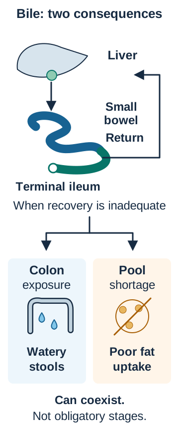
Terminal-ileal disease or resection can impair both active B12 uptake and bile-salt recovery. The bile-recycling diagram separates two consequences. Bile acids that escape ileal recovery can reach a connected colon and stimulate secretion, producing watery diarrhea. If net bile-salt losses exceed replacement sufficiently, the pool available for micellar fat absorption becomes inadequate and steatorrhea can develop. Steatorrhea means excess fat in stool. [14][19]
Predict the effect in each compartment. Excess colonic bile exposure can cause watery stools; losses exceeding replacement can leave inadequate bile for fat absorption. Neither consequence should be inferred from resection length alone. [14][19][22]
These are related mechanisms, not compulsory early and late stages. Watery bile-acid diarrhea can occur without severe fat malabsorption. Pool depletion depends on the remaining ileal function, disease extent and compensatory synthesis; an isolated resection-length cutoff is not a reliable substitute for the clinical pattern. Fat malabsorption makes vitamins A, D, E and K vulnerable even when their mucosal uptake sites remain anatomically present. [14][19]
Trace the recycling loop, then imagine that less bile returns to the liver. Predict two destinations for the unreclaimed bile: a connected colon can experience greater secretory exposure, while the next meal may receive a smaller effective bile supply if replacement is inadequate. These predictions concern different compartments and can coexist.
Predict why a bile-acid binder can help one pattern but worsen another
Binding excess bile acids can reduce their secretory effect in a connected colon. When extensive ileal disease or resection has already left too little bile for fat absorption, further binding can worsen steatorrhea. Guidance cautions against routine sequestrant use in that latter setting, although supporting evidence is limited and management remains individualized. [14]
After extensive jejunal loss, remaining healthy ileum can adapt and improve absorption. Adaptation supports recovery without recreating the original anatomy or guaranteeing independence from nutritional support. Keeping a functioning colon connected preserves water and electrolyte salvage and permits fermentation of unabsorbed carbohydrate into absorbable organic acids. Losing colon continuity forfeits those contributions; preserving it does not restore the missing ileal B12 receptor pathway. [15][16][17]
Bariatric procedures can alter acid exposure, intrinsic-factor contact, pancreatic mixing or access to proximal absorptive sites. Long-term follow-up therefore assesses more than the first deficiency found. Iron, folate, B12, calcium and vitamin D are important; copper, thiamine, zinc and fat-soluble vitamin assessment depend on the procedure, symptoms and monitoring plan. Persistent vomiting, rapid weight loss or neurologic symptoms require assessment before a routine surveillance appointment. Suspected thiamine deficiency is treated promptly rather than waiting for a delayed assay. [18][20]
Transfer the explanation: after colon exclusion, small-bowel length is unchanged but fluid losses increase. Name the contribution newly lost instead of attributing the change to new ileal damage. For fat loss with a very low fecal elastase on a formed specimen, examine pancreatic digestion instead of presuming every absorption defect requires a resection. [17][19]
Apply the absorption map to new cases
Case 1
Show answer and explanations for case 1
A. Reduced delivery of intrinsic factor to the ileum (Why this does not fit)
Intrinsic factor supports the active B12 pathway, and gastric surgery can affect its availability. The low ferritin and microcytosis identify iron depletion rather than establishing a B12 deficit. Identify the nutrient from the blood pattern before selecting an absorption dependency. [1] [18]
Reasoning steps for option A
How could intrinsic factor loss after bypass affect B12 uptake?
Intrinsic factor enables active B12 uptake in the ileum, and gastric surgery can reduce its availability.
Why do ferritin 6 and MCV 73 argue against B12 as the identified deficiency?
Ferritin 6 ng/mL with microcytosis indicates depleted iron stores, not a demonstrated B12 deficit.
Why should this bypass anemia not be assigned to the intrinsic factor pathway first?
This is iron depletion after proximal bypass; intrinsic factor concerns ileal B12 uptake, not the identified iron deficit.
B. Reduced contact with proximal absorptive mucosa (Best answer)
The duodenum and proximal jejunum are major iron-absorbing regions. Low ferritin establishes depleted iron stores, and the procedure diverts food from this region. A healthy segment contributes less when nutrients cannot reach it; other causes of iron loss still require assessment. [1] [18]
Reasoning steps for option B
Where does dietary iron normally encounter its main absorbing mucosa?
The duodenum and proximal jejunum are major sites of iron absorption.
How does duodenal exclusion combine with ferritin 6 to support this route?
Ferritin 6 documents depleted iron stores, and Roux-en-Y diverts ingested iron away from duodenal mucosa.
Why can an intact proximal segment still contribute less after Roux-en-Y?
Even healthy proximal mucosa absorbs less dietary iron if the bypassed food stream does not contact it; other sources of loss still warrant assessment.
C. Reduced recovery of bile salts in distal bowel (Why this does not fit)
Loss of ileal bile-salt recovery can impair fat handling. The documented alteration is proximal exclusion, and isolated iron depletion does not establish a bile-salt defect. Distinguish the region excluded by the operation from a different region's specialized role. [1] [18]
Reasoning steps for option C
What does impaired ileal bile-salt recovery principally disrupt?
Failed ileal bile-salt recovery can impair fat handling and absorption of fat-soluble nutrients.
Does this operative report document ileal loss or proximal food diversion?
The report documents duodenal exclusion, not ileal loss; low ferritin and microcytosis indicate iron depletion.
Why is bile-salt recycling a poorer match for isolated iron depletion?
Bile-salt recycling is an ileal function and does not best explain this proximal-bypass iron pattern.
D. Reduced pancreatic hydrolysis of dietary triglycerides (Why this does not fit)
Inadequate pancreatic fat digestion can cause steatorrhea and fat-soluble vitamin deficiencies. The supplied pattern is low iron stores after proximal bypass, not evidence of pancreatic fat maldigestion. A preparation defect and reduced contact with an absorptive surface are different explanations. [1] [18]
Reasoning steps for option D
What nutrient deficits would pancreatic fat maldigestion tend to produce?
Insufficient pancreatic fat digestion can cause oily stools and fat-soluble vitamin deficiencies.
What evidence here favors proximal iron deprivation over pancreatic insufficiency?
Low ferritin after duodenal bypass supports impaired access to iron-absorbing mucosa, not documented pancreatic fat maldigestion.
How does inadequate luminal fat digestion differ from lost contact with proximal iron mucosa?
Pancreatic enzymes prepare fat in the lumen; duodenal exclusion instead limits iron contact with proximal absorbing surface.
Takeaway: Healthy proximal mucosa cannot absorb its usual iron load when food is diverted away.
A. Reduced release of B12 from gastric dietary protein (Why this does not fit)
Poor gastric release can impair food-bound B12 absorption. The biochemical pattern lacks raised MMA and is accompanied by proximal intestinal mucosal injury rather than demonstrated gastric dysfunction. Use the metabolite pattern to distinguish B12-related dysfunction from a folate-related process. [2] [3] [5]
Reasoning steps for option A
Which gastric step is needed to liberate protein-bound B12?
Gastric acid and proteases release food-bound B12 from dietary protein before subsequent absorption.
How do normal MMA and proximal villous injury weaken a gastric B12 explanation?
Normal MMA with normal kidneys and proximal villous injury favor folate dysfunction over a demonstrated gastric B12 defect.
Why does raised homocysteine alone not establish defective gastric B12 release?
Raised homocysteine occurs with either folate or B12 dysfunction; normal MMA and proximal injury favor folate here.
B. Reduced intrinsic-factor complex uptake in terminal ileum (Why this does not fit)
The terminal ileum absorbs the prepared B12 complex. MMA is normal with preserved renal function, and the documented lesion is in proximal mucosa. Match both the suspected nutrient and the observed lesion to the proposed uptake site. [2] [3] [5]
Reasoning steps for option B
Which B12 complex is normally taken up in the terminal ileum?
The intrinsic-factor-B12 complex is taken up in the terminal ileum.
How do normal MMA with normal kidneys and proximal biopsies challenge an ileal lesion?
Normal MMA with preserved renal function and documented proximal villous injury do not favor ileal B12 failure.
Why must both metabolite pattern and lesion location match ileal B12 uptake?
An ileal B12 explanation would need concordant B12 biochemistry and ileal dysfunction, neither of which is shown.
C. Reduced ferrous iron uptake in proximal enterocytes (Why this does not fit)
Proximal villous injury can impair iron uptake and can coexist with other deficiencies. Iron depletion alone does not explain megaloblastic morphology and raised homocysteine with normal MMA. Choose the nutrient defect that explains the specific blood pattern, not every possible consequence of the lesion. [2] [3] [5]
Reasoning steps for option C
How could injured proximal villi also reduce iron absorption?
Proximal villous injury can reduce iron uptake and allow iron deficiency to coexist.
Why would iron deficiency alone fail to explain macro-ovalocytes and raised homocysteine?
Iron depletion alone does not account for macro-ovalocytes, hypersegmentation, raised homocysteine and normal MMA.
Which nutrient defect better unites this megaloblastic pattern with normal MMA?
Folate dysfunction better fits the megaloblastic findings and elevated homocysteine without elevated MMA.
D. Reduced folate uptake across proximal intestinal mucosa (Best answer)
Folate is absorbed in upper small bowel and supports normal DNA synthesis. The megaloblastic pattern and metabolite distinction favor folate dysfunction at the documented injured surface. A compatible biochemical pattern plus a matching lesion supports an absorptive explanation. [2] [3] [5]
Reasoning steps for option D
Which upper-small-bowel nutrient supports DNA synthesis in this blood picture?
Folate uptake occurs in upper small bowel, and folate supports normal DNA synthesis.
How do raised homocysteine, normal MMA and proximal villous damage converge?
Proximal villous damage with megaloblastic morphology, raised homocysteine and normal MMA favors impaired folate uptake.
Why does adequate dietary folate not rule out uptake failure at damaged villi?
Adequate intake cannot ensure folate uptake across extensively damaged duodenal and proximal jejunal villi.
Takeaway: Proximal mucosal disease can explain folate-related megaloblastic changes without implicating the ileal B12 receptor.
A. Reduced intestinal uptake is offset by hormonal compensation (Best answer)
Vitamin D supports active intestinal calcium absorption, while hormonal responses can defend circulating calcium. Low vitamin D and raised parathyroid hormone permit normal blood calcium despite an absorption problem. A regulated blood concentration is not a direct measure of nutrient absorption. [4] [18]
Reasoning steps for option A
What does low vitamin D predict for active intestinal calcium uptake?
Vitamin D deficiency reduces support for active intestinal calcium absorption.
How can raised PTH coexist with normal ionized calcium in this patient?
Raised PTH can defend circulating calcium despite low vitamin D and reduced intestinal calcium entry.
Why is a defended serum calcium concentration not proof of adequate absorption?
Normal ionized calcium may reflect PTH-mediated compensation, not adequate calcium absorption.
B. Increased intestinal uptake suppresses calcium release from reserves (Why this does not fit)
Greater absorbed calcium would reduce the need for compensatory parathyroid hormone signaling. The patient instead has low vitamin D and raised parathyroid hormone in a malabsorptive setting. Interpret the direction of the hormonal response rather than inferring absorption from serum calcium alone. [4] [18]
Reasoning steps for option B
What should greater intestinal calcium uptake do to compensatory PTH signaling?
Increased absorbed calcium would diminish the need for compensatory PTH release.
Do low 25-hydroxyvitamin D and raised PTH fit increased uptake?
Low vitamin D in fat malabsorption and raised PTH argue against increased intestinal calcium uptake.
Which direction of PTH response contradicts this proposed explanation?
Raised rather than suppressed PTH opposes a claim of increased intestinal calcium uptake.
C. Reduced renal excretion proves the proximal uptake pathway is intact (Why this does not fit)
Renal conservation can contribute to defending blood calcium. A compensatory renal response does not establish intact intestinal uptake, and renal failure is not present. Compensation in one organ cannot verify normal function in another. [4] [18]
Reasoning steps for option C
How can renal calcium conservation influence circulating calcium?
Renal calcium conservation can help maintain circulating calcium during compensation.
Does preserved kidney function establish normal intestinal calcium entry?
Normal kidney function and possible renal conservation do not measure or prove intestinal uptake.
Why can renal compensation not settle the malabsorption question?
Kidneys can conserve calcium while vitamin D-dependent intestinal absorption remains impaired.
D. Increased bile delivery replaces vitamin D-dependent calcium transport (Why this does not fit)
Adequate bile helps absorption of fat-soluble nutrients, including vitamin D. Bile does not substitute for vitamin D signaling in the calcium transport pathway. Separate absorption of a regulatory vitamin from the intestinal process that vitamin supports. [4] [18]
Reasoning steps for option D
What role does bile play in obtaining fat-soluble vitamin D?
Bile availability supports uptake of fat-soluble vitamin D.
Would bile availability replace vitamin D-mediated calcium transport once vitamin D is low?
Bile cannot replace deficient vitamin D signaling for intestinal calcium transport.
Why separate vitamin D absorption from its subsequent support of calcium uptake?
Absorbing vitamin D depends partly on fat handling, whereas vitamin D itself supports downstream calcium absorption.
Takeaway: Normal blood calcium can coexist with impaired absorption when hormonal compensation preserves the circulating level.
A. Reduced surface area after proximal villous destruction (Why this does not fit)
Mucosal injury can prevent uptake of adequately prepared nutrients. Biopsies are unremarkable, while the formed-stool elastase provides evidence of pancreatic exocrine dysfunction. Use direct evidence of the failed digestive function rather than assume every malabsorption syndrome is mucosal. [19] [22]
Reasoning steps for option A
How would proximal villous destruction interfere with vitamin uptake?
Destroyed proximal villi can reduce uptake even when luminal nutrients are properly prepared.
What do normal biopsies and elastase 48 on formed stool indicate instead?
Unremarkable biopsies oppose villous destruction; elastase 48 micrograms/g on formed stool supports exocrine pancreatic dysfunction.
Why should the tested digestive defect outweigh an unsupported mucosal lesion?
The interpretable low elastase and pancreatitis history support failed pancreatic digestion rather than an unobserved mucosal lesion.
B. Reduced bile-salt recovery after terminal-ileal loss (Why this does not fit)
Severe loss of ileal recovery can deplete the bile pool and impair fat-soluble vitamin uptake. No ileal resection is supplied; the pancreatic history and very low elastase support an upstream digestive defect. Similar deficiencies can arise from different compartments, so the discriminating history and test matter. [19] [22]
Reasoning steps for option B
How could terminal-ileal bile-salt loss cause low vitamins A and E?
Severe ileal bile-salt loss may deplete the bile pool and impede fat-soluble vitamin uptake.
Which history or test favors pancreatic failure over ileal bile-pool depletion?
No ileal resection is documented, while pancreatitis and very low formed-stool elastase favor pancreatic failure.
Why can matching fat-soluble deficits alone not localize the failed compartment?
Low vitamins A and E can arise through either route; pancreatic evidence, not the shared deficiency alone, identifies this defect.
C. Reduced luminal digestion before enterocyte uptake (Best answer)
Pancreatic enzymes make dietary fat available for intestinal absorption. Steatorrhea, low fat-soluble vitamins and elastase below 100 on formed stool support deficient pancreatic preparation. Intact mucosa and bile delivery do not compensate fully for inadequate pancreatic digestion. [19] [22]
Reasoning steps for option C
What do pancreatic enzymes contribute before fat can be absorbed?
Pancreatic enzymes digest dietary fat to make it available for intestinal uptake.
Why is elastase 48 on a formed specimen meaningful with oily stools?
Oily stools and elastase 48 micrograms/g, below 100 on a formed specimen, support substantial pancreatic exocrine insufficiency.
Why do normal bowel biopsies and unobstructed bile flow not rescue pancreatic fat digestion?
Normal biopsies and absent biliary obstruction do not replace pancreatic enzymes needed to prepare fat for absorption.
D. Reduced intrinsic-factor binding before ileal entry (Why this does not fit)
Intrinsic factor is required for the usual active B12 pathway. The deficient vitamins are fat soluble, and the supplied findings assess pancreatic fat digestion rather than intrinsic-factor availability. B12 and fat-soluble vitamins can both be affected by pancreatic disease, but not through identical digestive steps. [19] [22]
Reasoning steps for option D
Which nutrient uses intrinsic factor for active uptake?
Intrinsic factor supports active uptake of B12, not fat-soluble vitamins A and E.
Do low A and E and low fecal elastase point to that nutrient pathway?
Deficient A and E with steatorrhea and low elastase point to pancreatic fat digestion rather than intrinsic-factor shortage.
Why does possible pancreatic involvement in B12 processing not make intrinsic factor the mechanism here?
Pancreatic disease can also influence B12 processing, but the demonstrated A/E pattern reflects fat maldigestion, not intrinsic-factor loss.
Takeaway: Fat-soluble vitamin malabsorption can begin with failed pancreatic preparation rather than loss of the intestinal lining.
Ferroportin exports iron through the blood-facing enterocyte membrane. The requested protein binds oxidized iron in the soluble plasma compartment after export. A membrane export protein and a circulating carrier perform different jobs. [1] [24]
Reasoning steps for option A
At which enterocyte boundary does ferroportin move iron?
Ferroportin moves iron across the blood-facing enterocyte membrane.
Has the oxidized tracer already passed the ferroportin export step?
The tracer has already left the enterocyte and become ferric before binding a soluble plasma protein.
Why is a membrane exporter not the soluble ferric-iron plasma carrier?
Ferroportin performs membrane export; transferrin, not ferroportin, carries ferric iron in plasma.
B. Transferrin (Best answer)
Transferrin binds ferric iron and transports it in plasma. The tracer is already outside the enterocyte and has been oxidized, matching plasma iron carriage. Use the cargo, oxidation state and compartment together to select the carrier. [1] [24]
Reasoning steps for option B
Which iron oxidation state does plasma transferrin bind?
Transferrin binds ferric iron for soluble transport in plasma.
Where is the ferric tracer when the requested transport begins?
The exported tracer is now ferric and in the blood-facing compartment, matching transferrin carriage.
Why do ferric cargo and plasma location select transferrin rather than an enterocyte protein?
The ferric iron cargo outside the cell points to plasma transferrin rather than an enterocyte exporter or store.
C. Ferritin (Why this does not fit)
Ferritin stores iron within cells and can be measured in blood as a storage-related marker. The experiment asks about the main soluble delivery carrier, not iron storage. A laboratory marker of stores is not the same protein as the principal plasma transporter. [1] [24]
Reasoning steps for option C
What iron-handling function does intracellular ferritin serve?
Ferritin chiefly stores iron inside cells and serves as a storage-related blood marker.
Does this experiment ask for storage or soluble delivery in blood?
The experiment asks for the main soluble plasma transport protein after export, not storage.
Why does a marker of iron stores not identify the main plasma carrier?
Ferritin indicates or mediates iron storage, whereas transferrin is the principal plasma iron carrier.
D. Transcobalamin (Why this does not fit)
Transcobalamin transports newly absorbed B12 to tissues. The tracer is iron, not cobalamin, despite being in a postabsorptive compartment. The correct transport phase is insufficient unless the protein also matches the nutrient. [1] [24]
Reasoning steps for option D
What nutrient does transcobalamin carry after absorption?
Transcobalamin carries newly absorbed B12 to tissues.
Does a nonheme iron tracer match transcobalamin cargo?
The tracer is nonheme iron rather than cobalamin despite entering a postabsorptive compartment.
Why is being a postabsorptive carrier insufficient when the nutrient is iron?
Transcobalamin matches the delivery stage but carries B12, not the ferric iron in this experiment.
Takeaway: Ferroportin exports, ferritin stores, and transferrin carries iron in plasma.
Iron and folate have major absorption contributions in proximal small bowel. Both documented mucosal uptake defects overlap there while the specialized distal B12 pathway remains functional. Converging nutrient defects strengthen regional localization without identifying the disease by themselves. [1] [2] [3] [7] [22]
Reasoning steps for option A
Where do iron and folate absorption regions substantially overlap?
Major iron and folate uptake both occur in the duodenum and proximal jejunum.
Which measured uptake deficits point to the duodenum and proximal jejunum?
Measured mucosal defects in both iron and folate uptake converge in proximal small bowel.
How does preserved intrinsic-factor-dependent B12 uptake sharpen this proximal localization?
Preserved intrinsic-factor-dependent ileal B12 uptake fits a proximal, rather than distal, shared iron-folate lesion.
B. Distal ileum near the ileocecal junction (Why this does not fit)
Distal ileum is important for B12-complex uptake and bile-salt recovery. That B12 pathway is preserved and does not explain the paired proximal nutrient defects. A region's hallmark functions should fit the demonstrated uptake pattern. [1] [2] [3] [7] [22]
Reasoning steps for option B
What two specialized functions are associated with distal ileum?
Distal ileum supports intrinsic-factor-B12 complex uptake and bile-salt recovery.
Does preserved B12 uptake support a distal explanation for iron and folate loss?
The B12 pathway remains functional, while iron and folate uptake across diseased mucosa is reduced.
Why do the paired uptake defects not map to the ileocecal region?
Paired iron-folate defects map to proximal small bowel, not the distal ileum near the ileocecal junction.
C. Stomach near the parietal-cell glands (Why this does not fit)
Gastric acid and intrinsic factor influence preparation of selected nutrients. The defect is demonstrated across intestinal mucosa for iron and folate, not merely during gastric preparation. Distinguish luminal preparation from a measured intestinal entry defect. [1] [2] [3] [7] [22]
Reasoning steps for option C
Which gastric factors can prepare nutrients for later uptake?
Gastric acid and intrinsic factor help prepare selected nutrients for later uptake.
Where did the functional study actually demonstrate reduced iron and folate entry?
The functional study directly documents impaired iron and folate entry across intestinal mucosa.
Why is a gastric preparation site not the measured diseased mucosa?
The stomach prepares nutrients, but the measured defect here is uptake across proximal intestinal mucosa.
D. Colon near the ascending segment (Why this does not fit)
The colon salvages water, electrolytes and fermentation products. It is not the principal site for the paired iron and folate uptake processes measured here. Colonic salvage does not replace the usual proximal micronutrient absorption pathways. [1] [2] [3] [7] [22]
Reasoning steps for option D
What substances does colonic salvage mainly recover?
The colon chiefly salvages water, electrolytes and fermentation products.
Does the colon principally absorb the two nutrients deficient here?
Colonic salvage is not the major pathway for either measured iron or folate uptake.
Why cannot colonic salvage explain simultaneous iron and folate mucosal uptake loss?
A colonic site cannot account for the paired proximal iron and folate absorption defects.
Takeaway: An absorption map localizes a vulnerable function; it does not name the underlying disease.
A. Renewed destruction of proximal absorptive villi (Why this does not fit)
Proximal mucosal disease can reduce folate uptake. Repeat biopsies show healing rather than a new proximal lesion, while the medication exposure changed. Reassess current mucosal evidence rather than attributing every deficiency to the pre-existing bowel diagnosis. [3] [5]
Reasoning steps for option A
How could active proximal villous disease lower folate?
Active proximal villous disease can impede folate absorption.
What do healed repeat biopsies show four months after sulfasalazine began?
Repeat proximal biopsies show healed mucosa, while new sulfasalazine exposure precedes folate decline.
Why is past inflammatory bowel disease insufficient evidence of renewed villous damage?
Prior bowel disease alone does not establish recurrent villous injury when current biopsies show healing.
B. Progressive failure of terminal-ileal B12 uptake (Why this does not fit)
Ileal dysfunction can produce B12-related macrocytosis. B12 and MMA are normal and folate is low, so the specified biochemical deficit lies elsewhere. Macrocytosis alone does not identify the vitamin or the intestinal region. [3] [5]
Reasoning steps for option B
How could terminal-ileal disease produce macrocytosis?
Terminal-ileal dysfunction can cause B12-related macrocytosis.
How do normal B12 and MMA contrast with the measured low folate?
B12 and MMA are normal, whereas measured folate is low.
Why does macrocytosis alone not justify an ileal B12 explanation?
Macrocytosis occurs with folate as well as B12 dysfunction; normal B12 and MMA point away from ileal B12 failure.
C. Increased folate utilization from fetal growth (Why this does not fit)
Pregnancy increases folate requirements and can expose inadequate supply. Pregnancy is excluded and no new increased-demand state is supplied. A demand explanation requires a compatible clinical change rather than the deficiency alone. [3] [5]
Reasoning steps for option C
What physiological state increases folate requirements for fetal growth?
Pregnancy increases folate demand to support fetal growth.
Is a pregnancy-related demand increase present in this case?
The patient is not pregnant, and no other new high-demand state is supplied.
Why does low folate without pregnancy not support fetal utilization?
Low folate alone does not imply fetal consumption when pregnancy is explicitly excluded.
D. Medication interference with intestinal folate absorption (Best answer)
Sulfasalazine can impair folate absorption. Its introduction precedes low folate despite unchanged intake and healed proximal mucosa. A functional absorption defect can arise without new anatomical injury. [3] [5]
Reasoning steps for option D
How can new sulfasalazine exposure affect folate absorption?
Sulfasalazine can impair folate absorption.
Which stable diet and healed-biopsy findings favor this changed exposure?
Folate fell after sulfasalazine began despite unchanged diet and healed proximal biopsies.
Why can drug-related uptake impairment occur without renewed mucosal injury?
A drug can impair folate uptake functionally even when the proximal mucosa appears healed.
Takeaway: Low folate can reflect a medication-related uptake problem even when the intestinal lining has healed.
A. Pancreatic cleavage of the haptocorrin carrier (Why this does not fit)
Pancreatic proteases free B12 from R protein after gastric transit. Pancreatic function is preserved, whereas free crystalline B12 specifically bypasses the need for release from food protein. Compare what differs between the two preparations before selecting a shared downstream step. [5] [6]
Reasoning steps for option A
Which B12 binding protein do pancreatic proteases cleave downstream of the stomach?
Pancreatic proteases cleave haptocorrin, also called R protein, to release B12 after gastric transit.
How does preserved pancreatic function weigh against this cleavage defect?
Pancreatic function is preserved; free B12 specifically bypasses release from dietary protein.
Which preparation difference bypasses gastric food-protein release rather than pancreatic cleavage?
The preparations differ at gastric liberation from food protein, not at the later pancreatic cleavage of haptocorrin.
B. Receptor uptake of the intrinsic-factor complex (Why this does not fit)
Ileal receptors take up the prepared intrinsic-factor-B12 complex. Preserved uptake of free B12 through the usual low-dose pathway argues against a common terminal receiving defect. An upstream preparation difference can explain unequal uptake despite a shared receiving site. [5] [6]
Reasoning steps for option B
Where is the intrinsic-factor-bound B12 complex received?
Terminal-ileal receptors take up the intrinsic-factor-B12 complex.
What does preserved physiological-dose free-B12 uptake imply about ileal reception?
At physiological doses, uptake of free crystalline B12 is relatively preserved despite shared ileal reception.
Why would a shared ileal defect not selectively spare free crystalline B12?
A terminal-ileal receptor defect should affect both prepared B12 sources rather than selectively food-bound B12.
C. Gastric liberation of B12 from dietary protein (Best answer)
Acid and gastric proteases release food-bound cobalamin before its subsequent binding steps. Markedly reduced acid secretion affects the protein-bound preparation, while the free preparation does not require that release. Distinguish release from food protein from later release from haptocorrin. [5] [6]
Reasoning steps for option C
What must gastric acid and proteases do to food-protein-bound B12?
Acid and gastric proteases liberate cobalamin from food protein before later binding steps.
Which preparation avoids that step in this low-acid patient?
Markedly reduced acid hinders release of protein-bound B12; crystalline B12 is already free.
Why is liberation from dietary protein distinct from later haptocorrin cleavage?
Gastric liberation from dietary protein is bypassed by free B12, whereas pancreatic cleavage later removes haptocorrin.
D. Plasma carriage of B12 after epithelial entry (Why this does not fit)
Transcobalamin supports postabsorptive tissue delivery. A common blood-carriage defect would not preferentially explain impaired uptake of only the food-bound preparation. A difference established before absorption is not explained by the same downstream plasma step. [5] [6]
Reasoning steps for option D
When does transcobalamin act in B12 delivery?
Transcobalamin delivers absorbed B12 in blood to tissues.
Would a shared plasma carrier defect selectively lower uptake of food-bound B12?
Both preparations would require the same downstream carrier, so its defect does not explain their different uptake.
Why is downstream tissue carriage not the failed preparation-specific step?
A postabsorptive plasma-carriage defect cannot account for selectively reduced uptake of food-bound B12.
Takeaway: Free crystalline B12 bypasses release from food protein, not every subsequent requirement for absorption.
A. Supply pancreatic proteolytic activity during digestion (Best answer)
Pancreatic proteases digest haptocorrin and permit transfer of B12 to intrinsic factor. The measured persistence of haptocorrin-bound vitamin localizes the abnormality before the intact ileal receiving surface. Adequate intrinsic factor does not establish that B12 is available to bind it. [5] [6] [7] [8]
Reasoning steps for option A
What pancreatic action releases B12 from duodenal haptocorrin?
Pancreatic proteases digest haptocorrin in the duodenal lumen so B12 can transfer to intrinsic factor.
What does persistent haptocorrin binding show despite adequate intrinsic factor?
Why does proteolytic supplementation target the defect before intact ileal uptake?
Supplying pancreatic proteolytic activity addresses the measured luminal handoff while the ileal mucosa is intact.
B. Increase intrinsic-factor secretion from gastric glands (Why this does not fit)
Intrinsic factor is needed for the usual active ileal pathway. Its luminal availability is already adequate; B12 remains attached to a different binding protein. Adding the next binding partner does not directly correct failure to release the previous one. [5] [6] [7] [8]
Reasoning steps for option B
What role would additional intrinsic factor normally serve?
Intrinsic factor binds released B12 for subsequent active ileal uptake.
Is intrinsic factor scarce in the measured duodenal sample?
The duodenal sample already contains adequate intrinsic factor; B12 remains attached to haptocorrin.
Why can more intrinsic factor not free B12 still attached to haptocorrin?
More intrinsic factor cannot directly dislodge B12 from haptocorrin without the missing proteolytic release.
C. Increase cubilin expression at the ileal surface (Why this does not fit)
Cubilin participates in uptake of the prepared intrinsic-factor complex. The observed defect occurs in the duodenal preparation phase before the complex is available to the receptor. A stronger receiving capacity cannot directly substitute for the missing digestive handoff. [5] [6] [7] [8]
Reasoning steps for option C
What complex does the ileal cubilin receptor normally accept?
Cubilin participates in terminal-ileal uptake of the intrinsic-factor-B12 complex.
Has the haptocorrin-bound B12 reached a receptor-ready form?
B12 remains bound to haptocorrin in the duodenum, before an intrinsic-factor complex is ready for cubilin.
Why would increasing ileal reception leave the measured duodenal handoff unresolved?
Enhancing intact ileal reception cannot replace the failed pancreatic cleavage upstream in the duodenal lumen.
D. Supply transcobalamin for postabsorptive B12 carriage (Why this does not fit)
Transcobalamin delivers newly absorbed B12 to tissues after epithelial entry. The observed abnormality is persistent R-protein binding in the duodenal lumen, before the vitamin reaches a plasma carrier. A later delivery protein cannot directly correct the demonstrated luminal preparation defect. [5] [6] [7] [8]
Reasoning steps for option D
At what stage does transcobalamin deliver B12 to tissues?
Transcobalamin carries newly absorbed B12 to tissues after epithelial entry.
Where is the persistent B12 binding abnormality actually observed?
The defect is haptocorrin-bound B12 persisting in the duodenal lumen, before any plasma transport.
Why cannot a plasma carrier directly repair a duodenal luminal release defect?
A blood carrier acts after absorption and cannot directly free B12 from duodenal haptocorrin.
Takeaway: Pancreatic proteases make B12 available to intrinsic factor before the complex reaches the ileum.
A. Release of the vitamin from dietary animal protein (Why this does not fit)
Gastric acid and proteases release food-bound B12, and gastric disease can affect this earlier step. The question begins after the vitamin has already passed through the R-protein phase. Localize the requested event using the point already reached in the sequence. [5] [7] [10]
Reasoning steps for option A
Could oxyntic gland loss affect release of food-bound B12 before haptocorrin binding?
Gastric acid and proteases release food-bound B12, and gastric disease can affect this earlier step.
Why does the stipulated duodenal release from R protein make food-protein release an earlier event?
The question begins after the vitamin has already passed through the R-protein phase.
Does gastric food-protein release name the post-R-protein handoff asked about?
Because the question starts after duodenal release from R protein, earlier gastric release from food is not the requested failed binding step.
B. Digestion of the vitamin's haptocorrin covering (Why this does not fit)
Pancreatic proteases normally digest haptocorrin in the duodenum. That event is supplied as successful and pancreatic secretion is preserved. Do not assign a defect to a handoff that the stem has already completed. [5] [7] [10]
Reasoning steps for option B
Which duodenal enzyme action removes the haptocorrin covering from B12?
Pancreatic proteases normally digest haptocorrin in the duodenum.
What does preserved pancreatic secretion plus completed R-protein release establish about this candidate?
That event is supplied as successful and pancreatic secretion is preserved.
Can a completed pancreatic handoff be the event most directly impaired here?
Preserved pancreatic secretion and stated R-protein release rule out defective haptocorrin digestion; intrinsic-factor binding is next.
C. Binding to the plasma carrier after epithelial entry (Why this does not fit)
Transcobalamin carries newly absorbed B12 toward tissues. The identified gastric defect affects preparation for active uptake, not the later blood carrier itself. Intrinsic factor and transcobalamin are not interchangeable across luminal and plasma compartments. [5] [7] [10]
Reasoning steps for option C
Which carrier delivers B12 to tissues only after epithelial absorption?
How do intrinsic-factor antibodies locate this defect before plasma transcobalamin carriage?
The identified gastric defect affects preparation for active uptake, not the later blood carrier itself.
Why must a luminal binding partner not be confused with the blood carrier?
Oxyntic gland loss and intrinsic-factor antibodies disrupt the luminal partner before ileal uptake, not postabsorptive transcobalamin carriage.
D. Formation of the ligand for active ileal uptake (Best answer)
Intrinsic factor must bind available B12 to create the complex recognized by the ileal receptor system. Gland loss and intrinsic-factor antibodies impair availability or function of that partner despite successful pancreatic release. A prepared vitamin still needs the appropriate luminal partner before receptor-dependent uptake. [5] [7] [10]
Reasoning steps for option D
What ligand must free duodenal B12 form for active terminal-ileal uptake?
Intrinsic factor must bind available B12 to create the complex recognized by the ileal receptor system.
How do oxyntic gland loss and intrinsic-factor antibodies impair that ligand despite intact pancreatic digestion?
Gland loss and intrinsic-factor antibodies impair availability or function of that partner despite successful pancreatic release.
Why does preserved ileal mucosa not compensate for ineffective intrinsic-factor binding?
Despite intact pancreatic release and ileal mucosa, impaired intrinsic factor prevents formation of the B12 complex required for receptor-dependent uptake.
A. Acid-mediated release of food-bound cobalamin (Why this does not fit)
Gastric acid participates in release from dietary protein. This release remains functional and occurs before the vitamin reaches the resected region. A distal resection does not directly identify a gastric preparation failure. [5] [7] [10]
Reasoning steps for option A
Where does acid release cobalamin from food protein relative to the removed terminal ileum?
Gastric acid participates in release from dietary protein.
What does the assessment explicitly say about this upstream gastric release?
This release remains functional and occurs before the vitamin reaches the resected region.
Why would loss of distal receiving tissue not by itself abolish acid-mediated preparation?
Normal dietary B12 release establishes that gastric acid preparation remains intact; terminal-ileal loss removes a downstream uptake site.
B. Cubilin-amnionless uptake of the prepared complex (Best answer)
The terminal ileum provides specialized receptor-dependent entry of intrinsic-factor-bound B12. That receiving region is absent despite preserved luminal preparation and colonic continuity. Colonic salvage is not an equivalent active B12 uptake pathway. [5] [7] [10]
Reasoning steps for option B
Which ileal machinery normally takes up the intrinsic-factor-B12 complex?
The terminal ileum provides specialized receptor-dependent entry of intrinsic-factor-bound B12.
What happens to that receiving site when the entire terminal ileum is removed?
That receiving region is absent despite preserved luminal preparation and colonic continuity.
Why can the connected colon not substitute for cubilin-amnionless uptake?
Complete terminal-ileal resection removes cubilin-amnionless uptake of intrinsic-factor-bound B12, and the connected colon cannot replace it.
C. Protease-mediated digestion of the R-protein carrier (Why this does not fit)
Pancreatic digestion frees B12 from R protein in the duodenum. Intrinsic-factor-bound B12 has already completed that handoff before reaching the resection site. Successful formation of a later complex supports completion of the preceding release step. [5] [7] [10]
Reasoning steps for option C
What duodenal process permits B12 to leave R protein and bind intrinsic factor?
Pancreatic digestion frees B12 from R protein in the duodenum.
What does arrival as an intrinsic-factor-bound complex imply about this prior digestion?
Intrinsic-factor-bound B12 has already completed that handoff before reaching the resection site.
Why is the removed ileum downstream of the pancreatic protease step?
B12 already bound to intrinsic factor has passed pancreatic R-protein digestion before reaching the missing terminal ileum.
D. Transcobalamin carriage after epithelial absorption (Why this does not fit)
Transcobalamin transports newly absorbed B12 in blood. Loss of the ileal receiving surface reduces entry but does not directly eliminate the circulating transport protein. Less substrate reaching a downstream carrier is different from loss of the carrier itself. [5] [7] [10]
Reasoning steps for option D
At what stage does transcobalamin carry newly absorbed cobalamin?
Transcobalamin transports newly absorbed B12 in blood.
Does terminal-ileal resection remove the plasma transport protein or limit vitamin entry before it?
Loss of the ileal receiving surface reduces entry but does not directly eliminate the circulating transport protein.
How is reduced cargo reaching transcobalamin different from loss of transcobalamin itself?
The resection limits B12 entry into blood, but does not remove transcobalamin, its downstream plasma carrier.
Takeaway: Normal B12 preparation and colonic continuity do not replace the terminal-ileal receptor pathway.
A. Reduced gastric production of intrinsic factor (Why this does not fit)
Intrinsic-factor deficiency can lower active B12 absorption. The experimental change is the presence of microbes, and restoring availability does not require changing gastric secretion. Attribute a controlled comparison to the variable actually altered. [5] [25]
Reasoning steps for option A
How could deficient intrinsic factor ordinarily reduce active B12 uptake?
Intrinsic-factor deficiency can lower active B12 absorption.
Was gastric secretion manipulated when removal of stagnant-segment microbes restored tracer availability?
The experimental change is the presence of microbes, and restoring availability does not require changing gastric secretion.
Why does an unchanged gastric source fail to account for the controlled microbial comparison?
Removing microbes restores tracer without altering gastric intrinsic-factor production, so that option does not explain the comparison.
B. Reduced epithelial expression of ileal receptors (Why this does not fit)
Loss of receptor function can prevent uptake of an adequately prepared complex. The same functioning intestinal preparation receives more available tracer when microbes are excluded. Distinguish a receiving defect from a reduction in the substrate offered to the receiver. [5] [25]
Reasoning steps for option B
What would reduced ileal receptor expression do to available B12?
Loss of receptor function can prevent uptake of an adequately prepared complex.
What does restored tracer delivery to the same functioning intestinal preparation imply about its receptor machinery?
The same functioning intestinal preparation receives more available tracer when microbes are excluded.
Is the limiting factor here the receiver or the quantity of tracer offered to it?
Restored tracer delivery to the same functioning intestinal preparation points to reduced available cargo, not defective ileal receptors.
C. Reduced luminal B12 availability through bacterial uptake (Best answer)
Intestinal microbes can acquire B12 and compete with host availability. Tracer accumulates in the microbes, and eliminating their contribution restores vitamin available to the same host preparation. An absorption pathway can have intact host machinery but insufficient available cargo. [5] [25]
Reasoning steps for option C
Can microbes in the stagnant segment take up labeled B12 before the host receives it?
Intestinal microbes can acquire B12 and compete with host availability.
What does microbial tracer accumulation followed by recovery after their removal demonstrate?
Tracer accumulates in the microbes, and eliminating their contribution restores vitamin available to the same host preparation.
Why can intact host absorption coexist with too little luminal B12 cargo?
Microbial accumulation of labeled B12 and recovery after microbial removal identify competition for luminal vitamin despite intact host machinery.
D. Reduced pancreatic digestion of haptocorrin (Why this does not fit)
Pancreatic proteolysis makes R-bound B12 available for intrinsic factor. Pancreatic secretion is adequate, and the controlled comparison identifies microbial tracer uptake rather than altered proteolysis. Do not substitute a familiar upstream B12 defect for a directly demonstrated competing process. [5] [25]
Reasoning steps for option D
How do pancreatic proteases normally free R-bound B12?
Pancreatic proteolysis makes R-bound B12 available for intrinsic factor.
Does adequate pancreatic secretion explain the change produced solely by excluding microbes?
Pancreatic secretion is adequate, and the controlled comparison identifies microbial tracer uptake rather than altered proteolysis.
Why does direct microbial tracer capture favor competition over failed haptocorrin digestion?
Adequate pancreatic secretion and recovery after microbial removal implicate bacterial B12 uptake rather than failed haptocorrin proteolysis.
Takeaway: Bacterial uptake can reduce B12 available to otherwise functioning host absorption pathways.
Transcobalamin supports delivery of newly absorbed B12, while haptocorrin binds much of the total circulating pool. The two assays distinguish newly absorbed vitamin from all circulating vitamin rather than describing the same fraction. A carrier's delivery role and its share of the total blood pool are different properties. [8] [9]
Reasoning steps for option A
Which carrier handles the new post-dose B12 flux, and which holds most total circulating B12?
Transcobalamin supports delivery of newly absorbed B12, while haptocorrin binds much of the total circulating pool.
Why do the post-dose assay and total-pool assay need not identify the same dominant carrier?
The two assays distinguish newly absorbed vitamin from all circulating vitamin rather than describing the same fraction.
Does tissue delivery by transcobalamin imply it holds most of the blood B12 pool?
The post-dose tissue-delivery fraction travels on transcobalamin, while haptocorrin binds most total circulating B12; flux and pool size differ.
B. Haptocorrin; intrinsic factor (Why this does not fit)
Haptocorrin binds B12 in the stomach and in blood; intrinsic factor participates before ileal uptake. Intrinsic factor is not the major plasma binding protein, and the newly absorbed tissue-delivery role belongs to transcobalamin. Do not extend a luminal binding role into the plasma compartment. [8] [9]
Reasoning steps for option B
Where does haptocorrin bind B12, and where does intrinsic factor act?
Haptocorrin binds B12 in the stomach and in blood; intrinsic factor participates before ileal uptake.
Can intrinsic factor explain the majority of a measured plasma B12 pool?
Intrinsic factor is not the major plasma binding protein, and the newly absorbed tissue-delivery role belongs to transcobalamin.
Why does this pair mistake pre-ileal binding for postabsorptive tissue delivery?
Intrinsic factor binds B12 before ileal uptake, so it cannot account for the dominant circulating pool; haptocorrin also is not the new-delivery carrier.
C. Intrinsic factor; transcobalamin (Why this does not fit)
Intrinsic factor prepares B12 for receptor-dependent ileal uptake, and transcobalamin participates after absorption. The first assay is explicitly postabsorptive, while the larger total circulating pool is mainly haptocorrin-bound. Identify both the compartment and the particular pool being measured. [8] [9]
Reasoning steps for option C
What are the respective luminal and postabsorptive roles of intrinsic factor and transcobalamin?
Intrinsic factor prepares B12 for receptor-dependent ileal uptake, and transcobalamin participates after absorption.
Why can neither proposed position explain both the post-dose carrier and the dominant total-pool carrier?
The first assay is explicitly postabsorptive, while the larger total circulating pool is mainly haptocorrin-bound.
Which assay demands haptocorrin rather than transcobalamin in the second position?
The post-dose carrier is transcobalamin, not luminal intrinsic factor; most total circulating B12 is haptocorrin-bound, not transcobalamin-bound.
D. Transferrin; haptocorrin (Why this does not fit)
Transferrin is a plasma nutrient carrier and haptocorrin binds circulating B12. The first carrier must deliver cobalamin, not the iron carried by transferrin. A correct answer about one pool does not rescue a nutrient mismatch in the other. [8] [9]
Reasoning steps for option D
What nutrient does transferrin deliver in blood?
Transferrin delivers iron in blood, not the newly absorbed B12 measured in this study.
Although haptocorrin fits the total B12 pool, can transferrin carry the newly absorbed B12 fraction?
The first carrier must deliver cobalamin, not the iron carried by transferrin.
Why does getting the second carrier right not correct the iron-versus-cobalamin mismatch?
Haptocorrin fits the total B12 pool, but transferrin carries iron rather than newly absorbed cobalamin.
Takeaway: Newly absorbed B12 delivery on transcobalamin does not mean most circulating B12 is carried by it.
A. Replace iron and reassess the neurologic findings after hemoglobin normalizes (Why this does not fit)
Iron replacement is indicated for depleted stores and can improve iron-limited erythropoiesis. Progressive neurologic findings and low B12 require assessment and replacement without waiting for the iron response. One confirmed deficiency does not explain away a second time-sensitive nutrient problem. [1] [5] [10]
Reasoning steps for option A
What deficit does ferritin of 5 ng/mL justify correcting?
Iron replacement is indicated for depleted stores and can improve iron-limited erythropoiesis.
Why do worsening numbness, impaired vibration and B12 of 178 pg/mL rule out waiting for iron response?
Progressive neurologic findings and low B12 require assessment and replacement without waiting for the iron response.
Can iron replacement alone address this patient’s time-sensitive neurologic concern?
Ferritin of 5 supports iron replacement, but progressive numbness, impaired vibration and low B12 require prompt B12 attention rather than waiting for hemoglobin recovery.
B. Replace folate and use a rising hemoglobin to assess the sensory symptoms (Why this does not fit)
Folate can improve a folate-related megaloblastic blood pattern. Low B12 and impaired vibration sense make folate alone an inadequate response to the neurologic risk. A hematologic response does not establish that B12-related neurologic injury is being treated. [1] [5] [10]
Reasoning steps for option B
What blood finding might folate replacement improve if folate were deficient?
Folate can improve a folate-related megaloblastic blood pattern.
Why do measured low B12 and impaired vibration sense make a hemoglobin rise an unsafe neurologic endpoint?
Low B12 and impaired vibration sense make folate alone an inadequate response to the neurologic risk.
Would improved erythropoiesis prove that B12-associated sensory injury was addressed?
A rising hemoglobin after folate would not treat or assess this patient’s low-B12-associated sensory deficits.
C. Defer replacement while repeating the count to confirm persistent macrocytosis (Why this does not fit)
A repeat count can clarify uncertain laboratory results. Macrocytosis is not required when mixed cell populations and neurologic B12 deficiency are already supported. An average cell size can conceal opposing deficiencies and should not delay appropriate treatment. [1] [5] [10]
Reasoning steps for option C
Why might a repeat blood count be useful when macrocytosis is genuinely uncertain?
A repeat count can clarify uncertain laboratory results.
What do small cells plus macro-ovalocytes explain about MCV 90 fL in this patient?
Macrocytosis is not required when mixed cell populations and neurologic B12 deficiency are already supported.
Why should a normal average cell volume not postpone replacement in the face of these findings?
Small cells and macro-ovalocytes can average to MCV 90; low ferritin, low B12 and worsening sensory findings preclude waiting for overt macrocytosis.
D. Address both iron and B12 deficits with prompt B12 replacement (Best answer)
Mixed iron and B12 deficiency can yield a normal average cell volume despite abnormal cell populations. Low ferritin, low B12, megaloblastic features and progressive sensory findings support treating both rather than waiting for a high MCV. Interpret the full blood pattern and symptoms instead of using normal MCV to exclude B12 deficiency. [1] [5] [10]
Reasoning steps for option D
How can concurrent iron depletion and megaloblastic cells yield MCV within the reference range?
Mixed iron and B12 deficiency can yield a normal average cell volume despite abnormal cell populations.
Which ferritin, B12, smear and sensory findings call for addressing both deficits now?
Low ferritin, low B12, megaloblastic features and progressive sensory findings support treating both rather than waiting for a high MCV.
Why is prompt B12 replacement warranted alongside iron despite an MCV of 90 fL?
Low ferritin and B12 with mixed smear populations call for both deficits to be addressed, with prompt B12 replacement for progressive sensory findings despite normal MCV.
Takeaway: Normal MCV does not exclude B12 deficiency, especially when iron deficiency and neurologic findings coexist.
A. Ordinary nutrient-absorbing enterocyte (Why this does not fit)
Enterocytes absorb nutrients and participate in epithelial transport. Particulate cargo delivered inward to underlying immune cells identifies specialized sampling rather than ordinary nutrient uptake. The cargo and destination distinguish neighboring epithelial functions. [11] [23] [27]
Reasoning steps for option A
What cargo do ordinary absorptive enterocytes chiefly move from the ileal lumen?
Enterocytes absorb nutrients and participate in epithelial transport.
Do bacterial particles ending inside underlying antigen-presenting cells resemble nutrient entry into blood?
Particulate cargo delivered inward to underlying immune cells identifies specialized sampling rather than ordinary nutrient uptake.
Which destination separates follicular immune sampling from enterocyte absorption?
Labeled bacterial particles delivered to underlying antigen-presenting cells indicate M-cell immune sampling rather than nutrient uptake by enterocytes.
B. Crypt-associated Paneth cell (Why this does not fit)
Paneth cells contribute antimicrobial products from intestinal crypts. The described route is particulate transfer across follicle-associated epithelium into immune tissue, rather than crypt secretion. Distinguish antimicrobial secretion from specialized epithelial antigen delivery. [11] [23] [27]
Reasoning steps for option B
What do Paneth cells release from intestinal crypts?
Paneth cells contribute antimicrobial products from intestinal crypts.
Does transfer of labeled bacteria across follicle-associated epithelium describe crypt secretion?
The described route is particulate transfer across follicle-associated epithelium into immune tissue, rather than crypt secretion.
Why is antimicrobial output not the same as inward particulate delivery to immune cells?
C. Mucus-secreting goblet cell (Why this does not fit)
Goblet cells secrete mucus that supports the epithelial surface barrier. The specified bacterial-particle route across follicle-associated epithelium is the characteristic M-cell sampling pathway. Use the specified particulate cargo, epithelial region and immune destination, not a general barrier function. [11] [23] [27]
Reasoning steps for option C
How does a goblet cell support the epithelial mucus barrier?
Goblet cells secrete mucus that supports the epithelial surface barrier.
Does mucus secretion account for labeled particles recovered within antigen-presenting cells?
The specified bacterial-particle route across follicle-associated epithelium is the characteristic M-cell sampling pathway.
Which feature identifies a transcytotic sampling cell instead of a mucus-producing cell?
Goblet-cell mucus secretion cannot account for particulate bacteria recovered within underlying antigen-presenting cells; that inward sampling fits M cells.
D. Follicle-associated microfold cell (Best answer)
M cells transcytose luminal particulate antigen toward underlying immune tissue. The observed inward route from follicle-associated epithelium to antigen-presenting cells matches this sampling function. Incoming antigen transfer identifies M-cell sampling rather than outward antibody delivery. [11] [23] [27]
Reasoning steps for option D
What does an M cell transcytose over ileal lymphoid follicles?
M cells transcytose luminal particulate antigen toward underlying immune tissue.
How does recovery of luminal bacterial particles in underlying immune cells match that route?
The observed inward route from follicle-associated epithelium to antigen-presenting cells matches this sampling function.
Why does incoming antigen sampling, rather than secretion into blood, identify the microfold cell?
Follicle-associated M cells transfer the labeled bacterial particles inward to antigen-presenting cells rather than secreting them into blood.
Takeaway: M cells deliver sampled luminal antigen to immune tissue beneath the ileal lining.
A. Follicle-associated M-cell epithelium (Why this does not fit)
M cells occur in the specialized covering of intestinal lymphoid follicles. The label includes the underlying lymphoid tissue and is not restricted to the surface epithelial layer. Separate an organ-level lymphoid aggregate from its specialized covering cells. [23]
Reasoning steps for option A
Where are M cells situated relative to ileal lymphoid follicles?
M cells occur in the specialized covering of intestinal lymphoid follicles.
Does a label spanning lamina propria and submucosa stop at the M-cell epithelial covering?
The label includes the underlying lymphoid tissue and is not restricted to the surface epithelial layer.
Why is the covering alone narrower than the enclosed follicular aggregate?
The marked distal-ileal aggregate spans lamina propria and submucosa, whereas its M-cell covering occupies only the overlying epithelium.
B. Aggregated ileal lymphoid nodules (Best answer)
Peyer patches are organized ileal lymphoid aggregates with follicles in lamina propria and submucosa. The distal, antimesenteric location and the full tissue extent match the entire marked structure. Use the label boundary and tissue layers to identify what the anatomical annotation actually includes. [23]
Reasoning steps for option B
What organized ileal lymphoid structure occupies lamina propria and submucosa?
Peyer patches are organized ileal lymphoid aggregates with follicles in lamina propria and submucosa.
How do distal antimesenteric position and the full marked extent support a Peyer patch?
The distal, antimesenteric location and the full tissue extent match the entire marked structure.
Why does the boundary enclosing multiple follicles identify aggregated nodules rather than a cell type?
Multiple antimesenteric distal-ileal follicles enclosed across lamina propria and submucosa constitute a Peyer patch, not a single covering cell.
C. IgA-secreting plasma-cell population (Why this does not fit)
Plasma cells in lamina propria contribute to mucosal antibody production. A whole follicular aggregate extending into submucosa is not simply a population of individual antibody-secreting cells. A cellular component does not name the complete organized lymphoid structure. [23]
Reasoning steps for option C
What do lamina-propria plasma cells produce in mucosal immunity?
Plasma cells in lamina propria contribute to mucosal antibody production.
Does a group of antibody-secreting cells describe the entire marked submucosal follicular structure?
A whole follicular aggregate extending into submucosa is not simply a population of individual antibody-secreting cells.
Why cannot one functional cell population name the complete Peyer patch?
IgA-secreting plasma cells are a cell population, not the entire labeled follicular aggregate extending into submucosa.
D. Nutrient-absorbing villous epithelium (Why this does not fit)
Villous enterocytes provide an absorptive epithelial surface. The marked structure consists of lymphoid follicles beneath that surface rather than a layer of absorptive cells. Distinguish mucosal immune tissue from adjacent nutrient-absorbing epithelium. [23]
Reasoning steps for option D
Where do absorptive villous enterocytes sit in relation to underlying lymphoid tissue?
Villous enterocytes form the luminal absorptive epithelium, not the underlying lymphoid aggregate spanning lamina propria and submucosa.
Does the label trace a villous lining or multiple follicles through two deeper tissue layers?
The marked structure consists of lymphoid follicles beneath that surface rather than a layer of absorptive cells.
Why is an epithelial absorption surface not the marked aggregate?
The label encloses deep lymphoid follicles rather than the villous enterocyte layer that absorbs nutrients.
Takeaway: A Peyer patch is the underlying lymphoid aggregate, not its M-cell covering alone.
A. Antibody production by mucosal plasma cells (Best answer)
Plasma cells provide the dimeric IgA substrate for subsequent epithelial transport. The epithelial pathway transports supplied IgA normally, while endogenous antibody is already scarce beneath the lining. A successful substrate-addition experiment can distinguish deficient production from deficient delivery. [12] [13]
Reasoning steps for option A
Which cells supply endogenous dimeric IgA beneath the mucosal epithelium?
Plasma cells provide the dimeric IgA substrate for subsequent epithelial transport.
What does scarce tissue-side IgA despite normal transport of added IgA suggest about the source?
The epithelial pathway transports supplied IgA normally, while endogenous antibody is already scarce beneath the lining.
Why does successful rescue by purified substrate prioritize plasma-cell production?
Because added tissue-side IgA crosses normally but endogenous tissue-side IgA is scarce, investigate plasma-cell substrate production first.
B. Antigen transcytosis by follicular M cells (Why this does not fit)
M-cell sampling delivers luminal antigen to underlying immune tissue. Incoming antigen delivery is preserved, and the deficit appears in antibody availability after that step. Use the demonstrated intact upstream function to localize the remaining defect. [12] [13]
Reasoning steps for option B
What material do follicular M cells bring inward to immune cells?
M-cell sampling delivers luminal antigen to underlying immune tissue.
What does normal luminal antigen delivery say about this candidate upstream step?
Incoming antigen delivery is preserved, and the deficit appears in antibody availability after that step.
Why would impaired antigen sampling not explain the observed tissue-side IgA shortage here?
Normal antigen delivery to immune cells establishes preserved M-cell sampling; the shortage arises at antibody availability downstream.
C. Antibody transport through epithelial pIgR (Why this does not fit)
The polymeric immunoglobulin receptor mediates active outward transport of dimeric IgA. Normal transport of supplied dimeric IgA shows that this delivery step can function. Low luminal antibody is not sufficient evidence for a transport defect when little substrate is produced. [12] [13]
Reasoning steps for option C
What does epithelial pIgR do with dimeric IgA supplied on the tissue-facing side?
The polymeric immunoglobulin receptor mediates active outward transport of dimeric IgA.
What does normal transport of experimentally added IgA establish about this pathway?
Normal transport of supplied dimeric IgA shows that this delivery step can function.
Why is low endogenous luminal IgA not evidence of pIgR failure when substrate is scarce?
Normal luminal transport of added dimeric IgA demonstrates functioning epithelial pIgR despite low endogenous substrate beneath the lining.
D. Antibody protection by secretory component (Why this does not fit)
Secretory component helps protect IgA after transport to the lumen. The antibody deficit is present beneath the lining before luminal proteolysis could explain it. A post-delivery stability problem does not explain low pre-delivery antibody production. [12] [13]
Reasoning steps for option D
At what point does secretory component protect IgA against degradation?
Secretory component helps protect IgA after transport to the lumen.
Why does low IgA beneath the lining precede any proposed luminal stability problem?
The antibody deficit is present beneath the lining before luminal proteolysis could explain it.
Could post-transport proteolysis explain deficient endogenous antibody before transport?
Secretory component acts after IgA reaches the lumen and cannot explain the measured shortage beneath the epithelium.
Takeaway: Test antibody production separately from transport of an already available antibody.
A. M-cell transfer of sampled luminal antigen (Why this does not fit)
M cells provide an incoming route for antigen to mucosal immune tissue. Labeled particles still reach underlying follicles, whereas the abnormal cargo travels in the opposite direction. The direction and identity of cargo distinguish two epithelial transport routes. [7] [11] [12]
Reasoning steps for option A
Which direction does M-cell particulate-antigen transfer take across epithelium?
M cells provide an incoming route for antigen to mucosal immune tissue.
What does preserved particle entry into follicles show while outward IgA passage fails?
Labeled particles still reach underlying follicles, whereas the abnormal cargo travels in the opposite direction.
Why do opposite cargo and direction exclude M-cell sampling as the failed pathway?
Labeled luminal particles still enter follicles through M cells; the failing route is outward passage of dimeric IgA.
B. Plasma-cell synthesis of dimeric antibody (Why this does not fit)
Plasma cells generate the IgA substrate beneath the epithelium. Normal intracellular and secreted IgA are documented before the failed epithelial passage. Normal production does not establish normal delivery, but it localizes the problem after synthesis. [7] [11] [12]
Reasoning steps for option B
Where is dimeric IgA synthesized and secreted before epithelial export?
Plasma cells generate the IgA substrate beneath the epithelium.
How do normal plasma-cell intracellular and secreted IgA narrow the failure point?
Normal intracellular and secreted IgA are documented before the failed epithelial passage.
Why does preserved synthesis leave epithelial delivery as a separate possible defect?
Normal plasma-cell intracellular and secreted dimeric IgA places the defect after antibody synthesis, during epithelial passage.
C. Polymeric immunoglobulin receptor-mediated transport (Best answer)
pIgR carries polymeric antibody from the tissue-facing side toward the lumen. IgA is available in the correct starting compartment but fails to cross actively, matching this pathway. Compare the supplied production and transport findings before naming the failed step. [7] [11] [12]
Reasoning steps for option C
What receptor mediates active movement of tissue-side dimeric IgA toward the lumen?
pIgR carries polymeric antibody from the tissue-facing side toward the lumen.
How does reduced active passage despite available IgA implicate this epithelial pathway?
IgA is available in the correct starting compartment but fails to cross actively, matching this pathway.
Why does the observed outward transport defect fit pIgR rather than loss of antibody production?
Available tissue-side dimeric IgA fails active outward passage, identifying impaired pIgR-mediated epithelial transport.
D. Cubilin-mediated uptake of intrinsic-factor complexes (Why this does not fit)
Cubilin participates in active B12 uptake from the ileal lumen. The experimental cargo is dimeric IgA traveling outward, not a B12 complex entering from the lumen. Transport through the same intestinal region does not imply the same receptor. [7] [11] [12]
Reasoning steps for option D
Which luminal complex does cubilin help the ileum absorb?
Cubilin participates in uptake of the intrinsic-factor-B12 complex from the terminal-ileal lumen.
How does outward movement of tissue-side IgA differ from inward uptake of intrinsic-factor-B12?
The experimental cargo is dimeric IgA traveling outward, not a B12 complex entering from the lumen.
Why does shared ileal location not make a B12 receptor responsible for IgA export?
Cubilin imports intrinsic-factor-bound B12 from the lumen, whereas the failed cargo is tissue-side IgA moving outward.
Takeaway: Normal IgA synthesis can coexist with impaired active epithelial delivery to the lumen.
A. Secretory component determines which antigen the antibody recognizes (Why this does not fit)
Antibody binding specificity reflects the antigen-binding regions. Those regions and initial binding are matched; the measured difference appears after protease exposure. A change in post-exposure stability does not establish a change in antigen specificity. [13]
Reasoning steps for option A
If both IgA preparations have identical binding sites, can secretory component select a different antigen?
Antibody binding specificity reflects the antigen-binding regions.
What does equal antigen binding before protease exposure show about this specificity claim?
Those regions and initial binding are matched; the measured difference appears after protease exposure.
Does the 78% versus 21% intact fraction measure antigen choice or protease resistance?
Identical antigen-binding regions and equal baseline binding rule out a demonstrated change in antigen choice; the difference is post-protease integrity.
B. Secretory component improves antibody resistance to proteolytic injury (Best answer)
Attached secretory component can help protect IgA against degradation. Equal starting antibody and binding followed by unequal intact fractions support a protection effect under the tested conditions. Interpret what the controlled comparison measures without claiming complete protection against every protease. [13]
Reasoning steps for option B
What protective role could retained secretory component play during equal protease exposure?
Attached secretory component can help protect IgA against degradation.
How do the 78% and 21% intact fractions compare when starting IgA and binding are matched?
Equal starting antibody and binding followed by unequal intact fractions support a protection effect under the tested conditions.
Does this controlled comparison establish relative protection or immunity to all proteases?
The 78% versus 21% result supports relative protection under these assay conditions, not complete resistance to every protease.
C. Secretory component increases the rate of plasma-cell IgA synthesis (Why this does not fit)
Greater antibody synthesis could increase total antibody available in a living mucosa. This experiment begins with equal purified amounts and contains no plasma-cell production measurement. A cell-free stability comparison cannot demonstrate increased antibody synthesis. [13]
Reasoning steps for option C
Would increased plasma-cell synthesis alter antibody supply in living mucosa?
Greater antibody synthesis could increase total antibody available in a living mucosa.
Were unequal synthesis rates measured when equal purified IgA preparations entered the assay?
This experiment begins with equal purified amounts and contains no plasma-cell production measurement.
Can an in vitro proteolysis endpoint establish greater IgA production by plasma cells?
Equal purified starting amounts and no plasma cells in the comparison cannot demonstrate increased IgA synthesis.
D. Secretory component increases the rate of M-cell antigen sampling (Why this does not fit)
M-cell sampling determines delivery of luminal material to immune tissue. The assay tests purified antibody exposed to proteases, not transfer of antigen across epithelium. Do not infer a cellular transport effect from a molecular stability endpoint. [13]
Reasoning steps for option D
What epithelial process would M-cell antigen sampling change?
M-cell sampling determines delivery of luminal material to immune tissue.
Does protease exposure of purified IgA measure epithelial transfer of antigen?
The assay tests purified antibody exposed to proteases, not transfer of antigen across epithelium.
Why can the difference in intact IgA not be assigned to M-cell transport?
The measured endpoint is survival of purified IgA after proteases, not epithelial antigen transport by M cells.
Takeaway: Secretory component helps protect delivered IgA; the effect is not evidence of altered specificity or unlimited protease resistance.
A. Failure of micellar fat absorption from a depleted bile pool (Why this does not fit)
A severely inadequate bile pool can cause fat malabsorption and steatorrhea. The measured fecal fat is normal and weight is stable, while excess bile acids reach a connected colon. Do not equate excess fecal bile acids with proven depletion severe enough to impair fat absorption. [14] [19]
Reasoning steps for option A
What would a depleted bile pool do to micellar fat absorption after ileal resection?
A severely inadequate bile pool can cause fat malabsorption and steatorrhea.
Does fecal fat of 4 g/day with stable weight support clinically dominant steatorrhea?
The measured fecal fat is normal and weight is stable, while excess bile acids reach a connected colon.
Why does elevated fecal bile acid excretion alone not prove inadequate bile for micelles?
Bile-acid loss into stool can stimulate the colon without depleting the bile pool enough to cause steatorrhea; fecal fat remains normal.
B. Failure of colonic salvage from loss of bowel continuity (Why this does not fit)
Excluding the colon forfeits water and fermentation-product salvage. The colon is still connected, so loss of continuity is not the anatomical change explaining these findings. Read the remaining connections as carefully as the resected segment. [14] [19]
Reasoning steps for option B
How would disconnecting the colon alter water and fermentation-product salvage?
Excluding the colon forfeits water and fermentation-product salvage.
Was the colon actually excluded after this limited terminal-ileal resection?
The colon is still connected, so loss of continuity is not the anatomical change explaining these findings.
Why is an anatomically connected colon inconsistent with loss of colonic continuity?
The connected colon has not lost continuity, so its salvage capacity has not been removed by diversion.
C. Stimulation of colonic secretion by unreclaimed bile acids (Best answer)
Bile acids escaping ileal recovery can promote secretion in the colon. Raised fecal bile-acid excretion, watery stools and preserved fat absorption fit this dominant mechanism. Colonic bile-acid exposure and inadequate bile supply for fat absorption are distinct physiological questions. [14] [19]
Reasoning steps for option C
How can bile acids escaping the terminal ileum change secretion in the connected colon?
Bile acids escaping ileal recovery can promote secretion in the colon.
How do watery stools, high fecal bile acids, and normal fecal fat support this mechanism?
Raised fecal bile-acid excretion, watery stools and preserved fat absorption fit this dominant mechanism.
How does bile-acid-driven colonic secretion differ from bile-pool depletion causing steatorrhea?
Unreclaimed bile acids can trigger watery colonic secretion even while micellar fat absorption remains adequate.
D. Failure of pancreatic fat digestion from enzyme deficiency (Why this does not fit)
Pancreatic insufficiency can cause oily stools and fat-soluble nutrient loss. The stool fat result does not show the fat malabsorption expected from a dominant pancreatic digestive defect. Use stool composition to distinguish a secretory pattern from a fat-digestion pattern. [14] [19]
Reasoning steps for option D
What stool pattern would pancreatic enzyme deficiency tend to produce?
Pancreatic insufficiency can cause oily stools and fat-soluble nutrient loss.
Does fecal fat of 4 g/day indicate dominant pancreatic fat-digestion failure?
The stool fat result does not show the fat malabsorption expected from a dominant pancreatic digestive defect.
Which stool measurement distinguishes pancreatic steatorrhea from bile-acid-associated watery output?
Normal fecal fat on an adequate-fat diet weighs against pancreatic fat-digestion failure as the dominant stool mechanism.
Takeaway: Bile-acid diarrhea does not require established bile-pool depletion or steatorrhea.
A. Proximal iron uptake and folate uptake (Why this does not fit)
Proximal disease can impair both iron and folate absorption. That pair does not explain fat malabsorption plus raised MMA and sensory dysfunction after distal resection. Choose paired functions that explain both the nutrient pattern and the known operative location. [5] [7] [14] [19]
Reasoning steps for option A
Which nutrients would impaired proximal iron and folate uptake primarily affect?
Proximal disease can impair both iron and folate absorption.
Can that proximal pair explain oily stools, elevated MMA, and reduced vibration sense after distal resection?
That pair does not explain fat malabsorption plus raised MMA and sensory dysfunction after distal resection.
Why does the resected terminal ileum favor a different pair of functional losses?
Terminal-ileal loss links impaired bile-salt reclamation to oily stools and impaired active B12 uptake to raised MMA and sensory loss, unlike proximal iron and folate uptake.
B. Pancreatic fat digestion and haptocorrin cleavage (Why this does not fit)
Pancreatic insufficiency can affect fat digestion and B12 preparation. The pancreatic test is reassuring, whereas the operation directly affected the segment handling bile-salt recovery and active B12 uptake. A plausible unifying alternative must also fit the supplied localization and functional test. [5] [7] [14] [19]
Reasoning steps for option B
How might pancreatic insufficiency affect both lipid digestion and B12 preparation?
Insufficient pancreatic enzymes can impair fat digestion and reduce haptocorrin proteolysis, leaving less B12 available to bind intrinsic factor.
How does formed-sample fecal elastase of 360 compare with a pancreatic explanation?
The pancreatic test is reassuring, whereas the operation directly affected the segment handling bile-salt recovery and active B12 uptake.
Why prioritize the resected ileum over pancreatic digestion despite the oily stools?
Normal formed-sample fecal elastase and direct terminal-ileal resection favor ileal losses over pancreatic insufficiency.
C. Colonic water salvage and carbohydrate fermentation (Why this does not fit)
Colonic loss can worsen fluid and energy recovery after intestinal surgery. These functions do not provide the receptor-dependent B12 uptake whose failure fits the raised MMA and sensory findings. A general postoperative malabsorption explanation is weaker than one matching the specific nutrient deficits. [5] [7] [14] [19]
Reasoning steps for option C
What can a colon salvage after intestinal surgery?
Colonic loss can worsen fluid and energy recovery after intestinal surgery.
Would colonic water and fermentation salvage failure account for high MMA and sensory loss?
These functions do not provide the receptor-dependent B12 uptake whose failure fits the raised MMA and sensory findings.
Which missing specialized uptake step is not supplied by colonic salvage?
The colon cannot replace terminal-ileal receptor-dependent uptake of intrinsic-factor-bound B12; elevated MMA and sensory loss after ileal resection favor loss of that pathway.
D. Bile-salt reclamation and intrinsic-factor-B12 uptake (Best answer)
Terminal ileum reclaims bile salts and takes up the prepared B12 complex. Reduced bile availability can impair fat absorption, while a B12 deficit explains raised MMA and neurologic findings with normal kidney function. Two functions of the same resected region can explain different parts of a combined clinical pattern. [5] [7] [14] [19]
Reasoning steps for option D
What two absorptive functions are specialized in the resected terminal ileum?
Terminal ileum reclaims bile salts and takes up the prepared B12 complex.
How do oily stools and the elevated MMA with normal renal function map onto those two losses?
Reduced bile availability can impair fat absorption, while a B12 deficit explains raised MMA and neurologic findings with normal kidney function.
Why can one distal resection cause both fat malabsorption and neurologic B12 findings?
Terminal-ileal resection can diminish the bile pool needed for fat absorption and active B12 uptake, jointly explaining steatorrhea and neurologic B12 findings.
Takeaway: Terminal-ileal loss can impair both bile recycling and active B12 absorption, producing different but linked deficits.
A. Functional adaptation of the retained intestinal tissue (Best answer)
Remaining healthy bowel can improve its absorptive capacity over time after resection. Better energy balance and lower stool output occur without restoration of the original anatomy or a treatment change. Functional compensation can improve absorption without guaranteeing complete recovery. [15] [16] [17]
Reasoning steps for option A
Can healthy retained ileum increase effective absorption after jejunal resection without added length?
Remaining healthy bowel can improve its absorptive capacity over time after resection.
How do energy absorption rising from 46% to 69% and falling stool volume fit adaptation?
Better energy balance and lower stool output occur without restoration of the original anatomy or a treatment change.
Does functional improvement require restoration of the original jejunal surface?
The retained healthy bowel can compensate functionally, increasing absorbed energy and decreasing stool volume without regenerating resected jejunum.
B. Restoration of contact with the resected jejunal surface (Why this does not fit)
Restoring contact with viable excluded bowel could increase absorption. The jejunal tissue was resected and no operation restored a bypassed segment to continuity. Adaptation of retained tissue is different from anatomical restoration of absent tissue. [15] [16] [17]
Reasoning steps for option B
What tissue would need to be available for restored contact with jejunum?
Restoring contact with viable excluded bowel could increase absorption.
Was the jejunum bypassed or resected in this patient?
The jejunal tissue was resected and no operation restored a bypassed segment to continuity.
Why is adaptation of retained bowel distinct from restoring missing jejunum?
The missing jejunum was resected, not bypassed, so improved absorption reflects retained-bowel function rather than renewed contact with it.
C. Recovery of pancreatic secretion after enzyme replacement (Why this does not fit)
Improved pancreatic digestion can increase absorption when an exocrine defect is corrected. The supplied comparison does not involve a change in enzyme treatment and centers on retained bowel after resection. Do not attribute a longitudinal change to an intervention that did not occur. [15] [16] [17]
Reasoning steps for option C
When might pancreatic enzyme replacement improve energy absorption?
Improved pancreatic digestion can increase absorption when an exocrine defect is corrected.
Was enzyme treatment changed between the two matched balance assessments?
The supplied comparison does not involve a change in enzyme treatment and centers on retained bowel after resection.
Why is a change in pancreatic enzyme treatment unsupported as the cause of the longitudinal gain?
Pancreatic enzyme treatment was unchanged between matched assessments, so no new enzyme intervention explains the gain; adaptation of retained bowel better fits the comparison.
D. New colonic continuity after reversal of an intestinal diversion (Why this does not fit)
Reconnecting a colon can restore fluid and fermentation-product salvage. The colon was connected at both assessments, so a new anatomical connection cannot explain the difference. A preserved contributor can support recovery without having newly appeared. [15] [16] [17]
Reasoning steps for option D
What salvage contribution can a newly reconnected colon add?
Reconnecting a colon can restore fluid and fermentation-product salvage.
Was the colon disconnected at two months and reconnected by twelve months?
The colon was connected at both assessments, so a new anatomical connection cannot explain the difference.
How can the connected colon support recovery without a new continuity event?
The colon was connected at both assessments and may aid salvage, but no new colonic reconnection occurred.
Takeaway: Remaining bowel can adapt after jejunal loss without recreating the resected anatomy.
A. Active uptake of intrinsic-factor-bound B12 (Why this does not fit)
The terminal ileum provides the specialized active B12 uptake pathway. No additional ileum was resected, and the immediate change concerns fluid losses after colonic exclusion. Use the newly altered anatomy and timescale to identify the new functional deficit. [15] [16] [17] [22]
Reasoning steps for option A
Where does active uptake of intrinsic-factor-bound B12 normally occur?
The terminal ileum provides the specialized active B12 uptake pathway.
Did the diversion newly remove ileum or chiefly exclude the colon?
No additional ileum was resected, and the immediate change concerns fluid losses after colonic exclusion.
Can a new B12 uptake deficit explain a one-week rise in fluid loss as directly as colonic exclusion?
The newly excluded colon explains the abrupt fluid loss; no additional ileal resection occurred to newly disrupt specialized B12 uptake.
B. Pancreatic liberation of vitamins from dietary fat (Why this does not fit)
Pancreatic digestion helps make fat-soluble nutrients available for absorption. Pancreatic function has not changed, while the loss of colonic continuity directly alters downstream salvage. A stable upstream digestive function is not the best explanation for a new downstream anatomical effect. [15] [16] [17] [22]
Reasoning steps for option B
What role does pancreatic digestion play in making fat-soluble nutrients available?
Pancreatic digestion helps make fat-soluble nutrients available for absorption.
Did pancreatic function change when the colon was diverted?
Pancreatic function has not changed, while the loss of colonic continuity directly alters downstream salvage.
Why is unchanged upstream digestion a poor explanation for new downstream fluid losses?
Pancreatic function remained unchanged when the colon was excluded, whereas downstream fluid and energy salvage was lost.
C. Colonic recovery of water and fermentation products (Best answer)
A connected colon salvages fluid and absorbs products of bacterial carbohydrate fermentation. Excluding that previously functioning segment forfeits its contribution despite unchanged small-bowel length. The colon can materially support fluid and energy balance after small-bowel loss. [15] [16] [17] [22]
Reasoning steps for option C
What fluid and fermentation-product recovery can a connected colon provide after small-bowel loss?
A connected colon salvages fluid and absorbs products of bacterial carbohydrate fermentation.
What changed when the previously connected colon was excluded despite unchanged small-bowel length?
Excluding that previously functioning segment forfeits its contribution despite unchanged small-bowel length.
How does losing colonic salvage account for increased intestinal losses and reduced urine output?
Excluding the formerly connected colon removes water and fermentation-product recovery, worsening fluid loss even with identical small-bowel length.
D. Proximal uptake of ferrous iron and dietary folate (Why this does not fit)
Proximal small bowel provides important iron and folate absorption. That tissue is unchanged, and these micronutrient processes do not explain the abrupt increase in fluid loss. Match the affected compartment to the new physiological consequence, not only to a nutrient map. [15] [16] [17] [22]
Reasoning steps for option D
Where are iron and folate principally absorbed relative to the diverted colon?
Proximal small bowel provides important iron and folate absorption.
Did the diversion alter proximal small-bowel uptake?
That tissue is unchanged, and these micronutrient processes do not explain the abrupt increase in fluid loss.
Why do iron and folate processes poorly explain the abrupt fluid-balance change?
Proximal bowel was unchanged; its iron and folate uptake does not account for immediate excess intestinal fluid losses.
Takeaway: Colonic continuity matters for fluid and energy salvage even when small-bowel length is unchanged.
B12 deficiency can cause anemia and posterior-column-type sensory dysfunction after gastric surgery. Normal B12-related testing with normal kidneys and concurrent neutropenia makes another postoperative deficiency important to assess. A familiar neurologic pattern should not end the differential when the biochemical and blood-count pattern points elsewhere. [5] [18] [20]
Reasoning steps for option A
What blood and sensory findings can B12 deficiency cause after gastric bypass?
B12 deficiency can cause anemia and posterior-column-type sensory dysfunction after gastric surgery.
How do normal B12 and MMA with normal renal function and neutropenia affect this choice?
Normal B12-related testing with normal kidneys and concurrent neutropenia makes another postoperative deficiency important to assess.
Why should a B12-like gait syndrome not eclipse the concurrent cytopenia pattern?
Despite a B12-like sensory pattern, normal B12 and MMA alongside neutropenia and anemia call for assessing another postoperative deficiency.
B. Copper (Best answer)
Copper deficiency after proximal bypass can cause anemia, neutropenia and neurologic abnormalities. The combined cytopenias and gait or sensory symptoms fit despite normal iron, folate and B12-related measurements. Bariatric follow-up must consider nutrient deficits beyond the first commonly tested vitamins. [5] [18] [20]
Reasoning steps for option B
How can proximal bypass predispose to copper-related hematologic and neurologic deficits?
Bypassing proximal small bowel reduces contact with an important copper-absorbing region; copper deficiency can cause anemia, neutropenia and neurologic abnormalities.
Which combination of anemia, neutropenia, and progressive sensory or gait findings favors copper assessment?
The combined cytopenias and gait or sensory symptoms fit despite normal iron, folate and B12-related measurements.
Why does normal iron, folate, and B12-related testing leave copper important to investigate?
Copper deficiency after proximal bypass can unify the anemia, neutropenia, and sensory or gait syndrome despite normal first-line nutrient tests.
C. Folate (Why this does not fit)
Folate deficiency can cause megaloblastic anemia and other cytopenias. The folate result is normal and it does not adequately explain the progressive sensory and gait pattern. Choose a nutrient that accounts for both hematologic and neurologic findings in the surgical setting. [5] [18] [20]
Reasoning steps for option C
What blood-cell changes might folate deficiency produce?
Folate deficiency can cause megaloblastic anemia and other cytopenias.
How does the normal folate result and progressive sensory syndrome weigh against folate?
The folate result is normal and it does not adequately explain the progressive sensory and gait pattern.
Which proposed deficiency better links the postoperative blood counts with neurologic deficits?
Copper assessment fits both neurologic and hematologic findings better than folate, which is normal and does not adequately explain this sensory syndrome.
D. Vitamin E (Why this does not fit)
Vitamin E deficiency can produce neurologic dysfunction in malabsorptive settings. It does not best unify neutropenia, anemia and the sensory syndrome after proximal bypass; copper assessment is particularly important for that combination. When neurologic nutrient syndromes overlap, use the accompanying blood-cell pattern to identify the strongest unifying deficit. [5] [18] [20]
Reasoning steps for option D
What neurologic effects can vitamin E deficiency produce with malabsorption?
Vitamin E deficiency can cause sensory dysfunction and gait abnormalities in malabsorptive settings, making it a plausible neurologic alternative.
Does vitamin E best account for concurrent anemia and neutropenia after proximal bypass?
It does not best unify neutropenia, anemia and the sensory syndrome after proximal bypass; copper assessment is particularly important for that combination.
How do the paired cytopenias distinguish copper assessment from a neurologic-only nutrient explanation?
The concomitant anemia and neutropenia make copper a stronger unifying concern than vitamin E for this postoperative neurologic presentation.
Takeaway: Copper deficiency can combine cytopenias and neurologic dysfunction after proximal bypass even when B12 testing is reassuring.
Terminal ileum supports active B12 uptake, while proximal folate uptake can be affected by villous injury. A normal current B12 does not remove future ileal absorption risk; B's normal MMA and megaloblastic pattern favor folate dysfunction. Distinguish prospective anatomical risk from evidence of a current nutrient-related blood disorder. [2] [3] [5] [7]
Reasoning steps for option A
Which absorption sites link A’s terminal ileal Crohn disease to B12 and B’s proximal villous injury to folate?
Terminal ileum supports active B12 uptake, while proximal folate uptake can be affected by villous injury.
How can A’s normal current B12 coexist with future risk, while B’s macro-ovalocytes and normal MMA support folate?
A normal current B12 does not remove future ileal absorption risk; B's normal MMA and megaloblastic pattern favor folate dysfunction.
Why separate surveillance for A from the likely present blood-pattern cause in B?
A’s isolated ileal disease warrants prospective B12 surveillance despite normal current tests; B’s proximal injury, macro-ovalocytes, high homocysteine, and normal MMA favor current folate dysfunction.
B. A: folate; B: vitamin B12 (Why this does not fit)
Folate and B12 can each contribute to megaloblastic blood changes. The proposed pairing reverses their major absorption associations and does not use B's normal MMA. A shared hematologic effect does not make the nutrients interchangeable on the absorption map. [2] [3] [5] [7]
Reasoning steps for option B
Where are folate and intrinsic-factor-bound B12 chiefly taken up?
Folate uptake favors proximal small bowel, whereas intrinsic-factor-bound B12 is taken up in terminal ileum; this pairing reverses the patients' lesion-specific associations.
Why does assigning folate to A and B12 to B reverse their lesion-specific associations?
The proposed pairing reverses their major absorption associations and does not use B's normal MMA.
How does B's normal MMA further weaken B12 as the explanation for the megaloblastic pattern?
Ileal A fits B12 rather than folate surveillance; proximal B and normal MMA fit folate rather than B12 as the blood-pattern contributor.
C. A: iron; B: vitamin B12 (Why this does not fit)
Iron and B12 deficiencies are both important causes of anemia in intestinal disease. A has an isolated distal lesion rather than the principal iron-absorbing region, and B's metabolite pattern favors folate. Interpret the specific location and metabolite pattern rather than choose common deficiencies indiscriminately. [2] [3] [5] [7]
Reasoning steps for option C
Which intestinal locations would make iron risk in A and B12 risk in B more compelling?
A proximal small-bowel lesion would better match impaired iron uptake, and terminal-ileal disease would threaten active B12 uptake; the patients instead have the opposite lesion locations.
Does A’s isolated distal lesion fit principal iron uptake, and does B’s normal MMA favor B12?
A has an isolated distal lesion rather than the principal iron-absorbing region, and B's metabolite pattern favors folate.
Why are common anemia nutrients insufficient without matching each patient’s site and metabolites?
Iron uptake is principally proximal, not at A’s isolated ileal lesion; normal MMA and proximal injury favor folate over B12 for B.
D. A: folate; B: iron (Why this does not fit)
Folate and iron both have important proximal absorption contributions. That proximal geography does not match A's isolated ileal lesion, and iron alone does not explain B's megaloblastic pattern. A regionally plausible nutrient still has to match the particular patient's laboratory findings. [2] [3] [5] [7]
Reasoning steps for option D
How could proximal injury in B affect folate or iron absorption?
Folate and iron both have important proximal absorption contributions.
Why does a folate risk assigned to A mismatch isolated ileitis, and iron assigned to B mismatch macro-ovalocytes?
That proximal geography does not match A's isolated ileal lesion, and iron alone does not explain B's megaloblastic pattern.
How do lesion site and megaloblastic morphology argue against this folate-iron pairing?
A’s ileal lesion favors B12 surveillance, while B’s macro-ovalocytes and normal MMA favor folate rather than iron as the blood-pattern cause.
Takeaway: Normal current B12 does not erase ileal risk, and megaloblastic anemia still needs nutrient-specific interpretation.
A. Begin oral folate and reassess gait after the next blood count (Why this does not fit)
Folate replacement can address a documented folate-related blood disorder. Persistent vomiting with ocular and gait findings suggests a different urgent nutrient problem that oral folate does not treat. Choose treatment for the neurological risk and absorption context, not merely a commonly deficient postoperative vitamin. [18] [21]
Reasoning steps for option A
When would oral folate address a postoperative blood disorder?
Folate replacement can address a documented folate-related blood disorder.
Do repeated vomiting, nystagmus, and gait ataxia point to an urgent problem treated by oral folate?
Persistent vomiting with ocular and gait findings suggests a different urgent nutrient problem that oral folate does not treat.
Why is reassessment after a blood count too slow for this neurologic presentation?
Nystagmus and unsteady gait after weeks of vomiting demand prompt attention to thiamine risk, not delayed reassessment after oral folate.
B. Begin B12 injections and defer other replacement pending vitamin assays (Why this does not fit)
B12 treatment is appropriate when clinical or biochemical evidence supports B12-related disease. The acute vomiting-associated pattern and reassuring B12-related tests make thiamine risk urgent rather than a reason to wait. Covering one familiar vitamin does not address all time-sensitive postoperative neurological syndromes. [18] [21]
Reasoning steps for option B
When would B12 injection be indicated for neurologic or biochemical B12 disease?
B12 treatment is appropriate when clinical or biochemical evidence supports B12-related disease.
How do normal B12 and MMA compare with the acute vomiting-associated ocular and gait findings?
The acute vomiting-associated pattern and reassuring B12-related tests make thiamine risk urgent rather than a reason to wait.
Why must thiamine risk not be deferred merely because B12 is being replaced?
Normal B12 and MMA do not address the acute vomiting-associated thiamine risk, so B12 injections alone must not delay replacement.
C. Begin parenteral thiamine while assessing the ongoing vomiting (Best answer)
Prolonged vomiting after bariatric surgery can cause thiamine deficiency with ocular and gait abnormalities. The neurological findings and inability to sustain intake justify prompt treatment without waiting for the reference assay. Suspected thiamine-related neurological injury requires timely replacement alongside assessment of its cause. [18] [21]
Reasoning steps for option C
How can three weeks of vomiting after bariatric surgery precipitate thiamine-related neurologic injury?
Prolonged vomiting after bariatric surgery can cause thiamine deficiency with ocular and gait abnormalities.
Why do new nystagmus, unsteady gait, and inadequate intake justify parenteral treatment before assays return?
The neurological findings and inability to sustain intake justify prompt treatment without waiting for the reference assay.
Why pair prompt thiamine replacement with evaluation of persistent vomiting?
Treat the suspected thiamine-related neurologic syndrome promptly by a parenteral route while investigating and addressing ongoing vomiting.
D. Await the vitamin panel and use the results to select replacement (Why this does not fit)
Laboratory confirmation can refine a long-term nutrient plan. Waiting several days can postpone treatment of a clinically suspected time-sensitive neurological deficiency. A delayed assay should not determine the timing of urgent thiamine replacement. [18] [21]
Reasoning steps for option D
What might a reference-laboratory vitamin panel contribute to later management?
Laboratory confirmation can refine a long-term nutrient plan.
What risk arises from waiting several days with vomiting-associated ocular and gait signs?
Waiting several days can postpone treatment of a clinically suspected time-sensitive neurological deficiency.
Why should delayed assay results not set the start time for suspected urgent thiamine replacement?
Reference assays may later refine care but cannot safely determine when to start treatment for suspected thiamine-related neurologic injury.
Takeaway: Persistent postoperative vomiting plus compatible neurologic findings warrants prompt thiamine treatment without awaiting a delayed assay.
A. Improved pancreatic digestion accompanies reduced colonic salvage (Why this does not fit)
Improved pancreatic digestion can lower fat losses, while reduced colonic salvage can worsen watery output. Those predictions run opposite to the observed increase in fat loss and decrease in stool frequency. A proposed mechanism must explain the direction of both measured outcomes. [14] [19]
Reasoning steps for option A
How should improved pancreatic digestion affect fecal fat, and reduced colonic salvage affect watery output?
Improved pancreatic digestion can lower fat losses, while reduced colonic salvage can worsen watery output.
Are lower stool frequency and fecal fat rising from 17 to 29 g/day in those predicted directions?
Those predictions run opposite to the observed increase in fat loss and decrease in stool frequency.
Why must an explanation account for both post-sequestrant outcomes rather than only one?
Improved pancreatic digestion would lower fat loss and reduced colonic salvage would worsen watery output; both run opposite to the measured responses.
B. Reduced colonic bile exposure accompanies less bile available for micelles (Best answer)
Binding bile acids can lessen their colonic secretory effect while reducing the remaining effective bile supply for fat absorption. The patient already has significant fat malabsorption after extensive ileal loss, and further binding improves one symptom while worsening fat balance. A treatment can improve colonic secretion yet aggravate inadequate luminal bile availability. [14] [19]
Reasoning steps for option B
How does binding bile acids alter colonic secretory exposure and the effective bile supply for micelles?
Binding bile acids can lessen their colonic secretory effect while reducing the remaining effective bile supply for fat absorption.
Why could stool frequency fall while fecal fat rises from 17 to 29 g/day after sequestration?
The patient already has significant fat malabsorption after extensive ileal loss, and further binding improves one symptom while worsening fat balance.
How can relief of colonic secretion coexist with worsened fat absorption after extensive ileal loss?
Binding bile acids lowers their colonic secretory action but further restricts bile available for micelles, reducing stool frequency while worsening fecal fat loss.
C. Improved ileal bile recovery accompanies impaired intrinsic-factor binding (Why this does not fit)
Better ileal recovery would tend to preserve the bile pool, while intrinsic-factor dysfunction affects B12 uptake. The intervention does not restore ileal tissue, and a B12 binding change does not explain the short-term increase in fecal fat. Do not assign a new absorptive capacity to tissue that remains absent. [14] [19]
Reasoning steps for option C
What would improved ileal bile recovery do to the bile pool, and what does intrinsic factor bind?
Better ileal recovery would tend to preserve the bile pool, while intrinsic-factor dysfunction affects B12 uptake.
Did sequestration restore resected ileum or provide a short-term mechanism for increased fecal fat through B12 binding?
The intervention does not restore ileal tissue, and a B12 binding change does not explain the short-term increase in fecal fat.
Why cannot the intervention create new ileal recovery capacity?
A sequestrant neither restores the resected ileum nor makes B12 binding explain the short-term rise in fecal fat.
D. Reduced mucosal inflammation accompanies improved fat-soluble vitamin uptake (Why this does not fit)
Healing of mucosa can improve absorption of dietary fat and associated vitamins. Measured fat losses increase rather than decrease, and no anti-inflammatory treatment change is supplied. Improvement in stool frequency is not proof of improved overall nutrient absorption. [14] [19]
Reasoning steps for option D
How would reduced mucosal inflammation affect absorption of dietary fat?
Healing of mucosa can improve absorption of dietary fat and associated vitamins.
Does fecal fat increasing to 29 g/day support improved mucosal fat absorption?
Measured fat losses increase rather than decrease, and no anti-inflammatory treatment change is supplied.
Why is reduced stool frequency insufficient evidence for improved fat-soluble nutrient uptake?
Lower stool frequency can reflect reduced colonic bile-acid secretion, not improved fat absorption; fecal fat actually increased.
Takeaway: After extensive ileal loss, bile binding can reduce diarrhea while worsening fat malabsorption; reassessment must include nutritional effects.