Build lymphoma decisions from adequate tissue, lineage, architecture, tempo, genetics, staging, treatment intent, reassessment, and preventable toxicity.
A lymph node diagnosis is not made by memorizing a list of names. It is built in a fixed order: secure enough tissue, identify the malignant lineage and architecture, estimate tempo and extent, then choose therapy that fits the biology. A fast-growing CD20-positive mass, a sparse population of CD30-positive Hodgkin/Reed-Sternberg cells, and an indolent BCL2-rearranged follicle can all produce painless adenopathy, but they require different questions and different treatments.
Start with tissue, tempo, and threats
A persistent or suspicious node needs architecture whenever feasible. Excisional biopsy is ideal when accessible; a generous image-guided core can be appropriate when excision is unsafe or impractical. Fine-needle aspiration alone often cannot show the relationship between tumor cells, follicles, sinuses, fibrosis, and the reactive background. Preserve fresh material for flow cytometry when a non-Hodgkin lymphoma is possible, but remember that flow can miss classic Hodgkin lymphoma because the malignant cells are rare and fragile. [1][2]
Before naming the lymphoma, identify immediate threats: airway or superior vena cava compromise, spinal cord compression, tumor lysis, symptomatic hyperviscosity, severe cytopenias, infection, or organ failure. Steroids can rapidly shrink many lymphomas and may erase diagnostic tissue; in a stable patient, coordinate biopsy before empiric glucocorticoids. In an unstable patient, protect the threatened organ first and document why treatment could not wait.
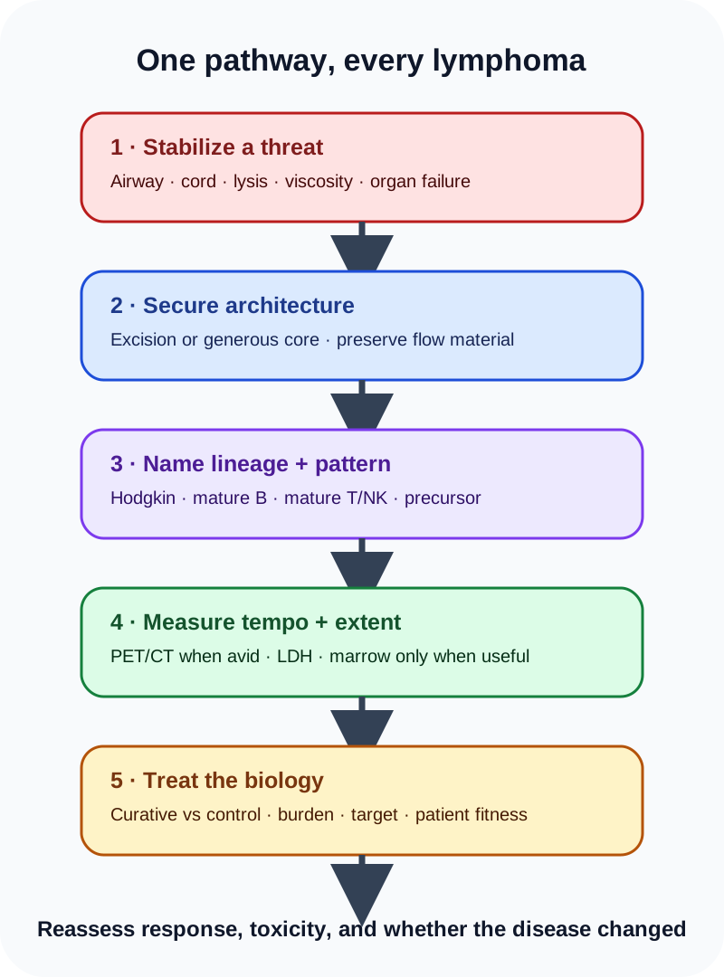
Use the same sequence every time: threat check, adequate tissue, lineage and architecture, tempo and extent, then biology-matched treatment. [1][2]
Tempo narrows the differential but does not replace tissue. Waxing and waning small nodes over years fit an indolent lymphoma; a mass doubling over weeks suggests aggressive disease or transformation. Fever, drenching sweats, and unintentional weight loss are constitutional “B symptoms” used most formally in Hodgkin staging. Pruritus, fatigue, alcohol-related node pain, elevated LDH, or extranodal symptoms can be useful, but none is diagnostic by itself.
Test the order on a stable patient with a rapidly enlarging accessible node and an inconclusive aspirate. Choose the step that protects diagnosis before therapy. Excisional biopsy preserves architecture; empiric steroids can create treatment-altered tissue and leave the lineage unresolved.
Try it here · Checkpoint 1 of 3
Make your prediction before reading the choices. A first attempt is just a starting point.
Case 1
Show answer and explanations for case 1
A. Repeat fine-needle aspiration with a larger needle (Why this does not fit)
Repeating aspiration may yield more cells, but it still does not reliably preserve nodal architecture. The unresolved question is the relationship among tumor cells, follicles, fibrosis, and background cells, so another architecture-poor sample is unlikely to settle it.
Reasoning steps for option A
Why is a second aspiration tempting after a nondiagnostic FNA?
It is quick and minimally invasive, and a larger needle may collect more cells.
What does a repeat aspiration still fail to provide?
It does not reliably preserve nodal architecture, which is what the unclassified atypical cells need.
B. Excisional biopsy of the accessible cervical node (Best answer)
An excisional specimen preserves architecture and supplies tissue for morphology, immunohistochemistry, flow cytometry, and molecular testing. Because the patient is stable and the node is safely accessible, this is the highest-yield route to a specific lymphoma diagnosis.
Reasoning steps for option B
What does the pathologist need that the FNA could not supply?
Lymphoma classification depends on architecture plus tissue for immunohistochemistry, flow cytometry, and molecular tests.
Why is excision the right procedure for this man?
He is stable and the cervical node is safely accessible, so an excisional biopsy gives the highest diagnostic yield.
C. Begin prednisone and repeat imaging in one week (Why this does not fit)
Glucocorticoids can transiently shrink lymphoma and may destroy the very cells needed for diagnosis. There is no immediate organ threat in this stem, so empiric steroid treatment before adequate tissue risks converting a clear biopsy into a nondiagnostic one.
Reasoning steps for option C
Why might a short steroid trial look attractive?
Steroids can shrink lymphoid masses quickly and might seem to test for lymphoma.
Why is prednisone harmful before tissue diagnosis here?
Steroids can destroy the diagnostic cells, and there is no organ threat that justifies treating before biopsy.
D. Order PET-CT and assign a lymphoma stage before biopsy (Why this does not fit)
PET-CT can map metabolically active disease and guide a biopsy site, but staging presumes that a lymphoma diagnosis exists. Imaging cannot replace histology when the lineage and entity remain unknown.
Reasoning steps for option D
Why does PET-CT seem useful with nodes in two regions?
PET-CT maps metabolically active disease and can point to the best biopsy site.
Why cannot staging come first?
Staging assumes a diagnosis; imaging cannot identify the lineage or entity that the biopsy must establish.
Takeaway: When an accessible suspicious node can be removed safely, preserve architecture before treatment changes the tissue.
Classic Hodgkin lymphoma is a microenvironment disease
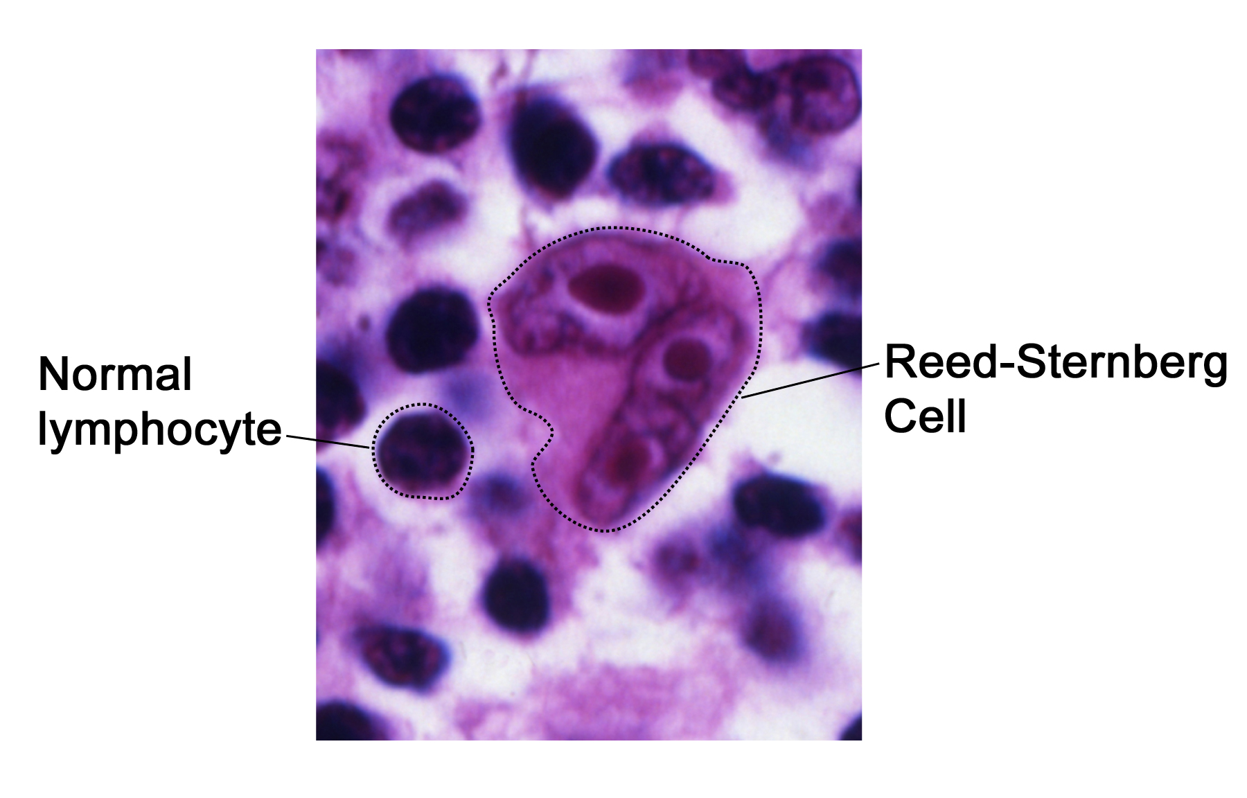
In classic Hodgkin lymphoma (cHL), the malignant Hodgkin/Reed-Sternberg population may be only a small fraction of the node. The surrounding lymphocytes, eosinophils, plasma cells, macrophages, and fibrosis are part of the disease biology. A classic binucleate cell with prominent nucleoli suggests the pattern, but morphology must be interpreted with immunophenotype and architecture because Hodgkin-like cells can occur in EBV-positive and immune-dysregulation disorders. "Owl eyes" are memorable; they are not a stand-alone molecular barcode. [1]
The size contrast makes the malignant cell conspicuous, but diagnosis still requires architecture and immunophenotype because Hodgkin-like cells have mimics. Image: U.S. National Cancer Institute; original source; Public domain (U.S. federal government work). [16].
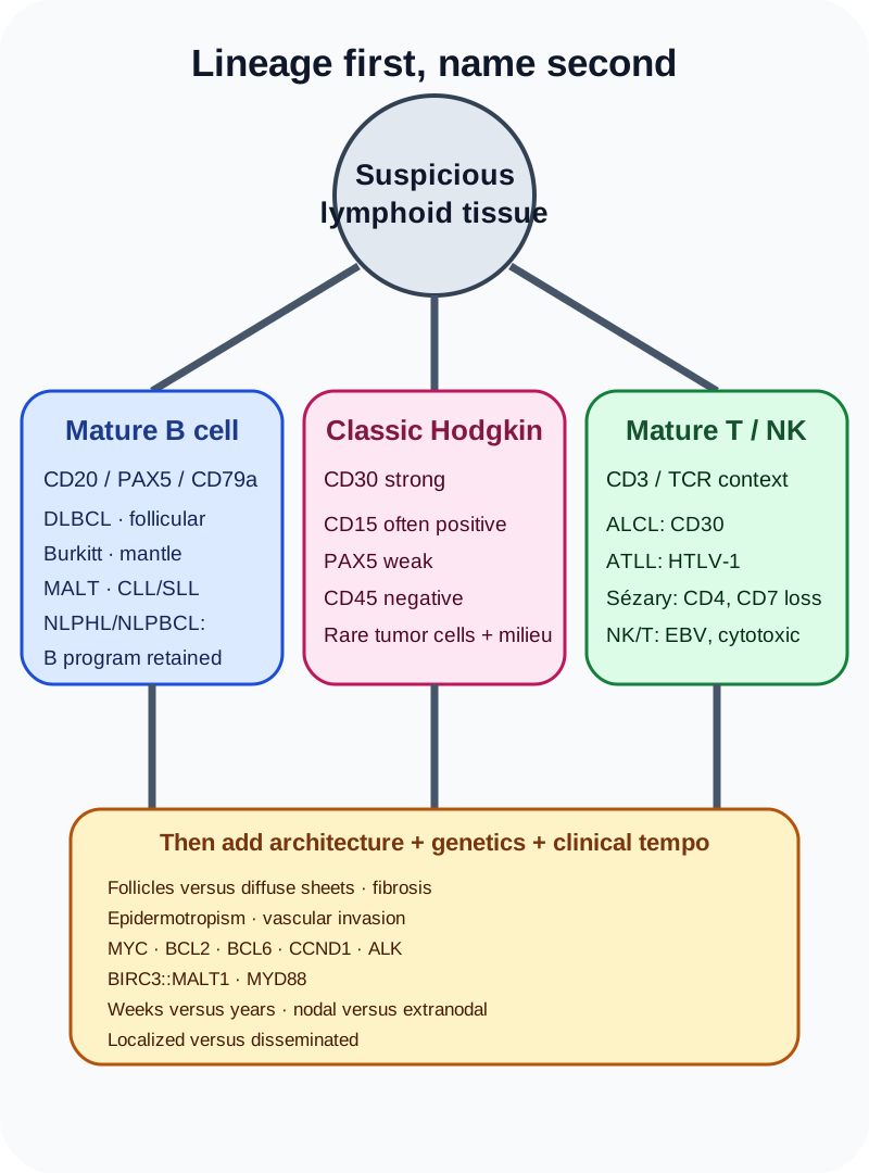
cHL is characteristically CD30 strong, often CD15 positive, weak PAX5 positive, and CD45 negative; CD20 may be absent or weak and variable. Nodular sclerosis cHL commonly affects adolescents and young adults, often with mediastinal disease, broad collagen bands, and lacunar-type cells. Mixed cellularity is more often associated with EBV and advanced disease. Lymphocyte-rich and lymphocyte-depleted patterns exist, but subtype labels do not substitute for stage, patient factors, and response.
Nodular lymphocyte-predominant Hodgkin lymphoma is biologically different enough that current sources also use the name nodular lymphocyte-predominant B-cell lymphoma. Its LP or “popcorn” cells retain a mature B-cell program: CD20, OCT2, BOB1, and BCL6 are typically expressed, while CD30 and CD15 are usually absent. It often behaves indolently, but variant growth patterns and transformation to large B-cell lymphoma matter. [1][15]
Stage describes distribution, not histologic aggressiveness. Stage I is one nodal region or one lymphatic structure; stage II involves multiple regions on one side of the diaphragm; stage III involves nodes on both sides of the diaphragm, or nodes above the diaphragm with splenic involvement; stage IV is diffuse noncontiguous extranodal disease such as marrow, liver, or lung involvement. Modern Lugano practice uses PET-CT for FDG-avid histologies and treats “limited” versus “advanced” disease as the clinically useful split. The older A/B and E/S suffix language is not applied identically across all lymphoma types. [2][18]
Compare two large-cell patterns before naming them. A CD30-strong, weak-PAX5 cell in an inflammatory background supports cHL, whereas a CD20-strong, OCT2/BOB1-positive popcorn cell supports NLPHL/NLPBCL. The consequence is a different disease category and treatment plan.
For mature B-cell lymphoma, combine architecture, phenotype, and tempo
Diffuse large B-cell lymphoma (DLBCL) produces sheets of large cells and usually behaves aggressively. CD20 establishes B-cell differentiation in many cases, but treatment planning also considers stage, IPI risk, cell-of-origin studies, and MYC, BCL2, and BCL6 rearrangements when morphology or phenotype raises concern for high-grade disease. A “double expressor” by immunohistochemistry is not the same as a rearrangement-defined lymphoma; FISH answers the rearrangement question.
Follicular lymphoma preserves a follicular growth pattern and usually expresses CD10, BCL6, CD20, and aberrant BCL2 within neoplastic follicles. The classic t(14;18) places BCL2 under immunoglobulin enhancer control, blocking apoptosis rather than directly accelerating mitosis. Asymptomatic low-burden advanced disease can be observed because immediate treatment has not consistently improved overall survival. Localized stage I disease can be treated with involved-site radiation in appropriate patients. New focal rapid growth, rising LDH, or new B symptoms requires PET-directed biopsy of the most suspicious site to evaluate histologic transformation; do not call follicular transformation “Richter syndrome.” [7]
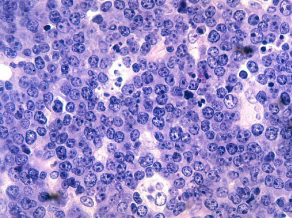
Burkitt lymphoma is a proliferation emergency. MYC rearrangement, classically t(8;14), drives near-continuous cycling, producing a very high Ki-67 index, brisk apoptosis, and the macrophage-rich “starry sky.” The same turnover creates tumor-lysis risk before and immediately after therapy. Treatment uses urgent intensive multiagent chemoimmunotherapy with CNS-directed therapy, not a standard DLBCL course. [5]
The pale stars are macrophages clearing apoptotic debris in a highly proliferative tumor. Pattern recognition supports, but does not replace, immunophenotype and MYC testing. Image: U.S. National Cancer Institute; original source; Public domain (U.S. federal government work). [17].
Mantle cell lymphoma usually coexpresses CD20 and CD5, overexpresses cyclin D1 from t(11;14), and is commonly SOX11 positive. CD23 is usually absent or weak, helping separate it from CLL/SLL, which is typically CD5 positive and CD23 positive with dim B-cell markers. The entities overlap enough that cyclin D1, SOX11, LEF1, FISH, and morphology may be needed rather than relying on one surface marker.
Extranodal marginal zone lymphoma of MALT often arises after chronic antigenic stimulation. Localized gastric MALT lymphoma associated with H. pylori is treated first with eradication therapy and then endoscopic/histologic reassessment; regression may take months. BIRC3::MALT1 from t(11;18) activates NF-κB independently of the organism and predicts a lower chance of antibiotic-only response, so persistent localized disease often moves to involved-site radiation. [13]
Primary mediastinal large B-cell lymphoma often affects a young adult with a bulky anterior mediastinal mass. Its biology overlaps with cHL, so architecture and a full panel matter. Primary CNS lymphoma is usually a DLBCL confined to the CNS/eye at diagnosis; treatment depends on CNS-penetrating high-dose methotrexate combinations, not ordinary R-CHOP alone. If the patient is stable, avoid pre-biopsy steroids when possible because they can obscure the lesion. [5][10]
A shared site or one marker never outranks the integrated lineage, architecture, genetics, and clinical tempo. [1]
Apply the framework to a mass that doubled in three weeks. Use tempo plus architecture to narrow the field, then use genetic testing to separate MYC-driven Burkitt lymphoma from other aggressive B-cell entities. The name changes the urgency, tumor-lysis preparation, and treatment program.
Translocations explain the behavior when you read the gene, not just the numbers
Do not use a translocation as a free-floating flash card. Ask what it does. BCL2 keeps cells alive; MYC makes them divide; cyclin D1 advances the cell cycle; ALK sends a constitutive growth signal; BIRC3::MALT1 supplies a survival signal that no longer depends on the inciting microbe. Also distinguish rearrangements from protein expression: MYC and BCL2 staining can be high without both genes being rearranged.
Test this: t(14;18) to BCL2 to what cell behavior?
BCL2 overexpression blocks apoptosis, so abnormal follicular B cells persist instead of dying on schedule.
Transfer the same method to t(8;14): MYC increases proliferation, producing rapid growth, high metabolic demand, and tumor-lysis risk.
Try it here · Checkpoint 2 of 3
Make your prediction before reading the choices. A first attempt is just a starting point.
Case 17
Show answer and explanations for case 17
A. Urgent Burkitt-directed therapy with tumor-lysis and CNS prophylaxis (Best answer)
The growth rate, germinal-center phenotype, near-universal proliferation, and IG::MYC rearrangement establish Burkitt lymphoma. Curative therapy is urgent and intensive, with aggressive hydration, urate-lowering therapy, electrolyte monitoring, and CNS-directed components built into the regimen.
Reasoning steps for option A
Which findings establish Burkitt lymphoma?
A CD10-positive B-cell proliferation with nearly 100% Ki-67 and an IG::MYC rearrangement.
What must the treatment plan include?
Urgent intensive therapy with tumor-lysis prevention, given LDH 6 times normal and uric acid 11.8, plus CNS-directed therapy.
B. Watchful waiting because the patient has no B symptoms (Why this does not fit)
Burkitt lymphoma can double in roughly a day and is rapidly fatal without treatment. B symptoms are not required to establish urgency; the tumor burden and biochemical tumor-lysis risk already demand immediate action.
Reasoning steps for option B
Why might the absence of B symptoms suggest a slow course?
B symptoms often signal aggressive disease.
Why is waiting dangerous here?
Burkitt lymphoma can double in about a day, and the metabolic findings already show high tumor-lysis risk.
C. Single-agent rituximab after the uric acid normalizes (Why this does not fit)
Rituximab contributes to modern Burkitt regimens but is not adequate alone. Waiting for spontaneous biochemical normalization would lose time and does not control the highly proliferative lymphoma.
Reasoning steps for option C
Why might rituximab seem a safe first step?
Rituximab is part of modern Burkitt regimens and is well tolerated.
Why is rituximab alone inadequate?
It cannot control a tumor with nearly 100% proliferation, and waiting for urate to settle loses time.
D. Standard six-cycle ABVD without CNS therapy (Why this does not fit)
ABVD treats classic Hodgkin lymphoma and does not match MYC-driven mature B-cell biology. Burkitt regimens are dose-intensive and include CNS-directed treatment because CNS relapse is a recognized risk.
Reasoning steps for option D
Why might ABVD be considered for a young patient with lymphoma?
ABVD is a well-known curative regimen for young adults with Hodgkin lymphoma.
Why is ABVD wrong for this tumor?
It treats classic Hodgkin lymphoma, not MYC-driven B-cell disease, and it lacks CNS-directed therapy.
Takeaway: Burkitt lymphoma is a treatment emergency: prevent tumor lysis, use an intensive disease-specific regimen, and include CNS-directed therapy.
Treatment follows curability, burden, and target expression
Early cHL is treated with a risk- and PET-adapted plan that balances disease control against late cardiac, pulmonary, endocrine, and second-cancer effects. A negative interim PET is evidence of chemosensitivity, not permission to invent a universal number of cycles or automatically omit radiation outside the protocol being followed. Advanced cHL is now commonly treated with nivolumab-AVD or brentuximab vedotin-AVD; S1826 favored nivolumab-AVD over BV-AVD in stage III-IV disease, while ABVD remains a viable option in some settings. [3][4]
For untreated higher-risk DLBCL, pola-R-CHP improves progression-free survival compared with R-CHOP and is a current first-line option; R-CHOP remains appropriate in other settings and when polatuzumab is unavailable or unsuitable. Early primary refractory disease or relapse within a year can proceed directly to CD19 CAR T-cell therapy in eligible patients, whereas later chemosensitive relapse may still use salvage therapy followed by autologous transplant. Bispecific CD20×CD3 antibodies and other targeted agents expand later-line options. [5][6]
Systemic ALCL is uniformly CD30 positive. Brentuximab vedotin plus cyclophosphamide, doxorubicin, and prednisone replaces vincristine with a CD30-directed antibody-drug conjugate and improved outcomes over CHOP in ECHELON-2. ALK positivity is biologically and prognostically important, especially in younger patients, but it does not make a symptomatic systemic lymphoma an observation-only disease. [8][9]
Relapsed cHL is unusually sensitive to PD-1 blockade because 9p24.1 alterations increase PD-L1/PD-L2 signaling. Response can bridge an eligible patient to autologous transplant or be incorporated into a specialist salvage plan. CD19 CAR T cells are genetically redirected T cells, not anti-CD20 antibodies; cytokine-release syndrome and ICANS require protocolized recognition and treatment.
Take two patients with DLBCL: one is untreated with IPI 3, and the other relapses seven months after first-line therapy. Preserve curative intent in both, but let relapse timing and treatment target separate first-line pola-R-CHP from an early CAR T-cell pathway.
Try it here · Checkpoint 3 of 3
Make your prediction before reading the choices. A first attempt is just a starting point.
Case 12
Show answer and explanations for case 12
A. Second-line CD19 CAR T-cell therapy evaluation (Best answer)
Primary refractory DLBCL or relapse within 12 months has poor outcomes with the traditional salvage-chemotherapy-to-autologous-transplant pathway. Randomized second-line data support CD19 CAR T-cell therapy for eligible patients in this early-relapse group.
Reasoning steps for option A
Why does the timing of relapse matter?
Relapse 7 months after first-line therapy places the patient in the early-relapse group with poor outcomes on standard salvage.
Which therapy has randomized support for this group?
Second-line CD19 CAR T-cell therapy improves outcomes over salvage chemotherapy and autologous transplant for eligible patients.
B. Routine salvage chemotherapy followed by autologous transplant regardless of response (Why this does not fit)
This was the historical pathway and remains useful for later chemosensitive relapse. Early relapse within a year is the group in which CAR T-cell therapy has displaced an automatic salvage-to-transplant sequence for eligible patients.
Reasoning steps for option B
Why is salvage chemotherapy with autologous transplant familiar?
It was the historical standard for relapsed DLBCL and still works for later chemosensitive relapse.
Why is it not preferred for this relapse?
Relapse within 12 months is where CAR T-cell therapy has replaced the automatic salvage-to-transplant route.
C. Observation with repeat PET-CT in three months (Why this does not fit)
Biopsy has confirmed an aggressive lymphoma recurrence. Waiting without treatment risks rapid progression and does not clarify an already established diagnosis.
Reasoning steps for option C
Why might repeat imaging seem reasonable first?
Surveillance imaging can clarify whether an abnormality is progressing.
Why is waiting harmful here?
The recurrence is already biopsy-confirmed aggressive lymphoma, so delay only allows progression.
D. Indefinite prednisone monotherapy (Why this does not fit)
Steroids can transiently reduce lymphoma burden but do not provide durable curative control. They can also complicate cellular-therapy collection and infection risk if used without a defined bridge plan.
Reasoning steps for option D
Why might steroids be used in relapsed lymphoma?
Steroids can shrink lymphoma temporarily and relieve symptoms.
Why is prednisone alone not a strategy?
It gives no durable control and may complicate cellular-therapy collection and infection risk.
Takeaway: Biopsy-confirmed DLBCL relapse within 12 months should trigger prompt second-line CAR T-cell evaluation in an eligible patient.
Reassess response, toxicity, and a changed disease
PET response is interpreted with the Deauville five-point scale for FDG-avid lymphomas. A new focal lesion, a discordantly growing node, or a sudden LDH rise should trigger a new diagnostic question rather than automatic escalation of the old regimen. Re-biopsy is especially important when an indolent lymphoma may have transformed or when relapse histology could change therapy. Routine surveillance imaging after remission is discouraged when it adds radiation and false positives without a clinical indication. [2][18]
Bleomycin can cause inflammatory pneumonitis and fibrosis. New cough, dyspnea, hypoxemia, or a falling diffusing capacity warrants prompt evaluation and usually holding further bleomycin while competing causes are assessed. Doxorubicin threatens cardiac function; vinca alkaloids threaten nerves; alkylators and radiation contribute to infertility and later malignancy. Toxicity monitoring is tied to the actual drugs received, not the lymphoma label.
Anti-CD20 therapy can reactivate hepatitis B even when HBsAg is negative and anti-HBc shows past infection. Screen with HBsAg, total anti-HBc, and anti-HBs before systemic therapy. Chronic infection needs antiviral therapy; past infection receiving anti-CD20 therapy generally needs prophylaxis during treatment and for at least 12 months afterward, with individualized longer follow-up. [14]
Some patterns sit outside the B-cell/Hodgkin split. Adult T-cell leukemia/lymphoma links HTLV-1, CD4-lineage cells, skin/nodal disease, hypercalcemia, and “flower” nuclei. Sézary syndrome combines erythroderma with a circulating clonal T-cell population, often CD4 positive with loss of CD7; modern criteria emphasize a demonstrable clone and substantial blood tumor burden. Waldenström macroglobulinemia combines lymphoplasmacytic marrow infiltration with an IgM paraprotein and commonly MYD88 mutation. Symptomatic hyperviscosity is treated immediately with plasmapheresis while disease-directed therapy is organized. [7][8][11]
The reusable sequence is: stabilize threats, obtain architecture, name lineage and biologic driver, stage with the right imaging, choose a regimen for that exact entity and risk group, then reassess both response and toxicity. When behavior changes, ask whether the disease changed before assuming the old diagnosis still explains it.
A formerly indolent lymphoma now has one rapidly enlarging, highly avid node. Do not simply intensify the old regimen. A new biopsy can establish transformation, while the changed behavior identifies the site most likely to answer the new question.
Apply the system
Case 2
Show answer and explanations for case 2
A. Classic Hodgkin lymphoma (Best answer)
Classic Hodgkin lymphoma places sparse Hodgkin/Reed-Sternberg cells in an abundant reactive background. Strong CD30, frequent CD15, weak B-cell transcription-factor expression, and loss of CD45 form the characteristic immunophenotypic constellation.
Reasoning steps for option A
Which morphologic feature suggests Hodgkin lymphoma?
Rare, very large binucleate cells with prominent nucleoli sit in a mixed inflammatory background.
Which marker panel confirms the classic Hodgkin program?
Strong CD30, CD15 positivity, weak PAX5, and absent CD45 form the classic Hodgkin constellation.
B. Nodular lymphocyte-predominant Hodgkin lymphoma (NLPHL/NLPBCL) (Why this does not fit)
LP cells can be large and multilobated, so morphology alone can attract this choice. They usually retain strong mature B-cell markers such as CD20, OCT2, and BOB1 and are generally CD30 and CD15 negative, unlike the supplied panel.
Reasoning steps for option B
Why can LP cells be confused with the large cells in this node?
LP cells are also large, rare, and multilobated in a reactive background.
Which markers separate NLPHL/NLPBCL from this panel?
LP cells keep a strong B-cell program and are usually CD30 and CD15 negative, the opposite of these cells.
C. Diffuse large B-cell lymphoma (Why this does not fit)
DLBCL can contain very large atypical B cells and may occasionally express CD30. It ordinarily forms sheets of malignant cells with a stronger B-cell program rather than rare CD45-negative cells embedded in a classic Hodgkin microenvironment.
Reasoning steps for option C
Why might DLBCL be considered with very large atypical cells?
DLBCL consists of large B cells and can occasionally express CD30.
What pattern argues against DLBCL here?
DLBCL forms sheets with a strong B-cell program, not rare CD45-negative cells in an inflammatory background.
D. Systemic anaplastic large-cell lymphoma (Why this does not fit)
Systemic ALCL is often strongly CD30 positive and can contain hallmark cells, making it a real mimic. The weak PAX5-positive, CD15-positive, CD45-negative profile in a Hodgkin-type background supports cHL rather than a mature T-cell lymphoma.
Reasoning steps for option D
Why is ALCL a genuine mimic of these cells?
Systemic ALCL is strongly CD30 positive and has large hallmark cells.
Which findings favor classic Hodgkin over ALCL?
CD15 positivity, weak PAX5, and a Hodgkin-type inflammatory background point to cHL rather than a T-cell lymphoma.
Takeaway: Diagnose classic Hodgkin lymphoma from the architecture plus marker constellation, not from an 'owl-eye' cell alone.
A. Nodular sclerosis classic Hodgkin lymphoma (Best answer)
The age, mediastinal presentation, collagen bands, nodular architecture, and lacunar cells form the classic nodular sclerosis pattern. CD30 expression supports the classic Hodgkin lineage within that architectural subtype.
Reasoning steps for option A
Which clinical features fit the nodular sclerosis subtype?
A young woman with supraclavicular nodes and a bulky anterior mediastinal mass is the typical presentation.
B. Mixed cellularity classic Hodgkin lymphoma (Why this does not fit)
Mixed cellularity cHL can show abundant inflammatory cells and classic HRS cells, often with EBV association. Broad fibrous bands and lacunar cells in a young patient with mediastinal disease point instead to nodular sclerosis.
Reasoning steps for option B
Why might mixed cellularity be considered for classic Hodgkin lymphoma?
It is a common cHL subtype with abundant inflammatory cells and classic HRS cells.
Which features point away from mixed cellularity?
Broad fibrous bands and lacunar cells in a young adult with mediastinal disease indicate nodular sclerosis.
C. Primary mediastinal large B-cell lymphoma (Why this does not fit)
PMBL also affects young adults with an anterior mediastinal mass and can show sclerosis. It is a large B-cell lymphoma with a preserved B-cell phenotype and sheets of tumor cells, not a lacunar-cell cHL pattern divided by broad collagen bands.
Reasoning steps for option C
Why is PMBL a serious competitor in this young woman?
PMBL also presents as an anterior mediastinal mass in young adults and can show sclerosis.
Which biopsy findings argue against PMBL?
PMBL shows sheets of large B cells, not CD30-positive lacunar cells in nodules divided by collagen bands.
D. Nodular lymphocyte-predominant Hodgkin lymphoma (NLPHL/NLPBCL) (Why this does not fit)
This indolent B-cell-pattern lymphoma often presents with peripheral nodes and LP cells in B-cell-rich nodules. Mediastinal disease, lacunar cells, collagen bands, and CD30 positivity are not its typical combination.
Reasoning steps for option D
Why might a nodular lymphoma name attract attention here?
The biopsy describes nodules, and NLPHL/NLPBCL is a nodular Hodgkin-related entity.
Why does NLPHL/NLPBCL not fit the combination?
It usually involves peripheral nodes with CD30-negative LP cells, not mediastinal disease with lacunar cells and collagen bands.
Takeaway: Young adult plus mediastinal disease plus fibrous bands and lacunar cells is the nodular sclerosis cHL pattern.
Stage II remains confined to two or more nodal regions on the same side of the diaphragm. The para-aortic and splenic disease place involved lymphatic tissue below the diaphragm as well as above it.
Reasoning steps for option A
Why might stage II be chosen for multiple nodal regions?
Stage II includes two or more nodal regions.
Which finding excludes stage II?
Stage II must stay on one side of the diaphragm, but para-aortic and splenic disease lie below it.
B. Stage III (Best answer)
Nodal disease on both sides of the diaphragm is stage III, and splenic involvement remains within this advanced lymphatic distribution. There is no diffuse noncontiguous extranodal organ disease to force stage IV.
Reasoning steps for option B
Where is disease located relative to the diaphragm?
Cervical and mediastinal nodes lie above it, and para-aortic nodes and splenic lesions lie below it.
Why does splenic involvement not push this to stage IV?
The spleen is lymphatic tissue, so nodal disease on both sides of the diaphragm with splenic involvement is stage III.
C. Stage IV (Why this does not fit)
Stage IV requires diffuse or disseminated noncontiguous extranodal involvement such as marrow, liver, lung, or bone disease. The spleen is a lymphoid organ and does not by itself convert this pattern to stage IV.
Reasoning steps for option C
Why might splenic lesions seem to mean stage IV?
The spleen is an organ outside lymph nodes, and organ disease often suggests stage IV.
What does stage IV actually require?
Diffuse noncontiguous extranodal disease such as marrow, liver, lung, or bone, none of which is present.
D. Stage IIE (Why this does not fit)
The E designation historically describes contiguous extension into an adjacent extranodal site, and stage II must remain on one side of the diaphragm. This patient crosses the diaphragm and has no described contiguous extranodal extension.
Reasoning steps for option D
Why might an E designation seem relevant to the spleen?
The E suffix marks extension beyond lymph nodes.
Why is stage IIE incorrect here?
Stage II cannot cross the diaphragm, and no contiguous extranodal extension is described.
Takeaway: Crossing the diaphragm makes lymphoma stage III; splenic disease is not automatically stage IV.
A. This is a complete metabolic response; continue the protocol's planned de-escalated pathway (Best answer)
Deauville 1 to 3 is generally treated as PET-negative in standard response frameworks, and a score of 2 indicates complete metabolic response. The treatment consequence should follow the validated protocol and baseline risk rather than a universal rule invented from the scan.
Reasoning steps for option A
What does a Deauville score of 2 mean?
Uptake no higher than mediastinal blood pool is a complete metabolic response.
How should the result change treatment?
It directs the validated protocol's planned de-escalated pathway rather than an improvised rule.
B. This is refractory disease; switch immediately to escalated BEACOPP (Why this does not fit)
Escalation is considered for inadequate response, not uptake below or equal to mediastinal blood pool. Calling a Deauville 2 scan refractory reverses the meaning of the five-point scale and adds toxicity without evidence of treatment failure.
Reasoning steps for option B
Why might residual uptake raise concern about treatment failure?
Any uptake after chemotherapy can look like persistent disease.
Why is escalation to BEACOPP wrong after this scan?
Deauville 2 is PET-negative, so there is no inadequate response to justify more toxic therapy.
C. Add involved-site radiation regardless of the protocol because every early-stage case requires it (Why this does not fit)
Radiation remains appropriate in several early-stage strategies, especially according to risk and protocol. It is not an automatic requirement in every PET-negative patient, so the scan must be interpreted within the selected response-adapted plan.
Reasoning steps for option C
Why might radiation seem mandatory in early-stage disease?
Many early-stage Hodgkin strategies include involved-site radiation.
What decides whether radiation is given here?
The chosen response-adapted protocol and baseline risk decide it, not a rule that every early-stage patient needs radiation.
D. Stop all therapy because PET negativity proves eradication after the initial cycles (Why this does not fit)
Interim PET is a powerful response marker, but it does not mean that the planned curative course can simply be abandoned. Complete the validated regimen or combined-modality pathway rather than equating an early response with finished treatment.
Reasoning steps for option D
Why might a negative interim scan suggest stopping therapy?
A PET-negative result after a few cycles shows the lymphoma is responding well.
Why is stopping all therapy unsafe?
Interim PET guides the planned course; it does not prove eradication or replace completion of the curative regimen.
Takeaway: A negative interim PET guides a validated response-adapted plan; it does not create one universal cycle or radiation rule.
The phase III S1826 trial showed superior progression-free survival and a favorable tolerability profile for nivolumab-AVD compared with BV-AVD in newly diagnosed stage III-IV cHL. In this stem there is no stated contraindication to PD-1 blockade and access is not limiting.
Reasoning steps for option A
Which trial compared the leading regimens for stage III-IV cHL?
S1826 compared nivolumab-AVD with brentuximab vedotin-AVD in newly diagnosed advanced cHL.
Why does N-AVD fit this patient?
N-AVD improved progression-free survival, and the patient has no contraindication to PD-1 blockade and full access.
B. ABVD followed automatically by mediastinal radiation (Why this does not fit)
ABVD remains a viable regimen in some settings, but advanced disease is primarily treated systemically and radiation is not automatic. The stem asks for the regimen best supported by the current head-to-head randomized evidence under conditions where N-AVD is available.
Reasoning steps for option B
Why is ABVD a familiar choice for classic Hodgkin lymphoma?
ABVD is a long-established regimen that remains viable in some settings.
Why is automatic mediastinal radiation wrong for stage IV disease?
Advanced disease is treated systemically, radiation is not automatic, and N-AVD has stronger current evidence.
C. R-CHOP (Why this does not fit)
R-CHOP targets mature CD20-positive B-cell lymphomas such as DLBCL. Classic Hodgkin cells have a different phenotype and microenvironment, so this familiar lymphoma regimen does not match the diagnosed entity.
Reasoning steps for option C
Why might R-CHOP come to mind for a new lymphoma?
R-CHOP is the best-known lymphoma regimen.
Why does R-CHOP not match cHL?
It targets CD20-positive mature B-cell lymphomas, while classic Hodgkin cells have a different phenotype.
D. Escalated BEACOPP for every patient younger than 30 (Why this does not fit)
Intensified chemotherapy can be used in selected strategies, but age alone does not make it mandatory. It adds infertility, marrow, and secondary-malignancy risks, and it is not the regimen favored by the specific contemporary comparison invoked in the stem.
Reasoning steps for option D
Why might a young patient be offered intensified chemotherapy?
Escalated BEACOPP is used in selected advanced Hodgkin strategies.
Why is age alone not a reason for BEACOPP?
It adds infertility, marrow, and secondary-cancer risk and is not the regimen favored by S1826.
Takeaway: For eligible stage III-IV cHL with access, N-AVD is a current evidence-supported front-line standard rather than an automatic ABVD reflex.
A. Give the next bleomycin dose on schedule while awaiting all test results (Why this does not fit)
Continuing the suspected offending drug can worsen a potentially serious pulmonary injury. Diagnostic uncertainty does not require re-exposure when the patient has new symptoms, hypoxemia, and compatible imaging.
Reasoning steps for option A
Why might a clinician want to keep ABVD on schedule?
Delays could compromise a curative Hodgkin regimen while tests are pending.
Why is re-exposure to bleomycin unsafe here?
New cough, hypoxemia, and ground-glass opacities suggest bleomycin lung injury that further doses could worsen.
B. Hold bleomycin and urgently evaluate pulmonary toxicity and competing causes (Best answer)
Bleomycin pneumonitis is a clinical diagnosis supported by new respiratory symptoms, hypoxemia, and compatible imaging after exposure. Holding further bleomycin prevents avoidable re-exposure while infection, embolism, cardiac disease, and other causes are assessed.
Reasoning steps for option B
Which findings raise concern for bleomycin toxicity?
After cycle 3 of ABVD the patient has dry cough, dyspnea, oxygen saturation of 91%, and new bilateral ground-glass opacities.
Why hold the drug while still testing for other causes?
Holding bleomycin prevents further injury while infection, embolism, and cardiac causes are evaluated.
C. Stop doxorubicin only because ground-glass opacities indicate cardiomyopathy (Why this does not fit)
Doxorubicin can cause cardiomyopathy, but the supplied pattern is an interstitial pulmonary syndrome temporally linked to bleomycin. Cardiac evaluation may be part of the workup, yet stopping only doxorubicin leaves the more likely offending drug in place.
Reasoning steps for option C
Why might doxorubicin be blamed for new dyspnea?
Doxorubicin can cause cardiomyopathy, which also causes exertional dyspnea.
Why is stopping only doxorubicin the wrong response?
Ground-glass opacities with hypoxemia fit a bleomycin-related interstitial pattern, so the likely offender would remain.
D. Diagnose bleomycin fibrosis only if DLCO has fallen by exactly 20% (Why this does not fit)
DLCO trends can support concern but are variable and not a universal binary stopping rule. Symptoms, oxygenation, imaging, infection assessment, and clinical trajectory matter more than waiting for one rigid percentage threshold.
Reasoning steps for option D
Why might a DLCO cutoff seem objective?
Falling DLCO can signal bleomycin lung injury and is easy to measure.
Why should action not wait for a fixed percentage?
DLCO is variable; symptoms, oxygenation, and imaging already justify holding the drug.
Takeaway: New pulmonary symptoms during bleomycin therapy justify holding bleomycin while the lung injury and its mimics are evaluated.
A. Autologous hematopoietic stem-cell transplant (Best answer)
For an eligible patient with relapsed cHL who responds to salvage therapy, high-dose therapy followed by autologous stem-cell rescue remains a standard curative consolidation. Checkpoint inhibition can be part of the route to response but does not by itself replace the consolidation decision.
Reasoning steps for option A
What makes this patient eligible for curative consolidation?
The patient is fit, relapse is biopsy-confirmed, and PET-CT shows chemosensitive disease after salvage.
Which consolidation is standard for that situation?
High-dose therapy with autologous stem-cell rescue is the standard curative consolidation for chemosensitive first relapse.
B. Observation because checkpoint response proves cure (Why this does not fit)
A metabolic response to salvage is favorable, but early relapse still carries substantial recurrence risk. Observation alone discards the established consolidative role of autologous transplant in a fit responder.
Reasoning steps for option B
Why might a good checkpoint-inhibitor response suggest observation?
A metabolic response after salvage looks like the disease is controlled.
Why is observation alone inadequate after early relapse?
Relapse within 8 months carries a high recurrence risk, and autologous transplant adds proven curative benefit.
C. Allogeneic transplant as the routine first transplant (Why this does not fit)
Allogeneic transplantation can be considered after further relapse or failure of autologous strategies in selected patients. It carries greater treatment-related morbidity and is not the routine first transplant for a chemosensitive first relapse.
Reasoning steps for option C
Why might allogeneic transplant seem like the stronger option?
It adds a graft-versus-lymphoma effect that could lower relapse risk.
Why is it not the routine first transplant here?
Its higher treatment-related morbidity reserves it for later relapse or failure after autologous transplant.
D. Rituximab maintenance for two years (Why this does not fit)
Rituximab targets CD20 and has roles in several mature B-cell lymphomas. Classic Hodgkin lymphoma is chiefly CD30 driven with PD-1-pathway biology, so rituximab maintenance is not the standard consolidation described here.
Reasoning steps for option D
Why might rituximab maintenance come to mind after lymphoma relapse?
Rituximab maintenance is used after response in several B-cell lymphomas.
Why does rituximab not suit classic Hodgkin lymphoma?
cHL is driven by CD30-positive HRS cells with PD-1 pathway biology, and rituximab maintenance is not its consolidation.
Takeaway: A fit patient with salvage-responsive relapsed cHL generally proceeds to autologous transplant rather than stopping at response.
A. Nodular lymphocyte-predominant Hodgkin lymphoma (NLPHL/NLPBCL) (Best answer)
LP cells have multilobated nuclei and retain a strong mature B-cell program, including CD20, OCT2, BOB1, and BCL6. Their usual lack of CD30 and CD15 separates this entity from classic Hodgkin lymphoma; current classifications acknowledge both Hodgkin and B-cell terminology.
Reasoning steps for option A
Which morphologic clue points to LP cells?
Scattered large cells with multilobated popcorn nuclei sit within nodules.
Which markers confirm NLPHL/NLPBCL rather than cHL?
CD20, OCT2, BOB1, and BCL6 positivity with absent CD30 and CD15 show a retained B-cell program.
B. Classic Hodgkin lymphoma, nodular sclerosis subtype (Why this does not fit)
Nodular architecture can make this choice attractive, but classic Hodgkin cells usually show strong CD30 with a weakened B-cell program. The supplied strong B-cell transcription factors and absent CD30/CD15 argue against cHL.
Reasoning steps for option B
Why might the nodular architecture suggest nodular sclerosis cHL?
Both entities have a nodular growth pattern.
Which markers exclude classic Hodgkin lymphoma?
Classic Hodgkin cells are CD30 positive with a weakened B-cell program, whereas these cells are CD30 and CD15 negative with strong B-cell factors.
C. Follicular lymphoma (Why this does not fit)
Follicular lymphoma can express CD20, CD10, and BCL6 within neoplastic follicles. It does not characteristically contain scattered LP 'popcorn' cells in the distinctive rosetting microenvironment described here.
Reasoning steps for option C
Why could follicular lymphoma be considered with a nodular B-cell pattern?
Follicular lymphoma forms neoplastic follicles and expresses CD20 and BCL6.
What feature of this biopsy is not typical of follicular lymphoma?
Follicular lymphoma lacks scattered large popcorn cells in a distinct reactive nodule.
D. T-cell/histiocyte-rich large B-cell lymphoma (Why this does not fit)
THRLBCL can overlap biologically and morphologically with variant NLPHL/NLPBCL patterns. The explicitly nodular architecture and classic LP-cell phenotype favor NLPHL/NLPBCL; diffuse areas or aggressive behavior would increase concern for THRLBCL-like transformation.
Reasoning steps for option D
Why is THRLBCL a genuine overlap with this entity?
It shares large B cells in a T-cell and histiocyte background and can resemble variant patterns.
What favors NLPHL/NLPBCL over THRLBCL here?
The biopsy is clearly nodular with a classic LP-cell phenotype and slow growth, without diffuse areas or aggressive behavior.
Takeaway: LP 'popcorn' cells retain a full B-cell program and are usually CD30/CD15 negative, unlike classic Hodgkin cells.
A. FISH for MYC, BCL2, and BCL6 rearrangements (Best answer)
Protein overexpression does not establish a chromosomal rearrangement. FISH directly evaluates MYC, BCL2, and BCL6 loci and separates a double-expressor phenotype from rearrangement-defined high-grade disease that may change classification and management.
Reasoning steps for option A
What does high MYC and BCL2 protein expression establish?
It defines a double-expressor phenotype, not a genetic rearrangement.
Which test answers the rearrangement question?
FISH for MYC, BCL2, and BCL6 directly detects rearrangements that define high-grade biology.
B. Repeat CD20 immunohistochemistry (Why this does not fit)
CD20 already establishes a mature B-cell phenotype and guides anti-CD20 treatment. Repeating the same stain does not answer whether MYC or BCL2/BCL6 genes are rearranged.
Reasoning steps for option B
Why might CD20 be rechecked in a B-cell lymphoma?
CD20 confirms B-cell lineage and guides anti-CD20 therapy.
Why does repeating CD20 not help?
CD20 is already positive and says nothing about MYC or BCL2 gene rearrangement.
C. Serum protein electrophoresis (Why this does not fit)
SPEP can identify a monoclonal immunoglobulin in plasma-cell or lymphoplasmacytic disorders. It does not determine the rearrangement status of a large B-cell lymphoma.
Reasoning steps for option C
Why could SPEP be ordered for a B-cell neoplasm?
SPEP detects monoclonal immunoglobulin from B-lineage and plasma-cell disorders.
What question does SPEP leave unanswered?
It cannot determine whether the lymphoma carries MYC, BCL2, or BCL6 rearrangements.
D. Peripheral-blood BCR::ABL1 PCR (Why this does not fit)
BCR::ABL1 testing is central to chronic myeloid and selected acute lymphoblastic leukemias. It is not the molecular discriminator for a MYC/BCL2-expressing DLBCL pattern.
Reasoning steps for option D
Why might a translocation test such as BCR::ABL1 seem relevant?
BCR::ABL1 is a classic diagnostic rearrangement in hematologic cancer.
Why is it the wrong molecular test here?
BCR::ABL1 defines CML and some ALL, not high-grade large B-cell lymphoma.
Takeaway: A double-expressor stain pattern is not a double-hit diagnosis; use rearrangement testing to answer the genetic question.
POLARIX replaced vincristine with polatuzumab vedotin and improved progression-free survival compared with R-CHOP in untreated intermediate- and high-risk DLBCL. Current NCI guidance lists pola-R-CHP for IPI 2 to 5, matching this patient's profile.
Reasoning steps for option A
Which features qualify this patient for pola-R-CHP?
The patient has untreated stage III DLBCL with IPI 3 and no contraindication to polatuzumab vedotin.
What evidence supports pola-R-CHP?
POLARIX showed better progression-free survival than R-CHOP, and NCI guidance lists it for IPI 2 to 5.
B. ABVD (Why this does not fit)
ABVD is a classic Hodgkin lymphoma regimen and does not provide the anti-CD20 and DLBCL-directed combination required here. The pathology is a mature large B-cell lymphoma, not cHL.
Reasoning steps for option B
Why might ABVD come up as a curative lymphoma regimen?
ABVD cures many patients with classic Hodgkin lymphoma.
Why does ABVD not treat DLBCL?
It lacks anti-CD20 therapy and is designed for cHL, not a mature large B-cell lymphoma.
C. Rituximab alone (Why this does not fit)
Single-agent rituximab may control selected indolent CD20-positive lymphomas. DLBCL is potentially curable but aggressive, so antibody monotherapy is inadequate for a fit patient with stage III disease.
Reasoning steps for option C
Why might rituximab alone seem gentle and targeted?
Rituximab targets CD20 and controls some indolent B-cell lymphomas.
Why is monotherapy inadequate here?
DLBCL is aggressive but curable, and stage III disease in a fit patient needs combination therapy.
D. Watchful waiting until B symptoms appear (Why this does not fit)
Observation is appropriate for selected asymptomatic indolent lymphomas. Untreated DLBCL progresses quickly and loses a curative window; stage III disease requires prompt systemic therapy regardless of B symptoms.
Reasoning steps for option D
Why might observation be considered without B symptoms?
Watchful waiting is standard for some asymptomatic indolent lymphomas.
Why is waiting dangerous in DLBCL?
DLBCL progresses quickly, and delay risks losing the curative window regardless of symptoms.
Takeaway: For untreated IPI 2-5 DLBCL with access and no contraindication, pola-R-CHP is a current front-line option; R-CHOP remains contextually appropriate.
The timing, fever, hypotension, and hypoxemia fit cytokine-release syndrome. Tocilizumab blocks the IL-6 receptor and is first-line targeted treatment for clinically significant CRS while infection is evaluated and supported in parallel.
Reasoning steps for option A
Which features point to cytokine-release syndrome?
Fever, hypotension needing vasopressor, and hypoxemia five days after CD19 CAR T infusion fit CRS.
Why is tocilizumab the targeted treatment?
It blocks the IL-6 receptor and is first-line for significant CRS while infection is still covered.
B. Rituximab (Why this does not fit)
Rituximab depletes CD20-positive B cells but does not directly interrupt IL-6 signaling in CRS. It could also worsen B-cell depletion without treating the acute inflammatory syndrome.
Reasoning steps for option B
Why might rituximab seem to target the problem cells?
It is a B-cell-directed antibody used across B-cell lymphoma care.
Why does rituximab not treat CRS?
It does not block IL-6 signaling and would only add B-cell depletion.
C. Plasmapheresis (Why this does not fit)
Plasmapheresis rapidly reduces circulating IgM in symptomatic hyperviscosity. CRS is driven by cellular activation and cytokine signaling, not a removable monoclonal immunoglobulin burden.
Reasoning steps for option C
Why could plasmapheresis seem to clear inflammatory mediators?
It removes circulating proteins quickly.
Why is plasmapheresis not the targeted therapy?
CRS is driven by ongoing cellular activation and cytokine signaling, not a removable paraprotein.
D. Intrathecal methotrexate (Why this does not fit)
Intrathecal therapy treats or prevents selected CNS lymphoma involvement. It does not address the systemic fever, shock, and oxygen requirement of CRS.
Reasoning steps for option D
Why might CNS-directed therapy be considered after CAR T?
Neurologic complications and CNS lymphoma both occur in this setting.
Why does intrathecal methotrexate not address the problem?
The patient has systemic fever, shock, and hypoxemia from CRS, not CNS lymphoma.
Takeaway: After CAR T therapy, fever plus hypotension or hypoxemia suggests CRS; treat the syndrome while still covering infection.
A. Active surveillance with scheduled clinical reassessment (Best answer)
Asymptomatic low-tumor-burden advanced follicular lymphoma can be observed without sacrificing overall survival. Surveillance is an active plan that monitors symptoms, examination, counts, and evidence of organ compromise rather than abandonment.
Reasoning steps for option A
Which findings show low tumor burden?
All nodes are under 3 cm, counts and organ function are normal, and there are no B symptoms or discomfort.
Why is active surveillance appropriate?
Observation of asymptomatic low-burden follicular lymphoma does not reduce overall survival and includes scheduled reassessment.
B. Immediate R-CHOP because any stage III lymphoma is aggressive (Why this does not fit)
Stage describes distribution, not tempo. Grade 1-2 follicular lymphoma is indolent, and the absence of burden or symptoms removes the usual trigger for immediate systemic treatment.
Reasoning steps for option B
Why might stage III disease seem to demand R-CHOP?
Involvement of several nodal regions sounds advanced.
Why is immediate R-CHOP not indicated?
Stage reflects distribution, not tempo; grade 1-2 follicular lymphoma is indolent and has no treatment trigger here.
C. Urgent allogeneic stem-cell transplant (Why this does not fit)
Allogeneic transplantation carries substantial morbidity and is reserved for selected multiply relapsed or high-risk situations. It is not appropriate for newly diagnosed asymptomatic low-burden disease.
Reasoning steps for option C
Why might transplant be raised for an incurable lymphoma?
Allogeneic transplant is one of the few potentially curative options in follicular lymphoma.
Why is it inappropriate now?
Its high morbidity reserves it for multiply relapsed or high-risk disease, not new asymptomatic disease.
D. Prophylactic cranial irradiation (Why this does not fit)
Routine CNS prophylaxis is not part of low-grade follicular lymphoma management. The disease pattern and risk described do not suggest a CNS-directed intervention.
Reasoning steps for option D
Why might CNS prophylaxis come up in lymphoma planning?
CNS prophylaxis is part of some aggressive lymphoma regimens.
Why is it unnecessary here?
Low-grade follicular lymphoma without high-risk features does not carry a CNS risk that warrants prophylaxis.
Takeaway: Observation is a treatment decision for asymptomatic low-burden follicular lymphoma, not a failure to treat.
Truly localized stage I follicular lymphoma can be treated with involved-site radiation and may achieve long remissions or cure. The key is careful staging that confirms disease is confined to one region.
Reasoning steps for option A
Which staging results define truly localized disease?
PET-CT and marrow assessment show only a single 2.5-cm cervical node.
Why is involved-site radiation the preferred approach?
Radiation to localized stage I follicular lymphoma can give long remissions or cure.
B. Observation is mandatory because follicular lymphoma is never curable (Why this does not fit)
Observation is reasonable for some low-burden presentations, but it is not mandatory. Localized follicular lymphoma is the important exception in which involved-site radiation can provide durable disease control with curative intent.
Reasoning steps for option B
Why might observation seem reasonable for follicular lymphoma?
Observation is appropriate for many low-burden, asymptomatic presentations.
What makes this case an exception?
Disease confined to one region can be treated with curative-intent radiation, so observation is not mandatory.
C. Six cycles of escalated BEACOPP (Why this does not fit)
BEACOPP is an intensive classic Hodgkin lymphoma regimen. This patient has an indolent mature B-cell lymphoma confined to one nodal region, so Hodgkin-directed multiagent therapy is inappropriate.
Reasoning steps for option C
Why might an intensive regimen seem to maximize cure?
Multiagent chemotherapy is used when aggressive treatment is needed.
Why is BEACOPP wrong for this patient?
It is a Hodgkin regimen, while this is an indolent B-cell lymphoma confined to one node.
D. Prophylactic intrathecal methotrexate (Why this does not fit)
Localized low-grade follicular lymphoma does not routinely require CNS prophylaxis. The disease is confined, indolent, and lacks the high-risk extranodal pattern that might prompt a separate CNS-risk discussion.
Reasoning steps for option D
Why might CNS prophylaxis be considered after a lymphoma diagnosis?
Some lymphomas carry a risk of CNS relapse.
Why is intrathecal therapy not needed here?
Localized low-grade follicular lymphoma does not carry that risk.
Takeaway: Localized follicular lymphoma is a treatment exception: involved-site radiation can be used with curative intent after complete staging.
A. PET-directed biopsy of the most metabolically active accessible site (Best answer)
Rapid focal growth, new B symptoms, and an LDH rise suggest histologic transformation, most often to DLBCL. A new biopsy is needed because treatment depends on the transformed histology; PET helps select the site most likely to contain it.
Reasoning steps for option A
Which changes suggest histologic transformation?
A node grew from 2 to 9 cm in 6 weeks with new night sweats and a marked LDH rise.
Why biopsy the PET-avid site?
Treatment depends on the transformed histology, and PET selects the site most likely to contain it.
B. Call this Richter transformation and start CLL therapy (Why this does not fit)
Richter transformation specifically describes transformation of CLL/SLL, usually to DLBCL or Hodgkin lymphoma. Follicular lymphoma can transform to an aggressive lymphoma, but that event should be called histologic transformation, not Richter syndrome.
Reasoning steps for option B
Why might Richter transformation seem the right name?
Richter transformation describes aggressive change arising from an indolent lymphoma.
Why is the term wrong here?
Richter transformation applies to CLL/SLL; follicular lymphoma undergoes histologic transformation.
C. Continue routine annual follow-up because waxing and waning is expected (Why this does not fit)
Indolent nodes can fluctuate slowly, but explosive growth in one site with new systemic symptoms and an LDH spike is a red-flag change in biology. Delaying biopsy risks missing an aggressive, potentially treatable transformation.
Reasoning steps for option C
Why might fluctuating nodes be accepted as usual follicular behavior?
Indolent follicular nodes can wax and wane.
Which findings break that pattern?
Rapid growth in one node with new B symptoms and an LDH spike signals changed biology.
D. Diagnose DLBCL from the LDH level alone (Why this does not fit)
LDH and growth tempo raise suspicion but do not establish histology. Infection, necrosis, and other lymphoma changes can also raise LDH; tissue is required before committing to a transformed-lymphoma regimen when the patient is stable.
Reasoning steps for option D
Why might LDH seem enough to diagnose DLBCL?
A marked LDH rise with rapid growth strongly suggests aggressive lymphoma.
Why is tissue still required?
LDH can rise with infection or necrosis, and treatment depends on confirmed histology.
Takeaway: Suspected follicular-lymphoma transformation requires a new biopsy; the term Richter transformation belongs to CLL/SLL.
Mantle cell lymphoma characteristically coexpresses B-cell markers and CD5, often lacks CD23, and overexpresses cyclin D1 because of the IGH::CCND1 rearrangement t(11;14). This combination separates it from the more common CD5-positive CLL phenotype.
Reasoning steps for option A
Which marker pair is shared with CLL?
Both mantle cell lymphoma and CLL are CD5-positive B-cell neoplasms.
Which results identify mantle cell lymphoma?
Bright CD20, absent CD23, cyclin D1 overexpression, and t(11;14) define it.
B. Chronic lymphocytic leukemia/small lymphocytic lymphoma (Why this does not fit)
CLL/SLL is usually CD5-positive but has dimmer CD20, usually expresses CD23, and does not carry the defining IGH::CCND1 rearrangement. A CD5-positive B-cell clone is therefore not automatically CLL.
Reasoning steps for option B
Why is CLL/SLL the first thought for CD5-positive lymphocytosis?
It is the most common CD5-positive B-cell neoplasm in older adults.
Which findings argue against CLL/SLL?
CLL usually has dim CD20 and positive CD23 and lacks t(11;14) with cyclin D1.
C. Follicular lymphoma (Why this does not fit)
Follicular lymphoma is usually a CD10-positive germinal-center B-cell neoplasm driven by BCL2 rearrangement rather than cyclin D1 overexpression. The CD5-positive t(11;14) pattern points elsewhere.
Reasoning steps for option C
Why might follicular lymphoma be considered with generalized adenopathy?
It is a common B-cell lymphoma that causes generalized nodes in older adults.
What separates follicular lymphoma from this clone?
Follicular lymphoma is CD10 positive with BCL2 rearrangement, not CD5 positive with t(11;14).
D. Classic Hodgkin lymphoma (Why this does not fit)
Classic Hodgkin lymphoma is diagnosed by rare Hodgkin/Reed-Sternberg cells in a reactive background, not by a circulating clonal CD5-positive B-cell population with t(11;14). Flow cytometry would not produce this mantle-cell signature.
Reasoning steps for option D
Why might Hodgkin lymphoma be listed for generalized adenopathy?
It is a classic cause of lymphadenopathy.
Why does the flow result exclude it?
cHL has rare HRS cells in tissue, not a circulating clonal CD5-positive B-cell population.
Takeaway: CD5 is shared; cyclin D1 and t(11;14) identify mantle cell lymphoma, whereas CD23 and dim B-cell markers favor CLL/SLL.
A. H. pylori eradication therapy followed by endoscopic reassessment (Best answer)
Chronic H. pylori-driven antigen stimulation sustains many localized gastric MALT lymphomas. Eradication is first-line because lymphoma regression can follow removal of the stimulus; response is assessed with repeat endoscopy and biopsy over time.
Reasoning steps for option A
What drives many localized gastric MALT lymphomas?
Chronic antigen stimulation from H. pylori sustains the lymphoma, and this patient's biopsies are positive.
Why start with eradication?
Removing the stimulus can make the lymphoma regress, with endoscopic biopsy to confirm the response.
B. Immediate total gastrectomy (Why this does not fit)
Gastrectomy is not routine first-line treatment for uncomplicated localized gastric MALT lymphoma. Organ-preserving antibiotic therapy, radiation, and systemic options usually control the disease without major surgery.
Reasoning steps for option B
Why might surgery seem definitive for a gastric tumor?
Gastrectomy removes the entire involved organ.
Why is gastrectomy not first-line?
Antibiotics, radiation, and systemic options control localized MALT lymphoma without major surgery.
C. Escalated BEACOPP (Why this does not fit)
BEACOPP is an intensive classic Hodgkin regimen and does not match an indolent marginal-zone lymphoma driven by a treatable gastric infection. It would add major toxicity without addressing the biologic driver.
Reasoning steps for option C
Why might multiagent chemotherapy seem thorough?
Intensive chemotherapy is used for many lymphomas.
Why is BEACOPP inappropriate here?
It is a Hodgkin regimen and does not address an indolent lymphoma driven by treatable infection.
D. No treatment because all MALT lymphomas regress spontaneously (Why this does not fit)
Spontaneous regression is not reliable. This patient has a defined microbial driver with an effective treatment, so observation alone would miss the opportunity to remove the antigenic stimulus and potentially induce remission.
Reasoning steps for option D
Why might MALT lymphoma seem harmless enough to leave alone?
Some MALT lymphomas are indolent and grow very slowly.
Why is observation wrong for this patient?
Regression is not reliable without treatment, and eradication targets a known driver.
Takeaway: Localized H. pylori-positive gastric MALT lymphoma starts with eradication therapy and planned biopsy-based reassessment.
BIRC3::MALT1 can activate NF-κB signaling independently of ongoing H. pylori stimulation and predicts a lower probability of antibiotic response. Persistent localized disease without transformation is highly radiosensitive and can be treated with involved-site radiation.
Reasoning steps for option A
What does BIRC3::MALT1 mean for the lymphoma?
It activates NF-κB signaling independently of H. pylori, which explains persistence after eradication.
Which treatment fits persistent localized disease?
Localized gastric MALT lymphoma without transformation is radiosensitive and is treated with involved-site radiation.
B. Repeat antibiotics indefinitely (Why this does not fit)
Once eradication is documented and the lymphoma persists with an antigen-independent signaling rearrangement, repeated antibiotic courses are unlikely to switch off the malignant clone. Continued infection-directed therapy would delay effective local treatment.
Reasoning steps for option B
Why might repeated antibiotics seem logical?
Eradication therapy is the usual first treatment for gastric MALT lymphoma.
Why will more antibiotics not help?
Eradication is already documented, and the rearrangement makes the clone independent of the infection.
C. Immediate CAR T-cell therapy (Why this does not fit)
CAR T therapy is reserved for selected relapsed or refractory aggressive B-cell lymphomas and certain heavily pretreated indolent diseases. Localized persistent gastric MALT can usually be controlled with far less toxic local therapy.
Reasoning steps for option C
Why might CAR T therapy be considered for persistent lymphoma?
It is a powerful option for B-cell lymphoma that has resisted earlier treatment.
Why is CAR T inappropriate here?
It is reserved for relapsed or refractory aggressive disease, while this localized lymphoma responds to radiation.
D. Prophylactic splenectomy (Why this does not fit)
The spleen is not the site of disease and its removal would not correct the gastric clone's NF-κB signaling. Splenectomy has no preventive role in this localized gastric presentation.
Reasoning steps for option D
Why might the spleen be linked to a marginal zone lymphoma?
Splenic marginal zone lymphoma is a related entity.
Why does splenectomy not help this patient?
The disease is gastric, and removing the spleen does not affect the NF-κB-driven gastric clone.
Takeaway: t(11;18)/BIRC3::MALT1 can make gastric MALT antigen-independent; persistent localized disease is treated with effective local therapy rather than endless antibiotics.
Systemic ALCL is uniformly CD30-positive, making the CD30-directed antibody-drug conjugate brentuximab a rational component. ECHELON-2 established brentuximab vedotin plus cyclophosphamide, doxorubicin, and prednisone as a preferred front-line approach for systemic ALCL.
Reasoning steps for option A
Which findings identify systemic ALCL?
Large pleomorphic CD30-strong T cells with horseshoe nuclei, ALK positivity, and systemic disease.
Why is brentuximab vedotin plus CHP preferred?
The drug targets CD30, and ECHELON-2 established BV-CHP as front-line therapy for systemic ALCL.
B. Rituximab plus bendamustine (Why this does not fit)
Rituximab targets CD20 on mature B cells. This tumor is a CD30-positive T-cell lymphoma, so a B-cell antibody does not match the lineage or defining target.
Reasoning steps for option B
Why might a B-cell regimen seem reasonable for a large-cell lymphoma?
Rituximab-based combinations are common front-line lymphoma therapy.
Why does rituximab not match this tumor?
The cells are CD30-positive T cells, so a CD20-directed antibody has no target.
C. H. pylori eradication alone (Why this does not fit)
Antibiotic eradication treats selected gastric MALT lymphomas driven by H. pylori. It has no role in systemic ALK-positive anaplastic large cell lymphoma.
Reasoning steps for option C
Why might an antibiotic regimen come to mind in lymphoma care?
H. pylori eradication treats some gastric MALT lymphomas.
Why is eradication irrelevant here?
Systemic ALK-positive ALCL is not driven by H. pylori and needs curative chemotherapy.
D. Watchful waiting because ALK positivity is favorable (Why this does not fit)
ALK positivity is associated with a better prognosis than ALK-negative systemic ALCL, especially in younger patients, but the disease remains an aggressive systemic lymphoma requiring curative-intent treatment.
Reasoning steps for option D
Why might ALK positivity seem reassuring?
ALK-positive ALCL has a better prognosis than ALK-negative disease.
Why is watchful waiting still wrong?
It remains an aggressive systemic lymphoma that needs curative-intent treatment.
Takeaway: Systemic ALCL is a CD30-positive T-cell lymphoma; front-line brentuximab vedotin plus CHP matches both the target and the evidence.
A. Primary mediastinal large B-cell lymphoma (Best answer)
PMBCL typically affects younger adults, often women, presents as a bulky anterior mediastinal mass, and shows large mature B cells with sclerosis. It can share molecular features with classic Hodgkin lymphoma, but retained B-cell markers and sheet-like large-cell architecture support PMBCL.
Reasoning steps for option A
Which clinical features suggest PMBCL?
A young woman with facial fullness and a 12-cm anterior mediastinal mass fits the typical presentation.
Which biopsy findings confirm it?
Sheets of large CD20-positive B cells with compartmentalizing fibrosis and no HRS cells indicate PMBCL.
B. Nodular sclerosis classic Hodgkin lymphoma (Why this does not fit)
Nodular sclerosis cHL also occurs in young adults with a mediastinal mass and fibrous bands, making it the major competitor. The biopsy here instead shows sheets of CD20-positive mature B cells and no Hodgkin/Reed-Sternberg population.
Reasoning steps for option B
Why is nodular sclerosis cHL the main competitor?
It also affects young adults with a mediastinal mass and fibrosis.
Which finding excludes classic Hodgkin lymphoma?
The biopsy shows sheets of CD20-positive B cells and no Hodgkin/Reed-Sternberg cells.
C. Thymoma (Why this does not fit)
Thymoma is an epithelial tumor that may contain many non-neoplastic T lymphocytes. It would be identified with epithelial markers and thymic architecture, not a monoclonal sheet of large CD20-positive B cells.
Reasoning steps for option C
Why might thymoma be considered for an anterior mediastinal mass?
Thymoma is a classic anterior mediastinal tumor and can contain many lymphocytes.
Why does the biopsy not fit thymoma?
Thymoma is epithelial, not a sheet of large CD20-positive B cells.
D. Mantle cell lymphoma (Why this does not fit)
Mantle cell lymphoma usually consists of smaller mature B cells, often coexpresses CD5 and cyclin D1, and is not defined by this young-adult bulky mediastinal presentation. The morphologic and anatomic pattern is PMBCL.
Reasoning steps for option D
Why might another mature B-cell lymphoma be considered?
Mantle cell lymphoma is also a CD20-positive mature B-cell neoplasm.
Why does mantle cell lymphoma not fit?
It has smaller CD5- and cyclin D1-positive cells and does not present as a bulky mediastinal mass in young adults.
Takeaway: A young adult with a bulky anterior mediastinal mass needs tissue discrimination: PMBCL retains a mature B-cell program, whereas cHL contains sparse Hodgkin/Reed-Sternberg cells in a reactive background.
A. Stereotactic biopsy before corticosteroids, if clinically safe (Best answer)
Primary CNS lymphoma can regress dramatically after corticosteroids, leaving nondiagnostic tissue. In a stable patient, obtain stereotactic biopsy first; once confirmed, induction is built around high-dose methotrexate rather than a standard systemic DLBCL regimen alone.
Reasoning steps for option A
Which features suggest primary CNS lymphoma?
A homogeneously enhancing periventricular lesion with restricted diffusion and no systemic disease.
Why biopsy before corticosteroids?
Steroids can make PCNSL regress and leave nondiagnostic tissue, and this patient is stable enough to wait.
B. Start high-dose dexamethasone immediately and biopsy after radiographic disappearance (Why this does not fit)
Steroids may be necessary for life-threatening mass effect, but routine pre-biopsy use in a stable patient can lyse lymphoma cells and erase the diagnostic target. Waiting for disappearance makes tissue confirmation harder, not easier.
Reasoning steps for option B
Why might dexamethasone be started at once for a brain lesion?
Steroids reduce edema and symptoms from brain masses.
Why is routine pre-biopsy dexamethasone harmful here?
It can lyse lymphoma cells and erase the diagnostic target in a patient who is stable.
C. Resect the entire lesion because surgery is routinely curative (Why this does not fit)
PCNSL is often multifocal or microscopically diffuse and is highly chemo-responsive. Surgery is mainly diagnostic rather than routinely curative; extensive resection is not the default strategy for a typical deep lesion.
Reasoning steps for option C
Why might resection seem definitive for a single lesion?
Surgery removes visible tumor and provides tissue.
Why is full resection not the strategy for PCNSL?
PCNSL is often diffuse and chemo-responsive, so surgery is mainly diagnostic.
D. Treat with R-CHOP alone without CNS-penetrating therapy (Why this does not fit)
Standard-dose CHOP components do not achieve adequate therapeutic CNS exposure. Confirmed PCNSL requires a CNS-penetrating high-dose methotrexate-based strategy, with consolidation individualized to the patient.
Reasoning steps for option D
Why might R-CHOP be considered for a large B-cell lymphoma?
Most PCNSL is a DLBCL, for which R-CHOP is standard systemically.
Why is R-CHOP alone inadequate?
Its drugs do not reach adequate CNS levels, so high-dose methotrexate-based therapy is needed.
Takeaway: Stable suspected PCNSL should be biopsied before steroids when possible; confirmed disease needs CNS-penetrating high-dose methotrexate-based treatment.
A. Biopsy the dominant PET-avid lesion for suspected Richter transformation (Best answer)
CLL can transform to an aggressive lymphoma, most commonly DLBCL and less often classic Hodgkin lymphoma. Rapid asymmetric growth, B symptoms, and an LDH rise are warning signs, but the transformed histology must be established with tissue from the most suspicious site.
Reasoning steps for option A
Which findings suggest Richter transformation?
Rapid growth of one cervical mass with fever, weight loss, and LDH four times normal in untreated CLL.
Why biopsy the dominant PET-avid lesion?
The transformed histology, usually DLBCL, must be confirmed from the most suspicious site.
B. Diagnose routine CLL progression and begin observation (Why this does not fit)
CLL progression is often gradual and symmetric. Explosive focal growth with new systemic symptoms and a major LDH rise is discordant and should trigger investigation for transformation rather than passive follow-up.
Reasoning steps for option B
Why might this be seen as ordinary CLL progression?
CLL commonly progresses with enlarging nodes.
Why is observation inappropriate?
Explosive focal growth with B symptoms and a major LDH rise is discordant with gradual CLL progression.
C. Call this follicular-lymphoma transformation (Why this does not fit)
The patient has CLL, not follicular lymphoma. Richter transformation is precisely the term for aggressive transformation arising from CLL/SLL; using another antecedent disease label ignores the established history.
Reasoning steps for option C
Why might follicular transformation be named for an aggressive change?
Follicular lymphoma is another indolent lymphoma that can transform.
Why is the label wrong?
This patient has CLL, and transformation from CLL/SLL is called Richter transformation.
D. Start antibiotics only because fever proves infection (Why this does not fit)
Infection remains an important parallel possibility in CLL, but fever alone does not explain the dominant rapidly growing mass and LDH rise. Cultures and infection assessment should not replace biopsy of the suspicious lesion.
Reasoning steps for option D
Why might fever point to infection in CLL?
Patients with CLL are prone to infection.
Why cannot antibiotics alone be the plan?
Infection does not explain a rapidly growing dominant mass and LDH rise, which need biopsy.
Takeaway: Richter transformation is a CLL/SLL-specific clinical suspicion that still requires tissue confirmation from the most suspicious lesion.
HTLV-1-associated ATLL classically presents after a long latency with a mature CD4-positive T-cell neoplasm, flower cells, skin and nodal disease, and often severe hypercalcemia. Geography, virology, morphology, and calcium all converge on this diagnosis.
Reasoning steps for option A
Which exposures and tests point to HTLV-1 disease?
The patient lived in southern Japan, an endemic region, and the HTLV-1 test is positive.
Which features complete the ATLL picture?
CD4-positive flower cells, skin and nodal disease, and calcium of 13.5 mg/dL fit adult T-cell leukemia/lymphoma.
B. Sézary syndrome (Why this does not fit)
Sézary syndrome also causes a circulating CD4-positive T-cell clone and skin disease, but it is characterized by diffuse erythroderma and cerebriform cells rather than HTLV-1-associated flower cells with prominent hypercalcemia.
Reasoning steps for option B
Why is Sézary syndrome a real competitor?
It also causes circulating CD4-positive T cells with skin disease.
Which findings favor ATLL instead?
HTLV-1 positivity, flower-like nuclei, and marked hypercalcemia point to ATLL rather than cerebriform Sézary cells.
C. Burkitt lymphoma (Why this does not fit)
Burkitt lymphoma is a mature B-cell neoplasm driven by MYC rearrangement. It does not produce an HTLV-1-positive CD4 T-cell population with flower-like nuclei.
Reasoning steps for option C
Why might Burkitt lymphoma be considered for aggressive disease with adenopathy?
Burkitt lymphoma is highly aggressive and can cause diffuse disease.
Why does Burkitt lymphoma not fit?
It is a B-cell tumor, not an HTLV-1-positive CD4 T-cell neoplasm.
D. Classic Hodgkin lymphoma (Why this does not fit)
Classic Hodgkin lymphoma is defined by Hodgkin/Reed-Sternberg cells in tissue and usually does not create this leukemic CD4-positive flower-cell picture. HTLV-1 and hypercalcemia instead point to ATLL.
Reasoning steps for option D
Why might Hodgkin lymphoma be considered with diffuse adenopathy?
cHL causes lymphadenopathy and can cause systemic symptoms.
Why is cHL a poor fit?
It does not produce circulating CD4-positive flower cells or an HTLV-1 association.
Takeaway: HTLV-1 plus flower cells, skin/nodal disease, and hypercalcemia is the classic ATLL pattern.
Sézary syndrome is an erythrodermic cutaneous T-cell lymphoma with a matching malignant T-cell clone in blood and skin. Cerebriform CD4-positive cells, antigen loss such as CD7, and substantial blood involvement support the diagnosis.
Reasoning steps for option A
Which skin and blood findings define Sézary syndrome?
Erythroderma over 90% of the body with clonal CD4-positive cerebriform T cells in blood.
Which values confirm substantial blood involvement?
CD7 loss and a Sézary-cell count above 1,000/µL indicate leukemic disease.
B. Patch-stage mycosis fungoides without blood involvement (Why this does not fit)
Mycosis fungoides can begin with patches and plaques limited to skin. This patient instead has near-generalized erythroderma and a large circulating clonal Sézary-cell population, which defines a leukemic erythrodermic presentation.
Reasoning steps for option B
Why might mycosis fungoides be considered for a cutaneous T-cell lymphoma?
Mycosis fungoides is the most common cutaneous T-cell lymphoma.
Which findings move this beyond patch-stage disease?
Near-total erythroderma and a large circulating clone define Sézary syndrome, not skin-limited patches.
C. Adult T-cell leukemia/lymphoma (Why this does not fit)
ATLL can involve skin and blood, but the classic clues are HTLV-1 association, flower cells, and often hypercalcemia. The supplied cerebriform cells, CD7 loss, and diffuse erythroderma are built for Sézary syndrome.
Reasoning steps for option C
Why is ATLL a genuine competitor here?
ATLL also involves skin and blood with a CD4-positive T-cell clone.
What separates this patient from ATLL?
The patient has cerebriform cells, CD7 loss, and erythroderma, and nothing in the stem points to HTLV-1, flower cells, or hypercalcemia.
D. CLL/SLL (Why this does not fit)
CLL/SLL is a mature B-cell neoplasm, usually CD5- and CD23-positive, and produces small lymphocytes rather than cerebriform CD4 T cells. It does not explain the erythrodermic cutaneous syndrome.
Reasoning steps for option D
Why might CLL/SLL be considered with lymphocytosis and adenopathy?
CLL is the most common cause of clonal lymphocytosis in older adults.
Why does CLL not fit?
CLL is a B-cell neoplasm and does not cause cerebriform CD4 T cells or erythroderma.
Takeaway: Sézary syndrome combines erythroderma with a substantial circulating clonal cerebriform CD4 T-cell population.
Symptomatic IgM hyperviscosity is an emergency. Plasmapheresis rapidly removes intravascular IgM and relieves viscosity-related neurologic, visual, and bleeding symptoms; disease-directed therapy is started or adjusted after immediate stabilization.
Reasoning steps for option A
Which findings show symptomatic hyperviscosity?
Blurred vision, headache, mucosal bleeding, retinal venous engorgement, and high serum viscosity with an IgM protein.
Why is plasmapheresis the urgent step?
It quickly removes intravascular IgM and relieves symptoms before disease-directed therapy.
B. Wait for a BTK inhibitor to lower the IgM over several weeks (Why this does not fit)
BTK inhibitors are effective disease-directed options, but they do not remove the current intravascular IgM burden quickly enough for an emergency with visual and neurologic manifestations.
Reasoning steps for option B
Why might a BTK inhibitor seem to treat the cause?
BTK inhibitors are effective disease-directed therapy for Waldenström macroglobulinemia.
Why is waiting for a BTK inhibitor unsafe?
It lowers IgM over weeks, too slowly for an emergency affecting vision and the brain.
C. Red-blood-cell transfusion to increase oxygen delivery before viscosity treatment (Why this does not fit)
Transfusion may be needed for severe symptomatic anemia, but adding cellular mass can worsen viscosity. The immediate problem is pathologic IgM thickness and should be relieved first unless another life-threatening indication changes priorities.
Reasoning steps for option C
Why might transfusion seem helpful for symptoms?
Anemia can contribute to fatigue and poor oxygen delivery.
Why should transfusion wait?
Adding red cells can raise viscosity further before the IgM is removed.
D. High-dose methotrexate (Why this does not fit)
High-dose methotrexate is a CNS-penetrating backbone for primary CNS lymphoma. It does not rapidly clear IgM or treat the hyperviscosity syndrome described here.
Reasoning steps for option D
Why might high-dose methotrexate be linked to neurologic symptoms in lymphoma?
It is the main therapy for primary CNS lymphoma.
Why does methotrexate not help here?
The neurologic symptoms come from IgM hyperviscosity, which methotrexate does not rapidly clear.
Takeaway: Symptomatic Waldenström hyperviscosity is treated immediately with plasmapheresis, followed by disease-directed therapy.
A. Start antiviral prophylaxis before rituximab and continue it after treatment (Best answer)
This serology indicates past HBV infection. Anti-CD20 therapy carries a high risk of reactivation even when HBsAg and baseline DNA are negative, so guidelines support prophylaxis with a high-barrier antiviral rather than serology-only reassurance.
Reasoning steps for option A
What does this serology pattern indicate?
Negative HBsAg with positive anti-HBc and anti-HBs indicates past HBV infection.
Why give antiviral prophylaxis before rituximab?
Anti-CD20 therapy carries a high risk of reactivation even with undetectable DNA, so prophylaxis starts before and continues after treatment.
B. No action because anti-HBs proves permanent sterilizing immunity (Why this does not fit)
Anti-HBs does not eliminate reactivation risk when anti-HBc documents prior infection. Rituximab causes prolonged B-cell depletion, and reactivation can occur during therapy or months afterward.
Reasoning steps for option B
Why might anti-HBs seem protective?
Anti-HBs is the antibody associated with immunity to HBV.
Why is it not enough during rituximab?
Anti-HBc shows prior infection, and B-cell depletion can allow reactivation during or months after therapy.
C. Delay all lymphoma treatment until HBsAg becomes positive (Why this does not fit)
Waiting for virologic reactivation would expose the patient to preventable hepatitis and delay curative lymphoma therapy. Prophylaxis allows treatment to proceed safely rather than using reactivation as a trigger.
Reasoning steps for option C
Why might a clinician wait for evidence of reactivation?
Treating only when HBsAg turns positive avoids giving antivirals to everyone.
Why is waiting for HBsAg positivity harmful?
It exposes the patient to preventable hepatitis and delays curative lymphoma treatment.
D. Give one hepatitis B vaccine dose instead of antiviral prophylaxis (Why this does not fit)
Vaccination does not treat latent intrahepatic HBV reservoirs and cannot substitute for prophylaxis in a previously infected patient receiving anti-CD20 therapy. Vaccine responses may also be blunted during B-cell depletion.
Reasoning steps for option D
Why might a vaccine dose seem to boost protection?
Vaccination raises anti-HBs levels in many people.
Why cannot vaccination replace prophylaxis?
It does not control latent HBV reservoirs, and responses are blunted during B-cell depletion.
Takeaway: Past HBV infection still carries high reactivation risk with rituximab; prophylaxis begins before anti-CD20 therapy and continues after it.