Vasculitis: read the vessel before naming the disease
Localize vessel size, organ pattern, pathology, and immune context to distinguish major vasculitides and choose the next diagnostic or treatment step.
A patient with purpura, kidney injury, or a weak pulse does not become diagnosable just because the word vasculitis appears. Start with one question: what vascular territory is failing? By the end of this lesson, you should be able to use vessel caliber, organ pattern, pathology, and immune pattern to separate the major vasculitides and choose the next diagnostic or treatment step.
Start with the damaged vascular territory
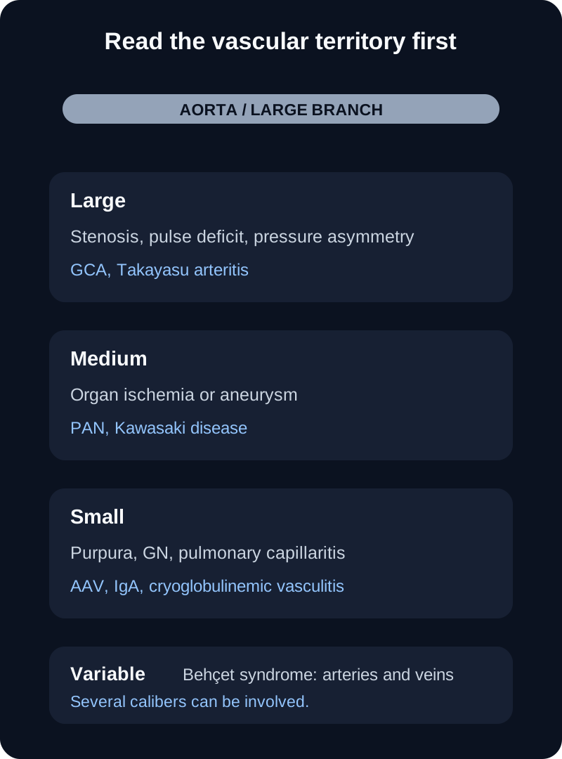
Large-vessel vasculitis centers on the aorta and its major branches. Medium-vessel disease targets named muscular arteries that supply organs. Small-vessel disease reaches arterioles, capillaries, and venules, so glomerular capillaries, pulmonary capillaries, and dermal postcapillary venules become especially informative. This framework is a starting map, not a rule that every disease respects one diameter at every site. Behçet syndrome is classified as variable-vessel vasculitis, and several named disorders can span adjacent calibers. [1]
Start with the vascular territory, then connect anatomy to ischemia, aneurysm, purpura, glomerular injury, or capillaritis. [1]
Ask what the lesion does to flow. Concentric wall inflammation and remodeling can narrow a large artery and produce asymmetric pulses or limb claudication. Necrotizing injury of a medium muscular artery can weaken the wall and create aneurysms while also reducing downstream perfusion. Capillary injury can leak red cells into alveoli or glomeruli. The same word, vasculitis, therefore produces very different organ patterns because anatomy controls the consequence.
Translate vessel territory into the finding you expect
High-yield examplesGPA, MPA, EGPA, IgA vasculitis, cryoglobulinemic vasculitis
TerritoryVariable
Typical consequenceArterial or venous disease of several calibers
High-yield examplesBehçet syndrome
Trace before naming: if the urine contains red cell casts, the glomerular capillary bed is involved. If hypertension occurs with renal-artery microaneurysms and a bland urine sediment, think upstream medium-vessel ischemia instead.
Check the renal comparison
PAN can injure renal muscular arteries and activate renin without directly inflaming glomerular capillaries. MPA and GPA can cause pauci-immune necrotizing glomerulonephritis, so hematuria and red cell casts fit them much better. [3][5]
Large vessels: separate ischemic territories before comparing histology
Takayasu arteritis commonly begins in younger patients and affects the aorta and major branches. Arm claudication, diminished pulses, a pressure difference between arms, bruits, and imaging that shows wall thickening or stenosis all point to a proximal arterial problem. Age and ancestry can alter prior probability, but neither should replace the vascular examination or imaging. Active disease is treated with glucocorticoids plus a nonglucocorticoid immunosuppressive strategy rather than reserving all steroid-sparing therapy only for late refractory disease. [2]
Giant cell arteritis occurs in adults age 50 or older. A new headache, scalp tenderness, jaw or tongue claudication, polymyalgia rheumatica symptoms, and ischemic visual symptoms can cluster together. Visual loss reflects ischemia of the ocular circulation and can become permanent. When GCA is strongly suspected with threatened vision, treatment is urgent; diagnostic testing should not create a harmful delay.
Temporal-artery biopsy remains useful, and vascular ultrasound or other imaging can add evidence depending on expertise and the territory involved. For newly diagnosed GCA, glucocorticoids combined with tocilizumab reduce relapse and cumulative steroid exposure; intravenous pulse glucocorticoids may be considered when vision is threatened. [2] Upadacitinib with a 26-week glucocorticoid taper improved sustained remission in a phase 3 trial and is FDA-approved for adults with GCA. [12][13]
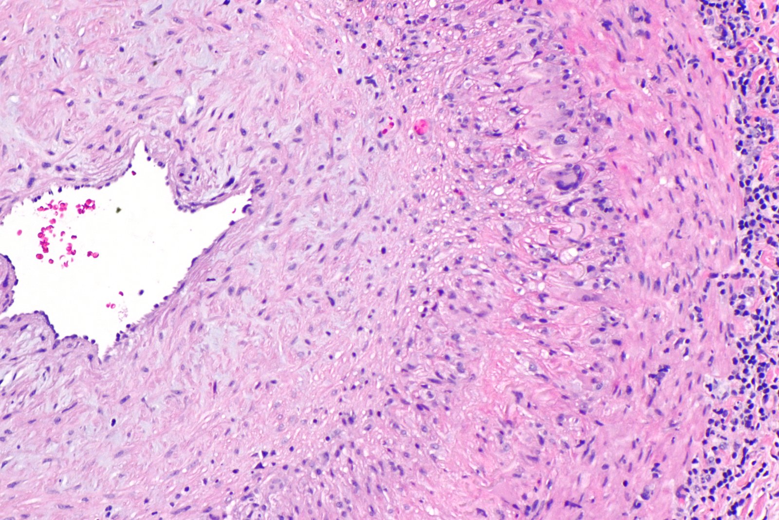
Use the clinical setting with the vessel-wall pattern. Histology can support GCA, but a negative sample does not erase a convincing ischemic phenotype. Image: Nephron; original source; CC BY-SA 3.0.
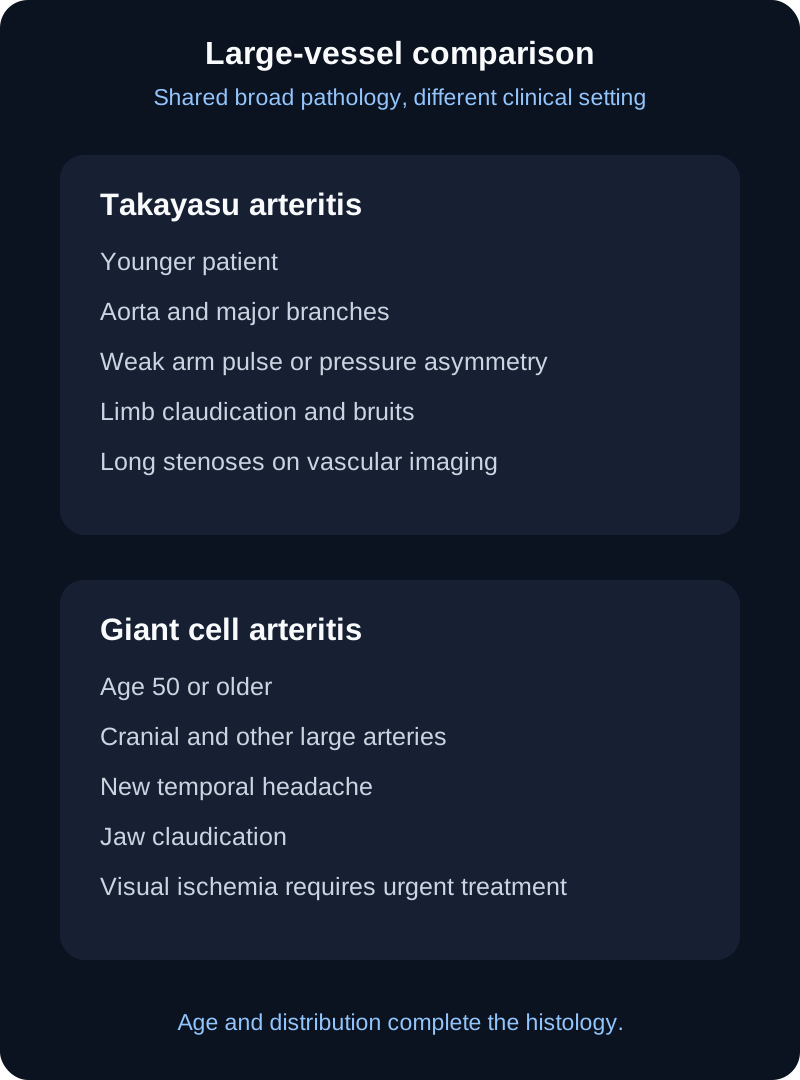
Both GCA and Takayasu arteritis can show granulomatous inflammation with giant cells and elastic-lamina injury, so histology alone does not always separate them. Put the tissue back into the patient: age 72 with jaw claudication and ischemic optic neuropathy fits GCA, while age 27 with subclavian stenoses and weak arm pulses fits Takayasu arteritis. Large-vessel imaging also matters because GCA can extend beyond the temporal artery. [2]
The comparison emphasizes that granulomatous large-vessel histology must be interpreted with age and vascular distribution. [2]
A high ESR or CRP supports inflammation but is not a disease label. The dangerous decision in suspected GCA is whether ocular ischemia is present, not whether an inflammatory marker crosses a memorized number.
Try it here · Checkpoint 1 of 3
Make your prediction before reading the choices. A first attempt is just a starting point.
Case 2
Show answer and explanations for case 2
A. Schedule temporal-artery biopsy before treatment (Why this does not fit)
Biopsy is useful, but waiting for tissue confirmation when ocular ischemia is present can risk permanent visual loss.
Reasoning steps for option A
Why is temporal-artery biopsy tempting before any treatment?
Temporal-artery biopsy is the classic confirmatory test, and tissue helps justify long-term glucocorticoid therapy.
Why is waiting for biopsy unsafe when transient visual loss is present?
Two episodes of monocular loss mean ocular perfusion is already failing, and a biopsy stays informative for about 2 weeks after glucocorticoids start, so treatment need not wait.
B. Start high-dose glucocorticoid treatment now (Best answer)
Threatened visual ischemia in suspected GCA is an emergency; treatment should begin promptly while diagnostic confirmation proceeds.
Reasoning steps for option B
What makes transient monocular visual loss an emergency in suspected GCA?
It often precedes permanent blindness from arteritic optic neuropathy or retinal artery occlusion, which is rarely reversible once established.
Which findings make GCA the presumed cause of the visual episodes?
Age 74, new temporal headache, scalp tenderness, jaw fatigue and high ESR and CRP make GCA highly likely, so the visual loss is treated as arteritic.
What rule governs treatment timing in suspected GCA with visual symptoms?
Start high-dose glucocorticoids the same day and arrange biopsy or ultrasound afterward, never the reverse.
C. Begin aspirin alone and reassess tomorrow (Why this does not fit)
Aspirin does not adequately suppress the arterial inflammation responsible for threatened ocular ischemia.
Reasoning steps for option C
Why might aspirin seem a cautious first step for ischemic symptoms?
Transient monocular visual loss resembles embolic amaurosis fugax, for which antiplatelet therapy is a standard first step.
Why is aspirin alone insufficient in this presentation?
Scalp tenderness, jaw fatigue and high inflammatory markers identify an arteritic cause, and only glucocorticoids quiet the granulomatous wall inflammation narrowing the ciliary arteries.
D. Obtain a routine brain MRI before treatment (Why this does not fit)
Brain MRI does not address the urgent arterial inflammatory process and should not delay treatment for threatened GCA vision loss.
Reasoning steps for option D
Why might imaging of the brain be requested after transient visual loss?
Transient visual loss in a 74-year-old raises concern for stroke or another intracranial cause.
What urgent problem would a routine brain MRI fail to address?
Jaw fatigue, scalp tenderness and high ESR and CRP point to arteritis of the vessels feeding the eye, which a delayed MRI would neither diagnose nor treat.
Takeaway: Suspected GCA with visual ischemia requires immediate glucocorticoid treatment while diagnostic testing continues.
Medium vessels: distinguish organ ischemia from coronary inflammation
Polyarteritis nodosa is a necrotizing arteritis of medium and small muscular arteries without glomerulonephritis or pulmonary capillaritis in its classic definition. Renal-artery disease can cause hypertension and renal ischemia; mesenteric disease can cause postprandial pain, bleeding, or infarction; vasa nervorum injury can produce mononeuritis multiplex. Angiography may show stenoses and aneurysms when biopsy is impractical. ANCA is not a defining feature. [1][3]
Hepatitis B can be associated with PAN, but modern epidemiology does not justify treating a fixed historical percentage as a diagnostic rule. In a patient with active HBV-associated disease, management must address the infection as well as the vasculitis. For non-HBV PAN, the ACR/Vasculitis Foundation guideline stratifies therapy by severity and supports glucocorticoids with an additional immunosuppressive agent for severe disease. [3]
Kawasaki disease is an acute pediatric vasculitis with a striking preference for the coronary arteries. Prolonged fever plus bilateral nonexudative conjunctival injection, oral changes, polymorphous rash, cervical lymphadenopathy, and extremity changes form the classic clinical pattern, but incomplete presentations occur. Coronary dimensions should be interpreted with body-size-adjusted Z scores rather than a single diameter cutoff. [4]
IVIG is the central initial anti-inflammatory therapy for Kawasaki disease, and aspirin is used as part of the regimen. IVIG 2 g/kg is given as a single infusion, ideally within the first 10 days of fever. The exact aspirin dose strategy varies, while the prevention of coronary injury cannot be credited to aspirin alone. The 2024 AHA statement also emphasizes additional anti-inflammatory therapy for patients at high risk of coronary artery aneurysm. [4]
Check the artery pattern
Renal and mesenteric microaneurysms with neuropathy point toward PAN. Fever with mucocutaneous inflammation in a young child directs attention to the coronary arteries and Kawasaki disease. Both involve medium arteries, but the organ pattern is different. [3][4]
Small-vessel pauci-immune disease: phenotype first, ANCA second
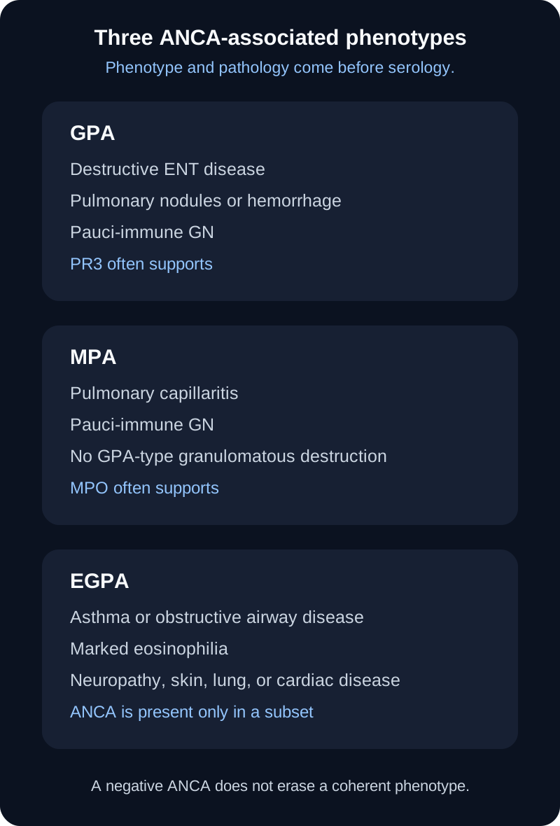
GPA, MPA, and EGPA are grouped as ANCA-associated vasculitides, but a positive ANCA is supportive rather than a substitute for the clinical pattern. PR3-ANCA is strongly associated with GPA and MPO-ANCA with MPA, while many patients with EGPA are ANCA negative. Tissue biopsy, when feasible and useful, can show the pattern of inflammation and help exclude mimics. [5][6]
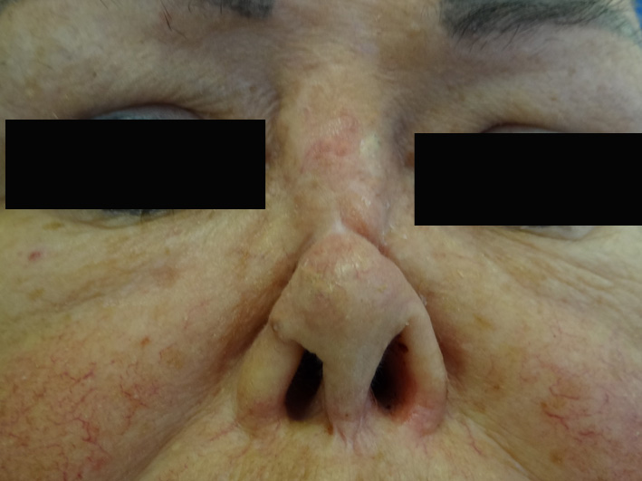
GPA links upper airway, lung, and kidney disease. Chronic destructive sinonasal disease, otitis, subglottic disease, pulmonary nodules or hemorrhage, and pauci-immune necrotizing glomerulonephritis form a recognizable combination. Granulomatous inflammation explains destructive respiratory lesions such as a saddle-nose deformity. PR3-ANCA increases confidence in the right phenotype, but absence of PR3 does not erase the phenotype. [1][5]
Destructive upper-airway disease helps separate GPA from MPA in the correct systemic context. Image: Simone Cristina Kawabata et al.; original source; CC BY 3.0.
MPA favors pulmonary capillaritis and glomerulonephritis without the granulomatous destructive upper-airway pattern typical of GPA. MPO-ANCA is common. A patient with hemoptysis, diffuse alveolar hemorrhage, red cell casts, and pauci-immune crescentic glomerulonephritis fits MPA far better than PAN, because PAN classically spares pulmonary capillaries and glomeruli. [1][5]
EGPA adds eosinophilic and airway biology. Asthma or other obstructive airway disease, marked eosinophilia, nasal polyps, eosinophilic tissue disease, neuropathy, skin findings, and sometimes cardiac involvement create the phenotype. The older three-phase story is useful as a memory scaffold, but real disease need not pass through three clean sequential stages. ANCA status also divides some clinical tendencies rather than defining every case. [7]
ANCA supports the diagnosis, but the organ phenotype and pathology determine how the pattern is interpreted. [5][6][7]
For organ-threatening GPA or MPA, current guidance supports rituximab or cyclophosphamide with a reduced-dose glucocorticoid taper; avacopan can be used to further reduce glucocorticoid exposure. Remission is then maintained, usually with rituximab, rather than with prolonged cyclophosphamide. For relapsing or refractory nonsevere EGPA, mepolizumab or benralizumab is an option. [5][6][14]
If a pulmonary-renal syndrome is present, ask what immunofluorescence shows. Pauci-immune means little or no immunoglobulin deposition, while anti-GBM disease has a linear IgG pattern and immune-complex disease has deposited immunoglobulin and complement.
Try it here · Checkpoint 2 of 3
Make your prediction before reading the choices. A first attempt is just a starting point.
Case 11
Show answer and explanations for case 11
A. Azathioprine alone (Why this does not fit)
Azathioprine is used in maintenance strategies, but it is not an adequate sole induction agent for life-threatening active GPA.
Reasoning steps for option A
Why might azathioprine seem a gentler option for GPA?
Azathioprine is an established steroid-sparing drug in AAV and is less toxic than cyclophosphamide.
Why is azathioprine alone insufficient now?
Azathioprine takes weeks to months to act, far too slow for alveolar hemorrhage and rapidly worsening nephritis.
B. Long-term cyclophosphamide alone without glucocorticoids (Why this does not fit)
Cyclophosphamide can be an induction agent, but severe AAV treatment uses it with glucocorticoids and limits exposure because of cumulative toxicity.
Reasoning steps for option B
Why might cyclophosphamide alone seem strong enough for severe GPA?
Cyclophosphamide is a proven induction agent for severe GPA, and avoiding glucocorticoids sounds safer.
What is missing from cyclophosphamide monotherapy?
Omitting glucocorticoids leaves the first weeks of inflammation uncontrolled, and prolonged cyclophosphamide raises bladder cancer, infertility and marrow toxicity.
C. Rituximab or cyclophosphamide with glucocorticoids (Best answer)
Current guidance supports glucocorticoids combined with rituximab or cyclophosphamide for organ-threatening GPA or MPA, with drug choice individualized.
Reasoning steps for option C
Which induction regimen fits organ-threatening GPA with alveolar hemorrhage?
Rituximab and cyclophosphamide achieved similar remission rates in severe GPA in trials, and both are given with glucocorticoids.
Which disease severity features justify rituximab or cyclophosphamide with glucocorticoids?
Diffuse alveolar hemorrhage and rapidly worsening pauci-immune glomerulonephritis are organ-threatening manifestations.
How does induction differ from maintenance in severe AAV?
Induction uses potent agents for about 3 to 6 months to reach remission; maintenance then switches to lower-toxicity drugs such as rituximab or azathioprine.
D. Mepolizumab alone (Why this does not fit)
Mepolizumab has a role in selected EGPA, especially nonsevere or relapsing eosinophilic disease, but it is not standard induction for this severe GPA presentation.
Reasoning steps for option D
Why might a biologic such as mepolizumab be considered in AAV?
Mepolizumab is a biologic approved for an ANCA-associated vasculitis, EGPA.
Why does mepolizumab not match this patient?
He has GPA without eosinophilia, and IL-5 blockade does not treat the neutrophil-driven capillaritis causing his hemorrhage and nephritis.
Takeaway: Severe GPA or MPA requires remission induction with glucocorticoids plus rituximab or cyclophosphamide, not maintenance monotherapy.
Immune complexes and variable-vessel disease: read the immune context
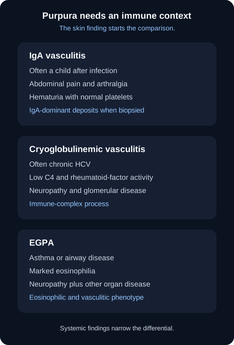
IgA vasculitis often follows an infection in children and combines nonthrombocytopenic palpable purpura with abdominal pain, arthralgia or arthritis, and renal involvement. IgA-dominant deposits can be demonstrated when biopsy is needed. A normal platelet count helps distinguish vascular inflammation from thrombocytopenic purpura. Gastrointestinal edema and hemorrhage can lead to intussusception, and kidney involvement determines much of the long-term follow-up. [8]
Cryoglobulinemic vasculitis is an immune-complex small-vessel process. Chronic hepatitis C is a major cause of mixed cryoglobulinemia, and the pattern can include palpable purpura, arthralgia, weakness, peripheral neuropathy, and glomerular disease. Low C4 and rheumatoid-factor activity support the diagnosis. In HCV-associated disease, direct-acting antiviral therapy treats the driver; severe organ disease can require additional immunomodulatory treatment. [11]
The same skin finding can arise from different small-vessel processes; associated systemic findings separate them. [7][8][11]
Behçet syndrome can affect arteries and veins of many sizes, which is why the Chapel Hill system places it in the variable-vessel category. Recurrent oral and genital ulcers, uveitis, skin lesions, venous thrombosis, arterial aneurysms, neurologic disease, and a positive pathergy response can occur in different combinations. HLA-B51 is an association, not a diagnostic test. Treatment is chosen by the organ threatened, especially ocular, vascular, neurologic, or gastrointestinal disease. [1][9]
Thromboangiitis obliterans, or Buerger disease, is strongly linked to tobacco exposure and causes inflammatory thrombotic disease of small and medium arteries and veins in the distal limbs. Distal ischemic pain, ulcers or gangrene, Raynaud phenomenon, and superficial thrombophlebitis in a younger tobacco user should prompt exclusion of atherosclerotic, embolic, autoimmune, and hypercoagulable alternatives. Complete tobacco cessation is the cornerstone of management. The exact biological pathway linking tobacco to the disease remains incompletely defined, so a single hypersensitivity mechanism should not be taught as established fact. [10]
Check the purpura pattern
Purpura plus abdominal pain and hematuria after an infection in a child favors IgA vasculitis. Purpura plus low C4, rheumatoid-factor activity, neuropathy, and chronic HCV favors cryoglobulinemic vasculitis. Purpura with asthma and eosinophilia should pull the comparison toward EGPA. [7][8][11]
Try it here · Checkpoint 3 of 3
Make your prediction before reading the choices. A first attempt is just a starting point.
Case 19
Show answer and explanations for case 19
A. Polyarteritis nodosa (Why this does not fit)
Viral association can suggest PAN, but low C4, rheumatoid-factor activity, purpura, neuropathy, and glomerular hematuria point toward immune-complex cryoglobulinemic disease.
Reasoning steps for option A
Why might PAN be considered in a patient with viral hepatitis?
Viral hepatitis is a classic PAN association, and PAN also causes neuropathy.
Which findings argue against polyarteritis nodosa despite a viral association?
PAN spares glomeruli and does not consume complement, whereas she has glomerular hematuria and low C4.
B. IgA vasculitis (Why this does not fit)
IgA vasculitis can cause purpura and hematuria, but chronic HCV with low C4 and rheumatoid-factor activity is much more characteristic of mixed cryoglobulinemia.
Reasoning steps for option B
Why might IgA vasculitis be considered for purpura with hematuria?
IgA vasculitis causes palpable purpura, arthralgia, and hematuria, all present here.
What context favors cryoglobulinemia over IgA vasculitis?
IgA vasculitis is mainly a childhood disease with normal complement; low C4 and rheumatoid-factor activity point to mixed cryoglobulins in a 59-year-old with HCV.
C. Cryoglobulinemic vasculitis (Best answer)
Chronic HCV plus purpura, neuropathy, glomerular disease, low C4, and rheumatoid-factor activity is the classic mixed cryoglobulinemic vasculitis pattern.
Reasoning steps for option C
Which findings in this woman with hepatitis C point to cryoglobulinemic vasculitis?
Chronic HCV drives B cells to make IgM with rheumatoid-factor activity, which binds polyclonal IgG to form mixed cryoglobulins.
How does low C4 fit the other findings?
Deposited cryoglobulins activate the classical complement pathway, consuming C4 while injuring skin, nerve and glomerular vessels.
D. Microscopic polyangiitis (Why this does not fit)
MPA can cause purpura, neuropathy, and glomerulonephritis, but it is pauci-immune and does not characteristically produce the low-C4, rheumatoid-factor pattern shown here.
Reasoning steps for option D
Why might MPA be raised for purpura, neuropathy, and hematuria?
MPA can cause purpura, neuropathy, and glomerulonephritis.
Why is microscopic polyangiitis less likely with low C4 and rheumatoid-factor activity?
Low C4 shows complement is being consumed by immune complexes, a mechanism absent in pauci-immune MPA.
Takeaway: HCV-associated purpura with low C4, rheumatoid-factor activity, neuropathy, and glomerular disease strongly supports cryoglobulinemic vasculitis.
Integrate four signals before committing to a name
Use four signals in order: territory, organ pattern, pathology, immune context. Territory tells you where to look. Organ pattern tests whether the proposed disease actually reaches those vessels. Pathology separates granulomatous, necrotizing, pauci-immune, and immune-complex injury. Immune context includes ANCA antigen specificity, IgA deposition, complement consumption, viral association, eosinophilia, and other disease-specific evidence.
Pulse deficit plus aortic-branch stenosis Large-vessel territory. Compare Takayasu arteritis with GCA using age, distribution, symptoms, and imaging.
Renal or mesenteric aneurysms plus neuropathy Medium-vessel ischemic pattern. PAN rises; glomerular casts or pulmonary capillaritis would argue against classic PAN.
Hemoptysis plus red cell casts Small-vessel pulmonary-renal pattern. Compare GPA, MPA, anti-GBM disease, and immune-complex causes using ENT findings, ANCA antigen, and immunofluorescence.
Purpura plus systemic immune findings Use age, platelets, abdominal symptoms, eosinophils, complement, viral history, and biopsy pattern to separate IgA, cryoglobulinemic, EGPA, and other causes.
Vasculitis questions become harder when several facts overlap, which is exactly why single buzzwords fail. A positive ANCA does not replace anatomy. A high ESR does not identify a disease. A viral association does not determine every case. The best answer is the diagnosis or action that explains the whole supplied pattern with the fewest contradictions.
Apply the framework
Case 1
Show answer and explanations for case 1
A. Takayasu arteritis (Best answer)
The age, limb ischemia, pulse asymmetry, and proximal aortic-branch stenoses form a large-vessel pattern that fits Takayasu arteritis.
Reasoning steps for option A
Which large-vessel disease fits a 27-year-old with pulse asymmetry and arch-branch stenoses?
Arm claudication with a 24 mmHg interarm gap localizes to the left subclavian artery, and wall thickening of the arch shows the narrowing is inflammatory rather than atherosclerotic.
How do the age and proximal aortic-branch lesions affect the Takayasu diagnosis?
A young adult with pulse asymmetry and long smooth stenoses of the arch branches has the expected large-vessel distribution.
What should an interarm pressure gap trigger in a young adult?
Imaging of the aorta and its branches, because a pressure difference or absent limb pulse may be the first sign of Takayasu arteritis.
B. Giant cell arteritis (Why this does not fit)
GCA can inflame large arteries, but it occurs in adults age 50 or older and more often presents with cranial ischemic symptoms in this comparison.
Reasoning steps for option B
Why is GCA considered when large arteries are inflamed?
GCA can involve the aorta and subclavian arteries, producing arm claudication and unequal arm pressures much like hers.
Which feature makes giant cell arteritis unlikely here?
GCA essentially does not begin before age 50, and at 27 she is decades below that threshold, with no headache or jaw claudication.
C. Fibromuscular dysplasia (Why this does not fit)
Fibromuscular dysplasia is noninflammatory and classically produces multifocal beading in renal or cervical arteries rather than inflammatory aortic-arch wall thickening.
Reasoning steps for option C
Could a noninflammatory arteriopathy produce these subclavian stenoses?
Fibromuscular dysplasia typically affects young women and narrows arteries, so her age, sex and a stenotic branch overlap with it.
What imaging feature conflicts with fibromuscular dysplasia?
Her arch wall is thickened and the subclavian lesion is a long smooth taper, whereas fibromuscular dysplasia leaves the wall thin and gives a string-of-beads contour in renal or carotid arteries.
D. Polyarteritis nodosa (Why this does not fit)
PAN affects medium muscular arteries and can cause organ ischemia or aneurysms, not this proximal aortic-branch stenosis pattern.
Reasoning steps for option D
Could a medium-vessel necrotizing arteritis explain limb ischemia in this woman?
PAN is a systemic necrotizing arteritis that can cause limb ischemia and hypertension in adults.
Why do the affected vessels argue against polyarteritis nodosa?
The lesions are in the aorta and proximal branches, not medium muscular arteries that develop organ ischemia or microaneurysms.
Takeaway: Pulse deficits plus proximal aortic-branch stenoses in a younger adult strongly support Takayasu arteritis.
Takayasu arteritis can share granulomatous large-vessel histology, but the patient's age and cranial ischemic presentation favor GCA.
Reasoning steps for option A
Why does Takayasu arteritis enter the comparison for a granulomatous arteritis?
Takayasu arteritis is also a granulomatous large-vessel vasculitis with giant cells, so the slide alone could fit it.
Why do the patient's age and cranial presentation favor GCA over Takayasu arteritis?
Takayasu arteritis typically begins before 40 and targets the aorta and its primary branches, whereas a 69-year-old with jaw claudication has cranial-artery disease.
B. Polyarteritis nodosa (Why this does not fit)
PAN is a necrotizing medium-artery disease with fibrinoid injury and aneurysms, not the granulomatous elastic-lamina pattern shown here.
Reasoning steps for option B
Why might a necrotizing arteritis be considered for an arterial biopsy abnormality?
The biopsy shows arterial wall destruction with elastic-lamina damage, and PAN is the classic necrotizing arteritis of muscular arteries.
Which tissue feature argues against polyarteritis nodosa?
Multinucleated giant cells mark granulomatous inflammation, whereas PAN shows fibrinoid necrosis with a mixed neutrophil-rich infiltrate and no giant cells.
C. Giant cell arteritis (Best answer)
Age over 50 plus jaw claudication and granulomatous arteritis with elastic-lamina injury is the classic GCA combination.
Reasoning steps for option C
Which clinical and tissue features converge on GCA in this 69-year-old?
Jaw claudication reflects ischemia of chewing muscles supplied by external carotid branches, and giant cells with intimal hyperplasia confirm granulomatous arteritis in the sampled temporal artery.
Which combination makes giant cell arteritis the best fit?
The patient is over 50 and has cranial ischemic symptoms, the two clinical features that separate GCA from the other granulomatous large-vessel arteritis.
Why is histology alone not enough to name the disease?
Takayasu arteritis produces the same giant-cell lesion, so the pathology report must be paired with the patient's age and the arteries involved.
D. Microscopic polyangiitis (Why this does not fit)
MPA is a nongranulomatous small-vessel vasculitis that often produces pauci-immune glomerulonephritis and pulmonary capillaritis.
Reasoning steps for option D
Why might an ANCA-associated vasculitis be raised for any vasculitic biopsy?
MPA is a common systemic necrotizing vasculitis in older adults and can involve small arteries as well as capillaries.
Why is microscopic polyangiitis inconsistent with this biopsy?
Granulomas and giant cells are absent in MPA by definition, and it does not cause jaw claudication from temporal-artery disease.
Takeaway: The same granulomatous pattern can occur in GCA and Takayasu arteritis, so age and vascular territory complete the diagnosis.
A. Granulomatosis with polyangiitis (Why this does not fit)
GPA can cause kidney and nerve disease, but it usually has glomerular hematuria and often respiratory granulomatous disease rather than mesenteric aneurysms.
Reasoning steps for option A
Why could GPA be considered with kidney and nerve involvement?
GPA is a necrotizing vasculitis that can cause mononeuritis multiplex and kidney disease, so foot drop plus hypertension could suggest it.
What two findings argue against granulomatosis with polyangiitis?
A bland urine sediment and mesenteric microaneurysms point away from glomerulonephritis and respiratory granulomatous disease.
B. Microscopic polyangiitis (Why this does not fit)
MPA can cause mononeuritis and renal disease, but pauci-immune glomerulonephritis and pulmonary capillaritis fit it better than medium-artery aneurysms.
Reasoning steps for option B
Why might MPA be raised for mononeuritis and hypertension with renal disease?
MPA commonly causes mononeuritis multiplex and kidney involvement, and hypertension could reflect renal disease.
Why are the aneurysms not typical of microscopic polyangiitis?
The urine has no red cell casts, so there is no glomerular capillaritis, and mesenteric aneurysms point to disease of muscular arteries rather than capillaries.
C. Cryoglobulinemic vasculitis (Why this does not fit)
Cryoglobulinemic disease can accompany viral hepatitis, especially HCV, but low complement and immune-complex small-vessel findings would be expected.
Reasoning steps for option C
Why does viral hepatitis bring cryoglobulinemic vasculitis to mind?
Chronic viral hepatitis is the classic trigger of cryoglobulinemic vasculitis, which also causes peripheral neuropathy.
Which expected immune-complex findings are missing for cryoglobulinemic vasculitis?
The case lacks low complement and glomerular immune-complex findings, and the viral association is hepatitis B rather than the usual hepatitis C context.
D. Polyarteritis nodosa (Best answer)
HBV association, mesenteric aneurysms, renal-artery hypertension, livedo, and mononeuritis without glomerulonephritis form a classic PAN pattern.
Reasoning steps for option D
Which features in this man with hepatitis B point to a medium-vessel arteritis?
Postprandial pain signals mesenteric ischemia, foot drop signals infarction of a peripheral nerve, and hypertension suggests renal-artery involvement, all ischemic effects of muscular-artery disease.
What does the angiogram add in a patient with hepatitis B?
Multiple small mesenteric aneurysms confirm necrotizing injury of muscular arteries, and HBV immune complexes are a recognized trigger of PAN.
What test does multiorgan ischemia with a clean urine sediment justify?
Angiography of the visceral arteries, looking for microaneurysms, is the most useful next step toward confirming PAN.
Takeaway: Medium-artery aneurysms plus multisystem ischemia and a bland glomerular sediment support PAN.
A. Renal ischemia from medium-artery disease (Best answer)
PAN injures renal muscular arteries, so reduced perfusion can activate renin and impair kidney function without directly producing glomerulonephritis.
Reasoning steps for option A
How can PAN damage the kidney without an active urine sediment?
Segmental renal arteries lie upstream of the glomeruli, so narrowing them lowers perfusion and filtration while the glomerular capillaries themselves stay uninflamed.
How does the bland sediment localize the renal injury?
No dysmorphic red cells or casts means the glomeruli are not inflamed, and the hypertension reflects renin release from underperfused kidney tissue.
Why does angiography rather than kidney biopsy usually document renal PAN?
Intrarenal aneurysms can bleed after a percutaneous needle biopsy, and angiography shows the arterial lesions directly.
B. Pauci-immune crescentic glomerulonephritis (Why this does not fit)
Crescentic glomerular injury should produce an active urine sediment and instead points toward small-vessel AAV.
Reasoning steps for option B
Why might crescentic glomerulonephritis be suspected with a rising creatinine?
A rapidly rising creatinine in a vasculitis patient raises concern for crescentic glomerulonephritis, a leading cause of rapid kidney failure in small-vessel vasculitis.
Which urine finding expected in crescentic glomerulonephritis is absent?
The urine shows no dysmorphic erythrocytes or casts, which crescentic glomerular injury almost always produces.
C. Mesangial IgA deposition (Why this does not fit)
IgA deposition causes a glomerular process with hematuria and often follows a different systemic pattern.
Reasoning steps for option C
Why might IgA deposition be considered in vasculitic kidney disease?
IgA nephropathy is the most common primary glomerulonephritis worldwide and often presents with hypertension and falling kidney function.
Why does mesangial IgA deposition not match the angiogram?
IgA disease is glomerular, whereas the demonstrated lesions are stenoses and aneurysms of segmental renal arteries.
D. Anti-GBM antibody injury (Why this does not fit)
Anti-GBM disease targets glomerular basement membranes and typically causes rapidly progressive glomerulonephritis with hematuria.
Reasoning steps for option D
Why could anti-GBM disease be raised when kidney function is falling?
Anti-GBM disease causes some of the fastest declines in kidney function of any glomerular disease.
What feature argues against anti-GBM injury?
The urine has only trace protein and no dysmorphic cells, whereas anti-GBM nephritis produces hematuria with red cell casts, and the angiogram shows arterial rather than glomerular lesions.
Takeaway: PAN can cause renal failure and hypertension by renal-artery ischemia while sparing the glomerular capillaries.
A. Oral antibiotics and observation (Why this does not fit)
The mucocutaneous pattern with coronary involvement is inflammatory Kawasaki disease, not a bacterial syndrome treated by routine antibiotics.
Reasoning steps for option A
Why might an infection be suspected in a febrile child with rash?
Fever with rash and red eyes in a young child is most often infectious, and streptococcal scarlet fever can mimic this picture.
Why do antibiotics not address this child's illness?
The prolonged fever, nonexudative conjunctivitis, mucosal changes, extremity inflammation, and coronary dilation identify an inflammatory vasculitis.
B. Intravenous immunoglobulin with aspirin (Best answer)
IVIG is the central initial anti-inflammatory treatment and aspirin is included in standard Kawasaki therapy; coronary risk also guides whether added anti-inflammatory treatment is needed.
Reasoning steps for option B
Which therapy is the core of initial Kawasaki treatment?
Seven days of fever with four principal mucocutaneous features and new coronary dilation establish Kawasaki disease, for which a single 2 g/kg IVIG infusion is the core therapy.
Which treatment element is central for reducing coronary complications?
IVIG given within the first 10 days of illness lowers the coronary aneurysm rate from about 25 percent to about 4 percent.
What is the overall initial treatment plan for Kawasaki disease?
Give IVIG with aspirin, and consider adding glucocorticoids at the start when coronary dilation or other high-risk features are already present.
C. Cyclophosphamide with glucocorticoids (Why this does not fit)
This regimen is reserved for selected severe systemic vasculitides and is not standard initial Kawasaki treatment.
Reasoning steps for option C
Why might strong immunosuppression seem justified with coronary involvement?
Coronary dilation is organ-threatening vasculitis, and cyclophosphamide with glucocorticoids is used for other organ-threatening vasculitides.
Why is cyclophosphamide not the standard initial choice?
Most children respond to IVIG, and cytotoxic induction has no established role in the initial treatment of Kawasaki disease.
D. Aspirin alone (Why this does not fit)
Aspirin is part of therapy, but it does not replace IVIG for initial treatment aimed at reducing coronary complications.
Reasoning steps for option D
Why might aspirin alone seem enough for a child with coronary dilation?
Aspirin is part of standard Kawasaki therapy and has both anti-inflammatory and antiplatelet effects.
What essential therapy is missing from aspirin alone?
Aspirin by itself has not been shown to lower the aneurysm rate; that benefit comes from IVIG, which this child with dilation needs now.
Takeaway: Kawasaki disease is treated initially with IVIG plus aspirin, with additional therapy considered according to coronary risk.
A. Aortic dissection from cystic medial degeneration (Why this does not fit)
Cystic medial degeneration underlies aortic dissection in Marfan-type aortopathy; Kawasaki disease preferentially injures the coronary arteries.
Reasoning steps for option A
Why might a major vessel wall catastrophe be feared after vasculitis?
Kawasaki disease is a systemic vasculitis, and dissection is the classic catastrophic arterial-wall complication.
Why does an inherited aortic-wall disorder not explain the reported Z score?
The measured abnormality is coronary dilation after Kawasaki disease, not a degenerative aortic-wall disorder such as Marfan-type aortopathy.
B. Pulmonary capillary hemorrhage (Why this does not fit)
Pulmonary capillaritis is characteristic of small-vessel disorders such as MPA or GPA, not the major vascular complication of Kawasaki disease.
Reasoning steps for option B
Why might pulmonary bleeding be listed as a vasculitis complication?
Pulmonary hemorrhage is a dangerous complication of several systemic vasculitides.
Which vascular bed makes pulmonary hemorrhage an implausible complication?
Kawasaki disease chiefly threatens the coronary arteries rather than pulmonary capillaries.
C. Coronary thrombosis within an aneurysmal segment (Best answer)
A high coronary Z score indicates important aneurysmal disease, which creates risk of thrombosis and myocardial ischemia and determines follow-up intensity.
Reasoning steps for option C
What does a coronary Z score of 7.2 imply for this child?
A Z score of 7.2 falls in the medium-aneurysm category (5 to below 10), where slow flow through the dilated segment favors thrombus.
How do Z scores shape follow-up after Kawasaki disease?
Higher Z-score categories call for more frequent imaging and stress testing and stronger antithrombotic therapy, such as adding anticoagulation for large or giant aneurysms.
D. Renal cortical infarction from PAN (Why this does not fit)
Medium-vessel renal ischemia is a PAN pattern; the measured abnormality in this child is specifically coronary.
Reasoning steps for option D
Why might renal infarction be considered after a medium-vessel vasculitis?
Kawasaki disease and PAN are both medium-vessel vasculitides, and PAN classically causes renal infarction.
Why is renal cortical infarction from PAN not the priority?
The documented lesion is a coronary aneurysm in a child with Kawasaki disease, not renal medium-artery involvement.
Takeaway: Kawasaki coronary aneurysms can thrombose and cause myocardial ischemia; Z scores help define coronary severity and follow-up.
A. Microscopic polyangiitis (Why this does not fit)
MPA can cause pulmonary-renal disease, but destructive upper-airway disease and granulomatous pulmonary nodules fit GPA better.
Reasoning steps for option A
Why is MPA a reasonable consideration in a pulmonary-renal syndrome?
MPA is a common cause of alveolar hemorrhage with red cell casts, so hemoptysis plus nephritis fits it.
Which findings distinguish GPA from microscopic polyangiitis?
Bloody nasal crusting and cavitary pulmonary nodules indicate destructive granulomatous airway and lung disease.
B. Eosinophilic granulomatosis with polyangiitis (Why this does not fit)
EGPA usually includes asthma, eosinophilia, and eosinophilic disease, which are absent from this presentation.
Reasoning steps for option B
Why might EGPA be considered with sinus disease and lung lesions?
EGPA commonly involves the sinuses and lungs and can be ANCA positive.
What defining EGPA findings are absent?
There is no asthma, marked eosinophilia, or eosinophilic organ disease.
C. Anti-GBM disease (Why this does not fit)
Anti-GBM disease can cause pulmonary hemorrhage and glomerulonephritis, but it does not explain destructive sinus disease and cavitary granulomatous nodules.
Reasoning steps for option C
Why does anti-GBM disease enter the differential for hemoptysis with casts?
Anti-GBM disease is a classic cause of hemoptysis with nephritic urine in a middle-aged adult.
Why does anti-GBM disease not explain the full presentation?
Anti-GBM disease spares the upper airway and does not form cavitating nodules, and PR3-ANCA points to GPA instead.
D. Granulomatosis with polyangiitis (Best answer)
Destructive upper-airway disease, cavitary lung lesions, pauci-immune renal injury, and PR3-ANCA form the classic GPA phenotype.
Reasoning steps for option D
Which organ pattern and serology point to GPA in this man?
Bloody crusting and recurrent otitis show upper-airway disease, cavitary nodules show necrotizing granulomas in the lung, and red cell casts show glomerulonephritis.
What does PR3-ANCA add to this organ pattern?
PR3-ANCA is the antibody most closely linked to GPA, so it strengthens the clinical diagnosis without replacing biopsy when tissue is accessible.
Which triad of sites is the signature of GPA?
Ear, nose and sinus disease, lung nodules and nephritis form the classic GPA triad, so finding any two should lead to a search for the third.
Takeaway: Upper-airway destruction plus pulmonary and glomerular disease strongly favors GPA.
Pulmonary capillaritis plus pauci-immune crescentic glomerulonephritis and MPO-ANCA, without granulomatous ENT disease or eosinophilia, fits MPA.
Reasoning steps for option A
Which features of this woman's pulmonary-renal syndrome point to MPA?
Alveolar hemorrhage and crescentic nephritis show capillaritis, scant immunoglobulin staining makes it pauci-immune, and MPO-ANCA is the antibody most often found in MPA.
Why do the negative findings matter as much as the positive ones?
No sinonasal destruction argues against GPA and no eosinophilia argues against EGPA, leaving MPA among the ANCA-associated diseases.
How is MPA summarized relative to GPA?
MPA shares the capillaritis of GPA but lacks granulomas, so it causes lung hemorrhage and nephritis without destructive airway disease.
B. Granulomatosis with polyangiitis (Why this does not fit)
GPA can cause pulmonary-renal disease, but destructive sinonasal or granulomatous respiratory disease would make it more likely.
Reasoning steps for option B
Why is GPA a strong competitor in pauci-immune pulmonary-renal disease?
GPA also causes alveolar hemorrhage and pauci-immune crescentic glomerulonephritis.
What expected GPA feature is missing?
She has no destructive sinonasal disease or lung nodules, and MPO rather than PR3-ANCA also favors MPA.
C. Eosinophilic granulomatosis with polyangiitis (Why this does not fit)
EGPA requires an eosinophilic and usually asthma-predominant phenotype that is not present here.
Reasoning steps for option C
Why might EGPA be raised in an ANCA-positive vasculitis?
EGPA is an ANCA-associated vasculitis, and when ANCA is present in EGPA it is usually MPO-ANCA.
Why does eosinophilic granulomatosis with polyangiitis not fit?
Neither asthma nor eosinophilia is present despite severe pulmonary and renal small-vessel disease.
D. Polyarteritis nodosa (Why this does not fit)
PAN classically spares pulmonary capillaries and glomeruli, so alveolar hemorrhage plus crescentic glomerulonephritis argues strongly against it.
Reasoning steps for option D
Why might PAN be considered for a systemic necrotizing vasculitis?
PAN is a systemic necrotizing vasculitis that can cause kidney failure in older adults.
Which involved structures exclude classic polyarteritis nodosa?
Alveolar and glomerular capillaries are directly injured, vascular beds that classic PAN spares.
Takeaway: MPA is a pauci-immune pulmonary-renal vasculitis often associated with MPO-ANCA and lacking GPA-type granulomatous ENT destruction.
A. Granulomatosis with polyangiitis (Why this does not fit)
GPA can involve upper and lower airways and nerves, but marked eosinophilia and asthma strongly redirect the pattern toward EGPA.
Reasoning steps for option A
Why might GPA be considered with sinus and lung involvement?
GPA involves the sinuses, lungs and peripheral nerves, all affected in this woman.
Which findings redirect the differential away from GPA?
Adult-onset asthma and marked eosinophilia are central to EGPA and are not defining features of GPA.
B. Eosinophilic granulomatosis with polyangiitis (Best answer)
Obstructive airway disease, marked eosinophilia, pulmonary infiltrates, purpura, and neuropathy form the characteristic EGPA phenotype.
Reasoning steps for option B
Which findings in this woman with adult-onset asthma point to EGPA?
Adult-onset asthma and sinus disease precede the vasculitic phase of EGPA, and a count of 4.0 × 10^9/L is marked eosinophilia.
What do the purpura and foot weakness add?
They show the disease has moved beyond eosinophilic tissue infiltration to vasculitis of skin vessels and the vasa nervorum.
Which combination should always raise concern for EGPA?
Any patient with asthma who develops new neuropathy, purpura or eosinophilia above 10 percent should be evaluated for EGPA.
C. Microscopic polyangiitis (Why this does not fit)
MPA can cause neuropathy and pulmonary disease, but marked eosinophilia and asthma are not its defining phenotype.
Reasoning steps for option C
Why might MPA be considered for neuropathy with lung infiltrates?
MPA commonly causes mononeuritis multiplex and lung involvement, and it is usually MPO-ANCA positive, like the ANCA-positive subset of EGPA.
Why is microscopic polyangiitis less likely in an asthma-eosinophilia presentation?
MPA can affect lungs, kidneys, skin, and nerves but does not explain the pronounced eosinophilic airway disease.
D. Cryoglobulinemic vasculitis (Why this does not fit)
Cryoglobulinemic vasculitis can cause purpura and neuropathy, but low complement, rheumatoid-factor activity, renal immune deposits, or chronic HCV would fit better than asthma with marked eosinophilia.
Reasoning steps for option D
Why could cryoglobulinemic vasculitis be raised for purpura with neuropathy?
Cryoglobulinemic vasculitis classically causes palpable purpura of the legs with peripheral neuropathy.
Which missing laboratory and exposure context weakens cryoglobulinemic vasculitis?
The case supplies asthma and eosinophilia instead of low complement, rheumatoid-factor activity, or chronic hepatitis C.
Takeaway: Asthma plus marked eosinophilia and systemic vasculitic findings should strongly suggest EGPA.
A. Indefinite daily oral cyclophosphamide (Why this does not fit)
Cyclophosphamide has substantial cumulative bladder, marrow, fertility, and malignancy toxicity and is not a preferred long-term maintenance drug.
Reasoning steps for option A
Why might continuing an induction drug seem safest after a prior relapse?
She relapsed after stopping azathioprine, so continuing the most potent immunosuppressant can seem the surest way to prevent another flare.
Why should cyclophosphamide not be continued indefinitely?
Rituximab maintenance prevents relapse better than azathioprine with far less toxicity, so there is no reason to accept cumulative cyclophosphamide exposure.
B. High-dose glucocorticoids alone for several years (Why this does not fit)
Prolonged high-dose glucocorticoid monotherapy carries major toxicity and does not match current maintenance strategies.
Reasoning steps for option B
Why might steroids alone seem enough to keep GPA quiet?
Glucocorticoids controlled her disease during induction, so continuing them alone can seem simple.
What makes prolonged high-dose glucocorticoid monotherapy a poor maintenance plan?
Years of high-dose glucocorticoids cause infection, diabetes, osteoporosis and cataracts, so they are tapered while a steroid-sparing agent maintains remission.
C. No therapy because ANCA is now negative (Why this does not fit)
ANCA level alone should not determine whether maintenance is stopped, especially in a patient with prior relapse.
Reasoning steps for option C
Why might a negative ANCA suggest that treatment can stop?
Disappearance of ANCA can seem to mark cure, and stopping therapy avoids drug toxicity.
Why does a negative ANCA result not justify stopping maintenance?
PR3 positivity and a previous relapse both predict further relapse, and a negative ANCA does not reliably predict sustained remission.
D. Rituximab maintenance (Best answer)
Rituximab is a recommended remission-maintenance option for GPA and is particularly reasonable in a relapsing PR3-associated phenotype.
Reasoning steps for option D
Which maintenance option suits relapsing PR3-positive GPA?
Her relapse after azathioprine withdrawal and PR3 positivity mark high relapse risk, and rituximab maintenance lowered relapse rates compared with azathioprine in trials.
Which current findings make rituximab maintenance safe to start?
Kidney function is stable and there is no active infection, so there is no contraindication to continued B-cell depletion.
What should guide maintenance decisions in GPA?
Choose maintenance by relapse risk, prior drug response and toxicity, and continue it for years rather than stopping on a single normal antibody test.
Takeaway: Maintenance decisions in GPA are based on relapse risk and treatment context, not ANCA status alone; rituximab is a major maintenance option.
Nonthrombocytopenic lower-extremity purpura with joint, gastrointestinal, and renal involvement after an infection is the classic IgA vasculitis pattern.
Reasoning steps for option A
Which organ pattern in this boy points to IgA vasculitis?
Purpura concentrated on the legs and buttocks with a normal platelet count means vessel inflammation rather than platelet deficiency.
Which other organs confirm a systemic small-vessel process?
Ankle arthralgia, cramping abdominal pain and microscopic hematuria add joint, gut and kidney involvement, the typical spread of IgA vasculitis.
Which constellation in a child points to IgA vasculitis?
IgA vasculitis is the most common vasculitis of childhood, so dependent palpable purpura with normal platelets in a school-age child should suggest it first.
B. Immune thrombocytopenia (Why this does not fit)
Immune thrombocytopenia can cause petechiae and purpura, but the platelet count should be low and the abdominal, joint, and renal pattern is not explained.
Reasoning steps for option B
Why might a platelet disorder be suspected with purpura after an infection?
Immune thrombocytopenia classically follows a viral infection in a child and presents with purpura.
Which laboratory result argues against immune thrombocytopenia?
A normal platelet count removes the mechanism by which ITP causes bleeding, and ITP does not inflame joints or the bowel.
C. Kawasaki disease (Why this does not fit)
Kawasaki disease is defined by prolonged fever and mucocutaneous inflammation with coronary risk, not this purpura-abdominal-renal syndrome.
Reasoning steps for option C
Why might Kawasaki disease be raised in a child with a systemic vasculitis?
Kawasaki disease is a common childhood vasculitis that can also cause rash, joint pain and abdominal symptoms.
What defining Kawasaki context is absent?
There is no prolonged febrile mucocutaneous syndrome or coronary involvement.
D. Microscopic polyangiitis (Why this does not fit)
MPA can cause glomerulonephritis and purpura, but this child's postinfectious abdominal and joint pattern with normal platelets is much more typical of IgA vasculitis.
Reasoning steps for option D
Why could MPA be considered for purpura with hematuria?
MPA causes palpable purpura and hematuria from small-vessel vasculitis, both present here.
Why is microscopic polyangiitis less likely in this child?
MPA is rare in children and usually causes rapidly progressive nephritis or lung hemorrhage, not dependent purpura with colicky pain after a cold.
Takeaway: Palpable purpura with normal platelets plus abdominal, joint, and renal findings in a child strongly supports IgA vasculitis.
Appendicitis can cause abdominal pain, but it does not explain the intermittent colicky pattern with a concentric-ring ultrasound finding.
Reasoning steps for option A
Why might appendicitis be suspected in a child with acute abdominal pain?
Appendicitis is the most common surgical cause of acute abdominal pain with vomiting in school-age children.
Which pain and imaging features argue against appendicitis?
The pain is episodic and colicky, and ultrasound shows concentric rings rather than a focal inflamed appendix.
B. Intussusception (Best answer)
Bowel-wall edema and hemorrhage in IgA vasculitis can provide a lead point for intussusception, which produces episodic pain and a target-like ultrasound appearance.
Reasoning steps for option B
What complication of IgA vasculitis explains colicky pain with a target sign?
Colicky episodic pain, bloody stool and concentric rings on ultrasound form the target sign of one bowel segment telescoped into another.
How does IgA vasculitis predispose to intussusception?
Submucosal hemorrhage and edema from vasculitis thicken a bowel segment, which peristalsis can then pull into the next segment.
How does the location differ from typical childhood intussusception?
In IgA vasculitis it is often ileoileal rather than ileocolic, so it can be missed if only the right lower quadrant is scanned.
C. Esophageal variceal bleeding (Why this does not fit)
Varices arise from portal hypertension and do not produce the supplied small-bowel sonographic pattern.
Reasoning steps for option C
Why might variceal bleeding be considered with bloody stool?
Bloody stool with vomiting can come from upper gastrointestinal bleeding, and varices bleed briskly.
Why do esophageal varices not explain the ultrasound?
Portal hypertensive bleeding does not create a concentric-ring lesion in the small bowel.
D. Mesenteric aneurysm rupture from PAN (Why this does not fit)
PAN can cause mesenteric ischemia or hemorrhage, but this child already has IgA vasculitis and the ultrasound directly supports intussusception.
Reasoning steps for option D
Why might a vasculitic aneurysm rupture be considered with abdominal pain?
Visceral aneurysm rupture causes sudden severe abdominal pain with gastrointestinal bleeding.
Which direct finding outweighs the alternative of PAN rupture?
PAN is rare in children, and a ruptured aneurysm would show free fluid or hematoma rather than a concentric-ring bowel segment.
Takeaway: Severe episodic abdominal pain with a target-like ultrasound in IgA vasculitis indicates intussusception.
A. Recent respiratory infection (Why this does not fit)
Both IgA vasculitis and immune thrombocytopenia can follow an infection, so this history alone does not separate them.
Reasoning steps for option A
Why might the preceding infection seem to separate the two disorders?
A respiratory infection often precedes IgA vasculitis, so it can seem to point toward vasculitis.
Why does a recent respiratory infection fail to distinguish the two disorders?
Childhood ITP also typically begins a few weeks after a viral illness, so the history occurs in both.
B. Purpuric skin lesions (Why this does not fit)
Both disorders can present with purpura, although the morphology and associated organs differ.
Reasoning steps for option B
Why might the skin lesions seem to identify the diagnosis?
The lesions are raised, and palpable purpura is a hallmark of small-vessel vasculitis.
Why is purpura alone nondiagnostic?
ITP also produces purpura and lesion texture can be hard to judge, so the skin is less decisive than a platelet count showing whether platelets are deficient.
C. Normal platelet count with renal and joint findings (Best answer)
The normal platelet count argues against thrombocytopenic bleeding, while hematuria and arthralgia fit systemic small-vessel IgA disease.
Reasoning steps for option C
Which findings most directly separate IgA vasculitis from immune thrombocytopenia?
A count of 285,000/mm3 is normal, so thrombocytopenia cannot explain the purpura.
What do the hematuria and knee pain add?
They show inflammation in the kidney and joints, organs that ITP does not inflame.
What separates vasculitic purpura from platelet-deficiency purpura?
Check the platelet count first; purpura with normal platelets directs the search toward vessel-wall disease.
D. Age younger than 10 years (Why this does not fit)
Both conditions occur in children, so age alone has little discriminatory value here.
Reasoning steps for option D
Why might the patient's young age seem to favor one diagnosis?
IgA vasculitis mostly affects children under 10, so her age fits it.
Why does the child's age add little discrimination?
ITP also peaks in early childhood, so a 6-year-old is typical for either disease.
Takeaway: Normal platelets plus renal and joint involvement separates IgA vasculitis from platelet-deficiency purpura.
Reactive arthritis can cause oral lesions and eye inflammation, but recurrent genital ulceration, pathergy, and venous thrombosis fit Behçet syndrome better.
Reasoning steps for option A
Why might reactive arthritis be considered with oral lesions and eye inflammation?
Reactive arthritis causes oral ulcers, eye inflammation and skin lesions in young men.
Which findings go beyond a reactive arthritis pattern?
Recurrent genital ulcers, pathergy, and venous thrombosis are much more characteristic of Behçet syndrome.
B. Systemic lupus erythematosus (Why this does not fit)
Lupus can cause mucosal ulcers and thrombosis in selected settings, but recurrent painful genital ulcers, uveitis, and pathergy form a different syndrome.
Reasoning steps for option B
Why might lupus be considered with mucosal ulcers and thrombosis?
Lupus causes oral ulcers and, with antiphospholipid antibodies, venous thrombosis.
Why does lupus not best unify the syndrome?
Lupus oral ulcers are usually painless, genital ulcers are uncommon, and pathergy is not a lupus feature.
C. Granulomatosis with polyangiitis (Why this does not fit)
GPA causes destructive respiratory granulomatous disease and glomerulonephritis; recurrent oral and genital aphthae with venous thrombosis are not its typical pattern.
Reasoning steps for option C
Why might GPA be raised for a multisystem vasculitis with eye disease?
GPA is a multisystem vasculitis that can cause ocular inflammation and oral ulceration.
Which expected GPA organs are not involved?
There is no destructive respiratory granulomatous disease or glomerulonephritis.
D. Behçet syndrome (Best answer)
Recurrent oral and genital ulcers, uveitis, characteristic skin findings, pathergy, and venous thrombosis are a classic multisystem Behçet presentation.
Reasoning steps for option D
Which combination of findings points to Behçet syndrome?
Recurrent painful oral aphthae are the anchor finding, and genital ulcers, eye disease, skin lesions and a positive pathergy test each add weight in Behçet classification.
What does the needle-puncture result show?
A sterile papule 24 to 48 hours after skin puncture is a positive pathergy test, a sign of neutrophil hyperreactivity that is highly specific for Behçet syndrome.
How is Behçet syndrome classified by vessel size?
It is the prototype variable-vessel vasculitis, affecting arteries and veins of any size, which is why venous thrombosis is a core vascular feature.
Takeaway: Behçet syndrome is variable-vessel disease that can combine mucocutaneous ulcers, uveitis, skin inflammation, and venous thrombosis.
A. Treat the underlying vascular inflammation with immunosuppression (Best answer)
Behçet venous thrombosis is driven by vessel-wall inflammation, so glucocorticoids and appropriate immunosuppressive therapy are central; anticoagulation decisions are individualized.
Reasoning steps for option A
Why is immunosuppression central to venous thrombosis in Behçet syndrome?
Behçet thrombi form on inflamed vein walls and adhere firmly, so the source is the vessel wall rather than hypercoagulable blood.
Why is immunosuppression central to this thrombosis?
Active ulcers and erythema nodosum accompany the deep venous thrombosis, indicating ongoing inflammatory vessel-wall disease.
What is the treatment principle for vascular Behçet disease?
Acute Behçet DVT is treated with glucocorticoids plus azathioprine, cyclosporine or cyclophosphamide; anticoagulation may be added only once bleeding risk is low and pulmonary artery aneurysms are excluded.
B. Use anticoagulation alone because inflammation is irrelevant (Why this does not fit)
Anticoagulation may be considered in selected patients, but treating only coagulation ignores the inflammatory vessel-wall process central to Behçet thrombosis.
Reasoning steps for option B
Why might anticoagulation alone seem the logical response to a DVT?
Anticoagulation is the standard treatment for a deep venous thrombosis in most patients.
What disease process would anticoagulation alone leave untreated?
It would not control the vascular inflammation driving Behçet-associated thrombosis.
C. Give IVIG because this is Kawasaki disease (Why this does not fit)
The adult ulcerative and venous-thrombotic phenotype is Behçet syndrome, not pediatric Kawasaki disease.
Reasoning steps for option C
Why might IVIG be proposed for a vasculitis with mucocutaneous findings?
IVIG treats Kawasaki disease, a mucocutaneous vasculitis with oral changes and rash.
Which features exclude Kawasaki disease?
This is an adult with recurrent ulcers and venous thrombosis, not a child with febrile mucocutaneous coronary disease.
D. Use antibiotics because pathergy proves infection (Why this does not fit)
Pathergy is an exaggerated sterile inflammatory response to minor trauma and does not establish bacterial infection.
Reasoning steps for option D
Why might a positive pathergy test be misread as infection?
A papule after needle puncture can look like a small skin infection.
Why does pathergy not justify antibiotics?
The papule is sterile and reflects neutrophil hyperreactivity; antibiotics would leave the venous inflammation untreated.
Takeaway: Vascular Behçet treatment targets vessel inflammation; anticoagulation is not a substitute for immunosuppressive control.
A. Long-term high-dose glucocorticoids (Why this does not fit)
Routine systemic glucocorticoids have not replaced tobacco cessation as the central disease-modifying intervention in thromboangiitis obliterans.
Reasoning steps for option A
Why might steroids seem reasonable for an inflammatory vascular disease?
Thromboangiitis obliterans involves inflammatory thrombus and he has superficial thrombophlebitis, so an anti-inflammatory drug seems logical.
Why are glucocorticoids not the central disease-modifying intervention?
The distal occlusive disease is tightly linked to tobacco exposure, and routine steroids do not remove that driver.
B. Complete tobacco cessation (Best answer)
Buerger disease is tightly linked to tobacco exposure, and complete cessation is the cornerstone of preventing continued vascular injury.
Reasoning steps for option B
What intervention addresses the driver of this smoker's distal occlusive disease?
A young heavy smoker with distal digital ischemia, superficial thrombophlebitis and segmental distal occlusions with collaterals has Buerger disease, a tobacco-driven disorder.
How complete must cessation be?
Even a few cigarettes a day or smokeless tobacco can keep the disease active, so all tobacco and nicotine products must stop.
What outcome follows when patients quit completely?
Patients who stop all tobacco rarely need major amputation, whereas those who keep smoking face a high amputation rate.
C. Immediate cyclophosphamide induction (Why this does not fit)
Cyclophosphamide is used for selected severe immune vasculitides but is not standard treatment for the tobacco-linked distal disease described here.
Reasoning steps for option C
Why might cytotoxic therapy be considered for an inflammatory arteritis?
Rest pain and digital ulcers signal threatened tissue, and cyclophosphamide is used for severe vasculitis.
Why is cyclophosphamide inappropriate as routine treatment?
The angiographic pattern is tobacco-linked distal occlusion, not a severe autoimmune vasculitis requiring cytotoxic induction.
D. Daily aspirin as the only therapy (Why this does not fit)
Antiplatelet therapy may address selected vascular risk considerations, but it does not substitute for complete tobacco cessation.
Reasoning steps for option D
Why might aspirin seem enough for occlusive arterial disease?
Aspirin is standard therapy for occlusive arterial disease and reduces thrombotic events.
Why is aspirin alone inadequate?
The occlusions are driven by ongoing tobacco exposure, and no drug, aspirin included, has been shown to halt progression while smoking continues.
Takeaway: In thromboangiitis obliterans, complete tobacco cessation is the central intervention for preventing progression.
A. Direct HCV infection of endothelial cells (Why this does not fit)
HCV drives cryoglobulin formation, but direct viral invasion of vessel walls is not the main mechanism of the immune-complex vasculitis described.
Reasoning steps for option A
Why might the virus itself be blamed for the vessel injury?
HCV is the root cause of this vasculitis, so direct viral damage to vessels can seem the simplest explanation.
Why is direct endothelial infection not the primary explanation?
The biopsy demonstrates deposited immunoglobulin and complement, while low C4 shows complement consumption.
B. PR3-ANCA activation of neutrophils (Why this does not fit)
PR3-ANCA is associated with GPA and a pauci-immune pattern, whereas this biopsy contains immune deposits and complement consumption.
Reasoning steps for option B
Why might ANCA-mediated neutrophil injury be considered for vasculitic nephritis?
ANCA-driven neutrophil activation is the classic mechanism of necrotizing glomerulonephritis in vasculitis.
Which pathology feature conflicts with PR3-ANCA injury?
PR3-associated disease is pauci-immune rather than rich in glomerular immune deposits.
C. Anti-GBM antibodies binding basement membrane (Why this does not fit)
Anti-GBM disease produces a linear immunofluorescence pattern and does not explain circulating cryoglobulins with low C4.
Reasoning steps for option C
Why might anti-GBM antibodies be considered for immune glomerular injury?
Anti-GBM antibodies are immunoglobulin deposited in glomeruli together with complement.
What immunofluorescence pattern would anti-GBM disease produce instead?
Anti-GBM antibodies create continuous linear staining, not cryoglobulin-associated granular immune deposits.
D. Deposition of circulating cryoglobulin immune complexes (Best answer)
Mixed cryoglobulins can deposit in small vessels and glomeruli, activate complement, and produce the low-C4 immune-complex pattern.
Reasoning steps for option D
How does the biopsy and complement pattern explain the injury?
Glomerular immunoglobulin with complement shows immune deposits, and low serum C4 shows complement is being consumed, the signature of immune-complex disease.
How do circulating cryoglobulins form in HCV infection?
Chronic antigen stimulation drives clonal B cells to make IgM rheumatoid factor that binds IgG, and these complexes precipitate in cooler small vessels.
What kind of disease is cryoglobulinemic vasculitis mechanistically?
It is a type III hypersensitivity, so treating HCV to remove the antigen source is a key part of management.
Takeaway: Cryoglobulinemic vasculitis is an immune-complex disease in which deposited cryoglobulins activate complement.
A. Patient 1 PAN; Patient 2 Takayasu arteritis (Why this does not fit)
The vascular geometry is reversed: proximal smooth stenoses fit Takayasu arteritis, while medium-artery aneurysms fit PAN.
Reasoning steps for option A
Why might a reversed pairing seem plausible at first glance?
Takayasu arteritis and PAN are the right two diagnoses for this pair, so a reversed option is easy to pick by recognition alone.
How are the vascular patterns reversed in this pairing?
Patient 1 has proximal large-vessel stenoses, while patient 2 has medium-artery visceral aneurysms.
B. Patient 1 Takayasu arteritis; Patient 2 PAN (Best answer)
Large aortic-branch stenosis in a younger adult fits Takayasu arteritis, while visceral medium-artery aneurysms with neuropathy fit PAN.
Reasoning steps for option B
Which features correctly assign the two patients?
A 26-year-old with weak arm pulses and smooth subclavian narrowing has large-vessel disease typical of Takayasu arteritis.
Which findings identify patient 2?
Abdominal pain, foot drop, hypertension and renal-artery microaneurysms show ischemia across several organs from necrotizing medium-artery disease, the PAN pattern.
What vessel-caliber lesson does the pairing teach?
Large-vessel vasculitis narrows vessels by thickening the wall, while medium-vessel necrosis weakens the wall and forms aneurysms.
C. Patient 1 GCA; Patient 2 MPA (Why this does not fit)
Patient 1 is too young for GCA and lacks cranial ischemia; patient 2 has medium-artery aneurysms rather than pulmonary-renal capillaritis.
Reasoning steps for option C
Why might GCA and MPA be considered for these two patients?
GCA is a large-vessel vasculitis that can cause arm pulse loss, and MPA causes neuropathy and kidney disease.
Why do GCA and MPA fail for these two patients?
GCA does not begin before 50, so it cannot explain a 26-year-old, and capillary disease such as MPA does not form renal-artery microaneurysms.
D. Patient 1 Buerger disease; Patient 2 Kawasaki disease (Why this does not fit)
Buerger disease is distal limb disease linked to tobacco, and Kawasaki disease is a pediatric coronary vasculitis; neither matches these two patterns.
Reasoning steps for option D
Why might Buerger and Kawasaki disease be considered for arterial occlusion and aneurysm?
Buerger disease causes limb arterial occlusion and Kawasaki disease causes aneurysms, matching one feature of each patient.
Which disease distributions rule out Buerger disease and Kawasaki disease?
Buerger disease occludes small distal limb arteries rather than the subclavian artery, and Kawasaki disease affects young children, not a 52-year-old.
Takeaway: Smooth proximal branch stenosis and visceral microaneurysms represent different vessel calibers and mechanisms.
A. Direct antibody destruction of acetylcholine receptors (Why this does not fit)
Neuromuscular-junction disease produces fatigable weakness rather than asymmetric, stepwise deficits in separate named peripheral nerves.
Reasoning steps for option A
Why might a neuromuscular-junction disorder be considered for new weakness?
Antibody-mediated neuromuscular disease is a common autoimmune cause of new motor weakness.
What weakness pattern would acetylcholine-receptor disease cause instead?
Myasthenic weakness fluctuates with use and causes no sensory loss, whereas this patient has numbness in a separate nerve territory.
B. Compression of a single lumbosacral root (Why this does not fit)
A single root lesion cannot explain deficits appearing in separate noncontiguous peripheral nerve territories.
Reasoning steps for option B
Why might a single root lesion be considered for foot drop?
An L5 root lesion is a common cause of foot drop.
Why can one lumbosacral root not explain the progression?
A second deficit weeks later in another nerve territory, with livedo and mesenteric aneurysms, points to a systemic process rather than one compressed root.
C. Ischemic injury of the vasa nervorum (Best answer)
Segmental vasculitis can interrupt blood flow to individual peripheral nerves at different times, producing mononeuritis multiplex.
Reasoning steps for option C
What mechanism produces nerve deficits that appear one after another?
The vasa nervorum are small arteries supplying peripheral nerves, and a vasculitis that occludes them one at a time infarcts one nerve at a time.
Which systemic clues identify the vasculitis?
Livedo reticularis and mesenteric aneurysms show medium-artery disease, pointing to PAN as the cause of the nerve infarcts.
How should new mononeuritis multiplex be investigated?
Asymmetric deficits in separate named nerves warrant a search for systemic vasculitis, and a nerve with muscle biopsy can confirm it.
D. Demyelination limited to the central nervous system (Why this does not fit)
Central demyelination would produce central tract findings rather than sequential asymmetric peripheral nerve infarction.
Reasoning steps for option D
Why might central demyelination be considered for new neurologic deficits?
Deficits separated in time and place suggest a relapsing demyelinating disease such as multiple sclerosis.
Which localization argues against central demyelination?
Foot drop with numbness in a named nerve distribution localizes to peripheral nerves, whereas multiple sclerosis affects only the brain and spinal cord.
Takeaway: Mononeuritis multiplex results from ischemia of the vessels that supply peripheral nerves and is classic in PAN and several other systemic vasculitides.
A. No significant immunoglobulin staining (Why this does not fit)
Little or no immunoglobulin staining is the pauci-immune pattern expected in GPA or MPA, not anti-GBM disease.
Reasoning steps for option A
Which disease category does absent immunoglobulin staining suggest?
Pulmonary-renal syndrome is most often caused by ANCA vasculitis, which shows this pattern.
Why does absent staining argue against anti-GBM disease?
Anti-GBM disease is defined by antibody bound to the basement membrane, so absent IgG staining argues strongly against it.
B. Granular mesangial IgA deposition (Why this does not fit)
Granular IgA deposition supports IgA-mediated immune-complex disease rather than anti-GBM disease.
Reasoning steps for option B
Why might IgA deposition be considered in glomerulonephritis?
IgA deposition is the most common immunofluorescence finding in glomerulonephritis worldwide.
What diagnosis would granular mesangial IgA support?
Mesangial IgA suggests IgA nephropathy or IgA vasculitis, which rarely cause alveolar hemorrhage and involve no antibody against the basement membrane.
C. Granular mixed immunoglobulin and complement deposition (Why this does not fit)
Granular immune deposits suggest immune-complex glomerulonephritis, not the classic anti-GBM pattern.
Reasoning steps for option C
Why might granular immune deposits be considered in pulmonary-renal disease?
Immune-complex disease such as lupus can also cause pulmonary hemorrhage with glomerulonephritis.
How should granular mixed immunoglobulin and complement be classified?
Granular deposits come from complexes trapped in clumps, whereas antibody bound to an evenly spread basement-membrane antigen stains as a smooth line.
D. Linear IgG staining along glomerular basement membranes (Best answer)
Anti-GBM antibodies bind basement-membrane targets in a continuous distribution, producing linear IgG staining.
Reasoning steps for option D
Which immunofluorescence result identifies anti-GBM disease?
The target is the α3 chain of type IV collagen, present along the whole basement membrane, so bound IgG outlines the capillary loops as a smooth ribbon.
Why does the same antibody cause lung hemorrhage?
Alveolar basement membranes contain the same collagen chain, so the antibody injures lung capillaries too, especially in smokers.
Takeaway: Pulmonary-renal syndromes separate sharply on immunofluorescence: pauci-immune AAV has little staining, while anti-GBM disease shows linear IgG.
ANCA testing contributes diagnostic evidence, but the characteristic multisite phenotype can still warrant imaging, biopsy, and specialist evaluation when PR3 is negative.
Reasoning steps for option A
How should a negative PR3-ANCA be weighed against this man's phenotype?
Chronic sinus inflammation, a cavitary nodule and glomerular hematuria involve the three classic GPA sites, which makes GPA likely before any antibody result.
What should follow the negative result?
Test for MPO-ANCA and obtain tissue from the most accessible involved site, such as the kidney, to confirm the diagnosis.
What role do ANCA results play in diagnosis?
A minority of GPA patients, more often those with limited disease, test ANCA negative, so a strong three-site phenotype outweighs one negative assay.
B. Negative PR3-ANCA excludes GPA (Why this does not fit)
A negative test cannot categorically exclude GPA; some clinically and pathologically established cases are ANCA negative.
Reasoning steps for option B
Why might a negative antibody test seem to rule out GPA?
PR3-ANCA is the typical antibody in GPA, so its absence can seem to exclude the disease.
What is wrong with treating a negative PR3-ANCA result as an exclusion test?
No ANCA assay is fully sensitive, and a three-site phenotype like this keeps the pretest probability high.
C. The negative result proves MPA (Why this does not fit)
MPA is often MPO-associated, but a negative PR3 result alone does not establish MPA, especially with granulomatous upper-airway disease.
Reasoning steps for option C
Why might a negative PR3 result seem to point toward MPA?
MPA is usually MPO-ANCA positive and PR3 negative, so a negative PR3 test can seem to shift toward it.
Which supplied feature argues against microscopic polyangiitis?
MPA rarely causes chronic sinus disease or cavitating nodules, and MPO-ANCA has not even been measured here.
D. The negative result proves PAN (Why this does not fit)
PAN is ANCA independent, but its medium-artery ischemic pattern does not explain destructive sinus disease, cavitary lung disease, and glomerular hematuria.
Reasoning steps for option D
Why might ANCA negativity seem to favor PAN?
PAN is the ANCA-negative necrotizing vasculitis, so a negative result can seem to shift toward it.
Why does ANCA independence not make PAN the answer?
PAN spares glomeruli and lungs, so it cannot explain a cavitating nodule with glomerular hematuria.
Takeaway: ANCA results modify probability; they do not replace clinical localization and tissue assessment.
A. The negative biopsy permanently excludes GCA (Why this does not fit)
Temporal-artery involvement can be segmental, so a negative specimen does not permanently exclude disease when the clinical and imaging evidence remains strong.
Reasoning steps for option A
Why might a negative biopsy seem to close the question?
A biopsy is often called the gold standard, so a negative result can seem definitive.
Why can a unilateral negative biopsy miss giant cell arteritis?
Biopsy sensitivity is well below 100 percent, and a halo on ultrasound is itself supportive evidence of GCA.
B. GCA remains possible despite the negative biopsy (Best answer)
Skip areas and sampling limitations can produce a negative biopsy, while the patient's cranial ischemic pattern and supportive vascular imaging preserve diagnostic concern.
Reasoning steps for option B
What evidence keeps GCA on the table after a negative biopsy?
New temporal headache, jaw claudication and high CRP in a 71-year-old give a high pretest probability that one negative biopsy cannot erase.
What does the halo sign add?
A hypoechoic halo around the temporal artery reflects wall edema and is a validated diagnostic finding for GCA.
What should happen next when suspicion stays high?
Consider biopsy of the other side or further vascular imaging, and keep treating while the evaluation continues.
C. The ultrasound proves Takayasu arteritis instead (Why this does not fit)
A temporal-artery halo in an older adult with jaw claudication supports GCA, not Takayasu arteritis.
Reasoning steps for option C
Why might a vascular ultrasound finding seem to suggest another large-vessel disease?
Vascular ultrasound is used to image large vessels in Takayasu arteritis, and a halo can appear in either disease.
Why does the halo sign not indicate Takayasu arteritis?
Takayasu arteritis starts before age 40 and spares the temporal arteries, while this patient is 71 with jaw claudication.
D. Treatment should be stopped until a second biopsy returns (Why this does not fit)
When GCA is strongly suspected, especially with ischemic risk, treatment should not be interrupted solely because one biopsy is negative.
Reasoning steps for option D
Why might stopping treatment seem reasonable while awaiting more tissue?
Glucocorticoids have significant side effects, so stopping them until tissue proof arrives can seem prudent.
Why should glucocorticoids not be stopped while awaiting more tissue?
Jaw claudication marks ischemic risk, and stopping glucocorticoids exposes her to sudden vision loss while a second biopsy is arranged.
Takeaway: A negative temporal-artery biopsy does not automatically exclude GCA when the clinical and imaging pattern remains convincing.
A. Stop follow-up after fever resolves (Why this does not fit)
Coronary complications can persist long after the acute inflammatory illness, so fever resolution does not end cardiovascular surveillance.
Reasoning steps for option A
Why might follow-up seem unnecessary once the child is afebrile?
Fever is the main marker of active Kawasaki inflammation, so its resolution can seem to signal recovery.
Why must follow-up continue after the fever resolves?
A giant coronary aneurysm can persist and remain thrombogenic long after acute inflammation subsides.
B. Use one absolute coronary diameter for every child (Why this does not fit)
Coronary measurements should be normalized to body size using Z scores; one diameter cutoff does not fit every child.
Reasoning steps for option B
Why might a single diameter cutoff seem simpler for coronary size?
An absolute diameter such as 8 mm is easy to remember and appears in the giant-aneurysm definition.
Why should coronary size be expressed as a Z score?
Normalization to body surface area allows the same measurement to be interpreted appropriately across children of different sizes.
C. Base antithrombotic intensity and surveillance on coronary involvement (Best answer)
Persistent coronary aneurysm size and thrombosis risk guide long-term cardiology follow-up and antithrombotic strategy.
Reasoning steps for option C
What should guide long-term care of a giant coronary aneurysm?
A giant aneurysm carries the highest risk of thrombosis and stenosis, so it calls for low-dose aspirin plus anticoagulation.
Which surveillance does a giant aneurysm need?
Lifelong cardiology follow-up with periodic echocardiography, stress testing and advanced coronary imaging to detect stenosis or thrombus.
D. Treat all persistent aneurysms with cyclophosphamide (Why this does not fit)
Cyclophosphamide is not standard long-term therapy for Kawasaki coronary aneurysm management.
Reasoning steps for option D
Why might immunosuppression be proposed for a persistent aneurysm?
A persistent aneurysm can seem like ongoing vasculitis that needs stronger immunosuppression.
Why is cyclophosphamide not routine long-term management?
After the acute phase the aneurysm is a structural lesion rather than active inflammation, so the threat is thrombosis, which antithrombotic therapy addresses.
Takeaway: Long-term Kawasaki care is determined by coronary artery involvement, with Z-score severity informing surveillance and antithrombotic decisions.
A. Microscopic polyangiitis (Why this does not fit)
MPA can cause neuropathy and kidney disease, but it usually involves glomerular small vessels and may cause pulmonary capillaritis; visceral microaneurysms with a bland sediment argue against it.
Reasoning steps for option A
Why might MPA be considered for neuropathy with kidney involvement?
MPA commonly causes mononeuritis multiplex, weight loss and kidney disease with hypertension in middle-aged adults.
Which findings argue against microscopic polyangiitis?
A bland urine sediment, normal chest imaging, and visceral microaneurysms point away from glomerular and pulmonary capillaritis.
B. Granulomatosis with polyangiitis (Why this does not fit)
GPA would be more likely with granulomatous ENT or lung disease and glomerulonephritis, none of which explains this angiographic pattern.
Reasoning steps for option B
Why might GPA be raised for a systemic necrotizing vasculitis?
GPA is a systemic necrotizing vasculitis that can cause weight loss, neuropathy and kidney disease.
What expected GPA features are missing?
There is no destructive ENT disease, cavitary lung disease, or glomerulonephritis.
C. Eosinophilic granulomatosis with polyangiitis (Why this does not fit)
EGPA would require an eosinophilic airway phenotype such as asthma and marked eosinophilia, which are absent here.
Reasoning steps for option C
Why might EGPA be considered with neuropathy and systemic symptoms?
EGPA frequently causes mononeuritis multiplex such as wrist drop.
Which defining EGPA context is absent?
The patient has no asthma, marked eosinophilia, or eosinophilic organ disease.
D. Polyarteritis nodosa (Best answer)
Visceral medium-artery aneurysms, renal-artery hypertension, mesenteric ischemia, livedo, and mononeuritis with no pulmonary capillaritis or glomerulonephritis strongly support PAN.
Reasoning steps for option D
Which findings in this man point to a medium-vessel arteritis?
Postprandial pain, wrist drop and hypertension reflect ischemia in mesenteric arteries, a nerve's blood supply and renal arteries, and CT angiography shows the aneurysms directly.
What do the negative results add?
Normal chest imaging, no red cell casts and negative ANCA remove the capillary features that would point to an ANCA-associated vasculitis.
Which cause of PAN should be tested for in every new case?
Test for hepatitis B, because HBV-associated PAN is treated with antivirals alongside short-course immunosuppression.
Takeaway: PAN is the medium-vessel diagnosis when visceral aneurysms and ischemic organ findings coexist without glomerular or pulmonary capillaritis.