Trace lipid A sensing to vascular failure, then use perfusion trends, resistance risk, and source control to reason through septic shock.
How can a bacterial membrane component produce low blood pressure, swollen tissues, and tiny intravascular clots at the same time? Follow lipid A from the bacterial surface to host sensing, then connect vascular tone, barrier function, and coagulation to the decisions made during septic shock.
By the end, you should be able to predict which response disappears when a signaling step is interrupted, explain why Gram status cannot be inferred from shock severity, and use changing perfusion findings to choose fluids, vasopressors, antimicrobials, and source control. Start with the central relationship: microbial recognition can provoke a harmful, dysregulated host response; low pressure is one consequence, not the whole disease. [1][2]
What changes when the sugar coat changes?
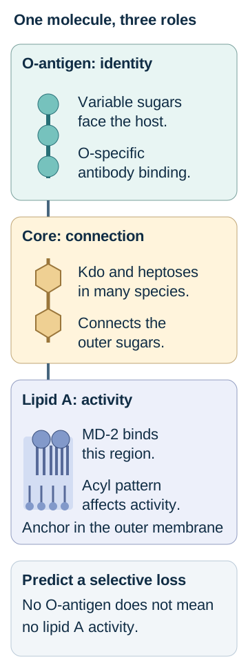
Picture one lipopolysaccharide molecule standing in the outer membrane of a Gram-negative bacterium. Its lipid anchor sits in the membrane, its core sugars connect the parts, and its outer repeating sugars face the host. The accompanying structural diagram separates these locations so that a change in one part does not automatically become a change in every function.
Lipid A is the membrane anchor and the principal endotoxic region recognized by the TLR4-MD-2 complex. The number and arrangement of its fatty-acyl groups influence receptor activation. Equal masses of LPS from different organisms need not produce equal responses. The well-studied six-acyl-group lipid A of E. coli is a useful model, not a universal blueprint for every Gram-negative species. [3]
The core oligosaccharide bridges lipid A and the O-antigen. Inner and outer core regions contribute to envelope organization. Kdo, or 3-deoxy-D-manno-oct-2-ulosonic acid, and heptoses are characteristic inner-core sugars in many familiar organisms. Their arrangement is not identical in all species. The O-antigen is the variable, outward-facing repeating polysaccharide that helps define the O serogroup and supplies accessible antibody targets. Do not confuse O-antigen with a capsule or with flagellar H antigen. [19]
The outer membrane is asymmetric: LPS predominates in the outer leaflet and phospholipids in the inner leaflet. Together with membrane proteins, this envelope limits permeability. The O-antigen can help protect the bacterial surface from complement-mediated killing. A rough mutant lacks the extended O-antigen and often loses some of that protection, but it can retain lipid A. Lipooligosaccharide, found in organisms such as Neisseria, also lacks an extended repeating O-antigen; this does not make it free of endotoxic activity. Interpret the specific organism and experiment rather than equating a short carbohydrate region with harmlessness. [3][19][21]
Try the comparison: point to the O-antigen on the diagram, then imagine only that region is absent. Predict the result of an O-specific antibody assay and a separate lipid A sensing assay before reading the reference comparison.
Point to the part detected by an O-specific antibody, then the part recognized by MD-2. Predict what remains after a selective O-antigen deletion. [3][19][21]
Different assays ask different structural questions
Change
Prediction when the other parts are preserved
ChangeLoss of the relevant O-antigen
Prediction when the other parts are preservedBinding by an antibody directed at that O structure decreases. Lipid A recognition can remain.
ChangeAltered lipid A acylation
Prediction when the other parts are preservedTLR4-MD-2 activation can change even when the O-specific antibody still binds.
ChangeDisrupted envelope integrity
Prediction when the other parts are preservedPermeability or complement susceptibility can change without directly measuring receptor signaling.
Apply it elsewhere: a mutant survives poorly in serum but its purified LPS still stimulates a receptor reporter. Those results are compatible: bacterial survival and the inflammatory activity of an isolated molecule are different outcomes. [3][19]
Where is LPS when the cell detects it?
Does endotoxin appear only after a bacterium dies? No. Lysis releases membrane material, but living Gram-negative bacteria also shed outer membrane vesicles containing LPS. Vesicles can deliver bacterial material to host cells. A sample without growing bacteria can therefore still contain an inflammatory stimulus. This biology is not a reason to postpone effective antibiotics during septic shock. [6][1]
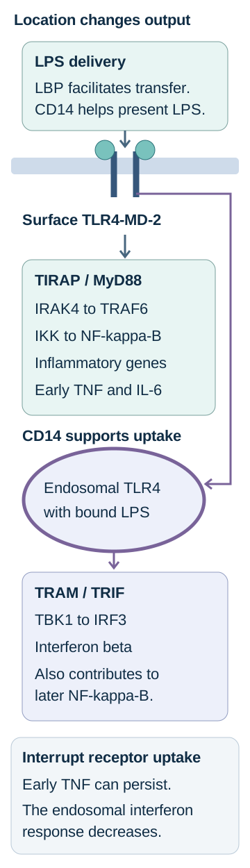
Follow the delivery route in the signaling diagram. Plasma LPS-binding protein, or LBP, facilitates transfer of LPS to CD14. CD14 can be soluble or attached to a cell membrane through a glycosylphosphatidylinositol anchor. It helps present LPS and organize its uptake, but does not supply the intracellular TIR signaling domain. LBP accelerates transfer; purified experimental systems should not be described as requiring LBP absolutely at every concentration. [16]
Trace each route from the shared surface receptor. Block uptake in your prediction: early surface output can persist while the endosomal interferon response falls. This is a compartment model, not a universal clinical clock. [3][4][5][16]
MD-2 directly accommodates lipid A beside TLR4. The activated receptor complex dimerizes. At the plasma membrane, the TLR4 TIR domains recruit TIRAP and MyD88. IRAK4 and downstream TRAF6-dependent signaling activate the IKK complex. IKK promotes phosphorylation and degradation of the inhibitor I-kappa-B, freeing NF-kappa-B to enter the nucleus and increase inflammatory gene transcription. TNF, IL-6, and the IL-1 precursor response are important outputs. Transcription of pro-IL-1 beta and processing of that precursor into mature IL-1 beta are distinct events; inflammasome-associated processing adds another control point. [3][5][7]
After CD14-supported endocytosis, TLR4 can signal from endosomes through TRAM and TRIF. TBK1 and IRF3 support type I interferon production, including interferon beta; TRIF also contributes to later NF-kappa-B activity. These are experimentally distinguishable branches, not a timetable that lets a clinician infer a patient's stage from one cytokine concentration. [4][5]
Trace and interrupt: use your finger to trace the surface route to NF-kappa-B, then the endosomal route to IRF3. Imagine receptor endocytosis is selectively blocked while surface binding remains intact. Predict which output persists. The visible reference below supplies the complete relationship; the optional questions let you rehearse it in smaller steps.
Compartment test: predicted results in the stated experimental model
Selective interruption
Early surface output
Endosomal interferon output
Selective interruptionTLR4-MD-2 activation
Early surface outputReduced
Endosomal interferon outputReduced
Selective interruptionReceptor endocytosis
Early surface outputCan remain
Endosomal interferon outputReduced
Selective interruptionTRIF signaling
Early surface outputMyD88 output can remain
Endosomal interferon outputReduced
Why can early TNF persist when receptor uptake is blocked?
The surface TIRAP-MyD88 route can still activate NF-kappa-B. Preventing entry into the endosomal compartment selectively compromises the TRAM-TRIF interferon response; it need not prevent the earlier surface response. [4][5]
Would destroying MD-2 produce the same pattern?
No. MD-2 acts before the two TLR4 branches separate. Disrupting productive lipid A recognition can impair both, rather than isolate the endosomal output. [3]
A second location matters: LPS delivered into the cytosol can be sensed by human caspases 4 and 5, with caspase 11 serving the corresponding role in mice. This noncanonical inflammasome response can trigger inflammatory cell death, or pyroptosis. TLR4-dependent gene expression and cytosolic LPS sensing are not interchangeable tests. Saying that LPS never contributes directly to a cell-death pathway would be incorrect. [7]
Transfer: if a TLR4-deficient cell responds to experimentally delivered cytosolic LPS but not extracellular LPS, first check the compartment and inflammatory caspases. Do not conclude that the surface receptor experiment was necessarily wrong. [7]
Can shock severity identify the microbial trigger?
A rapidly deteriorating patient has fever, confusion, and warm hypotensive extremities. That pattern does not tell you the Gram stain. Different microbial structures can initiate responses that converge on vascular dysfunction and organ injury. Both Gram-positive and Gram-negative infections can cause severe septic shock; microbial burden, source, host defenses, and treatment all matter. Do not use an alleged difference in cytokine explosiveness to choose antimicrobial coverage. [1][2]
The comparison below is a receptor map, not a rule assigning one receptor to each entire organism. A bacterium carries multiple signals. Gram-positive bacteria do not contain LPS. Their lipoproteins are important TLR2 stimuli, and lipoteichoic-acid preparations can show context-dependent TLR2 activity. Purified peptidoglycan fragments are better connected to intracellular NOD sensing than to a blanket claim that all peptidoglycan directly activates TLR2. NOD1 recognizes suitable diaminopimelate-containing fragments; NOD2 recognizes muramyl dipeptide. Lipoprotein contamination can confound experiments with crude cell-wall preparations. [8]
Compare the stimulus, its access, and the response
Stimulus
Useful recognition relationship
StimulusExtracellular LPS or LOS
Useful recognition relationshipLipid A delivery to TLR4-MD-2, aided by LBP and CD14.
StimulusBacterial lipoproteins
Useful recognition relationshipTLR2-containing receptor complexes; not exclusive to Gram-positive organisms.
StimulusPeptidoglycan fragments inside a cell
Useful recognition relationshipNOD-family recognition depends on the fragment.
StimulusA superantigen toxin
Useful recognition relationshipInteraction with MHC class II and selected T-cell receptor V-beta families, rather than conventional peptide-specific activation.
Sort the experiment: one preparation stimulates a TLR2 reporter until contaminating lipoproteins are depleted. A purified fragment still stimulates a cytosolic NOD2 reporter after intracellular delivery. Assign each response to the material that actually remains, rather than preserving the label on the crude bottle. The consequence is a more defensible receptor assignment: TLR2 activity and peptidoglycan recognition were separable in the experiment. [8]
Superantigens create another route to extensive immune activation by bridging MHC class II and particular T-cell receptor V-beta regions outside ordinary peptide-specific recognition. They should not be grouped under TLR2 merely because the producing organism is Gram positive. The resulting T-cell activation can be substantial, but not every T cell responds to every toxin. [17]
Transfer: a perforated bowel can expose the host to LPS, bacterial lipoproteins, and other microbial products together. Begin source-appropriate coverage, including anaerobes when indicated, while arranging control of contamination. Receptor diagrams explain biology; cultures, resistance risk, and the anatomic source guide treatment. [1][14]
Why do dilation, leakage, and clotting occur together?
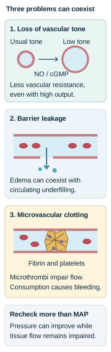
Would low blood pressure alone explain every injured organ? Consider three vessels in the accompanying diagram: one has lost tone, another leaks fluid into tissue, and a third contains fibrin-rich microthrombi. These abnormalities can coexist, so increasing a cuff pressure does not automatically restore every capillary's flow.
Match each physical change to a different observation: low resistance despite high output, edema with underfilling, or clotting with consumption. A better arterial pressure does not by itself prove restored tissue flow. [1][10][12][13][20]
Loss of tone: inflammatory signaling increases inducible nitric oxide synthase activity in several cell types. Nitric oxide stimulates soluble guanylate cyclase and cGMP signaling in vascular smooth muscle, promoting relaxation. Low systemic vascular resistance can lower arterial pressure even when cardiac output is high. Nitric oxide is one contributor, not the sole explanation for every episode of vasopressor-resistant shock. Experimental inhibition establishes a physiological role; it is not a recommendation for routine nitric oxide synthase inhibition in patients. [13][1]
Barrier failure: endothelial dysfunction permits fluid and proteins to leave the circulation. Interstitial edema can coexist with inadequate effective circulating volume. Venodilation also changes vascular capacity and venous return. Some patients can increase stroke volume when preload increases; others mainly accumulate additional edema. A patient's swelling, total fluid balance, and potential response to another bolus are related but different observations. [1][10]
Coagulation activation: inflammatory activation of monocytes and tissue-factor-bearing material promotes thrombin generation and fibrin deposition. Microvascular thrombosis and consumption of platelets and coagulation factors can occur together. Falling platelets, prolonged clotting times, increasing fibrin degradation products, and a falling fibrinogen concentration support disseminated intravascular coagulation in the appropriate setting. Fibrinogen may initially remain normal or high as an acute-phase reactant; one normal measurement does not exclude evolving disease. [12][20]
Connect the mediator to a measurable consequence
Mediator
Relationship worth predicting
MediatorTNF and IL-1
Relationship worth predictingInflammatory amplification, prostaglandin-associated fever, appetite changes, and vascular dysfunction. Experimental early release does not create a universal bedside minute-by-minute clock.
MediatorIL-6
Relationship worth predictingSignals hepatocytes to increase acute-phase protein production, including C-reactive protein. A high CRP does not identify a pathogen.
MediatorIL-10
Relationship worth predictingCounter-regulatory signaling limits inflammatory responses. Impaired immune function can coexist with ongoing inflammation and infection.
MediatorIL-5
Relationship worth predictingSupports eosinophil biology; it is not the principal explanation for the typical endotoxin-associated hepatic acute-phase response.
Human hepatocyte experiments support the IL-6 acute-phase relationship. Human sepsis studies also demonstrate impaired immune-cell function, but neither one cytokine nor a TNF-to-IL-10 ratio determines an individual patient's prognosis. Persistent infection, immune suppression, and inflammatory injury can overlap rather than occupy cleanly separated phases. [18][9]
Make a prediction: a patient has cardiac output 8 L/min, MAP 60 mm Hg, and central venous pressure 8 mm Hg. Using SVR = 80 x (MAP minus central venous pressure) / cardiac output gives 520 dyn s/cm5. The important finding is low resistance despite high output, not a universally low-output pump. If the same patient develops pulmonary edema and a falling platelet count, add barrier injury and coagulation activation to the explanation rather than trying to explain all three findings with low circulating volume.
Transfer: new cool extremities in a previously warm patient require reassessment for myocardial dysfunction, worsening hypoperfusion, excessive vasoconstriction, or another shock process. Septic shock is not permanently high-output, and a familiar diagnosis does not replace repeated examination. [1]
Does a better pressure mean perfusion has recovered?
First name what is being assessed. Sepsis is life-threatening organ dysfunction from a dysregulated response to infection; an acute SOFA increase of at least 2 operationalizes organ dysfunction in the Sepsis-3 framework. Septic shock is identified clinically by vasopressor requirement to maintain MAP at least 65 mm Hg and lactate greater than 2 mmol/L despite adequate volume resuscitation. A low pressure before resuscitation, an isolated high lactate, or a positive blood culture alone does not supply that complete definition. Do not postpone treatment until all classification criteria are documented. [2]
A patient can have serious infection-related kidney or brain dysfunction with systolic pressure above 90 mm Hg. Conversely, a single high lactate is not proof of tissue hypoxemia. Interpret infection likelihood, acute organ change, perfusion, treatment already delivered, and alternative causes together. qSOFA is not a stand-alone rule for excluding sepsis. [1][2]
Compare two questions before another fluid bolus: is perfusion inadequate, and is extra preload likely to increase useful flow without unacceptable harm? Passive leg raising briefly increases venous return; a reproducible change in measured stroke volume or cardiac output supports fluid responsiveness. A blood-pressure change alone is a weaker substitute. An isolated IVC measurement is not a universal fluid-response test. Pulse-pressure variation also has important validity limits, including irregular rhythm and spontaneous breathing. [10][1]
Changing the next decision with changing physiology
Assessment
Reasonable interpretation
AssessmentHypoperfusion with a reproducible stroke-volume increase during leg raising
Reasonable interpretationA monitored fluid trial may help when fluid tolerance permits; reassess flow, perfusion, and congestion afterward.
AssessmentPersistent hypotension without a stroke-volume response, with worsening pulmonary congestion
Reasonable interpretationFurther unselected boluses are unlikely to solve the main problem. Support vascular tone and reassess cardiac function and other causes.
AssessmentImproving capillary refill, mentation, and urine output with falling lactate
Reasonable interpretationThe trajectory is favorable, but ongoing vasopressor dependence or an uncontrolled source still matters.
The 2026 guideline conditionally suggests at least 30 mL/kg crystalloid in the first 3 hours for sepsis-induced hypoperfusion or septic shock, with individual context and repeated reassessment. This is not a required quantity to finish before starting a vasopressor, nor a command to repeat that quantity whenever lactate remains high. Balanced crystalloids are generally favored over saline, with exceptions such as traumatic brain injury. Heart failure, kidney disease, or diuretic use requires closer assessment of response and tolerance, not an automatic prohibition on resuscitation. [1]
Norepinephrine is the usual first vasopressor. In very unstable shock, begin it while initial fluids and reassessment are underway. Appropriate peripheral administration can avoid a harmful wait for central access, using local monitoring and extravasation safeguards. Vasopressin can be added as norepinephrine requirements rise; epinephrine can follow when the combination remains inadequate. Concurrent myocardial dysfunction may require a different hemodynamic assessment and selected inotropic support. [1]
Keep a definition separate from a treatment target. For most adults, the recommended initial MAP target is 65 mm Hg rather than a higher routine target. The 2026 guideline conditionally suggests an initial range of 60 to 65 mm Hg for adults aged 65 or older. These initial targets need reassessment against organ perfusion and individual circumstances; the age-specific treatment recommendation does not rewrite the Sepsis-3 research identification criteria above. [1][2]
Calculate, then qualify: lactate falling from 6 to 3 mmol/L is a 50% relative decrease: (6 minus 3) / 6 x 100. It is encouraging, not a universally required clearance percentage or proof that shock has ended. Lactate can reflect altered metabolism, adrenergic stimulation, impaired clearance, or hypoperfusion. Follow the trend alongside capillary refill, urine output, mentation, flow assessment, and support requirements. Giving fluid solely until lactate normalizes risks treating the number rather than the circulation. [1]
Oxygen delivery also depends on hemoglobin and arterial oxygenation. A restrictive red-cell strategy commonly uses a hemoglobin threshold around 7 g/dL in adults with septic shock, rather than transfusing automatically to 9 or 10. Active hemorrhage, myocardial ischemia, severe hypoxemia, and the overall clinical picture can change the decision. The TRISS trial compared 7 and 9 g/dL thresholds; similar mortality does not mean every individual must receive the same threshold without assessment. [11]
Transfer: an older patient whose MAP is 63 with improving urine output and mentation is different from one at MAP 68 with new oliguria and rising support needs. The numerical pressure alone cannot identify which circulation is recovering. [1]
What must happen while circulation is being supported?
Imagine an infected, obstructed bile duct while norepinephrine restores arterial pressure. The pressor supports circulation; it does not drain the duct. Antibiotics act against susceptible organisms; they do not correct mechanical obstruction. Successful care needs these tasks to proceed together, not as a sequence in which each team waits for the previous task to finish.
Obtain blood cultures promptly, ideally before antimicrobials, when doing so does not meaningfully delay treatment. In septic shock, start appropriate antimicrobials immediately, ideally within 1 hour of recognition; do not wait for identification or susceptibility results. The 2026 recommendations distinguish this emergency from possible sepsis without shock, where a time-limited diagnostic assessment may be appropriate. Severe infection is not a reason to await a perfectly complete microbiology report. [1]
Choose coverage from the patient and the source. Ceftriaxone is not an antipseudomonal drug. Cefepime and piperacillin-tazobactam can supply antipseudomonal activity when the isolate is susceptible, whereas resistant Gram-negative infection may require a different regimen. Neutropenia, prior resistant isolates, recent antimicrobial exposure, local susceptibility patterns, allergy, kidney function, and infection site change the selection. These drugs are not interchangeable merely because all are described as broad spectrum. [1][14]
For ESBL-producing Enterobacterales bloodstream infection in a critically ill patient, the 2026 IDSA guidance favors meropenem or imipenem. A laboratory report of piperacillin-tazobactam susceptibility does not by itself make it preferred for invasive ESBL disease. Broader empiric therapy should be reassessed and narrowed when organism, susceptibility, and clinical information allow. [14]
A bowel perforation usually requires Gram-negative and anaerobic coverage. Add Enterococcus, MRSA, or other resistant-organism coverage when source and patient risk justify it; not every community-acquired abdominal infection requires every possible agent. An infected catheter, necrotic tissue, a drainable collection, and an obstructed biliary system each present a different source-control problem. [1]
Pair an action with its purpose: for persistent shock from obstructed cholangitis, identify both the circulatory action and the anatomic action. Additional vasopressor support may be appropriate, while urgent biliary drainage is arranged. Neither increasing pressure alone nor simply changing antibiotics resolves the obstruction.
Match the persistent source to the intervention
Source problem
Assessment that cannot wait for a normal lactate
Source problemInfected biliary obstruction
Assessment that cannot wait for a normal lactateUrgent endoscopic or alternative drainage, coordinated with resuscitation.
Source problemPerforation with ongoing contamination
Assessment that cannot wait for a normal lactateSurgical assessment and control of the leak or contaminated space.
Source problemInfected device or necrotic tissue
Assessment that cannot wait for a normal lactateDevice removal when indicated or debridement, with procedural planning and antimicrobial treatment.
The 2026 guideline conditionally suggests early source control, ideally within 6 hours of diagnosing sepsis or septic shock when an anatomic source requires intervention. Anatomy, procedural availability, and patient stability affect the approach, but transient pressure improvement is not a reason to abandon a needed intervention. Avoid the absolute statement that no abscess can ever respond to antibiotics alone: selected small collections may be managed medically. A drainable focus driving ongoing septic shock is a different problem. [1]
Intravenous corticosteroids are an adjunct in adult septic shock, not a substitute for initial norepinephrine, effective antibiotics, or source control. Hydrocortisone 200 mg/day is a studied regimen. In ADRENAL, hydrocortisone shortened shock duration but did not significantly lower the primary 90-day mortality outcome. Selection and monitoring remain clinical decisions rather than a rigid rule that steroids must wait for a numbered third stage. [15][1]
Transfer: after drainage, a patient still needs norepinephrine but has improving perfusion and a susceptible isolate. Continue reassessment, optimize targeted antimicrobials, and consider appropriate adjuncts. Do not declare recovery from a single normal temperature, mandate another fluid bolus from a single lactate value, or assume that understanding lipid A supplies a proven endotoxin-specific rescue treatment. Routine polymyxin B hemoperfusion is not recommended by the current guideline. [1]
Apply the relationships to new cases
Commit to one answer before reading the explanations. For a missed option, compare the finding it would predict with the finding actually supplied.
Case 1
Show answer and explanations for case 1
A. Loss of receptor activation with retained surface protection (Why this does not fit)
It would reduce activation of the TLR4-MD-2 reporter. No. Reporter activity is similar, whereas survival in serum is lower, the opposite pairing. [3] [19] [21]
Reasoning steps for option A
What would loss of productive lipid A recognition change?
It would reduce activation of the TLR4-MD-2 reporter.
Do the reporter and serum results show that combination?
No. Reporter activity is similar, whereas survival in serum is lower, the opposite pairing.
B. Loss of surface protection with retained lipid A activity (Best answer)
The serum experiment measures bacterial survival and is compatible with loss of O-antigen-associated protection. The mutant retains the same lipid A, so a change in surface protection does not require loss of endotoxin signaling. [3] [19] [21]
Reasoning steps for option B
Which result measures the survival of intact bacteria?
The serum experiment measures bacterial survival and is compatible with loss of O-antigen-associated protection.
Why can reporter activation remain similar?
The mutant retains the same lipid A, so a change in surface protection does not require loss of endotoxin signaling.
C. Loss of capsule synthesis with retained O-specific binding (Why this does not fit)
A capsule defect can reduce protection against host defenses in an appropriate strain. The experiment specifies O-antigen loss, not capsule loss; preserved O-specific binding would not follow from that alteration. [3] [19] [21]
Reasoning steps for option C
How could a capsule defect affect survival?
A capsule defect can reduce protection against host defenses in an appropriate strain.
Which envelope alteration is actually established here?
The experiment specifies O-antigen loss, not capsule loss; preserved O-specific binding would not follow from that alteration.
D. Loss of lipid anchoring with retained receptor activation (Why this does not fit)
Its fatty-acyl groups anchor LPS within the outer membrane. The mutant retains the parental lipid A structure; the identified deletion concerns the outward polysaccharide instead. [3] [19] [21]
Reasoning steps for option D
What function does lipid A perform in the envelope?
Its fatty-acyl groups anchor LPS within the outer membrane.
Which supplied control argues against losing that anchor?
The mutant retains the parental lipid A structure; the identified deletion concerns the outward polysaccharide instead.
Takeaway: Separate the survival of an intact bacterium from the inflammatory activity of purified lipid A.
A. Reduced recognition of O-antigen by specific antibodies (Why this does not fit)
It tests recognition of the exposed repeating carbohydrate rather than the lipid A receptor pocket. O-antigen is unchanged and no O-specific antibody response is the measured pathway; altered receptor assembly tracks lipid A instead. [3]
Reasoning steps for option A
What does an O-specific antibody assay test?
It tests recognition of the exposed repeating carbohydrate rather than the lipid A receptor pocket.
Why is that not the best explanation for this assay?
O-antigen is unchanged and no O-specific antibody response is the measured pathway; altered receptor assembly tracks lipid A instead.
B. Failure of NF-kappa-B to function in the reporter cells (Why this does not fit)
A defective transcription factor could reduce an inflammatory transcriptional response downstream of IKK. The downstream response is intact and receptor dimerization already differs, locating the important change before that transcriptional machinery. [3]
Reasoning steps for option B
How could defective NF-kappa-B reduce TNF transcription?
A defective transcription factor could reduce an inflammatory transcriptional response downstream of IKK.
Which controls favor an earlier defect instead?
The downstream response is intact and receptor dimerization already differs, locating the important change before that transcriptional machinery.
C. Reduced delivery caused by a smaller LPS inoculum (Why this does not fit)
Less ligand can yield less receptor activation even when molecular potency is unchanged. The preparations are tested at equal molar concentrations, while their lipid A structures and receptor assembly differ. [3]
Reasoning steps for option C
Why would a smaller LPS dose be a competing explanation?
Less ligand can yield less receptor activation even when molecular potency is unchanged.
What comparison prevents using that explanation here?
The preparations are tested at equal molar concentrations, while their lipid A structures and receptor assembly differ.
D. Less effective assembly of the lipid A receptor complex (Best answer)
Receptor dimerization is reduced before downstream transcription is assessed. Altered lipid A geometry can impair productive receptor assembly, while preserved downstream responses argue against an intrinsic transcriptional defect. [3]
Reasoning steps for option D
Where does the first demonstrated response differ?
Receptor dimerization is reduced before downstream transcription is assessed.
How do the lipid analysis and downstream control localize the cause?
Altered lipid A geometry can impair productive receptor assembly, while preserved downstream responses argue against an intrinsic transcriptional defect.
Takeaway: Use dose controls and a downstream response to separate ligand potency from exposure or cell dysfunction.
A. LBP aids transfer but does not supply the signaling domain (Best answer)
No. Binding eventually occurs without LBP, so the experiment demonstrates acceleration rather than an absolute requirement. LBP facilitates LPS delivery to CD14; intracellular receptor signaling occurs through the responding cell, not through an LBP TIR domain. [16] [3]
Reasoning steps for option A
Does slow loading in the absence of LBP establish absolute dependence?
No. Binding eventually occurs without LBP, so the experiment demonstrates acceleration rather than an absolute requirement.
What does subsequent activation by the loaded preparation imply?
LBP facilitates LPS delivery to CD14; intracellular receptor signaling occurs through the responding cell, not through an LBP TIR domain.
B. LBP supplies the intracellular TIR domain needed for signaling (Why this does not fit)
The intracellular TLR4 TIR domain recruits signaling adaptors. No. The experiment isolates extracellular transfer kinetics, and LBP does not replace the cell-associated TLR4 signaling domain. [16] [3]
Reasoning steps for option B
What is the role of a TIR domain in this pathway?
The intracellular TLR4 TIR domain recruits signaling adaptors.
Does extracellular LBP loading demonstrate that role?
No. The experiment isolates extracellular transfer kinetics, and LBP does not replace the cell-associated TLR4 signaling domain.
C. CD14 must remain in a permanent three-protein complex with LBP (Why this does not fit)
It could explain ligand delivery if LBP had to remain attached throughout receptor engagement. CD14 can acquire LPS and support stimulation without LBP; the result supports facilitated transfer rather than a necessary permanent ternary complex. [16] [3]
Reasoning steps for option C
What could a stable transfer complex hypothetically explain?
It could explain ligand delivery if LBP had to remain attached throughout receptor engagement.
Which observation makes that requirement unsupported?
CD14 can acquire LPS and support stimulation without LBP; the result supports facilitated transfer rather than a necessary permanent ternary complex.
D. LBP must retain one molecule of itself with every transferred LPS (Why this does not fit)
It would require a stoichiometric amount of LBP for the whole pool of transferred LPS. The substoichiometric effect supports facilitated transfer rather than permanent retention of one LBP per LPS in the final loaded preparation. [16] [3]
Reasoning steps for option D
What would permanent one-to-one retention require?
It would require a stoichiometric amount of LBP for the whole pool of transferred LPS.
What does full transfer with one-twentieth as much LBP support instead?
The substoichiometric effect supports facilitated transfer rather than permanent retention of one LBP per LPS in the final loaded preparation.
Takeaway: A protein that accelerates ligand transfer is not necessarily obligatory in every purified assay.
A. Binding of lipid A within the extracellular MD-2 pocket (Why this does not fit)
A lesion before TLR4 activation can compromise both downstream signaling branches. Surface binding and early TNF transcription persist, whereas the defect tracks the uptake-dependent IRF3 response. [4] [5]
Reasoning steps for option A
What would loss of productive MD-2 recognition affect?
A lesion before TLR4 activation can compromise both downstream signaling branches.
Why does that predict a different response here?
Surface binding and early TNF transcription persist, whereas the defect tracks the uptake-dependent IRF3 response.
B. Recruitment of MyD88 to activated plasma-membrane TLR4 (Why this does not fit)
Early inflammatory transcription is a useful readout of the surface MyD88 route. It would reduce early TNF rather than selectively explain loss of the endosomal interferon output. [4] [5]
Reasoning steps for option B
Which output chiefly tests the early MyD88 route?
Early inflammatory transcription is a useful readout of the surface MyD88 route.
Does blocking that route fit the preserved response?
It would reduce early TNF rather than selectively explain loss of the endosomal interferon output.
C. Access of TLR4 to the TRAM-TRIF signaling compartment (Best answer)
Early TNF transcription despite the intervention supports continuing surface MyD88-dependent output. Preventing receptor uptake limits endosomal TRAM-TRIF signaling, separating the interferon branch from the preserved early surface response. [4] [5]
Reasoning steps for option C
Which intact observation preserves the surface pathway?
Early TNF transcription despite the intervention supports continuing surface MyD88-dependent output.
Why are IRF3 and interferon beta selectively impaired?
Preventing receptor uptake limits endosomal TRAM-TRIF signaling, separating the interferon branch from the preserved early surface response.
D. Availability of transcriptional machinery throughout the cell (Why this does not fit)
It could suppress multiple newly transcribed cytokine outputs, including TNF. TNF transcription and viability remain intact while IRF3 activation and interferon beta fall after an uptake-specific intervention. [4] [5]
Reasoning steps for option D
What would a general transcriptional defect do?
It could suppress multiple newly transcribed cytokine outputs, including TNF.
Which supplied result favors a selective compartment problem?
TNF transcription and viability remain intact while IRF3 activation and interferon beta fall after an uptake-specific intervention.
Takeaway: Preserved surface output with reduced IRF3 output localizes a defect to receptor uptake or endosomal signaling.
A. The MD-2 lipid-binding pocket before branch separation (Why this does not fit)
It would interfere before the activated TLR4 complex separates into signaling routes. Normal ligand binding, preserved endosomal signaling, and selective rescue by IRAK4 point downstream to the MyD88-associated route. [3] [4] [5]
Reasoning steps for option A
Where would a nonfunctional MD-2 pocket act?
It would interfere before the activated TLR4 complex separates into signaling routes.
Why is that less consistent with the controls?
Normal ligand binding, preserved endosomal signaling, and selective rescue by IRAK4 point downstream to the MyD88-associated route.
B. The TRAM-TRIF route after receptor internalization (Why this does not fit)
Endosomal IRF3 activation and interferon beta are important TRAM-TRIF outputs. They remain detectable, while IRAK4 restores the early inflammatory response, which is a different branch. [3] [4] [5]
Reasoning steps for option B
Which outputs evaluate this route?
Endosomal IRF3 activation and interferon beta are important TRAM-TRIF outputs.
What do the assay results show about them?
They remain detectable, while IRAK4 restores the early inflammatory response, which is a different branch.
C. The MyD88 pathway downstream of receptor activation (Best answer)
Ligand recognition and access to the endosomal compartment remain functional in the assay. They favor an IRAK4-dependent MyD88 signaling problem rather than a lesion that prevents all TLR4 activation or the TRIF response. [3] [4] [5]
Reasoning steps for option C
What do binding and internalization show?
Ligand recognition and access to the endosomal compartment remain functional in the assay.
How do the preserved interferon response and IRAK4 rescue localize the defect?
They favor an IRAK4-dependent MyD88 signaling problem rather than a lesion that prevents all TLR4 activation or the TRIF response.
D. The CD14-dependent delivery and uptake of extracellular LPS (Why this does not fit)
Impaired LPS delivery or receptor uptake can limit productive receptor signaling. Binding and internalization are normal, and the defect is corrected by a downstream MyD88-associated kinase. [3] [4] [5]
Reasoning steps for option D
How could defective CD14 affect the experiment?
Impaired LPS delivery or receptor uptake can limit productive receptor signaling.
Which observations argue for another site?
Binding and internalization are normal, and the defect is corrected by a downstream MyD88-associated kinase.
Takeaway: Use intact proximal functions and a rescue experiment to localize a selective pathway defect.
A. Impaired release of NF-kappa-B from the inhibitor in the cytoplasm (Best answer)
Phosphorylation and degradation of I-kappa-B frees the transcription factor from its inhibitor. Persistent I-kappa-B, absent nuclear accumulation, and reduced TNF transcription locate the defect at inhibitor release despite preserved upstream recognition. [3] [5] [7]
Reasoning steps for option A
What normally permits NF-kappa-B to enter the nucleus?
Phosphorylation and degradation of I-kappa-B frees the transcription factor from its inhibitor.
Which linked findings identify that failed step?
Persistent I-kappa-B, absent nuclear accumulation, and reduced TNF transcription locate the defect at inhibitor release despite preserved upstream recognition.
B. Impaired presentation of lipid A to the MD-2 complex (Why this does not fit)
It would reduce productive activation of the extracellular receptor complex. TLR4 dimerization is unchanged and IRF3 signaling remains intact, while the inhibitor-associated NF-kappa-B step is abnormal. [3] [5] [7]
Reasoning steps for option B
What would impaired presentation initially reduce?
It would reduce productive activation of the extracellular receptor complex.
Which intact measurement argues against this location?
TLR4 dimerization is unchanged and IRF3 signaling remains intact, while the inhibitor-associated NF-kappa-B step is abnormal.
C. Impaired endosomal signaling through TRIF and TBK1 (Why this does not fit)
TBK1-dependent IRF3 activation is an important interferon-pathway readout. IRF3 phosphorylation remains intact; NF-kappa-B nuclear accumulation fails with persistent I-kappa-B instead. [3] [5] [7]
Reasoning steps for option C
Which transcription factor reports this branch?
TBK1-dependent IRF3 activation is an important interferon-pathway readout.
Is that the factor that fails in this preparation?
D. Impaired processing of pro-IL-1 beta into mature IL-1 beta (Why this does not fit)
It changes maturation of a cytokine precursor after the precursor has been produced. A cytokine-processing defect does not account for the directly observed retention of NF-kappa-B with its inhibitor and reduced TNF transcription. [3] [5] [7]
Reasoning steps for option D
What does processing of pro-IL-1 beta change?
It changes maturation of a cytokine precursor after the precursor has been produced.
Why does that not explain the stated TNF result?
A cytokine-processing defect does not account for the directly observed retention of NF-kappa-B with its inhibitor and reduced TNF transcription.
Takeaway: Keep receptor activation, nuclear transcription, and cytokine maturation as separate steps.
A. Caspase 4 restores the extracellular LPS receptor complex (Why this does not fit)
It would permit a response to extracellular ligand through a functional receptor complex. No. Extracellular LPS remains ineffective in the TLR4-deficient cells, while the response depends on delivery into the cytosol. [7]
Reasoning steps for option A
How might restored surface sensing appear experimentally?
It would permit a response to extracellular ligand through a functional receptor complex.
Does the extracellular control demonstrate restoration?
No. Extracellular LPS remains ineffective in the TLR4-deficient cells, while the response depends on delivery into the cytosol.
B. MD-2 alone replaces TLR4 for extracellular LPS transcription (Why this does not fit)
MD-2 binds lipid A as part of the TLR4 recognition complex. The response requires cytosolic delivery and caspase 4; extracellular LPS does not restore the missing TLR4 response. [7]
Reasoning steps for option B
What role does MD-2 normally play?
MD-2 binds lipid A as part of the TLR4 recognition complex.
Which assay feature makes autonomous extracellular signaling an inadequate explanation?
The response requires cytosolic delivery and caspase 4; extracellular LPS does not restore the missing TLR4 response.
C. Endosomal TRIF signaling is the required death-triggering route (Why this does not fit)
It supports IRF3 activation and type I interferon production after receptor uptake. The responding cells lack TLR4 and depend on a cytosolic inflammatory caspase after direct intracellular ligand delivery. [7]
Reasoning steps for option C
What does endosomal TLR4-TRIF signaling ordinarily support?
It supports IRF3 activation and type I interferon production after receptor uptake.
Why is it not the required route demonstrated here?
The responding cells lack TLR4 and depend on a cytosolic inflammatory caspase after direct intracellular ligand delivery.
D. Cytosolic LPS sensing triggers inflammatory death here without TLR4 (Best answer)
Cytosolic delivery produced a response in cells unable to use the extracellular TLR4 pathway. It implicates a human inflammatory-caspase pathway in this assay, not restoration of the missing surface receptor. [7]
Reasoning steps for option D
What changed when the same ligand entered a different compartment?
Cytosolic delivery produced a response in cells unable to use the extracellular TLR4 pathway.
What does the additional caspase 4 deletion establish?
It implicates a human inflammatory-caspase pathway in this assay, not restoration of the missing surface receptor.
Takeaway: The ligand's compartment determines which sensor can respond; primed cytosolic-LPS experiments do not require intact surface TLR4.
A. The host response requires invasion by intact live bacteria (Why this does not fit)
Intact invading organisms can deliver several inflammatory stimuli to host cells. The active fraction is isolated from bacteria-free supernatant, so intact bacterial invasion is not required for the observed response. [6]
Reasoning steps for option A
When might invasion explain a host inflammatory response?
Intact invading organisms can deliver several inflammatory stimuli to host cells.
Which preparation excludes that explanation as a requirement here?
The active fraction is isolated from bacteria-free supernatant, so intact bacterial invasion is not required for the observed response.
B. Living bacteria release LPS-containing inflammatory material here (Best answer)
They argue against widespread bacterial lysis as the necessary source of this preparation. They support release of LPS-containing membrane material by living bacteria and an LPS contribution to this experimental response. [6]
Reasoning steps for option B
What do stable viability and the low lysis marker argue against?
They argue against widespread bacterial lysis as the necessary source of this preparation.
What do vesicle isolation and selective LPS depletion add?
They support release of LPS-containing membrane material by living bacteria and an LPS contribution to this experimental response.
C. LPS becomes available to host sensors after bacterial lysis alone (Why this does not fit)
Disruption of bacterial membranes can release LPS-containing material. No. Vesicles appear while bacterial counts remain stable and the cytosolic lysis marker stays low. [6]
Reasoning steps for option C
How can lysis make LPS available?
Disruption of bacterial membranes can release LPS-containing material.
Do the culture controls establish lysis as necessary?
No. Vesicles appear while bacterial counts remain stable and the cytosolic lysis marker stays low.
D. A secreted protein toxin fully accounts for the vesicle response (Why this does not fit)
Yes. Vesicles may carry several bacterial components, including proteins. The response decreases after selective LPS depletion, demonstrating an LPS contribution rather than a fully protein-dependent account. [6]
Reasoning steps for option D
Could vesicles contain molecules besides LPS?
Yes. Vesicles may carry several bacterial components, including proteins.
Why is a protein-only explanation not supported?
The response decreases after selective LPS depletion, demonstrating an LPS contribution rather than a fully protein-dependent account.
Takeaway: Bacteria-free does not mean free of bacterial products, and endotoxin release is not restricted to lysis.
A. The crude TLR2 response and purified-fragment sensing have different ligands (Best answer)
Removing contaminating lipoproteins reduced TLR2 activity, implicating those components in that response. It supports NOD2 recognition of muramyl dipeptide rather than assigning all crude cell-wall activity directly to peptidoglycan and TLR2. [8]
Reasoning steps for option A
What did depletion change in the crude preparation?
Removing contaminating lipoproteins reduced TLR2 activity, implicating those components in that response.
What does the cytosolic fragment assay establish separately?
It supports NOD2 recognition of muramyl dipeptide rather than assigning all crude cell-wall activity directly to peptidoglycan and TLR2.
B. Purified peptidoglycan directly stimulates TLR2 in both preparations (Why this does not fit)
A crude cell-wall preparation may contain more than one receptor-active microbial component. No. Lipoprotein depletion reduces TLR2 activity, while the defined intracellular fragment is tested through NOD2. [8]
Reasoning steps for option B
What makes the original crude result potentially misleading?
A crude cell-wall preparation may contain more than one receptor-active microbial component.
Does the purified intracellular experiment preserve the TLR2 assignment?
No. Lipoprotein depletion reduces TLR2 activity, while the defined intracellular fragment is tested through NOD2.
C. Loss of cell viability explains the reduced TLR2 signal (Why this does not fit)
Damaged reporter cells could produce a weaker response independent of ligand recognition. Cell viability and positive-control responses remain intact after the preparation is altered. [8]
Reasoning steps for option C
Why would viability be a necessary control?
Damaged reporter cells could produce a weaker response independent of ligand recognition.
What rules out that explanation in this experiment?
Cell viability and positive-control responses remain intact after the preparation is altered.
D. Gram-positive organisms activate NOD2 but not surface receptors (Why this does not fit)
A defined peptidoglycan fragment can activate NOD2 after reaching the appropriate compartment. The original lipoprotein-containing preparation activates TLR2; an organism can supply ligands for more than one receptor family. [8]
Reasoning steps for option D
What does cytosolic muramyl dipeptide demonstrate?
A defined peptidoglycan fragment can activate NOD2 after reaching the appropriate compartment.
Why does that not exclude surface recognition?
The original lipoprotein-containing preparation activates TLR2; an organism can supply ligands for more than one receptor family.
Takeaway: Identify the active component and its delivery compartment before assigning a receptor to a crude preparation.
A. Binding of bacterial lipoproteins to TLR2-containing receptor complexes (Why this does not fit)
They can stimulate innate signaling through TLR2-containing complexes. TLR2 blockade does not prevent the response, which instead depends on MHC class II and selective T-cell receptor V-beta families. [17] [3] [8]
Reasoning steps for option A
What response could bacterial lipoproteins cause?
They can stimulate innate signaling through TLR2-containing complexes.
Which experimental findings require a different explanation?
TLR2 blockade does not prevent the response, which instead depends on MHC class II and selective T-cell receptor V-beta families.
B. Presentation of a processed peptide in the MHC class II groove (Why this does not fit)
Recognition of a particular processed peptide in MHC class II selects compatible antigen-specific T cells. Processing is unnecessary and activation spans selected V-beta families, supporting superantigen activity rather than ordinary peptide specificity. [17] [3] [8]
Reasoning steps for option B
What normally determines conventional antigen-specific T-cell activation?
Recognition of a particular processed peptide in MHC class II selects compatible antigen-specific T cells.
What is atypical for that pathway here?
Processing is unnecessary and activation spans selected V-beta families, supporting superantigen activity rather than ordinary peptide specificity.
C. Bridging of MHC class II and selected T-cell receptor V-beta regions (Best answer)
It suggests activation based on a shared receptor variable-region family rather than ordinary recognition of one processed peptide. They support a superantigen bridge involving MHC class II and T-cell receptors, distinct from a TLR2-driven cell-wall response. [17] [3] [8]
Reasoning steps for option C
What does V-beta-selective proliferation suggest?
It suggests activation based on a shared receptor variable-region family rather than ordinary recognition of one processed peptide.
How do MHC class II dependence and preserved activity without peptide processing refine the mechanism?
They support a superantigen bridge involving MHC class II and T-cell receptors, distinct from a TLR2-driven cell-wall response.
D. Binding of extracellular lipid A to the TLR4-MD-2 receptor pocket (Why this does not fit)
It recognizes appropriate lipid A structures as part of an innate LPS response. The response depends on MHC class II and selected T-cell receptor families, not lipid A recognition by the TLR4 complex. [17] [3] [8]
Reasoning steps for option D
What does the TLR4-MD-2 pocket recognize?
It recognizes appropriate lipid A structures as part of an innate LPS response.
Why does that not explain the purified-protein experiment?
The response depends on MHC class II and selected T-cell receptor families, not lipid A recognition by the TLR4 complex.
Takeaway: A Gram-positive toxin can stimulate T cells through a superantigen mechanism without acting through TLR2.
A. Primary left ventricular pump failure with compensatory vasoconstriction (Why this does not fit)
Primary pump failure usually reduces forward output and can provoke compensatory systemic vasoconstriction. The output is high, contraction is vigorous, and calculated systemic resistance is low. [1] [10] [13]
Reasoning steps for option A
What pressure-flow pattern would a failing pump usually produce?
Primary pump failure usually reduces forward output and can provoke compensatory systemic vasoconstriction.
Which measurements conflict with that explanation?
The output is high, contraction is vigorous, and calculated systemic resistance is low.
B. Low systemic vascular resistance despite preserved high flow (Best answer)
SVR = 80 x (56 minus 8) / 8 = 480 dyn s/cm5, a low systemic resistance. High output with preserved contraction and little preload response favors vasoplegia over primary pump failure or a simple preload deficit. [1] [10] [13]
Reasoning steps for option B
What resistance follows from the supplied measurements?
SVR = 80 x (56 minus 8) / 8 = 480 dyn s/cm5, a low systemic resistance.
How do cardiac imaging and the leg-raise response refine the interpretation?
High output with preserved contraction and little preload response favors vasoplegia over primary pump failure or a simple preload deficit.
C. Acute pulmonary obstruction with reduced left ventricular filling (Why this does not fit)
Right ventricular pressure loading and reduced forward flow can cause hypotension. There is no right ventricular pressure overload and cardiac output is high rather than obstructed and low. [1] [10] [13]
Reasoning steps for option C
What findings could a major pulmonary obstruction produce?
Right ventricular pressure loading and reduced forward flow can cause hypotension.
Which supplied findings make it less explanatory here?
There is no right ventricular pressure overload and cardiac output is high rather than obstructed and low.
D. Persistent preload depletion with compensatory systemic vasoconstriction (Why this does not fit)
Vascular leakage and altered vascular capacity can reduce effective circulating volume. Stroke volume barely changes during leg raising, output is high, and calculated resistance is low rather than compensatorily high. [1] [10] [13]
Reasoning steps for option D
Why is a circulating-volume deficit plausible during infection?
Vascular leakage and altered vascular capacity can reduce effective circulating volume.
Which current measurements argue against it as the dominant explanation?
Stroke volume barely changes during leg raising, output is high, and calculated resistance is low rather than compensatorily high.
Takeaway: Calculate resistance from pressure and flow, then use dynamic and imaging findings to identify the dominant hemodynamic problem.
A. Isolated immune platelet destruction with preserved coagulation factors (Why this does not fit)
It can cause thrombocytopenia and bleeding without consuming fibrinogen or broadly prolonging clotting times. The rising PT, falling fibrinogen, and large increase in fibrin degradation products support systemic coagulation activation and consumption. [12] [20]
Reasoning steps for option A
What laboratory pattern can isolated immune thrombocytopenia produce?
It can cause thrombocytopenia and bleeding without consuming fibrinogen or broadly prolonging clotting times.
Which findings require more than isolated platelet destruction?
The rising PT, falling fibrinogen, and large increase in fibrin degradation products support systemic coagulation activation and consumption.
B. Platelet-rich microthrombi from severe ADAMTS13 deficiency (Why this does not fit)
Thrombocytopenia, microvascular injury, and renal or neurologic abnormalities can occur with platelet-rich microthrombi. Marked PT prolongation and falling fibrinogen indicate coagulation-factor consumption, unlike the usually preserved routine coagulation tests in TTP. [12] [20]
Reasoning steps for option B
Why does thrombotic thrombocytopenic purpura enter the differential?
Thrombocytopenia, microvascular injury, and renal or neurologic abnormalities can occur with platelet-rich microthrombi.
Which supplied pattern favors disseminated coagulation instead?
Marked PT prolongation and falling fibrinogen indicate coagulation-factor consumption, unlike the usually preserved routine coagulation tests in TTP.
C. Heparin-dependent antibody activation of circulating platelets (Why this does not fit)
It can combine thrombocytopenia with thrombosis after an appropriate heparin exposure. The abnormalities precede heparin exposure and include broad factor consumption during septic shock, favoring disseminated intravascular coagulation. [12] [20]
Reasoning steps for option C
What makes heparin-induced thrombocytopenia clinically important?
It can combine thrombocytopenia with thrombosis after an appropriate heparin exposure.
Which timing and laboratory findings point elsewhere?
The abnormalities precede heparin exposure and include broad factor consumption during septic shock, favoring disseminated intravascular coagulation.
D. Widespread fibrin formation with consumption of platelets and factors (Best answer)
It suggests consumptive coagulation rather than isolated platelet destruction. Microvascular fibrin deposition can impair organ perfusion while consumption of platelets and clotting factors produces bleeding. [12] [20]
Reasoning steps for option D
What does the combination of falling platelets, prolonged PT and falling fibrinogen suggest?
It suggests consumptive coagulation rather than isolated platelet destruction.
How can that cause bleeding and kidney injury together?
Microvascular fibrin deposition can impair organ perfusion while consumption of platelets and clotting factors produces bleeding.
Takeaway: Systemic coagulation activation can injure organs by thrombosis while simultaneously causing bleeding through consumption.
A. Eosinophil development and survival (Why this does not fit)
IL-5 supports eosinophil development and survival. The experiment measures new protein synthesis by hepatocytes, not eosinophil development, and specifically interrupts IL-6 signaling. [18] [5] [3]
Reasoning steps for option A
Which cytokine is strongly associated with eosinophil biology?
IL-5 supports eosinophil development and survival.
Does that explain the selected cell type and endpoint?
The experiment measures new protein synthesis by hepatocytes, not eosinophil development, and specifically interrupts IL-6 signaling.
B. Lipid A binding within MD-2 (Why this does not fit)
It occurs in the MD-2-associated TLR4 receptor complex before many inflammatory cytokines are produced. No. The intervention targets IL-6 signaling and the measured consequence is hepatic CRP synthesis, not lipid A binding. [18] [5] [3]
Reasoning steps for option B
Where does lipid A binding occur in the canonical recognition pathway?
It occurs in the MD-2-associated TLR4 receptor complex before many inflammatory cytokines are produced.
Is that the receptor manipulated in these hepatocytes?
No. The intervention targets IL-6 signaling and the measured consequence is hepatic CRP synthesis, not lipid A binding.
C. Hepatic acute-phase protein induction (Best answer)
New C-reactive protein synthesis is an acute-phase response of hepatocytes. It interrupts an important hepatic acute-phase signal without requiring TNF concentration to fall or the hepatocytes to die. [18] [5] [3]
Reasoning steps for option C
What does the measured endpoint represent?
New C-reactive protein synthesis is an acute-phase response of hepatocytes.
How does selective IL-6 receptor blockade connect to that endpoint?
It interrupts an important hepatic acute-phase signal without requiring TNF concentration to fall or the hepatocytes to die.
D. Endosomal induction of interferon beta (Why this does not fit)
TRAM-TRIF signaling supports TBK1-IRF3 activation and type I interferon production. The measured response is IL-6-dependent CRP production in hepatocytes, rather than an endosomal interferon assay. [18] [5] [3]
Reasoning steps for option D
Which pathway commonly links endosomal TLR4 with interferon beta?
TRAM-TRIF signaling supports TBK1-IRF3 activation and type I interferon production.
Which supplied endpoint distinguishes the present experiment?
The measured response is IL-6-dependent CRP production in hepatocytes, rather than an endosomal interferon assay.
Takeaway: Match cytokine action to the responding cell and the measured product; a high CRP alone does not name an organism.
A. High circulating IL-6 establishes intact monocyte response capacity (Why this does not fit)
It documents inflammatory signaling somewhere in the patient at the sampling time. The equal-cell ex vivo assay directly demonstrates reduced TNF production despite the systemic cytokine concentration. [9]
Reasoning steps for option A
Why might a circulating cytokine concentration appear reassuring about response capacity?
It documents inflammatory signaling somewhere in the patient at the sampling time.
Why does it not establish normal capacity in the tested monocytes?
The equal-cell ex vivo assay directly demonstrates reduced TNF production despite the systemic cytokine concentration.
B. Systemic inflammation coexists with reduced immune-cell responses (Best answer)
They measure different features: systemic inflammatory activity and the cellular response to an additional controlled stimulus. A patient can have ongoing inflammation and infection while immune cells show impaired responsiveness; the assay does not require inflammation to have ended. [9]
Reasoning steps for option B
What do circulating IL-6 and the standardized monocyte assay measure?
They measure different features: systemic inflammatory activity and the cellular response to an additional controlled stimulus.
Why need the results not contradict each other?
A patient can have ongoing inflammation and infection while immune cells show impaired responsiveness; the assay does not require inflammation to have ended.
C. The ex vivo result proves that the bloodstream infection has resolved (Why this does not fit)
It describes diminished response by the sampled cells under the test conditions. No. Persistent bacteremia is supplied, and reduced cellular responsiveness is not a negative blood culture. [9]
Reasoning steps for option C
What does weak cytokine production in the assay describe?
It describes diminished response by the sampled cells under the test conditions.
Does it establish microbiological clearance?
No. Persistent bacteremia is supplied, and reduced cellular responsiveness is not a negative blood culture.
D. A single cytokine ratio determines the need for immune-stimulating treatment (Why this does not fit)
They can characterize inflammatory and regulatory patterns in groups of patients. No validated individual treatment threshold or proven immune-stimulating indication follows from one ratio or this simplified functional assay. [9]
Reasoning steps for option D
What can cytokine measurements contribute to research?
They can characterize inflammatory and regulatory patterns in groups of patients.
What treatment conclusion is not established by these data?
No validated individual treatment threshold or proven immune-stimulating indication follows from one ratio or this simplified functional assay.
Takeaway: Systemic cytokine concentrations and cellular response capacity are different measurements; neither alone determines a treatment or prognosis.
A. Bacteremia without infection-related organ dysfunction (Why this does not fit)
It establishes bloodstream infection with a plausible urinary source. The acute creatinine rise supplies infection-related organ dysfunction rather than an uncomplicated positive culture. [2]
Reasoning steps for option A
What does a positive blood culture establish in this clinical setting?
It establishes bloodstream infection with a plausible urinary source.
Which additional finding prevents classifying this as bacteremia alone?
The acute creatinine rise supplies infection-related organ dysfunction rather than an uncomplicated positive culture.
B. Septic shock despite preserved perfusion pressure (Why this does not fit)
Yes. Organ dysfunction may occur even when arterial pressure is preserved. No. Vasopressor dependence and post-resuscitation lactate above 2 are not documented, so the evidence supports sepsis rather than that shock category. [2]
Reasoning steps for option B
Can serious infection occur without hypotension?
Yes. Organ dysfunction may occur even when arterial pressure is preserved.
Does that make the Sepsis-3 shock criteria present?
No. Vasopressor dependence and post-resuscitation lactate above 2 are not documented, so the evidence supports sepsis rather than that shock category.
C. A systemic inflammatory response without sepsis (Why this does not fit)
They can occur in infection or noninfectious illness without sufficient organ dysfunction to define sepsis. The documented infection is accompanied by substantial acute kidney dysfunction; normal pressure does not negate that organ change. [2]
Reasoning steps for option C
What might fever and an inflammatory response establish by themselves?
They can occur in infection or noninfectious illness without sufficient organ dysfunction to define sepsis.
What makes this presentation different?
The documented infection is accompanied by substantial acute kidney dysfunction; normal pressure does not negate that organ change.
D. Sepsis without demonstrated septic shock (Best answer)
Creatinine in this range represents an acute renal SOFA increase of at least 2 from the normal baseline in the stated context. No. The patient is not vasopressor-dependent and lactate is not above 2 mmol/L; infection-related organ dysfunction still supports sepsis. [2]
Reasoning steps for option D
What acute organ change is attributable to the infection?
Creatinine in this range represents an acute renal SOFA increase of at least 2 from the normal baseline in the stated context.
Are the septic shock identification criteria also supplied?
No. The patient is not vasopressor-dependent and lactate is not above 2 mmol/L; infection-related organ dysfunction still supports sepsis.
Takeaway: Infection-related organ dysfunction can establish sepsis before hypotension or a high lactate appears.
A. Norepinephrine required for MAP 66; lactate 3.6 mmol/L (Best answer)
Norepinephrine is still required to maintain a MAP of at least 65 after adequate initial volume assessment. Lactate is above 2 mmol/L, so both clinical identification components are present in the stated infection-related context. [2]
Reasoning steps for option A
Which part of this profile establishes vasopressor dependence?
Norepinephrine is still required to maintain a MAP of at least 65 after adequate initial volume assessment.
Does the metabolic criterion also apply?
Lactate is above 2 mmol/L, so both clinical identification components are present in the stated infection-related context.
B. No vasopressor required for MAP 72; lactate 3.6 mmol/L (Why this does not fit)
It exceeds 2 mmol/L and warrants interpretation in the perfusion and metabolic context. The patient does not require a vasopressor to maintain MAP, so lactate alone does not complete the Sepsis-3 shock criteria. [2]
Reasoning steps for option B
What does the lactate value contribute?
It exceeds 2 mmol/L and warrants interpretation in the perfusion and metabolic context.
Which separate shock identification requirement is missing?
The patient does not require a vasopressor to maintain MAP, so lactate alone does not complete the Sepsis-3 shock criteria.
C. Norepinephrine required for MAP 66; lactate 1.4 mmol/L (Why this does not fit)
Vasopressor dependence is present despite adequate initial volume resuscitation. Lactate is not above 2 mmol/L; clinically important vasopressor-dependent hypotension still needs treatment despite not meeting both Sepsis-3 components. [2]
Reasoning steps for option C
Which shock-related finding is present?
Vasopressor dependence is present despite adequate initial volume resuscitation.
Why does this profile not meet the complete specified research definition?
Lactate is not above 2 mmol/L; clinically important vasopressor-dependent hypotension still needs treatment despite not meeting both Sepsis-3 components.
D. No vasopressor required for MAP 72; lactate 1.4 mmol/L (Why this does not fit)
Yes. The stem supplies infection with organ dysfunction in every patient. Neither ongoing vasopressor dependence nor lactate above 2 mmol/L is supplied. [2]
Reasoning steps for option D
Can this patient still have sepsis?
Yes. The stem supplies infection with organ dysfunction in every patient.
What is absent from this particular shock profile?
Neither ongoing vasopressor dependence nor lactate above 2 mmol/L is supplied.
Takeaway: Apply both post-resuscitation shock components together without withholding care from patients who need treatment outside the complete classification.
A. Give another 30 mL/kg before considering vasopressor support (Why this does not fit)
It can improve circulation when inadequate effective volume contributes to poor perfusion. The patient has minimal stroke-volume response and new pulmonary congestion; a fixed-volume requirement should not postpone urgently needed pressure support. [1] [10]
Reasoning steps for option A
Why might initial crystalloid be useful in septic shock?
It can improve circulation when inadequate effective volume contributes to poor perfusion.
Why is a repeated large bolus poorly matched to these measurements?
The patient has minimal stroke-volume response and new pulmonary congestion; a fixed-volume requirement should not postpone urgently needed pressure support.
B. Begin loop diuresis instead of supporting arterial pressure (Why this does not fit)
It can help during an appropriate fluid-removal phase when circulation and renal tolerance permit. Severe hypotension and prolonged capillary refill show unresolved shock; reducing volume instead of supporting perfusion can worsen circulation. [1] [10]
Reasoning steps for option B
When can diuresis help an infected patient with fluid overload?
It can help during an appropriate fluid-removal phase when circulation and renal tolerance permit.
What immediate problem makes it an inadequate substitute here?
Severe hypotension and prolonged capillary refill show unresolved shock; reducing volume instead of supporting perfusion can worsen circulation.
C. Use vasopressin alone as the initial vasopressor infusion (Why this does not fit)
It can supplement norepinephrine when requirements rise or pressure remains inadequate. Norepinephrine remains the usual initial vasopressor; the comorbidities do not establish a reason to replace it with vasopressin monotherapy. [1] [10]
Reasoning steps for option C
What useful role can vasopressin have in septic shock?
It can supplement norepinephrine when requirements rise or pressure remains inadequate.
Why is it not the preferred first infusion in this presentation?
Norepinephrine remains the usual initial vasopressor; the comorbidities do not establish a reason to replace it with vasopressin monotherapy.
D. Start norepinephrine while reassessing perfusion and cardiac function (Best answer)
They suggest little current preload responsiveness and worsening fluid intolerance. Begin vascular support with norepinephrine and repeated perfusion and cardiac assessment rather than wait to complete a fixed fluid total. [1] [10]
Reasoning steps for option D
What do the leg-raise and lung findings say about another unselected bolus?
They suggest little current preload responsiveness and worsening fluid intolerance.
How should persistent severe hypotension be addressed?
Begin vascular support with norepinephrine and repeated perfusion and cardiac assessment rather than wait to complete a fixed fluid total.
Takeaway: Assess both responsiveness and tolerance; severe hypotension need not wait for completion of a fixed crystalloid volume.
A. Give a small balanced-crystalloid bolus and reassess flow and tolerance (Best answer)
The reproducible 20% increase supports current preload responsiveness, not simply a pressure fluctuation. They support a monitored fluid trial with reassessment; the positive cumulative balance alone does not establish that further fluid cannot help. [1] [10]
Reasoning steps for option A
What does the reversible stroke-volume increase show?
The reproducible 20% increase supports current preload responsiveness, not simply a pressure fluctuation.
How do ongoing hypoperfusion and the absence of new congestion affect the decision?
They support a monitored fluid trial with reassessment; the positive cumulative balance alone does not establish that further fluid cannot help.
B. Begin loop diuresis and reassess after achieving a negative balance (Why this does not fit)
Accumulated fluid can worsen tissue edema and eventually justify fluid-removal treatment. Ongoing hypoperfusion and a reproducible preload response suggest a potentially useful resuscitative effect, with no new pulmonary congestion supplied. [1] [10]
Reasoning steps for option B
Why might positive balance prompt consideration of fluid reduction?
Accumulated fluid can worsen tissue edema and eventually justify fluid-removal treatment.
Why does the present physiology argue against that as the immediate plan?
Ongoing hypoperfusion and a reproducible preload response suggest a potentially useful resuscitative effect, with no new pulmonary congestion supplied.
C. Raise the MAP target to 85 and avoid further preload assessment (Why this does not fit)
Yes. Increasing vascular tone can raise MAP in suitable circumstances. MAP is already near the usual initial target and the demonstrated unresolved problem includes preload responsiveness with poor perfusion, not a proven need for MAP 85. [1] [10]
Reasoning steps for option C
Could more vasopressor raise the measured pressure?
Yes. Increasing vascular tone can raise MAP in suitable circumstances.
Why is a high routine target not the best use of these findings?
MAP is already near the usual initial target and the demonstrated unresolved problem includes preload responsiveness with poor perfusion, not a proven need for MAP 85.
D. Hold the current plan until the next scheduled lactate measurement (Why this does not fit)
It can help assess the trajectory when combined with other perfusion findings. Current capillary refill, urine output, and reproducible flow response already provide actionable evidence of unresolved hypoperfusion. [1] [10]
Reasoning steps for option D
What can repeat lactate contribute during resuscitation?
It can help assess the trajectory when combined with other perfusion findings.
Why is waiting for that single measurement insufficient?
Current capillary refill, urine output, and reproducible flow response already provide actionable evidence of unresolved hypoperfusion.
Takeaway: A positive fluid balance does not by itself determine current preload responsiveness or the next fluid decision.
A. Using the reported pulse-pressure variation as a decisive fluid trigger (Why this does not fit)
Under suitable controlled ventilation and rhythm conditions it can help predict preload responsiveness. Atrial fibrillation and spontaneous breathing can distort the relationship, so the reported percentage is not a decisive stand-alone trigger. [1] [10]
Reasoning steps for option A
Why is pulse-pressure variation sometimes useful?
Under suitable controlled ventilation and rhythm conditions it can help predict preload responsiveness.
Which assumptions are not met in this patient?
Atrial fibrillation and spontaneous breathing can distort the relationship, so the reported percentage is not a decisive stand-alone trigger.
B. Using the narrow inferior vena cava as proof of fluid responsiveness (Why this does not fit)
It can contribute to a broader assessment of venous pressure and cardiopulmonary interactions. A narrow IVC alone does not demonstrate that stroke volume will increase with a bolus under the stated rhythm and respiratory conditions. [1] [10]
Reasoning steps for option B
What information can IVC imaging contribute?
It can contribute to a broader assessment of venous pressure and cardiopulmonary interactions.
What cannot be concluded from this isolated image?
A narrow IVC alone does not demonstrate that stroke volume will increase with a bolus under the stated rhythm and respiratory conditions.
C. Passive leg raising with repeated direct stroke-volume measurements (Best answer)
Irregular rhythm and spontaneous respiratory effort undermine the usual assumptions of that measurement. Passive leg raising with appropriately averaged, repeated stroke-volume or cardiac-output measurements directly assesses a flow response. [1] [10]
Reasoning steps for option C
Which supplied conditions weaken pulse-pressure variation?
Irregular rhythm and spontaneous respiratory effort undermine the usual assumptions of that measurement.
What alternative tests the response to a reversible preload change?
Passive leg raising with appropriately averaged, repeated stroke-volume or cardiac-output measurements directly assesses a flow response.
D. Raising central venous pressure to a preset endpoint with fluid (Why this does not fit)
It measures a right-sided filling pressure influenced by multiple cardiac and thoracic factors. No. A static pressure target does not replace testing whether cardiac output actually rises and whether the patient tolerates fluid. [1] [10]
Reasoning steps for option D
What does central venous pressure measure?
It measures a right-sided filling pressure influenced by multiple cardiac and thoracic factors.
Does achieving a fixed pressure establish improved forward flow?
No. A static pressure target does not replace testing whether cardiac output actually rises and whether the patient tolerates fluid.
Takeaway: Use a dynamic method whose assumptions fit the patient, and measure flow rather than infer it from a single pressure or image.
A. Give repeated boluses until the lactate concentration normalizes (Why this does not fit)
It can reflect ongoing hypoperfusion and should prompt reassessment rather than dismissal. Measured preload response is absent and bedside perfusion improves; multiple metabolic and clearance mechanisms can increase lactate. [1]
Reasoning steps for option A
Why can persistent lactate warrant concern?
It can reflect ongoing hypoperfusion and should prompt reassessment rather than dismissal.
Why is automatic fluid escalation not justified here?
Measured preload response is absent and bedside perfusion improves; multiple metabolic and clearance mechanisms can increase lactate.
B. Reassess lactate causes and perfusion before giving more fluid (Best answer)
Several bedside perfusion measures improve, while the measured preload response does not support a useful fluid effect. Adrenergic metabolic effects may contribute, but clearance and ongoing hypoperfusion also need assessment; lactate alone does not mandate another bolus. [1]
Reasoning steps for option B
What does the concurrent perfusion response show?
Several bedside perfusion measures improve, while the measured preload response does not support a useful fluid effect.
How should the lactate increase after epinephrine be interpreted?
Adrenergic metabolic effects may contribute, but clearance and ongoing hypoperfusion also need assessment; lactate alone does not mandate another bolus.
C. Conclude that tissue hypoxia has been excluded by the higher MAP (Why this does not fit)
It demonstrates improved supported arterial pressure. Macrovascular pressure does not establish every organ or microvascular region is adequately perfused, so the mixed trajectory still needs assessment. [1]
Reasoning steps for option C
What useful information does the higher MAP supply?
It demonstrates improved supported arterial pressure.
Why does that not exclude all tissue hypoperfusion?
Macrovascular pressure does not establish every organ or microvascular region is adequately perfused, so the mixed trajectory still needs assessment.
D. Attribute the rise to epinephrine and stop evaluating organ perfusion (Why this does not fit)
Adrenergic stimulation can increase lactate production even when some circulation measures improve. It is a competing explanation, not proof that hypoperfusion or impaired clearance is absent in this individual. [1]
Reasoning steps for option D
What makes epinephrine a plausible contributor?
Adrenergic stimulation can increase lactate production even when some circulation measures improve.
Why should that possibility not end the evaluation?
It is a competing explanation, not proof that hypoperfusion or impaired clearance is absent in this individual.
Takeaway: A discordant lactate trend requires causal reassessment, not automatic fluid escalation or automatic reassurance.
It is a carbapenem option for appropriate ESBL infections and offers convenient dosing. Critical illness and marked hypoalbuminemia favor meropenem or imipenem rather than applying routine ertapenem selection to this unstable bloodstream infection. [14]
Reasoning steps for option A
Why can ertapenem be an attractive ESBL treatment?
It is a carbapenem option for appropriate ESBL infections and offers convenient dosing.
Which supplied patient factors favor a different carbapenem now?
Critical illness and marked hypoalbuminemia favor meropenem or imipenem rather than applying routine ertapenem selection to this unstable bloodstream infection.
B. Meropenem (Best answer)
They support an ESBL-producing Enterobacterales bloodstream infection, not simply a susceptible uncomplicated urinary isolate. Current IDSA guidance favors meropenem or imipenem for critically ill or hypoalbuminemic patients; piperacillin-tazobactam susceptibility does not establish suitability for invasive ESBL disease. [14]
Reasoning steps for option B
How do CTX-M and the ceftriaxone phenotype affect the treatment category?
They support an ESBL-producing Enterobacterales bloodstream infection, not simply a susceptible uncomplicated urinary isolate.
Why does critical illness with hypoalbuminemia favor this option over ertapenem?
Current IDSA guidance favors meropenem or imipenem for critically ill or hypoalbuminemic patients; piperacillin-tazobactam susceptibility does not establish suitability for invasive ESBL disease.
C. Piperacillin-tazobactam (Why this does not fit)
The isolate is reported susceptible in vitro, and the drug covers many Gram-negative infections. IDSA does not suggest piperacillin-tazobactam for ESBL infections outside the urinary tract even with reported susceptibility; this patient has bloodstream infection and shock. [14]
Reasoning steps for option C
Why might the laboratory report make this option appear reasonable?
The isolate is reported susceptible in vitro, and the drug covers many Gram-negative infections.
Why does that not settle the invasive ESBL decision?
IDSA does not suggest piperacillin-tazobactam for ESBL infections outside the urinary tract even with reported susceptibility; this patient has bloodstream infection and shock.
D. Cefepime (Why this does not fit)
It has broad Gram-negative activity and can treat susceptible Pseudomonas and selected other pathogens. The documented ESBL bloodstream infection calls for a different definitive selection; routine cefepime is not preferred for invasive ESBL disease. [14]
Reasoning steps for option D
Why might cefepime initially be considered for Gram-negative sepsis?
It has broad Gram-negative activity and can treat susceptible Pseudomonas and selected other pathogens.
What organism-specific information makes that general coverage insufficient?
The documented ESBL bloodstream infection calls for a different definitive selection; routine cefepime is not preferred for invasive ESBL disease.
Takeaway: Combine resistance mechanism, infection site, and critical-illness pharmacology rather than selecting from a susceptibility label alone.
It covers many susceptible community Gram-negative pathogens and several important Gram-positive organisms. Ceftriaxone does not provide reliable Pseudomonas coverage, which is important in this neutropenic shock presentation. [1] [14]
Reasoning steps for option A
When can ceftriaxone be useful for bacterial infection?
It covers many susceptible community Gram-negative pathogens and several important Gram-positive organisms.
Which required activity is absent here?
Ceftriaxone does not provide reliable Pseudomonas coverage, which is important in this neutropenic shock presentation.
B. Ertapenem (Why this does not fit)
It can treat suitable infections caused by ESBL-producing Enterobacterales. Ertapenem does not supply reliable Pseudomonas coverage, unlike antipseudomonal carbapenems or the supported cefepime option. [1] [14]
Reasoning steps for option B
What can ertapenem contribute against resistant Enterobacterales?
It can treat suitable infections caused by ESBL-producing Enterobacterales.
Why is the carbapenem label not enough in this case?
Ertapenem does not supply reliable Pseudomonas coverage, unlike antipseudomonal carbapenems or the supported cefepime option.
C. Ampicillin-sulbactam (Why this does not fit)
It can cover susceptible organisms in selected respiratory, soft-tissue, and mixed infections. It does not provide the required reliable antipseudomonal component for this high-risk presentation. [1] [14]
Reasoning steps for option C
What infections might make ampicillin-sulbactam useful?
It can cover susceptible organisms in selected respiratory, soft-tissue, and mixed infections.
What patient-specific requirement does it fail to meet?
It does not provide the required reliable antipseudomonal component for this high-risk presentation.
D. Cefepime (Best answer)
Profound neutropenia, severe infection, and the recent Pseudomonas isolate make that coverage an important component of initial treatment. Cefepime has antipseudomonal activity and the recent isolate was susceptible; final therapy still requires current microbiology and clinical reassessment. [1] [14]
Reasoning steps for option D
Why is antipseudomonal activity important in this presentation?
Profound neutropenia, severe infection, and the recent Pseudomonas isolate make that coverage an important component of initial treatment.
Which supplied option supplies that component with relevant susceptibility support?
Cefepime has antipseudomonal activity and the recent isolate was susceptible; final therapy still requires current microbiology and clinical reassessment.
Takeaway: A broad-spectrum label does not guarantee Pseudomonas activity; organism risk and susceptibility history matter.
A. Add vasopressin and proceed with urgent biliary drainage (Best answer)
Persistent hypotension on substantial norepinephrine with no measured preload response favors another vasopressor rather than unselected fluid or an isolated inotrope. Urgent drainage relieves infected obstruction; the circulatory and anatomic treatments should proceed together. [1]
Reasoning steps for option A
Which hemodynamic findings guide the additional support?
Persistent hypotension on substantial norepinephrine with no measured preload response favors another vasopressor rather than unselected fluid or an isolated inotrope.
What separate intervention addresses the continuing source?
Urgent drainage relieves infected obstruction; the circulatory and anatomic treatments should proceed together.
B. Add a large crystalloid bolus and proceed with urgent drainage (Why this does not fit)
It treats an infected obstructed space that antibiotics and vasopressors do not mechanically decompress. The absent stroke-volume response argues against further unselected fluid as the best response to the current hypotension. [1]
Reasoning steps for option B
Why does urgent drainage belong in the plan?
It treats an infected obstructed space that antibiotics and vasopressors do not mechanically decompress.
Which measurement makes the additional large bolus less suitable?
The absent stroke-volume response argues against further unselected fluid as the best response to the current hypotension.
C. Add dobutamine and proceed with urgent biliary drainage (Why this does not fit)
It may help selected patients with cardiac dysfunction and persistent hypoperfusion despite adequate volume and pressure assessment. Vigorous left ventricular contraction and severe low pressure on norepinephrine favor additional vascular support rather than a primary low-contractility strategy. [1]
Reasoning steps for option C
When might an inotrope help a septic patient?
It may help selected patients with cardiac dysfunction and persistent hypoperfusion despite adequate volume and pressure assessment.
Does the current study establish that problem?
Vigorous left ventricular contraction and severe low pressure on norepinephrine favor additional vascular support rather than a primary low-contractility strategy.
D. Add vasopressin and postpone drainage until cultures finalize (Why this does not fit)
Adding vasopressin can help when norepinephrine requirements rise and MAP remains inadequate. Microbiology is not needed to establish the infected obstruction already demonstrated; feasible urgent source control should not wait for finalized cultures. [1]
Reasoning steps for option D
What appropriate pressure-related action is included?
Adding vasopressin can help when norepinephrine requirements rise and MAP remains inadequate.
Why does the proposed timing leave a major problem untreated?
Microbiology is not needed to establish the infected obstruction already demonstrated; feasible urgent source control should not wait for finalized cultures.
Takeaway: Treat vascular failure and an infected obstruction in parallel; improving one does not correct the other.
A. Transfuse now to maintain hemoglobin above 9 g/dL (Why this does not fit)
Increasing hemoglobin can increase arterial oxygen content, which is one component of oxygen delivery. The supplied patient lacks the stated exceptions, and TRISS did not demonstrate better mortality with the higher routine threshold. [11] [1]
Reasoning steps for option A
Why has a higher threshold been considered in shock?
Increasing hemoglobin can increase arterial oxygen content, which is one component of oxygen delivery.
Why does that physiologic rationale not make 9 g/dL necessary here?
The supplied patient lacks the stated exceptions, and TRISS did not demonstrate better mortality with the higher routine threshold.
B. Transfuse now because a vasopressor is still required (Why this does not fit)
It indicates continuing need for circulatory support rather than complete resolution of the episode. No. Oxygenation, bleeding, ischemia, hemoglobin, and perfusion must be assessed; residual pressor use does not independently require transfusion. [11] [1]
Reasoning steps for option B
Why does ongoing vasopressor use require continued clinical attention?
It indicates continuing need for circulatory support rather than complete resolution of the episode.
Does pressor use alone establish a red-cell indication at this hemoglobin?
No. Oxygenation, bleeding, ischemia, hemoglobin, and perfusion must be assessed; residual pressor use does not independently require transfusion.
C. Continue monitoring with a restrictive transfusion strategy (Best answer)
There is no active bleeding, myocardial ischemia, or severe hypoxemia, and the perfusion trajectory is improving. It is above the commonly used approximately 7 g/dL threshold, supporting monitoring rather than automatic transfusion to a higher target. [11] [1]
Reasoning steps for option C
Which supplied features argue against an exception requiring a higher immediate threshold?
There is no active bleeding, myocardial ischemia, or severe hypoxemia, and the perfusion trajectory is improving.
How does hemoglobin 7.6 fit the usual restrictive approach?
It is above the commonly used approximately 7 g/dL threshold, supporting monitoring rather than automatic transfusion to a higher target.
D. Use hemoglobin 10 g/dL as the target until cultures clear (Why this does not fit)
Microbiological and clinical response guide antimicrobial and source reassessment. No such routine target is established; culture status does not replace the clinical assessment underlying restrictive red-cell transfusion. [11] [1]
Reasoning steps for option D
Why should the infection still be followed?
Microbiological and clinical response guide antimicrobial and source reassessment.
How is a 10 g/dL transfusion target linked to culture clearance?
No such routine target is established; culture status does not replace the clinical assessment underlying restrictive red-cell transfusion.
Takeaway: Do not convert residual vasopressor use or persistent infection into an automatic higher hemoglobin target.
A. Add intravenous hydrocortisone to the existing treatment plan (Best answer)
Antimicrobials, source control, fluid assessment, and vasopressor support are established, with persistent shock despite them. It can be considered as an adjunct, commonly at a studied total dose of 200 mg/day, with monitoring rather than replacement of source treatment or pressors. [1] [15]
Reasoning steps for option A
What important treatments have already been addressed?
Antimicrobials, source control, fluid assessment, and vasopressor support are established, with persistent shock despite them.
What is the supported role of hydrocortisone in this setting?
It can be considered as an adjunct, commonly at a studied total dose of 200 mg/day, with monitoring rather than replacement of source treatment or pressors.
B. Replace both vasopressors with intravenous hydrocortisone (Why this does not fit)
It can shorten shock duration in some studied settings and is conditionally supported as an adjunct. It does not provide an immediate substitute for needed circulatory support in an unstable patient. [1] [15]
Reasoning steps for option B
What potential benefit can hydrocortisone provide?
It can shorten shock duration in some studied settings and is conditionally supported as an adjunct.
Why does that not justify replacing the current pressors?
It does not provide an immediate substitute for needed circulatory support in an unstable patient.
C. Require an abnormal ACTH stimulation test before any steroid decision (Why this does not fit)
It evaluates adrenal responsiveness in selected endocrine assessments. Routine ACTH testing is not the prerequisite for the guideline-supported adjunctive decision; persistent shock and clinical risk assessment are central. [1] [15]
Reasoning steps for option C
What clinical problem does ACTH testing usually address?
It evaluates adrenal responsiveness in selected endocrine assessments.
Is that result required to consider the septic shock adjunct described here?
Routine ACTH testing is not the prerequisite for the guideline-supported adjunctive decision; persistent shock and clinical risk assessment are central.
D. Use pulse-dose methylprednisolone to achieve faster inflammatory suppression (Why this does not fit)
A stronger anti-inflammatory exposure might appear to suppress inflammatory signaling more rapidly. Mechanistic intensity does not establish a better clinical outcome or safety; the cited septic shock evidence does not justify routine pulse-dose substitution. [1] [15]
A stronger anti-inflammatory exposure might appear to suppress inflammatory signaling more rapidly.
What prevents substituting that rationale for the supported regimen?
Mechanistic intensity does not establish a better clinical outcome or safety; the cited septic shock evidence does not justify routine pulse-dose substitution.
Takeaway: Corticosteroids are an adjunct with benefits and risks, not a substitute for perfusion support or infection treatment.
A. Complete resuscitation; discontinue norepinephrine before operating (Why this does not fit)
Both show improvement or an acceptable supported value compared with the initial presentation. MAP is maintained on unchanged vasopressor support and the perforation has not been controlled; the improved values do not establish unsupported stability. [1] [2]
Reasoning steps for option A
Why might the lactate and MAP seem reassuring?
Both show improvement or an acceptable supported value compared with the initial presentation.
What makes abrupt withdrawal and declaring completion unsupported?
MAP is maintained on unchanged vasopressor support and the perforation has not been controlled; the improved values do not establish unsupported stability.
B. Failed resuscitation; repeat fluid until lactate is below 2 mmol/L (Why this does not fit)
It remains abnormal and needs interpretation with the overall trajectory. Lactate is falling, refill is improving, and the measured preload response is absent; the persistent source and support requirement need attention instead. [1] [2]
Reasoning steps for option B
Why does residual lactate deserve follow-up?
It remains abnormal and needs interpretation with the overall trajectory.
Why does it not establish a need for another bolus here?
Lactate is falling, refill is improving, and the measured preload response is absent; the persistent source and support requirement need attention instead.
C. Improving physiology with ongoing shock support; proceed with source control (Best answer)
The concentration has decreased by 50%, accompanied by another favorable perfusion finding. The patient still requires norepinephrine and has ongoing anatomic contamination; a favorable trend does not replace source control or continued monitoring. [1] [2]
Reasoning steps for option C
What is the relative lactate change?
The concentration has decreased by 50%, accompanied by another favorable perfusion finding.
Why is recovery not yet established?
The patient still requires norepinephrine and has ongoing anatomic contamination; a favorable trend does not replace source control or continued monitoring.
D. Adequate infection control; postpone surgery while the trend continues (Why this does not fit)
They treat susceptible organisms and are essential alongside resuscitation. A perforated bowel remains an ongoing contamination source; antimicrobial activity and a better lactate do not repair the leak. [1] [2]
Reasoning steps for option D
What do active antibiotics contribute?
They treat susceptible organisms and are essential alongside resuscitation.
Which anatomic fact prevents using improvement to cancel source control?
A perforated bowel remains an ongoing contamination source; antimicrobial activity and a better lactate do not repair the leak.
Takeaway: A favorable percentage change describes a trajectory, not clearance of an uncontrolled source or freedom from circulatory support.
A. A MAP of 80 to 85 is routinely required in this age group (Why this does not fit)
Individual organ-perfusion concerns can justify reassessing the pressure needed by a particular patient. No. The current age-specific initial recommendation favors 60 to 65 rather than routinely higher ranges, and no separate indication is supplied. [1] [2]
Reasoning steps for option A
Why might a higher target sometimes be considered?
Individual organ-perfusion concerns can justify reassessing the pressure needed by a particular patient.
Does age alone establish the high routine target proposed here?
No. The current age-specific initial recommendation favors 60 to 65 rather than routinely higher ranges, and no separate indication is supplied.
B. This initial range fits, with continued perfusion reassessment (Best answer)
For adults aged 65 or older, the 2026 guideline conditionally suggests an initial MAP range of 60 to 65 mm Hg. They support continued reassessment within that initial approach rather than a routine higher target; the range is not a guarantee of adequate perfusion in every patient. [1] [2]
Reasoning steps for option B
Which population-specific recommendation applies?
For adults aged 65 or older, the 2026 guideline conditionally suggests an initial MAP range of 60 to 65 mm Hg.
How should the favorable findings affect ongoing care?
They support continued reassessment within that initial approach rather than a routine higher target; the range is not a guarantee of adequate perfusion in every patient.
C. The Sepsis-3 definition requires a treatment target above 75 (Why this does not fit)
They provide clinical identification criteria involving vasopressor requirement and lactate after volume assessment. No. Identification criteria and individualized initial treatment targets are different, and that higher number is not supplied by the definition. [1] [2]
Reasoning steps for option C
What purpose do Sepsis-3 shock criteria serve?
They provide clinical identification criteria involving vasopressor requirement and lactate after volume assessment.
Do those criteria impose a MAP treatment target above 75?
No. Identification criteria and individualized initial treatment targets are different, and that higher number is not supplied by the definition.
D. The pressure range establishes that organ perfusion has normalized (Why this does not fit)
It supplies an initial pressure goal for circulatory support. Perfusion, organ function, trajectory, and support requirements still need evaluation; an acceptable range alone is not a whole-patient endpoint. [1] [2]
Reasoning steps for option D
What useful role does a MAP target have?
It supplies an initial pressure goal for circulatory support.
Why can achieving it not establish complete recovery?
Perfusion, organ function, trajectory, and support requirements still need evaluation; an acceptable range alone is not a whole-patient endpoint.
Takeaway: An age-specific initial pressure recommendation is neither a fixed guarantee of perfusion nor a replacement for sepsis classification.
A. Wait for lactate to normalize before giving the bactericidal agent (Why this does not fit)
It might be interpreted as evidence that the circulation has recovered from the initial stress. Lactate may remain abnormal for several reasons, and ongoing untreated infection can sustain shock; resuscitation and antimicrobials should overlap. [1] [6]
Reasoning steps for option A
Why would lactate normalization appear to offer a safer starting point?
It might be interpreted as evidence that the circulation has recovered from the initial stress.
Why does that sequence conflict with the emergency?
Lactate may remain abnormal for several reasons, and ongoing untreated infection can sustain shock; resuscitation and antimicrobials should overlap.
B. Replace the antibiotic with endotoxin adsorption as the first treatment (Why this does not fit)
It attempts to reduce a circulating bacterial product contributing to inflammation. It does not supply adequate pathogen treatment or source control, and routine polymyxin B hemoperfusion is not recommended by the current guideline. [1] [6]
Reasoning steps for option B
What molecular target does endotoxin adsorption attempt to address?
It attempts to reduce a circulating bacterial product contributing to inflammation.
Why is that not a substitute for infection treatment?
It does not supply adequate pathogen treatment or source control, and routine polymyxin B hemoperfusion is not recommended by the current guideline.
C. Wait for final susceptibility results to limit treatment-related LPS release (Why this does not fit)
They can refine and narrow the antimicrobial regimen once available. The patient is deteriorating with shock; appropriate empiric treatment should begin immediately rather than wait for final identification or susceptibility. [1] [6]
Reasoning steps for option C
What useful information will final susceptibility results provide?
They can refine and narrow the antimicrobial regimen once available.
Why should they not postpone the ready source-appropriate initial therapy?
The patient is deteriorating with shock; appropriate empiric treatment should begin immediately rather than wait for final identification or susceptibility.
D. Give the antibiotic now while resuscitation and source assessment continue (Best answer)
Yes. Lysis can release LPS, and living organisms can also shed LPS-containing vesicles. No. Septic shock needs immediate appropriate antimicrobial therapy alongside resuscitation and source assessment; delaying control of the infection is not justified by that mechanism. [1] [6]
Reasoning steps for option D
Is the concern about release of membrane material biologically possible?
Yes. Lysis can release LPS, and living organisms can also shed LPS-containing vesicles.
Does that make lactate normalization a prerequisite for treatment?
No. Septic shock needs immediate appropriate antimicrobial therapy alongside resuscitation and source assessment; delaying control of the infection is not justified by that mechanism.
Takeaway: Mechanistic knowledge about LPS release must not become a reason to delay effective treatment of septic shock.