Virus classification: from genomes to clinical decisions
Predict viral replication needs, connect tissue injury to clinical findings, interpret hepatitis tests, and compare antiviral targets and prevention decisions.
A genome is not enough. The infected cell must obtain readable messenger RNA before its ribosomes can make viral proteins. Start there, then ask how the genome is copied, how the particle enters a cell, and which tissue is injured. By the end, you should be able to predict a virus's required equipment, interpret a hepatitis panel, and select a treatment target without relying on a family-name list alone.
What must a virus supply before a cell can make its proteins?
Keep four questions separate: What is the genome? Does the particle have a lipid envelope? Where does replication occur? Is the genome segmented? An envelope describes the particle's covering, not its genetic material. A capsid is its protein shell. Icosahedral, helical, and complex describe architecture; they do not identify a disease by themselves. The familiar DNA and RNA patterns describe medically important groups, not a literal percentage of every virus in nature. [1]
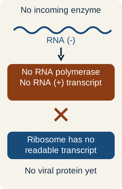
Start with the missing product, not the family name. A ribosome reads messenger RNA; it does not transcribe negative-sense RNA.
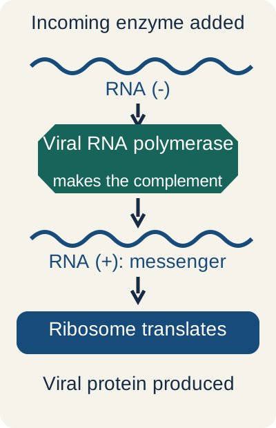
Try the RNA equipment test. Trace the route in the diagram. A negative-sense respiratory virus has entered a cell. Predict what happens when an incoming viral RNA polymerase is supplied, then open the comparison.
Supply the polymerase and inspect the new route
The added enzyme changes the outcome: transcription can now provide readable messenger RNA. Closing this comparison returns to the missing-enzyme state.
What changes: the supplied polymerase makes positive-sense messenger RNA, so translation becomes possible. Without that first transcription step, the virus cannot rely on making its polymerase after entry. Ordinary negative-sense RNA viruses therefore carry the enzyme in the incoming particle. The full explanation and the classification table remain readable with scripting disabled.
Predict the first route to messenger RNA
Genome carried into the cell
What must happen first?
Useful examples
Genome carried into the cellDouble-stranded DNA
What must happen first?Transcribe DNA into messenger RNA, usually in the nucleus. Poxviruses supply transcription machinery for cytoplasmic use.
What must happen first?Use an incoming viral RNA-dependent RNA polymerase to transcribe positive-sense messenger RNA.
Useful examplesInfluenza, RSV, measles, rabies
Genome carried into the cellDouble-stranded RNA
What must happen first?Use particle-associated RNA polymerase to produce messenger RNA. The double-stranded genome itself is not a ribosome substrate.
Useful examplesRotavirus
Genome carried into the cellRNA with reverse transcription
What must happen first?Use incoming reverse transcriptase to make DNA, then establish a nuclear DNA template.
Useful examplesHIV and HTLV-1
Genome carried into the cellPartially double-stranded DNA with reverse transcription
What must happen first?Repair DNA in the nucleus; transcribe RNA. Later, reverse-transcribe a pregenomic RNA into DNA inside a cytoplasmic capsid.
Useful examplesHBV
Apply it to a new situation: an intact rotavirus particle can supply an RNA polymerase even though an isolated double-stranded RNA genome cannot start translation on its own. Rotavirus produces transcripts within its core, rather than requiring the entire double-stranded genome to become exposed. Two RNA copies in HIV are a diploid genome, not separate influenza-like segments. [3][7]
Use the exceptions to understand the job
Most familiar DNA viruses use the nucleus and have double-stranded DNA. B19 has single-stranded DNA; HBV has partially double-stranded DNA and an RNA intermediate; poxviruses replicate in the cytoplasm. Most familiar RNA viruses use the cytoplasm, but influenza has nuclear transcription and replication steps, retroviruses establish nuclear proviral DNA, and hepatitis D uses host nuclear polymerases. HDV is an important exception to the usual requirement that a negative-sense RNA virus bring its own RNA polymerase. [2][7][9]
Among commonly taught DNA families, herpesviruses, hepadnaviruses, and poxviruses are enveloped. Adenovirus, papillomavirus, polyomavirus, and parvovirus are nonenveloped. Important nonenveloped RNA groups include picornaviruses, caliciviruses, astroviruses, and the rotavirus/reovirus groups. The older naked-RNA memory list also includes HEV, with the particle-compartment qualification below. HAV and HEV have nonenveloped particles in feces, although membrane-associated particles can circulate in blood. Lack of an envelope often improves environmental persistence; it does not prove acid resistance, fecal-oral transmission, or resistance to every disinfectant. Rhinovirus is a useful acid-labile counterexample. [1][18][60][90]
Translate older family names without preserving older errors
Human herpesviruses are classified in Orthoherpesviridae, with Herpesviridae familiar from older teaching. RSV and human metapneumovirus belong to Pneumoviridae, not Paramyxoviridae. Rubella belongs to Matonaviridae, not Togaviridae. HDV belongs to Kolmioviridae, genus Deltavirus. The older Reoviridae grouping is now divided: rotavirus is in Sedoreoviridae, while coltiviruses and orthoreoviruses are in Spinareoviridae. The older Bunyavirales grouping has been reorganized within the class Bunyaviricetes. Hantaviridae is now in Elliovirales, while Arenaviridae is in Hareavirales. Neither the historical umbrella nor the class is one uniformly three-segment family. [59][89][1][2][3][4][5][6][8]
Try it here · Checkpoint 1 of 3
Make your prediction before reading the choices. A first attempt is just a starting point.
Case 1
Show answer and explanations for case 1
A. A DNA-dependent RNA polymerase carried within the virion (Why this does not fit)
DNA-dependent RNA polymerase transcribes a DNA template, as in poxvirus early transcription. The measured genome is RNA complementary to the viral messenger RNA, with no DNA intermediate supplied. Identify the template before selecting a transcription enzyme.
Reasoning steps for option A
What template does a DNA-dependent RNA polymerase transcribe?
DNA-dependent RNA polymerase transcribes a DNA template, as in poxvirus early transcription.
Which measured genome feature conflicts with that template?
The measured genome is RNA complementary to the viral messenger RNA, with no DNA intermediate supplied.
What must be identified before choosing a transcription enzyme?
Identify the template before selecting a transcription enzyme.
B. An RNA-dependent RNA polymerase carried within the virion (Best answer)
The opposite polarity identifies a negative-sense RNA genome. Ribosomes cannot first translate this template to produce its own polymerase, so transcription requires enzyme delivered in the particle. Incoming RNA-dependent RNA polymerase supplies the initial messenger RNA.
Reasoning steps for option B
What does complementarity to messenger RNA establish?
The opposite polarity identifies a negative-sense RNA genome.
Why can translation not supply the first polymerase in this infection?
Ribosomes cannot first translate this template to produce its own polymerase, so transcription requires enzyme delivered in the particle.
What incoming activity supplies the first readable transcript?
Incoming RNA-dependent RNA polymerase supplies the initial messenger RNA.
C. An RNA-dependent DNA polymerase carried within the virion (Why this does not fit)
Reverse transcriptase produces DNA from RNA during retroviral replication. The complementary genome-to-messenger-RNA relationship instead identifies negative-sense RNA transcription. An RNA genome alone does not establish a reverse-transcribing life cycle.
Reasoning steps for option C
What product does reverse transcriptase make from RNA?
Reverse transcriptase produces DNA from RNA during retroviral replication.
Which RNA relationship instead points to direct RNA transcription?
The complementary genome-to-messenger-RNA relationship instead identifies negative-sense RNA transcription.
Why is an RNA genome insufficient to infer a retroviral life cycle?
An RNA genome alone does not establish a reverse-transcribing life cycle.
D. A DNA-dependent DNA polymerase carried within the virion (Why this does not fit)
DNA-dependent DNA polymerase copies DNA, as during herpesvirus genome replication. It does not generate the first readable RNA from the supplied RNA template. Distinguish genome replication from production of messenger RNA.
Reasoning steps for option D
What does a DNA-dependent DNA polymerase copy?
DNA-dependent DNA polymerase copies DNA, as during herpesvirus genome replication.
Does this activity provide messenger RNA from the measured RNA template?
It does not generate the first readable RNA from the supplied RNA template.
Which two cellular tasks must be kept separate?
Distinguish genome replication from production of messenger RNA.
Takeaway: Infer RNA polarity from its relationship to messenger RNA, then identify the enzyme needed before translation.
How does the same genome category produce such different diseases?
Compare two patients: one has progressive visual-field loss with white-matter lesions; another develops graft dysfunction after kidney transplantation. Both pathogens may be nonenveloped circular double-stranded DNA polyomaviruses. The family narrows the possibilities, but the infected cell and host immune state explain the illness. Predict the target cell before reading the comparison: JC virus injures oligodendrocytes; BK virus can injure renal tubular epithelium.
Adenovirus: surface infection, systemic risk, and delivery vectors
Adenovirus has a nonenveloped icosahedral particle and linear double-stranded DNA. Pharyngitis with conjunctivitis, including outbreaks in crowded settings or pools, is a familiar pattern. Other presentations include pneumonia in military recruits, hemorrhagic cystitis, and gastroenteritis from enteric types 40 and 41. Disease can disseminate in severely immunocompromised people. An oral live vaccine against types 4 and 7 is used for eligible military personnel, not as a routine civilian vaccine. [27]
A vector uses viral delivery machinery for a different purpose. Adenoviral vectors can transduce dividing and nondividing cells and generally maintain delivered DNA outside chromosomes; replication competence depends on the particular construct. Recombinant adeno-associated virus vectors have a small payload and usually persist mainly as episomal DNA. They do not inherit a guarantee of targeted chromosomal insertion from wild-type AAV biology.
Classical gammaretroviral vectors generally require dividing cells; lentiviral vectors can also transduce nondividing cells. Integration can make expression durable while creating insertion-related risks. Preexisting immunity, inflammatory responses, tissue targeting, dose, and the specific vector matter as much as its family label. [61][91][92]
Vector history illustrates why those distinctions matter. The fatal systemic reaction in the 1999 ornithine transcarbamylase gene-transfer trial was associated with an adenoviral vector, not proof that every viral vector has the same risk. Oncolytic viruses are intended to preferentially infect or replicate in tumors; the early description of ONYX-015 as simply a detector of absent p53 was incomplete. Viral RNA export and cellular context also matter. Modified HSV is used in talimogene laherparepvec for selected melanoma lesions. None of these examples justifies treating a whole family as uniformly safe or uniformly hazardous. [41][83][84]
Interpret a safety example: in a fictional trial, an adverse event occurs in 3 of 200 treated participants and 0 of 200 controls. The observed difference is 1.5 percentage points. A nonsignificant test does not establish zero risk; a small trial can miss rare harms. The reciprocal of the observed difference is approximately 67, but that point estimate is uncertain and is not a proven causal number needed to harm.
Herpesviruses: persistence changes the next illness
Herpesviruses have an envelope, an icosahedral capsid, a tegument layer, and linear double-stranded DNA. Nuclear replication and lifelong persistence explain why primary infection and reactivation can look different. HSV-1 and HSV-2 overlap in oral and genital disease. HSV establishes sensory-ganglion latency, commonly trigeminal or sacral depending on the site. Gingivostomatitis, recurrent oral or genital vesicles, keratitis, herpetic whitlow, and HSV-associated erythema multiforme are useful associations. Vesicle PCR is more specific than a Tzanck preparation, which cannot reliably distinguish HSV from VZV. Herpetic whitlow should not be treated as a bacterial abscess requiring incision and drainage. [8][31]
Fever, altered behavior, seizures, and temporal-lobe injury raise concern for HSV encephalitis. Start appropriate intravenous acyclovir promptly while diagnostic testing proceeds; an early negative CSF PCR does not always exclude the diagnosis. Neonatal HSV is often acquired around delivery and may produce skin, eye, mouth, CNS, or disseminated disease. Delivery management for active genital lesions or prodromal symptoms reduces risk but cannot guarantee prevention. [31][63]
VZV provides the latency experiment in everyday clinical practice. Primary varicella produces a widespread pruritic eruption with lesions at different stages, usually prominent on the trunk. Reactivation in a sensory ganglion sends virus along a nerve territory, producing the commonly unilateral dermatomal eruption of shingles. Persistent pain afterward is postherpetic neuralgia. Ear vesicles with ipsilateral facial palsy suggest Ramsay Hunt syndrome involving the facial nerve's geniculate ganglion.
Adult or pregnancy-associated varicella can cause serious pneumonia. Aspirin exposure during childhood influenza or varicella is associated with Reye syndrome, an acute encephalopathy with hepatic mitochondrial injury; avoid routine aspirin treatment of these illnesses in children. [23]
Look for healing rather than assuming this is the first day of the eruption. This photograph shows day-eight varicella; one photograph cannot establish the entire illness sequence or exclude other eruptions by itself. CDC, Public Health Image Library 4366, 1969; public domain. This material is freely available from CDC; its use does not imply CDC endorsement. [56][57]
EBV infects B cells, but the conspicuous blood cells are often reactive T cells. CD21 helps EBV enter B cells. Fever, pharyngitis, posterior cervical lymphadenopathy, fatigue, and splenomegaly support infectious mononucleosis; atypical Downey lymphocytes are reactive CD8-positive T cells. Heterophile testing can be negative early or in young children and occasionally falsely positive. It is not an absolute exclusion test for CMV, acute HIV, or other causes of a similar syndrome.
EBV-specific serology can clarify uncertainty: VCA IgM with absent EBNA antibody supports primary infection; EBNA antibody usually appears later. Neither the most conspicuous atypical blood cell nor a heterophile result alone identifies every infected cell. An aminopenicillin-associated rash does not automatically establish permanent penicillin allergy, but neither does it prove that true hypersensitivity is impossible.
Avoid unnecessary antibiotics and assess activity restrictions because an enlarged spleen can rupture. Oral hairy leukoplakia is an EBV-associated lesion, not a premalignant oral cancer. [64]
CMV can cause heterophile-negative mononucleosis. In congenital infection, hearing loss, petechiae, hepatosplenomegaly, and periventricular calcifications are important findings. In advanced HIV, particularly at CD4 counts below 50 cells/mm3, CMV can cause hemorrhagic retinitis, colitis, esophagitis with large ulcers, and other end-organ disease. These CD4 thresholds describe risk, not absolute permission or exclusion. Large intranuclear inclusions with a surrounding halo create the familiar owl-eye appearance. Ganciclovir activation in CMV-infected cells begins with UL97 kinase, not HSV thymidine kinase. [16][39]
HHV-6, especially HHV-6B, and HHV-7 can cause roseola: several days of high fever followed by a trunk-predominant rash as the fever resolves. Febrile seizures can occur. This timing is helpful but not an infallible diagnosis. HHV-7 has also been investigated in pityriasis rosea; an association is not proof of a single established cause. HHV-8 is associated with Kaposi sarcoma, primary effusion lymphoma, and some multicentric Castleman disease. Purple vascular lesions and spindle-cell histology describe Kaposi sarcoma, not all HHV-8 disease. [8][28]
Papillomavirus, parvovirus, and polyomavirus: similar names, different targets
HPV: nonenveloped circular double-stranded DNA infects basal squamous epithelium. Types 6 and 11 are associated with anogenital warts and recurrent respiratory papillomatosis; types 1, 2, and 4 are familiar cutaneous-wart associations. Persistent high-risk HPV, including types 16 and 18, contributes to cervical, anal, oropharyngeal, and other anogenital cancers. E6 promotes p53 loss; E7 interferes with Rb control of proliferation. Koilocytes have perinuclear clearing and abnormal nuclei. A wart or a koilocyte does not by itself identify a high-risk genotype. Vaccination prevents new infections; it is not treatment for an established lesion. [24][28]
Condylomata acuminata are often irregular or verrucous HPV-associated lesions. Condylomata lata are broad, moist lesions of secondary syphilis, which may coexist with a generalized rash involving palms and soles. The latter require syphilis evaluation and antibiotic treatment, not wart treatment. Appearance alone should not replace a sexual history, examination, and appropriate testing. [65]
B19: a nonenveloped single-stranded DNA virus targets erythroid precursors, with globoside (P antigen) participating in entry. The familiar child has a slapped-cheek eruption followed by a lacy body rash; adults may instead have symmetric small-joint arthralgia. In chronic hemolytic anemia, a temporary interruption of red-cell production causes a disproportionate hemoglobin fall with very few reticulocytes.
In the fetus, severe anemia can lead to high-output cardiac failure and hydrops. In immunocompromised people, persistent infection can cause chronic pure red-cell aplasia. The central event is loss of production, not simply destruction of mature circulating red cells. Giant pronormoblasts are a marrow finding. [40]
JC and BK: JC-associated PML causes progressive focal neurological deficits from demyelination. Typical MRI lesions involve white matter with little mass effect or enhancement, but enhancement can occur, particularly during immune reconstitution. Tissue can show infected oligodendrocytes with nuclear inclusions and bizarre astrocytes. In HIV-associated PML, restoring effective immunity with antiretroviral therapy is central even though no specific proven anti-JC drug exists. BK-associated nephropathy is especially important after kidney transplantation, and hemorrhagic cystitis after hematopoietic transplantation. Viral cytopathic changes in urine may produce decoy cells; these are not a diagnosis of cancer and require clinical and virological correlation. [30][66]
Poxviruses: identify the compartment, then the syndrome
Poxviruses are large, complex DNA viruses with cytoplasmic replication machinery. Variola caused smallpox, declared eradicated in 1980. Historically, a substantial febrile prodrome and centrifugal eruption with lesions at a similar stage within a body region contrasted with the mixed-stage, trunk-prominent eruption of varicella. Molluscum contagiosum causes localized, often painless, centrally depressed papules with large cytoplasmic Henderson-Patterson inclusions. Its distinction from smallpox is not a dependable rule about asynchronous lesion stages; the localized papules and lack of a smallpox-like systemic illness are more useful. [54]
Mpox may cause painful or umbilicated lesions and lymphadenopathy, but presentations vary and lesion testing matters. The broad viral clades are I and II. The modified vaccinia Ankara vaccine used for prevention is distinct from replicating smallpox vaccines. Tecovirimat is not a proven routine treatment for every uncomplicated case: randomized trials did not show faster lesion resolution in their studied populations. In the United States, investigational access remains available for eligible patients with severe disease or important risk factors, alongside supportive care. Do not generalize that trial result to every severely immunocompromised patient. [55][85]
Transfer: a cytoplasmic DNA-virus inclusion supports a different transcription strategy from a nuclear herpesvirus inclusion. It does not mean that every cytoplasmic inclusion is poxvirus: rabies is an RNA virus with cytoplasmic Negri bodies. Always combine compartment, cell type, host, and clinical course.
Try it here · Checkpoint 2 of 3
Make your prediction before reading the choices. A first attempt is just a starting point.
Case 7
Show answer and explanations for case 7
A. Severe anemia with high-output cardiac failure and hydrops (Best answer)
The abrupt anemia with very few reticulocytes indicates interrupted red-cell production rather than an appropriate marrow response to hemolysis. B19 targets erythroid precursors. The same target in the fetus can cause severe anemia, high-output failure, and hydrops.
Reasoning steps for option A
What does profound reticulocytopenia imply about the hemoglobin fall?
The abrupt anemia with very few reticulocytes indicates interrupted red-cell production rather than an appropriate marrow response to hemolysis.
Which cells does B19 target?
B19 targets erythroid precursors.
What follows from loss of those cells in a fetus?
The same target in the fetus can cause severe anemia, high-output failure, and hydrops.
B. Patent ductus arteriosus from congenital rubella syndrome (Why this does not fit)
Rubella can produce congenital cardiac and ocular disease. It does not best explain the child's selective reticulocytopenic production failure after a rash exposure. The fetal prediction should follow the erythroid target established by the blood counts.
Reasoning steps for option B
What congenital injury pattern can rubella produce?
Rubella can produce congenital cardiac and ocular disease.
Does it best explain selective red-cell production failure here?
It does not best explain the child's selective reticulocytopenic production failure after a rash exposure.
Which target should determine the fetal prediction?
The fetal prediction should follow the erythroid target established by the blood counts.
C. Hydrocephalus with diffuse intracranial calcifications from toxoplasmosis (Why this does not fit)
That congenital pattern can follow toxoplasma infection. Toxoplasma is not the virus associated with the child's transient red-cell production arrest. The option substitutes a different congenital syndrome rather than applying the identified cell target.
Reasoning steps for option C
Which congenital infection fits hydrocephalus and diffuse calcifications?
That congenital pattern can follow toxoplasma infection.
Does it explain this viral erythroid production arrest?
Toxoplasma is not the virus associated with the child's transient red-cell production arrest.
Does this option apply the demonstrated cell target?
The option substitutes a different congenital syndrome rather than applying the identified cell target.
D. Hearing loss with periventricular calcifications from CMV (Why this does not fit)
CMV can cause that congenital pattern and can cause cytopenias. Here, the exposure history and profound selective reticulocytopenia in a child with chronic hemolysis support B19. Its most direct fetal consequence is impaired erythropoiesis, not the proposed CMV pattern.
Reasoning steps for option D
What congenital findings can CMV cause?
CMV can cause that congenital pattern and can cause cytopenias.
Which combined findings more strongly support B19 here?
Here, the exposure history and profound selective reticulocytopenia in a child with chronic hemolysis support B19.
What is the direct fetal consequence of that target?
Its most direct fetal consequence is impaired erythropoiesis, not the proposed CMV pattern.
Takeaway: A low reticulocyte count identifies production failure and predicts B19-related fetal anemia.
Which RNA properties help explain an airway, intestinal, or exposure-related illness?
Begin with a comparison: an infant has wheeze and retractions, while an older sibling has barking cough and inspiratory stridor. Both illnesses may involve enveloped, nonsegmented negative-sense RNA viruses. Localize the dominant obstruction before naming a family: small-airway bronchiolitis suggests RSV among other causes; subglottic croup commonly involves parainfluenza. Genome category alone cannot decide between them.
Respiratory negative-sense RNA viruses
Influenza, Orthomyxoviridae: influenza A and B have eight RNA segments; C and D have seven. Hemagglutinin attaches to sialic acid and participates in fusion; neuraminidase helps newly formed particles separate from sialic-acid-containing surfaces. Influenza uses nuclear processes, including acquisition of capped RNA primers. The abrupt fever, myalgia, headache, and cough pattern is different from a diagnosis based solely on wheeze. A patient who improves and then develops renewed fever and focal pneumonia may have a bacterial complication, including Staphylococcus aureus, Streptococcus pneumoniae, or Haemophilus influenzae. [9][67]
Paramyxoviridae: measles, mumps, parainfluenza, and Hendra are enveloped, nonsegmented negative-sense RNA viruses. Fusion proteins explain the ability to form multinucleated cells. Measles combines fever, cough, coryza, conjunctivitis, and sometimes Koplik spots before a descending rash; Warthin-Finkeldey cells are multinucleated cells in lymphoid tissue. Persistent measles infection can later cause subacute sclerosing panencephalitis, with progressive neurological decline years after the initial illness.
Mumps can cause parotitis, aseptic meningitis, pancreatitis, and postpubertal orchitis. Meningitis need not be accompanied by parotitis; infertility is not inevitable after orchitis. Parainfluenza commonly causes croup, but neither a barking cough nor a steeple-shaped subglottic narrowing proves a specific viral cause. Hendra infection is associated with transmission from infected horses to people in Australia, with fruit bats sustaining the natural reservoir. [21][38][58]
Pneumoviridae: RSV and human metapneumovirus have enveloped, nonsegmented negative-sense RNA genomes. Their fusion activity can produce syncytia. RSV bronchiolitis is especially important in infants, but RSV and hMPV also cause lower respiratory disease in older adults and people with chronic lung disease. Hyperinflation and peribronchial thickening are compatible with bronchiolitis, not a pathogen-specific test. Routine care for uncomplicated bronchiolitis is supportive, including attention to breathing and hydration; prevention antibodies are not treatment for an active episode. [6][11][68]
Positive-sense viruses in the respiratory tract
Rhinoviruses are small nonenveloped picornaviruses that commonly cause upper respiratory illness. Many use ICAM-1, but receptor use differs among groups; an ICAM-1 rule does not cover every rhinovirus. Their relative acid lability helps explain why a nonenveloped picornavirus need not behave like an enteric virus. [53]
Coronaviruses have enveloped positive-sense RNA genomes and helical nucleocapsids. Common human coronaviruses include 229E, NL63, OC43, and HKU1. SARS-CoV and SARS-CoV-2 use ACE2 for entry; MERS-CoV uses DPP4, also called CD26. Spike binding and protease-dependent fusion are separate events, and protease use varies with the virus, variant, and cell. Camel exposure is important in MERS epidemiology. Receptor distribution contributes to tissue susceptibility but does not alone explain the inflammatory, vascular, and thrombotic complications of COVID-19. Coronaviruses are nonsegmented, yet mutation and recombination permit substantial evolution. A segmented genome is not a prerequisite for a pandemic. [45][69]
Messenger RNA vaccines supply a temporary cytoplasmic template for antigen production. They do not require a DNA intermediate or chromosomal integration. Lipid delivery and nucleoside modification help the message reach cells and support expression; they do not mean the immune system ignores the vaccine. Adenoviral vector vaccines use a different delivery platform, and immunity against a vector can influence responses. [70]
Intestinal infection is not one family
Picornaviridae includes poliovirus, echoviruses, coxsackieviruses, rhinoviruses, and HAV. Many enteroviruses survive gastrointestinal transit but cause disease outside the gut. Coxsackie A viruses, including A16 and A6, and enterovirus A71 can cause hand-foot-mouth disease; herpangina produces posterior oral lesions. Coxsackie B viruses are associated with myocarditis, pericarditis, and pleurodynia. A viral prodrome and lymphocytic myocarditis do not uniquely identify coxsackievirus, and myocarditis does not require ST depression. Pericarditis classically produces diffuse ST elevation and PR depression, whereas myocarditis has variable ECG findings. Echoviruses and other enteroviruses are important causes of aseptic meningitis. [53][71]
Poliovirus can injure anterior horn motor neurons, producing asymmetric flaccid weakness with preserved sensation. Inactivated injected polio vaccine cannot cause vaccine-associated paralytic poliomyelitis; live oral vaccine has important population benefits but can rarely be associated with paralytic or vaccine-derived virus disease. Postpolio weakness years later is not evidence that a latent poliovirus has reactivated. Contrast postinfectious Guillain-Barre syndrome, which often produces relatively symmetric weakness, sensory symptoms, areflexia, and increased CSF protein without a comparable cell increase.
Botulism is not viral: impaired acetylcholine release after SNARE protein cleavage commonly begins with cranial and autonomic dysfunction followed by descending weakness. Direction helps, but neither cranial involvement nor autonomic dysfunction alone separates every case. [25][72]
Compare the particle, then the intestinal consequence
Group
Useful distinction
GroupNorovirus and sapovirus, Caliciviridae
Useful distinctionNonenveloped positive-sense RNA. Norovirus causes abrupt vomiting and watery diarrhea across age groups, including institutional and food-associated outbreaks. Small infectious inocula and environmental persistence facilitate spread. Soap-and-water handwashing and effective surface disinfection matter; alcohol hand sanitizer is not an adequate substitute. Avoid food preparation and care of others while ill and for at least 48 hours after symptoms stop. Follow pathogen-specific public-health instructions as well as the product label. [18]
GroupRotavirus, Sedoreoviridae
Useful distinctionNonenveloped double-stranded RNA with 11 segments. Villous epithelial injury and NSP4 enterotoxin activity contribute to watery diarrhea and dehydration, especially in young children. Oral live vaccines substantially reduce severe disease, not every possible infection.
GroupAstrovirus, Astroviridae
Useful distinctionNonenveloped positive-sense RNA. Some particles have a star-like appearance on electron microscopy. Gastroenteritis often affects young children, older adults, or immunocompromised people. Morphology is a teaching association, not a requirement to order electron microscopy in routine diarrhea.
GroupEnteric adenovirus
Useful distinctionNonenveloped double-stranded DNA; types 40 and 41 are associated with childhood gastroenteritis. A negative test for one other pathogen does not establish adenovirus by exclusion.
These infections can overlap clinically. Vomiting does not reliably localize one virus to the proximal intestine while diarrhea localizes another to the distal intestine. Hydration status determines urgency even before the specific pathogen is known. HAV and HEV are also acquired through enteric routes but principally cause hepatitis rather than an isolated gastroenteritis syndrome. [18][26][27][52]
Match an exposure route to the organ at risk
Flaviviridae: dengue, yellow fever, Zika, and West Nile are enveloped positive-sense RNA viruses; HCV is another member but is not mosquito-borne. Dengue has four serotypes and is transmitted by Aedes mosquitoes. A heterotypic later infection can increase severe-disease risk through antibody-dependent enhancement, but severe disease can also occur in a first infection. Near defervescence, worsening abdominal pain, impaired perfusion, increasing hematocrit, or falling platelets can indicate the critical plasma-leak phase, not recovery.
Avoid aspirin and other NSAIDs when dengue is suspected. Yellow fever can cause jaundice, hemorrhage, and hepatic injury; Councilman bodies represent apoptotic hepatocytes rather than virus-specific inclusions. Zika can spread sexually and vertically as well as by mosquitoes; congenital neurological injury and adult Guillain-Barre syndrome are distinct risks. [36][47][48]
West Nile circulates in a bird-mosquito cycle, principally involving Culex mosquitoes. Most infections are not neuroinvasive. In an affected patient, encephalitis or asymmetric flaccid paralysis can reflect CNS and anterior horn injury. Direct contact with birds is not required, and neither a specific occupation nor one region defines the diagnosis. [37]
Togaviridae: the alphaviruses include chikungunya, with prominent and sometimes persistent arthralgia after Aedes exposure, and eastern, western, and Venezuelan equine encephalitis viruses. Regional ecology helps distinguish these encephalitis risks. Horses and humans are typically incidental hosts in eastern equine encephalitis, whereas equids can amplify epidemic Venezuelan equine encephalitis. Rubella is not an alphavirus; its current family is Matonaviridae. [49][51][5]
Rabies, Rhabdoviridae: a bullet-shaped enveloped negative-sense RNA virus reaches the CNS through peripheral nerves using retrograde axonal transport. Nicotinic acetylcholine receptors are among several implicated entry-related molecules, not a universal single-receptor explanation. Encephalitic disease may cause hydrophobia and aerophobia; paralytic rabies is another presentation. Negri bodies are cytoplasmic neuronal inclusions, but their absence does not exclude infection.
After a possible exposure, assess risk promptly with public health rather than waiting for symptoms. For a previously unvaccinated person, indicated prophylaxis includes wound care, rabies immunoglobulin, and vaccination on days 0, 3, 7, and 14; immunocompromised patients need an additional day-28 dose and response assessment. Previously vaccinated people receive vaccine on days 0 and 3 without immunoglobulin.
Filoviridae: Ebola and Marburg are filamentous enveloped negative-sense RNA viruses. Exposure to infected blood or other body fluids, including during care or handling remains, is central to transmission. Severe gastrointestinal losses, hepatic injury, shock, and coagulopathy can occur without conspicuous external bleeding. Egyptian fruit bats are an established Marburg reservoir; possible bat reservoirs for Ebola should not all be described as definitively proven. Treatment and vaccine specificity matter: Inmazeb contains atoltivimab, maftivimab, and odesivimab; Ebanga contains ansuvimab. These and the Ervebo vaccine target Zaire ebolavirus disease, not every filovirus. [29][73][86]
Historically grouped bunyaviruses and rodent exposures: many medically important members have segmented negative-sense or ambisense RNA. Hantaviruses have three segments. Sin Nombre virus is associated with deer-mouse exposure and a pulmonary capillary-leak syndrome; Hantaan, Seoul, and Puumala viruses are important in hemorrhagic fever with renal syndrome. Andes virus is an important exception to the usual rodent-to-human pattern because person-to-person transmission is documented.
Therefore, a general statement that every hantavirus lacks interpersonal spread is unsafe. La Crosse virus is mosquito-borne and can cause encephalitis in children. Crimean-Congo hemorrhagic fever involves tick exposure and infected animal or human blood; Rift Valley fever involves mosquitoes and exposure to infected livestock and can affect the eyes, liver, or CNS. These are different ecologies, not interchangeable reservoirs. [35][59][74][87][88]
Human mammarenaviruses in Arenaviridae, now placed in Hareavirales, have two ambisense RNA segments. [89] Packaged host ribosomes account for the historically described sandy appearance. Lymphocytic choriomeningitis virus can follow mouse or pet-hamster exposure and cause aseptic meningitis or congenital disease; exposure alone does not prove the diagnosis. Lassa virus is associated with Mastomys rodents and can also spread through infected body fluids. Supportive care is crucial; ribavirin should not be presented as an established universally effective cure, because clinical efficacy remains uncertain. [13][50]
Colorado tick fever is a useful counterexample to the assumption that every tick-associated fever is bacterial: a coltivirus in Spinareoviridae has 12 double-stranded RNA segments and is transmitted by Dermacentor ticks. Biphasic fever and leukopenia can occur. Orthoreoviruses have 10 segments, and rotavirus has 11; do not substitute the family-wide range for the exact rotavirus count. [4][75]
Compare outcomes: sketch an arrow from an exposure to a target organ. Mosquito exposure plus asymmetric motor weakness suggests a different route from rodent aerosol exposure plus pulmonary edema. The first can involve West Nile injury to motor neurons; the second can involve hantavirus-associated pulmonary capillary leak. For a new case, require the time course, examination, geography, and appropriate testing to support the proposed route.
Does a positive antibody mean the virus is gone?
Compare two immune patients: both have anti-HBs and no HBsAg. One also has total anti-HBc, while the other does not. Predict which person has evidence of prior natural HBV infection. Anti-HBc identifies exposure to core antigen, so its presence distinguishes the usual resolved-infection pattern from vaccine-only immunity. It does not, by itself, prove whether infection is current, recent, or remote. [10]
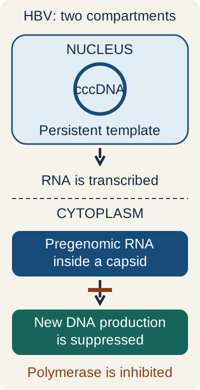
Trace the two compartments. Suppressing new DNA production is not equivalent to deleting the nuclear template.
HBV is an enveloped hepadnavirus with circular, partially double-stranded DNA. The complete Dane particle contains the nucleocapsid and surrounding HBsAg-bearing envelope. HBcAg is core antigen; HBeAg is a secreted marker often associated with replication. Nuclear repair generates covalently closed circular DNA, or cccDNA, which supplies RNA templates. Reverse transcription of pregenomic RNA occurs later inside cytoplasmic capsids. An HBV-active polymerase inhibitor can substantially suppress replication without reliably eliminating cccDNA. HBeAg negativity is not proof of absent replication or noninfectiousness; HBV DNA directly informs viral burden. [7][19]
Read HBV markers together, with timing
Pattern
Interpretation and limitation
PatternHBsAg negative; anti-HBs positive; total anti-HBc negative
Interpretation and limitationTypical vaccine-induced immunity. Vaccine does not generate anti-HBc.
PatternHBsAg negative; anti-HBs positive; total anti-HBc positive
Interpretation and limitationTypical resolved natural infection. Reactivation can still occur during important immunosuppression.
PatternHBsAg positive; IgM anti-HBc positive
Interpretation and limitationSupports acute infection in the appropriate history. IgM can also become positive during a flare or reactivation of chronic infection.
Interpretation and limitationCan represent the window period after surface antigen disappears but before surface antibody is detectable. Do not call IgM the only possible detectable marker under all testing conditions.
PatternHBsAg persists for at least six months
Interpretation and limitationSupports chronic infection, interpreted with clinical history and other tests.
PatternIsolated total anti-HBc
Interpretation and limitationConsider past infection with waning anti-HBs, a false-positive result, occult infection, or a window-period pattern depending on context and additional tests.
Chronicity depends strongly on age at acquisition: approximately 90% of infected infants develop chronic infection, compared with roughly 5% of adults. These are population estimates, not an individual prediction. Chronic HBV can cause cirrhosis and hepatocellular carcinoma, including carcinoma without established cirrhosis. Immune-complex associations include polyarteritis nodosa and membranous nephropathy. For an infant born to an HBsAg-positive mother, indicated HBV vaccine and HBIG should be given within 12 hours of birth; a negative maternal HBeAg does not cancel that need. Screening, treatment eligibility, and vaccination schedules require current guidance rather than an old family mnemonic. [19]
Hepatitis A through E: compare the route and the persistence mechanism
Same organ, different biology
Virus
Route and genome
What changes the clinical interpretation?
VirusHAV
Route and genomePicornavirus; positive-sense RNA; fecal-oral spread, including contaminated food or shellfish.
What changes the clinical interpretation?Acute infection without chronic carriage. Incubation is commonly 15 to 50 days. Severe acute hepatitis remains possible. Inactivated vaccine prevents infection. [20]
VirusHBV
Route and genomeHepadnavirus; partially double-stranded DNA with reverse transcription; blood, sexual, and perinatal spread.
What changes the clinical interpretation?Age-dependent chronicity, persistent nuclear template, cancer and immune-complex complications. Recombinant vaccines prevent acquisition.
VirusHCV
Route and genomeFlaviviridae; positive-sense RNA; primarily bloodborne.
What changes the clinical interpretation?Chronic infection is common but not inevitable. Direct-acting antivirals can cure infection because HCV does not require a persistent DNA template.
VirusHDV
Route and genomeKolmioviridae; circular negative-sense RNA; transmission associated with blood or body-fluid exposure in the presence of HBV.
What changes the clinical interpretation?Requires HBsAg for infectious-particle assembly and spread. New HDV on chronic HBV generally carries a greater risk of persistent and severe disease than simultaneous acquisition.
VirusHEV
Route and genomeHepeviridae; positive-sense RNA; contaminated water or zoonotic food exposure, including undercooked pork or game.
What changes the clinical interpretation?Severe acute disease is especially important during pregnancy in endemic waterborne settings. Persistent infection can occur in immunocompromised people. Vaccines exist in some countries, so a blanket claim that no HEV vaccine exists is incorrect.
HCV-associated mixed cryoglobulinemia can produce palpable purpura, arthralgia, neuropathy, low C4, and immune-complex glomerular injury, often with a membranoproliferative pattern. Porphyria cutanea tarda and some B-cell lymphoproliferative disorders are additional associations. [94] These are not explained by simply assigning HCV to renal tubular infection. After sustained virologic response, people with cirrhosis still need appropriate liver-cancer surveillance and can be reinfected; cure does not reverse every prior consequence or create immunity. [17][76]
HDV uses host nuclear polymerases and viral RNA processing functions for intracellular RNA replication. Its dependence on HBV is most specifically a dependence on HBsAg for packaging and spread. Coinfection means HBV and HDV were acquired together; superinfection means HDV was added to preexisting HBV. Establish the timeline rather than treating an IgM result as infallible proof of first infection. HBV vaccination prevents the setting needed for HDV acquisition in a susceptible person; it does not cure HDV in someone with chronic HBV. [2][15]
For HEV, connect host to outcome. A pregnant person with jaundice and coagulopathy after waterborne exposure needs urgent assessment for severe acute hepatitis. An immunosuppressed transplant recipient with persistent HEV RNA presents a different problem: chronic infection. The historically cited high pregnancy mortality comes from particular epidemiological settings, especially genotype-1 outbreaks, and should not be assigned to every genotype or patient. [14]
Test the persistence prediction. Point to the HBV step that a reverse-transcriptase inhibitor suppresses. Then predict whether a low plasma DNA result proves that nuclear cccDNA has disappeared.
Compare suppression with removal of the template
New DNA synthesis can fall while cccDNA remains. That explains why stopping therapy or changing immune control can permit renewed replication. HCV cure is biologically different because an obligatory persistent DNA form is absent.
Use it again: distinguish a negative viral-load test during suppressive therapy from proof that all reservoirs or future risks have disappeared. The specific virus determines what the negative test can establish.
Try it here · Checkpoint 3 of 3
Make your prediction before reading the choices. A first attempt is just a starting point.
Case 4
Show answer and explanations for case 4
A. Vaccine-induced immunity with an unrelated hepatitis episode (Why this does not fit)
Vaccination can produce anti-HBs without core antibody. This patient instead had documented surface antigen and core IgM during a recent hepatitis episode. Use evidence of natural infection before assigning vaccine-induced immunity.
Reasoning steps for option A
Which antibody pattern can vaccination produce?
Vaccination can produce anti-HBs without core antibody.
What prior findings establish natural infection here?
This patient instead had documented surface antigen and core IgM during a recent hepatitis episode.
Which evidence must be considered before calling this vaccine immunity?
Use evidence of natural infection before assigning vaccine-induced immunity.
B. Remote resolved infection with a waning surface antibody level (Why this does not fit)
Remote resolved infection can produce isolated total core antibody after anti-HBs wanes. The recent symptomatic antigen-positive episode and persisting core IgM instead establish a short acute timeline. Similar single panels can require different interpretations when prior results are available.
Reasoning steps for option B
How can remote resolved infection produce isolated core antibody?
Remote resolved infection can produce isolated total core antibody after anti-HBs wanes.
Which recent clinical and serological timeline supports another interpretation?
The recent symptomatic antigen-positive episode and persisting core IgM instead establish a short acute timeline.
Why can similar single panels need different interpretations?
Similar single panels can require different interpretations when prior results are available.
C. Established chronic infection with a new inflammatory flare (Why this does not fit)
Core IgM can recur during a severe chronic HBV flare. The supplied record does not establish chronicity and instead shows improvement with recent antigen clearance. Do not label an infection chronic solely because core IgM is present.
Reasoning steps for option C
Can core IgM recur during chronic HBV inflammation?
Core IgM can recur during a severe chronic HBV flare.
What longitudinal evidence favors an acute resolving episode here?
The supplied record does not establish chronicity and instead shows improvement with recent antigen clearance.
Why should core IgM not independently establish chronicity?
Do not label an infection chronic solely because core IgM is present.
D. Resolving acute infection before detectable surface antibody (Best answer)
Surface antigen has disappeared after documented acute infection, while surface antibody is not yet detected. Persisting core IgM supports the serologic window during recovery in this serial context. A single isolated-core result without this history would require a broader differential.
Reasoning steps for option D
What changed between the antigen-positive illness and the current sample?
Surface antigen has disappeared after documented acute infection, while surface antibody is not yet detected.
How does core IgM fit this documented short recovery timeline?
Persisting core IgM supports the serologic window during recovery in this serial context.
How would the interpretation change without that prior history?
A single isolated-core result without this history would require a broader differential.
Takeaway: Serial testing can distinguish a window-period pattern from an isolated marker interpreted without context.
What evidence separates a changed genome from a changed appearance?
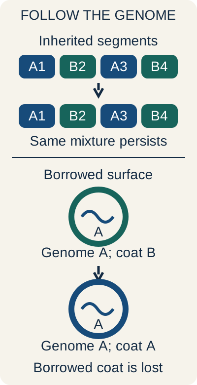
Predict the next generation. A particle carries genome A but temporarily displays surface proteins from virus B. It enters a cell using those proteins. With no further supply of B proteins, will its descendants necessarily keep the B surface? No: the unchanged A genome specifies the proteins subsequently produced. That is a different event from inheriting RNA segments from two parental viruses.
Follow the genetic material into the descendant. A surface change is not automatically a heritable genetic change. Four segments simplify this drawing; an influenza A genome actually has eight.
Five different explanations for an altered viral result
Process
What changes?
What should be predicted?
ProcessMutation and antigenic drift
What changes?Nucleotide substitutions can gradually alter an antigen.
What should be predicted?No exchange of whole segments is required. Influenza A and B undergo drift.
ProcessReassortment
What changes?Coinfecting compatible segmented viruses contribute different whole genome segments.
What should be predicted?The new segment combination can be inherited. A clinically important new influenza A antigenic subtype may represent antigenic shift, but not every reassortant spreads efficiently or causes a pandemic.
ProcessRecombination
What changes?Genetic information is exchanged within nucleic acid molecules, such as through template switching.
What should be predicted?A nonsegmented virus can acquire a heritable new sequence.
ProcessComplementation
What changes?One virus supplies a functional product missing from another.
What should be predicted?Replication can be rescued in that cell without permanently correcting the defective genome.
ProcessPhenotypic mixing
What changes?A particle incorporates proteins supplied by another virus in the same cell.
What should be predicted?Entry properties can differ temporarily while the genome remains unchanged.
Influenza A has a broad animal reservoir; influenza B circulates mainly in humans but should not be described as exclusively human under every circumstance. Reassortment can occur in both A and B, while the familiar pandemic antigenic-shift framework principally concerns influenza A. A pig can participate in influenza ecology but is not an obligatory location for every reassortment event. Human-preferring and avian-preferring sialic-acid interactions are useful patterns, not an absolute species barrier. [67]
Compare two reports: one describes a heritable mixture of whole influenza segments; another describes an unchanged genome with a temporary alternative envelope. Assign a process before checking the diagram. The first supports reassortment; the second supports phenotypic mixing. Transfer the same logic to a nonsegmented coronavirus: a heritable sequence change can result from recombination, but not from exchanging nonexistent influenza-like segments.
Receptors tell you about entry, not the whole illness
HIV gp120 binds CD4 and a coreceptor, commonly CCR5 or CXCR4; gp41 mediates fusion. That distinction explains why a CCR5 antagonist requires demonstration of CCR5-tropic virus. EBV uses CD21 in B-cell entry, HBV uses NTCP in hepatocyte entry, and HCV entry involves several factors including CD81 and tight-junction proteins. HPV attachment involves heparan sulfate before productive infection of basal epithelium. CMV uses multiple receptor systems according to cell type; heparan sulfate is not itself an integrin. A receptor can be necessary without being sufficient for the full tissue distribution, replication, or persistence of infection. [7][8][24][77]
Rash timing and congenital disease: do not stop at the color
Measles usually features substantial fever and respiratory symptoms with the rash; rubella is often milder, with postauricular or suboccipital nodes and arthralgia; roseola commonly produces a rash as fever subsides; B19 often produces facial erythema followed by a lacy eruption. In the old numbered-exanthem system, first disease was measles, second was scarlet fever, third rubella, fifth B19, and sixth roseola. Scarlet fever is toxin-mediated group A streptococcal disease, often with a sandpaper-like rash, strawberry tongue, and flexural accentuation; it is not viral. The proposed fourth disease is not a useful distinct modern diagnosis. [21][22][40][78]
Rubella is an enveloped positive-sense RNA virus in Matonaviridae. Early fetal infection can produce cataracts, sensorineural hearing loss, patent ductus arteriosus or pulmonary artery abnormalities, growth restriction, and a purpuric or blue-purple eruption related to fetal hematopoiesis and hematologic injury. Maternal mild illness does not mean fetal low risk. MMR is live attenuated and is not administered during pregnancy; nonimmune patients can be vaccinated postpartum according to guidance.
Congenital CMV can also cause hearing loss, petechiae, and hepatosplenomegaly, with periventricular calcifications supporting that diagnosis. Congenital toxoplasmosis more classically combines chorioretinitis, hydrocephalus, and more diffusely distributed intracranial calcifications. Neither a blue-purple rash nor hearing loss alone distinguishes all three. B19 instead threatens fetal red-cell production; congenital varicella can cause scarring and limb hypoplasia. [22][23][39][40][79]
Histology: identify what the named finding actually is
Cellular findings are evidence, not automatic diagnoses
Finding
Interpretation
FindingCowdry type A inclusions
InterpretationNuclear inclusions associated with HSV or VZV in the appropriate context; multinucleation and nuclear molding can accompany infection.
FindingOwl-eye inclusions
InterpretationLarge CMV-associated intranuclear inclusions with surrounding clearing; additional cytoplasmic inclusions may occur.
FindingHenderson-Patterson bodies
InterpretationLarge cytoplasmic molluscum inclusions in epidermal cells.
FindingGuarnieri bodies
InterpretationCytoplasmic inclusions associated with orthopoxvirus replication.
FindingNegri bodies
InterpretationCytoplasmic inclusions in rabies-infected neurons, classically hippocampal and Purkinje cells; absence does not exclude rabies.
FindingKoilocytes
InterpretationSquamous cells with perinuclear clearing and nuclear abnormalities associated with HPV; not a genotype test.
FindingGround-glass hepatocytes
InterpretationHBsAg accumulation in hepatocytes in the appropriate HBV context.
FindingCouncilman bodies
InterpretationApoptotic hepatocytes, not a specific viral inclusion.
FindingWarthin-Finkeldey cells and Downey cells
InterpretationThe former are multinucleated cells associated with measles; the latter are reactive atypical lymphocytes in mononucleosis. Neither term names a free-standing viral particle.
Use a testing example: DNA separated on a gel and detected with a complementary nucleic acid probe describes a Southern blot; RNA detection describes Northern blotting. Protein separation followed by antibody detection describes a Western blot. Southwestern methods assess DNA-binding proteins. An ELISA uses antigen-antibody recognition and can be designed to detect either antigen or antibody; it is not intrinsically an antibody-only test.
Modern HIV diagnostic algorithms generally begin with an antigen/antibody assay, proceed to HIV-1/HIV-2 differentiation testing, and use nucleic acid testing for unresolved or suspected early infection rather than relying on a historical Western-blot confirmation rule. [62][95][96][80]
Persistence, immune injury, and cancer are different routes to disease
EBV is associated with Burkitt lymphoma, some Hodgkin lymphomas, nasopharyngeal carcinoma, and post-transplant lymphoproliferative disease. The classic Burkitt translocation places MYC under immunoglobulin regulatory influence, often t(8;14); EBV association varies with the epidemiological subtype. HPV E6 and E7 interfere with tumor suppressors, while chronic HBV and HCV can contribute through persistent injury and additional virus-specific effects. HHV-8 is linked to Kaposi sarcoma and primary effusion lymphoma. HIV raises several cancer risks through immune dysfunction rather than serving as the direct oncogenic explanation for every associated tumor. [28]
HTLV-1 is a retrovirus transmitted through infected cells in blood, sexual contact, or breastfeeding. Adult T-cell leukemia/lymphoma may produce skin lesions, lymphadenopathy, hypercalcemia, and multilobated flower-like malignant T cells. Tax and HBZ contribute to altered cellular signaling and persistence; this is not adequately explained as random insertion next to a proto-oncogene. HTLV-1 can also cause an inflammatory myelopathy with chronic spastic paraparesis, a different consequence from leukemia and from the lower motor neuron weakness of poliomyelitis. Endemic regions include parts of Japan, the Caribbean, Africa, and other areas. [34][97]
Which step can treatment interrupt, and what will remain?
Start with a resistance problem: CMV no longer activates ganciclovir efficiently because of a UL97 alteration. Predict whether a drug that inhibits viral DNA polymerase without requiring that activation step could still work. Foscarnet can bypass the kinase requirement, although polymerase mutations can confer resistance to it as well. The missing activation step, not a memorized list of interchangeable drugs, explains the choice. [16]
DNA-virus targets and the cost of reaching them
Drug group
Target and activation
Important limitation
Drug groupAcyclovir and valacyclovir
Target and activationHSV or VZV thymidine kinase begins activation; host enzymes complete phosphorylation. The active form inhibits viral DNA polymerase and stops DNA extension.
Important limitationResistance can involve thymidine kinase or polymerase. Intravenous acyclovir can cause crystal-associated kidney injury; renal accumulation increases neurological toxicity risk. Renal dosing and appropriate hydration matter.
Drug groupGanciclovir and valganciclovir
Target and activationCMV UL97 begins activation; active drug inhibits viral DNA synthesis.
Important limitationMarrow suppression and reproductive toxicity precautions matter. UL97 and polymerase UL54 mutations have different resistance implications.
Drug groupFoscarnet
Target and activationA pyrophosphate analogue directly inhibits viral polymerase without prior kinase activation.
Important limitationKidney injury and electrolyte abnormalities, including calcium or magnesium disturbances, require monitoring.
Drug groupCidofovir
Target and activationA nucleotide analogue activated by host enzymes to a diphosphate form; it does not require viral thymidine kinase.
Important limitationProximal tubular toxicity is important. Appropriate use includes renal precautions, saline, and probenecid. Activity in laboratory systems does not establish routine efficacy for every DNA-virus illness.
Tenofovir and entecavir are important HBV polymerase inhibitors; they suppress new DNA production but do not reliably eradicate cccDNA. Selected patients may receive pegylated interferon-based treatment. Type I interferon acts through host signaling, including JAK-STAT-dependent antiviral responses, and can cause influenza-like symptoms, mood effects, cytopenias, and thyroid dysfunction. It is host-directed antiviral treatment, not simply a direct inhibitor of a viral enzyme. [19][43][44]
HIV: separate entry, DNA synthesis, integration, and maturation
HIV carries two positive-sense RNA copies, reverse transcriptase, integrase, and other proteins. Reverse transcription makes DNA; integrase inserts it into host chromosomal DNA; host transcription produces viral RNA; viral protease processing permits mature infectious particles. p24 is a capsid protein, while gp120 and gp41 participate in entry. Acute infection can resemble mononucleosis before all antibody tests become positive.
Later clinical silence does not mean viral replication has stopped. Advanced immune deficiency increases risks such as Pneumocystis pneumonia, toxoplasmosis, CMV disease, disseminated MAC, chronic cryptosporidiosis, PML, and associated malignancies. Thresholds such as CD4 below 200, 100, or 50 cells/mm3 organize risk but are not absolute diagnostic boundaries. [32][80]
Predict what changes after an antiretroviral is given
Class
Effect
A distinction worth retaining
ClassNucleoside or nucleotide reverse-transcriptase inhibitors
EffectActive phosphorylated forms interfere with DNA synthesis by reverse transcriptase.
A distinction worth retainingHost activation is important. Tenofovir disoproxil fumarate can cause proximal tubular dysfunction and bone effects; risk differs from tenofovir alafenamide. Zidovudine can suppress marrow. Abacavir requires HLA-B*5701 assessment because of hypersensitivity risk.
EffectAllosterically inhibit HIV-1 reverse transcriptase without nucleotide-like phosphorylation.
A distinction worth retainingThey are not interchangeable treatments for HIV-2 or HBV. Rash, hepatic effects, CNS effects, and interactions vary by agent.
ClassIntegrase strand-transfer inhibitors
EffectPrevent insertion of newly made viral DNA.
A distinction worth retainingThey do not excise an established provirus. Polyvalent-cation interactions can reduce absorption. These drugs are commonly combined with other classes, not used as stand-alone therapy.
ClassProtease inhibitors
EffectInterfere with processing needed for particle maturation.
A distinction worth retainingNew particles can remain immature. Metabolic effects and interactions matter; ritonavir or cobicistat boosting strongly affects CYP3A-mediated drug handling.
ClassEntry and fusion inhibitors
EffectMaraviroc blocks CCR5-mediated entry; enfuvirtide interferes with gp41-mediated fusion.
A distinction worth retainingMaraviroc requires susceptible CCR5-tropic virus and does not solve CXCR4 use. Enfuvirtide has injection-site limitations and a different target.
Some older nucleoside agents have important mitochondrial toxicities, including lactic acidosis, neuropathy, or lipoatrophy; these risks are not identical across all modern regimens. NNRTI effects also vary: efavirenz can cause CNS symptoms, nevirapine can cause severe rash or hepatitis, and rilpivirine has distinct interaction and QT considerations. Choose and monitor a complete regimen using current guidance rather than adding one drug solely because its suffix is familiar. [32][33]
HCV: recognize the target, not just the suffix
HCV medicines ending in -previr inhibit NS3/4A protease; -asvir agents inhibit NS5A functions involved in replication and assembly; -buvir agents inhibit NS5B RNA polymerase. Effective combinations do not all require one member of each category. Examples include sofosbuvir with an NS5A inhibitor and glecaprevir with pibrentasvir. Most treated patients can achieve cure with current oral regimens; regimen selection still depends on cirrhosis status, interactions, prior treatment, and other patient factors.
Ribavirin has several antiviral effects, including interference with nucleotide metabolism, but it is not the universal core of modern HCV treatment. Hemolytic anemia and reproductive toxicity are important limitations. [17][42]
Respiratory treatment: the clock is not the only variable
Oseltamivir, zanamivir, and peramivir inhibit influenza neuraminidase and have activity against influenza A and B. Baloxavir inhibits cap-dependent endonuclease. Amantadine and rimantadine are older influenza-A M2 inhibitors and are not recommended for routine influenza treatment because of resistance; influenza B has a different channel, not an absence of every ion channel. Inhaled zanamivir is problematic in patients at risk of bronchospasm. Although earlier treatment is preferable, hospitalized patients and patients with severe, progressive, or high-risk influenza may benefit when treatment begins after 48 hours. Do not withhold treatment from an ill hospitalized patient solely because day two has passed. [9]
For eligible high-risk outpatients with COVID-19, nirmatrelvir targets the main viral protease and is paired with ritonavir, which creates important drug-interaction issues; treatment is begun within five days of symptom onset. A three-day intravenous remdesivir course begun within seven days is another preferred outpatient option in appropriate patients. Molnupiravir is an alternative when preferred options are unsuitable, not a universally equivalent first choice.
Systemic corticosteroids have a role in oxygen-requiring hospitalized disease, not routine treatment of uncomplicated normoxemic infection. Selected patients with severe inflammatory disease may receive additional immunomodulation such as tocilizumab. Antiviral and anti-inflammatory treatment answer different questions. [46][93]
RSV prevention has changed. For the first season, an eligible infant younger than eight months may receive nirsevimab or clesrovimab when maternal RSV vaccination was not given, is unknown, or occurred too close to birth to provide expected protection. Most infants need either effective maternal vaccination or a long-acting infant antibody, not both. Nirsevimab is also used for selected high-risk children aged 8 through 19 months entering a second season.
Examples include chronic lung disease of prematurity requiring medical support within the preceding six months, severe immune compromise, qualifying cystic fibrosis, and American Indian or Alaska Native children; clesrovimab is not the second-season option. Palivizumab is a historical monthly option and, according to current CDC guidance, is no longer available beginning December 31, 2025. Eligibility, season, and local recommendations matter.
These antibodies prevent severe disease; they do not treat an established bronchiolitis episode. [11]
Finish with a paired decision: for severe influenza on illness day four, determine whether host risk and severity justify treatment despite the elapsed time. For an otherwise well toddler entering a second RSV season, determine whether an actual high-risk eligibility criterion exists. The consequence differs: treatment can remain appropriate in the first scenario, while prevention is not automatically indicated in every second-season child.
Use classification to predict a required function, then test the prediction against the patient's evidence. A family name cannot replace localization, timing, serology, severity, or the distinction between prevention and treatment.
Distinguish a CMV kinase target from polymerase inhibition
Maribavir inhibits the viral UL97 kinase, rather than the polymerase pyrophosphate site targeted by foscarnet. Its approval evidence concerns refractory post-transplant CMV; that does not establish it as interchangeable therapy for HIV-associated retinitis. Taste disturbance and drug interactions, including antagonism of ganciclovir activity, matter when considering this agent. [98][16]
Apply the lesson to independent cases
Commit to one answer before reading the explanations. Each explanation identifies the evidence that supports or contradicts the particular option. After an error, state what additional finding would have made your chosen option more plausible, then return to the relevant section.
Case 2
Show answer and explanations for case 2
A. Form a nuclear DNA intermediate before transcribing messenger RNA (Why this does not fit)
A DNA intermediate is central to retroviral replication. The segmented double-stranded RNA and active particle cores instead support rotavirus. Classify the genome before importing a reverse-transcribing mechanism.
Reasoning steps for option A
Which viral pathway uses a DNA intermediate?
A DNA intermediate is central to retroviral replication.
Which genome and particle findings favor rotavirus instead?
The segmented double-stranded RNA and active particle cores instead support rotavirus.
What classification step should precede selecting a reverse-transcribing pathway?
Classify the genome before importing a reverse-transcribing mechanism.
B. Translate the incoming genome directly before making a polymerase (Why this does not fit)
Direct translation is a useful starting model for many positive-sense RNA viruses. Here the genome is double-stranded and its transcripts emerge from particle cores. Readable messenger RNA must be produced before translation can begin.
Reasoning steps for option B
Which RNA genomes can commonly begin with direct translation?
Direct translation is a useful starting model for many positive-sense RNA viruses.
Which observed genome form and particle behavior differ here?
Here the genome is double-stranded and its transcripts emerge from particle cores.
What must precede translation of a double-stranded RNA virus?
Readable messenger RNA must be produced before translation can begin.
C. Transcribe messenger RNA within a viral core before translation (Best answer)
The 11-segment double-stranded RNA genome identifies the rotavirus pattern. Core-associated viral polymerase transcribes positive-sense messenger RNA while the double-stranded genome remains protected. Messenger RNA can then reach host ribosomes.
Reasoning steps for option C
Which viral pattern has eleven double-stranded RNA segments?
The 11-segment double-stranded RNA genome identifies the rotavirus pattern.
How does its core supply readable transcripts while retaining the genome?
Core-associated viral polymerase transcribes positive-sense messenger RNA while the double-stranded genome remains protected.
What can happen once messenger RNA reaches host ribosomes?
Messenger RNA can then reach host ribosomes.
D. Import the genome into the nucleus and obtain caps from host RNA (Why this does not fit)
Nuclear transcription with cap acquisition describes influenza rather than rotavirus. The observed transcription in partially uncoated cytoplasmic particles supports a core-associated polymerase. Segmentation does not determine the replication compartment.
Reasoning steps for option D
Which respiratory virus uses nuclear transcription and cap acquisition?
Nuclear transcription with cap acquisition describes influenza rather than rotavirus.
What does transcription in partially uncoated particles support here?
The observed transcription in partially uncoated cytoplasmic particles supports a core-associated polymerase.
Why does segmentation not determine the replication compartment?
Segmentation does not determine the replication compartment.
Takeaway: Rotavirus carries the equipment to transcribe messenger RNA from double-stranded templates.
A. Cytoplasm, using a viral DNA-dependent RNA polymerase (Best answer)
Umbilicated papules with large cytoplasmic inclusions support molluscum contagiosum, a poxvirus. Poxvirus early transcription occurs in the cytoplasm using transcription machinery carried by the virus. A DNA genome does not imply reliance on nuclear host transcription.
Reasoning steps for option A
Which virus fits the papules and cytoplasmic inclusions?
Umbilicated papules with large cytoplasmic inclusions support molluscum contagiosum, a poxvirus.
Which compartment and supplied enzyme support its early transcription?
Poxvirus early transcription occurs in the cytoplasm using transcription machinery carried by the virus.
Why is a DNA genome not enough to infer host nuclear transcription?
A DNA genome does not imply reliance on nuclear host transcription.
B. Nucleus, using the host DNA-dependent RNA polymerase (Why this does not fit)
Host nuclear transcription is the usual model for many DNA viruses. The lesion morphology and cytoplasmic inclusions instead identify the poxvirus exception. Use the tissue findings to choose the relevant DNA-virus pathway.
Reasoning steps for option B
For which broad viral group is host nuclear transcription a common starting model?
Host nuclear transcription is the usual model for many DNA viruses.
Which tissue findings identify a different pathway here?
The lesion morphology and cytoplasmic inclusions instead identify the poxvirus exception.
How should the clinical and microscopic observations modify the general rule?
Use the tissue findings to choose the relevant DNA-virus pathway.
C. Cytoplasm, using the host DNA-dependent RNA polymerase (Why this does not fit)
The cytoplasmic location fits poxvirus factories. Host RNA polymerase II is nuclear rather than the supplied cytoplasmic transcription system. Poxviruses must bring machinery that can transcribe their DNA outside the nucleus.
Reasoning steps for option C
Which part of the proposed compartment assignment fits poxvirus?
The cytoplasmic location fits poxvirus factories.
Why does the proposed host enzyme not supply this cytoplasmic route?
Host RNA polymerase II is nuclear rather than the supplied cytoplasmic transcription system.
What equipment must the virus provide outside the nucleus?
Poxviruses must bring machinery that can transcribe their DNA outside the nucleus.
D. Nucleus, using a viral RNA-dependent RNA polymerase (Why this does not fit)
An RNA-dependent RNA polymerase requires an RNA template. Both the identified DNA genome and cytoplasmic factories contradict this nuclear RNA-transcription route. Keep genome type and compartment separate when evaluating a proposed mechanism.
Reasoning steps for option D
What kind of template would an RNA-dependent RNA polymerase require?
An RNA-dependent RNA polymerase requires an RNA template.
Which two supplied observations conflict with that proposal?
Both the identified DNA genome and cytoplasmic factories contradict this nuclear RNA-transcription route.
Which independent classification features must be checked together?
Keep genome type and compartment separate when evaluating a proposed mechanism.
Takeaway: Use lesion morphology and the inclusion compartment together before inferring a replication strategy.
A. Transcription of viral RNA from the nuclear template (Why this does not fit)
This would reduce the supply of pregenomic RNA itself. The assay instead finds that pregenomic RNA remains available while new capsid DNA declines. The major measured interruption lies after transcription.
Reasoning steps for option A
What would inhibition of transcription reduce?
This would reduce the supply of pregenomic RNA itself.
What substrate remains available in the actual assay?
The assay instead finds that pregenomic RNA remains available while new capsid DNA declines.
Where does the measured interruption therefore lie?
The major measured interruption lies after transcription.
B. Reverse transcription of pregenomic RNA within assembling capsids (Best answer)
The RNA template remains available but its newly synthesized DNA product is reduced. HBV polymerase normally reverse-transcribes pregenomic RNA inside cytoplasmic capsids. Inhibition at that step explains the assay pattern without requiring elimination of nuclear cccDNA.
Reasoning steps for option B
Which product falls while its RNA template persists?
The RNA template remains available but its newly synthesized DNA product is reduced.
Does blocking that reaction require deleting cccDNA?
Inhibition at that step explains the assay pattern without requiring elimination of nuclear cccDNA.
C. Repair of incoming viral DNA into nuclear cccDNA (Why this does not fit)
Repair establishes the nuclear transcription template. That template is still detected and supplies pregenomic RNA in this established infection. A later defect converting capsid RNA into DNA better explains the specific measured change.
Reasoning steps for option C
What does incoming-DNA repair establish?
Repair establishes the nuclear transcription template.
Is that template still functional in this experiment?
That template is still detected and supplies pregenomic RNA in this established infection.
Which later reaction more directly explains the reduced product?
A later defect converting capsid RNA into DNA better explains the specific measured change.
D. Release of completed DNA-containing particles from the hepatocyte (Why this does not fit)
A selective release defect could lower extracellular DNA while leaving completed intracellular DNA available. Here, the assay directly shows less newly synthesized DNA within capsids. That localizes the major inhibition to DNA production rather than release alone.
Reasoning steps for option D
What intracellular product might remain after a selective release defect?
A selective release defect could lower extracellular DNA while leaving completed intracellular DNA available.
What intracellular measurement argues against release alone?
Here, the assay directly shows less newly synthesized DNA within capsids.
Where is the major inhibition localized instead?
That localizes the major inhibition to DNA production rather than release alone.
Takeaway: Suppressed plasma HBV DNA does not establish elimination of the nuclear transcription template.
A. New simultaneous acquisition of HBV and HDV (Why this does not fit)
Coinfection means acquisition of HBV and HDV during the same new infection period. Four years of documented surface-antigen positivity establish HBV before the recent HDV acquisition. Prior records take precedence over treating core IgM as proof of newly acquired HBV.
Reasoning steps for option A
What does simultaneous HBV-HDV coinfection mean?
Coinfection means acquisition of HBV and HDV during the same new infection period.
Which historical result shows that HBV preceded the new episode?
Four years of documented surface-antigen positivity establish HBV before the recent HDV acquisition.
Which evidence is more reliable than core IgM alone for dating HBV acquisition?
Prior records take precedence over treating core IgM as proof of newly acquired HBV.
B. Reactivation of previously established chronic HDV infection (Why this does not fit)
HDV can persist and contribute to later liver inflammation. Here the negative HDV testing followed by a new exposure and new positive results supports recent acquisition instead. Use the documented HDV timeline when distinguishing superinfection from reactivation.
Reasoning steps for option B
Can established HDV contribute to later inflammation?
HDV can persist and contribute to later liver inflammation.
How do the earlier negative tests and new exposure alter that possibility?
Here the negative HDV testing followed by a new exposure and new positive results supports recent acquisition instead.
Which timeline distinguishes new superinfection from reactivation?
Use the documented HDV timeline when distinguishing superinfection from reactivation.
C. New HDV infection superimposed on chronic HBV (Best answer)
HBV was already chronic before the new HDV infection. The new HDV RNA and antibody results support superinfection, while core IgM can occur during a severe chronic HBV flare. Combine serial testing with the pre-existing infection rather than classifying from one antibody.
Reasoning steps for option C
What was the HBV state before HDV appeared?
HBV was already chronic before the new HDV infection.
How can new HDV results and current core IgM coexist in this setting?
The new HDV RNA and antibody results support superinfection, while core IgM can occur during a severe chronic HBV flare.
Which records must be combined to classify the episode?
Combine serial testing with the pre-existing infection rather than classifying from one antibody.
D. An HBV inflammatory flare without a second active infection (Why this does not fit)
An HBV flare can explain the core IgM result and jaundice. It cannot by itself explain newly detectable HDV RNA. A compatible second active infection must be incorporated into the interpretation.
Reasoning steps for option D
Which findings could an HBV flare explain?
An HBV flare can explain the core IgM result and jaundice.
Which new viral result is not explained by an HBV-only process?
It cannot by itself explain newly detectable HDV RNA.
What additional active infection must be included in the interpretation?
A compatible second active infection must be incorporated into the interpretation.
Takeaway: Define HBV/HDV coinfection or superinfection from the chronology, not one IgM result.
A. Infected B cells that have acquired a cytotoxic CD8 phenotype (Why this does not fit)
EBV classically infects B cells, making their identity initially plausible. However, the measured atypical cells express CD8 and represent a reactive T-cell response rather than B cells changing lineage. Distinguish the infected population from the cells expanding in response.
Reasoning steps for option A
Why is an infected B-cell explanation initially relevant to EBV?
EBV classically infects B cells, making their identity initially plausible.
What does the measured CD8 phenotype actually identify?
However, the measured atypical cells express CD8 and represent a reactive T-cell response rather than B cells changing lineage.
Which two cell populations must be distinguished?
Distinguish the infected population from the cells expanding in response.
B. Infected CD8 T cells that account for the circulating response (Why this does not fit)
CD8 identifies the prominent responding T-cell population. EBV infection of B cells elicits that response; their prominence does not establish CD8 cells as the principal infected population. An immune phenotype does not by itself identify the pathogen reservoir.
Reasoning steps for option B
Which responding lymphocyte population expresses CD8?
CD8 identifies the prominent responding T-cell population.
Does prominence of those cells establish them as the main infected population?
EBV infection of B cells elicits that response; their prominence does not establish CD8 cells as the principal infected population.
What can an immune phenotype not establish by itself?
An immune phenotype does not by itself identify the pathogen reservoir.
C. Malignant CD4 T cells with a secondary reactive B-cell response (Why this does not fit)
HTLV-1-associated adult T-cell leukemia can involve malignant CD4 cells. This acute syndrome, VCA IgM-positive and EBNA-negative serology, and CD8 expansion instead support primary EBV infection. Interpret lineage together with the clinical course and antigen-specific antibody timing.
Reasoning steps for option C
Which human retroviral malignancy can involve CD4 cells?
HTLV-1-associated adult T-cell leukemia can involve malignant CD4 cells.
Which clinical, serological and cell-lineage features support a different process?
This acute syndrome, VCA IgM-positive and EBNA-negative serology, and CD8 expansion instead support primary EBV infection.
How should lineage be interpreted with the illness course?
Interpret lineage together with the clinical course and antigen-specific antibody timing.
D. Reactive CD8 T cells responding to predominantly infected B cells (Best answer)
VCA IgM with absent EBNA supports primary EBV infection. The atypical circulating CD8 cells are responding to the infection, whose major lymphocyte target is the B cell. A conspicuous reactive population need not be the population carrying the virus.
Reasoning steps for option D
What do VCA IgM and absent EBNA suggest?
VCA IgM with absent EBNA supports primary EBV infection.
How are the circulating CD8 cells related to the infected B cells?
The atypical circulating CD8 cells are responding to the infection, whose major lymphocyte target is the B cell.
Why need the most conspicuous blood cell not be the viral reservoir?
A conspicuous reactive population need not be the population carrying the virus.
Takeaway: The most conspicuous circulating cell may be the host response rather than the infected cell.
A. Valganciclovir with monitoring for bone marrow suppression (Why this does not fit)
Valganciclovir produces ganciclovir after absorption. Although marrow suppression is a genuine concern, changing the delivery form does not bypass the UL97 activation defect. A prodrug substitution is not necessarily a different resistance pathway.
Reasoning steps for option A
What active drug does valganciclovir provide?
Valganciclovir produces ganciclovir after absorption.
Why does changing delivery form fail to bypass the measured activation defect?
Although marrow suppression is a genuine concern, changing the delivery form does not bypass the UL97 activation defect.
Why is a prodrug substitution not necessarily a new resistance pathway?
A prodrug substitution is not necessarily a different resistance pathway.
B. Cidofovir with monitoring for proximal tubular dysfunction (Why this does not fit)
Cidofovir bypasses viral kinase activation and has important proximal tubular toxicity. It is a nucleotide analogue rather than the pyrophosphate-site inhibitor described. Distinguish agents that share a resistance bypass by their actual molecular targets.
Reasoning steps for option B
Which activation requirement does cidofovir bypass, and what toxicity matters?
Cidofovir bypasses viral kinase activation and has important proximal tubular toxicity.
How does its molecular action differ from the specified pyrophosphate-site drug?
It is a nucleotide analogue rather than the pyrophosphate-site inhibitor described.
How should drugs that bypass the same defect be distinguished?
Distinguish agents that share a resistance bypass by their actual molecular targets.
C. Foscarnet with monitoring for kidney and electrolyte abnormalities (Best answer)
Foscarnet directly inhibits viral polymerase without phosphorylation, so an isolated UL97 activation defect can leave its activity intact. Its pyrophosphate-site action fits the proposed alternative. Kidney injury and electrolyte abnormalities, including calcium or magnesium disturbances, require close monitoring.
Reasoning steps for option C
Why can foscarnet retain activity after an isolated UL97 defect?
Foscarnet directly inhibits viral polymerase without phosphorylation, so an isolated UL97 activation defect can leave its activity intact.
Which stated molecular target identifies this agent?
Its pyrophosphate-site action fits the proposed alternative.
Which toxicities require close monitoring with that choice?
Kidney injury and electrolyte abnormalities, including calcium or magnesium disturbances, require close monitoring.
D. Maribavir with monitoring for taste disturbance and interactions (Why this does not fit)
Maribavir inhibits UL97 and can cause dysgeusia and drug interactions. It is not a direct pyrophosphate-site polymerase inhibitor, and its approval evidence concerns post-transplant CMV rather than this HIV-associated retinitis setting. Match the stated mechanism and disease setting rather than selecting any resistant-CMV agent.
Reasoning steps for option D
What is the target of maribavir, and what adverse effects are relevant?
Maribavir inhibits UL97 and can cause dysgeusia and drug interactions.
Why is it not the polymerase-site agent described in this retinitis question?
It is not a direct pyrophosphate-site polymerase inhibitor, and its approval evidence concerns post-transplant CMV rather than this HIV-associated retinitis setting.
Which disease-site and mechanism constraints should govern the comparison?
Match the stated mechanism and disease setting rather than selecting any resistant-CMV agent.
Takeaway: Resistance at an activation step can favor a bypass drug, but the bypass introduces its own toxicity profile.
A. Continue intravenous acyclovir and repeat HSV PCR after several days (Best answer)
Fever, focal seizures and temporal-lobe injury sustain concern for HSV encephalitis. A CSF sample obtained very early can yield a false-negative PCR. Continue appropriate intravenous treatment and repeat CSF PCR, commonly after 3 to 7 days, while investigating alternatives.
Reasoning steps for option A
Which clinical features maintain concern for HSV encephalitis?
Fever, focal seizures and temporal-lobe injury sustain concern for HSV encephalitis.
Why can a very early CSF sample be falsely negative?
A CSF sample obtained very early can yield a false-negative PCR.
What treatment and repeat-testing approach preserves appropriate coverage?
Continue appropriate intravenous treatment and repeat CSF PCR, commonly after 3 to 7 days, while investigating alternatives.
B. Replace intravenous acyclovir with oral suppression while awaiting recovery (Why this does not fit)
Oral suppression is useful in other HSV settings. It does not provide the appropriate acute treatment strategy for suspected encephalitis. A single early negative result does not reduce the compatible CNS syndrome to a suppression-only indication.
Reasoning steps for option B
Where can oral HSV suppression be useful?
Oral suppression is useful in other HSV settings.
Why is that strategy insufficient for this acute CNS syndrome?
It does not provide the appropriate acute treatment strategy for suspected encephalitis.
How much reassurance should this early negative result provide?
A single early negative result does not reduce the compatible CNS syndrome to a suppression-only indication.
C. Stop acyclovir and attribute the temporal abnormalities to seizures alone (Why this does not fit)
Seizures can themselves cause imaging abnormalities. Here, fever and inflammatory CSF with the temporal syndrome still support infection, and the sample was obtained early. An alternative explanation should be substantiated before stopping time-sensitive antiviral treatment.
Reasoning steps for option C
Could seizures contribute to temporal imaging abnormalities?
Seizures can themselves cause imaging abnormalities.
Which accompanying findings still support infection despite that alternative?
Here, fever and inflammatory CSF with the temporal syndrome still support infection, and the sample was obtained early.
What must be established before stopping urgent antiviral coverage?
An alternative explanation should be substantiated before stopping time-sensitive antiviral treatment.
D. Replace acyclovir with corticosteroids for presumed autoimmune encephalitis (Why this does not fit)
Autoimmune encephalitis is part of the differential for seizures and altered cognition. No established alternative diagnosis is supplied, and early negative HSV PCR does not adequately exclude infection. Investigate competing causes without prematurely substituting immunosuppression for indicated antiviral coverage.
Reasoning steps for option D
Why does autoimmune encephalitis remain a differential consideration?
Autoimmune encephalitis is part of the differential for seizures and altered cognition.
Has the record established an alternative that adequately excludes infection?
No established alternative diagnosis is supplied, and early negative HSV PCR does not adequately exclude infection.
How can competing diagnoses be investigated without prematurely replacing treatment?
Investigate competing causes without prematurely substituting immunosuppression for indicated antiviral coverage.
Takeaway: A high-risk clinical syndrome and an early negative test must be interpreted together.
A. Start antiretroviral therapy with routine adjunctive cidofovir (Why this does not fit)
Cidofovir has activity against several DNA viruses and has been studied in PML. It has not shown sufficient clinical benefit to recommend routine use for JC-associated PML. Restoring immune control is not a reason to add an unproven antiviral with renal toxicity.
Reasoning steps for option A
Why might cidofovir initially appear relevant to a DNA-virus disease?
Cidofovir has activity against several DNA viruses and has been studied in PML.
What has its clinical evidence shown in JC-associated PML?
It has not shown sufficient clinical benefit to recommend routine use for JC-associated PML.
Why should immune restoration not be paired routinely with an unproven toxic agent?
Restoring immune control is not a reason to add an unproven antiviral with renal toxicity.
B. Delay antiretroviral therapy to reduce the chance of immune-reconstitution inflammation (Why this does not fit)
Immune reconstitution can cause inflammatory worsening in PML. Nevertheless, delaying effective HIV treatment leaves the major permissive immune defect untreated. Monitor for inflammatory complications rather than routinely postponing immune restoration.
Reasoning steps for option B
What inflammatory complication can follow immune recovery?
Immune reconstitution can cause inflammatory worsening in PML.
What problem remains untreated when effective ART is postponed?
Nevertheless, delaying effective HIV treatment leaves the major permissive immune defect untreated.
How should that risk alter monitoring rather than prompt routine delay?
Monitor for inflammatory complications rather than routinely postponing immune restoration.
C. Start antiretroviral therapy with routine high-dose corticosteroids (Why this does not fit)
Corticosteroids may be considered for selected severe inflammatory PML presentations. This patient has no described immune-reconstitution syndrome or mass-effect complication. Routine corticosteroids can impede the immune response that is needed for viral control.
Reasoning steps for option C
When might corticosteroids be considered in PML?
Corticosteroids may be considered for selected severe inflammatory PML presentations.
Does this patient have the inflammatory complication that would justify them?
This patient has no described immune-reconstitution syndrome or mass-effect complication.
Why can routine corticosteroids work against the desired immune response?
Routine corticosteroids can impede the immune response that is needed for viral control.
D. Start effective antiretroviral therapy with clinical monitoring for inflammatory worsening (Best answer)
The white-matter disease and JC DNA establish PML in a profoundly immunodeficient untreated host. Effective antiretroviral therapy addresses the major modifiable factor permitting infection. Follow for clinical response and possible immune-reconstitution inflammation rather than withholding treatment.
Reasoning steps for option D
What establishes PML in this profoundly immunodeficient patient?
The white-matter disease and JC DNA establish PML in a profoundly immunodeficient untreated host.
Which modifiable host factor is addressed by effective ART?
Effective antiretroviral therapy addresses the major modifiable factor permitting infection.
What should be monitored as immune control improves?
Follow for clinical response and possible immune-reconstitution inflammation rather than withholding treatment.
Takeaway: No specific proven anti-JC drug does not mean there is no disease-directed intervention.
A. Alloreactive T-cell injury without intrarenal viral replication (Why this does not fit)
Cellular rejection can impair renal graft function and may resemble some inflammatory changes. Increasing BK DNA and viral nuclear changes in tubular cells, however, provide direct evidence for a viral process. Graft dysfunction alone is insufficient to classify all injury as rejection.
Reasoning steps for option A
Why can rejection initially fit impaired graft function?
Cellular rejection can impair renal graft function and may resemble some inflammatory changes.
Which virological and tissue findings instead demonstrate infection?
Increasing BK DNA and viral nuclear changes in tubular cells, however, provide direct evidence for a viral process.
Why is graft dysfunction insufficient to label the injury rejection?
Graft dysfunction alone is insufficient to classify all injury as rejection.
B. Active polyomavirus replication with injury to renal tubular epithelium (Best answer)
The rising BK viral load and tubular polyomavirus staining are concordant findings. They support active viral replication in the renal tubular compartment rather than an isolated hemodynamic or glomerular disorder. Connect virologic evidence to the cells showing injury.
Reasoning steps for option B
How do the viral load and tissue staining relate?
The rising BK viral load and tubular polyomavirus staining are concordant findings.
What cellular compartment do the findings identify?
They support active viral replication in the renal tubular compartment rather than an isolated hemodynamic or glomerular disorder.
How should virological evidence be connected to the injured cells?
Connect virologic evidence to the cells showing injury.
C. HCV-associated immune-complex deposition within glomerular capillaries (Why this does not fit)
HCV-associated mixed cryoglobulinemia can produce glomerular immune deposits. The demonstrated abnormalities here localize to tubular epithelial cells with increasing BK DNA. A glomerular immune-complex explanation does not account for those viral tissue findings.
Reasoning steps for option C
Which renal compartment can HCV-associated immune complexes affect?
HCV-associated mixed cryoglobulinemia can produce glomerular immune deposits.
Where are the demonstrated abnormalities actually localized?
The demonstrated abnormalities here localize to tubular epithelial cells with increasing BK DNA.
Why does a glomerular explanation not account for the tissue findings?
A glomerular immune-complex explanation does not account for those viral tissue findings.
D. Calcineurin-inhibitor vasoconstriction without tubular viral cytopathic injury (Why this does not fit)
Calcineurin-inhibitor toxicity can contribute to graft dysfunction through vascular and tubular effects. It does not explain rising BK DNA together with viral nuclear changes and polyomavirus antigen. Consider drug toxicity in context without discarding direct evidence of infection.
Reasoning steps for option D
How can calcineurin inhibitors impair graft function?
Calcineurin-inhibitor toxicity can contribute to graft dysfunction through vascular and tubular effects.
Which linked viral observations remain unexplained by toxicity alone?
It does not explain rising BK DNA together with viral nuclear changes and polyomavirus antigen.
How can drug toxicity be considered without disregarding demonstrated infection?
Consider drug toxicity in context without discarding direct evidence of infection.
Takeaway: The affected cell compartment and virological context give meaning to an inclusion or antigen stain.
A. More Rb-E2F binding; fewer cells entering S phase (Best answer)
E7 disrupts the Rb-E2F interaction, so reducing E7 permits functional Rb to restrain E2F. Less available E2F reduces transcription supporting entry into S phase. Predict both the restored protein interaction and its downstream cell-cycle consequence.
Reasoning steps for option A
What happens to Rb-E2F restraint when E7 activity falls?
E7 disrupts the Rb-E2F interaction, so reducing E7 permits functional Rb to restrain E2F.
How does restrained E2F affect entry into S phase?
Less available E2F reduces transcription supporting entry into S phase.
Which two linked outcomes must the prediction include?
Predict both the restored protein interaction and its downstream cell-cycle consequence.
B. Less Rb-E2F binding; more cells entering S phase (Why this does not fit)
E7 activity promotes release of E2F from Rb rather than strengthening their association. Reducing that activity should increase Rb-E2F restraint, not decrease it. This option predicts the effect of continued E7 activity rather than its reduction.
Reasoning steps for option B
Does E7 normally restrain or release E2F?
E7 activity promotes release of E2F from Rb rather than strengthening their association.
Which direction should Rb-E2F binding take after E7 reduction?
Reducing that activity should increase Rb-E2F restraint, not decrease it.
Which experimental condition does this distractor actually resemble?
This option predicts the effect of continued E7 activity rather than its reduction.
C. More Rb-E2F binding; more cells entering S phase (Why this does not fit)
More Rb-E2F binding correctly predicts restoration of the checkpoint. Bound E2F is restrained, so greater binding does not predict more cells entering S phase through this pathway. The protein interaction is correct, but its downstream consequence is reversed.
Reasoning steps for option C
Which part of this paired prediction fits restored Rb function?
More Rb-E2F binding correctly predicts restoration of the checkpoint.
Does binding to Rb increase E2F-driven S-phase entry?
Bound E2F is restrained, so greater binding does not predict more cells entering S phase through this pathway.
Where does this explanation reverse the mechanism?
The protein interaction is correct, but its downstream consequence is reversed.
D. Less Rb-E2F binding; fewer cells entering S phase (Why this does not fit)
Fewer cells entering S phase fits restoration of an Rb-dependent brake. Less Rb-E2F binding would instead leave more E2F available to support DNA synthesis. The proposed upstream change does not produce the stated downstream result.
Reasoning steps for option D
Which cell-cycle outcome fits an effective Rb checkpoint?
Fewer cells entering S phase fits restoration of an Rb-dependent brake.
What would reduced Rb-E2F binding actually favor?
Less Rb-E2F binding would instead leave more E2F available to support DNA synthesis.
Are the proposed upstream and downstream changes consistent?
The proposed upstream change does not produce the stated downstream result.
Takeaway: Use E7 disruption of Rb-E2F binding to predict the direction of cell-cycle changes; E6 activity is a separate variable.
A. Left trigeminal ganglion; left V1 sensory input (Why this does not fit)
The trigeminal ganglion carries facial sensory pathways, including corneal input through V1. Normal sensation and a right blink after left corneal stimulation demonstrate preserved left sensory input. The missing left blink with either stimulus instead identifies a left facial motor deficit.
Reasoning steps for option A
Which sensory pathway passes through the trigeminal ganglion?
The trigeminal ganglion carries facial sensory pathways, including corneal input through V1.
What does a right blink after left corneal stimulation establish?
Normal sensation and a right blink after left corneal stimulation demonstrate preserved left sensory input.
Which limb fails when the left eyelid never closes?
The missing left blink with either stimulus instead identifies a left facial motor deficit.
B. Left geniculate ganglion; left V1 sensory input (Why this does not fit)
The ear eruption and peripheral facial palsy support VZV reactivation in the geniculate region. A left V1 input defect would not explain preserved sensation and failure of left blinking when the right cornea is stimulated. The persistence site fits, but the reflex deficit is motor rather than sensory.
Reasoning steps for option B
Which part of this localization fits the ear and facial findings?
The ear eruption and peripheral facial palsy support VZV reactivation in the geniculate region.
Why does a left V1 lesion fail to explain the observed reflex pattern?
A left V1 input defect would not explain preserved sensation and failure of left blinking when the right cornea is stimulated.
Which component of the reflex is actually impaired?
The persistence site fits, but the reflex deficit is motor rather than sensory.
C. Left geniculate ganglion; left VII motor output (Best answer)
The ear vesicles and same-sided upper and lower facial weakness support Ramsay Hunt syndrome with geniculate-region VZV reactivation. A right blink with either corneal stimulus shows that sensory input reaches the reflex circuit from both sides. Failure of the left eyelid to close with either stimulus localizes the impaired output to the left facial nerve.
Reasoning steps for option C
What syndrome connects the ear eruption and whole-face weakness?
The ear vesicles and same-sided upper and lower facial weakness support Ramsay Hunt syndrome with geniculate-region VZV reactivation.
What do the right-sided blinks establish about sensory input?
A right blink with either corneal stimulus shows that sensory input reaches the reflex circuit from both sides.
Which motor output accounts for the consistently absent left blink?
Failure of the left eyelid to close with either stimulus localizes the impaired output to the left facial nerve.
D. Left trigeminal ganglion; left VII motor output (Why this does not fit)
A left VII motor deficit correctly explains absent left blinking despite preserved corneal input. The associated ear vesicles and peripheral facial palsy localize the reactivation syndrome to the geniculate region rather than the trigeminal ganglion. The reflex localization is correct, but it must be combined with the eruption to identify the persistence site.
Reasoning steps for option D
Which part of this option correctly interprets the reflex?
A left VII motor deficit correctly explains absent left blinking despite preserved corneal input.
What does the associated eruption add to the localization?
The associated ear vesicles and peripheral facial palsy localize the reactivation syndrome to the geniculate region rather than the trigeminal ganglion.
Why is motor localization alone insufficient for the paired answer?
The reflex localization is correct, but it must be combined with the eruption to identify the persistence site.
Takeaway: Combine the ear eruption with the reflex pattern: identify the reactivation syndrome, then separate intact corneal input from failed facial motor output.
A. Clesrovimab before the second season because recent oxygen use confers eligibility (Why this does not fit)
Clesrovimab is a long-acting RSV antibody, but its indication is for the first season. A qualifying second-season risk history does not make every antibody product interchangeable. Use nirsevimab for an eligible child in the second-season group.
Reasoning steps for option A
For which season is clesrovimab an infant-prevention option?
Clesrovimab is a long-acting RSV antibody, but its indication is for the first season.
Does a second-season risk history make all antibody products interchangeable?
A qualifying second-season risk history does not make every antibody product interchangeable.
Which product fits an eligible second-season child?
Use nirsevimab for an eligible child in the second-season group.
B. Nirsevimab before the second season because recent lung support confers eligibility (Best answer)
The child is 14 months old and entering a second season. Chronic lung disease of prematurity requiring oxygen within the preceding six months supplies the qualifying recent-support criterion. Age, season and documented support history together support nirsevimab.
Reasoning steps for option B
Which age and seasonal group does this child enter?
The child is 14 months old and entering a second season.
How does oxygen support three months earlier satisfy the relevant history?
Chronic lung disease of prematurity requiring oxygen within the preceding six months supplies the qualifying recent-support criterion.
Which combination of findings supports nirsevimab?
Age, season and documented support history together support nirsevimab.
C. Defer antibody prevention because prematurity-related risk ends after the first season (Why this does not fit)
Many otherwise healthy former preterm children do not require second-season antibody prevention. This child has chronic lung disease with recent oxygen support, which is a specific high-risk criterion. Do not collapse all former preterm children into the same risk group.
Reasoning steps for option C
Do all formerly premature children need second-season antibody prevention?
Many otherwise healthy former preterm children do not require second-season antibody prevention.
Which specific high-risk history is present in this child?
This child has chronic lung disease with recent oxygen support, which is a specific high-risk criterion.
Why should former prematurity not be treated as one uniform risk group?
Do not collapse all former preterm children into the same risk group.
D. Defer antibody prevention because the child is no longer receiving oxygen (Why this does not fit)
Current oxygen dependence is not the only relevant history. The guidance considers medical support during the preceding six months, and oxygen was needed three months ago. A resolved support requirement may still fall within the eligibility window.
Reasoning steps for option D
Must oxygen support still be ongoing for the history to count?
Current oxygen dependence is not the only relevant history.
Which recent-support interval is relevant to eligibility?
The guidance considers medical support during the preceding six months, and oxygen was needed three months ago.
Why can a discontinued support requirement still affect the decision?
A resolved support requirement may still fall within the eligibility window.
Takeaway: Second-season RSV prevention requires a qualifying risk history and the appropriate product, not prematurity alone.
A. Withhold influenza antivirals because more than 48 hours have elapsed (Why this does not fit)
The 48-hour emphasis is not an absolute cutoff for hospitalized or severe influenza. This patient has both high-risk comorbidity and hypoxemic pneumonia requiring admission. Elapsed time alone does not justify withholding treatment.
Reasoning steps for option A
Is 48 hours an absolute cutoff in hospitalized or severe influenza?
The 48-hour emphasis is not an absolute cutoff for hospitalized or severe influenza.
Which severity and host-risk findings are present?
This patient has both high-risk comorbidity and hypoxemic pneumonia requiring admission.
Can elapsed time alone justify withholding treatment?
Elapsed time alone does not justify withholding treatment.
B. Use amantadine as preferred monotherapy because influenza A has an M2 channel (Why this does not fit)
M2 is a biologically relevant influenza A target, but widespread resistance limits amantadine's clinical role. A valid target does not guarantee a recommended treatment. Current guidance favors an appropriate neuraminidase inhibitor for this hospitalized patient.
Reasoning steps for option B
Does a valid M2 target overcome current resistance limitations?
M2 is a biologically relevant influenza A target, but widespread resistance limits amantadine's clinical role.
What distinction separates biological activity from a recommended treatment?
A valid target does not guarantee a recommended treatment.
Which class better fits this hospitalized episode?
Current guidance favors an appropriate neuraminidase inhibitor for this hospitalized patient.
C. Begin inhaled zanamivir as first-choice treatment for this hospitalized episode (Why this does not fit)
Zanamivir has activity against influenza, but inhaled treatment is not the preferred routine route for severe hospitalized disease. Enteral medication is feasible in this patient. Oral or enteral oseltamivir better matches the clinical setting and current guidance.
Reasoning steps for option C
Is inhaled zanamivir the preferred routine route for severe hospitalized disease?
Zanamivir has activity against influenza, but inhaled treatment is not the preferred routine route for severe hospitalized disease.
Can this patient receive enteral treatment?
Enteral medication is feasible in this patient.
Which agent and route best match the available route and setting?
Oral or enteral oseltamivir better matches the clinical setting and current guidance.
D. Begin oral or enterally administered oseltamivir promptly (Best answer)
Hospitalization and severe, progressive influenza support treatment even when presentation occurs after 48 hours. The patient can receive the preferred oral or enteral agent. Prompt oseltamivir addresses viral neuraminidase activity without treating the timing threshold as an exclusion rule.
Reasoning steps for option D
What supports treatment despite presentation after day two?
Hospitalization and severe, progressive influenza support treatment even when presentation occurs after 48 hours.
Is a preferred oral or enteral option feasible?
The patient can receive the preferred oral or enteral agent.
Which action follows without misusing the timing threshold?
Prompt oseltamivir addresses viral neuraminidase activity without treating the timing threshold as an exclusion rule.
Takeaway: Severity and host risk can sustain the indication for influenza treatment after day two.
A. Reassortment, with inherited whole segments originating from different lineages (Best answer)
Segments retain distinct whole-lineage ancestries and the combination persists in later isolates. That pattern supports reassortment of a segmented genome rather than a transient borrowed coat. It does not, by itself, establish efficient global spread or pandemic potential.
Reasoning steps for option A
Which inherited ancestry pattern is demonstrated by the complete segments?
Segments retain distinct whole-lineage ancestries and the combination persists in later isolates.
Why does persistence in later isolates support a genomic rather than temporary surface change?
That pattern supports reassortment of a segmented genome rather than a transient borrowed coat.
What larger epidemiological conclusion cannot be established from these data alone?
It does not, by itself, establish efficient global spread or pandemic potential.
B. Recombination, with inherited crossover sequences inside each segment (Why this does not fit)
Recombination can join sequences within a genome or segment. The reported boundaries align with complete segments rather than crossover points inside them. Use the unit of inherited change to distinguish reassortment from recombination.
Reasoning steps for option B
What sequence pattern can recombination create?
Recombination can join sequences within a genome or segment.
How do the reported ancestry boundaries differ from within-segment crossover points?
The reported boundaries align with complete segments rather than crossover points inside them.
Which unit of inherited change distinguishes reassortment from recombination?
Use the unit of inherited change to distinguish reassortment from recombination.
C. Antigenic drift, with independent mutations accounting for each segment ancestry (Why this does not fit)
Antigenic drift reflects accumulated mutations and can alter recognition of viral antigens. The supplied result instead groups complete segments with distinct pre-existing lineages. Independent point changes are not the best explanation for that whole-segment ancestry pattern.
Reasoning steps for option C
What kind of change underlies antigenic drift?
Antigenic drift reflects accumulated mutations and can alter recognition of viral antigens.
What does the lineage analysis demonstrate instead of isolated new substitutions?
The supplied result instead groups complete segments with distinct pre-existing lineages.
Why are independent point changes not the best explanation here?
Independent point changes are not the best explanation for that whole-segment ancestry pattern.
D. Phenotypic mixing, with borrowed surface proteins accounting for the sequence pattern (Why this does not fit)
Phenotypic mixing can change the surface of a particle without changing its genome. Here sequencing establishes an inherited genomic combination across subsequent isolates. A borrowed protein coat cannot account for inherited segment sequences.
Reasoning steps for option D
What can phenotypic mixing alter without changing a genome?
Phenotypic mixing can change the surface of a particle without changing its genome.
Which finding establishes an inherited genomic combination here?
Here sequencing establishes an inherited genomic combination across subsequent isolates.
Why cannot a borrowed protein coat explain that sequence pattern?
A borrowed protein coat cannot account for inherited segment sequences.
Takeaway: Heritable whole-segment exchange supports reassortment; pandemic behavior is a separate evidentiary question.
A. Reassortment of separate RNA genome segments (Why this does not fit)
Reassortment would produce an inherited combination of genome segments. Sequencing instead finds only genome A, and the surface change disappears when B proteins are no longer supplied. The evidence concerns particle proteins rather than segment inheritance.
Reasoning steps for option A
What would reassortment produce in the inherited genome?
Reassortment would produce an inherited combination of genome segments.
What happens to the coat despite unchanged sequencing?
Sequencing instead finds only genome A, and the surface change disappears when B proteins are no longer supplied.
Is the altered property carried by segments or proteins?
The evidence concerns particle proteins rather than segment inheritance.
B. Stable recombination introducing B's surface-protein sequence into genome A (Why this does not fit)
Stable recombination would alter the inherited nucleic acid sequence. The described sequencing and return to A-associated proteins contradict that explanation. A transient protein-level effect better accounts for the findings.
Reasoning steps for option B
What should stable recombination change?
Stable recombination would alter the inherited nucleic acid sequence.
Which sequence and descendant findings contradict that proposal?
The described sequencing and return to A-associated proteins contradict that explanation.
What kind of effect better fits the data?
A transient protein-level effect better accounts for the findings.
C. Phenotypic mixing that changes a particle's surface without changing its genome (Best answer)
The initial surface reflects a supplied protein rather than an inherited B sequence. Its loss in later particles despite an unchanged A genome is the decisive observation. Phenotypic mixing explains temporary altered entry without a stable genetic change.
Reasoning steps for option C
What supplies the initial altered surface?
The initial surface reflects a supplied protein rather than an inherited B sequence.
Why is loss of the effect in later particles decisive?
Its loss in later particles despite an unchanged A genome is the decisive observation.
Which process explains changed entry without stable genetic change?
Phenotypic mixing explains temporary altered entry without a stable genetic change.
D. Selection of a permanent receptor-binding mutation in genome A (Why this does not fit)
A permanent selected mutation should be reflected in the inherited genome and could persist without B-protein supply. Here, the altered entry property is temporary and sequencing remains A. The result does not establish a receptor-binding mutation.
Reasoning steps for option D
What evidence should accompany a permanent selected mutation?
A permanent selected mutation should be reflected in the inherited genome and could persist without B-protein supply.
Does the measured sequence change with the entry phenotype?
Here, the altered entry property is temporary and sequencing remains A.
Do the results establish a stable binding mutation?
The result does not establish a receptor-binding mutation.
Takeaway: Follow both sequence inheritance and the duration of the surface effect before assigning a genetic process.
A. Loss of isotonic fluid from ongoing gastrointestinal symptoms (Why this does not fit)
Gastrointestinal fluid loss can cause hypovolemia and increased hematocrit. The critical-phase timing, falling platelets and warning symptoms in confirmed dengue favor vascular plasma leakage as the unifying process. Assess external losses, but do not infer recovery merely because fever has resolved.
Reasoning steps for option A
How can gastrointestinal fluid loss affect perfusion and hematocrit?
Gastrointestinal fluid loss can cause hypovolemia and increased hematocrit.
Which dengue timing and accompanying changes favor vascular leakage instead?
The critical-phase timing, falling platelets and warning symptoms in confirmed dengue favor vascular plasma leakage as the unifying process.
Why should resolving fever not be treated as proof of recovery?
Assess external losses, but do not infer recovery merely because fever has resolved.
B. Vascular plasma leakage with hemoconcentration and threatened shock (Best answer)
The rising hematocrit indicates concentration of red cells as circulating plasma volume falls. Narrowing pulse pressure and delayed refill indicate threatened circulatory compromise at the characteristic critical-phase timing. Dengue-associated vascular leakage best connects these observations.
Reasoning steps for option B
What does the rising hematocrit suggest about circulating plasma volume?
The rising hematocrit indicates concentration of red cells as circulating plasma volume falls.
How do pulse pressure, refill and timing indicate a critical circulatory phase?
Narrowing pulse pressure and delayed refill indicate threatened circulatory compromise at the characteristic critical-phase timing.
Which mechanism connects the laboratory and bedside observations?
Dengue-associated vascular leakage best connects these observations.
C. Occult hemorrhage with loss of red cells exceeding loss of plasma (Why this does not fit)
Bleeding can complicate severe dengue and must be assessed. Loss of red cells exceeding plasma loss does not explain the observed rising hematocrit. Interpret bleeding risk separately from the current evidence for hemoconcentration.
Reasoning steps for option C
Why must bleeding still be assessed in severe dengue?
Bleeding can complicate severe dengue and must be assessed.
Would predominant red-cell loss explain the rising hematocrit?
Loss of red cells exceeding plasma loss does not explain the observed rising hematocrit.
How should bleeding risk be separated from the current hemoconcentration pattern?
Interpret bleeding risk separately from the current evidence for hemoconcentration.
D. Primary myocardial dysfunction with reduced forward cardiac output (Why this does not fit)
Dengue can involve the myocardium and reduce cardiac output. The supplied hemoconcentration and critical-phase pattern more directly support a loss of intravascular plasma. Do not attribute shock to pump failure without corresponding cardiac evidence.
Reasoning steps for option D
Can dengue involve the myocardium?
Dengue can involve the myocardium and reduce cardiac output.
Which supplied changes more directly support intravascular plasma loss?
The supplied hemoconcentration and critical-phase pattern more directly support a loss of intravascular plasma.
What additional evidence would be needed before attributing shock to pump failure?
Do not attribute shock to pump failure without corresponding cardiac evidence.
Takeaway: In dengue, falling temperature with rising hematocrit and impaired perfusion can signal plasma leakage.
A. A peripheral sensory neuropathy affecting the right lower limb (Why this does not fit)
Peripheral sensory neuropathy would be expected to disturb sensation or sensory conduction. The affected limb retains sensation despite weakness and an absent reflex, while the CSF demonstrates infection. Localize the motor-predominant deficit rather than treating all areflexia as sensory neuropathy.
Reasoning steps for option A
What deficit would primary peripheral sensory injury usually produce?
Peripheral sensory neuropathy would be expected to disturb sensation or sensory conduction.
How do preserved sensation and inflammatory CSF differ from that pattern?
The affected limb retains sensation despite weakness and an absent reflex, while the CSF demonstrates infection.
Which dominant functional deficit should guide localization?
Localize the motor-predominant deficit rather than treating all areflexia as sensory neuropathy.
B. A corticospinal tract lesion above the lumbosacral motor neurons (Why this does not fit)
A corticospinal tract lesion typically produces an upper motor neuron pattern after the acute phase. The asymmetric flaccid weakness with absent reflex and preserved sensation fits a lower motor neuron process more closely. The inflammatory West Nile syndrome supplies the relevant anterior horn injury mechanism.
Reasoning steps for option B
What examination pattern usually follows a corticospinal tract lesion?
A corticospinal tract lesion typically produces an upper motor neuron pattern after the acute phase.
Which observed signs instead favor a lower motor-neuron process?
The asymmetric flaccid weakness with absent reflex and preserved sensation fits a lower motor neuron process more closely.
How does the documented infection connect to anterior horn injury?
The inflammatory West Nile syndrome supplies the relevant anterior horn injury mechanism.
C. A presynaptic neuromuscular transmission defect without neural inflammation (Why this does not fit)
A presynaptic transmission defect can cause weakness and reduced reflexes with preserved sensation. It does not account well for fever, encephalopathy and West Nile-associated CSF inflammation. Integrate the motor examination with the evidence for neuroinvasive infection.
Reasoning steps for option C
Which findings can a presynaptic transmission disorder share with this case?
A presynaptic transmission defect can cause weakness and reduced reflexes with preserved sensation.
Which fever, cognitive and CSF findings are not explained by a purely junctional disorder?
It does not account well for fever, encephalopathy and West Nile-associated CSF inflammation.
Why should localization incorporate the evidence of neural infection?
Integrate the motor examination with the evidence for neuroinvasive infection.
D. Anterior horn motor-neuron injury associated with neuroinvasive infection (Best answer)
The clinical and CSF findings support neuroinvasive West Nile disease. Injury to anterior horn motor neurons explains asymmetric motor weakness, reduced reflexes and preserved sensory function. Viral infection can mimic paralytic poliomyelitis without being poliovirus.
Reasoning steps for option D
Which findings establish neuroinvasive West Nile disease?
The clinical and CSF findings support neuroinvasive West Nile disease.
How does anterior horn injury explain weakness, reflex loss and preserved sensation?
Injury to anterior horn motor neurons explains asymmetric motor weakness, reduced reflexes and preserved sensory function.
Why does a poliomyelitis-like motor pattern not uniquely identify poliovirus?
Viral infection can mimic paralytic poliomyelitis without being poliovirus.
Takeaway: Use symmetry, reflexes, sensation, and CSF inflammation together to localize viral weakness.
A. Enveloped positive-sense RNA in Matonaviridae (Best answer)
Cataracts, hearing loss, and patent ductus arteriosus with the maternal history support congenital rubella. Rubella is a positive-sense RNA virus with an envelope. Its current family is Matonaviridae, rather than its older placement in Togaviridae.
Reasoning steps for option A
Which congenital infection fits the combined ocular, auditory, cardiac, and maternal findings?
Cataracts, hearing loss, and patent ductus arteriosus with the maternal history support congenital rubella.
What genome and covering does that virus have?
Rubella is a positive-sense RNA virus with an envelope.
Which current family replaces its older assignment?
Its current family is Matonaviridae, rather than its older placement in Togaviridae.
B. Enveloped double-stranded DNA in Orthoherpesviridae (Why this does not fit)
CMV can cause congenital hearing loss and other systemic findings. The combined cataract, ductal, and maternal-rash pattern more strongly supports rubella. Rubella does not have a herpesvirus DNA genome.
Reasoning steps for option B
Which finding could make congenital CMV tempting?
CMV can cause congenital hearing loss and other systemic findings.
Which combined pattern better supports rubella?
The combined cataract, ductal, and maternal-rash pattern more strongly supports rubella.
Does rubella have a herpesvirus DNA genome?
Rubella does not have a herpesvirus DNA genome.
C. Nonenveloped single-stranded DNA in Parvoviridae (Why this does not fit)
B19 threatens fetal erythroid production and can cause anemia with hydrops. It does not best account for this combination of cataracts and ductal disease. The congenital syndrome points to rubella rather than parvovirus.
Reasoning steps for option C
What fetal injury most directly follows B19 infection?
B19 threatens fetal erythroid production and can cause anemia with hydrops.
Does that injury best explain cataracts with ductal disease?
It does not best account for this combination of cataracts and ductal disease.
Which congenital syndrome better integrates the findings?
The congenital syndrome points to rubella rather than parvovirus.
D. Enveloped negative-sense RNA in Pneumoviridae (Why this does not fit)
RSV and hMPV are pneumoviruses associated mainly with respiratory disease. They do not explain the characteristic congenital ocular, auditory, and cardiac pattern. The genome-family pairing is therefore mismatched to the patient evidence.
Reasoning steps for option D
Which major syndromes are associated with pneumoviruses?
RSV and hMPV are pneumoviruses associated mainly with respiratory disease.
Do those viruses explain this congenital multisystem pattern?
They do not explain the characteristic congenital ocular, auditory, and cardiac pattern.
Is the proposed family and genome matched to the patient?
The genome-family pairing is therefore mismatched to the patient evidence.
Takeaway: Recognize the combined congenital syndrome, then apply current taxonomy rather than an older mnemonic.
A. A positive-sense RNA genome translated directly without a supplied RNA polymerase (Why this does not fit)
That describes an ordinary positive-sense RNA strategy. Measles is a negative-sense RNA virus, so its genome is not immediately readable by ribosomes. It requires an incoming transcription apparatus.
Reasoning steps for option A
Which RNA strategy allows ordinary immediate genome translation?
That describes an ordinary positive-sense RNA strategy.
Does measles have that polarity?
Measles is a negative-sense RNA virus, so its genome is not immediately readable by ribosomes.
What initial apparatus is therefore required?
It requires an incoming transcription apparatus.
B. A single-stranded DNA genome converted into a nuclear double-stranded template (Why this does not fit)
That strategy fits parvoviruses rather than measles. The delayed neurological syndrome follows persistent measles infection, which has an RNA genome. A DNA-template explanation does not fit the virus identified by the history and testing.
Reasoning steps for option B
Which viruses fit the proposed single-stranded DNA strategy?
That strategy fits parvoviruses rather than measles.
What virus is implicated by the delayed syndrome and childhood history?
The delayed neurological syndrome follows persistent measles infection, which has an RNA genome.
Does a DNA-template explanation fit that infection?
A DNA-template explanation does not fit the virus identified by the history and testing.
C. A nonsegmented negative-sense RNA genome with particle-associated RNA polymerase (Best answer)
The delayed progressive neurological illness is compatible with subacute sclerosing panencephalitis after measles. Measles has a nonsegmented negative-sense RNA genome. A supplied RNA polymerase is needed to make the initial readable messenger RNAs.
Reasoning steps for option C
Which delayed neurological illness follows persistent measles?
The delayed progressive neurological illness is compatible with subacute sclerosing panencephalitis after measles.
What polarity and segmentation does measles have?
Measles has a nonsegmented negative-sense RNA genome.
Why must readable transcripts be supplied before new proteins?
A supplied RNA polymerase is needed to make the initial readable messenger RNAs.
D. Eleven double-stranded RNA segments retained in a rotavirus core (Why this does not fit)
Rotavirus has that architecture and causes predominantly enteric disease. It is not the cause of the documented childhood measles illness and delayed syndrome. The option confuses two different reasons for carrying an RNA polymerase.
Reasoning steps for option D
Which virus has eleven double-stranded RNA segments?
Rotavirus has that architecture and causes predominantly enteric disease.
Does it explain the prior respiratory rash illness and delayed syndrome?
It is not the cause of the documented childhood measles illness and delayed syndrome.
What distinction prevents equating all polymerase-carrying RNA viruses?
The option confuses two different reasons for carrying an RNA polymerase.
Takeaway: A delayed clinical consequence can still be linked to the original virus's transcription requirement.
A. The protein change is transient; whole-segment reassortment is available (Why this does not fit)
A transiently borrowed coat need not be encoded by the genome inside a particle. Here the measured nucleotide substitution encodes the altered protein, so progeny retain that instruction if the sequence does not change again. The continuous genome also lacks the separate segments needed for influenza-style reassortment.
Reasoning steps for option A
How does a borrowed coat differ from an encoded surface protein?
A transiently borrowed coat need not be encoded by the genome inside a particle.
What does the measured coding substitution predict for progeny?
Here the measured nucleotide substitution encodes the altered protein, so progeny retain that instruction if the sequence does not change again.
What additional constraint follows from the continuous genome?
The continuous genome also lacks the separate segments needed for influenza-style reassortment.
B. The protein change is inherited; whole-segment reassortment is unavailable (Best answer)
The substitution changes an encoded protein, so the genetic instruction persists in progeny under the stated assumption. Whole-segment reassortment requires separate genomic segments, which hMPV does not possess. An inherited antigen change therefore does not establish influenza-style segment exchange.
Reasoning steps for option B
Why does the changed protein instruction persist in progeny?
The substitution changes an encoded protein, so the genetic instruction persists in progeny under the stated assumption.
What genomic prerequisite for reassortment is missing?
Whole-segment reassortment requires separate genomic segments, which hMPV does not possess.
Does inherited antigen change itself establish segment exchange?
An inherited antigen change therefore does not establish influenza-style segment exchange.
C. The protein change is transient; whole-segment reassortment is unavailable (Why this does not fit)
A nonsegmented genome correctly excludes influenza-style whole-segment reassortment. It does not make an encoded sequence change transient, because progeny copy the altered genome. Distinguish loss of a borrowed surface protein from inheritance of a changed coding sequence.
Reasoning steps for option C
Which mechanism is correctly excluded by this option?
A nonsegmented genome correctly excludes influenza-style whole-segment reassortment.
Why would the encoded change not disappear in the next generation?
It does not make an encoded sequence change transient, because progeny copy the altered genome.
Which two types of surface change must be distinguished?
Distinguish loss of a borrowed surface protein from inheritance of a changed coding sequence.
D. The protein change is inherited; whole-segment reassortment is available (Why this does not fit)
Inheritance of the changed protein sequence follows from the altered genomic instruction. Whole-segment reassortment does not follow, because one continuous RNA molecule is not a collection of exchangeable segments. Genetic inheritance and the mechanism producing variation are separate questions.
Reasoning steps for option D
Which part of this prediction follows from the coding change?
Inheritance of the changed protein sequence follows from the altered genomic instruction.
Why is whole-segment reassortment inconsistent with this genome?
Whole-segment reassortment does not follow, because one continuous RNA molecule is not a collection of exchangeable segments.
Which two different questions are being conflated?
Genetic inheritance and the mechanism producing variation are separate questions.
Takeaway: First decide whether a change is encoded in the genome; then ask whether the genome architecture permits the proposed exchange mechanism.
A. Donor had resolved infection; recipient findings reflect repeated acquisition (Why this does not fit)
Absent donor antibodies do not establish resolved infection when HEV RNA is present. Repeated recipient RNA detection under immunosuppression supports persistence rather than several undocumented acquisitions. This interpretation misreads both the donor virologic marker and the recipient time course.
Reasoning steps for option A
Does negative donor serology establish clearance when RNA is detectable?
Absent donor antibodies do not establish resolved infection when HEV RNA is present.
What does sustained recipient viremia under immunosuppression favor?
Repeated recipient RNA detection under immunosuppression supports persistence rather than several undocumented acquisitions.
Which two parts of this interpretation conflict with the record?
This interpretation misreads both the donor virologic marker and the recipient time course.
B. Donor had active infection; recipient findings reflect repeated acquisition (Why this does not fit)
Detectable donor HEV RNA supports active infection despite negative antibody tests. Five months of continuous recipient RNA detection with no identified new exposure supports impaired clearance rather than repeated acquisition. The donor interpretation is sound, but the explanation of the recipient course is not.
Reasoning steps for option B
Which marker supports active donor infection?
Detectable donor HEV RNA supports active infection despite negative antibody tests.
Why is repeated acquisition a poorer account of the recipient course?
Five months of continuous recipient RNA detection with no identified new exposure supports impaired clearance rather than repeated acquisition.
Which half of this paired interpretation needs correction?
The donor interpretation is sound, but the explanation of the recipient course is not.
C. Donor had resolved infection; recipient findings reflect impaired clearance (Why this does not fit)
Immunosuppression provides a mechanism for persistent HEV infection in the recipient. The donor RNA result indicates active infection rather than evidence that the infection had resolved. A correct explanation of persistence does not rescue an incorrect reading of the donor panel.
Reasoning steps for option C
What host factor supports impaired clearance in the recipient?
Immunosuppression provides a mechanism for persistent HEV infection in the recipient.
Why does the donor panel not indicate resolved infection?
The donor RNA result indicates active infection rather than evidence that the infection had resolved.
Must both parts of a paired interpretation fit the evidence?
A correct explanation of persistence does not rescue an incorrect reading of the donor panel.
D. Donor had active infection; recipient findings reflect impaired clearance (Best answer)
Donor HEV RNA identifies active infection even without detectable IgM or IgG. Sustained recipient RNA detection for more than three months under immunosuppression supports persistent infection from impaired clearance. The exposure investigation and matching sequences support donor-associated acquisition without requiring repeated new exposures.
Reasoning steps for option D
What does detectable donor RNA mean despite negative antibodies?
Donor HEV RNA identifies active infection even without detectable IgM or IgG.
How do duration and immune status explain recipient persistence?
Sustained recipient RNA detection for more than three months under immunosuppression supports persistent infection from impaired clearance.
How does the exposure investigation affect the acquisition interpretation?
The exposure investigation and matching sequences support donor-associated acquisition without requiring repeated new exposures.
Takeaway: Interpret viral RNA separately from antibody status, then use serial results and immune status to distinguish persistence from repeated acquisition.
A. Pauci-immune small-vessel inflammation with little renal immune deposition (Why this does not fit)
Pauci-immune vasculitis can cause neuropathy, purpura and glomerulonephritis. The conspicuous immune deposits, low C4 and chronic HCV setting instead support immune-complex disease. Use the tissue staining pattern to distinguish similar systemic vasculitic syndromes.
Reasoning steps for option A
Which systemic findings could make pauci-immune vasculitis plausible?
Pauci-immune vasculitis can cause neuropathy, purpura and glomerulonephritis.
Which complement and tissue observations favor immune-complex disease instead?
The conspicuous immune deposits, low C4 and chronic HCV setting instead support immune-complex disease.
How can renal staining distinguish similar vasculitic syndromes?
Use the tissue staining pattern to distinguish similar systemic vasculitic syndromes.
B. Deposition of circulating immune complexes associated with mixed cryoglobulinemia (Best answer)
HCV can drive mixed cryoglobulin-associated immune complexes. Their deposition can explain purpura, neuropathy and a membranoproliferative renal pattern with complement consumption. The low C4 and immune deposits connect the systemic findings to the kidney lesion.
Reasoning steps for option B
What immune products can chronic HCV promote?
HCV can drive mixed cryoglobulin-associated immune complexes.
How could their deposition connect the skin, nerve and kidney findings?
Their deposition can explain purpura, neuropathy and a membranoproliferative renal pattern with complement consumption.
Which complement and tissue results reinforce this mechanism?
The low C4 and immune deposits connect the systemic findings to the kidney lesion.
C. Anti-basement-membrane antibody binding with linear capillary-wall staining (Why this does not fit)
Anti-basement-membrane disease can cause rapidly progressive glomerular injury. Its characteristic linear staining does not match the described immune-deposit membranoproliferative pattern and systemic cryoglobulinemic features. Match the renal pathology to the proposed antibody mechanism.
Reasoning steps for option C
What kind of renal injury can anti-basement-membrane disease cause?
Anti-basement-membrane disease can cause rapidly progressive glomerular injury.
How does its usual staining differ from the supplied biopsy pattern?
Its characteristic linear staining does not match the described immune-deposit membranoproliferative pattern and systemic cryoglobulinemic features.
Why should the proposed antibody mechanism match the renal pathology?
Match the renal pathology to the proposed antibody mechanism.
D. Reduced renal perfusion from advanced portal hypertension without intrinsic nephritis (Why this does not fit)
Portal-hypertensive circulatory dysfunction can reduce renal filtration in advanced liver disease. Hematuria, proteinuria and immune-deposit glomerulonephritis demonstrate intrinsic renal injury rather than a purely hemodynamic syndrome. Viral liver disease can have a separate immune-mediated renal complication.
Reasoning steps for option D
How can advanced portal hypertension reduce renal function?
Portal-hypertensive circulatory dysfunction can reduce renal filtration in advanced liver disease.
Which urine and tissue findings demonstrate intrinsic nephritis instead?
Hematuria, proteinuria and immune-deposit glomerulonephritis demonstrate intrinsic renal injury rather than a purely hemodynamic syndrome.
Why can liver disease coexist with a separate immune-mediated renal complication?
Viral liver disease can have a separate immune-mediated renal complication.
Takeaway: HCV can injure organs through immune complexes rather than direct infection of the visibly injured cell.
A. Stop liver surveillance after one normal ultrasound because viral RNA is absent (Why this does not fit)
Viral clearance lowers future risk and a normal ultrasound is reassuring for the current examination. Neither observation erases the established cirrhosis-associated risk. Long-term surveillance decisions depend on the underlying liver stage, not a single normal image.
Reasoning steps for option A
What reassurance do viral clearance and a normal ultrasound provide?
Viral clearance lowers future risk and a normal ultrasound is reassuring for the current examination.
Do these findings erase risk from established cirrhosis?
Neither observation erases the established cirrhosis-associated risk.
Which underlying condition should govern long-term surveillance?
Long-term surveillance decisions depend on the underlying liver stage, not a single normal image.
B. Continue a maintenance direct-acting antiviral indefinitely to prevent cancer (Why this does not fit)
Direct-acting antivirals can cure HCV by stopping viral replication. Routine indefinite maintenance therapy is not the strategy for residual cirrhosis-associated cancer risk after sustained virologic response. Separate antiviral treatment from surveillance of prior structural injury.
Reasoning steps for option B
What does direct-acting antiviral treatment accomplish?
Direct-acting antivirals can cure HCV by stopping viral replication.
Is indefinite antiviral maintenance the response to residual cirrhosis-related cancer risk?
Routine indefinite maintenance therapy is not the strategy for residual cirrhosis-associated cancer risk after sustained virologic response.
Which two types of follow-up must be distinguished?
Separate antiviral treatment from surveillance of prior structural injury.
C. Continue cirrhosis-based cancer surveillance despite sustained virologic response (Best answer)
Undetectable RNA 12 weeks after treatment supports HCV cure. Established cirrhosis can still confer hepatocellular carcinoma risk after viral clearance. Continue recommended cirrhosis-based surveillance and counsel that cure does not confer immunity against reinfection.
Reasoning steps for option C
What does undetectable RNA twelve weeks after treatment support?
Undetectable RNA 12 weeks after treatment supports HCV cure.
What risk can persist after cure when cirrhosis is established?
Established cirrhosis can still confer hepatocellular carcinoma risk after viral clearance.
Which surveillance and reinfection counseling remain relevant?
Continue recommended cirrhosis-based surveillance and counsel that cure does not confer immunity against reinfection.
D. Use serial HCV antibody levels instead of imaging to monitor cancer risk (Why this does not fit)
HCV antibody can remain positive after cure and is not a measure of liver-cancer development. It cannot replace cirrhosis-based imaging surveillance or RNA testing when reinfection is suspected. Select a test that measures the outcome being assessed.
Reasoning steps for option D
What can happen to HCV antibody after cure?
HCV antibody can remain positive after cure and is not a measure of liver-cancer development.
Which imaging and RNA assessments cannot be replaced by antibody levels?
It cannot replace cirrhosis-based imaging surveillance or RNA testing when reinfection is suspected.
How should a monitoring test be matched to its intended outcome?
Select a test that measures the outcome being assessed.
Takeaway: Virologic cure, reversal of organ injury, and immunity to reinfection are different outcomes.
A. New proviral integration is inhibited while previously integrated DNA can persist (Best answer)
Integrase strand-transfer inhibitors prevent a newly made viral DNA product from being inserted into host DNA. They do not excise a provirus already present in a resting cell. Suppressed circulating virus can therefore coexist with a persistent infected-cell reservoir.
Reasoning steps for option A
Which step is directly blocked by an integrase strand-transfer inhibitor?
Integrase strand-transfer inhibitors prevent a newly made viral DNA product from being inserted into host DNA.
Does the drug remove a provirus that was already inserted?
They do not excise a provirus already present in a resting cell.
How can undetectable plasma RNA coexist with an infected-cell reservoir?
Suppressed circulating virus can therefore coexist with a persistent infected-cell reservoir.
B. New Gag polyprotein cleavage is inhibited while previously integrated DNA can persist (Why this does not fit)
Protease inhibitors interfere with cleavage of viral polyproteins during maturation. That is not the direct function of the integrase inhibitor specified here. Persistent proviral DNA does not make distinct antiretroviral targets interchangeable.
Reasoning steps for option B
Which step is targeted by protease inhibitors?
Protease inhibitors interfere with cleavage of viral polyproteins during maturation.
Does that match the named integrase inhibitor?
That is not the direct function of the integrase inhibitor specified here.
Why does persistence of proviral DNA not make these drug targets interchangeable?
Persistent proviral DNA does not make distinct antiretroviral targets interchangeable.
C. New viral DNA synthesis is inhibited while previously integrated DNA can persist (Why this does not fit)
Reverse-transcriptase inhibitors impede new viral DNA production. The named integrase inhibitor acts at the subsequent strand-transfer step instead. Locate the treatment target within the DNA-to-provirus sequence before interpreting persistence.
Reasoning steps for option C
Which agents impede production of new viral DNA?
Reverse-transcriptase inhibitors impede new viral DNA production.
At which later step does the named integrase inhibitor act?
The named integrase inhibitor acts at the subsequent strand-transfer step instead.
How should the treatment target be located before interpreting persistence?
Locate the treatment target within the DNA-to-provirus sequence before interpreting persistence.
D. New envelope-mediated membrane fusion is inhibited while previously integrated DNA can persist (Why this does not fit)
Entry or fusion inhibitors interfere before intracellular reverse transcription and integration. The specified drug targets integration, not membrane fusion. Different ways to suppress new infection can still leave pre-existing proviruses intact.
Reasoning steps for option D
At which stage do entry or fusion inhibitors act?
Entry or fusion inhibitors interfere before intracellular reverse transcription and integration.
Which different stage is targeted by the specified drug?
The specified drug targets integration, not membrane fusion.
Why can distinct suppressive strategies all leave pre-existing proviruses intact?
Different ways to suppress new infection can still leave pre-existing proviruses intact.
Takeaway: Identify the exact replication stage blocked before interpreting a persistent reservoir assay.
A. Insulin deficiency causing filtered glucose overload and ketoacidosis (Why this does not fit)
Insulin-deficient ketoacidosis usually combines hyperglycemia with an increased anion gap. This patient has normal serum glucose and a normal-gap acidosis. Urinary loss of several normally reclaimed solutes points to a renal transport defect.
Reasoning steps for option A
What glucose and anion-gap pattern would ketoacidosis usually produce?
Insulin-deficient ketoacidosis usually combines hyperglycemia with an increased anion gap.
Which actual measurements contradict that pattern?
This patient has normal serum glucose and a normal-gap acidosis.
What does loss of several normally reclaimed solutes suggest?
Urinary loss of several normally reclaimed solutes points to a renal transport defect.
B. Isolated distal hydrogen-ion secretion failure with otherwise normal proximal transport (Why this does not fit)
A distal acidification defect can cause a normal-gap acidosis. It does not explain simultaneous normoglycemic glycosuria and phosphate wasting. The combined losses localize more proximally.
Reasoning steps for option B
Which acid-base change can distal secretion failure cause?
A distal acidification defect can cause a normal-gap acidosis.
Does it account for normoglycemic glycosuria and phosphate wasting together?
It does not explain simultaneous normoglycemic glycosuria and phosphate wasting.
Where do the combined losses localize?
The combined losses localize more proximally.
C. HCV-associated immune-complex glomerular injury as the sole explanation (Why this does not fit)
Glomerular immune-complex disease more typically produces hematuria, proteinuria, and relevant complement or biopsy findings. The defining data here are urinary loss of glucose, phosphate, and bicarbonate-related acid-base disturbance. That combination supports proximal tubular dysfunction.
Reasoning steps for option C
Which findings ordinarily support glomerular immune-complex disease?
Glomerular immune-complex disease more typically produces hematuria, proteinuria, and relevant complement or biopsy findings.
Which transport abnormalities dominate this case?
The defining data here are urinary loss of glucose, phosphate, and bicarbonate-related acid-base disturbance.
Which renal compartment is therefore implicated?
That combination supports proximal tubular dysfunction.
D. Drug-associated proximal tubular dysfunction with generalized solute wasting (Best answer)
Glucose in urine despite normal blood glucose indicates failed renal reclamation rather than filtered overload. Phosphate loss and normal-gap acidosis add evidence of proximal tubular dysfunction. Tenofovir disoproxil fumarate is a relevant exposure for this Fanconi-type pattern.
Reasoning steps for option D
What does glucose in urine despite normal serum glucose imply?
Glucose in urine despite normal blood glucose indicates failed renal reclamation rather than filtered overload.
What do phosphate loss and normal-gap acidosis add?
Phosphate loss and normal-gap acidosis add evidence of proximal tubular dysfunction.
Which relevant drug exposure fits a Fanconi-type pattern?
Tenofovir disoproxil fumarate is a relevant exposure for this Fanconi-type pattern.
Takeaway: Normoglycemic glycosuria plus phosphate wasting and normal-gap acidosis localizes injury to the proximal tubule.
A. Neither of the two identified populations (Why this does not fit)
A shared requirement for CD4 does not imply shared susceptibility to maraviroc. The drug blocks CCR5, leaving the measured CXCR4 route available. Check the inhibited coreceptor rather than assuming that any entry inhibitor blocks every entry pathway.
Reasoning steps for option A
Why does shared CD4 dependence not establish shared maraviroc susceptibility?
A shared requirement for CD4 does not imply shared susceptibility to maraviroc.
Which alternative coreceptor is left available by CCR5 inhibition?
The drug blocks CCR5, leaving the measured CXCR4 route available.
Which part of the entry pathway must be checked when judging coverage?
Check the inhibited coreceptor rather than assuming that any entry inhibitor blocks every entry pathway.
B. The CXCR4-using population only (Best answer)
Maraviroc blocks CCR5-mediated entry rather than CD4 or CXCR4. The measured CXCR4-using population therefore retains a route into susceptible cells. This explains why mixed tropism does not support counting maraviroc as active against the entire detected population.
Reasoning steps for option B
Which receptor interaction does maraviroc inhibit?
Maraviroc blocks CCR5-mediated entry rather than CD4 or CXCR4.
Which measured viral population can still use its available coreceptor?
The measured CXCR4-using population therefore retains a route into susceptible cells.
Why can this drug not be counted as covering the entire mixed population?
This explains why mixed tropism does not support counting maraviroc as active against the entire detected population.
C. The CCR5-using population only (Why this does not fit)
CCR5-dependent entry is the pathway that maraviroc is designed to block. Nothing in this record establishes drug-resistant CCR5 use, while CXCR4 use is directly documented. Identify the bypass pathway actually measured rather than reversing the drug target.
Reasoning steps for option C
Which viral population uses the pathway targeted by maraviroc?
CCR5-dependent entry is the pathway that maraviroc is designed to block.
Which bypass is directly documented, and which resistance mechanism is not?
Nothing in this record establishes drug-resistant CCR5 use, while CXCR4 use is directly documented.
How can reversing the drug target lead to an incorrect population prediction?
Identify the bypass pathway actually measured rather than reversing the drug target.
D. Both of the two identified populations (Why this does not fit)
Entry-inhibitor resistance could permit additional routes in a different case. Here, the expected target-based effect is inhibition of CCR5 use but not CXCR4 use. Mixed tropism predicts incomplete coverage, not absence of any target effect.
Reasoning steps for option D
What could alter the target-based expectation in a different resistant infection?
Entry-inhibitor resistance could permit additional routes in a different case.
What does the measured tropism predict in the record actually supplied?
Here, the expected target-based effect is inhibition of CCR5 use but not CXCR4 use.
Why does incomplete coverage not mean that the drug has no effect on either population?
Mixed tropism predicts incomplete coverage, not absence of any target effect.
Takeaway: A treatment's receptor target must match the viral population's available entry routes.
A. Patient-virus binding decreases with X; reference-virus binding decreases with Y (Best answer)
Camel-associated coronavirus pneumonia with negative SARS-CoV-2 testing makes MERS coronavirus the leading diagnosis. MERS uses DPP4, whereas the SARS-CoV-2 reference uses ACE2. Blocking the corresponding attachment surface predicts decreased patient-virus binding with X and reference-virus binding with Y.
Reasoning steps for option A
Which virus best fits the patient exposure and testing?
Camel-associated coronavirus pneumonia with negative SARS-CoV-2 testing makes MERS coronavirus the leading diagnosis.
Which receptors distinguish the two viruses?
MERS uses DPP4, whereas the SARS-CoV-2 reference uses ACE2.
How do the receptor assignments predict the effects of X and Y?
Blocking the corresponding attachment surface predicts decreased patient-virus binding with X and reference-virus binding with Y.
B. Patient-virus binding decreases with Y; reference-virus binding decreases with X (Why this does not fit)
DPP4 and ACE2 distinguish MERS coronavirus from SARS-CoV-2 in this model. Assigning the patient virus to Y reverses the MERS-DPP4 relationship, and assigning the reference to X reverses the SARS-CoV-2-ACE2 relationship. Identify each virus before translating its receptor requirement into a predicted binding result.
Reasoning steps for option B
Which receptor comparison is relevant to the model?
DPP4 and ACE2 distinguish MERS coronavirus from SARS-CoV-2 in this model.
How does this option reverse both assignments?
Assigning the patient virus to Y reverses the MERS-DPP4 relationship, and assigning the reference to X reverses the SARS-CoV-2-ACE2 relationship.
What must be established before interpreting the blocking reagents?
Identify each virus before translating its receptor requirement into a predicted binding result.
C. Patient-virus binding decreases with X; reference-virus binding decreases with X (Why this does not fit)
Reduced patient-virus binding with X fits MERS attachment to DPP4. The SARS-CoV-2 reference instead requires ACE2, which X leaves unaffected. Correctly assigning the patient virus does not justify giving both viruses the same receptor.
Reasoning steps for option C
Which patient result fits the expected receptor?
Reduced patient-virus binding with X fits MERS attachment to DPP4.
Why does X not target the reference virus attachment surface?
The SARS-CoV-2 reference instead requires ACE2, which X leaves unaffected.
Why must the reference be assessed separately?
Correctly assigning the patient virus does not justify giving both viruses the same receptor.
D. Patient-virus binding decreases with Y; reference-virus binding decreases with Y (Why this does not fit)
Reduced reference-virus binding with Y fits SARS-CoV-2 attachment to ACE2. The likely MERS patient virus uses DPP4, which Y leaves unaffected. The reference result is correct, but the patient result must reflect the different receptor.
Reasoning steps for option D
Which reference result is consistent with ACE2 use?
Reduced reference-virus binding with Y fits SARS-CoV-2 attachment to ACE2.
Why would Y not block the expected patient-virus receptor?
The likely MERS patient virus uses DPP4, which Y leaves unaffected.
Which half of the proposed result needs correction?
The reference result is correct, but the patient result must reflect the different receptor.
Takeaway: Use exposure and testing to identify the likely coronavirus, then predict the result of blocking its own receptor rather than a relative's receptor.
A. Caregiver: exclude from assessment; companion: include in assessment (Why this does not fit)
The companion has a relevant rodent-associated exposure from cleaning the infested cabin. The caregiver also has a relevant exposure because close personal contact can transmit Andes virus. Excluding a contact solely for lacking rodent exposure overlooks the species-specific interpersonal route.
Reasoning steps for option A
Which exposure is correctly recognized in the companion?
The companion has a relevant rodent-associated exposure from cleaning the infested cabin.
Why does the caregiver also meet a relevant exposure pathway?
The caregiver also has a relevant exposure because close personal contact can transmit Andes virus.
What error results from requiring rodent contact in every case?
Excluding a contact solely for lacking rodent exposure overlooks the species-specific interpersonal route.
B. Caregiver: include in assessment; companion: exclude from assessment (Why this does not fit)
The caregiver has a relevant close-contact exposure to a confirmed Andes-virus case. The companion also has a relevant exposure from contact with rodent droppings, even without caring for the ill patient. Recognizing interpersonal spread does not replace the usual rodent-associated exposure assessment.
Reasoning steps for option B
Which interpersonal exposure is correctly recognized?
The caregiver has a relevant close-contact exposure to a confirmed Andes-virus case.
What independent route applies to the companion?
The companion also has a relevant exposure from contact with rodent droppings, even without caring for the ill patient.
Does interpersonal transmission replace rodent-associated risk assessment?
Recognizing interpersonal spread does not replace the usual rodent-associated exposure assessment.
C. Caregiver: include in assessment; companion: include in assessment (Best answer)
The companion has a relevant rodent-associated exposure, while the caregiver has a relevant interpersonal exposure to Andes virus. Absence of symptoms does not erase either exposure history. Both histories warrant public-health assessment, but exposure alone does not establish that either person is infected.
Reasoning steps for option C
Which distinct routes apply to the two contacts?
The companion has a relevant rodent-associated exposure, while the caregiver has a relevant interpersonal exposure to Andes virus.
Does being asymptomatic remove a documented exposure?
Absence of symptoms does not erase either exposure history.
What is the difference between exposure follow-up and an infection diagnosis?
Both histories warrant public-health assessment, but exposure alone does not establish that either person is infected.
D. Caregiver: exclude from assessment; companion: exclude from assessment (Why this does not fit)
The companion's cabin-cleaning history identifies a rodent-associated exposure. The caregiver's close personal care identifies an interpersonal exposure recognized for Andes virus. Neither lack of current symptoms nor absence of a shared exposure route justifies excluding both histories.
Reasoning steps for option D
What exposure does the companion's history identify?
The companion's cabin-cleaning history identifies a rodent-associated exposure.
Which transmission feature makes the caregiver exposure relevant?
Andes virus is unusual among hantaviruses because person-to-person spread has been documented during close contact.
Why should neither history be dismissed on the supplied facts?
Neither lack of current symptoms nor absence of a shared exposure route justifies excluding both histories.
Takeaway: Assess the usual rodent route and the Andes-virus close-contact exception separately; distinguish a relevant exposure from a confirmed infection.
A. A 1.5-percentage-point estimate with strong evidence for a causal excess risk (Why this does not fit)
The observed event-rate difference is correctly calculated as 3/200 minus 0/200. With only three events and a two-sided P value near 0.25, the reported test does not provide strong evidence for an excess. A correct point estimate does not make its statistical precision strong.
Reasoning steps for option A
How is the observed risk difference calculated?
The observed event-rate difference is correctly calculated as 3/200 minus 0/200.
How much evidence does a P value near 0.25 provide with only three events?
With only three events and a two-sided P value near 0.25, the reported test does not provide strong evidence for an excess.
Why does a correct point estimate not establish strong precision?
A correct point estimate does not make its statistical precision strong.
B. A 1.5-percentage-point estimate with imprecise evidence about a causal excess risk (Best answer)
The observed difference is 0.015, or 1.5 percentage points. A nonsignificant result in this sparse study does not demonstrate equivalence or exclude clinically important harm. Separate the point estimate from uncertainty about the true causal effect.
Reasoning steps for option B
What is the treated-minus-control event-rate difference in percentage points?
The observed difference is 0.015, or 1.5 percentage points.
What can a nonsignificant sparse study not demonstrate about safety?
A nonsignificant result in this sparse study does not demonstrate equivalence or exclude clinically important harm.
Which distinction is essential when interpreting the estimated causal effect?
Separate the point estimate from uncertainty about the true causal effect.
C. A 0.75-percentage-point estimate with imprecise evidence about a causal excess risk (Why this does not fit)
The value 3/400 equals the pooled event proportion across both groups. It is not the treated-minus-control risk difference. Calculate the comparison requested before interpreting its uncertainty.
Reasoning steps for option C
What does dividing three events by all four hundred participants calculate?
The value 3/400 equals the pooled event proportion across both groups.
Why is that quantity not the requested between-group risk difference?
It is not the treated-minus-control risk difference.
What must be calculated correctly before assessing uncertainty?
Calculate the comparison requested before interpreting its uncertainty.
D. A 3.0-percentage-point estimate with strong evidence for a causal excess risk (Why this does not fit)
Three is the number of observed events rather than the percentage-point difference. The denominators give 1.5 percentage points, and the supplied test also does not provide strong evidence for an excess. Both the effect measure and the precision require correct interpretation.
Reasoning steps for option D
What does the number three represent in these data?
Three is the number of observed events rather than the percentage-point difference.
How do the denominators and test result correct this proposed interpretation?
The denominators give 1.5 percentage points, and the supplied test also does not provide strong evidence for an excess.
Which two parts of the reported conclusion both require correction?
Both the effect measure and the precision require correct interpretation.
Takeaway: A nonsignificant result does not prove safety, and a reciprocal risk estimate is not a deterministic event schedule.
A. Alcohol sanitizer, norovirus-active disinfection, 48-hour exclusion from food handling (Why this does not fit)
Norovirus-active disinfection and a 48-hour food-handling exclusion address two gaps. Alcohol sanitizer does not substitute for soap-and-water handwashing in a norovirus outbreak. A combined plan must adequately address hand hygiene as well as surfaces and food handling.
Reasoning steps for option A
Which two parts of this proposed outbreak plan are appropriate?
Norovirus-active disinfection and a 48-hour food-handling exclusion address two gaps.
Which hand-hygiene limitation remains unaddressed?
Alcohol sanitizer does not substitute for soap-and-water handwashing in a norovirus outbreak.
Why must a combined control plan correct each described gap?
A combined plan must adequately address hand hygiene as well as surfaces and food handling.
B. Soap and water, antibacterial-only disinfection, 48-hour exclusion from food handling (Why this does not fit)
Soap-and-water handwashing and the longer food-handling exclusion are appropriate. An antibacterial-only label does not establish activity against norovirus. Select a product with the relevant virucidal claim and follow its instructions.
Reasoning steps for option B
Which handwashing and food-handling components are appropriate?
Soap-and-water handwashing and the longer food-handling exclusion are appropriate.
What does an antibacterial-only product label fail to establish?
An antibacterial-only label does not establish activity against norovirus.
How should the environmental product be selected and used?
Select a product with the relevant virucidal claim and follow its instructions.
C. Soap and water, norovirus-active disinfection, 18-hour exclusion from food handling (Why this does not fit)
The proposed handwashing and norovirus-active disinfection are appropriate. Eighteen symptom-free hours is too soon for an ill food worker to resume food preparation under CDC advice. Symptom resolution does not immediately remove transmission risk.
Reasoning steps for option C
Which proposed handwashing and surface measures are appropriate?
The proposed handwashing and norovirus-active disinfection are appropriate.
Why is the proposed food-handling return time premature?
Eighteen symptom-free hours is too soon for an ill food worker to resume food preparation under CDC advice.
Why does symptom resolution not immediately remove transmission risk?
Symptom resolution does not immediately remove transmission risk.
D. Soap and water, norovirus-active disinfection, 48-hour exclusion from food handling (Best answer)
The nonenveloped pathogen and failed sanitizer-only approach support soap-and-water handwashing plus effective environmental disinfection. Food preparation should be avoided for at least 48 hours after symptoms stop. The integrated plan corrects the hand, surface and timing problems rather than fixing only one.
Reasoning steps for option D
How do the pathogen and failed control approach guide hand and surface measures?
The nonenveloped pathogen and failed sanitizer-only approach support soap-and-water handwashing plus effective environmental disinfection.
How long should food preparation be avoided after symptoms stop?
Food preparation should be avoided for at least 48 hours after symptoms stop.
What makes this the integrated plan for the three supplied problems?
The integrated plan corrects the hand, surface and timing problems rather than fixing only one.
Takeaway: Match hygiene and disinfection to the pathogen rather than assuming every virus behaves like an enveloped respiratory virus.
A. HTLV-1-associated alterations involving Tax and HBZ in infected T cells (Best answer)
The T-cell phenotype, flower-like nuclei, hypercalcemia, and epidemiological context support adult T-cell leukemia/lymphoma. HTLV-1 contributes through viral regulatory functions including Tax and HBZ. This is more specific than treating every retroviral cancer as a simple insertion next to a proto-oncogene.
Reasoning steps for option A
Which malignancy fits the T-cell phenotype, flower-like nuclei, and hypercalcemia?
The T-cell phenotype, flower-like nuclei, hypercalcemia, and epidemiological context support adult T-cell leukemia/lymphoma.
Which viral regulatory functions contribute to it?
HTLV-1 contributes through viral regulatory functions including Tax and HBZ.
Why is a generic insertion-only explanation incomplete?
This is more specific than treating every retroviral cancer as a simple insertion next to a proto-oncogene.
B. HPV E6 and E7 effects in persistently infected squamous epithelial cells (Why this does not fit)
E6 and E7 are important in high-risk HPV-associated epithelial malignancy. This patient has a malignant CD4 T-cell process rather than a squamous lesion. HPV also is not the retrovirus identified in the stem.
Reasoning steps for option B
Which tissues are involved in the classic E6/E7 cancer mechanism?
E6 and E7 are important in high-risk HPV-associated epithelial malignancy.
What malignant lineage is demonstrated here?
This patient has a malignant CD4 T-cell process rather than a squamous lesion.
Does HPV match the identified viral category?
HPV also is not the retrovirus identified in the stem.
C. EBV-associated B-cell proliferation with an immunoglobulin-MYC translocation (Why this does not fit)
That pathway is relevant to Burkitt lymphoma in an appropriate setting. The patient's malignant cells are CD4-positive T cells with an adult T-cell leukemia/lymphoma pattern. EBV is a DNA virus, so this option also conflicts with the confirmed retroviral association.
Reasoning steps for option C
Which lymphoma is associated with the immunoglobulin-MYC pathway?
That pathway is relevant to Burkitt lymphoma in an appropriate setting.
What lineage and morphology distinguish this case?
The patient's malignant cells are CD4-positive T cells with an adult T-cell leukemia/lymphoma pattern.
Does EBV match the confirmed retroviral association?
EBV is a DNA virus, so this option also conflicts with the confirmed retroviral association.
D. HHV-8-associated endothelial proliferation producing Kaposi sarcoma (Why this does not fit)
Kaposi sarcoma can produce skin lesions, but its vascular spindle-cell process differs from the circulating malignant T cells here. HHV-8 is a herpesvirus rather than a retrovirus. Skin findings alone cannot outweigh the lineage and blood-cell evidence.
Reasoning steps for option D
Which part of the presentation might suggest Kaposi sarcoma?
Kaposi sarcoma can produce skin lesions, but its vascular spindle-cell process differs from the circulating malignant T cells here.
Does its vascular process explain the circulating malignant T cells?
HHV-8 is a herpesvirus rather than a retrovirus.
Why must lineage and virological evidence outweigh a skin-only comparison?
Skin findings alone cannot outweigh the lineage and blood-cell evidence.
Takeaway: Cell lineage and systemic findings distinguish different virus-associated cancers even when skin lesions overlap.