Neural Tube Defects: when the neuropore won't close
Embryology · Week 3-4

Neural Tube Defects: when the neuropore won't close

Two ends, two failures. Rostral fails: anencephaly. Caudal fails: spina bifida spectrum. Skin on top or skin off the top decides whether the AFP screen catches it.

An 18-week pregnant patient comes in for routine second-trimester screening. MSAFP is 2.7 multiples of the median. Anatomic ultrasound shows a lumbar lesion with herniation of the spinal cord and meninges, no skin covering. Lemon sign and banana sign are seen on the head.

What is the most likely diagnosis?
Spina bifida occulta
Meningocele
Myelomeningocele
Encephalocele
Anencephaly
Open caudal lesion + cord and meninges herniated + elevated MSAFP = myelomeningocele.

The three pieces stack. Open (no skin) explains the elevated MSAFP because fetal protein leaks freely into the amniotic fluid and then the maternal serum. Cord in the sac (not just meninges) is what makes it myelomeningocele rather than the simpler meningocele. Lumbar plus lemon plus banana on the head means the brain is being tugged caudally: classic Arnold-Chiari II that travels with myelomeningocele.

Spina bifida occulta is closed (skin intact, MSAFP normal). Meningocele has meninges only without cord involvement. Encephalocele is rostral, not lumbar. Anencephaly has no brain or skull cap.
Arnold-Chiari II pairing › · CNS embryology / neurulation · Antiepileptic teratogens
Open vs Closed: the four-quadrant map

One axis is location (rostral = head, caudal = tail). The other axis is skin (open = neural tissue exposed, closed = skin covered). The skin column is the AFP column. Tap any cell.

Open · AFP up
Closed · AFP normal
Rostral (head)
Caudal (spine)
Open / Rostral
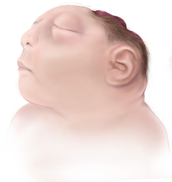
AnencephalyNo brain, no skull cap. Frog-eye facies.
Rostral neuropore fails. No forebrain develops. Fatal in utero or shortly after birth.
MSAFP ↑↑↑
Closed / Rostral
EncephaloceleSkin-covered skull defect, brain herniates out.
Most often occipital. Skin intact, so MSAFP is normal. Ultrasound catches it.
MSAFP normal
Open / Caudal
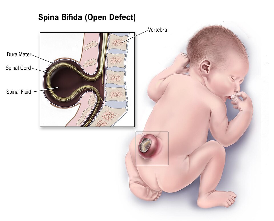
Spina bifida (open)Three flavors, increasing severity.
Meningocele · meninges out, no cord. Relatively mild.
Myelomeningocele · meninges + cord. Most severe common form. Deficits below the lesion.
Rachischisis · cord exposed, no covering at all. Catastrophic.
MSAFP ↑↑
Closed / Caudal
Spina bifida occultaVertebral arch defect, skin intact.
Skin markers over the spine: tuft of hair, dimple, lipoma, hyperpigmented patch. Usually asymptomatic; may have tethered cord. MSAFP normal, so prenatal screen misses it.
MSAFP normal
Open · neural tissue exposed · AFP leaks into amniotic fluid Closed · skin covers the defect · AFP stays normal
Spina bifida illustration
Spina bifida (illustrated) · tap to expand
Spina bifida neonate
Open lumbar lesion · tap to expand
Anencephaly illustration
Anencephaly · tap to expand
Tap the spine: where the defect lands

The caudal neuropore can fail at any level. Tap a region: severity ramps up as the lesion goes from closed-and-tiny (occulta) to wide-open-with-cord-out (rachischisis).

Lateral spine with tappable regions Cervical, thoracic, lumbar, and sacral spine levels each open a readout describing the neural tube defect at that level. C-spine T-spine L-spine Sacral ROSTRAL CAUDAL
Lumbar Myelomeningocele Severe
Most common location for myelomeningocele. Meninges + spinal cord herniate through an open vertebral defect with no skin covering. Motor and sensory deficits start below the level of the lesion. Pairs with Arnold-Chiari II about 90% of the time. MSAFP is high; ultrasound shows lemon (frontal bone scalloping) and banana (cerebellum pulled caudally).
AFP screening: what makes it go up, what makes it go down

Maternal serum alpha-fetoprotein is drawn in the second trimester (16-20 weeks). Each scenario shifts the bar. Tap a scenario to highlight.

AFP bar chart by scenario, multiples of median 0.0 0.5 1.0 1.5 2.0 2.5+ MoM 2.0 MoM cutoff Normal Open NTD Abdom wall Twins T21 Closed NTD
Normal pregnancy: MSAFP sits around 1.0 multiples of the median. The baseline. Everything below is read off this.
Risk factors: what blocks the neuropore from closing

Folate runs through almost all of them. The teratogens either antagonize folate directly or steal it through enzyme induction.

Top driver
Folate deficiency
Folate provides one-carbon units for DNA synthesis. Neural tube cells are dividing fast at week 3-4. No folate, no closure. Preconception supplementation is the only proven prevention.
Antimetabolite
Methotrexate
Blocks dihydrofolate reductase. Pulls folate out of circulation entirely. Contraindicated in pregnancy. Used as an abortifacient for ectopics for this exact reason.
Antiepileptic
Valproate
The worst NTD offender among antiepileptics. Interferes with folate metabolism and inhibits methionine synthase. NTD risk roughly 1 to 2 percent on monotherapy.
Antiepileptic
Phenytoin
Induces hepatic CYP enzymes that burn through folate stores faster. Adds cleft lip/palate and fetal hydantoin syndrome to the NTD risk list.
Antiepileptic
Carbamazepine
Same folate-depleting mechanism as phenytoin via CYP induction. Spina bifida risk roughly 1 percent. Counsel on switching agents before pregnancy.
Maternal
Maternal diabetes
Hyperglycemia at conception is teratogenic across body plans. NTDs, caudal regression, cardiac defects. Pre-pregnancy A1c is the lever.
Maternal
Maternal obesity
Independent NTD risk roughly doubled at BMI above 30. Mechanism includes lower folate bioavailability and undiagnosed glucose intolerance.
Maternal
Hyperthermia (week 3-4)
Early-pregnancy fever or hot tub exposure raises NTD risk. The neuropore is heat-sensitive during the window it should be fusing. Avoid saunas and untreated fevers in the first trimester.
Prevention · the only proven move
Folate before conception, not after the test turns positive
By the time most patients know they're pregnant, the neuropore has already closed (or failed to close). Folate has to be on board before week 3-4. Start it at the start of any reproductive plan.
0.4 mg/day All reproductive-age patients planning or capable of pregnancy
4 mg/day Prior pregnancy affected by NTD, or on folate-antagonist antiepileptic
Keep going

These two pages stack on this one.

Test yourself

Six vignettes. No timer. Explanations show the clue, the chain, and the high-yield discriminator.

Before you reveal an answer: elevated MSAFP, correct dates, singleton pregnancy, and a nondiagnostic ultrasound. What has to prove neural tissue is leaking?
Amniotic acetylcholinesterase is the specific neural-tissue leak signal when ultrasound has not shown the defect.
Medically reviewed by Kaitlyn Cocuzzo, MD and Fatima Ali, DO · Last updated June 30, 2026 at 10:02 AM ET
Bone Wizardry is an independent educational resource for visual learning in the medical sciences. It is not affiliated with, endorsed by, or sponsored by any licensing or examination board, contains no real or recalled examination questions, and does not guarantee any educational or examination outcome.