Sciatica Localizer: Root, Nerve, Plexus, or Canal?
Localize leg pain by integrating motor, sensory, reflex, electrodiagnostic, and imaging evidence while recognizing mimics and urgent neurologic danger.
Pain traveling down a leg is a symptom pattern, not a finished localization. The useful question is whether the patient has radicular pain alone, objective radiculopathy, a named nerve or plexus lesion, a canal process, or a nonneurologic competitor. This lesson builds that answer from findings that should agree with one another, then shows how to act when they do not.
Start with the kind of evidence
On a narrow screen, scroll inside this figure to read every label.
A proximal L5 process can affect actions carried by several named nerves. A common fibular process stays within its downstream distribution. This is a relationship map, not a complete plexus.
Radicular pain and radiculopathy are related but not interchangeable. Radicular pain is pain generated by abnormal activity in a spinal nerve or its roots. Radiculopathy requires objective loss of neurologic function, such as myotomal weakness, reflex asymmetry, or a defensible sensory deficit. A patient can have severe radiating pain without measurable radiculopathy, and a patient with weakness may describe little pain. [1][2]
Localize with a concordant set. Ask which weak actions share a root but travel through different named nerves. Compare reflexes side to side. Map sensation without treating a dermatome as a crisp border. Myotomal weakness and reflex loss are generally more persuasive than pain geography alone, especially when several findings converge. [3]
Evidence
What it can support
What it cannot prove alone
EvidenceFamiliar pain below the knee during straight leg raise
What it can supportMechanical sensitivity of lumbosacral neural tissue
What it cannot prove aloneThe exact root or structural cause
EvidenceWeak actions from different peripheral nerves
What it can supportA shared root or plexus level
What it cannot prove aloneCause without the rest of the examination
EvidenceOne abnormal sensory nerve response
What it can supportA postganglionic lesion in that sensory pathway
What it cannot prove aloneThat every symptom comes from one nerve
A straight leg raise is most useful when it reproduces the patient's familiar radiating symptoms, not when it merely stretches the hamstrings or causes nonspecific posterior thigh discomfort. Even a convincing response supports neural mechanosensitivity rather than proving a disc lesion. Structural differentiation and the full neurologic examination determine how much weight it deserves. [13]
Build a root pattern from overlapping functions
On a narrow screen, scroll inside this figure to read every label.
Approximate L4, L5, and S1 sensory anchors can guide comparison. They overlap and should be interpreted with the rest of the examination.
Roots share muscles, skin, and reflex arcs. Think of L4 as contributing to knee extension and ankle dorsiflexion, L5 to great toe and ankle dorsiflexion, inversion, and hip abduction, and S1 to plantar flexion, eversion, and hip extension. None of those actions belongs to one root alone, so the pattern across several actions matters more than a single weak muscle. [2][6]
Candidate root
Useful motor combination
Reflex support
Common sensory emphasis
Candidate rootL4
Useful motor combinationKnee extension with some ankle dorsiflexion contribution
Reflex supportPatellar arc, mainly L3 and L4
Common sensory emphasisMedial leg
Candidate rootL5
Useful motor combinationGreat toe extension, dorsiflexion, inversion, hip abduction
Reflex supportNo routinely dependable standard reflex; medial hamstring may add support
Common sensory emphasisDorsum of foot, with overlap
Candidate rootS1
Useful motor combinationPlantar flexion, eversion, hip extension
Reflex supportAchilles arc, mainly S1 and S2
Common sensory emphasisLateral foot and posterior calf, with overlap
Normal knee and ankle jerks do not erase a strong L5 pattern. L5 lacks a routinely reliable deep tendon reflex. An asymmetric medial hamstring reflex can contribute useful evidence, but it remains one part of a combined examination rather than a solitary verdict. [4]
Foot drop is not automatically L5 radiculopathy. Dorsiflexion and eversion weakness with preserved inversion and hip abduction fits common fibular neuropathy better. Weak inversion tests tibialis posterior through the tibial nerve, while weak hip abduction tests superior gluteal output; involvement of those separate pathways supports an L5 process above the named nerves. Broader plantar flexion or hamstring weakness raises sciatic, plexus, or multiroot possibilities. Symmetric length-dependent loss points toward polyneuropathy. [5][6][7]
Case 1
Show answer and explanations for case 1
A. Right common fibular neuropathy at the fibular head (Why this does not fit)
Which findings resemble fibular neuropathy?
Foot slap, dorsiflexion loss, and eversion weakness all occur with a common fibular lesion.
Which findings escape that nerve?
Tibialis posterior inversion and superior gluteal hip abduction are also weak, while fibular sensation is preserved.
Read the complete option explanation
Tempting because foot drop and eversion weakness fit a fibular lesion; however, weak inversion and hip abduction involve L5 pathways outside the common fibular nerve, and the superficial fibular sensory response is preserved.
B. Right sciatic neuropathy proximal to the hamstrings (Why this does not fit)
Why can sciatic neuropathy cause foot slap?
The sciatic nerve carries the fibular division that powers dorsiflexion and toe extension.
What contradicts a sciatic-only lesion?
Plantar flexion is full, while hip abduction is weak above and outside the sciatic nerve.
Read the complete option explanation
Tempting because a sciatic lesion can produce foot drop; however, preserved plantar flexion and the added hip abduction weakness do not fit a lesion confined to the sciatic nerve.
C. Right L5 radiculopathy (Best answer)
Which root unites the weak actions?
L5 contributes to dorsiflexion, toe extension, inversion, and hip abduction.
Why do several nerves matter?
Those actions travel through fibular, tibial, and superior gluteal nerves, placing disease proximal to them.
What does preserved sensory conduction add?
It supports a preganglionic L5 lesion rather than fibular or plexus sensory axon loss.
Read the complete option explanation
Tempting and correct because several weak actions share L5 input; the discriminator is weakness across fibular, tibial, and superior gluteal pathways with preserved distal sensory responses.
D. Right lumbosacral plexopathy (Why this does not fit)
Why is plexopathy initially plausible?
A plexus process can weaken muscles supplied by several named nerves.
Why is it not the best fit?
The deficit is tightly L5-weighted and both distal sensory responses remain intact.
Read the complete option explanation
Tempting because a plexus lesion can cross named nerves; however, the focused L5 pattern and preserved sensory responses favor a preganglionic root lesion over a postganglionic plexopathy.
E. Length-dependent polyneuropathy (Why this does not fit)
How can polyneuropathy produce foot drop?
Length-dependent motor axon loss can weaken distal ankle and toe muscles.
What distribution argues against it here?
The supplied right-sided deficit spans proximal and distal L5 actions with preserved sensory responses.
Read the complete option explanation
Tempting because polyneuropathy can cause foot drop; however, the supplied right-sided weakness spans proximal and distal L5 actions while distal sensory responses remain preserved, rather than forming a symmetric length-dependent pattern.
Takeaway: Foot drop becomes L5 when weakness crosses named nerves, especially inversion and hip abduction, while sensory responses remain preserved.
On a narrow screen, scroll inside this figure to read every label.
At L4-5, a foraminal or far lateral lesion can affect the exiting L4 root; a paracentral lesion commonly affects the traversing L5 root. These maps explain relationships rather than reproducing axial anatomy.
Image: Bone Wizardry. Reference: InjuryMap. Reference license: CC BY-SA 4.0. No source outline was copied.
A disc space is named for the vertebra above and below it, while a root follows its own path. In the lumbar spine, the root matching the lower vertebral number usually traverses behind the disc before exiting at the next foramen. A paracentral L4 to L5 herniation therefore usually affects the traversing L5 root. A foraminal or far lateral lesion at the same disc can affect the exiting L4 root. [8][9]
L4 to L5 paracentral predicts traversing L5 findings.
L4 to L5 foraminal or far lateral predicts exiting L4 findings.
L5 to S1 paracentral predicts traversing S1 findings.
L5 to S1 foraminal or far lateral predicts exiting L5 findings.
The relationship is a starting prediction, not permission to ignore the scan or examination. A large lesion can span zones and affect more than one root. Foraminal narrowing can be chronic and clinically silent. State the side, disc space, zone, root, and matching deficit each time. This prevents the common error of converting a disc name directly into a root number.
Use anatomy to forecast a consequence before seeing the answer. A left far lateral L4 to L5 lesion should fit left L4 function, such as knee extension weakness or patellar asymmetry, better than isolated great toe weakness. A left paracentral lesion at that disc should reverse the expectation toward L5. If the patient's deficits disagree, reconsider whether the image is causal.
Case 12
Show answer and explanations for case 12
A. Weak hip abduction and great toe extension with preserved patellar reflex (Why this does not fit)
Which root pattern do these actions imply?
Hip abduction and great toe extension are weighted toward L5.
Which zone would usually produce it at L4 to L5?
A paracentral or lateral-recess lesion, not an isolated far lateral fragment, would target traversing L5.
Read the complete option explanation
Tempting because these are common findings in lower lumbar radiculopathy; however, they emphasize L5, the traversing root affected by a paracentral rather than a far lateral L4 to L5 lesion.
B. Weak knee extension with a reduced patellar reflex and medial-leg sensory loss (Best answer)
Which root occupies the L4 to L5 foramen?
The exiting L4 root passes through that foramen.
Which three findings converge on L4?
Knee extension loss, patellar reduction, and medial-leg sensory change form an L4 set.
Why does this confirm causation better than pain alone?
The objective deficit matches the side, zone, and root contacted on imaging.
Read the complete option explanation
Tempting and correct because those findings form an L4 pattern; the discriminator is that the exiting L4 root occupies the L4 to L5 foramen where the fragment lies.
C. Weak plantar flexion with a reduced Achilles reflex and lateral-foot sensory loss (Why this does not fit)
Which root does this motor-reflex-sensory set represent?
Plantar weakness, Achilles reduction, and lateral-foot loss converge on S1.
Why is S1 anatomically discordant?
S1 traverses behind L5 to S1 and does not exit through L4 to L5.
Read the complete option explanation
Tempting because a disc can produce a root-reflex-sensory set; however, this is an S1 pattern and does not match the exiting root at L4 to L5.
D. Weak toe flexion with abnormal plantar sensory responses and full knee extension (Why this does not fit)
Which peripheral nerve does this set suggest?
Toe flexion plus abnormal plantar responses suggests tibial neuropathy.
Why does that fail to validate the scan?
A distal tibial lesion neither reflects exiting L4 function nor proves foraminal L4 compression.
Read the complete option explanation
Tempting because distal weakness can accompany radiating pain; however, abnormal plantar responses point to a tibial lesion and do not verify an exiting L4 compression.
E. Symmetric distal weakness with bilateral absent Achilles reflexes (Why this does not fit)
Which alternative process creates this bilateral pattern?
Polyneuropathy can produce symmetric distal weakness with absent Achilles reflexes.
Why is it not concordant with this image?
A unilateral foraminal fragment should not create a symmetric generalized distal syndrome.
Read the complete option explanation
Tempting because a structural scan can coexist with a separate distal neurologic process; however, symmetry and bilateral Achilles loss would suggest polyneuropathy rather than confirm the unilateral foraminal root lesion.
Takeaway: A foraminal or far lateral L4 to L5 lesion can affect the exiting L4 root, producing an L4 rather than L5 pattern.
Historical labels distinguish the tibial (internal popliteal), common fibular (external popliteal), and posterior femoral cutaneous (small sciatic) nerves. Compare the deep sciatic course with cutaneous branches; this plate is not current diagnostic guidance.
Henry Gray, Anatomy (1860); digitized by Internet Archive Book Images. Source record. No known copyright restrictions. Proportionally resized reference copy, 1280 by 2335 pixels, retained from the verified local cache. The original Commons source is 2268 by 4138 pixels; no image field was cropped or annotated.
Lumbar stenosis often produces leg symptoms during standing or walking and relief with sitting or lumbar flexion. It may affect one leg or both, and neurologic deficits can occur. Posture is informative, but it is not exclusive to stenosis because joint, muscle, and vascular symptoms can also vary with activity or position. Symptoms and anatomic narrowing must correlate. [10]
Vascular disease can coexist with spinal disease. Palpable pulses or a seemingly normal resting vascular examination lower some probabilities but do not exclude peripheral artery disease. Reproducible exertional calf symptoms, vascular risk, wounds, or discordant recovery should prompt objective vascular assessment such as an ankle brachial index. A resting index of 0.90 or less is abnormal, 0.91 to 0.99 is borderline, 1.00 to 1.40 is normal, and greater than 1.40 is noncompressible. When symptoms persist despite a normal or borderline resting value, exercise testing can uncover exertional disease. [11]
Buttock pain and a positive FAIR maneuver do not prove piriformis syndrome. Deep gluteal syndrome is a broader category of nondiscogenic sciatic entrapment, and diagnosis rests on a coherent history, several examination findings, and appropriate testing. Because the sciatic nerve carries motor and reflex pathways, a sufficiently significant lesion can cause weakness, sensory response abnormalities, or reduced Achilles support. Those findings do not automatically force the lesion back to a root. [12][7]
Root pattern can cross named nerves, usually preserves distal sensory responses, and may involve paraspinal muscles.
Named nerve pattern follows that nerve, can reduce its sensory response, and should spare muscles supplied outside it.
Plexus pattern crosses several nerves and roots, often reduces sensory responses, and usually spares paraspinal muscles.
Polyneuropathy is usually bilateral, length dependent, and supported by abnormalities in several distal nerves.
Read electrodiagnosis as anatomy plus time
A typical root lesion lies proximal to the dorsal root ganglion, so distal sensory nerve action potentials are usually preserved. Sciatic, fibular, tibial, and most plexus lesions are postganglionic and can reduce relevant sensory responses. This is a strong localizing principle, not an absolute law, because anatomy, technical factors, and mixed lesions create exceptions. [14]
Needle examination looks for motor axon loss in a distribution. Evidence in two muscles sharing a root but supplied by different peripheral nerves supports radiculopathy; matching paraspinal abnormalities add support. Posterior tibialis is especially useful when weak dorsiflexion could reflect either L5 radiculopathy or a fibular lesion. Conversely, abnormalities confined to one named nerve should redirect the localization. [14][6]
A normal study is not a universal exclusion. Denervation appears first in paraspinal muscles and later in limb muscles, so an early needle examination can be normal. A purely sensory root process may also evade a motor axon study. Report timing, muscles sampled, sensory responses, and the clinical question before deciding what a negative result means. [14]
MRI answers a structural question, not a causation question. Disc protrusions and other degenerative findings are common in people without symptoms and become more prevalent with age. A report matters when side, level, zone, and root agree with the neurologic pattern. A dramatic but contralateral or functionally discordant bulge should not displace a better localization. [15]
Let danger and trajectory set the pace
New urinary retention, saddle sensory loss, rapidly progressive weakness, or a compatible cluster of bowel, bladder, sexual, and bilateral neurologic symptoms requires emergency assessment for cauda equina or conus compression. Preserved limb strength or reflexes do not make sacral deficits safe. Do not reinterpret a quoted twenty-four or forty-eight hour window as permission to wait; the patient needs urgent evaluation now. [17]
Progressive focal weakness also needs prompt reassessment and appropriate imaging or specialist evaluation. Fever, immunosuppression, injection exposure, known cancer, unexplained weight loss, or destructive night pain changes the imaging threshold because infection or malignancy must be considered. Urgency comes from the whole pattern, not from pain intensity alone. [2][16]
For an uncomplicated acute course without red flags or progressive deficit, explain the expected trajectory, encourage tolerable activity, and arrange serial neurologic assessment. Immediate MRI is usually unnecessary because early structural findings may be incidental and often do not change initial care. Persistent disabling symptoms, worsening function, or a plan for intervention can justify imaging when the result will answer a focused clinical question. [2][16]
Before choosing an action, say the localization aloud. Name the likely site, cite the motor and reflex or sensory evidence, identify the closest competitor, and state the finding that defeats it. Then check whether the time course or red flags change urgency.
Case 25
Show answer and explanations for case 25
A. Arrange same-day outpatient lumbar MRI with telephone review after the scan (Why this does not fit)
Why does same-day outpatient MRI sound responsive?
It accelerates imaging without relying on the preserved limb examination.
What does that setting fail to provide?
It fails to provide monitored emergency evaluation during an acute sacral syndrome.
Read the complete option explanation
Tempting because it accelerates structural imaging while the limb examination is preserved; however, objective retention plus saddle sensory loss requires monitored emergency assessment rather than an outpatient sequence.
B. Transfer for emergency lumbar MRI and immediate spine surgical evaluation (Best answer)
What does a 650 mL postvoid residual establish?
It establishes objective urinary retention.
What does new perineal sensory loss add?
It adds a second sacral neurologic deficit.
Which setting can image and act without delay?
The emergency department can coordinate lumbar MRI with immediate spine evaluation.
Read the complete option explanation
Tempting and correct because the high postvoid residual and new saddle sensory deficit form an acute sacral compression pattern; preserved limb strength and reflexes test different pathways and do not lower the emergency priority.
C. Obtain urgent urologic assessment and catheterization before deciding on spine imaging (Why this does not fit)
Why is urologic care relevant?
A large residual may require prompt bladder decompression.
Why can it not precede the spine decision?
The paired saddle deficit already makes acute neural compression plausible.
Read the complete option explanation
Tempting because a large postvoid residual needs bladder management and can have nonspinal causes; however, the paired perineal sensory loss requires simultaneous emergency spinal evaluation rather than a urology-first decision.
D. Arrange expedited electrodiagnostic localization followed by lumbar MRI (Why this does not fit)
What can electrodiagnosis localize?
It can localize motor axon injury among roots and peripheral nerves.
What can it not promptly exclude?
It cannot promptly exclude acute canal compression.
Read the complete option explanation
Tempting because electrodiagnosis can separate roots from peripheral lesions; however, it cannot promptly exclude acute canal compression and adds delay before the available definitive structural study.
E. Perform a repeat sacral examination and bladder ultrasound in an urgent clinic (Why this does not fit)
Why can repeating the measurements feel careful?
It can confirm that an unexpected finding is reproducible.
Why is confirmation already sufficient here?
The stem supplies both objective retention and a new sacral sensory deficit.
Read the complete option explanation
Tempting because confirmation can reduce unnecessary emergency imaging; however, the stem already supplies objective urinary retention and a new sacral sensory deficit, so repetition cannot replace emergency evaluation.
Takeaway: Objective urinary retention plus new saddle sensory loss requires emergency MRI and spine evaluation even when limb strength and reflexes remain symmetric.
Dorsiflexion, toe extension, and eversion all travel through the common fibular nerve.
Which alternate L5 outputs are spared?
Inversion through the tibial nerve and hip abduction through the superior gluteal nerve remain full.
How much weight should the sensory result carry?
The reduced superficial fibular response adds postganglionic support to the nerve-confined motor pattern.
What exposure completes the localization?
Prolonged squatting supplies a focal compression mechanism at the fibular head.
Read the complete option explanation
Tempting and correct because the nerve-confined motor and sensory deficit follows focal fibular-head compression. Preserved inversion and hip abduction plus the abnormal superficial fibular response converge on common fibular neuropathy; the sensory response is supportive rather than decisive alone.
B. Left L5 radiculopathy (Why this does not fit)
Why does L5 enter the differential?
L5 supplies dorsiflexion and great toe extension and can cause foot drop.
Which L5 actions outside the fibular nerve remain intact?
Inversion and hip abduction are full.
Does one reduced superficial fibular SNAP exclude L5?
No, distal sensory responses can occasionally be abnormal in root disease.
Why is fibular neuropathy still the best fit?
The focal compression history and nerve-confined motor pattern converge with the sensory result.
Read the complete option explanation
Tempting because L5 contributes to dorsiflexion and toe extension; however, inversion and hip abduction are spared, the exposure is focal at the fibular head, and the abnormal superficial fibular response adds postganglionic support. A single abnormal SNAP does not independently exclude an uncommon root-level exception.
C. Left sciatic neuropathy (Why this does not fit)
Why can sciatic injury resemble this pattern?
Its fibular division is often more visibly affected than its tibial division.
What makes a more distal site stronger?
Inversion and plantar flexion remain full after a focal fibular-head compression exposure.
Read the complete option explanation
Tempting because the fibular division can be preferentially affected in sciatic lesions; however, inversion and plantar flexion are full, and the deficit follows a focal fibular-head compression with fibular sensory loss.
D. Left L4 radiculopathy (Why this does not fit)
Which finding could suggest L4 involvement?
L4 can contribute to ankle dorsiflexion.
Which supplied pattern makes the fibular nerve a stronger fit?
Eversion and toe extension are affected while inversion and hip abduction remain full, following focal compression.
Read the complete option explanation
Tempting because L4 can contribute to ankle dorsiflexion; however, knee extension, the patellar reflex, and medial-leg sensation are intact while the deficit follows the fibular distribution.
E. Distal symmetric polyneuropathy (Why this does not fit)
Why consider a distal polyneuropathy?
Generalized distal axon loss can weaken ankle dorsiflexors.
What defeats a length-dependent process?
Onset is abrupt and unilateral after local compression, with one sensory nerve implicated.
Read the complete option explanation
Tempting because distal neuropathy can weaken ankle muscles; however, the abrupt unilateral deficit after fibular-head compression and the single-nerve sensory abnormality are not length dependent.
Takeaway: The compression history, fibular motor-sensory distribution, preserved alternate L5 actions, and abnormal superficial fibular response together support common fibular neuropathy.
Why is fibularis longus a plausible muscle to sample?
Fibularis longus contributes to an S1-weighted action.
Why should it remain normal here?
Its motor fibers travel in the common fibular nerve rather than the injured tibial nerve.
Read the complete option explanation
Tempting because fibularis longus contributes to an S1-weighted action; however, it travels through the common fibular division, and eversion is full in this tibial pattern.
B. Right gluteus maximus (Why this does not fit)
What does gluteus maximus sample?
It samples proximal S1 output through the inferior gluteal nerve.
Why is it spared by the stated lesion?
The inferior gluteal nerve branches proximal to the popliteal tibial injury.
Read the complete option explanation
Tempting because gluteus maximus samples proximal S1 function; however, hip extension is full and the suspected lesion is distal to the inferior gluteal nerve.
C. Right short head of biceps femoris (Why this does not fit)
Why can short-head biceps femoris help near the sciatic bifurcation?
It receives motor fibers from the common fibular division of the sciatic nerve.
What does full knee flexion predict?
That proximal sciatic muscle should remain spared.
Read the complete option explanation
Tempting because this hamstring muscle helps test a lesion near the sciatic bifurcation; however, it uses the common fibular division and knee flexion is full.
D. Right gluteus medius (Why this does not fit)
Why is gluteus medius useful in a root differential?
It samples an L5 pathway above the sciatic nerve.
Which supplied finding predicts sparing?
Right hip abduction remains full.
Read the complete option explanation
Tempting because gluteus medius is useful when a root or plexus lesion is possible; however, it is supplied by the superior gluteal nerve and hip abduction is full.
E. Right tibialis posterior (Best answer)
Which nerve supplies tibialis posterior?
Tibialis posterior receives motor supply from the tibial nerve.
Where is its motor branch relative to the injury?
Its branch lies distal to the stated proximal popliteal lesion.
What does gastrocnemius denervation add?
It supports tibial motor axon injury proximal to major calf branches but does not locate the lesion alone.
Which other data make the proximal tibial site coherent?
Reduced plantar responses and sparing outside the tibial pathway converge with the popliteal laceration.
Read the complete option explanation
Tempting and correct because tibialis posterior is supplied by the tibial nerve distal to the stated popliteal lesion; the postganglionic plantar sensory loss and sparing of fibular, hamstring, gluteal, and paraspinal pathways keep the prediction within the tibial nerve.
Takeaway: Within this tibial-only pattern, Achilles loss and gastrocnemius denervation support motor axon injury proximal to the calf branches; plantar sensory loss and spared non-tibial pathways then predict tibialis posterior involvement.
A. Combined L5 and S1 radiculopathies (Why this does not fit)
Why could two roots explain the weakness?
L5 contributes to dorsiflexion and S1 contributes to plantar flexion and the Achilles reflex.
What makes two roots less coherent?
At four weeks, two reduced sensory responses and normal lumbar paraspinals add postganglionic support to the sciatic motor pattern.
Read the complete option explanation
Tempting because dorsiflexion and plantar flexion span L5 and S1; however, at four weeks the two abnormal distal sensory responses, normal paraspinals, and sciatic motor distribution collectively favor a postganglionic lesion.
B. Proximal sciatic neuropathy (Best answer)
Which sciatic components are affected?
Fibular dorsiflexion, tibial plantar flexion, and proximal hamstring knee flexion are all weak.
Why does the four-week interval matter?
It allows sensory-amplitude decline and needle denervation time to evolve after axonal injury.
What do the two abnormal sensory responses add?
They add postganglionic support when interpreted with the motor and needle pattern.
What narrows the site from plexus to sciatic nerve?
Gluteal and femoral functions remain intact outside the sciatic distribution.
Read the complete option explanation
Tempting and correct because four weeks allows the sensory-amplitude and needle patterns time to evolve. Fibular and tibial divisions plus knee flexion are involved, while spared gluteal and femoral functions, abnormal sural and superficial fibular responses, and normal paraspinals converge on the proximal sciatic nerve.
C. Lumbosacral plexopathy (Why this does not fit)
Why is plexopathy plausible after hip surgery?
Pelvic-region injury can damage several postganglionic nerve divisions.
Which outputs are unexpectedly spared?
Hip abduction, hip extension, and knee extension remain full outside the sciatic distribution.
Read the complete option explanation
Tempting because a plexus lesion is postganglionic and can span multiple nerves; however, preserved gluteal and femoral functions with a focused sciatic distribution make the lesion more distal.
D. Common fibular neuropathy (Why this does not fit)
Which visible deficit fits the fibular nerve?
Foot drop is a common presentation of common fibular neuropathy.
Which abnormalities exceed fibular territory?
Plantar flexion, knee flexion, Achilles support, and sural conduction also fail.
Read the complete option explanation
Tempting because foot drop is prominent; however, plantar flexion, knee flexion, the Achilles reflex, and the sural response extend beyond the common fibular nerve.
E. Distal symmetric polyneuropathy (Why this does not fit)
Why can polyneuropathy reduce both sensory responses?
Generalized distal axon loss often affects sural and superficial fibular nerves together.
What pattern rejects it?
The deficit is unilateral and postoperative and includes proximal hamstring weakness.
Read the complete option explanation
Both distal sensory responses are abnormal, but the postoperative pattern includes proximal sciatic weakness with spared gluteal and femoral functions. That focused distribution is more coherent with sciatic injury than a generalized length-dependent neuropathy.
Takeaway: Four weeks after onset, combined fibular, tibial, and hamstring deficits with spared gluteal muscles, abnormal sensory responses, and normal paraspinals support proximal sciatic neuropathy.
A. Right multilevel L2 through L5 radiculopathies (Why this does not fit)
Why do right L2 through L5 roots enter the differential?
The weak actions span several lumbar myotomes.
What makes separate roots less coherent?
Two right sensory responses are reduced despite normal lumbar paraspinals.
Read the complete option explanation
Tempting because several root functions are weak; however, reduced saphenous and superficial fibular sensory responses with normal paraspinals place the lesion distal to the dorsal root ganglia.
B. Right femoral neuropathy within the pelvis (Why this does not fit)
Which findings make right femoral neuropathy plausible?
Knee extension and the patellar reflex are impaired on the right.
Which supplied weakness exceeds femoral territory?
Right hip adduction requires obturator output.
Read the complete option explanation
Tempting because knee extension and the patellar reflex are affected after a pelvic procedure; however, hip abduction, adduction, and dorsiflexion extend beyond the femoral nerve.
C. Right proximal sciatic neuropathy (Why this does not fit)
Why might right sciatic neuropathy be considered?
Sciatic disease can weaken ankle dorsiflexion.
Which sensory result lies outside the sciatic nerve?
The reduced right saphenous response belongs to femoral territory.
Read the complete option explanation
Tempting because dorsiflexion weakness can follow sciatic injury; however, knee extension, patellar loss, and saphenous sensory involvement are outside the sciatic nerve.
D. Right lumbosacral plexopathy (Best answer)
What does weakness across several right named nerves require?
It requires a shared site proximal to those peripheral nerves.
What do the reduced right sensory responses establish?
They place the shared lesion distal to the dorsal root ganglia.
Which structure supplies that shared site?
The right pelvic hematoma occupies the lumbosacral plexus region.
Read the complete option explanation
Tempting and correct because the pelvic process can affect several divisions; the discriminator is weakness across femoral, obturator, superior gluteal, and fibular pathways with postganglionic sensory loss and spared paraspinals.
E. Length-dependent sensorimotor polyneuropathy (Why this does not fit)
Why can polyneuropathy reduce several sensory responses?
Generalized axon loss can affect more than one distal nerve.
Which spatial fact argues against it?
Every new deficit is right-sided after a focal right pelvic event.
Read the complete option explanation
Tempting because more than one sensory response is reduced; however, the acute asymmetric proximal and distal deficit tracks a focal pelvic hematoma rather than a bilateral distal process.
Takeaway: A right-sided postganglionic pattern crossing several named nerves with normal paraspinals and a right pelvic lesion supports lumbosacral plexopathy.
A. Vastus medialis, adductor longus, tibialis anterior, and L4 paraspinals abnormal (Why this does not fit)
What localization does this broad needle pattern suggest?
It places denervation across several L4 peripheral pathways.
Which sampled site makes it a root pattern?
L4 paraspinal denervation places the lesion proximal to the plexus.
Read the complete option explanation
Tempting because this set explains knee extension and could reproduce an L4 pattern; however, abnormalities across femoral, obturator, deep fibular, and paraspinal sites would support L4 radiculopathy rather than femoral mononeuropathy.
B. Vastus medialis and sartorius abnormal; adductor longus and L4 paraspinals normal (Best answer)
Which nerve supplies both proposed abnormal muscles?
The femoral nerve supplies vastus medialis and sartorius.
What does normal adductor longus show?
The obturator pathway is spared.
What does normal L4 paraspinal sampling show?
The mature needle pattern does not extend to the root region.
Read the complete option explanation
Tempting and correct because vastus medialis and sartorius share femoral supply; sparing of obturator adductor longus and L4 paraspinals confines the mature motor-axon pattern to the femoral nerve.
C. Vastus medialis and adductor longus abnormal; L4 paraspinals normal (Why this does not fit)
Why is this paired pattern a plausible postoperative competitor?
A plexus lesion can affect femoral and obturator muscles together.
Which examination finding defeats it?
Right hip adduction is full.
Read the complete option explanation
Tempting because a postganglionic upper lumbar plexus lesion can affect femoral and obturator outputs; however, hip adduction is full and the sensory and motor abnormalities supplied in the stem remain femoral.
D. Tibialis anterior and fibularis longus abnormal; vastus medialis normal (Why this does not fit)
Why is this a real perioperative alternative?
Positioning can injure the common fibular nerve near the fibular head.
Which supplied action contradicts it?
Right ankle dorsiflexion is full.
Read the complete option explanation
Tempting because perioperative positioning can injure the common fibular nerve; however, ankle dorsiflexion is full while the supplied quadriceps, patellar, and saphenous findings are abnormal.
E. Bilateral tibialis anterior and gastrocnemius abnormal; paraspinals normal (Why this does not fit)
What diagnosis does a bilateral distal needle pattern raise?
It raises a length-dependent polyneuropathy.
Which distribution in the stem defeats it?
The new deficits remain unilateral within right femoral territory.
Read the complete option explanation
Tempting because a generalized neuropathy can produce a mature distal needle pattern; however, the clinical and conduction abnormalities are acute, unilateral, and restricted to the right femoral distribution.
Takeaway: At an appropriate electrodiagnostic interval, abnormalities in two femoral muscles with sparing of obturator and paraspinal sites distinguish femoral mononeuropathy from plexus and L4 root lesions.
Quadriceps weakness, patellar reduction, and medial-leg symptoms are common femoral findings.
Which abnormalities cannot come from femoral nerve injury?
Tibialis anterior, adductor longus, and lumbar paraspinal denervation lie outside it.
Read the complete option explanation
Tempting because quadriceps weakness and patellar loss fit the femoral nerve; however, hip adduction, tibialis anterior, and paraspinal denervation lie outside that nerve.
B. Obturator neuropathy (Why this does not fit)
Which muscle makes obturator neuropathy tempting?
Adductor longus is supplied through the obturator nerve.
What remains unexplained by obturator disease?
Quadriceps, tibialis anterior, patellar, and paraspinal abnormalities remain outside that nerve.
Read the complete option explanation
Tempting because adductor longus is abnormal; however, knee extension, dorsiflexion, and the patellar reflex require broader L4 involvement.
C. Upper lumbar plexopathy (Why this does not fit)
Why does upper lumbar plexopathy fit several muscles?
It can span femoral, obturator, and other postganglionic outputs.
Which two data place disease before the plexus?
The saphenous response is preserved and matching lumbar paraspinals show denervation.
Read the complete option explanation
Tempting because femoral and obturator muscles are involved; however, preserved sensory responses and abnormal paraspinals place the lesion before the plexus.
D. Common fibular neuropathy (Why this does not fit)
Which abnormal muscle uses the deep fibular nerve?
Tibialis anterior is supplied through the deep fibular branch.
Why is fibular neuropathy far too narrow?
It cannot affect quadriceps, adductor longus, the patellar arc, or lumbar paraspinals.
Read the complete option explanation
Tempting because tibialis anterior weakness can cause foot slap; however, quadriceps, adductor, patellar, and paraspinal abnormalities cannot be explained by the common fibular nerve.
E. L4 radiculopathy (Best answer)
Which root links the three limb muscles?
L4 contributes to vastus medialis, tibialis anterior, and adductor longus across three nerves.
What does the preserved sensory response show?
It supports disease proximal to the dorsal root ganglion.
What confirms the proximal level?
L3 to L4 paraspinal denervation places the process at the root region.
Read the complete option explanation
Tempting and correct because the weak muscles share L4 across femoral, deep fibular, and obturator pathways; the discriminator is preserved sensory response with matching paraspinal denervation.
Takeaway: L4 radiculopathy crosses femoral, obturator, and fibular pathways while usually preserving the distal sensory response.
A. Bilateral L5 radiculopathies (Why this does not fit)
Which weakness resembles bilateral L5 disease?
Toe extension and ankle dorsiflexion receive strong L5 contributions on both sides.
What makes paired roots unlikely?
Stocking loss and bilateral sural and superficial fibular response reduction show distal peripheral disease.
Read the complete option explanation
Tempting because toe extension and dorsiflexion include L5; however, symmetric stocking loss and bilateral reduced sensory responses are postganglionic and length dependent.
B. Lumbar central canal stenosis (Why this does not fit)
Why does age plus bilateral leg trouble suggest stenosis?
Central stenosis can produce bilateral symptoms and gait limitation in older adults.
Which evidence favors neuropathy instead?
Symptoms are not posture dependent, and several distal sensory nerves are abnormal bilaterally.
Read the complete option explanation
Tempting because central stenosis can produce bilateral leg symptoms; however, back position does not change the symptoms and several distal sensory responses are reduced bilaterally in a stocking pattern.
C. Length-dependent sensorimotor polyneuropathy (Best answer)
What is the spatial pattern?
Weakness and sensory loss are symmetric and greatest distally in both legs.
What objective data match that pattern?
Sural and superficial fibular responses are reduced bilaterally with absent Achilles reflexes.
Which proximal actions remain protected?
Hip abduction and inversion remain full, reinforcing a length-dependent rather than root-focused process.
Read the complete option explanation
Tempting and correct because distal weakness, sensory loss, and reflex loss are symmetric; the discriminator is bilateral reduction of multiple distal sensory responses with proximal sparing.
D. Bilateral common fibular neuropathies (Why this does not fit)
Which weakness could paired fibular lesions explain?
Bilateral dorsiflexion and toe extension loss can arise from both common fibular nerves.
Tempting because dorsiflexion is weak and superficial fibular responses are reduced; however, sural loss, stocking distribution, and bilateral Achilles loss extend beyond both fibular nerves.
E. Cauda equina compression (Why this does not fit)
Why might cauda equina disease affect several distal functions?
Multiple lumbosacral roots can be compressed within the canal.
What argues against that central process?
Several distal sensory responses are reduced bilaterally in a stocking distribution.
Read the complete option explanation
Tempting because a canal process can affect multiple distal root functions; however, bilateral reduction of several distal sensory responses places the measured lesion after the dorsal root ganglia, and the stocking distribution is length dependent.
Takeaway: Symmetric length-dependent weakness, stocking loss, reflex reduction, and abnormalities in several distal sensory nerves support polyneuropathy.
Plantar flexion, eversion, and hip extension all receive meaningful S1 input.
Which reflex and sensory data agree?
The Achilles reflex is reduced while the sural sensory response stays preserved.
What makes the lesion preganglionic?
S1 paraspinal and gluteus maximus denervation place disease proximal to the sciatic nerve.
Read the complete option explanation
Tempting and correct because distal and proximal S1 actions plus the Achilles arc are involved; the discriminator is preserved sural response with denervation in gluteus maximus and S1 paraspinals.
B. Tibial neuropathy (Why this does not fit)
Which findings could tibial neuropathy explain?
Plantar flexion weakness and reduced Achilles support involve tibial motor fibers.
Which findings escape the tibial nerve?
Eversion, hip extension, and paraspinal denervation require other S1 pathways.
Read the complete option explanation
Tempting because plantar flexion and Achilles support travel through the tibial nerve; however, eversion, hip extension, and paraspinal denervation lie outside it.
C. Sciatic neuropathy (Why this does not fit)
Why does sciatic neuropathy resemble S1 disease?
The sciatic nerve carries tibial and fibular fibers for plantar flexion and eversion.
What places the lesion above the sciatic nerve?
Gluteus maximus and S1 paraspinals are abnormal while sural conduction is preserved.
Read the complete option explanation
Tempting because plantar flexion and eversion are sciatic functions; however, gluteus maximus and paraspinal abnormalities place the lesion proximal to the sciatic nerve, and the sural response is preserved.
D. L5 radiculopathy (Why this does not fit)
Which action could suggest L5?
Eversion often has substantial L5 contribution.
What makes S1 dominant?
Plantar flexion, hip extension, Achilles reduction, and lateral-foot symptoms converge on S1.
Read the complete option explanation
Tempting because eversion can receive L5 input; however, the dominant plantar flexion, hip extension, lateral-foot symptoms, and Achilles loss form an S1 pattern.
E. Common fibular neuropathy (Why this does not fit)
Which weak action enters fibular territory?
Eversion travels through the superficial fibular nerve.
Which findings make fibular neuropathy impossible?
Plantar flexion, hip extension, Achilles, and paraspinal abnormalities lie outside that nerve.
Read the complete option explanation
Tempting because eversion is weak; however, plantar flexion, hip extension, Achilles loss, and paraspinal denervation are outside the common fibular nerve.
Takeaway: Proximal and distal S1 weakness with Achilles loss, preserved sensory responses, and paraspinal denervation supports S1 radiculopathy.
Posterior symptoms, plantar weakness, and reduced Achilles support all overlap with S1.
Which physiology redirects postganglionically?
The sural response is abnormal while lumbar paraspinals are normal.
Read the complete option explanation
Tempting because posterior symptoms, plantar flexion weakness, and Achilles loss fit S1; however, an abnormal sural response and normal paraspinals favor a postganglionic sciatic lesion.
B. Isolated piriformis muscle strain (Why this does not fit)
Why can a local piriformis injury reproduce FAIR pain?
That position loads deep gluteal muscles as well as nearby neural tissue.
What proves the syndrome is not muscle pain alone?
Tempting because buttock pain and FAIR provocation can arise from local muscle injury; however, objective sciatic weakness, reflex loss, and an abnormal sural response require neural involvement.
C. Lumbosacral plexopathy (Why this does not fit)
Why does pelvic trauma raise plexus concern?
Trauma can injure postganglionic lumbosacral elements before named nerves form.
What limits the lesion to sciatic territory?
Hip abduction and extension are full while the supplied weak actions remain within sciatic output.
Read the complete option explanation
Tempting because pelvic trauma and postganglionic findings can indicate plexus injury; however, preserved gluteal functions and a focused sciatic distribution favor the nerve itself.
D. Deep-gluteal sciatic neuropathy (Best answer)
Which motor branches are involved?
Hamstring, fibular, and tibial functions all fail within the sciatic distribution.
What shows the lesion is postganglionic?
The sural sensory response is abnormal and lumbar paraspinals are normal.
What supports the deep gluteal site?
Buttock symptoms follow pelvic injury and are reproduced locally while gluteal muscles remain strong.
Read the complete option explanation
Tempting and correct because buttock provocation accompanies objective sciatic dysfunction; the discriminator is combined hamstring, fibular, and tibial weakness with sural loss, spared gluteals, and normal paraspinals.
E. Common fibular neuropathy (Why this does not fit)
Which deficit resembles common fibular neuropathy?
Dorsiflexion weakness can dominate either a fibular or sciatic lesion.
Which failures extend above and beyond it?
Knee flexion, plantar flexion, Achilles support, and sural conduction also fail.
Read the complete option explanation
Tempting because dorsiflexion is weak; however, knee flexion, plantar flexion, Achilles loss, and sural abnormality extend well beyond the common fibular nerve.
Takeaway: Buttock pain and FAIR provocation do not settle the diagnosis; objective postganglionic sciatic findings can localize deep gluteal entrapment.
A. Weak knee extension with a reduced patellar reflex (Why this does not fit)
Which root does this pattern represent?
Knee extension weakness with patellar reduction forms an L4-weighted pattern.
Why does the disc zone reject it?
A paracentral L4 to L5 fragment usually reaches traversing L5 rather than exiting L4.
Read the complete option explanation
Tempting because the scan is labeled L4 to L5; however, knee extension and patellar loss predict the exiting L4 root, which is not usually the root compressed by a paracentral lesion at this disc.
B. Weak plantar flexion with a reduced Achilles reflex (Why this does not fit)
Which root does plantar weakness plus Achilles loss represent?
That motor-reflex pair points toward S1.
At which disc would S1 usually traverse?
S1 normally traverses behind L5 to S1, one disc below the lesion described.
Read the complete option explanation
Tempting because lower lumbar discs can cause leg symptoms; however, plantar flexion and Achilles loss predict S1, usually traversing behind L5 to S1 rather than L4 to L5.
C. Weak knee flexion with abnormal sural sensory response (Why this does not fit)
Why can knee flexion suggest a proximal neural lesion?
Hamstring weakness can occur with sciatic or sacral root involvement.
What makes this option the wrong lesion type?
An abnormal sural response is postganglionic and does not match the requested disc-root prediction.
Read the complete option explanation
Tempting because posterior-leg function can fail with disc symptoms; however, an abnormal sural response favors a postganglionic sciatic process, not the expected root lesion.
D. Weak great toe extension, inversion, and hip abduction with routine reflexes preserved (Best answer)
Which root traverses behind L4 to L5?
The L5 root descends through the lateral recess behind that disc.
Which actions sample L5 through separate nerves?
Great toe extension, inversion, and hip abduction use fibular, tibial, and superior gluteal routes.
Why can routine reflexes stay normal?
There is no consistently dependable standard deep tendon reflex for L5.
Read the complete option explanation
Tempting and correct because those actions cross named nerves yet share L5; the discriminator is that the traversing L5 root lies behind a paracentral L4 to L5 disc and lacks a dependable routine reflex.
E. Weak dorsiflexion and eversion with an abnormal superficial fibular sensory response (Why this does not fit)
Which portion resembles an L5 deficit?
Dorsiflexion and eversion can weaken with L5 dysfunction.
What changes the lesion to a fibular nerve?
The pattern is confined to fibular actions and includes abnormal fibular sensory conduction.
Read the complete option explanation
Tempting because L5 contributes to dorsiflexion and eversion; however, the abnormal sensory response and restriction to fibular actions would instead favor common fibular neuropathy.
Takeaway: A paracentral L4 to L5 lesion usually reaches the traversing L5 root, not the exiting L4 root named by the upper vertebra.
A. Weak plantar flexion and hip extension with a reduced Achilles reflex (Best answer)
Which root travels in this lateral recess?
S1 traverses behind the L5 to S1 disc before exiting at the sacrum.
Which motor functions should fall?
Plantar flexion and hip extension both receive important S1 contribution.
Which reflex supplies additional agreement?
A reduced Achilles reflex supports S1 while acknowledging S2 contribution.
Read the complete option explanation
Tempting and correct because these findings converge on S1; the discriminator is that S1 is the traversing root behind a paracentral L5 to S1 disc.
B. Weak great toe extension and hip abduction with no reliable reflex change (Why this does not fit)
Which root does great toe and hip abduction weakness suggest?
Those actions are commonly used to test L5.
What imaging statement defeats L5?
The exiting L5 root is explicitly free in the foramen while the descending root is displaced.
Read the complete option explanation
Tempting because L5 deficits are common near this disc space; however, L5 is the exiting root at L5 to S1 and is reported free in the foramen.
C. Weak knee extension with a reduced patellar reflex (Why this does not fit)
Which root uses knee extension and the patellar arc?
Those findings are weighted toward L4.
Why is L4 too high?
L4 is neither the exiting nor the traversing root implicated at L5 to S1.
Read the complete option explanation
Tempting because disc disease can reduce a root-associated reflex; however, knee extension and the patellar arc indicate L4, two root levels above the traversing root here.
D. Weak dorsiflexion and eversion with an abnormal superficial fibular response (Why this does not fit)
Why can dorsiflexion weakness seem relevant near L5 to S1?
The exiting L5 root can be affected by a foraminal lesion at that disc.
What makes this a nerve rather than root pattern?
Eversion-only pairing with abnormal superficial fibular sensation is postganglionic and fibular-restricted.
Read the complete option explanation
Tempting because foot weakness can accompany an L5 to S1 lesion; however, this postganglionic fibular pattern does not match preserved sensory responses or traversing S1 anatomy.
E. Weak knee flexion and plantar flexion with an abnormal sural response (Why this does not fit)
Which overlap makes sciatic neuropathy plausible?
Knee flexion and plantar flexion both travel through the sciatic nerve.
What contradicts the expected preganglionic lesion?
The sural response is abnormal even though the stem states distal sensory responses are preserved.
Read the complete option explanation
Tempting because sciatic and S1 patterns overlap; however, an abnormal sural response would favor sciatic neuropathy rather than a preganglionic disc-root lesion.
Takeaway: A paracentral L5 to S1 lesion usually affects the traversing S1 root and should correlate with S1 motor and Achilles findings.
A. Weak plantar flexion with Achilles loss and lateral-foot numbness (Why this does not fit)
Why is S1 an easy association at this disc?
Paracentral L5 to S1 lesions commonly compress traversing S1.
What does the actual zone change?
The lesion is foraminal and the S1-containing lateral recess is spared.
Read the complete option explanation
Tempting because S1 symptoms often arise at the L5 to S1 disc; however, S1 is the traversing root and the lateral recess is spared.
B. Weak knee extension with patellar loss and medial-leg numbness (Why this does not fit)
Which root does this set represent?
Quadriceps weakness, patellar loss, and medial-leg numbness indicate L4.
Where does L4 exit?
L4 exits through the L4 to L5 foramen, one level above this fragment.
Read the complete option explanation
Tempting because a foraminal lesion affects an exiting root; however, L4 exits one level above and does not occupy the L5 to S1 foramen.
C. Weak plantar flexion with abnormal plantar sensory responses (Why this does not fit)
Which named nerve fits plantar weakness and plantar sensory loss?
Those findings align with a tibial neuropathy.
Why is that not the imaged mechanism?
A distal postganglionic tibial lesion does not match exiting L5 foraminal compression.
Read the complete option explanation
Tempting because lower-leg weakness can accompany an L5 to S1 disc syndrome; however, plantar weakness with abnormal plantar responses is a postganglionic tibial pattern rather than the exiting L5 root compressed on MRI.
D. Weak dorsiflexion and eversion with preserved inversion and hip abduction (Why this does not fit)
Which weak actions can carry L5 input?
Dorsiflexion and eversion can both weaken in L5 disease.
What sparing redirects to fibular neuropathy?
Normal inversion and hip abduction confine the weakness to the fibular pathway.
Read the complete option explanation
Tempting because dorsiflexion and eversion receive L5 input; however, sparing other L5 actions makes common fibular neuropathy more likely than the foraminal root lesion.
E. Weak great toe extension, inversion, and hip abduction with preserved routine reflexes (Best answer)
Which root exits through L5 to S1?
The L5 root leaves through the L5 to S1 foramen.
Which actions show L5 across named nerves?
Great toe extension, inversion, and hip abduction span fibular, tibial, and superior gluteal routes.
Why can knee and ankle reflexes stay intact?
L5 has no routinely reliable standard reflex expected to change.
Read the complete option explanation
Tempting and correct because those cross-nerve actions share L5; the discriminator is that L5 is the exiting root compressed within the L5 to S1 foramen.
Takeaway: A foraminal L5 to S1 lesion can affect exiting L5, while a paracentral lesion at that disc more often affects traversing S1.
A. Isolated exiting L4 root involvement (Why this does not fit)
Which findings does isolated L4 explain?
Knee extension weakness and patellar reduction align with exiting L4.
Which findings demand another root?
Great toe extension, inversion, and hip abduction add a coherent L5 set.
Read the complete option explanation
Tempting because knee extension and patellar loss fit L4 and the lesion enters the foramen; however, great toe extension and hip abduction add an L5 deficit not explained by isolated L4 disease.
B. Isolated traversing L5 root involvement (Why this does not fit)
Which findings does isolated L5 explain?
Great toe extension, inversion, and hip abduction align with traversing L5.
Which findings remain outside L5?
Quadriceps weakness and patellar reduction require substantial L4 involvement.
Read the complete option explanation
Tempting because inversion, great toe extension, and hip abduction fit L5 and the lesion enters the recess; however, patellar loss and knee extension weakness require L4 involvement.
C. Combined L4 and L5 root involvement (Best answer)
Which root can the foraminal component affect?
It can compress the exiting L4 root at L4 to L5.
Which root can the recess component affect?
It can compress the traversing L5 root behind the same disc.
How does the examination verify both?
It combines an L4 motor-reflex set with L5 weakness through three peripheral nerves.
Read the complete option explanation
Tempting and correct because a lesion spanning foramen and lateral recess can reach both roots; the discriminator is the combined L4 motor-reflex set and cross-nerve L5 motor set.
D. Combined femoral and common fibular neuropathies (Why this does not fit)
Why might two mononeuropathies mimic this pattern?
Femoral injury can weaken quadriceps and fibular injury can weaken toe extension.
Which actions break that two-nerve explanation?
Inversion and hip abduction add tibial and superior gluteal L5 pathways with preserved sensation.
Read the complete option explanation
Tempting because knee extension and great toe extension could be split between two nerves; however, weak inversion and hip abduction cross those nerves, and preserved sensory responses favor roots.
E. Lumbosacral plexopathy (Why this does not fit)
Why does plexopathy enter the differential?
The deficits cross several named nerves and root levels.
What makes two roots more economical?
Distal sensory responses are preserved and the scan spans exactly the foraminal and recess zones for L4 and L5.
Read the complete option explanation
Tempting because several nerve territories are involved; however, the precise two-root pattern, preserved sensory responses, and concordant disc zones make a plexus lesion less coherent.
Takeaway: A disc lesion spanning foraminal and paracentral zones can affect both the exiting root above and the traversing root below.
A. Weak hip flexion and adduction with a preserved patellar reflex (Why this does not fit)
Why does this pattern initially fit the pain region?
Hip flexion and adduction can accompany an upper lumbar syndrome.
Which imaging fact defeats exiting L3?
The left L3 root is free within its foramen.
Read the complete option explanation
Tempting because anterior-thigh pain and the L3 to L4 disc label can suggest L3; however, the exiting L3 root is free and this pattern does not test the displaced traversing root.
B. Weak great toe extension and hip abduction with symmetric routine reflexes (Why this does not fit)
Which root do these two actions emphasize?
Great toe extension and hip abduction emphasize L5.
Why is L5 too caudal here?
The displaced root behind L3 to L4 is L4.
Read the complete option explanation
Tempting because this is a coherent lower-lumbar root pattern with preserved sensory responses; however, it predicts L5, one root below the root traversing behind L3 to L4.
C. Weak plantar flexion and hip extension with a reduced Achilles reflex (Why this does not fit)
Which root does this motor-reflex pair test?
Plantar flexion with Achilles loss tests an S1-weighted pattern.
At which disc does S1 usually traverse?
S1 usually traverses behind L5 to S1.
Read the complete option explanation
Tempting because a paracentral disc can affect a descending root; however, this is an S1 motor-reflex set and is anatomically too caudal for an L3 to L4 lesion.
D. Weak knee flexion and dorsiflexion with a reduced sural sensory response (Why this does not fit)
Why is a proximal sciatic pattern a plausible alternative?
Knee flexion and dorsiflexion both travel through sciatic pathways.
Which supplied result argues against it?
Distal sensory responses are preserved.
Read the complete option explanation
Tempting because a proximal sciatic lesion can combine posterior-thigh and foot weakness; however, the reduced sural response conflicts with the preserved sensory responses and the supplied preganglionic disc-root mechanism.
E. Weak knee extension and ankle dorsiflexion with a reduced patellar reflex (Best answer)
Which root descends behind the L3 to L4 disc?
L4 traverses through that lateral recess.
Which movement pair samples L4 across separate nerves?
Knee extension and ankle dorsiflexion both receive meaningful L4 input.
Which reflex supplies concordant support?
A reduced left patellar reflex supports L4 dysfunction.
Read the complete option explanation
Tempting and correct because knee extension, ankle dorsiflexion, and the patellar arc form an L4-weighted set; the paracentral zone selects traversing L4 while the exiting L3 root is explicitly free.
Takeaway: A paracentral L3 to L4 lesion predicts traversing L4, so the downstream examination should combine L4-weighted motor findings with patellar asymmetry rather than simply repeat the disc label.
Which supplied action argues against that extension?
Right plantar flexion remains full.
Read the complete option explanation
Tempting because gastrocnemius samples a proximal tibial pathway and can be abnormal in sciatic disease; however, plantar flexion is full and the supplied pattern is L5 rather than S1.
B. Right vastus medialis (Why this does not fit)
What root does vastus medialis emphasize?
Vastus medialis is weighted toward L4 through the femoral nerve.
Which supplied action argues against L4?
Right knee extension remains full.
Read the complete option explanation
Tempting because vastus medialis provides another proximal limb sample; however, it is L4-weighted through the femoral nerve and knee extension is full.
C. Right gluteus medius (Best answer)
Which root already links both abnormal tibialis muscles?
Tibialis anterior and tibialis posterior share strong L5 input.
Why is gluteus medius the decisive next sample?
Its L5 fibers travel through the superior gluteal nerve.
What would denervation there establish?
It would extend the L5 pattern outside the sciatic nerve.
Read the complete option explanation
Tempting and correct because gluteus medius receives strong L5 input through the superior gluteal nerve; denervation there would extend the pattern outside both divisions of the sciatic nerve and match the traversing L5 lesion.
D. Right gluteus maximus (Why this does not fit)
Why sample gluteus maximus in a proximal differential?
It tests a gluteal pathway above the sciatic nerve.
Why is it the wrong segment?
Gluteus maximus is weighted toward S1 rather than L5.
Read the complete option explanation
Tempting because a gluteal muscle would place the lesion proximal to the sciatic nerve; however, gluteus maximus is S1-weighted and plantar flexion is full.
E. Left tibialis anterior (Why this does not fit)
What would contralateral tibialis anterior denervation suggest?
It would raise a bilateral or generalized motor process.
Which spatial pattern argues against it?
The clinical and imaging abnormalities are confined to the right side.
Read the complete option explanation
Tempting because bilateral tibialis anterior abnormalities could suggest a generalized motor process; however, the symptoms, weakness, needle findings, and MRI are all right-sided.
Takeaway: After tibialis anterior and tibialis posterior establish a cross-nerve L5 pattern, gluteus medius is the useful downstream sample because it tests L5 outside the sciatic nerve.
A. Repeat comprehensive EMG and nerve conduction at three to four weeks (Why this does not fit)
Why would a three-to-four-week study be more sensitive?
Limb denervation has more time to appear by that interval.
Why is it not the next investigation?
Motor strength is worsening before that physiologic window.
Read the complete option explanation
Tempting because a later study has greater sensitivity for limb denervation; however, it would answer physiology after the clinically progressive weakness has already created a need for structural assessment.
B. Urgent lumbar MRI for a compressive L5 lesion (Best answer)
Which root pattern spans the three weak actions?
Dorsiflexion, inversion, and hip abduction create an L5-weighted pattern.
Why can the seven-day needle study remain normal?
Spontaneous activity may not yet be present in sampled limb muscles.
What question must be answered now?
Lumbar MRI must assess for a compressive L5 lesion.
Read the complete option explanation
Tempting and correct because the worsening weakness spans three L5 pathways and the seven-day needle study is too early to exclude axonal injury; urgent lumbar MRI addresses a potentially compressive cause without waiting for denervation to mature.
C. High-resolution ultrasound of the right common fibular nerve (Why this does not fit)
When is fibular ultrasound useful?
It can assess a focal lesion near the fibular head.
Which weak action lies outside that nerve?
Right hip abduction lies outside the common fibular nerve.
Read the complete option explanation
Tempting because fibular ultrasound can evaluate a common cause of foot drop; however, inversion and hip abduction are weak, so the supplied deficit is not confined to the common fibular nerve.
D. Pelvic MR neurography for a right lumbosacral plexus lesion (Why this does not fit)
Why can plexus imaging be reasonable in cross-nerve weakness?
A plexus lesion can affect several peripheral outputs.
Which physiology lowers that priority here?
Distal sensory responses remain preserved.
Read the complete option explanation
Tempting because weakness across named nerves can arise in the plexus; however, preserved sensory responses and the focused L5 pattern favor a preganglionic lesion without a supplied pelvic mechanism.
E. Lumbar CT myelography despite available MRI (Why this does not fit)
When is CT myelography a useful structural test?
It is useful when lumbar MRI cannot be performed.
Which supplied condition removes that advantage?
MRI is available without a contraindication.
Read the complete option explanation
Tempting because CT myelography can evaluate root compression when MRI cannot be used; however, the stem provides no MRI contraindication and MRI is the appropriate noninvasive first structural study.
Takeaway: A normal limb needle examination at seven days does not exclude axonal radiculopathy; progressive cross-nerve weakness makes lumbar imaging urgent before a repeat study matures.
A. More intense below-knee pain during straight leg raise with full heel raises (Why this does not fit)
What does stronger straight-leg pain add?
It adds support for neural mechanosensitivity.
Why does full heel-raise function still matter?
It leaves S1 motor function intact.
Read the complete option explanation
Tempting because stronger reproduction increases confidence in neural mechanosensitivity; however, pain intensity with full heel raises still does not document loss of S1 function.
B. A larger left L5 to S1 protrusion with symmetric Achilles reflexes (Why this does not fit)
Why does a larger protrusion increase concern?
It creates stronger structural contact with the traversing S1 root.
What remains absent?
The Achilles reflexes remain symmetric.
Read the complete option explanation
Tempting because a larger paracentral lesion is more structurally concordant with S1; however, imaging severity without a functional deficit still does not establish objective radiculopathy.
C. A reduced left sural response with weak toe flexion (Why this does not fit)
Why can weak toe flexion resemble S1 disease?
Toe flexors receive substantial S1 input.
Where does a reduced sural response redirect the lesion?
It redirects the lesion to postganglionic sciatic territory.
Read the complete option explanation
Tempting because the new weakness is compatible with distal S1 function; however, a reduced sural response redirects the lesion toward postganglionic sciatic or tibial tissue rather than an isolated root.
D. A reduced left Achilles reflex with weak repetitive heel raises (Best answer)
Which function does repeated heel raising test?
It tests functional S1-weighted plantar flexion.
Which reflex tests the same candidate root?
The Achilles reflex supplies additional S1 support.
Why can this pair outweigh a repeat normal needle study?
It documents objective motor and reflex dysfunction on the symptomatic side.
Read the complete option explanation
Tempting and correct because Achilles asymmetry and impaired repetitive heel raises supply independent reflex and motor evidence of left S1 dysfunction; objective loss can establish radiculopathy even when a motor-axon study remains negative.
E. Weak left hip abduction with preserved plantar flexion (Why this does not fit)
Which root does hip abduction emphasize?
Hip abduction emphasizes L5.
Why does preserved plantar flexion fail to confirm S1?
The proposed S1 motor action remains intact.
Read the complete option explanation
Tempting because new objective weakness would establish neurologic dysfunction; however, hip abduction is L5-weighted and does not confirm the proposed S1 process when plantar flexion remains full.
Takeaway: Sensory-predominant radicular pain can remain electrodiagnostically normal; new concordant motor and reflex loss, not larger imaging or stronger pain alone, establishes objective radiculopathy.
A. Coexisting neurogenic and vascular claudication; assess the functional contribution of each (Best answer)
Which feature supports a neurogenic contributor?
Forward flexion accelerates relief in the setting of central stenosis.
Which measurement supports a vascular contributor?
Both resting ankle brachial indices are 0.90 or less.
What do palpable pulses change?
They do not erase the abnormal vascular measurements.
Read the complete option explanation
Tempting and correct because flexion-linked relief plus central stenosis supports a neurogenic component, while both resting ankle brachial indices objectively support peripheral artery disease despite palpable pulses.
B. Predominantly neurogenic claudication; begin canal-directed care and observe the PAD symptoms (Why this does not fit)
Why is canal-directed care plausible?
The symptoms improve with flexion in the setting of central stenosis.
Why is the PAD not merely incidental?
The exertional calf symptoms accompany bilateral abnormal resting indices.
Read the complete option explanation
Tempting because posture-linked relief and concordant stenosis make the canal clinically relevant; however, reproducible exertional symptoms plus bilateral abnormal ankle brachial indices do not justify treating the vascular disease as incidental.
C. Predominantly vascular claudication; begin PAD-directed care and observe the stenosis symptoms (Why this does not fit)
Why is artery-directed care plausible?
A fixed walking exposure reproduces symptoms with objectively abnormal indices.
Why is the stenosis not merely incidental?
Leaning forward shortens the recovery despite the same walking trigger.
Read the complete option explanation
Tempting because the walking limit and abnormal ankle brachial indices establish a vascular contributor; however, faster relief with flexion and concordant central stenosis preserve a neurogenic contributor even without weakness.
D. Neurogenic claudication with unreliable noncompressible-vessel indices; obtain toe pressures before assigning PAD (Why this does not fit)
When do toe pressures help interpret ankle indices?
They help when ankle arteries are noncompressible.
Which number pattern defines noncompressibility?
A resting ankle brachial index above 1.40 defines it.
Read the complete option explanation
Tempting because toe pressures help when calcified arteries make an ankle brachial index noncompressible; however, noncompressibility raises the index above 1.40 rather than producing the supplied low values.
E. PAD plus nonspecific deconditioning; reserve lumbar correlation for objective neurologic loss (Why this does not fit)
Why does full strength make deconditioning tempting?
There is no fixed motor deficit at rest.
Which positional feature still requires lumbar correlation?
Forward flexion produces faster relief.
Read the complete option explanation
Tempting because full strength makes fixed motor radiculopathy unlikely and PAD is established; however, neurogenic claudication can occur without weakness, and the flexion-linked relief matches the central stenosis.
Takeaway: Flexion-linked symptoms and an abnormal resting ankle brachial index can support coexisting neurogenic and vascular claudication; palpable pulses and full strength do not force a single-process answer.
A. Preserved inversion and hip abduction with an abnormal superficial fibular sensory response (Best answer)
Which L5 actions remain intact in this counterfactual?
Inversion and hip abduction remain intact through tibial and superior gluteal pathways.
Where does the sensory abnormality place disease?
A reduced superficial fibular response places the lesion after the dorsal root ganglion.
What localization now contains every deficit?
The common fibular nerve contains the weak motor actions and abnormal sensory territory.
Read the complete option explanation
Tempting and correct because it tests both a spared alternate L5 pathway and postganglionic sensory function; the discriminator is confinement of motor and sensory loss to the common fibular nerve.
B. Weak inversion and hip abduction with a preserved superficial fibular sensory response (Why this does not fit)
What do weak inversion and hip abduction add?
They add L5 deficits carried outside the common fibular nerve.
What does preserved sensory conduction support?
It supports a preganglionic L5 root lesion rather than the requested fibular redirection.
Read the complete option explanation
Tempting because it adds strong objective findings; however, weakness outside the fibular nerve and preserved sensory response strengthen a preganglionic L5 localization.
C. Weak plantar flexion and knee flexion with an abnormal sural sensory response (Why this does not fit)
What broader motor territory is added?
Plantar flexion and knee flexion add tibial and proximal sciatic involvement.
Where would this counterfactual redirect?
The abnormal sural response plus both divisions would favor sciatic neuropathy, not common fibular neuropathy.
Read the complete option explanation
Tempting because an abnormal sensory response redirects away from a root; however, the broader tibial and hamstring deficits would redirect to sciatic neuropathy, not common fibular neuropathy.
D. Weak knee extension with a reduced patellar reflex and medial-leg numbness (Why this does not fit)
Which neurologic set is being added?
Quadriceps weakness, patellar reduction, and medial-leg numbness form an L4 or femoral set.
Why does it not isolate the fibular nerve?
Every added finding lies outside the common fibular distribution.
Read the complete option explanation
Tempting because it supplies a coherent neurologic set; however, it adds L4 or femoral territory rather than isolating the common fibular nerve.
E. Bilateral stocking loss with reduced sural responses on both sides (Why this does not fit)
What distribution does bilateral stocking loss imply?
It implies a symmetric length-dependent peripheral neuropathy.
Why is that not a focal fibular redirection?
Bilateral sural loss involves nerves and the opposite limb beyond one common fibular nerve.
Read the complete option explanation
Tempting because distal sensory abnormalities argue against an isolated root; however, bilateral length-dependent loss redirects to polyneuropathy rather than a focal common fibular lesion.
Takeaway: Preserved inversion and hip abduction plus an abnormal superficial fibular sensory response can flip a foot-drop localization from L5 root to common fibular nerve.
A. New weakness of left knee extension and hip flexion (Why this does not fit)
Which root does knee extension emphasize?
Knee extension emphasizes L4.
Which imaging fact defeats a new L4 deficit?
The new fragment has no foraminal component.
Read the complete option explanation
Tempting because these actions can reflect the former exiting-root syndrome; however, the far lateral component has resolved and the new fragment occupies the L5-containing lateral recess.
B. New weakness of left plantar flexion and hip extension (Why this does not fit)
Which root do plantar flexion and hip extension emphasize?
Both actions emphasize S1.
Why is S1 one level too low?
S1 traverses behind L5 to S1 rather than L4 to L5.
Read the complete option explanation
Tempting because a paracentral fragment affects a traversing lower root; however, plantar flexion and hip extension form an S1 pattern, which is expected one disc lower.
C. New reduction of the left saphenous response with quadriceps weakness (Why this does not fit)
What site does a reduced saphenous response suggest?
It suggests postganglionic femoral involvement.
Why does that site not follow the new MRI zone?
The new fragment compresses a preganglionic traversing root.
Read the complete option explanation
Tempting because it offers objective evidence for recurrent anterior-leg dysfunction; however, a reduced saphenous response with quadriceps weakness is a postganglionic femoral pattern rather than traversing L5 compression.
D. New weakness of left great toe extension and hip abduction (Best answer)
Which root lies in the L4 to L5 lateral recess?
The L5 root traverses there.
Which new distal action tests L5?
Great toe extension tests an L5-weighted fibular pathway.
Which new proximal action confirms a cross-nerve pattern?
Hip abduction tests L5 through the superior gluteal nerve.
Read the complete option explanation
Tempting and correct because great toe extension and hip abduction sample L5 through different named nerves; the new paracentral L4 to L5 fragment targets traversing L5 without requiring the old patellar asymmetry to recover.
E. New reduction of the left plantar responses with toe flexion weakness (Why this does not fit)
Which nerve combines toe flexion with plantar sensory loss?
Tempting because it is a coherent new distal motor-sensory deficit; however, it localizes to the postganglionic tibial nerve rather than the traversing L5 root.
Takeaway: A new paracentral L4 to L5 fragment predicts new L5 weakness; an old patellar reflex asymmetry may persist and need not reverse on the imaging timetable.
A. Reduced right plantar responses; gastrocnemius and abductor hallucis denervation only (Why this does not fit)
Which nerve contains both abnormal muscles?
The tibial nerve supplies gastrocnemius and abductor hallucis.
What do reduced plantar responses add?
They add postganglionic support but do not locate the lesion by themselves.
What completes the proximal tibial pattern?
The needle abnormalities remain restricted to tibial muscles.
Read the complete option explanation
Tempting because gastrocnemius involvement could reduce the Achilles reflex; however, reduced plantar sensory responses and restriction to tibial muscles support a postganglionic proximal tibial lesion.
B. Reduced right sural and superficial fibular responses; short-head biceps and gastrocnemius denervation (Why this does not fit)
Which nerve level includes short-head biceps plus gastrocnemius?
A proximal sciatic lesion can include both muscles.
What do the reduced distal sensory responses add?
They support a postganglionic sciatic site.
Read the complete option explanation
Tempting because the needle findings include proximal and distal sciatic muscles. Although an individual SNAP can occasionally be abnormal in root disease, two reduced distal sensory responses combined with a sciatic-restricted needle pattern support a postganglionic sciatic lesion.
C. Reduced right saphenous and superficial fibular responses; adductor longus and tibialis anterior denervation (Why this does not fit)
Why does this pattern suggest a shared proximal lesion?
It crosses saphenous and superficial fibular sensory territories and different motor nerves.
Which site can explain this multi-nerve postganglionic pattern?
A lumbosacral plexus lesion is a stronger candidate than an isolated S1 root lesion.
Read the complete option explanation
Tempting because abnormalities across several nerves can indicate a proximal lesion; however, the postganglionic sensory pattern and mixed lumbar outputs fit lumbosacral plexopathy rather than an isolated S1 root.
D. Reduced bilateral sural responses; symmetric distal gastrocnemius and tibialis anterior denervation (Why this does not fit)
What distribution do bilateral sural abnormalities create?
They create a symmetric distal sensory pattern.
What does symmetric distal denervation add?
It supports length-dependent polyneuropathy.
Read the complete option explanation
Tempting because the distal weakness and reduced Achilles reflex can occur in generalized neuropathy; however, bilateral sensory loss with symmetric distal denervation defines a length-dependent process rather than a unilateral S1 lesion.
E. Preserved right sural and plantar responses; gastrocnemius, gluteus maximus, and S1 paraspinal denervation (Best answer)
What do preserved sural and plantar responses imply?
They support a lesion proximal to the dorsal root ganglion.
Which muscle carries S1 outside the sciatic nerve?
Gluteus maximus carries S1 through the inferior gluteal nerve.
How should S1 paraspinal denervation be weighted?
It adds proximal support, but isolated paraspinal activity can occur without symptomatic radiculopathy.
What makes S1 root the best fit?
Preserved sensory responses and denervation across tibial, gluteal, and paraspinal S1 pathways converge on the root.
Read the complete option explanation
Tempting and correct because preserved distal sensory responses support a preganglionic site; denervation across tibial, inferior gluteal, and paraspinal S1 pathways extends beyond the tibial and sciatic nerves.
Takeaway: At a mature interval, preserved sensory responses plus denervation across tibial, gluteal, and paraspinal S1 pathways support a root lesion; no single component proves the level alone.
A. The same thigh tightness begins at 70 degrees instead of 80 degrees (Why this does not fit)
Why can an earlier angle seem more abnormal?
Less hip flexion may appear to indicate greater tissue sensitivity.
What essential feature is still absent?
The response remains nonspecific thigh tightness rather than the patient's familiar distal symptom.
Read the complete option explanation
Tempting because earlier onset seems more abnormal; however, angle alone does not transform nonspecific hamstring tightness into a reproduced neural symptom pattern.
B. Low back pressure appears without any leg symptom (Why this does not fit)
What does isolated back pressure show?
The maneuver loads lumbar and pelvic structures but does not reproduce the leg complaint.
Why is neural support still weak?
No familiar radiating symptom or response to structural differentiation appears.
Read the complete option explanation
Tempting because the maneuver stresses lumbosacral structures; however, isolated back pressure does not reproduce the patient's radiating leg complaint.
C. Familiar below-knee pain intensifies with ankle dorsiflexion at a fixed hip angle (Best answer)
What symptom becomes more specific?
The patient's familiar pain now radiates below the knee instead of remaining as diffuse thigh tightness.
What does ankle dorsiflexion test?
At a fixed hip angle it changes neural loading rather than simply increasing hip stretch.
What can the revised maneuver support?
It supports lumbosacral neural mechanosensitivity without proving a particular structural cause.
Read the complete option explanation
Tempting and correct because it reproduces the patient's distal symptom and changes with neural sensitization; the discriminator is familiar below-knee radiation under structural differentiation rather than simple stretch discomfort.
D. Both hamstrings feel equally tight at the same angle (Why this does not fit)
What does symmetric hamstring tightness suggest?
It suggests a bilateral muscle-length response rather than a focal symptomatic neural response.
Which patient-specific feature remains absent?
The patient's familiar radiating leg symptom is still not reproduced.
Read the complete option explanation
Tempting because symmetry provides a comparison; however, bilateral equal muscle tightness makes a focal lumbosacral neural response less likely.
E. Buttock discomfort appears only when FAIR positioning is added (Why this does not fit)
What region does FAIR discomfort emphasize?
It emphasizes the buttock and deep gluteal region rather than the straight leg raise response.
Why is this insufficient?
A separate provocative maneuver cannot by itself convert nonspecific straight-leg tightness into lumbosacral neural evidence.
Read the complete option explanation
Tempting because buttock provocation may suggest a sciatic pathway; however, FAIR discomfort is not the same test and cannot establish lumbosacral neural mechanosensitivity by itself.
Takeaway: Straight leg raise gains value when it reproduces familiar distal symptoms and responds to structural differentiation, not merely when it stretches the posterior thigh.
A. Continue active care with a short-interval neurologic examination because pain is improving (Why this does not fit)
Why does active care remain tempting?
The pain has improved substantially over five days.
Which trajectory overrides pain relief?
Objective motor strength has declined over two days.
Read the complete option explanation
Tempting because improving pain and absent sacral red flags often support outpatient care; however, rapidly progressive cross-nerve weakness makes the neurologic trajectory worse despite symptom relief.
B. Schedule EMG and nerve conduction at three to four weeks before selecting imaging (Why this does not fit)
Why is the later electrodiagnostic interval reasonable?
Limb denervation becomes easier to detect after axonal injury matures.
Why must imaging come first?
The weakness is progressing now.
Read the complete option explanation
Tempting because that interval improves detection of limb denervation and can confirm L5 physiology; however, a later physiologic study should not precede structural assessment of motor function that is deteriorating now.
C. Arrange same-day neurologic reassessment, lumbar MRI, and spine consultation (Best answer)
Which root links dorsiflexion with inversion?
L5 links those actions across fibular and tibial nerves.
Which proximal action strengthens that localization?
Hip abduction adds a superior gluteal L5 pathway.
What response matches rapid motor decline?
Same-day reassessment and lumbar structural imaging are appropriate.
Read the complete option explanation
Tempting and correct because dorsiflexion, inversion, and hip abduction create a progressive L5 pattern across named nerves; objective decline sets urgency even though pain is better and sacral findings are absent.
D. Obtain common fibular nerve ultrasound and fit an ankle-foot orthosis (Why this does not fit)
Why are ultrasound and bracing reasonable for some foot drops?
They can evaluate and support a focal common fibular lesion.
Which weak action escapes that nerve?
Hip abduction escapes the common fibular nerve.
Read the complete option explanation
Tempting because ultrasound and bracing are reasonable for a focal fibular foot drop; however, weak inversion and hip abduction place the supplied deficit outside the common fibular nerve.
E. Obtain pelvic MR neurography for a lumbosacral plexus lesion before lumbar imaging (Why this does not fit)
Why can plexus imaging fit cross-nerve weakness?
A plexus lesion can span several named nerves.
Which expected context is absent?
The stem provides no pelvic mechanism or postganglionic sensory evidence.
Read the complete option explanation
Tempting because weakness across several nerves can arise in the plexus; however, the stem supplies no pelvic mechanism or postganglionic sensory finding, while the urgent question is compressive root disease.
Takeaway: Improving pain does not offset progressive objective weakness; a worsening cross-nerve L5 pattern warrants same-day reassessment and structural evaluation.
A. Noncontrast lumbar CT followed by routine outpatient review (Why this does not fit)
What can noncontrast CT show well?
It can show destructive osseous change.
What urgent process may remain poorly defined?
An epidural soft-tissue process may remain poorly defined.
Read the complete option explanation
Tempting because CT can identify destructive bone change and is rapidly available; however, it is less suited to defining an epidural soft-tissue process and routine review is unsafe with fever and new weakness.
B. Technetium bone scan followed by infectious-disease clinic review (Why this does not fit)
Why can a bone scan help with occult infection?
It can detect increased osseous activity.
Why is it not the best first study here?
It does not directly define neural compression in a new motor deficit.
Read the complete option explanation
Tempting because nuclear imaging can detect increased osseous activity in occult infection; however, it does not define urgent neural compression as directly as MRI in a patient with a new motor deficit.
C. Electrodiagnostic testing at three to four weeks followed by targeted imaging (Why this does not fit)
What could mature electrodiagnosis clarify?
It could clarify the distribution of motor axon injury.
Which suspected process cannot await that interval?
A compressive spinal infection cannot await it.
Read the complete option explanation
Tempting because mature electrodiagnosis can localize the ankle weakness; however, it cannot exclude an epidural infectious process and would postpone the structural test needed now.
D. Noncontrast lumbar MRI followed by review after cultures finalize (Why this does not fit)
Why is noncontrast MRI a plausible test?
It can show marrow and canal abnormalities.
What makes the proposed sequence insufficient?
It postpones action despite current fever and new weakness.
Read the complete option explanation
Tempting because noncontrast MRI can reveal marrow, disc, and canal abnormalities; however, the bacteremia, fever, inflammation, and new weakness call for immediate evaluation with contrast-enhanced characterization when feasible.
E. Urgent lumbar MRI without and with contrast plus immediate spine and infection evaluation (Best answer)
Which exposure raises the leading concern?
Recent Staphylococcus aureus bacteremia raises concern for spinal infection.
Which finding raises compression urgency?
New left ankle weakness indicates neurologic involvement.
Which study defines the canal and infected soft tissue?
Urgent lumbar MRI without and with contrast defines both.
Read the complete option explanation
Tempting and correct because recent bacteremia, fever, marked inflammation, constant pain, and new weakness converge on possible spinal infection with neural compromise; urgent contrast-enhanced MRI defines the canal and soft tissues while specialist evaluation proceeds.
Takeaway: Recent bacteremia, fever, inflammatory elevation, constant back pain, and new weakness require urgent contrast-enhanced spinal MRI and immediate evaluation for infection.
A. Continue tolerable activity and education with scheduled repeat neurologic assessment (Best answer)
What does the neurologic examination establish?
It shows no objective deficit.
What does the ten-day trajectory establish?
The radiating pain is already improving.
What plan matches both facts?
Tolerable activity with planned neurologic follow-up matches them.
Read the complete option explanation
Tempting and correct because the course is short, improving, and free of objective deficit or red flags; active care with a planned repeat examination preserves function while monitoring for a change in status.
B. Begin supervised physical therapy after obtaining lumbar MRI to select exercises (Why this does not fit)
Why is supervised exercise a plausible action?
It can support an active recovery plan.
Why is MRI not a prerequisite?
The examination is normal during an improving course.
Read the complete option explanation
Tempting because guided exercise is compatible with active recovery; however, the supplied improvement and normal examination do not make pretreatment MRI necessary, and incidental findings could misdirect care.
C. Schedule EMG and nerve conduction at three to four weeks if pain remains (Why this does not fit)
Why does three-to-four-week timing fit electrodiagnosis?
Motor denervation is more detectable at that interval.
What focused question is missing?
No objective motor deficit requires localization.
Read the complete option explanation
Tempting because that timing improves detection of motor axon loss; however, persistent pain alone without objective dysfunction gives the study no focused localization or management question.
D. Obtain lumbar MRI now because the pain extends below the knee (Why this does not fit)
Why does below-knee radiation make MRI tempting?
It can occur with disc-root irritation.
Which supplied facts lower immediate imaging value?
The neurologic examination is normal during spontaneous improvement.
Read the complete option explanation
Tempting because distal radiation can accompany disc-root irritation; however, distribution alone does not create a red flag, and the normal improving course makes immediate imaging low value.
E. Refer now for a transforaminal injection while continuing tolerable activity (Why this does not fit)
When can a transforaminal injection become reasonable?
It can be considered for persistent function-limiting radicular symptoms.
Which trajectory argues against it now?
The symptoms are improving after only ten days.
Read the complete option explanation
Tempting because an injection can be considered for function-limiting persistent radicular symptoms; however, symptoms are already improving after ten days and no targeted structural level has been established.
Takeaway: An improving acute course without objective deficit or red flags supports tolerable activity, education, and serial neurologic examination rather than routine early testing.
A. Repeat lumbar MRI with intravenous contrast (Why this does not fit)
Why is repeat MRI tempting?
The current image does not match the measured motor pattern.
Why does contrast not settle the physiologic level?
Contrast does not assign the weakness to L4 or L5.
Read the complete option explanation
Tempting because repeat imaging can reassess the discordant anatomy before a procedure; however, contrast does not directly determine whether the measured motor deficit follows L4, L5, or a peripheral nerve.
B. Targeted L5 electrodiagnostic testing with paraspinal sampling (Best answer)
Which root does the examination predict?
Great toe extension, inversion, and hip abduction predict L5.
Which root does the far lateral image predict?
The far lateral L4 to L5 fragment predicts exiting L4.
What can a targeted mature study adjudicate?
It can test L5 physiology across nerves and paraspinals before intervention.
Read the complete option explanation
Tempting and correct because the examination predicts L5 while MRI predicts exiting L4; a mature targeted study can test L5 muscles across named nerves, assess sensory physiology, and compare paraspinals before the invasive level is chosen.
C. Noncontrast CT of the lumbar spine (Why this does not fit)
What can lumbar CT clarify?
It can clarify osseous foraminal narrowing.
What can it not directly test?
It cannot directly test the physiologic L5 pattern.
Read the complete option explanation
Tempting because CT can clarify osseous foraminal narrowing near the L4 root; however, it does not directly adjudicate the supplied L5 motor pattern against fibular, plexus, or root physiology.
D. Diagnostic left L4 transforaminal injection (Why this does not fit)
Why could an L4 injection be diagnostically attractive?
It targets the root contacted on MRI.
Why should it not come first?
It is invasive before the discordant L5 weakness is adjudicated.
Read the complete option explanation
Tempting because the injection could test the MRI-contacted root while treating pain; however, it is already invasive and targets L4 before the discordant L5-pattern weakness has been physiologically adjudicated.
E. High-resolution ultrasound of the left common fibular nerve (Why this does not fit)
Why is fibular ultrasound a plausible competitor test?
Common fibular neuropathy can cause great toe weakness.
Which weak action lies outside that nerve?
Hip abduction lies outside the common fibular nerve.
Read the complete option explanation
Tempting because common fibular neuropathy is an important foot-drop competitor; however, weak inversion and hip abduction already extend outside that nerve, so ultrasound alone cannot resolve the root-level discordance.
Takeaway: Before an invasive procedure, discordant imaging and examination should be adjudicated with a focused physiologic study rather than assigning causation to the visible lesion.
A. Right tibialis posterior and gluteus medius denervation (Why this does not fit)
What root links tibialis posterior with gluteus medius?
L5 links those muscles through separate nerves.
What would denervation in both do to the localization?
It would redirect the case toward right L5 radiculopathy.
Read the complete option explanation
Tempting because both muscles test pathways outside the common fibular nerve; however, their shared L5 contribution would redirect the localization to right L5 radiculopathy rather than confirm fibular neuropathy.
B. Right L5 paraspinal denervation with preserved sensory responses (Why this does not fit)
What does L5 paraspinal denervation establish?
It establishes motor axon loss near the root level.
Why would that oppose fibular neuropathy?
The common fibular nerve cannot denervate lumbar paraspinals.
Read the complete option explanation
Tempting because paraspinal denervation can identify a proximal lesion despite discordant MRI; however, this result would support L5 radiculopathy and oppose the inferred postganglionic fibular site.
C. Bilateral sural response loss with symmetric distal slowing (Why this does not fit)
What pattern do bilateral sural abnormalities suggest?
They suggest a generalized distal sensory process.
Why would symmetric slowing defeat a focal lesion?
It would establish disease beyond the right common fibular nerve.
Read the complete option explanation
Tempting because bilateral sensory loss with symmetric distal slowing would be valid evidence for polyneuropathy; however, it would establish a generalized process rather than confirm the focal right fibular-head localization inferred from this examination.
D. Right fibular motor conduction block across the fibular head (Best answer)
Which actions are weak in the examination?
Dorsiflexion and eversion are confined to common fibular motor output.
Which exposure identifies a likely compression site?
Frequent leg crossing exposes the fibular head.
Which finding directly tests that site?
Motor conduction block across the fibular head directly tests it.
Read the complete option explanation
Tempting and correct because a focal conduction block across the fibular head directly tests the compression site suggested by leg crossing; it also fits weakness confined to fibular actions while inversion and hip abduction remain full.
E. Right sural response loss with short-head biceps femoris denervation (Why this does not fit)
What site combines sural loss with hamstring denervation?
A proximal sciatic lesion can combine them.
Which supplied functions argue against that site?
Knee flexion and plantar flexion remain full.
Read the complete option explanation
Tempting because these postganglionic abnormalities can accompany foot drop; however, sural and proximal sciatic involvement would localize above the common fibular nerve and conflict with full knee flexion and plantar flexion.
Takeaway: When examination findings point to common fibular neuropathy despite discordant lumbar MRI, focal conduction block across the fibular head directly tests the competing localization.

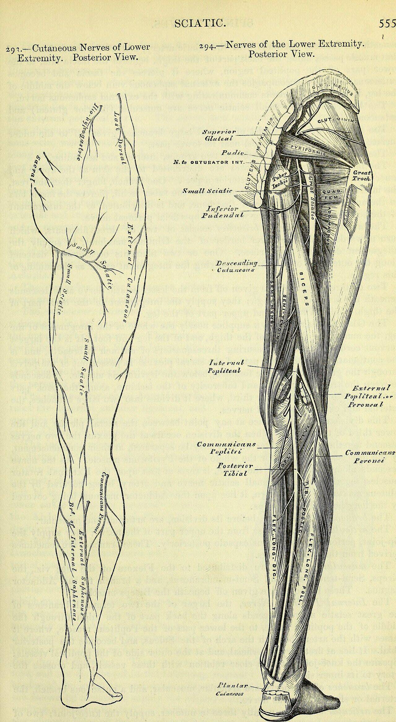


_(1860)_(14741895766).jpg)