Localize spinal cord lesions by tract and syndrome patterns, distinguish urgent mimics, and connect the neurologic examination to appropriate emergency imaging.
A cord syndrome is not a memorized silhouette. Predict which pathways should fail, where each pathway crosses, and whether findings at the lesion differ from those below it.
Track three pathways and their crossings
On a narrow screen, scroll the diagram sideways to read every label.
Trace the arrows from body or brain. Motor and position pathways cross in the medulla. Pain and temperature cross near the spinal entry level before ascending. Original non-scale teaching schematic.
Dorsal-column vibration and position fibers ascend ipsilaterally in the cord and cross in the medulla. Corticospinal fibers descend from cortex, cross in the caudal medulla, then travel ipsilaterally in the lateral cord to control the same side of the body below a spinal lesion.
Spinothalamic pain and temperature fibers enter the cord, travel a short distance, cross through the anterior white commissure, and then ascend contralaterally. This creates a small offset between a cord lesion and the first contralateral pain-temperature deficit. [1]
Damage at a cord segment can injure anterior horn cells or exiting roots, producing lower motor neuron signs at that level, while corticospinal injury produces upper motor neuron signs below. Immediately after severe cord injury, reflexes may be depressed during spinal shock before hyperreflexia appears. [2]
Translate a pathway into a bedside finding
Upper motor neuron signs include spasticity, brisk reflexes, and an extensor plantar response when the acute shock phase has passed. Lower motor neuron signs include reduced tone and reflexes, weakness, and possible denervation wasting or fasciculations. Neither weakness alone nor muscle size alone determines which system is injured. [1][2][11]
To test posterior-column function, compare vibration and the direction of a small joint-position change. Sensory ataxia becomes more apparent when visual compensation is removed: a patient may stand more steadily with eyes open than closed. A positive Romberg finding supports impaired sensory or vestibular input; it is not by itself a named spinal disease. [1][10]
Pain and temperature follow a different route from vibration and position. A cord lesion can therefore affect one set of modalities while sparing another. Light touch alone may miss that separation, so describing only "sensation present" can hide the information needed to identify the pathway. [1]
The cross-section and the crossing diagram answer different questions. The cross-section shows where pathways lie at one spinal level; the arrows show where information changes sides along the nervous system. Use both: place the lesion locally, then trace the affected information back to the body region it represents. [1]
Case 3
Show answer and explanations for case 3
A. Pain fibers cross in the medulla and descend two levels (Why this does not fit)
Do body pain-temperature fibers wait until the medulla to cross?
No; they decussate within the spinal cord shortly after entry.
What would a medullary crossing fail to explain here?
It would not account for the small segmental offset below the T10 lesion.
Read the full explanation
Body pain-temperature fibers cross within the spinal cord rather than waiting for the medulla.
B. Pain fibers enter, travel briefly, then cross through the anterior white commissure (Best answer)
What happens immediately after pain fibers enter the dorsal horn region?
They may travel briefly through nearby segments before synapsing.
Where do second-order fibers cross to the opposite side?
They cross through the anterior white commissure within the cord.
Why does left-sided loss begin around T12 after a right T10 lesion?
The right tract contains already-crossed left-sided input entering slightly below.
Read the full explanation
Short segmental travel before crossing makes contralateral loss begin slightly below the cord lesion.
C. Dorsal-column fibers cross twice inside the cord (Why this does not fit)
Which modalities belong to the dorsal columns instead?
Vibration and joint position ascend there on the same side.
Why is a double spinal crossing irrelevant to this case?
The lost modalities use the spinothalamic system, not the dorsal columns.
Read the full explanation
Dorsal-column fibers do not cross in the spinal cord and do not carry primary pain-temperature sensation.
D. The corticospinal tract carries contralateral pain (Why this does not fit)
What is the lateral corticospinal tract carrying at T10?
It carries descending motor commands after their medullary crossing.
Can interruption of that tract erase contralateral pain sensation?
No; pain-temperature information ascends in the anterolateral system.
Read the full explanation
The corticospinal tract is a descending motor pathway, not the pain-temperature pathway.
Takeaway: Spinothalamic fibers cross after short in-cord travel, so contralateral pain-temperature loss may begin below the lesion.
On a narrow screen, scroll the diagram sideways to read every label.
Compare dorsal, lateral and anterolateral pathways. Original conceptual cross-section, not patient imaging.
A lateral hemicord lesion causes ipsilateral weakness and loss of vibration or position below the lesion, plus contralateral pain-temperature loss beginning slightly below it. At-level segmental weakness can sit beside those long-tract findings.
Real injuries are often incomplete and asymmetric, so use the pattern as a localization model rather than demanding a perfect textbook triad. Formal complete versus incomplete spinal cord injury depends on sacral sensory and motor sparing, not merely on how many named tracts appear abnormal. [3]
Work one right-sided example from the crossings
Start with a right thoracic hemicord lesion. The descending motor fibers have already crossed in the medulla, so the right spinal pathway now serves the right side below. The right posterior column is carrying right-sided position and vibration information that has not yet reached its crossing. Both findings therefore stay on the lesion side. [1]
Now trace pain information from the left leg. Those fibers enter on the left, cross near their entry level, and ascend on the right. A right-sided spinal lesion can interrupt them after the crossing. That explains why the pain-temperature deficit is opposite to the weakness rather than making the examination internally inconsistent. [1]
Short ascending or descending travel before the pain fibers cross creates an offset that is often taught as roughly one or two segments. Do not use that approximation to calculate an exact structural lesion from a single pinprick line. Partial lesions, examination variability, and the distribution of damaged tissue can complicate the idealized pattern. [1][3]
A hemicord pattern describes the affected pathways, not the cause. Penetrating injury is a familiar example, but focal demyelination and other structural lesions can create related patterns. The history and imaging determine why those pathways were affected and what management is required. [1][14]
Sacral sparing is a separate question. Preserved S4-S5 sensation, deep anal pressure, or voluntary anal contraction affects formal completeness classification. A patient can have profound limb weakness yet remain neurologically incomplete. This lesson introduces that principle; formal grading requires the complete standardized examination. [3]
Case 6
Show answer and explanations for case 6
A. Anterior cord syndrome (Why this does not fit)
What modality should an anterior cord syndrome relatively preserve?
Dorsal-column vibration and position are often spared by an anterior pattern.
Which current finding directly contradicts that expectation?
The right toe-position deficit confirms ipsilateral dorsal-column involvement.
Read the full explanation
Anterior cord syndrome is usually bilateral and preserves dorsal-column modalities rather than losing them ipsilaterally.
B. Posterior cord syndrome (Why this does not fit)
Which deficit could posterior cord injury explain?
It could produce the right-sided loss of toe position.
Which two findings require additional lateral tract damage?
Right spastic weakness and left pain-temperature loss extend beyond posterior columns.
Read the full explanation
Posterior cord syndrome does not explain ipsilateral weakness plus contralateral pain-temperature loss.
C. Right hemicord syndrome (Best answer)
Why is the weakness on the side of the cord wound?
Corticospinal fibers already crossed in the medulla before descending on the right.
Why is toe-position loss also right-sided?
Dorsal-column fibers remain ipsilateral until they reach the medulla.
Why is pain-temperature loss found on the left?
Spinothalamic fibers from the left have crossed into the right cord.
What explains its onset below the wound level?
Short segmental travel before crossing creates the lower sensory offset.
Read the full explanation
Ipsilateral motor and dorsal-column loss with contralateral pain-temperature loss is the expected hemicord pattern.
D. Cauda equina syndrome (Why this does not fit)
What motor signs would cauda equina roots produce?
They would cause lower motor neuron weakness with reduced reflexes.
Why do crossed sensory findings oppose that location?
Separate roots cannot create ipsilateral dorsal loss plus contralateral pain loss.
Read the full explanation
Cauda equina disease produces root-level lower motor neuron findings, not crossed long-tract deficits.
Takeaway: Hemicord injury combines ipsilateral motor and dorsal-column loss with contralateral pain-temperature loss below.
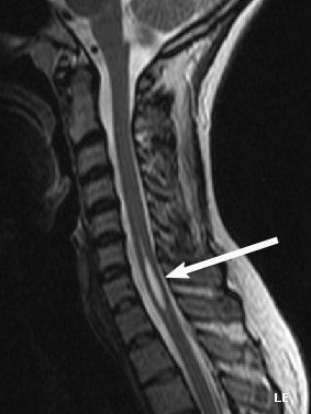
The source identifies this cervical MRI finding as syringomyelia. The arrow marks the cavity inside the spinal cord. An image alone does not establish the cause or predict an individual patient's examination.
Cyborg Ninja; arrow added by SUM1. Original image. CC BY 4.0. Original image retained without modification.
Traumatic central cord syndrome often presents after cervical injury with upper-limb motor impairment greater than lower-limb impairment, but simple medial-arm and lateral-leg corticospinal lamination is not an established explanation. Diagnose the clinical syndrome and structural injury without teaching that disputed map as fact. [4]
A syrinx is a fluid-filled spinal cord cavity that often evolves gradually. It can interrupt crossing pain-temperature fibers and anterior horn cells, causing segmental dissociated sensory loss and hand weakness, but presentations vary and MRI defines the cavity. [5]
Traumatic central cord does not require a cape-shaped sensory deficit, and a syrinx is not simply a chronic version of traumatic central cord. Time course, motor distribution, at-level findings, and imaging separate them.
Predict what an enlarging cavity can reach
In the small-cavity model, the first structure highlighted is the crossing zone near the central canal. Interrupting pain-temperature fibers there can produce loss on both sides at the affected segments, with relatively preserved vibration and position. Dissociated sensory loss means that some modalities are impaired while others remain available. [1][5]
A cervical cavity may create a shawl-like distribution across the upper trunk or arms, but the affected levels and extent determine the actual pattern. If it extends into anterior horn cells, weakness and wasting can develop in muscles supplied at those segments. More extensive disease can affect long pathways and function below the cavity. [1][5]
Syringomyelia can occur with altered cerebrospinal-fluid flow, including Chiari malformation, and can also follow spinal injury or accompany a tumor. The association is not a diagnosis from the skin pattern alone. MRI shows the cavity and helps investigate the anatomy responsible for it. [5]
In traumatic central cord syndrome, an older person with cervical narrowing may develop much greater hand and arm weakness than leg weakness after a hyperextension injury. Bladder symptoms and sensory findings vary. The absence of a fracture on an initial study does not dismiss a neurologic deficit; appropriate trauma assessment and cord imaging remain important. [4][17]
The useful comparison is therefore acute traumatic motor pattern versus an intramedullary cavity. Do not assign the same simple tract diagram to both. A model that explains crossing-fiber injury from a syrinx should not be presented as proof of a disputed mechanism for every arm-predominant traumatic injury. [4][5]
Recognize anterior and posterior patterns
The anterior spinal artery supplies much of the anterior two-thirds of the cord. It arises from vertebral-artery branches and is reinforced by radiculomedullary arteries, including the artery of Adamkiewicz, rather than originating directly from the aorta. [6]
Anterior cord ischemia classically causes abrupt bilateral motor and pain-temperature loss with relative preservation of vibration and position. Posterior cord ischemia is less common and more strongly affects proprioception and vibration; real vascular lesions can be incomplete. Sudden spinal deficits demand urgent imaging and vascular evaluation. [7]
Use the preserved modality as an anatomical control
Consider abrupt postoperative paraplegia with absent pinprick but preserved toe position. The preserved position sense is not an incidental detail: it argues that posterior sensory pathways are working better than the motor and anterolateral systems. That contrast supports an anterior-cord pattern, while the timing directs attention to ischemia or another acute complication. [6][7]
The anterior spinal artery runs along the ventral cord and receives reinforcement from segmental vessels. Aortic disease or surgery can compromise important feeders or perfusion without the anterior spinal artery literally arising from the aorta. This distinction connects the clinical association to the correct anatomy. [6]
Posterior spinal circulation supports the posterior cord. A posterior-column-predominant lesion may leave strength and pain-temperature sensation relatively intact while producing severe sensory imbalance. A posterior pattern is not the same as B12-related combined degeneration, which can additionally involve lateral motor pathways. [6][8]
Avoid using a syndrome name as a guaranteed prognosis. Outcomes depend on the level, severity, cause, extent of spared function, and subsequent care. An abrupt unexplained cord deficit requires urgent investigation even when its examination is incomplete or an initial image is not yet definitive. [6][7]
Test disorders that imitate tract patterns
Vitamin B12 neurologic disease can affect posterior and lateral columns even without anemia or macrocytosis. Methylmalonic acid can support an indeterminate diagnosis, but renal impairment can raise it, so it is an adjunct rather than a universal definitive test. [8][9]
Tabes dorsalis favors dorsal-root and posterior-column dysfunction with lightning pains and sensory ataxia. ALS can combine upper and lower motor neuron signs with relative sensory preservation, but that combination is not pathognomonic and cognitive or behavioral changes can occur. SMA and poliomyelitis are lower motor neuron disorders, while multiple sclerosis can produce central demyelinating lesions with sensory, optic, brainstem, or cord findings. [10][11][12][13][14]
Separate combined disease from a pure posterior pattern
Subacute combined degeneration links posterior-column dysfunction with lateral corticospinal involvement. Impaired position sense can make walking in darkness difficult, while corticospinal disease can add stiffness, brisk reflexes, or extensor plantar responses. Associated peripheral neuropathy can reduce ankle reflexes even when knee reflexes are brisk. [8][9]
Ask about impaired B12 intake or absorption, autoimmune gastritis, prior gastric surgery, and relevant medicines or exposures. Dietary history alone does not settle the mechanism, and folate deficiency does not explain the same classic neurologic pattern. Current B12 assessment uses clinical findings and appropriate laboratory testing rather than the historical Schilling test as a routine first step. [8][9]
Tabes dorsalis affects dorsal sensory structures and roots in late neurosyphilis. Lightning pains, impaired position sense, sensory ataxia, and reduced reflexes fit that combination. Argyll Robertson pupils can show a near response with an impaired light response, and loss of protective sensation may accompany neuropathic joint damage. These findings need the relevant clinical evaluation, not a diagnosis based on one pupil or gait sign. [10]
ALS, poliomyelitis, and SMA all involve motor neurons, but the surrounding pattern differs. ALS can combine upper and lower motor neuron findings; poliomyelitis is classically an asymmetric lower motor neuron illness; SMN1-related SMA is a genetic motor neuron disorder. Preserved sensation is useful in these comparisons, but mixed motor findings still require exclusion of mimics such as structural cervical disease. [11][12][13]
In multiple sclerosis, a spinal plaque may be one event among attacks affecting different parts of the central nervous system. Prior optic neuritis or a brainstem syndrome changes the interpretation of a focal cord pattern. An isolated symptom such as neck-flexion tingling is not specific enough to establish MS, and a short illustrative case does not replace formal diagnostic criteria. [14]
Localize while protecting the patient
On a narrow screen, scroll the diagram sideways to read every label.
A lesion can injure motor neurons or roots at its own level and long pathways serving levels below. Acute spinal shock can temporarily mask the expected upper motor neuron pattern. Original non-scale teaching schematic.
Conus medullaris and cauda equina syndromes overlap. Saddle sensory change, bladder or bowel dysfunction, bilateral leg symptoms, or progressive weakness warrants emergency imaging for compression rather than reassurance based on a supposedly gradual course. [15]
In suspected traumatic injury, protect the spine, address airway, breathing, and circulation, document the neurologic examination, and obtain urgent imaging under the local trauma pathway. In adults, CT evaluates suspected bony injury; MRI follows when a neurologic abnormality may reflect cord injury even if CT is unrevealing. [16][17]
An anatomic cord level and a vertebral level are not interchangeable. Cord segments sit progressively rostral to same-numbered lower vertebrae, so communicate whether a label came from examination, vertebral imaging, or formal neurologic classification.
Communicate the level, the pattern, and the immediate risk
In adults, the cord usually ends around the L1-L2 vertebral region as the conus medullaris. Below it, descending cauda equina roots continue toward their exit levels. A lesion near the cord ending can involve both neural structures, which helps explain why conus and cauda syndromes do not always divide into neat separate boxes. [1][15]
A root-predominant cauda presentation may have severe radicular pain and asymmetric flaccid weakness; a conus presentation may be more symmetric with early sacral dysfunction. These are tendencies, not permission to delay care when the opposite pattern appears. New retention, altered saddle sensation, or progressive leg deficits warrants urgent assessment for compression. [15]
Spinal shock and neurogenic shock are not synonyms. Spinal shock refers to transient loss or depression of neurologic reflex activity after acute cord injury. Neurogenic shock concerns circulatory failure from loss of sympathetic control, often with hypotension and relative bradycardia after higher injuries. They can coexist, and other causes of shock still require assessment. [2][16]
During traumatic evaluation, stabilization comes before an elaborate localization exercise. Document motor function, sensory modalities, sacral findings when appropriate, and changes over time. A patient whose CT does not explain new neurologic findings may still need MRI for cord injury; a normal bony study is not a normal neurologic examination. [16][17]
For a handoff, say what was observed: "right leg weakness and position loss with left-sided pain-temperature loss below a thoracic level" is more informative than an unsupported exact vertebral number. Then add the time course, imaging findings, and urgent concern. This preserves the reasoning without making the examination more precise than it really was. [1][3]
Case 25
Show answer and explanations for case 25
A. Symmetry proves conus medullaris syndrome (Why this does not fit)
Can symmetric symptoms occur with cauda equina compression?
Yes; extensive root compression can affect both sides.
What does symmetry legitimately contribute?
It may suggest conus involvement but cannot prove it.
Read the full explanation
Symmetry can suggest conus involvement but cannot reliably separate conus from cauda equina.
B. Reduced reflexes prove cauda equina syndrome (Why this does not fit)
Can conus disease reduce lower-limb reflexes?
Yes; conus lesions may involve segmental motor neurons or nearby roots.
Why do reduced reflexes fail to settle the distinction?
Conus and cauda syndromes share lower motor neuron features.
Read the full explanation
Conus and cauda findings overlap, and acute cord-related states can also reduce reflexes.
C. Conus and cauda syndromes overlap, so urgent MRI is required (Best answer)
Which findings make this an emergency before exact localization?
Urinary retention, saddle numbness, and bilateral weakness signal compressive neural dysfunction.
Can bedside symmetry reliably separate conus from cauda?
No; their bedside patterns frequently overlap in acute presentations.
What investigation should follow immediately?
Urgent MRI is required to locate a treatable compression.
Read the full explanation
Bladder dysfunction, saddle loss, and bilateral weakness are compression red flags regardless of an imperfect bedside distinction.
D. A gradual course is required before imaging (Why this does not fit)
Must compressive conus or cauda symptoms develop gradually?
No; disc herniation or other compression may present abruptly.
What would waiting for a gradual course risk?
Delay could permit progression of bladder and motor injury.
Read the full explanation
Compression can be sudden or progressive and should not wait for a gradual course.
Takeaway: Conus and cauda equina findings overlap; bladder, saddle, and bilateral leg deficits require urgent imaging.
For each patient, select the localization or interpretation that explains the tract pattern, time course, and level-specific findings. Each option includes a case-specific explanation.
Case 1
Show answer and explanations for case 1
A. Corticospinal fibers cross in the caudal medulla before descending in the cord (Best answer)
Where do the major lateral corticospinal fibers decussate?
They cross at the pyramidal decussation in the caudal medulla.
On which side do they descend after entering the cord?
After crossing, the fibers descend ipsilateral to the muscles they control.
What follows from interrupting the right tract at cervical level?
The right body develops upper motor neuron weakness below the lesion.
Read the full explanation
After crossing in the caudal medulla, corticospinal fibers descend ipsilaterally, so a right cord lesion weakens the right body below.
B. Corticospinal fibers first cross at each spinal segment (Why this does not fit)
Do major corticospinal fibers wait for each spinal segment to cross?
No; their principal decussation has already occurred in the caudal medulla.
Which pathway more closely fits short in-cord segmental crossing?
Body pain-temperature fibers cross through the anterior white commissure.
Read the full explanation
The major corticospinal crossing occurs in the caudal medulla, not separately at each spinal level.
C. Corticospinal fibers remain uncrossed from cortex to muscle (Why this does not fit)
Can the observed right-sided weakness prove that motor fibers never cross?
No; a prior medullary crossing also produces ipsilateral cord findings.
What established anatomy contradicts the uncrossed proposal?
Most corticospinal fibers switch sides before descending through the lateral cord.
Read the full explanation
Most corticospinal fibers cross before entering the lateral spinal cord.
D. Corticospinal fibers cross in the dorsal columns (Why this does not fit)
What modalities travel in the dorsal columns?
They chiefly carry vibration and conscious position sensation toward the medulla.
Why can their crossing not explain this weakness?
They are ascending sensory pathways rather than descending motor fibers.
Read the full explanation
Dorsal columns carry vibration and position, not the primary descending motor pathway.
Takeaway: Corticospinal fibers cross in the caudal medulla, then a spinal lesion causes ipsilateral upper motor neuron signs below.
A. The findings are compatible with ALS but are not pathognomonic (Best answer)
Which findings demonstrate lower motor neuron involvement?
Hand atrophy and fasciculations indicate active denervation of lower motor neurons.
Which findings simultaneously demonstrate upper motor neuron involvement?
Brisk knees and extensor plantar responses indicate corticospinal dysfunction.
Does relative sensory preservation uniquely identify ALS?
It supports a motor-system disorder but is not specific to ALS.
How should the mild executive and behavioral change affect interpretation?
Frontotemporal cognitive or behavioral impairment can coexist with ALS.
Read the full explanation
Mixed upper and lower motor neuron signs with relative sensory preservation support ALS, while cognitive or behavioral changes may coexist.
B. Cognitive change excludes ALS (Why this does not fit)
Can ALS involve cognition or behavior?
Yes; frontotemporal cognitive and behavioral impairment can accompany ALS.
What does the mild executive change therefore do diagnostically?
It remains compatible rather than excluding the motor neuron syndrome.
Read the full explanation
ALS can coexist with frontotemporal cognitive or behavioral impairment.
C. Atrophy proves a pure lower motor neuron disorder (Why this does not fit)
What does atrophy establish by itself?
It shows muscle volume loss but does not identify a single mechanism.
Which findings disprove a purely lower motor neuron pattern?
Brisk knees and extensor plantar responses provide clear upper motor neuron signs.
Read the full explanation
ALS combines lower motor neuron denervation with upper motor neuron signs, and disuse can also contribute to atrophy.
D. The pattern proves a spinal hemicord lesion (Why this does not fit)
What sensory geometry should a hemicord lesion create?
It should produce a spinal level with side-specific tract asymmetry.
Which features instead favor diffuse motor system disease?
Mixed motor neuron signs occur with relatively preserved sensation and no level.
Read the full explanation
A hemicord lesion should create a level and sensory tract asymmetry, which are absent.
Takeaway: ALS can combine upper and lower motor neuron signs with relative sensory preservation, but the pattern is not unique and cognition may change.

.png)