Trace neonatal airflow, localize nasal obstruction, prioritize ventilation, distinguish airway lesions, and plan repair and follow-up for choanal atresia.
A newborn becomes blue while sucking, then pinker during a cry. Before asking which scan will confirm the diagnosis, ask which route still lets air reach the lungs. Bilateral nasal obstruction can make feeding dangerous. A temporary improvement during crying is an observation, not a secure airway. [1]
Explain the route that can ventilate the infant, distinguish anterior from posterior obstruction, decide when respiratory support must precede investigation, and plan surveillance after repair.
Newborns strongly favor nasal breathing, especially while sucking. Calling them obligate nasal breathers is a useful warning, but it should not imply that oral airflow is impossible. Their ability to compensate varies. The classic alternating pattern follows airway access: a closed or occupied mouth leaves the infant dependent on the nose; crying opens an oral route. Bilateral choanal atresia blocks the posterior nasal exits into the nasopharynx. Air entering either nostril cannot reach the pharynx normally. [1]
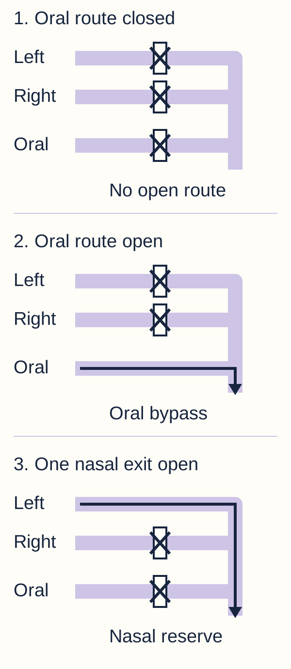
In the route diagram, trace the nasal and oral paths until they join. First cover the oral inlet with your finger. With both posterior nasal exits closed, neither route supplies effective airflow. Then uncover the oral inlet and trace a path that avoids the nasal plates. The obstruction has not disappeared; a different entrance is available. This explains why crying can improve color without curing the problem. The drawing represents airflow, not a ventilation device or an instruction to provoke crying.
Trace Left and Right as nasal routes, then Oral as the separate mouth route. Cover the oral inlet and compare which nasal routes still reach the pharynx. No route is dependable unless the infant ventilates effectively. This is an airflow model, not a device diagram. [1]
Mouth occupied during a feed: nasal blockage limits breathing, sucking competes with ventilation, and oxygenation can deteriorate.
Mouth open during a cry: oral airflow can bypass the nose and oxygenation may improve. Exhaustion, apnea or additional airway disease can still defeat this compensation.
Predict what changes when only one posterior exit is blocked. A patent opposite side can still supply nasal airflow, so a stable infant may feed and grow without neonatal cyclic cyanosis. Later unilateral congestion or persistent discharge may be the first recognized problem. Conversely, new swelling or secretions in the previously patent side can reduce this reserve. Laterality describes the anatomy; current airflow determines urgency. [1]
Check the airflow prediction
The oral route bypasses both anterior and posterior nasal obstruction. Improvement with crying therefore supports a nasal-level problem but does not identify its exact site. Neither absent cyclic cyanosis nor transient oral compensation excludes serious obstruction. Confirm the location after supporting ventilation. [1][4]
Find the obstruction, then map the anatomy
Does a catheter that stops in a nostril prove choanal atresia? No. The choana is the posterior opening, not the external nostril. Atresia reflects abnormal development of that connection. Bony obstruction, membranous obstruction and combinations have been described. Mixed bony and soft-tissue obstruction is common. A precise anatomical map is more useful than memorizing a contested percentage. [1][7]
A trained clinician can gently assess both nasal passages with an appropriately sized soft catheter after addressing accessible secretions. Record which side is affected and where resistance occurs. Secretions, a deviated septum, turbinates, anterior narrowing or technique can prevent passage. Do not repeatedly force an instrument through resistance. A catheter screen assesses passage; it does not identify tissue composition or justify blind perforation. [1]
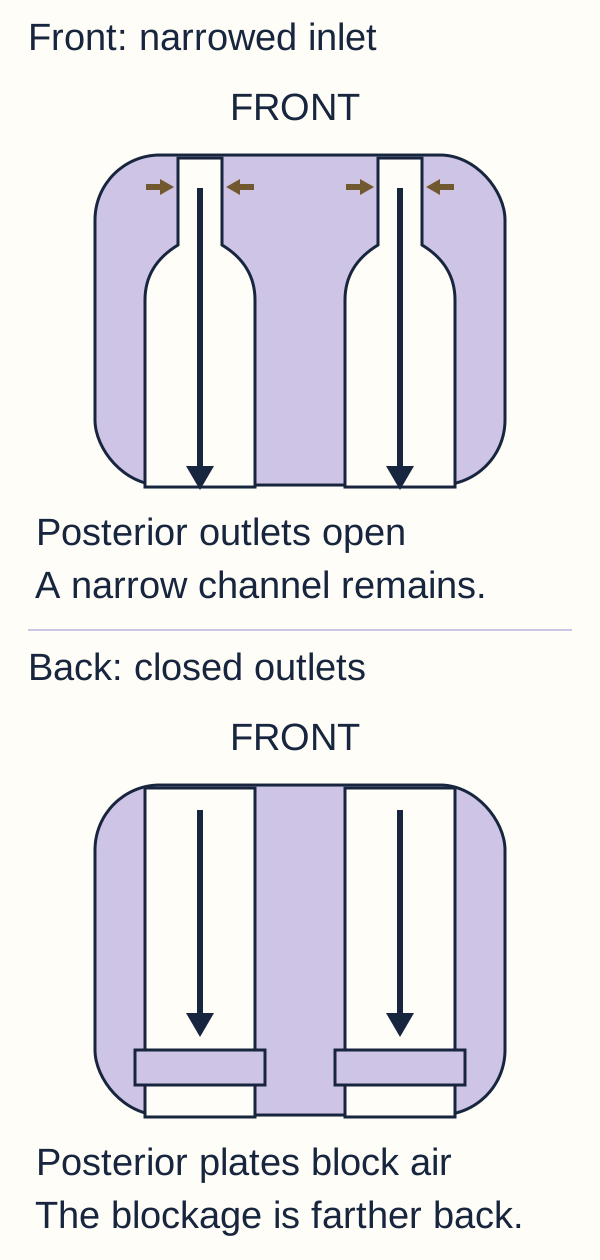
Compare the front and back of the nasal cavity in the localization diagram. Resistance near the inlet suggests an anterior problem. Passage along the nasal floor followed by posterior resistance suggests a choanal-level problem. Depth is approximate and depends on patient size and technique. Flexible nasal endoscopy, performed by the airway team, directly examines the level and character of the blockage. A small catheter passing does not exclude clinically important partial narrowing. [1][4]
Compare the position of the restriction, not just the breathing symptoms. The upper model retains narrow anterior channels and open posterior outlets; the lower model has patent inlets but closed posterior exits. Sizes are not diagnostic measurements. [1][4]
Now predict which test answers a different question. Once ventilation is reliable, CT of the nasal cavity and relevant facial/skull-base anatomy defines the bony boundaries for surgery. The posterior vomer and medial pterygoid region may be thickened; the atretic plate narrows or closes the posterior opening. CT helps distinguish anterior from posterior narrowing and characterize the operative anatomy. It does not treat hypoxemia and should not delay airway support or justify unsafe transport. Endoscopy and CT are complementary. [1][2]
Retained mucus can resemble a soft-tissue abnormality on CT. Appropriate nasal preparation and correlation with endoscopy prevent an image from being interpreted without context. An unusual mass or concern about skull-base continuity needs specialist-directed imaging rather than traumatic probing. In a stable unilateral case with a clear endoscopic diagnosis, the timing of CT can be coordinated with surgery to avoid unnecessary repeated radiation. [1]
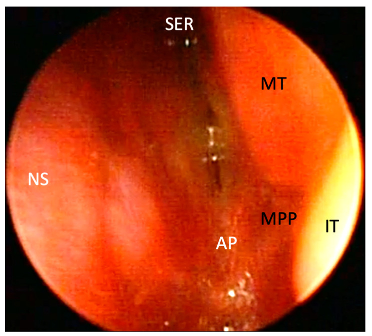
In the clinical endoscopic photograph, locate AP, the atretic plate, between the nasal septum labeled NS and the lateral structures. Predict whether air can reach the nasopharynx through a posterior outlet that is closed by this tissue. The visible plate prevents normal posterior passage. The photograph shows the left cavity of a neonate reported to have bilateral disease; this one view alone does not establish the opposite side. [1]
Locate AP between the septal and lateral structures. This is the left cavity of a three-day-old neonate reported to have bilateral bony choanal atresia. This single view alone cannot establish the other side. Figure 2 from Urbancic and colleagues, Children 2023, 10(1):91, used unchanged under CC BY 4.0. Image: Jure Urbančič and colleagues; original source; CC BY 4.0.Check the test sequence
Localize gently, visualize the obstruction, and map the anatomy when the airway is safe. When secretions are cleared and passage returns, reassess before diagnosing a fixed plate. When posterior obstruction persists, obtain direct visualization and the required preoperative imaging. [1][2]
Similar symptoms can start at different levels
A baby with a small jaw and a baby with blocked posterior nasal exits may both struggle to feed. Which finding separates them? Use the site of resistance, the sound of breathing, the oral examination and direct airway visualization together. A single feeding symptom or response to crying cannot settle the diagnosis. [1][4][5][6]
Compare the suspected site with the finding that tests it
Site
Pattern
Useful discriminator
SiteAnterior nasal inlet
PatternPyriform aperture stenosis narrows the bony entrance.
PatternLaryngomalacia produces inspiratory collapse above the vocal folds.
Useful discriminatorInspiratory stridor and dynamic findings during flexible laryngeal examination.
Pyriform aperture stenosis may produce apnea, feeding difficulty and cyanosis just as posterior nasal obstruction can. The key distinction is where the nasal passage is narrow, not whether the child becomes pinker with an open mouth. Management depends on severity and specialist assessment; do not operate on the posterior choana to correct an anterior inlet lesion. [4]
In Robin sequence, micrognathia reduces space for the tongue and glossoptosis compromises the pharyngeal airway. A cleft palate is common but is not required to recognize the jaw-tongue relationship. Improvement with professionally supported jaw or tongue positioning suggests that level of obstruction. Any therapeutic positioning in an affected neonate requires an individualized airway-team plan and monitoring; this is not advice for unsupervised prone sleep. [5]
Laryngomalacia is dynamic rather than a fixed posterior nasal plate. Symptoms can become more evident during feeding or when supine. Severe respiratory effort, apnea, cyanosis or poor growth requires prompt evaluation rather than reassurance based on the name alone. Patent nasal passages do not establish that the rest of the airway is safe. [6]
Apply the comparison to an infant with easy bilateral nasal catheter passage but persistent inspiratory noise. Shift attention toward the larynx rather than repeating nasal instrumentation. Conversely, a visibly narrow anterior inlet with patent posterior openings directs attention to the pyriform aperture. Nasal edema, secretions, septal deviation and masses remain alternatives. Persistent cyanosis despite effective ventilation also requires assessment for pulmonary, cardiac or other illness. Several abnormalities may coexist. [1][4][6]
Support breathing before seeking a perfect diagnosis
Would a more detailed CT image help a newborn who is becoming exhausted? Not before oxygenation and ventilation are supported. Call neonatal and pediatric airway expertise, stop oral feeding during distress, monitor the infant, and establish an effective route for ventilation. Gentle clearance of accessible secretions and oxygen may assist, but oxygen delivered against a complete nasal obstruction is not a substitute for airflow. [1]
An appropriately selected oral airway or a specialist-managed McGovern-type nipple can maintain an oral route temporarily in a spontaneously breathing infant. The latter is an airway aid, not an ordinary feeding nipple, a bottle-feeding test or a home improvisation. Its presence does not prove adequate ventilation. Observe respiratory effort, chest expansion, oxygenation and clinical trajectory while the team organizes definitive care. [1]
Breathing effectively through an oral route: continue close monitoring and specialist airway planning. The nasal lesion still needs assessment and treatment.
Persistent hypoxemia, ineffective breaths, apnea or exhaustion: provide assisted ventilation and escalate to an appropriately secured airway, including endotracheal intubation when required. Do not wait for imaging.
Reliable ventilation established: complete endoscopic assessment, necessary imaging and associated-anomaly evaluation without abandoning airway surveillance.
Consider a newborn whose color briefly improves when the mouth is open but who then becomes limp with little chest expansion. Name the variable that has changed: spontaneous ventilation is no longer dependable. Holding the mouth open cannot generate breaths. The trained resuscitation team must support ventilation, verify its effectiveness and secure the airway as needed. An oral airway is a bridge, not a reason to defer escalation. Nasal-only pressure support does not bypass complete bilateral posterior obstruction. [1]
Do not test the diagnosis by repeatedly feeding a distressed infant. Sucking competes with breathing and can add aspiration risk. After stabilization, select a nutrition route with the neonatal and feeding teams; a tube intended to pass through an atretic nostril will not solve the problem. Return to oral feeding only after airway reliability and feeding safety have been assessed. For persistent problems, consider an additional swallowing or airway disorder rather than assuming nasal repair is sufficient. [1][3]
Check the immediate priority
A planned scan, genetic test or operation is not the next priority when ventilation is failing. Support effective breaths first. A normal saturation during one brief quiet observation does not establish stability across sleep and feeding. [1]
Use physiology to judge timing
Should every infant with an atretic choana have emergency surgery? Compare two patients rather than treating the anatomical label as a clock. An infant with bilateral obstruction who needs an oral airway to breathe has a different immediate risk from a thriving infant with one patent nasal passage. Recurrent desaturation, respiratory exhaustion and unsafe feeding take priority over the convenience of a scheduled investigation. [1][2]
After stabilization, bilateral choanal atresia generally needs early repair by the specialist team. Associated cardiac disease, prematurity, additional airway lesions and the infant's ability to maintain ventilation affect preparation and timing. Stabilization is not a reason to discharge an infant who still depends on a tenuous temporary airway. A secure transport and monitoring plan is needed when transfer is required. [1]
A stable unilateral case can often be managed with planned evaluation and elective repair. The international consensus recommends delaying unilateral repair until at least six months of age when feasible. This is not a promise that every infant can safely wait or an instruction to ignore impaired feeding, growth or breathing. Apply the recommendation to the patient's functional airway and associated problems, then arrange follow-up. [2]
Transnasal endoscopic repair is generally preferred. It restores the posterior connection while allowing direct visualization of the surgical field. The desired result is a durable, adequately sized opening, not simply a temporary hole. A repaired opening can become narrowed by crust, granulation or scar. Anatomy, associated conditions, tissue handling and aftercare all matter; one successful early examination does not guarantee lifelong patency. [1][2]
Predict the tradeoff of placing a stent across the new opening. It may support early patency, but contact with tissue can cause irritation, ulceration or granulation, and the device itself can become obstructed. Evidence and protocols do not support teaching one universal stent policy. Retrospective comparisons can be confounded when more complex cases are preferentially stented; equal observed rates alone do not prove equal treatment effects. [7] Selection, duration, cleaning and any change in the plan belong to the operating team. Do not convert a particular center's regimen for stents or postoperative medication into a rule for every patient. [1]
Compare the two timing decisions
Stable unilateral anatomy permits planning because another nasal route functions. Bilateral dependence on an oral bridge calls for early definitive care after stabilization. New desaturation changes the assessment regardless of which label was assigned earlier. [1][2]
Check the child beyond the repaired opening
Does opening the choana finish the evaluation? No. Choanal atresia may be isolated or accompany CHARGE and other craniofacial or genetic disorders. CHARGE describes a pattern involving coloboma, heart defects, choanal atresia, growth and developmental concerns, genital differences, and ear or hearing abnormalities. Not every feature is present in every child. Do not wait for all six features, or for poor growth to become apparent, before arranging an appropriate assessment. [1][3]
Compare a repaired infant who coughs during every feed with one whose nasal breathing becomes progressively obstructed between feeds. Both need evaluation, but they may have different problems. Cranial nerve dysfunction in CHARGE can impair swallowing and airway protection despite patent choanae. Ear structure and hearing, eye findings including internal coloboma, congenital heart disease, growth and endocrine concerns, and the wider phenotype guide multidisciplinary evaluation. Genetics consultation can guide CHD7 testing and assessment of alternative diagnoses. A negative family history does not exclude a usually de novo disorder. [1][3]
Cardiac assessment matters before anesthesia because a heart defect can change perioperative planning. Audiology and ophthalmology assess functions that a quick external examination may miss. Feeding and swallowing evaluation addresses aspiration and nutrition, while neonatal, airway and genetics teams coordinate other investigations. An urgent airway intervention should not wait for the full syndrome workup. A stable child still deserves planned screening rather than a diagnosis based only on appearance. [1][3]
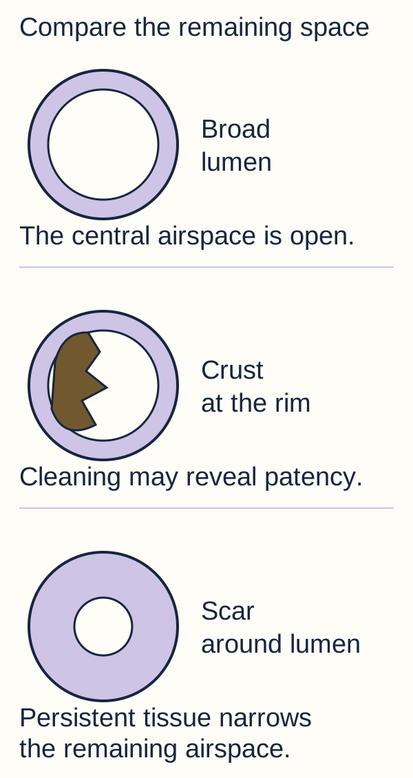
Study the postoperative lumen diagram. Trace the open central space, then compare a narrowing caused by adherent crust with a narrowing caused by circumferential scar. Similar symptoms do not prove identical treatment needs. New congestion, discharge, difficulty feeding or recurrent desaturation should prompt assessment of airway safety and direct visualization of patency. Apnea, severe distress or cyanosis needs emergency care, not a routine future clinic slot. Endoscopy can identify crust, granulation and recurrent narrowing; routine repeated CT is not the default surveillance method. [1][2]
Predict what remains after gentle clinician-directed cleaning. Crust can conceal a patent opening, whereas persistent tissue narrowing needs a different assessment. These are simplified models, not endoscopic photographs or measured operative results. [1]
Postoperative care commonly includes saline and gentle cleaning or suction according to the surgical team's protocol, with debridement or further treatment when indicated. Families need explicit instructions about device care when a stent is present and clear escalation advice. Do not advise forceful home probing or independent stent changes. The international consensus recommends at least a year of endoscopic follow-up; the schedule is individualized and may continue longer. Assess breathing during sleep and feeding as well as the appearance of the opening. [1][2]
Apply the follow-up distinction
Patent choanae with coughing during feeds directs attention to swallowing and possible aspiration. A progressively smaller posterior lumen with obstruction between feeds supports recurrent stenosis. Persistent inspiratory stridor despite nasal patency directs attention to an additional laryngeal problem. Reassess the affected function rather than assuming every symptom is recurrent atresia. [1][3][6]
Apply the lesson
Case 1
Show answer and explanations for case 1
A. Obtain urgent CT before selecting an airway procedure. (Why this does not fit)
CT maps the structural obstruction for operative planning. Minimal chest expansion and poor responsiveness indicate failing ventilation; a diagnostic image cannot supply breaths.
Reasoning steps for option A
What information does CT provide?
CT maps the structural obstruction for operative planning.
What prevents imaging from being the immediate priority?
Minimal chest expansion and poor responsiveness indicate failing ventilation; a diagnostic image cannot supply breaths.
B. Repeat bilateral catheter passage after nasal saline. (Why this does not fit)
Clearing accessible secretions can distinguish a reversible blockage from persistent resistance. The infant is no longer ventilating effectively through an available oral route, so repeating the screen delays respiratory support.
Reasoning steps for option B
When can gentle nasal preparation be informative?
Clearing accessible secretions can distinguish a reversible blockage from persistent resistance.
What has changed beyond nasal patency in this infant?
The infant is no longer ventilating effectively through an available oral route, so repeating the screen delays respiratory support.
C. Assist ventilation and secure the airway as required. (Best answer)
An open oral route is not producing effective breaths. The resuscitation team must provide effective assisted ventilation and secure an appropriate airway, including intubation when required, before imaging.
Reasoning steps for option C
What does minimal chest expansion indicate?
An open oral route is not producing effective breaths.
What follows when spontaneous ventilation fails?
The resuscitation team must provide effective assisted ventilation and secure an appropriate airway, including intubation when required, before imaging.
D. Continue oral airway observation until ENT examination. (Why this does not fit)
It can help a spontaneously breathing infant bypass nasal obstruction under close monitoring. Shallow breaths and reduced responsiveness show that the temporary bridge is failing.
Reasoning steps for option D
When can an oral airway serve as a temporary bridge?
It can help a spontaneously breathing infant bypass nasal obstruction under close monitoring.
Why is observation alone insufficient here?
Shallow breaths and reduced responsiveness show that the temporary bridge is failing.
Takeaway: A patent oral entrance cannot compensate for absent or ineffective respiratory effort.
A. Plan elective repair with anatomical assessment. (Best answer)
The opposite choana is patent and breathing and feeding remain effective. Persistent unilateral atresia requires specialist planning and appropriate preoperative imaging rather than indefinite dismissal.
Reasoning steps for option A
What supports elective rather than emergency treatment?
The opposite choana is patent and breathing and feeding remain effective.
What still needs attention despite clinical stability?
Persistent unilateral atresia requires specialist planning and appropriate preoperative imaging rather than indefinite dismissal.
B. Arrange immediate intubation followed by emergency repair. (Why this does not fit)
An infant unable to oxygenate or ventilate because of bilateral obstruction needs urgent support. Normal oxygenation, uninterrupted feeds and a patent left outlet do not support emergency intubation.
Reasoning steps for option B
Which presentation can require immediate airway intervention?
An infant unable to oxygenate or ventilate because of bilateral obstruction needs urgent support.
Do the observed functions show that problem?
Normal oxygenation, uninterrupted feeds and a patent left outlet do not support emergency intubation.
C. Treat rhinitis alone and defer structural follow-up. (Why this does not fit)
Rhinitis can produce congestion and secretions. Direct visualization has established a fixed closed posterior outlet.
Reasoning steps for option C
What might explain isolated nasal discharge without an anatomical lesion?
Rhinitis can produce congestion and secretions.
What finding requires more than rhinitis treatment?
Direct visualization has established a fixed closed posterior outlet.
D. Schedule repeat CT surveillance without surgical consultation. (Why this does not fit)
CT defines anatomy when needed for diagnosis and operative planning. It neither resolves the fixed obstruction nor replaces specialist planning and functional follow-up.
Reasoning steps for option D
What is the useful role of CT in atresia?
CT defines anatomy when needed for diagnosis and operative planning.
Why is serial imaging alone inadequate?
It neither resolves the fixed obstruction nor replaces specialist planning and functional follow-up.
Takeaway: A patent opposite passage changes urgency but does not erase a fixed unilateral lesion.
A. Posterior vomer and atretic choanal plates. (Why this does not fit)
It would interrupt the posterior connection from nasal cavity to nasopharynx. Both posterior connections are open; the described narrowing is at the maxillary inlet.
Reasoning steps for option A
Where would a choanal plate obstruct airflow?
It would interrupt the posterior connection from nasal cavity to nasopharynx.
Does that match this CT?
Both posterior connections are open; the described narrowing is at the maxillary inlet.
B. Tongue base behind a small lower jaw. (Why this does not fit)
Micrognathia with a posterior tongue can compromise the pharyngeal airway. CT localizes marked narrowing to the anterior bony nasal entrance.
Reasoning steps for option B
What supports tongue-base obstruction?
Micrognathia with a posterior tongue can compromise the pharyngeal airway.
Which objective finding identifies a different site here?
CT localizes marked narrowing to the anterior bony nasal entrance.
C. Supraglottic tissue above the vocal folds. (Why this does not fit)
Tissue falls inward during inspiration at the laryngeal entrance. A laryngeal lesion does not cause a narrowed anterior nasal inlet on CT.
Reasoning steps for option C
How does supraglottic collapse obstruct breathing?
Tissue falls inward during inspiration at the laryngeal entrance.
Why does it not explain the catheter and CT findings?
A laryngeal lesion does not cause a narrowed anterior nasal inlet on CT.
D. Maxillary margins of the pyriform aperture. (Best answer)
The pyriform aperture is the anterior bony inlet bounded partly by the maxilla. Early catheter resistance and anterior narrowing with open posterior choanae support pyriform aperture stenosis.
Reasoning steps for option D
Which nasal structure forms the anterior bony entrance?
The pyriform aperture is the anterior bony inlet bounded partly by the maxilla.
How do the two observations identify this site?
Early catheter resistance and anterior narrowing with open posterior choanae support pyriform aperture stenosis.
Takeaway: Similar neonatal symptoms can result from different nasal levels; locate the narrow segment before selecting a target.
A. Posterior nasal plates are displaced during jaw support. (Why this does not fit)
A fixed choanal plate is a structural posterior nasal obstruction, not a mobile jaw-dependent barrier. Both catheters pass into the pharynx, while endoscopy identifies tongue contact posteriorly.
Reasoning steps for option A
Can jaw support open a congenital bony choanal plate?
A fixed choanal plate is a structural posterior nasal obstruction, not a mobile jaw-dependent barrier.
Which finding also argues against it?
Both catheters pass into the pharynx, while endoscopy identifies tongue contact posteriorly.
B. Anterior tongue positioning opens the pharyngeal airway. (Best answer)
A posteriorly positioned tongue can narrow the pharyngeal airway in an infant with micrognathia. It alters the jaw-tongue relationship and reduces the observed tongue-base obstruction, consistent with Robin sequence.
Reasoning steps for option B
What does the endoscopic tongue position do?
A posteriorly positioned tongue can narrow the pharyngeal airway in an infant with micrognathia.
Why does supporting the jaw improve airflow?
It alters the jaw-tongue relationship and reduces the observed tongue-base obstruction, consistent with Robin sequence.
C. The maxillary nasal inlet widens during jaw support. (Why this does not fit)
It is the anterior bony entrance to the nasal cavity, not the lower-jaw segment. The nasal routes are patent and the demonstrated obstruction is at the tongue base.
Reasoning steps for option C
Where is the maxillary inlet relative to the mandible?
It is the anterior bony entrance to the nasal cavity, not the lower-jaw segment.
Why is that an unlikely explanation here?
The nasal routes are patent and the demonstrated obstruction is at the tongue base.
D. Vocal-fold paralysis resolves during jaw support. (Why this does not fit)
Impaired vocal-fold mobility would need to be demonstrated at the larynx. The visible obstruction and its response are at the tongue base, not the vocal folds.
Reasoning steps for option D
What would vocal-fold paralysis require on examination?
Impaired vocal-fold mobility would need to be demonstrated at the larynx.
What does the examination instead demonstrate?
The visible obstruction and its response are at the tongue base, not the vocal folds.
Takeaway: Patent nares do not guarantee a patent pharynx; link an intervention to the anatomical segment it affects.
A. CT mapping of the posterior vomer. (Why this does not fit)
It defines the bony anatomy of a suspected choanal-level lesion. Patent nasal passages and persistent inspiratory noise direct attention farther down the airway.
Reasoning steps for option A
What question does posterior nasal CT answer?
It defines the bony anatomy of a suspected choanal-level lesion.
Why is that not the leading question here?
Patent nasal passages and persistent inspiratory noise direct attention farther down the airway.
B. Flexible laryngeal examination during breathing. (Best answer)
It raises concern for an upper-airway lesion such as dynamic supraglottic collapse. Slowed growth and feed-related symptoms require evaluation of severity as well as visualization of the cause.
Reasoning steps for option B
What does inspiratory noise suggest about the affected level?
It raises concern for an upper-airway lesion such as dynamic supraglottic collapse.
Why is direct laryngeal assessment warranted rather than simple reassurance?
Slowed growth and feed-related symptoms require evaluation of severity as well as visualization of the cause.
C. Repeat nasal catheter testing after each feed. (Why this does not fit)
It screens for resistance within the nasal passages. The passages are already patent, and repeated nasal testing does not show dynamic supraglottic behavior.
Reasoning steps for option C
What does nasal catheter testing assess?
It screens for resistance within the nasal passages.
Why would repeating it miss the suspected problem?
The passages are already patent, and repeated nasal testing does not show dynamic supraglottic behavior.
D. Observation alone until the sound resolves with age. (Why this does not fit)
A mild presentation with satisfactory breathing, feeding and growth may be monitored by the care team. Weight gain is slowing, so severity and feeding safety need assessment.
Reasoning steps for option D
When might conservative observation fit laryngomalacia?
A mild presentation with satisfactory breathing, feeding and growth may be monitored by the care team.
What finding makes unassessed observation inappropriate?
Weight gain is slowing, so severity and feeding safety need assessment.
Takeaway: A common and often mild disorder still requires escalation of evaluation when growth or feeding is affected.
A. A lower work of breathing as oral resistance increases. (Why this does not fit)
In A, the nasal outlets are closed, leaving oral access as the remaining potential route. It can make effective ventilation harder rather than reduce the work of breathing.
Reasoning steps for option A
What happens when oral access becomes restricted?
In A, the nasal outlets are closed, leaving oral access as the remaining potential route.
Would increasing oral resistance lower breathing effort?
It can make effective ventilation harder rather than reduce the work of breathing.
B. Similar nasal reserve because their lungs are unaffected. (Why this does not fit)
Normal downstream lungs still require a patent route for ventilation. A has no patent nasal outlet, while B can breathe through the left side during sucking.
Reasoning steps for option B
Do healthy lungs establish adequate airflow to those lungs?
Normal downstream lungs still require a patent route for ventilation.
What important difference remains between these infants?
A has no patent nasal outlet, while B can breathe through the left side during sucking.
C. Less desaturation because sucking creates an oral bypass. (Why this does not fit)
Crying opens an oral route, while sucking occupies the mouth. It restricts the route that could otherwise bypass both closed nasal exits.
Reasoning steps for option C
Which behavior in the classic pattern provides oral access?
Crying opens an oral route, while sucking occupies the mouth.
Why would sucking not provide the same compensation in A?
It restricts the route that could otherwise bypass both closed nasal exits.
D. Greater desaturation as the oral route becomes restricted. (Best answer)
B retains the left nasal route, while A has no patent nasal outlet. A has less ability to compensate and is more likely to develop desaturation.
Reasoning steps for option D
Which infant retains a nasal route during sucking?
B retains the left nasal route, while A has no patent nasal outlet.
What follows when sucking restricts oral airflow?
A has less ability to compensate and is more likely to develop desaturation.
Takeaway: Predict physiology from the remaining patent routes, not from the diagnostic label alone.
A. Reversible left obstruction; the right side still needs localization. (Best answer)
The return of passage shows that the first left-sided resistance was not proof of a fixed complete plate. It needs further anatomical assessment; a failed catheter screen alone cannot distinguish posterior atresia from anterior narrowing or another cause.
Reasoning steps for option A
What did secretion clearance demonstrate on the left?
The return of passage shows that the first left-sided resistance was not proof of a fixed complete plate.
What can be concluded about the persistently resistant right side?
It needs further anatomical assessment; a failed catheter screen alone cannot distinguish posterior atresia from anterior narrowing or another cause.
B. Bilateral fixed atresia; improvement reflects oral compensation. (Why this does not fit)
Both passages initially resisted a catheter in a newborn with nasal symptoms. The left catheter now traverses the nasal passage, so oral compensation alone does not explain the improved assessment.
Reasoning steps for option B
Why might bilateral atresia initially have been suspected?
Both passages initially resisted a catheter in a newborn with nasal symptoms.
Which later observation argues against complete fixed bilateral closure?
The left catheter now traverses the nasal passage, so oral compensation alone does not explain the improved assessment.
C. No fixed nasal disease; better airflow excludes a structural lesion. (Why this does not fit)
The newly patent left passage provides a functioning nasal route. The right-sided resistance and obstruction persist despite clearance of visible mucus.
Reasoning steps for option C
Why is improved overall airflow reassuring?
The newly patent left passage provides a functioning nasal route.
Why does that not settle the right-sided diagnosis?
The right-sided resistance and obstruction persist despite clearance of visible mucus.
D. Right pyriform stenosis; catheter resistance establishes its site. (Why this does not fit)
Yes, pyriform aperture stenosis is one possible cause of nasal resistance. The resistance has not been localized by direct examination or imaging, so a specific anterior diagnosis is not established.
Reasoning steps for option D
Can anterior inlet narrowing cause catheter resistance?
Yes, pyriform aperture stenosis is one possible cause of nasal resistance.
What needed localization information is missing?
The resistance has not been localized by direct examination or imaging, so a specific anterior diagnosis is not established.
Takeaway: Reassess each side separately after preparation; improvement on one side can conceal a persistent problem on the other.
A. Bilateral fixed atresia accounts for both posterior findings. (Why this does not fit)
Both posterior regions appear obstructed before secretion clearance. The right outlet is seen to be patent after preparation, with mobile mucus rather than fixed closure.
Reasoning steps for option A
Why might the first scan raise concern for both sides?
Both posterior regions appear obstructed before secretion clearance.
Which direct finding argues against complete right atresia?
The right outlet is seen to be patent after preparation, with mobile mucus rather than fixed closure.
B. Neither side has a fixed lesion after nasal preparation. (Why this does not fit)
Clearance reveals a patent right posterior outlet. A left bony plate and fixed posterior closure remain on imaging and direct examination.
Reasoning steps for option B
What changed after preparation on the right?
Clearance reveals a patent right posterior outlet.
Which finding prevents applying that conclusion to the left?
A left bony plate and fixed posterior closure remain on imaging and direct examination.
C. Left atresia persists; right density likely reflected secretions. (Best answer)
The left posterior bony plate persists on CT and endoscopy. A patent outlet after clearance with mobile mucus favors retained secretions rather than complete right atresia.
Reasoning steps for option C
Which side has demonstrated fixed anatomical closure?
The left posterior bony plate persists on CT and endoscopy.
What best explains the changing right-sided appearance?
A patent outlet after clearance with mobile mucus favors retained secretions rather than complete right atresia.
D. Left inlet stenosis coexists with right posterior atresia. (Why this does not fit)
Different sites can produce obstruction, so each side requires localization. The anterior entrances are not narrowed, the left lesion is posterior, and the right outlet is patent.
Reasoning steps for option D
Can anterior and posterior nasal lesions coexist?
Different sites can produce obstruction, so each side requires localization.
Why does this proposed pairing fail the supplied observations?
The anterior entrances are not narrowed, the left lesion is posterior, and the right outlet is patent.
Takeaway: Interpret each side and each tissue finding separately; secretions can mimic a second structural lesion.
A. Serial chest radiographs during feeding. (Why this does not fit)
It can evaluate thoracic disease, not detailed posterior nasal bone. It cannot map the vomer, choanal plate and adjacent skull-base relationships.
Reasoning steps for option A
What would a chest radiograph assess?
It can evaluate thoracic disease, not detailed posterior nasal bone.
Does it answer the surgeon's remaining question?
It cannot map the vomer, choanal plate and adjacent skull-base relationships.
B. Repeated catheter passage to measure resistance. (Why this does not fit)
It provides a limited screen of patency and approximate resistance. It does not map bony thickness or the operative boundaries.
Reasoning steps for option B
What can catheter passage establish?
It provides a limited screen of patency and approximate resistance.
What information remains unavailable from that screen?
It does not map bony thickness or the operative boundaries.
C. Routine cranial ultrasound as the sole nasal study. (Why this does not fit)
It can assess selected intracranial findings in a neonate. It does not provide the required detailed map of the posterior nasal bony anatomy.
Reasoning steps for option C
What is a useful role of cranial ultrasound?
It can assess selected intracranial findings in a neonate.
Why is it insufficient as the sole study here?
It does not provide the required detailed map of the posterior nasal bony anatomy.
D. CT of the nose and relevant skull-base anatomy. (Best answer)
The team needs the dimensions and relationships of the bony obstruction for surgery. It addresses that anatomical question after a reliable airway has been established.
Reasoning steps for option D
What question remains after posterior closure is visualized?
The team needs the dimensions and relationships of the bony obstruction for surgery.
Why is CT appropriately timed now?
It addresses that anatomical question after a reliable airway has been established.
Takeaway: The correct investigation depends on both the unanswered question and the infant's current stability.
A. Limit follow-up to nasal patency because growth is normal. (Why this does not fit)
The CHARGE phenotype varies, and growth problems need not already be apparent. The combined eye, ear and choanal findings warrant a broader assessment.
Reasoning steps for option A
Can growth abnormalities be absent at this age?
The CHARGE phenotype varies, and growth problems need not already be apparent.
Why is nasal-only follow-up inadequate?
The combined eye, ear and choanal findings warrant a broader assessment.
B. Arrange coordinated assessment with clinical genetics. (Best answer)
It supports evaluation for CHARGE or a related syndromic condition. Features vary and CHD7-related cases are commonly de novo, so neither observation excludes the possibility.
Reasoning steps for option B
What does the combination of choanal, eye and ear findings suggest?
It supports evaluation for CHARGE or a related syndromic condition.
Why do normal growth and unaffected parents not end the evaluation?
Features vary and CHD7-related cases are commonly de novo, so neither observation excludes the possibility.
C. Exclude a genetic condition unless a parent has atresia. (Why this does not fit)
A pathogenic variant may arise de novo. The infant has a syndromic pattern for which a negative family history is common.
Reasoning steps for option C
How can a genetic disorder occur without an affected parent?
A pathogenic variant may arise de novo.
What makes the family-history rule unsuitable here?
The infant has a syndromic pattern for which a negative family history is common.
D. Wait for all six CHARGE features before further assessment. (Why this does not fit)
It summarizes characteristic domains rather than requiring every feature in every child. Their combination already justifies coordinated evaluation instead of waiting for additional disease.
Reasoning steps for option D
Is the acronym a requirement for six simultaneous findings?
It summarizes characteristic domains rather than requiring every feature in every child.
What should the present abnormalities prompt?
Their combination already justifies coordinated evaluation instead of waiting for additional disease.
Takeaway: Use a pattern of findings to initiate evaluation; missing features and family history do not exclude a variable de novo condition.
A. Specialist swallowing assessment for aspiration risk. (Best answer)
It shows that a recurrent posterior nasal blockage is not demonstrated at this examination. Liquid swallowing triggers cough and desaturation, raising concern for dysphagia and aspiration despite successful nasal repair.
Reasoning steps for option A
What does normal nasal patency establish?
It shows that a recurrent posterior nasal blockage is not demonstrated at this examination.
What function remains abnormal during the observed events?
Liquid swallowing triggers cough and desaturation, raising concern for dysphagia and aspiration despite successful nasal repair.
B. Repeat CT measurement of the posterior nasal bone. (Why this does not fit)
It would assess the structural nasal anatomy. Direct endoscopy already shows open outlets, while symptoms are specifically linked to swallowing.
Reasoning steps for option B
What would repeat nasal CT primarily assess?
It would assess the structural nasal anatomy.
Why is it less directed than a swallowing assessment?
Direct endoscopy already shows open outlets, while symptoms are specifically linked to swallowing.
C. Immediate revision surgery for presumed choanal restenosis. (Why this does not fit)
Persistent or recurrent narrowing would need to be demonstrated in context. The outlets are widely patent, and breathing is satisfactory outside feeds.
Reasoning steps for option C
What evidence would support revision for restenosis?
Persistent or recurrent narrowing would need to be demonstrated in context.
What conflicts with that explanation here?
The outlets are widely patent, and breathing is satisfactory outside feeds.
D. Routine respiratory cultures as the sole feeding investigation. (Why this does not fit)
They may identify infection when the clinical presentation supports it. They do not assess the feeding-related failure of airway protection suggested by cough and desaturation.
Reasoning steps for option D
What can respiratory cultures help assess?
They may identify infection when the clinical presentation supports it.
What problem would cultures alone leave unanswered?
They do not assess the feeding-related failure of airway protection suggested by cough and desaturation.
Takeaway: Restored nasal patency does not establish safe swallowing, particularly in a child with cranial nerve dysfunction.
A. Continue larger oral feeds to reduce the number of feeding episodes. (Why this does not fit)
Sucking competes with the only effective entrance for airflow. They can prolong the same unsafe competition rather than establish reliable ventilation.
Reasoning steps for option A
What is the physiological problem during each feed?
Sucking competes with the only effective entrance for airflow.
Would larger feeds address that problem?
They can prolong the same unsafe competition rather than establish reliable ventilation.
B. Deliver all nutrition through the obstructed nasal passage. (Why this does not fit)
It must traverse a patent nasal route. Both posterior nasal outlets are closed, so a tube should not be forced through the obstruction.
Reasoning steps for option B
What is required for a nasal feeding tube to reach the pharynx?
It must traverse a patent nasal route.
Why is this route not a solution here?
Both posterior nasal outlets are closed, so a tube should not be forced through the obstruction.
C. Pause oral feeding and plan safe nutrition with the neonatal team. (Best answer)
The feeding state compromises an already limited route for breathing. The team should reassess airway reliability and feeding safety and select appropriate nutritional support.
Reasoning steps for option C
What does recovery after stopping the feed suggest?
The feeding state compromises an already limited route for breathing.
What follows before another oral attempt?
The team should reassess airway reliability and feeding safety and select appropriate nutritional support.
D. Continue the same feeding technique with nasal oxygen alone. (Why this does not fit)
Oxygen concentration cannot compensate for a completely blocked passage. The oral route is again occupied and the observed ventilatory compromise is not corrected.
Reasoning steps for option D
Can oxygen correct absent airflow through an atretic route?
Oxygen concentration cannot compensate for a completely blocked passage.
What remains unsafe with the same feeding technique?
The oral route is again occupied and the observed ventilatory compromise is not corrected.
Takeaway: A normal quiet saturation is not proof that the infant can safely coordinate sucking, swallowing and breathing.
A. Unilateral atresia reliably resolves without anatomical treatment. (Why this does not fit)
It is a fixed structural abnormality rather than a transient secretion. A functioning opposite passage supports stability, not spontaneous resolution of the lesion.
Reasoning steps for option A
Is a congenital atretic plate expected to disappear during infancy?
It is a fixed structural abnormality rather than a transient secretion.
Why can surgery still be planned rather than immediate?
A functioning opposite passage supports stability, not spontaneous resolution of the lesion.
B. Stable function permits planned repair and follow-up. (Best answer)
A patent right passage supports normal breathing and feeding with appropriate growth. Unilateral repair can often wait until at least six months when feasible, with reassessment if function deteriorates.
Reasoning steps for option B
Which observations support delaying emergency intervention?
A patent right passage supports normal breathing and feeding with appropriate growth.
How does consensus timing apply?
Unilateral repair can often wait until at least six months when feasible, with reassessment if function deteriorates.
C. Normal oxygenation excludes important associated abnormalities. (Why this does not fit)
It describes oxygenation during the observed conditions. It does not exclude hearing, ocular, cardiac or other associated abnormalities that merit appropriate assessment.
Reasoning steps for option C
What does a normal saturation primarily describe?
It describes oxygenation during the observed conditions.
What does it not exclude?
It does not exclude hearing, ocular, cardiac or other associated abnormalities that merit appropriate assessment.
D. The timing should be decided from plate thickness without follow-up. (Why this does not fit)
It contributes to surgical planning. Feeding, growth, respiratory stability and associated disease also affect timing and require follow-up.
Reasoning steps for option D
Why might plate thickness matter?
It contributes to surgical planning.
Why is it not the sole timing criterion?
Feeding, growth, respiratory stability and associated disease also affect timing and require follow-up.
Takeaway: Elective timing is conditional on stable function and follow-up, not simply on unilateral anatomy.
A. Reversible obstruction from superficial mucus alone. (Why this does not fit)
The underlying lumen should again be visible as patent without a persistent constricting tissue ring. A smaller lumen bounded by circumferential tissue persists, favoring a structural narrowing.
Reasoning steps for option A
How would mucus-only obstruction respond to effective clearance?
The underlying lumen should again be visible as patent without a persistent constricting tissue ring.
What remains after cleaning in this case?
A smaller lumen bounded by circumferential tissue persists, favoring a structural narrowing.
B. Dynamic supraglottic collapse during inspiration. (Why this does not fit)
At the laryngeal entrance during inspiration. Serial nasal endoscopy shows a new fixed narrowing at the repaired posterior opening.
Reasoning steps for option B
Where would supraglottic collapse be demonstrated?
At the laryngeal entrance during inspiration.
What observed change instead accounts for nasal obstruction?
Serial nasal endoscopy shows a new fixed narrowing at the repaired posterior opening.
C. Persistence of an unchanged congenital atretic plate. (Why this does not fit)
The posterior connection would not have been broadly patent after surgery. Earlier endoscopy documented a broad opening before the subsequent narrowing.
Reasoning steps for option C
What would an unchanged complete plate imply?
The posterior connection would not have been broadly patent after surgery.
Which observation contradicts that account?
Earlier endoscopy documented a broad opening before the subsequent narrowing.
D. Postoperative restenosis of the repaired choana. (Best answer)
A previously patent operative opening has narrowed over time. The persistent tissue ring after superficial crust clearance supports recurrent structural stenosis rather than debris alone.
Reasoning steps for option D
What does comparison with the earlier examination show?
A previously patent operative opening has narrowed over time.
Why does the response to cleaning matter?
The persistent tissue ring after superficial crust clearance supports recurrent structural stenosis rather than debris alone.
Takeaway: Use the postoperative baseline and the response to cleaning to distinguish recurrent tissue narrowing from superficial obstruction.
A. Repeated blind catheter passage through both nares. (Why this does not fit)
It would again screen an obstruction whose location is already established. The infant is ventilated through a secured airway that bypasses the nose, yet hypoxemia persists.
Reasoning steps for option A
What would nasal catheter passage reassess?
It would again screen an obstruction whose location is already established.
Why does it not address the remaining physiology?
The infant is ventilated through a secured airway that bypasses the nose, yet hypoxemia persists.
B. CT confirmation of the already documented atretic plates. (Why this does not fit)
It may assist future operative planning. Effective ventilation is already bypassing those plates, and the murmur raises a separate concern.
Reasoning steps for option B
What would confirming plate anatomy add?
It may assist future operative planning.
Why is it not the most direct assessment of the persistent hypoxemia?
Effective ventilation is already bypassing those plates, and the murmur raises a separate concern.
C. Urgent cardiac evaluation including echocardiography. (Best answer)
The nasal lesion is not a sufficient explanation for the remaining oxygenation problem. A prominent murmur and the association with congenital anomalies support prompt cardiac assessment alongside continued stabilization.
Reasoning steps for option C
What does persistent hypoxemia after effective bypass imply?
The nasal lesion is not a sufficient explanation for the remaining oxygenation problem.
Which additional finding directs an investigation?
A prominent murmur and the association with congenital anomalies support prompt cardiac assessment alongside continued stabilization.
D. Genetic testing before further physiological investigation. (Why this does not fit)
It can clarify an underlying syndrome and guide longer-term evaluation. A genetic result does not characterize the current cardiac physiology or stabilize persistent hypoxemia.
Reasoning steps for option D
What can genetic testing contribute?
It can clarify an underlying syndrome and guide longer-term evaluation.
Why should it not precede assessment of this immediate problem?
A genetic result does not characterize the current cardiac physiology or stabilize persistent hypoxemia.
Takeaway: When a bypass corrects airflow but not oxygenation, reassess for additional disease rather than anchoring on the nasal diagnosis.
A. Arrange operating-team reassessment of stent care and duration. (Best answer)
The stent spans a patent opening but also has contact-related granulation and accumulating secretions. The operating team should reassess cleaning, patency and the device plan in this patient rather than apply an automatic duration rule.
Reasoning steps for option A
What competing effects are visible?
The stent spans a patent opening but also has contact-related granulation and accumulating secretions.
How should those effects influence care?
The operating team should reassess cleaning, patency and the device plan in this patient rather than apply an automatic duration rule.
B. Treat the secretions alone and retain the original stent plan. (Why this does not fit)
The device spans a patent opening and can support early patency. Increasing contact-related granulation indicates a tissue response that also needs assessment of the device plan.
Reasoning steps for option B
Why might the original stent plan initially seem reasonable?
The device spans a patent opening and can support early patency.
What would secretion treatment alone leave unaddressed?
Increasing contact-related granulation indicates a tissue response that also needs assessment of the device plan.
C. Plan revision for presumed recurrent bony choanal closure. (Why this does not fit)
It can raise concern for restenosis and sometimes the need for a further procedure. The opening is still patent, and the observed problems are device secretions and contact-related granulation rather than a demonstrated bony plate.
Reasoning steps for option C
What can recurrent obstruction after repair suggest?
It can raise concern for restenosis and sometimes the need for a further procedure.
Which finding argues against presuming recurrent bony closure here?
The opening is still patent, and the observed problems are device secretions and contact-related granulation rather than a demonstrated bony plate.
D. Extend stent retention until the contact granulation resolves. (Why this does not fit)
The stent provides temporary structural support across the operative site. The granulation is developing at tissue-device contact, so continued exposure may perpetuate the problem instead of resolving it.
Reasoning steps for option D
Why might longer retention appear to protect the opening?
The stent provides temporary structural support across the operative site.
Why should longer retention not be automatic in this case?
The granulation is developing at tissue-device contact, so continued exposure may perpetuate the problem instead of resolving it.
Takeaway: Stent decisions balance a supported opening against device obstruction and tissue injury in the individual patient.
A. Schedule immediate revision for a recurrent bony plate. (Why this does not fit)
A clinically relevant persistent narrowing or closure would need to be identified. The lumen is broad after cleaning and symptoms return to baseline.
Reasoning steps for option A
What finding would support structural revision?
A clinically relevant persistent narrowing or closure would need to be identified.
What conflicts with that requirement?
The lumen is broad after cleaning and symptoms return to baseline.
B. Discontinue follow-up because the first obstruction was reversible. (Why this does not fit)
It showed that crust contributed to the current congestion. It does not guarantee that granulation or scar will not narrow the opening later.
Reasoning steps for option B
What did the cleaning establish about this episode?
It showed that crust contributed to the current congestion.
What does that not guarantee?
It does not guarantee that granulation or scar will not narrow the opening later.
C. Use forceful daily probing to maintain the new opening. (Why this does not fit)
It can irritate or injure mucosa and complicate healing. Prescribed saline and gentle care with specialist surveillance address crust without blind traumatic instrumentation.
Reasoning steps for option C
What can trauma do to a healing operative site?
It can irritate or injure mucosa and complicate healing.
What alternative addresses the demonstrated problem?
Prescribed saline and gentle care with specialist surveillance address crust without blind traumatic instrumentation.
D. Continue prescribed gentle nasal care and endoscopic follow-up. (Best answer)
Reversible crust rather than persistent structural narrowing explains this episode. Gentle protocol-based care and continued endoscopic surveillance are still needed because later narrowing remains possible.
Reasoning steps for option D
What does restored patency after cleaning indicate?
Reversible crust rather than persistent structural narrowing explains this episode.
What follow-up remains appropriate?
Gentle protocol-based care and continued endoscopic surveillance are still needed because later narrowing remains possible.
Takeaway: A reversible episode can be treated conservatively without abandoning surveillance for later structural narrowing.
A. Continue scheduled clinical and endoscopic follow-up. (Best answer)
Restenosis can develop after initially satisfactory repair. Clinical assessment and nasal endoscopy are the recommended surveillance approach; international consensus advises at least one year of follow-up.
Reasoning steps for option A
Why is some follow-up still appropriate?
Restenosis can develop after initially satisfactory repair.
Which method follows patency without routine repeated CT?
Clinical assessment and nasal endoscopy are the recommended surveillance approach; international consensus advises at least one year of follow-up.
B. Obtain monthly CT despite unchanged clinical findings. (Why this does not fit)
It can define bony anatomy for diagnosis or operative planning when needed. Endoscopy can assess patency directly, and consensus does not recommend systematic CT surveillance.
Reasoning steps for option B
When can CT add useful information?
It can define bony anatomy for diagnosis or operative planning when needed.
Why is routine monthly imaging not the preferred plan?
Endoscopy can assess patency directly, and consensus does not recommend systematic CT surveillance.
C. End follow-up now that three months of growth are normal. (Why this does not fit)
They support current satisfactory function. No; early success does not replace the recommended longer-term assessment of patency.
Reasoning steps for option C
What do normal growth and feeding indicate at present?
They support current satisfactory function.
Do they exclude later restenosis?
No; early success does not replace the recommended longer-term assessment of patency.
D. Replace direct assessment with intermittent home catheter testing. (Why this does not fit)
It does not define granulation, scar or the entire operative opening. Blind instrumentation adds trauma risk and cannot replace scheduled specialist visualization.
Reasoning steps for option D
What does a catheter screen fail to characterize?
It does not define granulation, scar or the entire operative opening.
Why is this not a suitable home surveillance substitute?
A. The catheter result excludes clinically important nasal obstruction. (Why this does not fit)
It shows that a channel large enough for that instrument exists. It does not establish that the channel provides enough airflow during the infant's physiological demands.
Reasoning steps for option A
What does passage of a small catheter prove?
It shows that a channel large enough for that instrument exists.
What does it fail to prove?
It does not establish that the channel provides enough airflow during the infant's physiological demands.
B. The symptoms establish isolated laryngeal disease despite the imaging. (Why this does not fit)
Nasal catheter passage does not exclude disease elsewhere in the airway. Direct visualization and CT show a substantial posterior nasal narrowing that can contribute to symptoms.
Reasoning steps for option B
Why might a distal airway lesion enter the differential?
Nasal catheter passage does not exclude disease elsewhere in the airway.
Why is isolated laryngeal disease not established here?
Direct visualization and CT show a substantial posterior nasal narrowing that can contribute to symptoms.
C. Severe choanal stenosis can coexist with catheter passage. (Best answer)
Stenosis leaves a narrowed lumen, whereas complete atresia closes the connection. A tiny residual channel can admit a catheter without supporting adequate airflow during feeding.
Reasoning steps for option C
How do stenosis and complete atresia differ?
Stenosis leaves a narrowed lumen, whereas complete atresia closes the connection.
Why can this infant remain symptomatic despite passage?
A tiny residual channel can admit a catheter without supporting adequate airflow during feeding.
D. An open channel means that surgery timing depends on age alone. (Why this does not fit)
Anatomy, size and maturity can affect procedural planning. Respiratory effort, desaturation and feeding safety indicate clinically significant dysfunction that must guide care.
Reasoning steps for option D
Why is age relevant to operative planning?
Anatomy, size and maturity can affect procedural planning.
Why can it not be the only timing variable here?
Respiratory effort, desaturation and feeding safety indicate clinically significant dysfunction that must guide care.
Takeaway: A positive passage test is not a measurement of adequate airflow or feeding safety.
A. The negative test establishes that the three findings are unrelated. (Why this does not fit)
It means the initial assay did not identify a causal variant. No; the observed pattern still warrants genetics interpretation and coordinated evaluation.
Reasoning steps for option A
What does a nondiagnostic test establish?
It means the initial assay did not identify a causal variant.
Does that prove the clinical findings have separate causes?
No; the observed pattern still warrants genetics interpretation and coordinated evaluation.
B. The result does not negate the need for phenotype-directed care. (Best answer)
A minority of clinically affected individuals have no identified CHD7 variant. The demonstrated eye, hearing and choanal abnormalities still require appropriate coordinated assessment and follow-up.
Reasoning steps for option B
Can a clinical CHARGE-like pattern lack an identified CHD7 variant?
A minority of clinically affected individuals have no identified CHD7 variant.
What should guide present care while genetics remains unresolved?
The demonstrated eye, hearing and choanal abnormalities still require appropriate coordinated assessment and follow-up.
C. Nasal patency should replace hearing assessment as the follow-up target. (Why this does not fit)
It assesses the nasal passage and repaired outlet. No; hearing impairment is a separate demonstrated problem that persists irrespective of nasal patency.
Reasoning steps for option C
Which function does nasal endoscopy assess?
It assesses the nasal passage and repaired outlet.
Can it substitute for hearing assessment?
No; hearing impairment is a separate demonstrated problem that persists irrespective of nasal patency.
D. Further genetics assessment depends on finding an affected parent. (Why this does not fit)
It can identify inherited patterns and help select further investigations. CHD7-related disease is commonly de novo, so an affected parent is not required for a clinically suggestive presentation.
Reasoning steps for option D
Why is family history useful in a genetics assessment?
It can identify inherited patterns and help select further investigations.
Why should a negative family history not end this assessment?
CHD7-related disease is commonly de novo, so an affected parent is not required for a clinically suggestive presentation.
Takeaway: A molecular result must be interpreted alongside the phenotype; a nondiagnostic test does not cancel care for established abnormalities.
A. Secure a reliable airway and appropriate transport. (Best answer)
It depends on continuous manual maintenance of the oral route. The route is not reliably maintained without support, so the team must establish sustainable airway management and appropriate transport capability before travel.
Reasoning steps for option A
What does the normal saturation depend on?
It depends on continuous manual maintenance of the oral route.
Why does that alter readiness for transfer?
The route is not reliably maintained without support, so the team must establish sustainable airway management and appropriate transport capability before travel.
B. Proceed immediately because the most recent saturation is normal. (Why this does not fit)
It describes oxygenation during the supported observation. Recurrent obstruction when support stops shows that the infant may not tolerate interruption during transfer.
Reasoning steps for option B
What does one normal saturation document?
It describes oxygenation during the supported observation.
What critical risk does it omit?
Recurrent obstruction when support stops shows that the infant may not tolerate interruption during transfer.
C. Use nasal oxygen alone for transport and complete airway planning later. (Why this does not fit)
It must pass through a patent nasal route. Both posterior outlets are closed, so oxygen delivery alone does not ensure ventilation.
Reasoning steps for option C
What must nasal oxygen traverse to reach the pharynx?
It must pass through a patent nasal route.
Why is it not a substitute for the present support?
Both posterior outlets are closed, so oxygen delivery alone does not ensure ventilation.
D. Delay all airway decisions until CT defines the plate thickness. (Why this does not fit)
It contributes to operative planning after stabilization. Current recurrent hypoxemia is already sufficient evidence that a reliable airway is needed.
Reasoning steps for option D
What does plate thickness contribute to?
It contributes to operative planning after stabilization.
Why should airway planning precede that information?
Current recurrent hypoxemia is already sufficient evidence that a reliable airway is needed.
Takeaway: Stability means dependable ventilation across the next care transition, not a single normal reading.
A. Higher patency without stents; routine stenting is harmful. (Why this does not fit)
Twelve of twenty and fifteen of twenty-five are both 60%. The observed rates are equal, and nonrandom anatomical selection also prevents a simple causal conclusion.
Reasoning steps for option A
What happens when each count is divided by its denominator?
Twelve of twenty and fifteen of twenty-five are both 60%.
Why is a harm conclusion unsupported?
The observed rates are equal, and nonrandom anatomical selection also prevents a simple causal conclusion.
B. Higher patency with stents; routine stenting is beneficial. (Why this does not fit)
It is 12/20, or 60%, the same as 15/25 in the unstented group. It cannot isolate a causal stent effect without an appropriate comparison.
Reasoning steps for option B
What is the observed patency proportion in the stented group?
It is 12/20, or 60%, the same as 15/25 in the unstented group.
Can preferential use in complex anatomy establish benefit?
It cannot isolate a causal stent effect without an appropriate comparison.
C. Equal observed patency; the techniques are proven equivalent. (Why this does not fit)
No; a small nonrandom comparison is not an equivalence trial. The groups differ in anatomical complexity because treatment selection was not random.
Reasoning steps for option C
Do identical observed proportions prove equivalence?
No; a small nonrandom comparison is not an equivalence trial.
Which design feature complicates comparison?
The groups differ in anatomical complexity because treatment selection was not random.
D. Equal observed rates; causal effects remain uncertain. (Best answer)
Both groups have observed patency of 60%. Anatomy influenced treatment selection, so equal observed rates do not establish equal causal effects.
Reasoning steps for option D
What proportions do the two counts represent?
Both groups have observed patency of 60%.
Why does that not settle stent efficacy?
Anatomy influenced treatment selection, so equal observed rates do not establish equal causal effects.
Takeaway: Compare proportions before interpreting counts, then assess selection bias before making a universal treatment rule.
A. The left congenital plate has necessarily extended across the septum. (Why this does not fit)
It would require a new structural change, which has not been demonstrated. Secretions now obstruct the previously functioning right passage.
Reasoning steps for option A
What would extension of a fixed plate require?
It would require a new structural change, which has not been demonstrated.
What observed change already explains deterioration?
Secretions now obstruct the previously functioning right passage.
B. The remaining nasal route has become temporarily compromised. (Best answer)
The right nasal passage supplied the remaining nasal airflow. With the left side already closed, loss of right-sided airflow leaves the infant functionally obstructed on both sides and requires urgent reassessment.
Reasoning steps for option B
Which route supported earlier feeding and sleep?
The right nasal passage supplied the remaining nasal airflow.
Why does right-sided secretion obstruction matter now?
With the left side already closed, loss of right-sided airflow leaves the infant functionally obstructed on both sides and requires urgent reassessment.
C. The unilateral diagnosis makes a nasal cause of desaturation unlikely. (Why this does not fit)
It depends on the opposite passage continuing to function. New secretions obstruct it, eliminating the reserve that previously supported breathing.
Reasoning steps for option C
Does unilateral atresia guarantee adequate airflow forever?
It depends on the opposite passage continuing to function.
What has changed in that opposite passage?
New secretions obstruct it, eliminating the reserve that previously supported breathing.
D. The initial atresia diagnosis is disproved by the symptom-free interval. (Why this does not fit)
Yes; a patent opposite passage can compensate. It affects the functioning side and can expose the consequence of the pre-existing fixed obstruction.
Reasoning steps for option D
Can unilateral atresia be compatible with earlier satisfactory function?
Yes; a patent opposite passage can compensate.
Why does the new illness not invalidate the earlier diagnosis?
It affects the functioning side and can expose the consequence of the pre-existing fixed obstruction.
Takeaway: Reassess current effective airflow when symptoms change; historical laterality does not guarantee present stability.
A. Proceed without cardiac evaluation because the obstruction is nasal. (Why this does not fit)
Yes; associated syndromes can involve congenital heart abnormalities. The murmur and additional anomalies raise a specific perioperative cardiac concern.
Reasoning steps for option A
Can choanal atresia coexist with important cardiac disease?
Yes; associated syndromes can involve congenital heart abnormalities.
Why does this infant need more than a nasal assessment?
The murmur and additional anomalies raise a specific perioperative cardiac concern.
B. Wait for complete genetic confirmation before arranging cardiac assessment. (Why this does not fit)
It could clarify the broader syndrome and guide long-term care. The clinical findings already justify cardiac evaluation relevant to planned anesthesia.
Reasoning steps for option B
What would a genetic diagnosis add?
It could clarify the broader syndrome and guide long-term care.
Why is it not a prerequisite for assessing this murmur?
The clinical findings already justify cardiac evaluation relevant to planned anesthesia.
C. Coordinate cardiac assessment while maintaining the supported airway. (Best answer)
It can affect anesthetic and perioperative management. Ventilation is reliable, allowing coordinated evaluation of the murmur before planned anesthesia without interrupting airway support.
Reasoning steps for option C
What can a cardiac defect change about the planned operation?
It can affect anesthetic and perioperative management.
Why is assessment appropriate at this point?
Ventilation is reliable, allowing coordinated evaluation of the murmur before planned anesthesia without interrupting airway support.
D. End airway monitoring while the cardiac workup is completed. (Why this does not fit)
A monitored oral route is compensating for bilateral nasal obstruction. The nasal obstruction remains and the supported airway still requires surveillance.
Reasoning steps for option D
What makes the infant currently stable?
A monitored oral route is compensating for bilateral nasal obstruction.
Why must monitoring continue during the workup?
The nasal obstruction remains and the supported airway still requires surveillance.
Takeaway: Associated-anomaly assessment follows urgent stabilization and can meaningfully change planned anesthesia.
A. Oral airflow bypassed nasal obstruction at different levels. (Best answer)
Opening the mouth permitted oral airflow that did not need to traverse the nose. An oral route bypasses both an anterior inlet obstruction and a posterior choanal obstruction, so the response does not localize the nasal lesion.
Reasoning steps for option A
Which route became available during crying?
Opening the mouth permitted oral airflow that did not need to traverse the nose.
Why can the same response occur with two different lesions?
An oral route bypasses both an anterior inlet obstruction and a posterior choanal obstruction, so the response does not localize the nasal lesion.
B. The improvement establishes posterior choanal atresia in both infants. (Why this does not fit)
No; it can also bypass obstruction at the anterior nasal inlet. A has anterior narrowing and open choanae, whereas B has posterior plates.
Reasoning steps for option B
Is improvement with oral airflow specific to the posterior nasal outlet?
No; it can also bypass obstruction at the anterior nasal inlet.
Which evidence distinguishes these infants?
A has anterior narrowing and open choanae, whereas B has posterior plates.
C. Both infants need the same posterior nasal surgical target. (Why this does not fit)
It must address the anatomical segment responsible for obstruction. Only B has a posterior plate; A's demonstrated lesion is anterior.
Reasoning steps for option C
What must a corrective procedure address?
It must address the anatomical segment responsible for obstruction.
Why is the same posterior target unsupported?
Only B has a posterior plate; A's demonstrated lesion is anterior.
D. The improvement excludes a nasal explanation in infant A. (Why this does not fit)
Yes; severe anterior narrowing can compromise neonatal nasal breathing. Oral airflow avoids the anterior nasal inlet just as it avoids a posterior nasal plate.
Reasoning steps for option D
Can an anterior nasal lesion impair feeding-related airflow?
Yes; severe anterior narrowing can compromise neonatal nasal breathing.
Why can crying still improve the physiology?
Oral airflow avoids the anterior nasal inlet just as it avoids a posterior nasal plate.
Takeaway: A physiological response can identify a bypassable route without identifying the exact obstructed segment.