
renal
Drug-induced kidney injury
Locate drug-related kidney problems, compare blood and urine findings, and test safer decisions using medication timelines, visual mechanisms, and clinical cases.
A creatinine rise is a measurement, not a culprit's name. Start by locating the failed kidney function, compare the medication timeline with the illness timeline, and ask which observation would change the next decision. Keep the whole lesson available while you test each prediction.
Find the kidney job that changed
Imagine blood entering a filter, filtered fluid passing through a reclaiming tube, and final urine leaving through an open drain. The glomerulus filters plasma. The proximal tubule returns much of the filtered glucose, phosphate, bicarbonate, water, and other solutes to blood. Distal segments fine-tune electrolytes and acid excretion. The collecting duct adjusts water recovery in response to vasopressin. The interstitium is the tissue around the tubules, not their hollow interior.
Now ask what can fail. Too little effective blood flow can reduce filtration without initially destroying tubular cells. A damaged tubule may fail to reclaim solutes even before creatinine changes much. Interstitial inflammation can involve the adjacent tubular epithelium. Material inside the tubule, collecting system, or ureter can impede flow. More than one process can coexist. Drug labels describe possible injury patterns; they do not turn every exposure into a diagnosis. [5] [9] [12] [14]
Trace a glucose molecule. It passes through the glomerular filter and should ordinarily be reclaimed proximally. Urinary glucose with a normal simultaneous plasma glucose suggests failed reclamation rather than excessive glucose delivery. Add inappropriate phosphate loss during low serum phosphate and the pattern now spans two proximal transport functions. The second observation matters because isolated glucose transport changes can occur without a generalized proximal disorder. [9]
Urine sediment supplies localization evidence, not an automatic drug verdict. Renal tubular epithelial cells and granular casts support acute tubular injury, which can accompany ischemia, sepsis, or drug exposure. Red-cell casts direct attention toward renal parenchymal, often glomerular, bleeding. White-cell casts can accompany interstitial inflammation or upper urinary infection. Interpret casts with the clinical course and other tests; a cast description is not a photograph-recognition exercise. [23] [4]
Could normal creatinine exclude a proximal transport problem?
No. Creatinine is primarily used to assess filtration, whereas glucose and phosphate loss test tubular reclamation. Compare blood and urine together before deciding that a normal filtration marker means every kidney function is intact.
The diagrams in this lesson are conceptual maps, not clinical histology or urine microscopy. Use the labels and direction of transport to explain a mechanism; do not learn a diagnostic image pattern from a fabricated specimen.
Separate filtration pressure from creatinine secretion
Creatinine reaches urine through glomerular filtration and a smaller tubular secretion pathway. If filtration falls, serum creatinine can rise. If secretion is inhibited, serum creatinine can also rise even when measured filtration is unchanged. Trimethoprim can produce this second effect. An independent filtration assessment and the clinical course help distinguish the possibilities; a presumed secretion effect must not be used to dismiss an otherwise concerning AKI episode. [10]
Trimethoprim can also reduce renal potassium excretion. Thus a secretion-related creatinine rise and real hyperkalemia can coexist. Repeated nonhemolyzed potassium measurements, symptoms, electrocardiographic assessment when indicated, renal function, and interacting medications determine urgency. Calling one result a marker effect does not make the other result harmless. [11]
Follow the inlet and outlet separately. The afferent arteriole brings blood into the glomerulus. Prostaglandins help preserve its dilation when perfusion is threatened. NSAIDs inhibit prostaglandin production, weakening that support. Angiotensin II helps maintain filtration pressure by constricting the efferent outlet; ACE inhibitors and ARBs reduce that influence. Diuretic-associated volume loss, vomiting, or another perfusion problem can make these adaptations more important. This is a qualitative pressure model, not a calculator of a patient's GFR. [13] [1]
Predict the effect of stopping an NSAID while ACE inhibition continues. Afferent vasodilator support can recover, but the efferent effect of ACE inhibition remains. Restoring volume addresses another part of the problem. Do not swap the two vessels or assume that changing one prescription reverses every simultaneous influence.
An early creatinine increase after starting indicated RAAS blockade is not automatically toxic tubular injury. KDIGO recommends checking blood pressure, creatinine, and potassium within 2 to 4 weeks after starting or increasing the dose, with timing informed by baseline function and potassium. Its CKD practice point supports continuing ACE inhibitor or ARB treatment unless creatinine rises by more than 30% within four weeks. A larger rise prompts reassessment; it does not itself diagnose renal artery stenosis. Symptomatic hypotension or uncontrolled hyperkalemia despite treatment can justify a different plan even below that percentage. [1]
Calculate before concluding. From 1.0 to 1.2 mg/dL is a 20% increase. From 1.2 to 1.6 is approximately 33%, not 30% or less. A stable modest increase with acceptable potassium and blood pressure differs from a progressive increase during dehydration. Review the actual trend, indication, volume status, NSAIDs, and other contributors with the treating team.
Why is a stable 20% creatinine increase not enough to justify continuation by itself?
The percentage addresses only one part of tolerance. Clinically important hyperkalemia or symptomatic hypotension can require action even when the creatinine increase remains modest.
Compare what blood needs with what urine loses
Aminoglycosides such as gentamicin can injure proximal tubular cells. Reduced renal clearance can also cause drug accumulation. Establish whether AKI started before or after the first dose, then examine correctly timed drug concentrations and the current dosing interval. Granular casts support tubular injury but cannot distinguish gentamicin from simultaneous shock. Preserved urine output does not establish safety, and an excessive concentration matters even if the drug did not initiate AKI. Renal assessment, protocol-based exposure monitoring, and attention to ototoxic symptoms address different risks. [5] [23]
Cisplatin can cause cumulative kidney injury and renal electrolyte wasting. Hydration and electrolyte monitoring belong to the actual oncology protocol and the patient's fluid tolerance. Compare a low serum magnesium with urine magnesium obtained before replacement. A kidney that is conserving a scarce solute should excrete less of it. Continued loss during depletion points to a renal conservation problem rather than isolated gastrointestinal loss. [6]
Apply the same comparison to potassium. A substantial pretreatment urinary potassium loss during hypokalemia supports renal loss rather than redistribution alone. Magnesium depletion can favor potassium secretion through renal potassium channels, so persistent hypokalemia calls for assessment of magnesium as well as potassium. Urine collected after replacement or a diuretic answers a different question from urine collected before those interventions. Avoid treating one fractional-excretion threshold as universal across changing GFR and treatment conditions. [26] [32]
Amphotericin B can impair renal function and distal tubular handling, with potassium and magnesium loss and renal tubular acidosis. A liposomal formulation can reduce nephrotoxicity compared with conventional deoxycholate, but it does not abolish renal risk. An improving creatinine and continuing electrolyte wasting can therefore coexist. Formulation selection, administration, hydration, and replacement need their own monitoring plan. [7] [8]
Locate an acidification failure. First establish the blood disturbance. Sodium minus chloride minus bicarbonate estimates the serum anion gap. Acidemia with low bicarbonate and a normal gap suggests a normal-anion-gap metabolic acidosis. Then ask whether the kidney is responding. Persistently alkaline urine during bicarbonate depletion can support impaired distal acidification, but urine pH alone is insufficient. The urine anion gap, urine sodium plus potassium minus chloride, is a rough surrogate for ammonium-associated acid excretion in a suitable context. A positive value can support reduced ammonium excretion; a negative value often supports an appropriate response to gastrointestinal bicarbonate loss. Unmeasured urinary anions, treatment, and altered distal sodium delivery can complicate interpretation. [27]
Tenofovir disoproxil fumarate can produce proximal tubular dysfunction with glucose and phosphate loss, sometimes a broader Fanconi pattern including bicarbonate and amino-acid wasting. Check simultaneous blood values, urine glucose and protein, estimated clearance, and phosphate when indicated. Near-baseline creatinine does not exclude this transport injury. Do not infer that a different tenofovir formulation has zero renal risk, and do not interrupt hepatitis B-active treatment without a plan for hepatitis monitoring and replacement coverage. [9] [22]
Ifosfamide illustrates why the injury site matters. Proximal solute wasting is distinct from bladder mucosal bleeding. Mesna is used for uroprotection, not as proof that proximal transport is protected. Normoglycemic glycosuria and phosphate wasting can identify a tubular problem even when the bladder has been assessed separately. [19]
Lithium can impair collecting-duct responsiveness to vasopressin. Hyperosmolar plasma should promote water conservation. Persistently dilute urine is inappropriate in that setting; a poor concentration response to supervised desmopressin supports renal resistance rather than isolated hormone deficiency. This is not an instruction to perform unsupervised water deprivation. Long-term lithium can also be associated with chronic structural kidney disease. Monitor renal trends, water balance, electrolytes, and properly timed lithium concentrations; a previously therapeutic level does not exclude a concentrating defect. [16] [31]
Which finding would separate isolated urinary glucose loss from a broader proximal disorder?
Look for another independently impaired proximal function, such as inappropriate phosphate loss during hypophosphatemia. More urine glucose is not a second transport function.
Test the inflammatory explanation
Acute interstitial nephritis, or AIN, is inflammation of the kidney interstitium that can involve tubular epithelium. Antibiotics, PPIs, and NSAIDs are important medication considerations, but the diagnosis cannot be assigned from a drug list alone. Review all exposures, including long-standing treatment, and establish whether each was present before kidney dysfunction began. The WHO safety-database study identifies reporting associations, not a patient's incidence of injury or proof of causation. [21]
Do not wait for a complete allergy picture. Fever, rash, and peripheral eosinophilia may be absent. The omeprazole label specifically describes interstitial nephritis diagnosed without the familiar extra-renal manifestations. Urine eosinophils are a separate test from blood eosinophilia. In the biopsy-referenced Muriithi study, urine eosinophils performed poorly as a standalone discriminator. A positive test does not confirm AIN; a negative test does not safely exclude it. [12] [4]
Compare three observations before choosing an explanation. Interstitial edema and tubulitis on biopsy localize tissue inflammation. A medication that began after the initial creatinine rise cannot have initiated that earlier injury. Fever, urinary symptoms, cultures, and imaging may identify infection or obstruction instead. White-cell casts do not resolve that competition by themselves. Kidney biopsy may be appropriate when uncertainty persists and the result will change management; medication withdrawal and any immunosuppression decision require the full clinical assessment. [4] [24]
NSAIDs can have overlapping renal effects. Heavy proteinuria with podocyte foot-process effacement indicates a glomerular permeability lesion; simultaneous interstitial inflammation is a second tissue finding. Do not force every NSAID case into either hemodynamic dysfunction or AIN based only on duration of use. Similarly, vancomycin-associated injury is not restricted to one histologic mechanism. [13] [28] [29]
What would make an automatic steroid decision unsafe in an apparent AIN case?
A demonstrated infection or infected obstruction may require antimicrobial treatment and source control. Nonspecific urine findings or a new prescription do not justify leaving those problems untreated.
The interstitium can be inflamed without obvious eosinophils
Find the arrows in a and b. Is the prominent inflammation centered between tubules and within tubular epithelium, or inside glomerular capillary loops?
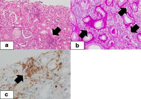
Koda et al. (2018), Figure 1. CC BY 4.0. Re-encoded as PNG; image content unchanged.
Compare your observation with the reported finding
The arrows highlight interstitial inflammation and tubulitis. This supports a tissue-injury pattern, not a uniquely identified culprit drug. Medication history and other evidence are still required; an eosinophil-poor specimen does not exclude this pattern.
Distinguish cell injury from blocked flow
A drug can become poorly soluble in tubular fluid. A high delivered concentration, low urine flow, or an unfavorable urine pH can promote precipitation, but the important factors differ by drug. Crystalluria supports a possibility; it does not automatically establish that every crystal caused the AKI. A normal upper-tract ultrasound also does not exclude obstruction within microscopic tubules.
IV acyclovir should be infused over at least one hour rather than delivered as a rapid bolus. Its label links renal precipitation risk to administration, hydration, renal function, and other nephrotoxins. During AKI, review both renal-function-based dosing and fluid tolerance. Do not substitute routine urine alkalinization for correcting an excessively rapid infusion. [14]
High-dose methotrexate needs a protocol-directed plan for hydration, urine alkalinization, drug concentrations, and rescue. Acidic urine reduces solubility. Delayed clearance prolongs systemic antifolate exposure. Leucovorin rescue and urine alkalinization therefore address different parts of toxicity. Persistent drug levels require clearance-guided specialist reassessment, not stopping rescue merely because a set number of hours has passed. Levoleucovorin is a related rescue formulation with its own labeled dosing; these formulations must not be assumed interchangeable milligram for milligram. No rescue dose is specified here. [15] [25]
Indinavir is a historical example of drug-associated crystalluria and stones. Keep the general lesson about drug solubility, but do not transplant one drug's pH or hydration regimen to every crystalline injury. The medication identity, formulation, renal course, and actual evidence of impaired drainage matter. [35]
Obstructing material need not be a crystal. NSAID-associated papillary injury can release necrotic papillary tissue into the urine pathway. Hydronephrosis identifies a drainage problem; a recovered tissue fragment identifies the material. In a solitary functioning kidney, an obstructed ureter can substantially reduce total filtration. Long-term exposure is a risk context, not proof that every obstruction is a sloughed papilla. [13] [30]
Why could correcting urine pH leave methotrexate toxicity unresolved?
Improving urinary solubility does not by itself reverse persistent systemic antifolate exposure. Measured clearance and clinical toxicity still determine the rescue plan.