Trace venous clot to pulmonary vascular obstruction, choose probability-based testing, assess right-heart stress, and individualize VTE treatment.
A swollen calf and sudden breathlessness raise concern for venous thromboembolism. Two questions must stay separate. How likely is a clot before testing, and how much physiological trouble is it causing? A large-looking clot can occur with stable circulation; a patient in shock needs more than a probability score.
Follow the clot, then explain the physiology
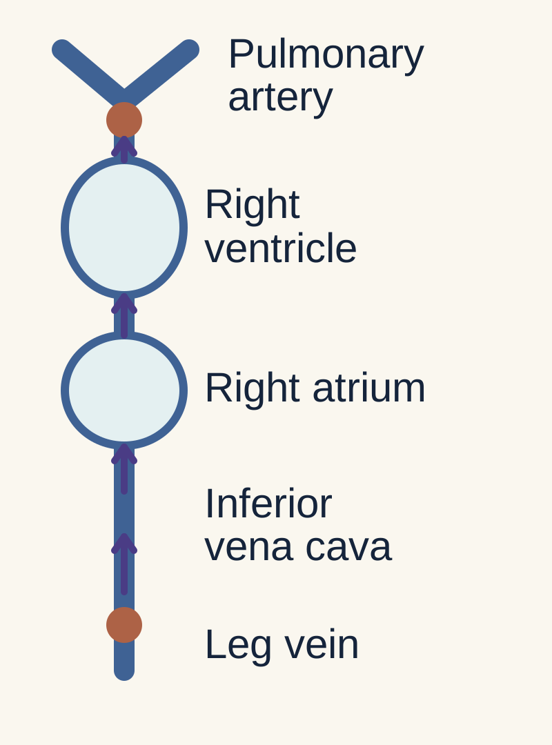
A detached leg-vein clot travels through the inferior vena cava, right atrium and right ventricle into the pulmonary arteries. Ventilation brings air; perfusion brings blood. A blocked artery reduces perfusion despite continuing ventilation and raises the load against which the right ventricle pumps.
Through the inferior vena cava, right atrium and right ventricle into the pulmonary arteries.
What is lost downstream when the artery is blocked?
Blood delivery. Air can still reach the alveoli, but ventilation there cannot efficiently exchange gas with blood.
Why can the right ventricle fail?
Obstruction increases pulmonary vascular resistance. The right ventricle must eject against a greater load.
Deep vein thrombosis and pulmonary embolism are related manifestations of VTE. A leg-vein thrombus can detach and pass through the right heart to the pulmonary arteries. DVT can begin in distal calf veins and extend proximally; it does not always start in the proximal leg. Popliteal, femoral and iliac thromboses carry greater embolic concern than an isolated distal calf thrombus. Catheter-associated upper-extremity DVT is another source. [3][4]
Deep vein
Stasis, vessel injury or increased coagulation favors thrombus formation.
Vena cava and right heart
An embolus travels with venous blood through the right atrium and ventricle.
Pulmonary artery
Obstruction reduces downstream perfusion while alveoli may remain ventilated. This creates high V/Q and wasted ventilation.
Right ventricle under pressure
Increased afterload can dilate the RV, impair coronary perfusion and shift the septum toward the LV. Reduced LV filling lowers systemic output.
The location of the obstruction explains both wasted ventilation and obstructive shock. The systemic effect depends on vascular burden and cardiopulmonary reserve, not the word saddle.
Hypoxemia in PE is multifactorial, including redistribution of blood flow to other lung regions and low mixed venous oxygen when output falls. A normal oxygen saturation or arterial gas does not exclude PE. Tachypnea often lowers PaCO2, but no blood-gas pattern is diagnostic. Pulmonary infarction can cause pleuritic pain and hemoptysis. [3]
Virchow's triad organizes the history
Stasis accompanies immobility, hospitalization and venous compression. Endothelial injury accompanies surgery, trauma and venous catheters. Hypercoagulability can be acquired through cancer, pregnancy, estrogen exposure or APS, or inherited through factor V Leiden, prothrombin G20210A, antithrombin deficiency or protein C/S deficiency. Several contributors often coexist after surgery. Atherosclerotic plaque rupture is principally an arterial-thrombosis mechanism, not the usual explanation for venous clot. [3][5]
Migratory superficial thrombophlebitis can accompany malignancy, historically termed Trousseau syndrome. [12] It is distinct from proving a deep clot or pulmonary embolus. Recurrent or unexplained thrombosis warrants attention to the clinical context, while an unprovoked VTE alone does not mandate indiscriminate whole-body cancer imaging. [1]
Factor V Leiden resists inactivation by activated protein C. Prothrombin G20210A increases prothrombin availability. Antithrombin deficiency can reduce heparin responsiveness. Protein C/S deficiency impairs natural anticoagulation. These mechanisms explain risk without proving that every person with VTE needs a laboratory panel. [5]
Choose one PE probability pathway
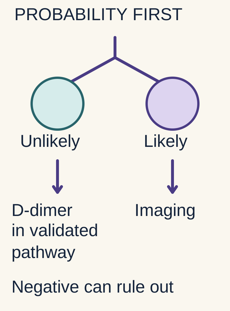
Clinical probability is the estimated chance of PE before testing. D-dimer reflects fibrin turnover; a positive result does not locate a clot. A negative result can exclude PE only within a suitable validated pathway. PERC, YEARS and NICE use different entry criteria and rules; keep each pathway intact.
Read down each branch. A rule-out test is useful only within its validated population and cutoff. The diagram summarizes the lesson; PERC, YEARS and NICE are distinct pathways.
Why decide probability before interpreting D-dimer?
The same test result has different meaning in different probability groups. A negative rule-out pathway is intended for selected patients.
Does a positive D-dimer locate a pulmonary clot?
No. It indicates fibrin turnover and can rise for other reasons; appropriate imaging may still be needed.
Can you combine the easiest cutoff from several pathways?
No. Use one validated pathway with its own criteria, assay units and thresholds.
First assess stability. Shock, syncope with poor perfusion or major respiratory compromise requires urgent evaluation and support. In a stable patient, estimate clinical probability using a validated pathway. This lesson uses the two-level Wells PE scheme. Its cutoffs must not be mixed with the separate three-level scheme. [1]
Two-level Wells PE score
Finding
Points
FindingClinical signs of DVT
Points3
FindingAn alternative diagnosis is less likely than PE
Points3
FindingHeart rate above 100/min
Points1.5
FindingImmobilization over 3 days or surgery in the previous 4 weeks
Points1.5
FindingPrevious DVT or PE
Points1.5
FindingHemoptysis
Points1
FindingMalignancy treated within 6 months or receiving palliation
Points1
More than 4 points means PE likely. Obtain CTPA directly when suitable. A high probability cannot be dismissed using D-dimer. At 4 points or fewer, a sensitive D-dimer can determine the need for imaging. A positive result raises the need for further evaluation but does not diagnose PE. Surgery, infection, cancer, pregnancy and age can increase D-dimer without a pulmonary embolus. Numerical test performance depends on assay and population. [1]
For patients older than 50, an age-adjusted threshold is commonly age multiplied by 10 ng/mL FEU, equivalent to micrograms/L FEU, for assays whose conventional threshold is 500. A 74-year-old therefore has a threshold of 740 ng/mL FEU. Do not transfer this number to a DDU assay without the laboratory's conversion and validated pathway. [3]
PERC can avoid even D-dimer in a patient already judged to have a very low clinical probability in an appropriate emergency-care population. All eight must be negative. The patient is younger than 50, pulse below 100/min, saturation at least 95%, and has no unilateral leg swelling, hemoptysis, recent qualifying surgery or trauma, prior VTE or exogenous estrogen. One positive feature does not diagnose PE; it means PERC cannot end the evaluation. Do not use ordinary PERC as a pregnancy-specific rule. [1][3]
If imaging will be delayed and probability is substantial, assess bleeding risk and consider interim therapeutic anticoagulation according to the pathway. Obtain baseline blood counts and renal/coagulation testing without creating an unnecessary treatment delay. A negative CTPA that conflicts strongly with the clinical picture may require reassessment or additional testing, rather than automatic closure. [1][2]
Case 2
Show answer and explanations for case 2
A. Reassess probability and use D-dimer or imaging as the resulting pathway indicates (Best answer)
Which newly disclosed PERC criterion is positive?
A history of objectively confirmed VTE.
Can the all-negative PERC exit still be used?
No. One positive criterion removes that exit.
Does that positive criterion itself diagnose PE or mandate CT?
No. The complete probability assessment determines the next pathway.
What follows?
Reassess probability, then use the appropriate D-dimer or imaging branch.
Read the complete explanation
A positive PERC item removes that no-testing exit but does not determine the whole next pathway. Prior VTE matters even with normal pulse and oxygen saturation. Failure of a rule-out screen is a reason for further assessment, not a diagnosis.
B. Proceed directly to CT solely because of the positive PERC item (Why this does not fit)
What assessment can justify proceeding directly to CT?
High clinical probability or other pathway findings can justify immediate imaging.
Does one positive PERC item establish that probability by itself?
One positive PERC item alone does not establish high probability.
How should this distinction guide a similar patient?
Choose the next test from the complete probability assessment.
Read the complete explanation
High clinical probability or other pathway findings can justify immediate imaging. One positive PERC item alone does not establish high probability. Choose the next test from the complete probability assessment.
C. Begin anticoagulation solely on the revised screening result (Why this does not fit)
When might treatment begin before imaging?
Empiric treatment may be appropriate when suspicion, delay and bleeding risk justify it.
Does failure of PERC exclusion itself prove an acute clot?
The new history invalidates PERC exclusion but does not establish an acute clot.
How should this distinction guide a similar patient?
Separate screening eligibility from the threshold for treatment.
Read the complete explanation
Empiric treatment may be appropriate when suspicion, delay and bleeding risk justify it. The new history invalidates PERC exclusion but does not establish an acute clot. Separate screening eligibility from the threshold for treatment.
D. End the PE evaluation using the unchanged low-risk rule (Why this does not fit)
What must be true of all PERC criteria to use its exit?
A very-low-risk patient can avoid testing when all PERC criteria are negative.
What did the newly disclosed prior DVT change?
The newly disclosed prior DVT makes one criterion positive.
How should this distinction guide a similar patient?
Recheck rule eligibility when the history changes.
Read the complete explanation
A very-low-risk patient can avoid testing when all PERC criteria are negative. The newly disclosed prior DVT makes one criterion positive. Recheck rule eligibility when the history changes.
Takeaway: Failure of a rule-out screen is a reason for further assessment, not a diagnosis.
The leg pathway and the lung images answer different questions
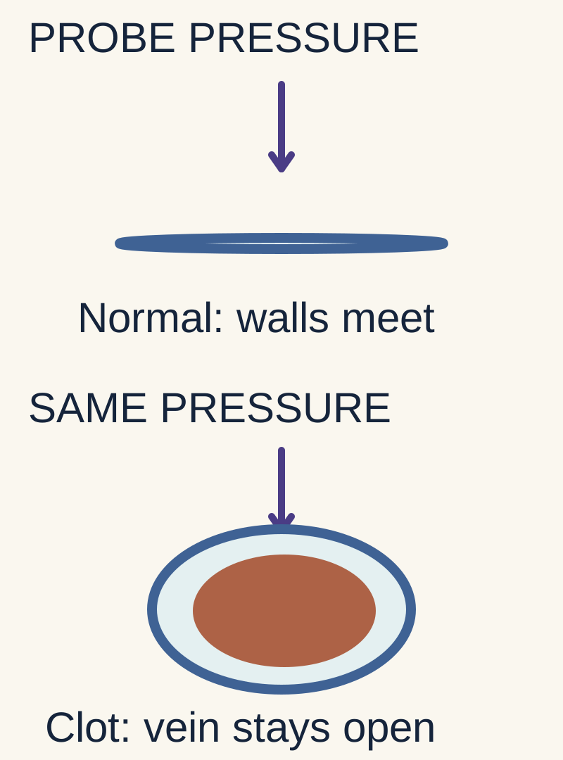
A normal vein collapses under ultrasound probe pressure. Failure to compress supports clot in the examined vein; it does not locate a lung clot. V/Q imaging compares ventilation with perfusion. A perfusion defect with preserved ventilation is a mismatch whose meaning depends on the full scan and clinical probability.
The cross-sections isolate the compression sign. Ultrasound interpretation also requires anatomy, technique and the full examination. A leg-vein finding does not locate a pulmonary embolus.
What happens to a normal vein under ultrasound probe pressure?
Its walls meet as the vein compresses. Failure to compress supports thrombosis in the examined segment.
Does finding a leg clot show where a lung clot is?
No. Leg ultrasound examines veins; pulmonary imaging answers the lung question.
What does a perfusion defect with preserved ventilation suggest?
A ventilation-perfusion mismatch. Interpret the complete scan and clinical probability; an isolated picture is not a diagnosis.
The two-level DVT Wells score is likely at 2 or more and unlikely at 1 or fewer. Each positive feature contributes one point. These include active cancer; paralysis, paresis or recent leg plaster immobilization; bed rest for at least 3 days or major surgery within 12 weeks requiring general/regional anesthesia; deep-vein tenderness; whole-leg swelling; calf difference at least 3 cm measured 10 cm below the tibial tuberosity; unilateral pitting edema; nonvaricose collateral veins; and previous documented DVT. Subtract two when another diagnosis is at least as likely. [1]
Likely DVT warrants proximal compression ultrasound. A noncompressible vein supports thrombosis. If that scan is negative but D-dimer is positive, the NICE proximal-scan pathway repeats ultrasound in 6 to 8 days. A whole-leg ultrasound pathway has different follow-up rules. For unlikely DVT, a negative sensitive D-dimer can end testing. Homans sign is not a reliable rule-in or rule-out maneuver. Severe swelling with cyanosis and threatened pulses suggests phlegmasia cerulea dolens, a limb-threatening outflow obstruction requiring urgent vascular assessment. [1][4]
CTPA
A contrast filling defect identifies clot in a pulmonary artery. CT can also show RV enlargement. Neither a saddle location nor RV enlargement alone establishes shock.
V/Q imaging
A perfusion defect with relatively preserved ventilation supports a vascular obstruction. Matched defects may reflect parenchymal disease and do not independently confirm PE. A nondiagnostic study requires further assessment.
Echo and ECG
RV dilation, septal flattening and McConnell's regional pattern support right-heart stress. Sinus tachycardia is common; S1Q3T3 is neither sensitive nor specific enough to establish PE.
Echo usually stratifies confirmed PE but has a special role at the bedside when an unstable patient cannot safely undergo CT. Absence of RV strain does not exclude PE. McConnell sign can occur in other RV disease, including RV infarction. Troponin signals myocardial injury and BNP signals wall stress; neither identifies the clot by itself. [3]
V/Q is useful when iodinated contrast poses a substantial problem, especially with a chest radiograph that permits interpretation. Pregnancy is not an absolute contraindication to CTPA. Both V/Q and CTPA can be used when indicated; chest radiograph, availability and maternal/fetal radiation considerations guide selection. If leg symptoms are present, ultrasound can establish VTE without thoracic imaging. Pregnancy-adapted YEARS is a validated alternative pathway using its own clinical items and D-dimer thresholds, not ordinary Wells with improvised pregnancy cutoffs. [6][7]
Case 6
Show answer and explanations for case 6
A. Proximal DVT; assess and initiate therapeutic anticoagulation (Best answer)
Which vessel fails to compress?
The femoral vein, a proximal deep vein.
Does a patent adjacent artery remove that venous finding?
No. Arterial inflow and venous outflow are separate questions.
What diagnosis fits the acute symptoms and positive compression finding?
Proximal DVT.
What treatment direction follows after routine suitability assessment?
Therapeutic anticoagulation, because no major constraint is supplied.
Read the complete explanation
A noncompressible proximal deep vein in this acute context supports DVT. The femoral finding matches the symptoms and no major treatment constraint is supplied. Confirm location, then assess bleeding and drug suitability before treatment.
B. Isolated superficial thrombophlebitis; use local symptom treatment (Why this does not fit)
Which vascular compartment defines superficial thrombophlebitis?
Superficial thrombophlebitis affects superficial veins, rather than the proximal deep venous system.
Is the femoral vein in that compartment?
The femoral vein is a deep proximal vein.
How should this distinction guide a similar patient?
Name the affected vessel before choosing the treatment pathway.
Read the complete explanation
Superficial-vein thrombosis has a different anatomic extent and treatment assessment. The femoral vein is a deep proximal vein. Name the affected vessel before choosing the treatment pathway.
C. An inconclusive venous study; repeat ultrasound before treatment (Why this does not fit)
When does an inconclusive examination call for repeat imaging?
A technically limited or equivocal study can need repeat imaging.
Is the symptomatic proximal vein finding equivocal here?
The supplied examination demonstrates noncompressibility in the symptomatic proximal deep vein.
How should this distinction guide a similar patient?
Use a clear positive finding differently from an incomplete negative study.
Read the complete explanation
A technically limited or equivocal study can need repeat imaging. The supplied examination demonstrates noncompressibility in the symptomatic proximal deep vein. Use a clear positive finding differently from an incomplete negative study.
D. Acute limb ischemia; prioritize arterial embolectomy (Why this does not fit)
What lesion leads to arterial embolectomy?
Arterial occlusion can threaten a limb and require urgent revascularization.
Which vessel is abnormal and which remains patent?
The abnormality is in the vein and the adjacent artery is patent.
How should this distinction guide a similar patient?
Separate venous obstruction from arterial inflow failure.
Read the complete explanation
Arterial occlusion can threaten a limb and require urgent revascularization. The abnormality is in the vein and the adjacent artery is patent. Separate venous obstruction from arterial inflow failure.
E. Isolated distal DVT; consider surveillance based on extension risk (Why this does not fit)
Where is an isolated distal DVT located?
Isolated distal DVT involves calf deep veins below the popliteal vein.
Does a femoral clot belong to the calf-vein category?
The clot is in the femoral vein, proximal to the calf-vein territory.
How should this distinction guide a similar patient?
Proximal and distal DVT are different treatment categories.
Read the complete explanation
Selected calf-vein thromboses can be managed with serial imaging. The clot is in the femoral vein, proximal to the calf-vein territory. Proximal and distal DVT are different treatment categories.
Takeaway: Confirm location, then assess bleeding and drug suitability before treatment.
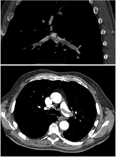
Saddle names a clot at the main pulmonary artery bifurcation, not a shock category. Assess circulation, breathing and right-ventricular injury. Worsening perfusion can precede hypotension. Reperfusion decisions balance physiological severity against major bleeding risk; a positive troponin alone does not mandate thrombolysis.
Enlarge the complete imageThe dark material within contrast-filled pulmonary arteries is thrombus. The bifurcation location is called saddle; physiological severity still requires clinical assessment. Aung Myat and Arif Ahsan. Image: Original source. CC BY 2.0.
Does saddle describe physiology or location?
Location: a clot straddles the main pulmonary artery bifurcation. It does not by itself establish shock.
Can a patient deteriorate before persistent hypotension appears?
Yes. Worsening perfusion or respiratory support needs can signal incipient cardiopulmonary failure.
Why is thrombolysis not automatic for every positive troponin?
Escalation weighs physiological severity against major bleeding risk. RV injury warrants assessment and monitoring, not an automatic lysis order.
The historical terms massive and submassive describe physiological severity, not clot dimensions. Massive generally refers to hemodynamic instability. Submassive describes normotension with RV dysfunction or biomarker injury, but that label contains patients with different risks. Use clinical trajectory alongside formal severity assessment. [3]
The 2026 AHA/ACC guideline introduces categories A through E. Category A includes incidental/asymptomatic PE; B includes symptomatic PE with low clinical severity; C includes higher clinical severity with possible RV or biomarker abnormalities; D identifies incipient cardiopulmonary failure; E identifies cardiopulmonary failure with persistent hypotension. Categories C through E generally require hospitalization. These categories do not map one-to-one onto old clot-size labels. [2][11]
Anticoagulation is the foundation for most confirmed PE. Carefully selected low-risk patients may receive outpatient treatment when oxygenation, bleeding risk, comorbidities, medication access and follow-up are suitable. RV dysfunction plus troponin elevation in a normotensive patient warrants monitoring and reassessment, not automatic thrombolysis. PEITHO found less decompensation with fibrinolysis but more major bleeding and stroke, without a demonstrated mortality reduction. [2][8]
Shock attributed to PE requires urgent reperfusion assessment. Systemic thrombolysis is an option when benefit outweighs bleeding risk. Surgical embolectomy or catheter-based therapy may be appropriate when lysis is contraindicated, has failed, or another strategy is selected by the experienced team. Catheter procedures are not restricted to stable patients. Reduced-dose lysis is not a universal default for intermediate-risk PE. [2][3]
Active major bleeding, previous intracranial hemorrhage, recent major intracranial disease or surgery and other major bleeding hazards matter. No rule makes active intracranial pathology the only contraindication. During immediately life-threatening collapse the team weighs otherwise prohibitive risks, but this requires explicit individualized judgment. Support systemic pressure, often with norepinephrine. Large indiscriminate fluid loads can distend the RV further. Intubation and positive pressure can precipitate cardiovascular deterioration and require careful planning. [3]
Case 11
Show answer and explanations for case 11
A. Give systemic thrombolysis immediately followed by anticoagulation (Why this does not fit)
When can systemic lysis have a favorable benefit–risk balance?
Systemic lysis can be lifesaving in appropriately selected high-risk PE.
Does this patient currently have shock or hypoperfusion?
The patient has no current shock or hypoperfusion, and routine lysis adds major bleeding risk.
How should this distinction guide a similar patient?
RV injury alone does not establish that immediate lysis has a favorable balance.
Read the complete explanation
Systemic lysis can be lifesaving in appropriately selected high-risk PE. The patient has no current shock or hypoperfusion, and routine lysis adds major bleeding risk. RV injury alone does not establish that immediate lysis has a favorable balance.
B. Discharge with a DOAC and early routine outpatient review (Why this does not fit)
What profile supports outpatient PE treatment?
Outpatient care is appropriate for carefully selected low-risk PE.
How do RV dilation and troponin change that assessment?
RV dilation and positive troponin make a low-risk outpatient assumption inappropriate.
How should this distinction guide a similar patient?
Normal blood pressure is only one part of disposition assessment.
Read the complete explanation
Outpatient care is appropriate for carefully selected low-risk PE. RV dilation and positive troponin make a low-risk outpatient assumption inappropriate. Normal blood pressure is only one part of disposition assessment.
C. Anticoagulate in a monitored inpatient setting and reassess for deterioration (Best answer)
What findings prevent a simple low-risk assumption?
RV dilation and a positive troponin.
What do lactate, warm extremities and blood pressure say about current perfusion?
No current hypoperfusion or shock is demonstrated.
Which treatment is foundational now?
Therapeutic anticoagulation.
What setting and contingency fit the increased risk?
Monitored inpatient care with reassessment and rescue escalation if deterioration develops.
Read the complete explanation
RV dysfunction and injury markers raise concern even before shock develops. Current perfusion is stable, supporting anticoagulation and monitoring rather than routine immediate reperfusion. Use both current physiology and deterioration risk to choose the setting of care.
D. Arrange catheter thrombectomy before starting anticoagulation (Why this does not fit)
What severity or trajectory can justify catheter intervention?
Catheter intervention is considered for selected severe or deteriorating PE presentations.
Is that intervention need established by the present stable perfusion?
Stable perfusion without escalating support does not establish a routine need for upfront thrombectomy.
How should this distinction guide a similar patient?
Match an invasive intervention to the severity and trajectory demonstrated.
Read the complete explanation
Catheter intervention is considered for selected severe or deteriorating PE presentations. Stable perfusion without escalating support does not establish a routine need for upfront thrombectomy. Match an invasive intervention to the severity and trajectory demonstrated.
E. Use prophylactic anticoagulation until RV size normalizes (Why this does not fit)
What situation is prophylactic dosing designed for?
Prophylactic dosing prevents VTE in selected patients without established events.
Does confirmed PE fit that preventive indication?
PE is confirmed and requires therapeutic anticoagulation unless contraindicated.
How should this distinction guide a similar patient?
The presence of a clot determines treatment intensity, not the absence of hypotension.
Read the complete explanation
Prophylactic dosing prevents VTE in selected patients without established events. PE is confirmed and requires therapeutic anticoagulation unless contraindicated. The presence of a clot determines treatment intensity, not the absence of hypotension.
Takeaway: Use both current physiology and deterioration risk to choose the setting of care.
Anticoagulants limit clot propagation and recurrence while the body clears clot. They do not instantly dissolve it. Pregnancy, kidney function, APS, cancer and drug interactions can change the best drug. Treatment duration depends on persistent risk, recurrence, bleeding and preferences; continuing symptoms need reassessment.
Does anticoagulation instantly dissolve the current clot?
No. It limits clot propagation and recurrence while the body clears clot. Reperfusion is a separate treatment question.
Why can two patients with DVT need different drugs?
Pregnancy, kidney function, antiphospholipid syndrome, cancer, bleeding risk and interactions can change suitability.
What determines treatment beyond the initial course?
Whether risk persists, recurrence history, bleeding risk and patient preferences. Reassess the plan and investigate persistent symptoms.
For eligible nonpregnant adults, DOACs are generally preferred over vitamin K antagonists. Apixaban and rivaroxaban have oral acute-treatment regimens; dabigatran and edoxaban require an initial parenteral course. Kidney function, drug interactions, body size, adherence and cost influence selection. Parenteral therapy is often useful in hospitalized patients; LMWH is generally preferred when suitable, while UFH retains specific roles such as severe renal impairment or situations needing closely controlled procedural interruption. Do not automatically assign every patient with RV strain to UFH. [2][4]
When treating acute VTE with warfarin, overlap parenteral anticoagulation for at least 5 days and until INR is therapeutic at 2 or above for at least 24 hours. An early INR rise mainly reflects the short half-life of factor VII and does not prove that factor II has fallen sufficiently. Early reduction of protein C contributes to the transient procoagulant concern. [4]
LMWH is preferred for VTE during pregnancy because it does not cross the placenta. Warfarin crosses and can harm the fetus. DOACs are avoided because adequate pregnancy safety evidence is lacking, not because human teratogenicity has been definitively established for every agent. Active cancer can be treated with a suitable DOAC or LMWH; gastrointestinal/genitourinary bleeding hazards, interactions and absorption matter. [6][10]
APS combines a compatible clinical event with persistent antiphospholipid antibodies. Lupus anticoagulant can prolong phospholipid-dependent assays despite a tendency toward thrombosis. A noncorrecting mixing test suggests an inhibitor, but is not by itself diagnostic of APS. Antibody confirmation at least 12 weeks apart helps establish persistence. For thrombotic APS, especially triple-positive disease, warfarin is preferred to rivaroxaban; a usual venous-event INR target is 2 to 3. Pregnancy with thrombotic APS requires an obstetric/hematology plan using heparin-based treatment, often with low-dose aspirin. [3][6][9]
Most proximal DVT and PE require at least three months of anticoagulation. A resolved major transient trigger such as surgery often permits stopping after that course. Unprovoked events, persistent risks and recurrent VTE favor extended treatment if bleeding risk permits. Cancer-associated treatment is reassessed over time while cancer remains active. The 2026 PE guidance describes an initial 3 to 6 month phase and extended treatment for patients without a major reversible trigger or with a persistent risk. [1][4][11]
An IVC filter is not a routine addition to effective anticoagulation. A retrievable filter may be appropriate for acute VTE when anticoagulation is temporarily impossible because of major bleeding, with a retrieval and restart plan. Suspected recurrence during treatment first requires confirmation and review of adherence, dose, interactions and underlying disease. Isolated distal DVT and selected subsegmental PE need individualized assessment; their anatomical labels alone neither demand nor prohibit anticoagulation. [4]
Order thrombophilia testing only if a result could change care. Routine screening before combined oral contraception is not recommended. A family history of an identified high-risk deficiency differs from an unspecified family clot history. Acute thrombosis and anticoagulants can distort functional assays; coordinate timing with the laboratory rather than stopping necessary treatment merely to obtain a panel. Persistent breathlessness after PE needs evaluation for chronic thromboembolic disease and other causes. [5][11]
For a stable patient, choose the validated probability pathway before the test. Once VTE is confirmed, determine circulatory risk, bleeding risk and the reason the clot formed. Those findings decide treatment setting, anticoagulant, reperfusion and follow-up.
Apply the lesson
Case 1
Show answer and explanations for case 1
A. Obtain CT pulmonary angiography promptly (Best answer)
Which probability branch do the recent surgery, tachycardia and DVT signs support?
With PE judged more likely than an alternative, the supplied assessment is PE likely.
Is D-dimer the exclusion step for that branch?
No. The likely-PE pathway proceeds to appropriate imaging.
What supplied constraints favorCTPA?
CT is available, kidney function is adequate and there is no contrast allergy.
What is the next action?
Obtain CTPA promptly while continuing clinical assessment.
Read the complete explanation
CTPA directly evaluates pulmonary arteries in a likely-PE pathway when suitable. Multiple clinical risk findings establish the pathway, and the stated constraints permit CT. A likely-PE assessment leads to appropriate imaging rather than screening D-dimer.
B. Use ventilation-perfusion imaging as the initial test (Why this does not fit)
When is V/Q a useful initial imaging choice?
V/Q imaging is useful when CT contrast is unsuitable or other circumstances favor it.
Are the relevant CT constraints present here?
CT is available and no kidney or contrast limitation is supplied.
How should this distinction guide a similar patient?
Match imaging to probability, patient constraints and availability.
Read the complete explanation
V/Q imaging is useful when CT contrast is unsuitable or other circumstances favor it. CT is available and no kidney or contrast limitation is supplied. Match imaging to probability, patient constraints and availability.
C. Obtain D-dimer and reserve imaging for a positive result (Why this does not fit)
In which probability group can D-dimer help exclude PE?
D-dimer exclusion is useful in selected low or unlikely clinical-probability pathways.
Which probability branch do these combined findings establish?
The supplied findings place this patient in the likely-PE pathway.
How should this distinction guide a similar patient?
Establish probability before selecting a rule-out assay.
Read the complete explanation
D-dimer exclusion is useful in selected low or unlikely clinical-probability pathways. The supplied findings place this patient in the likely-PE pathway. Establish probability before selecting a rule-out assay.
D. Use bedside echocardiography as the initial exclusion test (Why this does not fit)
What does echo assess in PE?
Echo helps assess RV stress, especially in an unstable patient.
Could a normal echo reliably exclude clot in this stable patient?
This stable patient needs diagnostic pulmonary imaging; a normal echo would not reliably exclude PE.
How should this distinction guide a similar patient?
Distinguish hemodynamic assessment from clot exclusion.
Read the complete explanation
Echo helps assess RV stress, especially in an unstable patient. This stable patient needs diagnostic pulmonary imaging; a normal echo would not reliably exclude PE. Distinguish hemodynamic assessment from clot exclusion.
E. Repeat compression ultrasound in one week before pulmonary testing (Why this does not fit)
What uncertainty does serial leg ultrasound address?
Serial ultrasound can resolve selected negative initial DVT studies.
Would waiting resolve the current likely-PE chest presentation safely?
Acute chest symptoms and a likely-PE assessment require a pulmonary diagnostic plan now.
How should this distinction guide a similar patient?
Do not substitute a delayed leg pathway for current suspected PE.
Read the complete explanation
Serial ultrasound can resolve selected negative initial DVT studies. Acute chest symptoms and a likely-PE assessment require a pulmonary diagnostic plan now. Do not substitute a delayed leg pathway for current suspected PE.
Takeaway: A likely-PE assessment leads to appropriate imaging rather than screening D-dimer.
A. Obtain leg ultrasound before ending the PE pathway (Why this does not fit)
What additional question can leg ultrasound answer?
Ultrasound is useful for suspected DVT and some alternative diagnostic strategies.
Is an unresolved leg question supplied after this eligible negative pathway?
No leg finding is supplied and the selected PE exclusion pathway is satisfied.
How should this distinction guide a similar patient?
Order the next test for a remaining clinical question, not an isolated flagged laboratory value.
Read the complete explanation
Ultrasound is useful for suspected DVT and some alternative diagnostic strategies. No leg finding is supplied and the selected PE exclusion pathway is satisfied. Order the next test for a remaining clinical question, not an isolated flagged laboratory value.
B. Obtain CTPA because the result exceeds 500 ng/mL FEU (Why this does not fit)
When is the conventional 500 threshold the applicable cutoff?
The conventional cutoff is used where age adjustment is not part of the selected pathway.
Which validated cutoff does this service use for this 74-year-old?
The validated age-adjusted cutoff here is 74 × 10 = 740 ng/mL FEU; 680 is below it.
How should this distinction guide a similar patient?
Apply one validated pathway consistently.
Read the complete explanation
The conventional cutoff is used where age adjustment is not part of the selected pathway. This service explicitly uses a validated age-adjusted pathway for this patient. Apply one validated pathway consistently.
C. Repeat D-dimer the next day before deciding on imaging (Why this does not fit)
When would a repeat assay resolve a genuine uncertainty?
Repeat testing may be needed when a sample or clinical assessment is unreliable.
Is a sample problem or clinical change supplied?
No sample problem or new clinical change is supplied; the current eligible result is below threshold.
How should this distinction guide a similar patient?
Do not add serial tests without a defined unresolved question.
Read the complete explanation
Repeat testing may be needed when a sample or clinical assessment is unreliable. No sample problem or new clinical change is supplied; the current eligible result is below threshold. Do not add serial tests without a defined unresolved question.
D. End PE-specific testing and investigate other causes of dyspnea (Best answer)
Is this patient in the pathway's eligible probability group?
Yes. The Wells assessment is PE unlikely.
What is 74 times 10?
The age-adjusted threshold is 740 ng/mL FEU.
How does 680 compare with 740?
It is below the validated threshold.
What pathway endpoint follows?
End PE-specific testing and investigate other causes of dyspnea, unless the clinical situation changes.
Read the complete explanation
A result below the validated adjusted threshold can exclude PE in an eligible unlikely-probability pathway. The threshold is 740 and the measured value is 680 ng/mL FEU. Check probability, age, assay units and threshold together.
Takeaway: Check probability, age, assay units and threshold together.
A. Assess alternative diagnoses and advise return for persistent or worsening symptoms (Best answer)
What does a two-level DVT Wells score 1 mean?
DVT is unlikely in the stated pathway.
Is the negative assay interpretable here?
Yes. No prior anticoagulation or sample problem is supplied.
What does that combination permit?
Stop this DVT investigation and consider alternative diagnoses.
What advice preserves safety as symptoms evolve?
Return for persistent or worsening symptoms that require reassessment.
Read the complete explanation
A negative D-dimer in an eligible unlikely-DVT pathway makes DVT unlikely enough to stop that investigation. Both the probability score and assay support the pathway endpoint. End a completed rule-out pathway while preserving follow-up for a changing presentation.
B. Schedule serial proximal ultrasound at six to eight days (Why this does not fit)
Which result combination calls for serial proximal scanning?
Serial scanning is used after specific combinations of likely probability, negative proximal scan and positive D-dimer.
Does this patient have that combination?
That combination is absent; this is an unlikely-DVT negative-assay pathway.
How should this distinction guide a similar patient?
Use the follow-up schedule belonging to the pathway actually entered.
Read the complete explanation
Serial scanning is used after specific combinations of likely probability, negative proximal scan and positive D-dimer. That combination is absent; this is an unlikely-DVT negative-assay pathway. Use the follow-up schedule belonging to the pathway actually entered.
C. Begin interim therapeutic anticoagulation while awaiting a repeat assay (Why this does not fit)
When can diagnostic delay justify interim anticoagulation?
Interim treatment can be indicated when required diagnostic tests are delayed.
Is the required exclusion test still pending here?
The supplied pathway already has a timely interpretable negative result.
How should this distinction guide a similar patient?
Diagnostic delay and a completed negative pathway are different situations.
Read the complete explanation
Interim treatment can be indicated when required diagnostic tests are delayed. The supplied pathway already has a timely interpretable negative result. Diagnostic delay and a completed negative pathway are different situations.
D. Obtain immediate proximal compression ultrasound (Why this does not fit)
Which branches call for proximal ultrasound?
Ultrasound is indicated after a positive D-dimer in the unlikely pathway or in a likely-DVT pathway.
Does a Wells score 1 with a negative assay enter one of them?
This patient has an interpretable negative assay in the unlikely group.
How should this distinction guide a similar patient?
Combine the test with its intended probability category.
Read the complete explanation
Ultrasound is indicated after a positive D-dimer in the unlikely pathway or in a likely-DVT pathway. This patient has an interpretable negative assay in the unlikely group. Combine the test with its intended probability category.
Takeaway: End a completed rule-out pathway while preserving follow-up for a changing presentation.
A. Repeat D-dimer in six to eight days instead of ultrasound (Why this does not fit)
Does D-dimer localize an evolving proximal clot?
D-dimer indicates fibrin turnover but does not localize evolving thrombosis.
What uncertainty must the follow-up actually resolve?
The unresolved question is whether proximal thrombosis becomes demonstrable.
How should this distinction guide a similar patient?
Use imaging to resolve a serial-imaging question.
Read the complete explanation
D-dimer indicates fibrin turnover but does not localize evolving thrombosis. The unresolved question is whether proximal thrombosis becomes demonstrable. Use imaging to resolve a serial-imaging question.
B. Obtain CT venography immediately as routine follow-up (Why this does not fit)
When can CT venography help with venous assessment?
Cross-sectional venous imaging can help selected anatomic or technically limited cases.
Is a technical or pelvic-anatomic limitation supplied?
No technical limitation or pelvic-obstruction concern is supplied; the stated pathway uses repeat proximal ultrasound.
How should this distinction guide a similar patient?
Reserve alternative imaging for a specific unresolved limitation.
Read the complete explanation
Cross-sectional venous imaging can help selected anatomic or technically limited cases. No technical limitation or pelvic-obstruction concern is supplied; the stated pathway uses repeat proximal ultrasound. Reserve alternative imaging for a specific unresolved limitation.
C. Repeat proximal compression ultrasound in six to eight days (Best answer)
Which ultrasound strategy is being used?
The NICE proximal-ultrasound pathway, not a whole-leg strategy.
What remains unresolved after a negative proximal scan and positiveD-dimer?
Proximal thrombosis may need serial reassessment.
When should the repeat proximal scan occur?
Six to eight days later.
What makes the interval meaningful?
It belongs to this specific result combination and imaging pathway.
Read the complete explanation
NICE specifies a repeat proximal scan for this result combination. The likely score, positive D-dimer and negative proximal scan match that branch. Interpret a negative scan in the context of both technique and pathway.
D. End DVT follow-up after the single proximal scan (Why this does not fit)
What decision is reasonable after the entire validated pathway is negative?
A completed negative diagnostic pathway can allow testing to stop.
What role does the positive D-dimer have after this proximal scan?
Positive D-dimer after a negative proximal scan in this pathway calls for repeat imaging.
How should this distinction guide a similar patient?
Know whether the chosen ultrasound strategy requires serial assessment.
Read the complete explanation
A completed negative diagnostic pathway can allow testing to stop. Positive D-dimer after a negative proximal scan in this pathway calls for repeat imaging. Know whether the chosen ultrasound strategy requires serial assessment.
Takeaway: Interpret a negative scan in the context of both technique and pathway.
A. Use bedside echocardiography to end the diagnostic workup if normal (Why this does not fit)
What does a normal RV assessment tell you about a stable PE?
Normal RV findings do not reliably exclude stable PE; echo primarily assesses cardiac consequences.
Would it resolve the pulmonary clot question?
A stable PE can occur without diagnostic echo abnormalities.
How should this distinction guide a similar patient?
A consequence test is not a reliable exclusion test for the clot itself.
Read the complete explanation
Echo assesses RV consequences and helps in unstable presentations. A stable PE can occur without diagnostic echo abnormalities. A consequence test is not a reliable exclusion test for the clot itself.
B. Use CTPA after routine contrast premedication as the first choice (Why this does not fit)
Does premedication remove all the supplied contrast constraints?
Premedication does not remove all contrast-related risk or resolve this patient’s severe renal impairment.
Is a suitable contrast-sparing alternative already available?
A suitable contrast-sparing alternative is available with favorable chest imaging.
How should this distinction guide a similar patient?
Do not treat premedication as removal of all contrast-related risk.
Read the complete explanation
CTPA is useful when its diagnostic benefit justifies contrast risk; premedication may be considered in selected cases. A suitable contrast-sparing alternative is available with favorable chest imaging. Do not treat premedication as removal of all contrast-related risk.
C. Obtain a ventilation-perfusion scan (Best answer)
What two supplied findings make iodinated contrast problematic?
Severe renal impairment and prior contrast anaphylaxis.
Which available pulmonary test avoids iodinated contrast?
V/Q scanning.
What makes its interpretation more favorable here?
A near-normal chest radiograph.
Which initial strategy fits both constraints and availability?
Obtain the available V/Q study.
Read the complete explanation
V/Q imaging evaluates regional ventilation and perfusion without iodinated contrast. Contrast risks are substantial, the film is near normal and V/Q is available. Choose the diagnostic modality using both contraindications and likely interpretability.
D. Use compression ultrasound alone and stop if it is negative (Why this does not fit)
What does a positive leg study establish?
A positive leg study can establish VTE in some diagnostic strategies.
Would a negative leg study exclude clot in the lung?
A negative leg scan would not exclude PE; an appropriate pulmonary test is available.
How should this distinction guide a similar patient?
A negative study of one vascular territory does not exclude clot in another.
Read the complete explanation
A positive leg study can establish VTE in some diagnostic strategies. A negative leg scan would not exclude PE; an appropriate pulmonary test is available. A negative study of one vascular territory does not exclude clot in another.
E. Use unenhanced chest CT to exclude pulmonary arterial clot (Why this does not fit)
What can unenhanced chest CT assess?
Noncontrast CT can evaluate some alternative thoracic disease.
Does it provide the arterial opacification needed for this exclusion study?
It does not provide the required contrast opacification for a reliable pulmonary arterial exclusion study.
How should this distinction guide a similar patient?
Select imaging that answers the actual vascular question.
Read the complete explanation
Noncontrast CT can evaluate some alternative thoracic disease. It does not provide the required contrast opacification for a reliable pulmonary arterial exclusion study. Select imaging that answers the actual vascular question.
Takeaway: Choose the diagnostic modality using both contraindications and likely interpretability.
A. Repeat the same V/Q study after a bronchodilator dose (Why this does not fit)
When might improving ventilation change a scan's usefulness?
Improving reversible ventilation may help some lung-function measures.
Does repeating the limited modality best resolve the remaining question here?
Marked underlying abnormalities have already limited the test, while another suitable modality is available.
How should this distinction guide a similar patient?
Resolve a known diagnostic limitation with an appropriate alternative test.
Read the complete explanation
Improving reversible ventilation may help some lung-function measures. Marked underlying abnormalities have already limited the test, while another suitable modality is available. Resolve a known diagnostic limitation with an appropriate alternative test.
B. Obtain CT pulmonary angiography (Best answer)
Has the V/Q report resolved the PE question?
No. It is indeterminate in an abnormal lung.
Does a negative proximal leg study exclude pulmonary clot?
No. It examines a different vascular territory.
What suitable alternative can answer the pulmonary question?
CTPA, because kidney function and contrast history permit it.
What is the general pathway rule?
Resolve meaningful residual uncertainty rather than call an indeterminate test negative or positive.
Read the complete explanation
CTPA can resolve a nondiagnostic V/Q result when contrast is suitable. The unresolved pulmonary question remains after the negative leg study, and CT constraints are absent. Continue until the clinical question is resolved by a valid diagnostic pathway.
C. End PE testing because the defects are matched (Why this does not fit)
What can matched defects suggest about underlying lung disease?
Matched defects may reflect underlying lung disease rather than a classic embolic mismatch.
Has the complete scan actually excluded PE?
The report remains indeterminate and clinical concern persists.
How should this distinction guide a similar patient?
Interpret the complete scan category and probability, not one pattern in isolation.
Read the complete explanation
Matched defects may reflect underlying lung disease rather than a classic embolic mismatch. The report remains indeterminate and clinical concern persists. Interpret the complete scan category and probability, not one pattern in isolation.
D. Diagnose PE from the perfusion abnormalities and start a long-term plan (Why this does not fit)
What scan interpretation can support a positive PE diagnosis?
High-probability mismatched patterns can support PE in the appropriate context.
Is this scan in that diagnostic category?
The scan is explicitly indeterminate with matched defects.
How should this distinction guide a similar patient?
Do not promote an indeterminate result into a positive diagnosis.
Read the complete explanation
High-probability mismatched patterns can support PE in the appropriate context. The scan is explicitly indeterminate with matched defects. Do not promote an indeterminate result into a positive diagnosis.
Takeaway: Continue until the clinical question is resolved by a valid diagnostic pathway.
A. Proceed to appropriately optimized CTPA after counseling (Best answer)
Has the negative leg study resolved the pulmonary concern?
No. PE remains under consideration.
What makes the available V/Q study less useful?
Substantial parenchymal abnormalities on the chest radiograph limit expected interpretation.
Is aCTPA constraint supplied?
No. Kidney function is normal and no contrast allergy is present.
What plan follows after counseling?
Proceed to appropriately optimized CTPA rather than defer indicated pulmonary imaging because of pregnancy alone.
Read the complete explanation
CTPA is a diagnostic option in pregnancy when pulmonary imaging is indicated. The abnormal radiograph limits V/Q usefulness and no CT contrast barrier is supplied. Choose a test likely to answer the question while discussing its risks.
B. Use echocardiography and end testing if RV size is normal (Why this does not fit)
What cardiac consequence can echo detect?
Echo can detect RV dilation or dysfunction associated with an increased pulmonary vascular load.
Could a normal RV exclude the stable pulmonary clot being considered?
A normal RV would not exclude a stable pulmonary embolus.
How should this distinction guide a similar patient?
Evaluate the clot with a suitable diagnostic pathway, not only its cardiac consequences.
Read the complete explanation
Echo can assess hemodynamic consequences of PE. A normal RV would not exclude a stable pulmonary embolus. Evaluate the clot with a suitable diagnostic pathway, not only its cardiac consequences.
C. Use the available V/Q study regardless of the expected limitation (Why this does not fit)
What chest-film context often favors V/Q in pregnancy?
V/Q is often useful in pregnancy, particularly with a normal chest radiograph.
What supplied finding instead limits its expected quality?
Substantial parenchymal abnormalities make the supplied service expect poor interpretability.
How should this distinction guide a similar patient?
Pregnancy imaging choices depend on the actual film and local test quality.
Read the complete explanation
V/Q is often useful in pregnancy, particularly with a normal chest radiograph. Substantial parenchymal abnormalities make the supplied service expect poor interpretability. Pregnancy imaging choices depend on the actual film and local test quality.
D. Repeat leg ultrasound in one week before pulmonary imaging (Why this does not fit)
What diagnostic pathway can use serial leg imaging?
Serial leg ultrasound can be appropriate in a DVT pathway.
Does it answer the current pulmonary question after a negative leg scan?
The current unresolved problem is suspected pulmonary embolism in pregnancy.
How should this distinction guide a similar patient?
Do not let a negative test in another territory postpone necessary pulmonary evaluation.
Read the complete explanation
Serial leg ultrasound can be appropriate in a DVT pathway. The current unresolved problem is suspected pulmonary embolism in pregnancy. Do not let a negative test in another territory postpone necessary pulmonary evaluation.
Takeaway: Choose a test likely to answer the question while discussing its risks.
A. Oral warfarin adjusted to a therapeutic INR (Why this does not fit)
Can warfarin effectively treat VTE outside pregnancy?
Warfarin is effective for many nonpregnant VTE indications.
What separate fetal concern changes its suitability here?
Fetal risks make it unsuitable for routine pregnancy-associated VTE treatment here.
How should this distinction guide a similar patient?
Maternal efficacy does not settle fetal safety.
Read the complete explanation
Warfarin is effective for many nonpregnant VTE indications. Fetal risks make it unsuitable for routine pregnancy-associated VTE treatment here. Maternal efficacy does not settle fetal safety.
B. Intravenous unfractionated heparin throughout the remaining pregnancy (Why this does not fit)
Which clinical constraints can favor UFH?
Severe renal impairment or a need for rapid interruption and reversal, such as an imminent procedure or delivery, can favor UFH.
Are those constraints present at 20 weeks with normal kidneys?
Normal kidneys and no imminent delivery make prolonged infusion less suitable than LMWH.
How should this distinction guide a similar patient?
Select heparin formulation using renal function, timing and reversibility needs.
Read the complete explanation
UFH can be useful when rapid reversal or severe renal impairment is important. Normal kidneys and no imminent delivery make prolonged infusion less suitable than LMWH. Select heparin formulation using renal function, timing and reversibility needs.
C. Oral rivaroxaban at the standard VTE treatment dose (Why this does not fit)
For which population is routine DOAC treatment well established?
DOACs are useful for many nonpregnant adults with VTE.
What does pregnancy change about that usual choice?
Pregnancy lacks the necessary safety basis for this choice.
How should this distinction guide a similar patient?
Reassess usual oral-drug preferences during pregnancy.
Read the complete explanation
DOACs are useful for many nonpregnant adults with VTE. Pregnancy lacks the necessary safety basis for this choice. Reassess usual oral-drug preferences during pregnancy.
D. Weight-adjusted therapeutic low-molecular-weight heparin (Best answer)
Does confirmed proximal DVT require prevention-dose or treatment-dose anticoagulation?
Treatment-dose anticoagulation.
Which drug class is generally preferred for pregnancy-associated VTE?
Low-molecular-weight heparin.
What supplied details favor LMWH over a reversible UFH infusion?
Renal function is normal and delivery is not imminent.
What makes the plan practical?
The patient can self-inject, with no supplied heparin allergy or active bleeding.
Read the complete explanation
LMWH is preferred for most acute VTE treatment during pregnancy. Adequate renal function, no imminent delivery and ability to self-inject favor it. Combine pregnancy status with renal and peripartum considerations.
E. Prophylactic-dose low-molecular-weight heparin (Why this does not fit)
What is prophylactic dosing intended to do?
Prophylactic dosing is used to prevent events in selected risk settings.
Does confirmed proximal DVT require prevention or treatment intensity?
A proximal clot is already confirmed and requires therapeutic treatment.
How should this distinction guide a similar patient?
Distinguish prevention dosing from treatment of established VTE.
Read the complete explanation
Prophylactic dosing is used to prevent events in selected risk settings. A proximal clot is already confirmed and requires therapeutic treatment. Distinguish prevention dosing from treatment of established VTE.
Takeaway: Combine pregnancy status with renal and peripartum considerations.
A. Transfer for catheter therapy before using any available reperfusion (Why this does not fit)
When can catheter treatment be especially useful?
Catheter therapy can be valuable in selected severe PE, especially when lysis is unsuitable.
What cost would transfer impose when immediate lysis is suitable?
A transfer delay is imposed despite severe shock and no supplied lysis contraindication.
How should this distinction guide a similar patient?
Balance intervention choice against the time needed to restore perfusion.
Read the complete explanation
Catheter therapy can be valuable in selected severe PE, especially when lysis is unsuitable. A transfer delay is imposed despite severe shock and no supplied lysis contraindication. Balance intervention choice against the time needed to restore perfusion.
B. Transfer for surgical embolectomy as the first reperfusion option (Why this does not fit)
When can surgical embolectomy be the preferred rescue route?
Surgery is an option when lysis is contraindicated or unsuccessful and expertise is available.
Is a lysis contraindication or failed lysis supplied here?
No such constraint is supplied, while transfer delays treatment of profound shock.
How should this distinction guide a similar patient?
Select rescue routes using contraindications, local capability and urgency.
Read the complete explanation
Surgery is an option when lysis is contraindicated or unsuccessful and expertise is available. No such constraint is supplied, while transfer delays treatment of profound shock. Select rescue routes using contraindications, local capability and urgency.
C. Continue anticoagulation alone and reassess in several hours (Why this does not fit)
What can anticoagulation do immediately to clot propagation?
Anticoagulation is foundational PE treatment and limits further clot propagation.
Is that enough to address the current persistent shock without reperfusion assessment?
Persistent shock requires consideration of urgent reperfusion beyond its slower indirect effects.
How should this distinction guide a similar patient?
Escalate according to perfusion failure rather than clot diagnosis alone.
Read the complete explanation
Anticoagulation is foundational PE treatment and limits further clot propagation. Persistent shock requires consideration of urgent reperfusion beyond its slower indirect effects. Escalate according to perfusion failure rather than clot diagnosis alone.
D. Administer systemic thrombolysis urgently with appropriate resuscitation (Best answer)
What establishes cardiopulmonary failure beyond a positive CT?
Persistent profound hypotension with cool extremities and rising lactate.
Does the RV/LV pattern fit PE-associated obstructive shock?
Yes. Acute RV dilation accompanies an underfilled LV.
Is a major systemic-lysis contraindication supplied?
No.
Which reperfusion route can act without the transfer delay?
Urgent systemic thrombolysis with appropriate resuscitation and monitoring.
Read the complete explanation
Systemic lysis is an established reperfusion option for PE with hemodynamic instability when bleeding risk permits. The patient is in shock, has no stated major contraindication and can receive treatment immediately. In PE shock, assess both bleeding risk and the fastest appropriate reperfusion route.
Takeaway: In PE shock, assess both bleeding risk and the fastest appropriate reperfusion route.
A. Give full-dose systemic thrombolysis before consulting the procedural teams (Why this does not fit)
What must be weighed before systemic thrombolysis?
Systemic lysis is often considered in PE shock when bleeding risk permits.
How does intracranial surgery five days ago change that balance?
Very recent intracranial surgery creates a major intracranial bleeding hazard and alternatives are available now.
How should this distinction guide a similar patient?
Reperfusion urgency does not remove the need to assess the method's bleeding risk.
Read the complete explanation
Systemic lysis is often considered in PE shock when bleeding risk permits. Very recent intracranial surgery creates a major intracranial bleeding hazard and alternatives are available now. Reperfusion urgency does not remove the need to assess the method's bleeding risk.
B. Obtain urgent multidisciplinary assessment for surgical or catheter-based reperfusion (Best answer)
Why is reperfusion assessment urgent?
Shock and worsening perfusion persist despite initial resuscitation.
What makes systemic lysis particularly hazardous?
Intracranial surgery occurred only five days earlier.
What relevant resources are available now?
Surgical and catheter teams can assess immediately.
What plan combines urgency with the bleeding constraint?
Obtain urgent multidisciplinary selection of a surgical or catheter-based rescue strategy.
Read the complete explanation
Mechanical and surgical options can be considered when systemic lysis is contraindicated or hazardous. Severe ongoing shock and recent brain surgery make immediate expert selection appropriate. Match rescue technique to both circulatory failure and bleeding constraints.
C. Use anticoagulation alone while waiting for postoperative bleeding risk to fall (Why this does not fit)
What can anticoagulation accomplish without immediate clot removal?
Anticoagulation can limit clot propagation when tolerated.
Does ongoing shock permit waiting for postoperative risk to decline?
Persistent shock makes waiting for days of risk reduction inadequate without urgent rescue assessment.
How should this distinction guide a similar patient?
Address the immediate life-threatening physiology while selecting the least harmful feasible rescue.
Read the complete explanation
Anticoagulation can limit clot propagation when tolerated. Persistent shock makes waiting for days of risk reduction inadequate without urgent rescue assessment. Address the immediate life-threatening physiology while selecting the least harmful feasible rescue.
D. Use reduced-dose systemic thrombolysis as the default safer substitute (Why this does not fit)
Does a lower lytic dose establish safety after recent brain surgery?
No. Reducing the lytic dose does not establish acceptable intracranial safety after very recent brain surgery.
Which major bleeding constraint remains despite dose reduction?
Dose reduction does not establish acceptable intracranial safety five days after surgery.
How should this distinction guide a similar patient?
A smaller dose does not erase a major contraindication.
Read the complete explanation
Lower lytic exposure may be studied or considered in selected settings. Dose reduction does not establish acceptable intracranial safety five days after surgery. A smaller dose does not erase a major contraindication.
Takeaway: Match rescue technique to both circulatory failure and bleeding constraints.
A. Add systemic thrombolysis to clear the central clot promptly (Why this does not fit)
What physiological state can justify systemic lysis?
Systemic lysis can restore perfusion in appropriately selected severe PE.
Which such findings are absent despite the central clot?
This patient has no supplied shock, RV injury or respiratory failure to justify its bleeding risk.
How should this distinction guide a similar patient?
Match reperfusion to physiological severity rather than a location label.
Read the complete explanation
Systemic lysis can restore perfusion in appropriately selected severe PE. This patient has no supplied shock, RV injury or respiratory failure to justify its bleeding risk. Match reperfusion to physiological severity rather than a location label.
B. Refer for urgent surgical embolectomy as the initial definitive treatment (Why this does not fit)
When can surgical rescue offer a favorable balance?
Surgery can provide reperfusion in selected high-risk situations.
Does this patient have the physiological compromise that supports it?
The supplied physiological assessment does not establish that level of urgency or benefit.
How should this distinction guide a similar patient?
Assess the patient's functional compromise before selecting surgical rescue.
Read the complete explanation
Surgery can provide reperfusion in selected high-risk situations. The supplied physiological assessment does not establish that level of urgency or benefit. Assess the patient's functional compromise before selecting surgical rescue.
C. Continue therapeutic anticoagulation and reassess clinical risk (Best answer)
What does saddle describe on CT?
Thrombus at the main pulmonary artery bifurcation.
What physiological indication for immediate reperfusion is demonstrated?
None is supplied: perfusion, troponin,RV size and breathing are reassuring.
What remains the treatment foundation?
Therapeutic anticoagulation.
How should the central anatomy affect reassessment?
It informs the clinical picture, but does not alone justify adding reperfusion's bleeding and procedural risks.
Read the complete explanation
Anticoagulation is the foundation for confirmed PE without a demonstrated need for immediate reperfusion. Reassuring perfusion and RV findings remain relevant despite the saddle anatomy. Use anatomy to understand the clot and physiology to guide escalation.
D. Arrange immediate catheter thrombectomy before further anticoagulation (Why this does not fit)
Which presentations can justify urgent catheter removal?
Catheter intervention can be considered in selected severe or deteriorating PE.
Does the supplied trajectory demonstrate one of them?
No current deterioration or cardiopulmonary failure is demonstrated.
How should this distinction guide a similar patient?
An invasive clot-removal procedure needs more than an impressive CT location.
Read the complete explanation
Catheter intervention can be considered in selected severe or deteriorating PE. No current deterioration or cardiopulmonary failure is demonstrated. An invasive clot-removal procedure needs more than an impressive CT location.
Takeaway: Use anatomy to understand the clot and physiology to guide escalation.
A. The RV pattern is nonspecific; urgently evaluate both thromboembolic and ischemic explanations (Best answer)
Can RV apical sparing with free-wall hypokinesis occur in PE?
Yes, but the pattern is not specific to PE.
Which supplied findings create an urgent competing concern?
Inferior ST elevation and pressure-like chest pain raise concern for acute coronary ischemia.
Can the PE differential postpone that coronary assessment?
No. Urgent acute-coronary evaluation must proceed without that delay.
What is the interpretive rule?
Integrate overlapping RV findings with the full presentation rather than treating a named echo pattern as diagnostic proof.
Read the complete explanation
RV infarction and PE can produce overlapping regional RV findings. Inferior ST elevation with pressure-like pain requires urgent acute-coronary assessment; considering PE must not delay that care. Let discordant evidence broaden the urgent assessment instead of forcing a single-sign diagnosis.
B. The regional RV pattern establishes PE, so coronary evaluation can wait (Why this does not fit)
Why can this RV pattern raise concern for PE?
This echo pattern is associated with acute PE and RV strain.
Which independent findings require urgent coronary assessment too?
Inferior ischemic ECG findings and pressure-like pain provide a meaningful competing cause of RV dysfunction.
How should this distinction guide a similar patient?
A suggestive sign must be interpreted with the rest of the presentation.
Read the complete explanation
This echo pattern is associated with acute PE and RV strain. Inferior ischemic ECG findings and pressure-like pain provide a meaningful competing cause of RV dysfunction. A suggestive sign must be interpreted with the rest of the presentation.
C. The inferior ECG pattern excludes PE as a concurrent consideration (Why this does not fit)
Why is inferior ST elevation clinically urgent?
Inferior ST elevation warrants urgent assessment for coronary ischemia.
Does it erase the separate thromboembolic evidence?
It does not by itself negate the immobilization history and acute dyspnea.
How should this distinction guide a similar patient?
Evaluate a strong alternative without turning it into an unsupported exclusion rule.
Read the complete explanation
Inferior ST elevation warrants urgent assessment for coronary ischemia. It does not by itself negate the immobilization history and acute dyspnea. Evaluate a strong alternative without turning it into an unsupported exclusion rule.
D. The preserved RV apex makes clinically important RV disease unlikely (Why this does not fit)
Can preserved function in one segment coexist with serious disease elsewhere?
Regional preservation can coexist with substantial dysfunction elsewhere.
What is abnormal in this RV and the overall presentation?
The free wall is hypokinetic and symptoms plus ECG are abnormal.
How should this distinction guide a similar patient?
Preserved function in one segment does not neutralize dysfunction in another.
Read the complete explanation
Regional preservation can coexist with substantial dysfunction elsewhere. The free wall is hypokinetic and symptoms plus ECG are abnormal. Preserved function in one segment does not neutralize dysfunction in another.
Takeaway: Let discordant evidence broaden the urgent assessment instead of forcing a single-sign diagnosis.
A. Perform coronary angiography based solely on the troponin rise (Why this does not fit)
Should a troponin rise select coronary angiography by itself?
No. The decision needs evidence supporting acute coronary ischemia, such as compatible symptoms and ischemic ECG changes.
What does this complete presentation and ECG instead support investigating?
The stated symptoms, strain pattern and probability favor a pulmonary evaluation; no diagnostic ST-elevation pattern is present.
How should this distinction guide a similar patient?
Interpret troponin as myocardial injury whose cause still requires clinical evidence.
Read the complete explanation
Coronary angiography is indicated when the complete presentation supports an acute coronary process. The stated symptoms, strain pattern and probability favor a pulmonary evaluation; no diagnostic ST-elevation pattern is present. Interpret troponin as myocardial injury whose cause still requires clinical evidence.
B. Obtain CTPA while assessing the patient for evolving RV compromise (Best answer)
What can S1Q3T3 suggest?
Right-heart strain in the appropriate clinical setting.
Does that pattern or a mild troponin rise establish clot location?
No. Both are supportive or consequence findings, not direct clot confirmation.
Which independent assessment determines the test branch?
The supplied likely-PE clinical probability.
What imaging fits that branch and these constraints?
CTPA, while assessing for evolving RV compromise.
Read the complete explanation
CTPA is appropriate in a likely-PE pathway when suitable. Symptoms and probability support imaging, while ECG and troponin alert the team to possible RV stress. Separate confirmation of PE from assessment of its physiological consequences.
C. Diagnose PE from the ECG and use echo only to grade severity (Why this does not fit)
What can right-heart strain findings contribute to a PE assessment?
Right-heart strain findings can increase concern for PE.
Do they directly demonstrate a pulmonary clot?
Neither S1Q3T3 nor a mild troponin rise directly establishes pulmonary arterial clot.
How should this distinction guide a similar patient?
Use supportive signs to guide urgency, then complete the diagnostic pathway.
Read the complete explanation
Right-heart strain findings can increase concern for PE. Neither S1Q3T3 nor a mild troponin rise directly establishes pulmonary arterial clot. Use supportive signs to guide urgency, then complete the diagnostic pathway.
D. Use D-dimer to decide whether CT is necessary (Why this does not fit)
Where does D-dimer exclusion fit in a probability pathway?
D-dimer can exclude PE in selected unlikely-probability pathways.
Has this patient already entered a different branch?
The supplied clinical assessment is already likely PE.
How should this distinction guide a similar patient?
Do not let a nonspecific ECG move a patient into the wrong diagnostic branch.
Read the complete explanation
D-dimer can exclude PE in selected unlikely-probability pathways. The supplied clinical assessment is already likely PE. Do not let a nonspecific ECG move a patient into the wrong diagnostic branch.
Takeaway: Separate confirmation of PE from assessment of its physiological consequences.
A. Reduced antithrombin activity impairs inhibition of thrombin (Why this does not fit)
Which anticoagulant pathway does antithrombin regulate?
Antithrombin inhibits thrombin and factor Xa as part of endogenous anticoagulant regulation.
Is its measured activity the abnormal finding here?
Antithrombin activity is normal, while the demonstrated abnormality concerns the activated-protein-C response.
How should this distinction guide a similar patient?
Match the assay abnormality to the affected anticoagulant pathway.
Read the complete explanation
Antithrombin deficiency can increase thrombotic risk. Antithrombin activity is normal, while the demonstrated abnormality concerns the activated-protein-C response. Match the assay abnormality to the affected anticoagulant pathway.
B. Reduced production of protein C leaves its plasma concentration low (Why this does not fit)
What would protein C deficiency reduce?
Protein C deficiency reduces its amount or functional activity, weakening anticoagulant regulation.
What do the normal concentration and poor response to added activated protein C imply?
Normal concentration with little response to added activated protein C points toward target resistance rather than inadequate protein C supply.
How should this distinction guide a similar patient?
Separate inhibitor deficiency from target resistance.
Read the complete explanation
Protein C deficiency can impair an anticoagulant pathway. Protein C concentration is normal and the assay adds activated protein C without restoring the expected response. Separate inhibitor deficiency from target resistance.
C. Increased prothrombin synthesis raises its circulating concentration (Why this does not fit)
What does the prothrombin variant change?
The prothrombin G20210A variant can increase prothrombin levels.
Which functional response instead points to altered factor V regulation?
The response to added activated protein C instead points to impaired target inactivation.
How should this distinction guide a similar patient?
Different inherited risks require different mechanistic explanations.
Read the complete explanation
The prothrombin G20210A variant can increase prothrombin levels. The response to added activated protein C instead points to impaired target inactivation. Different inherited risks require different mechanistic explanations.
D. Reduced cleavage of activated factor V prolongs its procoagulant activity (Best answer)
Did the patient lack measurable protein C?
No. Its concentration is normal.
What happened when activated protein C was added?
Clotting time prolonged much less than expected.
What does that suggest about the target of activated protein C?
Resistance to its anticoagulant effect rather than simply an inadequate supply.
What factor V change explains this pattern?
Reduced cleavage of activated factor V allows its procoagulant activity to persist.
Read the complete explanation
Activated protein C normally helps inactivate factor Va. Added activated protein C has little effect because the variant factor V is relatively resistant to cleavage. A normal inhibitor concentration does not guarantee that its target responds normally.
Takeaway: A normal inhibitor concentration does not guarantee that its target responds normally.
A. Dabigatran after adequate heparin lead-in (Why this does not fit)
Which treatment phase precedes dabigatran in an acute VTE regimen?
Dabigatran can treat selected VTE after initial parenteral therapy.
Would fulfilling that sequence resolve the APS-specific drug concern?
Completing a lead-in does not remove the APS-related concern about oral-agent choice.
How should this distinction guide a similar patient?
A correct initiation sequence does not establish suitability for every thrombophilia.
Read the complete explanation
Dabigatran can treat selected VTE after initial parenteral therapy. Completing a lead-in does not remove the APS-related concern about oral-agent choice. A correct initiation sequence does not establish suitability for every thrombophilia.
B. Warfarin with an INR target of 2 to 3 (Best answer)
Is this merely a transient positive antibody result?
No. Triple positivity persists across tests 14 weeks apart.
What clinical events make the antibody pattern relevant?
Recurrent thrombosis and pregnancy morbidity.
Which long-term oral-drug preference changes in high-risk thrombotic APS?
A vitamin K antagonist is preferred over routine DOAC use.
Which supplied strategy fits the venous setting?
Warfarin targetingINR2 to 3, with feasible monitoring.
Read the complete explanation
A vitamin K antagonist is preferred for this high-risk thrombotic APS profile. Persistent triple positivity, recurrent venous events and feasible INR monitoring support this choice. Persistent antibody pattern and clinical events together change the long-term drug plan.
C. Apixaban at a therapeutic VTE dose (Why this does not fit)
When is apixaban a usual VTE option?
Apixaban is suitable for many patients with VTE.
Which persistent antibody pattern changes that general preference?
The supplied persistent triple-positive APS favors a vitamin K antagonist rather than extrapolating routine DOAC use.
How should this distinction guide a similar patient?
Check disease-specific exceptions before choosing a familiar oral agent.
Read the complete explanation
Apixaban is suitable for many patients with VTE. The supplied persistent triple-positive APS favors a vitamin K antagonist rather than extrapolating routine DOAC use. Check disease-specific exceptions before choosing a familiar oral agent.
D. Rivaroxaban at a therapeutic VTE dose (Why this does not fit)
Why is rivaroxaban attractive in many VTE settings?
Rivaroxaban is convenient and effective for many VTE populations.
What trial-relevant disease profile creates an exception here?
High-risk triple-positive APS is an important exception with unfavorable trial evidence.
How should this distinction guide a similar patient?
General DOAC preference does not apply uniformly to thrombotic APS.
Read the complete explanation
Rivaroxaban is convenient and effective for many VTE populations. High-risk triple-positive APS is an important exception with unfavorable trial evidence. General DOAC preference does not apply uniformly to thrombotic APS.
Takeaway: Persistent antibody pattern and clinical events together change the long-term drug plan.
A. Continue heparin until ultrasound shows complete clot disappearance (Why this does not fit)
How do anticoagulants relate to eventual clot resolution?
Anticoagulants prevent propagation and recurrence while endogenous processes clear clot.
Is complete ultrasound normalization the bridge endpoint?
Imaging resolution can lag far beyond the required initiation overlap.
How should this distinction guide a similar patient?
Use validated treatment milestones rather than waiting for an anatomic endpoint.
Read the complete explanation
Anticoagulants prevent propagation and recurrence while endogenous processes clear clot. Imaging resolution can lag far beyond the required initiation overlap. Use validated treatment milestones rather than waiting for an anatomic endpoint.
B. Continue overlap for at least five days and until INR is therapeutic for at least 24 hours (Best answer)
How long has the heparin–warfarin overlap lasted?
Only two days.
Why can INR rise before full antithrombotic effect develops?
Shorter-lived factors fall before longer-lived procoagulant factors are adequately reduced.
What minimum overlap duration remains necessary?
At least five days.
What additional INR condition is required before stopping heparin?
A therapeutic INR for at least 24 hours, alongside that minimum duration.
Read the complete explanation
Warfarin takes time to reduce longer-lived procoagulant factors adequately. An early INR of 2.2 does not replace the minimum treatment overlap. Interpret the INR in relation to time since warfarin initiation.
C. Stop heparin now because the INR has entered range (Why this does not fit)
Which stopping criterion does an INR in range satisfy?
A therapeutic INR is part of the criterion for ending overlap.
Which separate criterion remains unmet on day 2?
Only two days have elapsed, before the minimum overlap needed for acute VTE treatment.
How should this distinction guide a similar patient?
Satisfy duration and INR criteria together.
Read the complete explanation
A therapeutic INR is part of the criterion for ending overlap. Only two days have elapsed, before the minimum overlap needed for acute VTE treatment. Satisfy duration and INR criteria together.
D. Stop heparin after one more therapeutic INR tomorrow (Why this does not fit)
What does another therapeutic INR demonstrate?
Repeated INR confirmation adds evidence of a stable measured response.
Would a day 3 result meet the minimum overlap duration?
Day 3 would still fall short of the minimum five-day overlap.
How should this distinction guide a similar patient?
Repetition of one criterion does not satisfy a separate timing criterion.
Read the complete explanation
Repeated INR confirmation adds evidence of a stable measured response. Day 3 would still fall short of the minimum five-day overlap. Repetition of one criterion does not satisfy a separate timing criterion.
Takeaway: Interpret the INR in relation to time since warfarin initiation.
A. Reduce anticoagulation to prophylactic dosing as definitive VTE treatment (Why this does not fit)
Which indications use prophylactic rather than therapeutic dosing?
Lower dosing is used for prevention and some later extended-treatment contexts.
Would that dose provide established acute-PE treatment during this bleed?
It is not an established substitute for therapeutic acute VTE treatment during uncontrolled major bleeding.
How should this distinction guide a similar patient?
A reduced dose must still match the indication and current bleeding safety.
Read the complete explanation
Lower dosing is used for prevention and some later extended-treatment contexts. It is not an established substitute for therapeutic acute VTE treatment during uncontrolled major bleeding. A reduced dose must still match the indication and current bleeding safety.
B. Use catheter-delivered thrombolysis to avoid systemic bleeding risk (Why this does not fit)
What bleeding mechanism remains with catheter-delivered lysis?
Local delivery may reduce systemic exposure but still introduces a fibrinolytic bleeding hazard.
Does active major GI bleeding cease to matter with local delivery?
Active major GI bleeding remains a major constraint.
How should this distinction guide a similar patient?
A different delivery route does not remove the drug's fundamental bleeding risk.
Read the complete explanation
Local delivery may reduce systemic exposure but still introduces a fibrinolytic bleeding hazard. Active major GI bleeding remains a major constraint. A different delivery route does not remove the drug's fundamental bleeding risk.
C. Insert a permanent filter and discontinue future anticoagulation plans (Why this does not fit)
When might a persistent contraindication justify a long-term filter?
Long-term filters may be unavoidable in selected persistent contraindications.
Is the stated contraindication expected to persist?
The supplied contraindication is expected to resolve.
How should this distinction guide a similar patient?
A temporary barrier to treatment does not justify abandoning reassessment.
Read the complete explanation
Long-term filters may be unavoidable in selected persistent contraindications. The supplied contraindication is expected to resolve. A temporary barrier to treatment does not justify abandoning reassessment.
D. Assess a retrievable IVC filter with a plan to restart anticoagulation and remove it (Best answer)
What currently prevents therapeutic anticoagulation?
Ongoing major GI bleeding requiring transfusion.
Is that contraindication expected to be permanent?
No. It is expected to resolve after bleeding control.
What temporary mechanical option can be assessed?
A retrievableIVC filter in this acute PE/proximal DVT setting.
What must be planned at the same time?
Reassess for anticoagulation restart and remove the filter when it is no longer needed.
Read the complete explanation
A filter may be considered when acute VTE coexists with a contraindication to anticoagulation. The active bleed creates that temporary gap, so restart and retrieval planning are essential. Treat filter use as a time-limited exception with follow-up ownership.
E. Continue therapeutic anticoagulation at the same dose during active bleeding (Why this does not fit)
Why is therapeutic anticoagulation central to acute VTE?
Therapeutic anticoagulation limits clot propagation and reduces recurrent thromboembolism.
What new bleeding event changes its immediate safety?
Ongoing major bleeding requiring transfusion is a current contraindication.
How should this distinction guide a similar patient?
Reassess treatment immediately when major bleeding changes the risk balance.
Read the complete explanation
Anticoagulation is the central treatment for most acute VTE. Ongoing major bleeding requiring transfusion is a current contraindication. Reassess treatment immediately when major bleeding changes the risk balance.
Takeaway: Treat filter use as a time-limited exception with follow-up ownership.
A. Order routine inherited-thrombophilia testing before deciding whether to stop (Why this does not fit)
When can a hereditary panel improve a management decision?
Testing can matter in selected scenarios where results change management.
Does this first surgery-provoked event routinely require it?
A first surgery-provoked event does not routinely require a panel to decide duration.
How should this distinction guide a similar patient?
Ask how a test would change the plan before ordering it.
Read the complete explanation
Testing can matter in selected scenarios where results change management. A first surgery-provoked event does not routinely require a panel to decide duration. Ask how a test would change the plan before ordering it.
B. Consider stopping anticoagulation after reviewing recurrence and bleeding risks (Best answer)
Was the event unprovoked or linked to a major transient factor?
It followed knee replacement.
Does that provoking factor still persist?
No. Mobility has returned and the surgical trigger has resolved.
Has an adequate primary course been completed?
Yes. Three months are complete.
What duration discussion follows with no continuing indication?
Consider stopping after reviewing recurrence risk, bleeding risk and preferences.
Read the complete explanation
A first event provoked by a resolved major transient factor often needs a finite course. Three months are complete and the supplied ongoing-risk assessment is reassuring. Reassess duration using the trigger, persistence of risk and patient circumstances.
C. Extend treatment until D-dimer is repeatedly undetectable (Why this does not fit)
What valid roles can D-dimer have?
D-dimer can have diagnostic and selected prognostic roles.
Is repeated undetectability a required treatment endpoint here?
This plan substitutes an unrequired laboratory endpoint for the clinical duration assessment.
How should this distinction guide a similar patient?
Do not create an indefinite course from an assay without a validated decision role.
Read the complete explanation
D-dimer can have diagnostic and selected prognostic roles. This plan substitutes an unrequired laboratory endpoint for the clinical duration assessment. Do not create an indefinite course from an assay without a validated decision role.
D. Continue full-dose anticoagulation indefinitely because the clot was proximal (Why this does not fit)
What does proximal location establish about initial treatment?
Does it independently establish a lifelong indication after a resolved surgical trigger?
Location alone does not establish indefinite benefit after a resolved major surgical trigger.
How should this distinction guide a similar patient?
Separate the need for acute treatment from the decision about extended prevention.
Read the complete explanation
Proximal location warrants adequate initial treatment. Location alone does not establish indefinite benefit after a resolved major surgical trigger. Separate the need for acute treatment from the decision about extended prevention.
E. Continue until follow-up ultrasound shows a completely normal vein (Why this does not fit)
Can venous abnormalities persist after adequate treatment?
Residual venous abnormalities may persist after an adequately treated DVT.
Must the vein become entirely normal before a finite course can end?
Anatomical normalization is not the routine endpoint for this finite-course decision.
How should this distinction guide a similar patient?
Duration depends on recurrence prevention, not mandatory disappearance of every imaging abnormality.
Read the complete explanation
Residual venous abnormalities may persist after an adequately treated DVT. Anatomical normalization is not the routine endpoint for this finite-course decision. Duration depends on recurrence prevention, not mandatory disappearance of every imaging abnormality.
Takeaway: Reassess duration using the trigger, persistence of risk and patient circumstances.
A. Use anticoagulation only during future travel or immobilization (Why this does not fit)
When does episodic prophylaxis match the pattern of risk?
Episodic prophylaxis can be relevant to discrete high-risk exposures.
Does the supplied history limit the problem to future travel or immobilization?
The stem does not link either event solely to travel or immobilization; that narrow schedule would not address the supplied recurrence history.
How should this distinction guide a similar patient?
Match prevention duration to whether the recurrence risk is episodic or persistent.
Read the complete explanation
Episodic prophylaxis can be relevant to discrete high-risk exposures. The stem does not link either event solely to travel or immobilization; that narrow schedule would not address the supplied recurrence history. Match prevention duration to whether the recurrence risk is episodic or persistent.
B. Stop anticoagulation after the completed primary-treatment course (Why this does not fit)
What circumstances can favor a finite course?
Finite therapy may be appropriate after some resolved transient triggers or when bleeding risk dominates.
Does the absence of a major transient trigger support a low-recurrence assumption after two events?
This is recurrent PE without a major transient trigger, with low stated bleeding risk.
How should this distinction guide a similar patient?
Repeated events without a major transient trigger shift the balance toward continued prevention.
Read the complete explanation
Finite therapy may be appropriate after some resolved transient triggers or when bleeding risk dominates. This is recurrent PE without a major transient trigger, with low stated bleeding risk. Repeated events without a major transient trigger shift the balance toward continued prevention.
C. Replace anticoagulation with aspirin for secondary prevention (Why this does not fit)
How does aspirin compare with anticoagulation for VTE prevention?
Aspirin is less effective than anticoagulation for secondary prevention of VTE in a patient able to receive anticoagulation.
Is it the stronger option for this treatment-tolerant patient?
It is less effective than anticoagulation for recurrent VTE prevention in a suitable patient.
How should this distinction guide a similar patient?
Do not equate a weaker preventive option with therapeutic anticoagulation.
Read the complete explanation
Aspirin may reduce some recurrence risk when anticoagulation is not used. It is less effective than anticoagulation for recurrent VTE prevention in a suitable patient. Do not equate a weaker preventive option with therapeutic anticoagulation.
D. Continue extended anticoagulation with periodic reassessment (Best answer)
What distinguishes this from a first event after a resolved major transient trigger?
It is a second PE with no major transient trigger identified for either event.
Does the supplied history justify limiting prevention to future travel or immobilization?
No. That narrow schedule would not address this recurrence history.
What supplied factor favors continued treatment's balance?
Bleeding risk is low and anticoagulation is tolerated.
What strategy follows?
Extended anticoagulation with periodic reassessment rather than an automatic finite stop.
Read the complete explanation
Extended therapy reduces recurrent VTE while treatment continues. Recurrent events without a major transient trigger and low bleeding risk favor ongoing prevention. Continued treatment still requires regular review of bleeding, renal function and preferences.
Takeaway: Continued treatment still requires regular review of bleeding, renal function and preferences.
Which anticoagulant class does apixaban belong to?
Apixaban is an oral factor Xa inhibitor used for VTE treatment in suitable patients, including selected patients with cancer.
Does this patient's regimen remove or add an interaction concern?
This regimen has a relevant Xa-inhibitor interaction, and the gastric lesion adds a bleeding concern.
How should this distinction guide a similar patient?
A drug's general evidence does not remove patient-specific constraints.
Read the complete explanation
Apixaban is an option for many patients with cancer-associated VTE. This regimen has a relevant Xa-inhibitor interaction, and the gastric lesion adds a bleeding concern. A drug's general evidence does not remove patient-specific constraints.
B. Rivaroxaban (Why this does not fit)
Which patient factors matter before choosing rivaroxaban in cancer?
Bleeding risk, kidney function and drug interactions are important selection factors before using rivaroxaban for cancer-associated VTE.
Which mucosal and interaction findings reduce its suitability here?
A friable luminal GI lesion plus an important oral-Xa interaction makes it less suitable here.
How should this distinction guide a similar patient?
Choose the agent after checking both tumor bleeding risk and drug interactions.
Read the complete explanation
Rivaroxaban can treat cancer-associated VTE in selected patients. A friable luminal GI lesion plus an important oral-Xa interaction makes it less suitable here. Choose the agent after checking both tumor bleeding risk and drug interactions.
C. Dabigatran after a heparin lead-in (Why this does not fit)
What initiation sequence is used with dabigatran?
Dabigatran treats selected VTE after parenteral initiation.
Does that sequence resolve the supplied cancer and luminal GI concerns?
The cancer-associated evidence and active luminal-GI concerns favor LMWH in this presentation.
How should this distinction guide a similar patient?
A valid initiation sequence does not make an oral agent the best choice for every cancer patient.
Read the complete explanation
Dabigatran treats selected VTE after parenteral initiation. The cancer-associated evidence and active luminal-GI concerns favor LMWH in this presentation. A valid initiation sequence does not make an oral agent the best choice for every cancer patient.
D. Warfarin (Why this does not fit)
How is warfarin treatment monitored and adjusted?
Warfarin treatment is adjusted using serial INR measurements.
Why does LMWH fit this initial cancer-associated setting better?
LMWH is generally favored over a vitamin K antagonist for this initial cancer-associated setting and avoids the supplied oral-agent problems.
How should this distinction guide a similar patient?
Compare suitability in the actual cancer-treatment context.
Read the complete explanation
Warfarin can be used when patient circumstances favor a monitored vitamin K antagonist. LMWH is generally favored over a vitamin K antagonist for this initial cancer-associated setting and avoids the supplied oral-agent problems. Compare suitability in the actual cancer-treatment context.
E. Low-molecular-weight heparin (Best answer)
What tumor feature increases bleeding concern?
A friable luminal gastric lesion with recent occult bleeding.
What additional constraint affects the contemplated oral Xa inhibitors?
Important interactions with the cancer regimen.
What supplied organ and laboratory findings permit LMWH?
Kidney function and platelets are suitable.
Which initial agent best fits those combined constraints?
Therapeutic LMWH, with continued review as cancer treatment and bleeding risk change.
Read the complete explanation
LMWH is useful when mucosal bleeding concerns or interactions complicate oral treatment. Renal function and platelets permit it, while the supplied oral-agent constraints are important. Reassess the drug as cancer treatment and bleeding risk change.
Takeaway: Reassess the drug as cancer treatment and bleeding risk change.
A. Assess clinical risk and contraceptive options without routine testing (Best answer)
How strong is the supplied family signal for a known inherited defect?
It is limited: one elderly second-degree relative had a surgery-provoked event.
Is a specific familial thrombophilia known?
No.
Does this create a routine pre-contraception screening indication?
No. It remains a setting where broad routine screening is not recommended.
What counseling still matters?
Assess clinical risk and discuss contraceptive options and estrogen-related risk without promising that testing would guarantee safety.
Read the complete explanation
General-population screening before combined oral contraception is not recommended. The supplied personal and family history does not create a specific high-risk testing indication. Discuss estrogen-related risk even when hereditary testing is not indicated.
B. Order antithrombin activity first and expand testing if abnormal (Why this does not fit)
When can selective antithrombin testing change counseling?
Selective testing for a known high-risk familial defect can change counseling.
Is a known familial deficiency or strong relevant history supplied?
There is no known antithrombin deficiency or strong unprovoked family pattern here.
How should this distinction guide a similar patient?
Choose a targeted test for a supported familial question, not a nonspecific history.
Read the complete explanation
Selective testing for a known high-risk familial defect can change counseling. There is no known antithrombin deficiency or strong unprovoked family pattern here. Choose a targeted test for a supported familial question, not a nonspecific history.
C. Order factor V Leiden testing alone before prescribing (Why this does not fit)
Would known low-risk familial factor V Leiden alone routinely justify pre-contraception testing?
Even known familial low-risk (heterozygous) factor V Leiden alone does not routinely justify testing before combined oral contraception under ASH guidance.
Has a familial factor V variant been identified here?
No familial variant is known here, and the weak provoked family history gives no additional indication for this test.
How should this distinction guide a similar patient?
Distinguish low-risk factor V Leiden from high-risk familial deficiencies when judging whether testing changes contraceptive counseling.
Read the complete explanation
Even known familial low-risk (heterozygous) factor V Leiden alone does not routinely justify testing before combined oral contraception under ASH guidance. No familial variant is known here, and the weak provoked family history gives no additional indication for this test. Distinguish low-risk factor V Leiden from high-risk familial deficiencies when judging whether testing changes contraceptive counseling.
D. Order a broad hereditary thrombophilia panel before prescribing (Why this does not fit)
What management consequence should be specified before ordering a thrombophilia panel?
Specify how a positive or negative result would change contraceptive selection or other care; obtaining a result alone is not a sufficient purpose.
Does this weak provoked family history create that screening indication?
This weak provoked family history does not establish an indication for general screening.
How should this distinction guide a similar patient?
Evaluate the relationship, age and trigger behind a reported family clot.
Read the complete explanation
A panel can be useful when a result has a defined management consequence in selected situations. This weak provoked family history does not establish an indication for general screening. Evaluate the relationship, age and trigger behind a reported family clot.
Takeaway: Discuss estrogen-related risk even when hereditary testing is not indicated.
A. Stop anticoagulation immediately to obtain an uncontaminated repeat panel (Why this does not fit)
How could drug timing be addressed in a planned testing strategy?
Removing a confounder may be part of a planned specialist testing strategy.
Would abrupt interruption of acute treatment be justified solely by this panel?
Abrupt interruption during VTE treatment creates risk and is not justified solely by these results.
How should this distinction guide a similar patient?
Plan testing around safe care rather than interrupting effective treatment for an assay.
Read the complete explanation
Removing a confounder may be part of a planned specialist testing strategy. Abrupt interruption during VTE treatment creates risk and is not justified solely by these results. Plan testing around safe care rather than interrupting effective treatment for an assay.
B. Attribute the findings to factor V Leiden despite the prior normal result (Why this does not fit)
What mechanism defines factor V Leiden?
Factor V Leiden causes resistance of factor V to activated protein C.
Does that explain the timing of the low protein C and protein S levels?
It does not explain this treatment-timed reduction in measured protein C and S activities.
How should this distinction guide a similar patient?
Distinguish abnormal inhibitor levels from resistance of the inhibitor's target.
Read the complete explanation
Factor V Leiden causes resistance of factor V to activated protein C. It does not explain this treatment-timed reduction in measured protein C and S activities. Distinguish abnormal inhibitor levels from resistance of the inhibitor's target.
C. Treat the results as warfarin-confounded and reassess testing only if it would change care (Best answer)
When were the low protein C andS values measured?
During therapeutic warfarin treatment.
What does warfarin do to these vitamin K-dependent proteins?
It lowers their measured activity.
What does the earlier normal protein C add?
It supports caution against diagnosing an inherited deficiency from the treatment-timed result.
What is the appropriate testing plan?
Revisit testing only if it changes care and can be arranged safely, without abrupt interruption solely for the panel.
Read the complete explanation
Warfarin reduces vitamin K-dependent protein C and protein S. The low values follow treatment, while pretreatment protein C was normal. Interpret functional thrombophilia assays using timing, drug exposure and management relevance.
D. Diagnose combined inherited protein C and protein S deficiency now (Why this does not fit)
What is needed before assigning an inherited functional deficiency?
Inherited deficiencies require valid interpretation of appropriate assays.
Were these samples obtained under interpretable conditions?
Both results were obtained during a drug exposure that lowers these proteins.
How should this distinction guide a similar patient?
Exclude acquired and treatment effects before assigning an inherited label.
Read the complete explanation
Inherited deficiencies require valid interpretation of appropriate assays. Both results were obtained during a drug exposure that lowers these proteins. Exclude acquired and treatment effects before assigning an inherited label.
Takeaway: Interpret functional thrombophilia assays using timing, drug exposure and management relevance.
A. Start an oral anticoagulant and arrange routine outpatient follow-up (Why this does not fit)
Which DVT presentations may be managed outside hospital?
Oral anticoagulation is appropriate for many uncomplicated stable DVT cases.
Do cyanosis, a cool foot and sensory change fit that group?
Cyanosis, threatened perfusion and sensory change indicate a limb emergency.
How should this distinction guide a similar patient?
Escalate the setting of care when venous thrombosis threatens tissue viability.
Read the complete explanation
Oral anticoagulation is appropriate for many uncomplicated stable DVT cases. Cyanosis, threatened perfusion and sensory change indicate a limb emergency. Escalate the setting of care when venous thrombosis threatens tissue viability.
B. Perform arterial embolectomy based on the faint pulse alone (Why this does not fit)
What lesion does arterial embolectomy remove?
Arterial embolectomy removes an obstructing arterial embolus to restore arterial inflow.
Is arterial inflow obstruction the demonstrated primary lesion?
The demonstrated lesion is massive venous outflow obstruction with cyanotic swelling.
How should this distinction guide a similar patient?
Identify whether inflow or outflow is the primary obstruction before selecting a procedure.
Read the complete explanation
Arterial occlusion can cause a cool pulseless limb. The demonstrated lesion is massive venous outflow obstruction with cyanotic swelling. Identify whether inflow or outflow is the primary obstruction before selecting a procedure.
C. Use compression and elevation as the sole initial treatment for several hours (Why this does not fit)
What can elevation and compression provide?
Supportive measures can reduce symptoms in selected venous disease.
Would symptom support alone address the threatened perfusion?
They do not address a rapidly threatened limb from extensive acute obstruction.
How should this distinction guide a similar patient?
Supportive care must not delay time-critical specialist treatment.
Read the complete explanation
Supportive measures can reduce symptoms in selected venous disease. They do not address a rapidly threatened limb from extensive acute obstruction. Supportive care must not delay time-critical specialist treatment.
D. Begin urgent anticoagulation assessment and obtain immediate vascular/interventional review (Best answer)
What vascular lesion is demonstrated?
Extensive acute iliofemoral DVT.
What findings show more than uncomplicated venous swelling?
Cyanosis, a cool foot, faint pulses and emerging sensory change indicate threatened perfusion.
What does that imply about the setting and urgency of care?
Immediate vascular or interventional assessment is needed.
What treatment direction accompanies that review?
Urgent anticoagulation assessment and consideration of thrombus removal for the threatened limb.
Read the complete explanation
Limb-threatening iliofemoral venous obstruction may require urgent thrombus-removal assessment in addition to anticoagulation. The blue, painful swollen leg with impaired perfusion fits this high-risk presentation. Treat the threatened limb, not only the diagnosis of DVT.
Takeaway: Treat the threatened limb, not only the diagnosis of DVT.
A. Right-heart catheterization as the screening test for obstruction (Why this does not fit)
What does right-heart catheterization directly measure?
Catheterization confirms pulmonary hemodynamics and can characterize pulmonary hypertension.
Does pressure confirmation identify regional thromboembolic perfusion defects?
Pressure measurements alone do not screen regional thromboembolic perfusion defects.
How should this distinction guide a similar patient?
Distinguish hemodynamic confirmation from identifying a thromboembolic cause.
Read the complete explanation
Catheterization confirms pulmonary hemodynamics and can characterize pulmonary hypertension. Pressure measurements alone do not screen regional thromboembolic perfusion defects. Distinguish hemodynamic confirmation from identifying a thromboembolic cause.
B. Ventilation-perfusion scintigraphy (Best answer)
What makes this a chronic post-PE question?
Symptoms persist four months after the event, with possible pulmonary hypertension.
Did the CT establish that no chronic perfusion problem exists?
No. It found no new acute embolus.
Which test screens for regional perfusion obstruction?
Ventilation-perfusion scintigraphy.
What distinct role can catheterization have afterward?
Confirming and characterizing pulmonary hemodynamics when the diagnostic pathway calls for it.
Read the complete explanation
V/Q imaging is useful for screening suspected chronic thromboembolic disease. Persistent symptoms and pulmonary-pressure findings justify examining residual perfusion despite no new acute clot on CT. Use a chronic-disease pathway when post-PE recovery remains incomplete.
C. Repeat routine CTPA with the same acute-PE protocol (Why this does not fit)
What vascular information does CT provide?
CT directly visualizes pulmonary arterial anatomy and may show chronic changes.
What screening question remains after acute recurrence has been assessed?
The present question is a sensitive screen for persistent perfusion obstruction after acute recurrence has been assessed.
How should this distinction guide a similar patient?
Choose the modality for the unresolved question rather than repeat the prior acute study.
Read the complete explanation
CT directly visualizes pulmonary arterial anatomy and may show chronic changes. The present question is a sensitive screen for persistent perfusion obstruction after acute recurrence has been assessed. Choose the modality for the unresolved question rather than repeat the prior acute study.
D. Cardiac magnetic resonance measurement of RV volumes (Why this does not fit)
What can cardiac MRI measure accurately?
Cardiac MRI can assess RV size and function accurately.
Does RV size alone answer the pulmonary-obstruction question?
It does not directly answer the regional pulmonary perfusion-screening question asked here.
How should this distinction guide a similar patient?
Select a test for cause as well as for cardiac consequences.
Read the complete explanation
Cardiac MRI can assess RV size and function accurately. It does not directly answer the regional pulmonary perfusion-screening question asked here. Select a test for cause as well as for cardiac consequences.
E. Exercise spirometry as the initial obstruction screen (Why this does not fit)
What alternative cause can spirometry help assess?
Spirometry can identify airflow obstruction as an alternative contributor to breathlessness.
Does an airway test screen the vascular mechanism under investigation?
The specific unresolved concern is chronic pulmonary vascular obstruction with pulmonary hypertension.
How should this distinction guide a similar patient?
Evaluate alternative causes without substituting an airway test for a vascular question.
Read the complete explanation
Pulmonary function testing can help evaluate alternative causes of breathlessness. The specific unresolved concern is chronic pulmonary vascular obstruction with pulmonary hypertension. Evaluate alternative causes without substituting an airway test for a vascular question.
Takeaway: Use a chronic-disease pathway when post-PE recovery remains incomplete.
A. Category B: symptomatic PE with low clinical severity (Why this does not fit)
What physiological profile belongs in category B?
CategoryB covers symptomatic presentations with a low clinical-severity profile.
Do these worsening organ findings fit low clinical severity?
Worsening perfusion and increasing oxygen needs are incompatible with that low-risk state.
How should this distinction guide a similar patient?
Use the whole physiological profile when assigning severity.
Read the complete explanation
CategoryB covers symptomatic presentations with a low clinical-severity profile. Worsening perfusion and increasing oxygen needs are incompatible with that low-risk state. Use the whole physiological profile when assigning severity.
B. Category C: symptomatic PE with elevated severity but stable cardiopulmonary function (Why this does not fit)
What can category C include while function remains stable?
CategoryC includes elevated severity scores and may include RV dysfunction or biomarkers.
What new organ-level findings show progression beyond stable injury markers?
This patient is progressing beyond stable injury markers to hypoperfusion and worsening respiration.
How should this distinction guide a similar patient?
Distinguish a risk marker from an evolving organ-failure trajectory.
Read the complete explanation
CategoryC includes elevated severity scores and may include RV dysfunction or biomarkers. This patient is progressing beyond stable injury markers to hypoperfusion and worsening respiration. Distinguish a risk marker from an evolving organ-failure trajectory.
C. Category D: incipient cardiopulmonary failure (Best answer)
Which findings show hypoperfusion despite blood pressure 112/70?
Rising lactate, cool extremities and oliguria.
What respiratory trend adds concern?
Increasing oxygen requirements.
Is this only stable RV injury, or a progression toward failure?
The evolving organ-perfusion and respiratory abnormalities indicate incipient failure.
Which 2026 category fits before persistent hypotension is established?
The framework recognizes impending failure from evolving perfusion or respiratory abnormalities. Rising lactate, oliguria, cool limbs and increasing oxygen needs support this state despite preserved pressure. Escalate according to perfusion and support trajectory as well as blood pressure.
D. Category E: cardiopulmonary failure with persistent hypotension (Why this does not fit)
What hemodynamic finding characterizes the advanced category E state?
Persistent hypotension, refractory shock or arrest characterizes the more advanced failure category.
Is persistent hypotension supplied at this assessment?
Systolic pressure remains 112, while the evidence supports failure developing before persistent hypotension.
How should this distinction guide a similar patient?
Recognize an earlier severe state rather than wait for a later defining pressure change.
Read the complete explanation
Persistent hypotension, refractory shock or arrest characterizes the more advanced failure category. Systolic pressure remains 112, while the evidence supports failure developing before persistent hypotension. Recognize an earlier severe state rather than wait for a later defining pressure change.
Takeaway: Escalate according to perfusion and support trajectory as well as blood pressure.