Solitary Pulmonary Nodules and Lung Cancer Red Flags
Interpret pulmonary nodule density, growth and cancer risk; distinguish screening from incidental findings and choose surveillance, PET or tissue sampling.
A spot on lung CT is a finding, not a diagnosis. The central decision is whether to observe it, investigate it, or obtain tissue. First identify why the scan was obtained; then combine density, reliable change, patient risk and the consequences of testing. By the end, you should be able to choose a defensible pathway and explain what would change it.
First choose the pathway, then describe the nodule
Does a 9 mm finding mean the same thing on every CT? No. A baseline screening examination, an incidental finding and a scan investigating unexplained hemoptysis answer different questions. Obtain earlier images whenever possible; a report saying "stable" without a reliable comparison is not equivalent to demonstrated long-term stability.
A pulmonary nodule is a focal opacity no larger than 3 cm. A classic solitary pulmonary nodule is surrounded by aerated lung without associated collapse, hilar enlargement or pleural effusion. A larger lesion is a mass; a small lesion with suspicious nodes still needs a cancer diagnostic pathway rather than an isolated-nodule timetable. [2]
Fleischner 2017 concerns incidental nodules in adults at least 35 years old. It is not the routine pathway for screening, known cancer or immunocompromise. Screening uses a system such as ACR Lung-RADS. Symptoms concerning for cancer or acute infection need their own evaluation, even when an incidental nodule is small. [1][3][9]
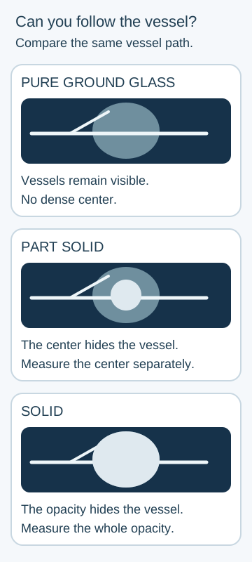
Can you still see the vessels through the opacity?
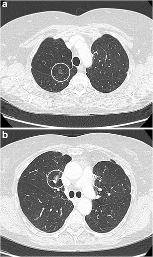
On thin-section CT, ground glass preserves underlying vessel outlines; solid opacity obscures them. A part-solid nodule contains both appearances. Subsolid is the umbrella term for pure ground-glass and part-solid nodules. Compare the density schematic with the published clinical CT pair: a translucent region and a dense center are different observations, not merely different names for a white spot. [4][10]
Panel a illustrates pure ground glass and panel b part-solid tissue. These are recognition examples from a patient with prior lung cancer, not cases eligible for routine incidental-nodule surveillance. Snoeckx et al., Figure 2, CC BY 4.0; converted losslessly from static GIF to PNG. Image: Annemie Snoeckx, Pieter Reyntiens, Damien Desbuquoit, Maarten J. Spinhoven, Paul E. Van Schil, Jan P. van Meerbeeck and Paul M. Parizel; original source; CC BY 4.0. [10].
Persistent ground-glass lesions can reflect atypical adenomatous hyperplasia or adenocarcinoma with lepidic growth along alveolar walls. Some are inflammatory, and some cancers have invasive tissue despite a ground-glass appearance. A growing solid component increases concern for invasion, but CT does not directly measure microscopic invasion. Transient infection, fibrosis and partial-volume effects can also produce solid-appearing tissue. [1][10]
Solid nodules include granulomas, hamartomas and several malignant histologies. Density changes risk and follow-up, but it does not create a universal ranking in which every part-solid nodule is more dangerous than every solid nodule. Size, persistence, morphology and clinical setting remain necessary.
Trace a vessel across each schematic opacity. Predict which region would be measured separately when the opacity contains a dense center.
Follow the vessel from left to right before naming the density. These are conceptual opacities, not simulated CT scans; density alone does not determine pathology. [1][4][10]Check the density comparison
The dense center is the solid component. Record its size separately from total nodule size; the surrounding ground-glass region does not cancel its significance.
Now change the setting: the same mixed opacity appears during fever after chemotherapy. The immediate question becomes infection in an immunocompromised patient, not which routine incidental follow-up interval to select. An appearance must be interpreted inside the correct clinical pathway. [1]
Measure change before interpreting growth
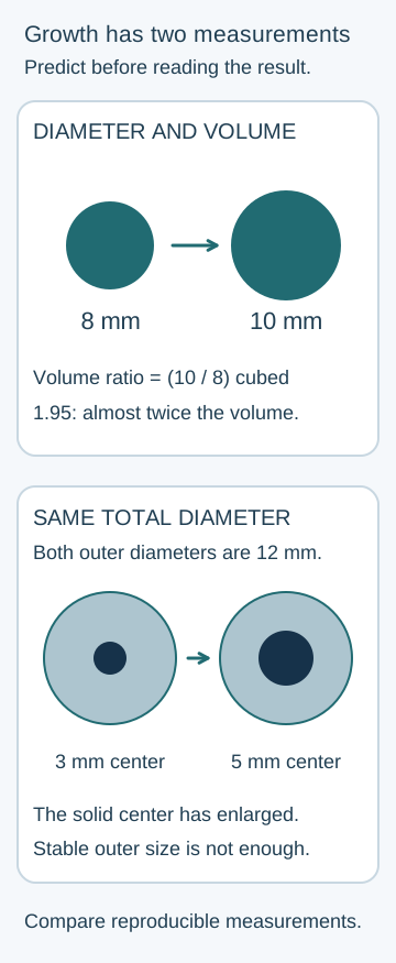
Is an increase from 8 to 10 mm too small to matter? For an approximately spherical nodule, volume is proportional to diameter cubed. The ratio is (10/8)3 = 1.95: almost twice the volume despite only 2 mm of diameter change. This geometric example assumes the same shape and comparable measurements, not that every nodule is a sphere. [2]
Compare thin sections using similar acquisition, reconstruction, plane and window settings. Small nodules are commonly described using the average of long and perpendicular short diameters; document the solid component separately when present. An apparent 1.5 mm difference is not automatically biological growth. Differences around 1.5 to 2 mm can be poorly reproducible; a reproducible change of at least 2 mm is a practical diameter threshold for concern. Volumetry has its own segmentation and reproducibility limits. [2][4]
For measured volumes, doubling time = elapsed time × ln(2) / ln(final volume / initial volume). If volume doubles over 180 days, the volume doubling time is 180 days. Diameter doubling would represent eight times the volume, not one volume doubling.
Many malignant solid nodules have volume doubling times around 20 to 400 days, but this is not a diagnostic cutoff. Rapid growth can occur with infection; slow-growing cancer exists, especially among subsolid lesions. A nodule that gets smaller but does not disappear may still require follow-up. [2]
Two years of reliable stability is reassuring for a solid nodule, not a universal clearance rule. Persistent subsolid nodules can change much later. Compare both the outer diameter and the solid center; an unchanged outer boundary can conceal an enlarging solid component. [1]
Use the growth comparison. First predict the volume change from 8 to 10 mm. Then compare two 12 mm part-solid outlines whose centers measure 3 and 5 mm. Decide whether stable total size is sufficient reassurance.
The circles use proportional diameters. First calculate the volume ratio, then compare the dense centers independently of their unchanged outer borders. Use measured CT dimensions rather than assuming every nodule is spherical. [1][2][4]Check the two growth predictions
The first nodule nearly doubles in volume. In the second pair, the solid center enlarges despite a stable outer diameter. Reproducible solid-component growth warrants reassessment even before it reaches 6 mm.
For transfer, imagine the reported change is only 1 mm and the first scan used much thicker slices. Request a comparable image review before labeling progression. Conversely, do not dismiss consistent growth on several comparable studies because each individual increase looks small. [3][4]
Probability determines what the next test should accomplish
Would the same negative PET result reassure you equally at a starting cancer probability of 5% and 80%? No. A test changes an existing probability; it does not erase the history and CT findings. Increasing size raises concern, but a size band alone cannot assign a reliable individual percentage. [2]
The Brock model incorporates age, sex, family history of lung cancer, emphysema, nodule size, density, upper-lobe location, spiculation and nodule count. Its development cohorts were people undergoing screening. The Mayo model uses age, smoking, diameter, spiculation, upper-lobe location and a history of extrathoracic cancer more than five years earlier. Active or recent cancer is not just another interchangeable entry in that original model. [5][6]
Use the full specified equation or a validated implementation, not a remembered subset of variables. Check units, how binary findings are coded and whether the patient resembles the validation population. Good discrimination between groups does not guarantee accurate calibration for one patient, and neither model substitutes for review of a changing nodule.
A logistic model converts its score x to probability with p = ex / (1 + ex). If a correctly completed Mayo calculation gives x = -1.5, ex is about 0.223 and p is about 18%. For a 12 mm solid nodule, that is an intermediate-probability problem, not an 82% probability requiring immediate surgery. This is a worked conversion, not an independently validated risk calculator. [2]
ACCP framework for an indeterminate solid nodule larger than 8 mm
Estimated probability
Typical decision
Estimated probabilityBelow 5%
Typical decisionCT surveillance is generally appropriate.
Estimated probability5% through 65%
Typical decisionPET can refine risk; tissue sampling depends on the result, discordance and patient preferences.
Estimated probabilityAbove 65%
Typical decisionDiscuss definitive diagnosis or treatment. An operable patient may choose surgical diagnosis; PET may still be useful for staging.
These are named ACCP probability thresholds, not Fleischner size categories or Lung-RADS categories. The decision also depends on surgical fitness, biopsy risk and what the patient values. Very high procedural risk can change the preferred strategy even when cancer probability is substantial. [2]
Start with probability 20%. The odds are 0.20/0.80 = 0.25. With a hypothetical negative likelihood ratio of 0.20, calculate the new odds before choosing surveillance or another test.
Check the probability update
The new odds are 0.25 × 0.20 = 0.05. Probability is 0.05/1.05, about 4.8%. A reassuring test can support surveillance, but it does not make the probability zero.
Now keep that likelihood ratio but start at 80%. Odds 4 become 0.8, corresponding to about 44%. The same negative result leaves substantial concern. Actual PET performance varies with lesion size and biology; the example does not assign every negative PET a likelihood ratio of 0.20.
Suspicious appearance is evidence, not a tissue diagnosis
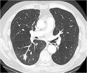
Does a spiculated border prove cancer? It raises concern but does not prove it. In the clinical CT, examine the irregular extensions around the nodule rather than its brightness alone. Spiculation, sometimes called corona radiata, can reflect a tumor-associated stromal response or infiltration; scarring and inflammation can resemble it. The published example had malignant pathology, but its CT appearance alone does not establish a histologic subtype. [10]
Inspect the fine extensions at the nodule border. The original report documents malignant pathology, but this morphology alone does not identify a histologic subtype. The published patient had a cancer history; this image teaches recognition, not a routine incidental surveillance assignment. Snoeckx et al., Figure 11, CC BY 4.0; unchanged publisher image. Image: Annemie Snoeckx, Pieter Reyntiens, Damien Desbuquoit, Maarten J. Spinhoven, Paul E. Van Schil, Jan P. van Meerbeeck and Paul M. Parizel; original source; CC BY 4.0. [10].
Upper-lobe location, pleural retraction, reproducible growth and a new or enlarging solid component increase concern. An irregular thick-walled cavity is concerning, but cavitation also occurs in infection. Upper-lobe location is an observed risk association; no unverified explanation about preferential carcinogen deposition is needed to use it appropriately. [1][2]
Calcification must be described, not merely noticed. Diffuse, central or laminated calcification often supports a healed granuloma. Popcorn calcification with macroscopic fat supports a hamartoma. Eccentric or punctate calcification is not reliably benign; cancer can surround an older calcified focus. A smooth border alone also cannot exclude malignancy. [2][10]
Compare two otherwise similar solid nodules: one contains central layered calcification, while the other has an eccentric calcified dot within a growing spiculated margin. Predict which description leaves a malignant process unresolved.
Check the calcification comparison
The second pattern remains suspicious. The location and distribution of calcium matter; finding any calcium is not sufficient to cancel documented growth and spiculation.
Symptoms can change the pathway independently of nodule size. Unexplained hemoptysis, persistent or changing cough with weight loss, recurrent focal chest infections and suspicious lymphadenopathy require diagnostic attention. In NICE NG12, unexplained hemoptysis at age 40 or older warrants a suspected lung cancer referral. A low-risk 4 mm nodule does not explain away such symptoms or justify delaying their assessment. [9]
For transfer, an otherwise stable patient develops recurrent blood-streaked sputum after earlier CT documented a tiny nodule. Reassess the symptom rather than simply shortening or lengthening the old nodule timetable. Significant ongoing bleeding, respiratory compromise or circulatory instability requires emergency assessment, not an outpatient surveillance appointment.
Choose a test that changes the decision
Must the lung nodule itself always be the first biopsy target? No. Review imaging before sampling and select a safe target that provides useful diagnosis and stage. A suspicious accessible node may answer both questions in one procedure; removing the lung lesion first can leave a treatment-changing staging question unanswered. [8]
FDG-PET measures glucose uptake, not cancer cells directly. Infection and granulomatous inflammation can be avid. Small lesions, ground-glass lesions and some indolent or low-uptake tumors can be nonavid despite malignancy. PET is most useful for characterization of a sufficiently large solid target when its result can change management; a negative scan cannot reliably clear a small solid focus within a subsolid nodule. [2]
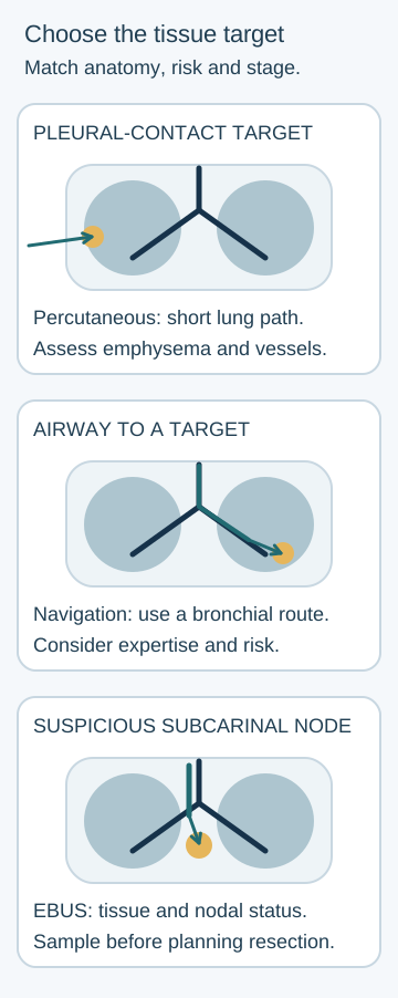
For a peripheral lesion near the pleura, CT-guided transthoracic sampling may offer a direct route. A longer path through aerated or emphysematous lung raises pneumothorax concern. An airway leading to a lesion can support bronchoscopic sampling, including navigation and radial endobronchial ultrasound. These methods also reach selected peripheral lesions; they are not restricted to central nodules. Convex-probe EBUS-guided needle aspiration targets accessible hilar or mediastinal nodes, including station 7 in the subcarinal region. [2][8]
The 2025 VERITAS trial compared navigational bronchoscopy with transthoracic biopsy for selected peripheral nodules measuring 10 to 30 mm. Diagnostic accuracy was 79.0% versus 73.6%, meeting the trial's noninferiority criterion, not demonstrating superiority. Pneumothorax occurred in 3.3% versus 28.3%. These are trial-specific results with particular techniques and expertise, not universal complication probabilities for every center or nodule. [7]
Trace the three routes in the sampling diagram. For a hilar lesion with a suspicious station 7 node, choose the route that can provide both tissue and nodal staging before an operation is planned.
Trace the route to each gold target. A suspicious accessible node can establish diagnosis and nodal involvement together. Compare which accessible target yields both diagnosis and staging information. [2][7][8]Check the sampling route
EBUS-guided aspiration of the accessible node can establish diagnosis and nodal involvement. A transthoracic sample of the primary lesion alone would not answer the nodal question.
A nondiagnostic biopsy is not a benign diagnosis. Reconcile it with imaging, estimated probability and whether the specimen actually sampled the lesion. High suspicion despite nondiagnostic sampling can justify another approach or surgical diagnosis. A fit patient with high probability who prefers definitive diagnosis may choose thoracoscopic diagnostic excision, with an oncologic and nodal-staging plan if cancer is confirmed. Operative extent requires the full clinical assessment; it is not determined solely by a 65% threshold. [2]
For transfer, change the CT to include a possible solitary adrenal metastasis. Confirm that treatment-changing finding with appropriate imaging or tissue rather than declaring metastatic cancer from uptake alone. Similarly, negative endosonographic nodal sampling may need further staging when suspicion remains high and nodal status would alter treatment. [8]
Match surveillance to the correct kind of nodule
Why is there no single follow-up interval for an 8 mm opacity? Density, clinical setting and change determine what the next scan needs to establish. Use low-dose, noncontrast thin-section CT for routine nodule surveillance when appropriate. The schedules below summarize single incidental nodules in eligible adults, not screening findings, patients with known cancer, or immunocompromised patients. Multiple nodules require their separate recommendations. [1][2]
Solid incidental nodules
Fleischner 2017: size and patient risk
Finding
Follow-up approach
FindingSmaller than 6 mm
Follow-up approachNo routine CT in low-risk patients. In high-risk patients, a CT at 12 months is optional, particularly with suspicious morphology or upper-lobe location.
Finding6 to 8 mm
Follow-up approachCT at 6 to 12 months. In low-risk patients, consider another at 18 to 24 months; in high-risk patients, obtain that second study.
FindingLarger than 8 mm
Follow-up approachConsider CT at about 3 months, PET-CT or tissue sampling according to probability and clinical circumstances, rather than automatically waiting 6 to 12 months.
No routine follow-up for a low-risk tiny nodule means the expected benefit of repeated testing is small; it does not mean the nodule is a normal variant, impossible to be malignant, or a reason to disregard future symptoms. Conversely, size above 8 mm does not make biopsy mandatory for every patient. [1]
Subsolid incidental nodules
Fleischner 2017: persistence and the solid component
Finding
Follow-up approach
FindingPure ground glass smaller than 6 mm
Follow-up approachGenerally no routine CT. Selected suspicious or near-threshold lesions can justify individualized longer-term follow-up.
FindingPure ground glass at least 6 mm
Follow-up approachCT at 6 to 12 months to confirm persistence, then every 2 years until 5 years if unchanged.
FindingPart solid, total size at least 6 mm
Follow-up approachCT at 3 to 6 months to confirm persistence. If unchanged and the solid component remains below 6 mm, annual CT for 5 years.
FindingPersistent part solid with solid component at least 6 mm, or reproducible solid-component growth
Follow-up approachReassess promptly for malignancy with specialist-directed diagnostic planning. Do not treat this as routine stable-subsolid surveillance.
A pure ground-glass lesion does not become an automatic surgical target merely because its total diameter crosses 10 mm. Persistence, growth, emerging solid tissue, suspicious features and patient factors inform diagnostic escalation. Follow-up beyond the usual 5-year subsolid schedule is individualized rather than a universal instruction. [1][2]
Screening has its own clock
Under Lung-RADS v2022, a baseline solid nodule measuring 8 to less than 15 mm is category 4A: repeat low-dose CT in 3 months, with PET-CT an option when the solid nodule or component is at least 8 mm. A new 6 to less than 8 mm solid nodule is also category 4A, although a baseline 6 to less than 8 mm solid nodule is category 3 with 6-month LDCT. Newness matters. [3]
Compare two incidental 10 mm subsolid nodules. One remains entirely ground glass; the other has a 4 mm solid center. Predict which needs an earlier first persistence check and, if unchanged, more frequent long-term CT.
Check the two surveillance clocks
The part-solid nodule needs its initial check at 3 to 6 months and then annual CT if stable with a solid component below 6 mm. The pure ground-glass nodule starts at 6 to 12 months and then uses the every-2-year schedule.
For transfer, a category 4A screening nodule is unchanged at 3 months. Do not declare it cleared: Lung-RADS v2022 uses stepped management, generally category 3 with another LDCT in 6 months for this situation, excluding its special airway-nodule pathway. Name the guideline and document the next scan rather than switching between screening and incidental schedules. [3]
Apply the lesson
Case 1
Show answer and explanations for case 1
A. Category 2; LDCT in 12 months (Why this does not fit)
Baseline solid nodules smaller than 6 mm commonly fit category 2. This lesion measures 9 mm, so its smooth margin does not justify the category 2 interval.
Reasoning steps for option A
Which baseline solid nodules fit category 2?
Baseline solid nodules smaller than 6 mm commonly fit category 2.
Does the measured lesion fit that size?
This lesion measures 9 mm, so its smooth margin does not justify the category 2 interval.
B. Category 3; LDCT in 6 months (Why this does not fit)
A baseline solid nodule from 6 to less than 8 mm fits category 3. Nine millimeters exceeds the category 3 baseline size band.
Reasoning steps for option B
Which baseline solid size fits category 3?
A baseline solid nodule from 6 to less than 8 mm fits category 3.
How does 9 mm change that assignment?
Nine millimeters exceeds the category 3 baseline size band.
C. Category 4A; LDCT in 3 months (Best answer)
A baseline solid nodule from 8 to less than 15 mm meets category 4A size criteria. Three-month LDCT is the category 4A follow-up; PET may also be considered because the solid target is at least 8 mm.
Reasoning steps for option C
Which baseline size band applies?
A baseline solid nodule from 8 to less than 15 mm meets category 4A size criteria.
What scheduled imaging follows here?
Three-month LDCT is the category 4A follow-up; PET may also be considered because the solid target is at least 8 mm.
D. Category 4B; diagnostic CT now (Why this does not fit)
A baseline solid nodule at least 15 mm meets category 4B size criteria. The nodule is 9 mm and lacks supplied additional findings requiring that higher category.
Reasoning steps for option D
Which baseline solid size meets category 4B?
A baseline solid nodule at least 15 mm meets category 4B size criteria.
Does this examination establish that category?
The nodule is 9 mm and lacks supplied additional findings requiring that higher category.
E. Category 0; LDCT in 1 month (Why this does not fit)
Category 0 can apply when evaluation is incomplete or findings suggest an acute inflammatory process. The lesion is fully characterized and no acute inflammatory findings are supplied; lack of an earlier scan alone does not invalidate a baseline examination.
Reasoning steps for option E
When can an examination be incomplete?
Category 0 can apply when evaluation is incomplete or findings suggest an acute inflammatory process.
Is the present scan incomplete?
The lesion is fully characterized and no acute inflammatory findings are supplied; lack of an earlier scan alone does not invalidate a baseline examination.
Takeaway: Choose the screening framework before assigning the size-based interval.
A. No routine nodule-surveillance CT (Best answer)
A single solid nodule smaller than 6 mm in an eligible low-risk adult generally needs no routine follow-up. The incidental 5 mm smooth lesion and low-risk history fit; this is a risk-based decision, not proof that cancer is impossible.
Reasoning steps for option A
Which combination permits no routine CT?
A single solid nodule smaller than 6 mm in an eligible low-risk adult generally needs no routine follow-up.
Does this patient fit that combination?
The incidental 5 mm smooth lesion and low-risk history fit; this is a risk-based decision, not proof that cancer is impossible.
B. Thin-section CT in 6 to 12 months (Why this does not fit)
Six-to-eight-millimeter solid nodules generally receive CT at 6 to 12 months. The measured diameter is 5 mm, and no high-risk feature changes its low-risk classification.
Reasoning steps for option B
Which solid size usually uses this interval?
Six-to-eight-millimeter solid nodules generally receive CT at 6 to 12 months.
Does this lesion enter that band?
The measured diameter is 5 mm, and no high-risk feature changes its low-risk classification.
C. Thin-section CT in 3 months (Why this does not fit)
A solid nodule larger than 8 mm or concerning clinical change can justify a short diagnostic interval. The lesion is much smaller and the patient has no concerning trajectory or symptoms.
Reasoning steps for option C
When is short-interval evaluation useful?
A solid nodule larger than 8 mm or concerning clinical change can justify a short diagnostic interval.
Is that indication present?
The lesion is much smaller and the patient has no concerning trajectory or symptoms.
D. FDG-PET/CT for characterization (Why this does not fit)
PET is more useful for sufficiently large solid targets when risk refinement changes management. A 5 mm low-risk nodule provides a small target and a low probability of benefit from PET.
Reasoning steps for option D
What target makes PET more informative?
PET is more useful for sufficiently large solid targets when risk refinement changes management.
Why is PET poorly matched here?
A 5 mm low-risk nodule provides a small target and a low probability of benefit from PET.
E. CT-guided percutaneous biopsy (Why this does not fit)
Biopsy may resolve an indeterminate lesion when cancer probability and management consequences justify procedural risk. The tiny low-risk lesion offers little expected benefit from an invasive test and can be technically difficult to sample.
Reasoning steps for option E
When can tissue sampling be worthwhile?
Biopsy may resolve an indeterminate lesion when cancer probability and management consequences justify procedural risk.
What weakens that balance here?
The tiny low-risk lesion offers little expected benefit from an invasive test and can be technically difficult to sample.
Takeaway: No routine surveillance is not the same as declaring a nodule biologically irrelevant.
Larger or substantially suspicious lesions may need short-interval CT or another diagnostic test. The question asks for optional surveillance of a single sub-6-mm solid nodule, for which the guideline interval is later.
Reasoning steps for option A
Which lesions commonly need a 3-month diagnostic assessment?
Larger or substantially suspicious lesions may need short-interval CT or another diagnostic test.
What schedule is asked for here?
The question asks for optional surveillance of a single sub-6-mm solid nodule, for which the guideline interval is later.
B. Repeat CT in 6 months (Why this does not fit)
A 6-to-8-mm solid nodule is ordinarily first followed at 6 to 12 months. It does not; high-risk features permit an optional 12-month scan in this smaller-size category.
Reasoning steps for option B
Which solid size band commonly starts around 6 months?
A 6-to-8-mm solid nodule is ordinarily first followed at 6 to 12 months.
Does a 5 mm diameter meet that band?
It does not; high-risk features permit an optional 12-month scan in this smaller-size category.
C. Repeat CT in 18 months (Why this does not fit)
Eighteen to 24 months is a later follow-up window for selected larger solid nodules. It is not the recommended optional first interval for a high-risk sub-6-mm solid nodule.
Reasoning steps for option C
Where does an 18-month interval usually appear?
Eighteen to 24 months is a later follow-up window for selected larger solid nodules.
Is it the optional first scan for this lesion?
It is not the recommended optional first interval for a high-risk sub-6-mm solid nodule.
D. Repeat CT in 24 months (Why this does not fit)
Persistent pure ground-glass nodules can have scans separated by 2 years after persistence is established. This is a solid nodule, not a persistent pure ground-glass lesion.
Reasoning steps for option D
What follow-up pattern can include 2-year spacing?
Persistent pure ground-glass nodules can have scans separated by 2 years after persistence is established.
Does that density pattern apply?
This is a solid nodule, not a persistent pure ground-glass lesion.
E. Repeat CT in 12 months (Best answer)
High-risk history, suspicious morphology or upper-lobe location can support an optional CT at 12 months. She has those risk features and has chosen surveillance, so the optional 12-month interval fits.
Reasoning steps for option E
What changes the otherwise no-routine-follow-up approach?
High-risk history, suspicious morphology or upper-lobe location can support an optional CT at 12 months.
How does that apply after shared decision-making?
She has those risk features and has chosen surveillance, so the optional 12-month interval fits.
Takeaway: For a high-risk single solid nodule below 6 mm, the optional scan is at 12 months.
A. End CT surveillance after this scan (Why this does not fit)
Two-year stability is strong evidence of benignity for an otherwise indeterminate solid nodule. This nodule is pure ground glass, so the solid-nodule stability rule does not apply.
Reasoning steps for option A
For which density is 2-year stability strongly reassuring?
Two-year stability is strong evidence of benignity for an otherwise indeterminate solid nodule.
Does the density here support ending follow-up?
This nodule is pure ground glass, so the solid-nodule stability rule does not apply.
B. Continue CT every 2 years through year 5 (Best answer)
Persistence was confirmed on the 9-month CT. An unchanged lesion follows the every-2-year surveillance approach through 5 years rather than ending at 2 years.
Reasoning steps for option B
What has the earlier scan already established?
Persistence was confirmed on the 9-month CT.
What does persistent pure ground glass at least 6 mm require?
An unchanged lesion follows the every-2-year surveillance approach through 5 years rather than ending at 2 years.
C. Begin annual CT for the next 5 years (Why this does not fit)
Annual surveillance is used for a qualifying part-solid nodule whose solid component remains below 6 mm. There is no solid component, and the surveillance horizon does not restart automatically at the current scan.
Reasoning steps for option C
Which persistent subsolid lesion uses annual CT?
Annual surveillance is used for a qualifying part-solid nodule whose solid component remains below 6 mm.
What feature is missing here?
There is no solid component, and the surveillance horizon does not restart automatically at the current scan.
D. Obtain PET-CT before ending surveillance (Why this does not fit)
Ground-glass lesions can have low uptake despite malignancy. A negative result would not reliably exclude indolent cancer or replace the recommended CT follow-up.
Reasoning steps for option D
What can a negative PET establish in a ground-glass lesion?
Ground-glass lesions can have low uptake despite malignancy.
Would PET justify stopping this schedule?
A negative result would not reliably exclude indolent cancer or replace the recommended CT follow-up.
E. Arrange diagnostic resection after this scan (Why this does not fit)
Growth, emerging solid tissue or other suspicious evolution can prompt diagnostic escalation. Neither size nor density has changed, so persistence of this small stable pure ground-glass lesion alone does not mandate resection.
Reasoning steps for option E
Which changes would increase concern for intervention?
Growth, emerging solid tissue or other suspicious evolution can prompt diagnostic escalation.
What has changed in this patient?
Neither size nor density has changed, so persistence of this small stable pure ground-glass lesion alone does not mandate resection.
Takeaway: Stable pure ground glass requires a longer surveillance horizon than stable solid tissue.
A. No further scheduled nodule CT (Why this does not fit)
Some transient inflammatory subsolid nodules resolve on short-interval CT. It persisted with a solid component, so resolution has not provided reassurance.
Reasoning steps for option A
Does persistence establish benignity?
Some transient inflammatory subsolid nodules resolve on short-interval CT.
What happened to this nodule?
It persisted with a solid component, so resolution has not provided reassurance.
B. CT every 2 years through year 5 (Why this does not fit)
Persistent pure ground-glass nodules at least 6 mm generally use every-2-year follow-up. The 4 mm solid center makes this part solid, requiring the annual rather than the pure ground-glass schedule.
Reasoning steps for option B
Which density uses this longer spacing?
Persistent pure ground-glass nodules at least 6 mm generally use every-2-year follow-up.
Does the measured solid component matter?
The 4 mm solid center makes this part solid, requiring the annual rather than the pure ground-glass schedule.
C. CT at year 2, then end surveillance (Why this does not fit)
Stable solid nodules can generally finish routine follow-up after an adequate 2-year comparison. Part-solid nodules can evolve slowly and use a longer observation period.
Reasoning steps for option C
Which nodules often finish after 2-year stability?
Stable solid nodules can generally finish routine follow-up after an adequate 2-year comparison.
Why is that horizon insufficient here?
Part-solid nodules can evolve slowly and use a longer observation period.
D. Annual CT surveillance for 5 years (Best answer)
The part-solid lesion is persistent and unchanged, with a solid component below 6 mm. Annual CT for 5 years is the Fleischner approach for this qualifying stable part-solid pattern.
Reasoning steps for option D
What has the 4-month CT confirmed?
The part-solid lesion is persistent and unchanged, with a solid component below 6 mm.
Which ongoing schedule follows?
Annual CT for 5 years is the Fleischner approach for this qualifying stable part-solid pattern.
E. CT every 3 months for 5 years (Why this does not fit)
The initial 3-to-6-month CT determines whether a part-solid finding persists. No; after persistence is confirmed without suspicious evolution, the recommended schedule becomes annual.
Reasoning steps for option E
Why was the first interval short?
The initial 3-to-6-month CT determines whether a part-solid finding persists.
Must that short interval continue unchanged?
No; after persistence is confirmed without suspicious evolution, the recommended schedule becomes annual.
Takeaway: A stable small solid center changes a subsolid nodule from every-2-year to annual follow-up.
A. Low PET uptake supports ending nodule follow-up (Why this does not fit)
A negative PET can reduce probability for a suitable solid target in an appropriate risk setting. The small solid focus and subsolid biology limit PET reassurance, while the measured solid component has enlarged.
Reasoning steps for option A
When can low uptake lower cancer probability?
A negative PET can reduce probability for a suitable solid target in an appropriate risk setting.
Does it settle this evolving subsolid lesion?
The small solid focus and subsolid biology limit PET reassurance, while the measured solid component has enlarged.
B. Solid growth supports diagnostic tissue planning (Best answer)
The solid component has reproducibly enlarged and now exceeds 6 mm. The structural progression remains suspicious despite low uptake and warrants specialist-directed tissue or definitive diagnostic planning.
Reasoning steps for option B
What does the comparable CT series establish?
The solid component has reproducibly enlarged and now exceeds 6 mm.
How should that interact with the PET result?
The structural progression remains suspicious despite low uptake and warrants specialist-directed tissue or definitive diagnostic planning.
C. Stable total size supports CT again in 2 years (Why this does not fit)
The total outer diameter is only one measure of subsolid behavior. Growth of the dense component can signal progression even when the outer diameter changes little.
Reasoning steps for option C
Which change is important in part-solid lesions?
The total outer diameter is only one measure of subsolid behavior.
Why is a 2-year delay poorly supported?
Growth of the dense component can signal progression even when the outer diameter changes little.
D. Persistent ground glass supports annual CT alone (Why this does not fit)
A stable persistent nodule with a solid component below 6 mm can follow the annual schedule. The component has grown to 7 mm, so the stable small-component recommendation no longer describes it.
Reasoning steps for option D
Which part-solid pattern can use annual CT?
A stable persistent nodule with a solid component below 6 mm can follow the annual schedule.
Does this patient remain in that pattern?
The component has grown to 7 mm, so the stable small-component recommendation no longer describes it.
E. Low PET uptake supports an inflammatory diagnosis (Why this does not fit)
Small or indolent malignant lesions may be nonavid as well as benign lesions. Progressive solid growth over a year without an acute infectious syndrome leaves malignancy unresolved.
Reasoning steps for option E
What processes can have low FDG uptake?
Small or indolent malignant lesions may be nonavid as well as benign lesions.
What prevents attributing this lesion to inflammation?
Progressive solid growth over a year without an acute infectious syndrome leaves malignancy unresolved.
Takeaway: A nonavid PET does not cancel reproducible solid-component growth.
A. Volume rises about 25%; routine stability is established (Why this does not fit)
Diameter increases from 8 to 10 mm, a 25% increase. Volume depends on the cube of diameter, and the reproducible diameter change does not establish stability.
Reasoning steps for option A
Which quantity rises by 25%?
Diameter increases from 8 to 10 mm, a 25% increase.
Does the same percentage describe volume or stability?
Volume depends on the cube of diameter, and the reproducible diameter change does not establish stability.
B. Volume rises about 56%; a benign course is established (Why this does not fit)
Squaring 10/8 gives 1.56, which approximates a cross-sectional area ratio. Volume requires cubing that ratio, and growth alone cannot identify a benign cause.
Reasoning steps for option B
What calculation produces about 56%?
Squaring 10/8 gives 1.56, which approximates a cross-sectional area ratio.
Does area establish nodule volume or benignity?
Volume requires cubing that ratio, and growth alone cannot identify a benign cause.
C. Volume increases eightfold; urgent infection treatment is indicated (Why this does not fit)
Doubling diameter increases volume by a factor of eight. Diameter increased only from 8 to 10 mm, and there is no supplied evidence establishing an infectious process.
Reasoning steps for option C
When does sphere volume increase eightfold?
Doubling diameter increases volume by a factor of eight.
Did diameter double or infection become established?
Diameter increased only from 8 to 10 mm, and there is no supplied evidence establishing an infectious process.
D. Volume is unchanged; the difference is below CT resolution (Why this does not fit)
Small differences on dissimilar CT examinations can reflect technique and reader variability. The same thin-section technique and independent confirmation support a real 2 mm increase.
Reasoning steps for option D
When can apparent size change be measurement error?
Small differences on dissimilar CT examinations can reflect technique and reader variability.
What supports true change here?
The same thin-section technique and independent confirmation support a real 2 mm increase.
E. Volume nearly doubles; malignancy risk needs reassessment (Best answer)
The ratio (10/8) cubed is about 1.95, nearly one volume doubling. Growth over 180 days in a smoker raises concern and warrants diagnostic reassessment, without proving cancer.
Reasoning steps for option E
What is the volume ratio?
The ratio (10/8) cubed is about 1.95, nearly one volume doubling.
What follows from that reliable growth?
Growth over 180 days in a smoker raises concern and warrants diagnostic reassessment, without proving cancer.
Takeaway: A 25% diameter increase can represent almost twice the tissue volume.
A. Classify confirmed growth and arrange surgical diagnosis (Why this does not fit)
Clear malignant-pattern growth can justify tissue or surgical diagnosis when overall risk supports it. The 1.2 mm difference was measured with different slice thicknesses and planes, so progression is not yet securely established.
Reasoning steps for option A
When can surgery become reasonable after surveillance?
Clear malignant-pattern growth can justify tissue or surgical diagnosis when overall risk supports it.
Has comparable growth actually been demonstrated?
The 1.2 mm difference was measured with different slice thicknesses and planes, so progression is not yet securely established.
B. Classify 2-year stability and end follow-up (Why this does not fit)
A reliable approximately 2-year comparison can support ending follow-up for a solid nodule. Only six months and inconsistent measurements are available; neither duration nor technique establishes that endpoint.
Reasoning steps for option B
What observation supports a solid-nodule stability decision?
A reliable approximately 2-year comparison can support ending follow-up for a solid nodule.
What comparison is available here?
Only six months and inconsistent measurements are available; neither duration nor technique establishes that endpoint.
C. Review both scans using comparable measurements (Best answer)
Small diameter differences can reflect reconstruction thickness, plane and observer variability. Reconstruction and side-by-side review can test whether the reported 1.2 mm difference represents reproducible growth.
Reasoning steps for option C
Why compare technique before interpreting this difference?
Small diameter differences can reflect reconstruction thickness, plane and observer variability.
What can the available original images resolve?
Reconstruction and side-by-side review can test whether the reported 1.2 mm difference represents reproducible growth.
D. Obtain PET to determine whether diameter increased (Why this does not fit)
PET provides metabolic information that can refine risk in selected adequately sized nodules. It does not resolve the technical size-comparison problem, and an approximately 8 mm target also limits sensitivity.
Reasoning steps for option D
What does PET measure?
PET provides metabolic information that can refine risk in selected adequately sized nodules.
Can it validate these mismatched diameter measurements?
It does not resolve the technical size-comparison problem, and an approximately 8 mm target also limits sensitivity.
E. Treat presumed infection and rescan after antibiotics (Why this does not fit)
An infectious syndrome with compatible imaging may justify treatment and targeted reassessment. No infectious symptoms or supporting imaging pattern is supplied; the immediate uncertainty is measurement comparability.
Reasoning steps for option E
When would infection-directed treatment be justified?
An infectious syndrome with compatible imaging may justify treatment and targeted reassessment.
Which supplied facts support that diagnosis?
No infectious symptoms or supporting imaging pattern is supplied; the immediate uncertainty is measurement comparability.
Takeaway: Resolve technical measurement uncertainty before treating it as biological progression.
A. The solid lesion can finish follow-up; the ground-glass lesion needs longer observation (Best answer)
Reliable stability beyond two years strongly supports a benign solid lesion. The persistent ground-glass nodule can evolve more slowly, so the solid-nodule endpoint should not end its longer-term observation.
Reasoning steps for option A
How does prolonged stability inform a solid nodule?
Reliable stability beyond two years strongly supports a benign solid lesion.
Why does the second lesion need a different horizon?
The persistent ground-glass nodule can evolve more slowly, so the solid-nodule endpoint should not end its longer-term observation.
B. Both lesions can finish follow-up because each is stable (Why this does not fit)
An unchanged lesion over an adequate time interval is less concerning than a growing one. Adequate observation depends on density; 30-month stability does not complete the usual subsolid surveillance horizon.
Reasoning steps for option B
What makes stability reassuring?
An unchanged lesion over an adequate time interval is less concerning than a growing one.
Why is one common stopping point inappropriate?
Adequate observation depends on density; 30-month stability does not complete the usual subsolid surveillance horizon.
C. The solid lesion needs PET; the ground-glass lesion can finish follow-up (Why this does not fit)
A newly detected solid nodule larger than 8 mm can warrant probability-guided PET evaluation. The solid nodule has prolonged reliable stability, while the ground-glass lesion still has the longer observation requirement.
Reasoning steps for option C
Why might a new 9 mm solid lesion prompt PET consideration?
A newly detected solid nodule larger than 8 mm can warrant probability-guided PET evaluation.
What reverses that reasoning here?
The solid nodule has prolonged reliable stability, while the ground-glass lesion still has the longer observation requirement.
D. Both lesions require diagnostic resection because they exceed 6 mm (Why this does not fit)
That threshold changes follow-up recommendations for several incidental nodule types. No; stable behavior and density determine surveillance, and neither lesion has supplied progressive or highly suspicious findings.
Reasoning steps for option D
What does crossing 6 mm often change?
That threshold changes follow-up recommendations for several incidental nodule types.
Does the threshold itself establish a need for resection?
No; stable behavior and density determine surveillance, and neither lesion has supplied progressive or highly suspicious findings.
E. The ground-glass lesion needs PET before either lesion can finish follow-up (Why this does not fit)
Metabolic imaging may refine cancer probability for suitable solid targets. Low uptake cannot reliably clear a ground-glass lesion; its remaining question is long-term CT behavior.
Reasoning steps for option E
What is PET intended to add?
Metabolic imaging may refine cancer probability for suitable solid targets.
Why is it not the missing endpoint test here?
Low uptake cannot reliably clear a ground-glass lesion; its remaining question is long-term CT behavior.
Takeaway: Judge the surveillance horizon of each lesion by its density, not by the oldest scan alone.
A. About 82%; discuss surgical diagnosis (Why this does not fit)
It approximates one minus the calculated probability. No; using the complement incorrectly converts an intermediate-probability lesion into a high-probability one.
Reasoning steps for option A
What does 82% represent in this calculation?
It approximates one minus the calculated probability.
Is the complement the malignancy estimate?
No; using the complement incorrectly converts an intermediate-probability lesion into a high-probability one.
B. About 22%; end nodule follow-up (Why this does not fit)
It is e^x, the odds term before dividing by 1 + e^x. Neither 22% nor the correctly calculated probability is sufficiently low to end evaluation of this indeterminate 12 mm nodule.
Reasoning steps for option B
What is the meaning of 0.223?
It is e^x, the odds term before dividing by 1 + e^x.
Would that number justify ending follow-up?
Neither 22% nor the correctly calculated probability is sufficiently low to end evaluation of this indeterminate 12 mm nodule.
C. About 4%; use CT surveillance (Why this does not fit)
A very low probability below 5% often supports CT surveillance. No; 0.223 divided by 1.223 is about 0.182, not 0.04.
Reasoning steps for option C
Which probability would favor surveillance alone?
A very low probability below 5% often supports CT surveillance.
Does the supplied formula produce that value?
No; 0.223 divided by 1.223 is about 0.182, not 0.04.
D. About 18%; obtain FDG-PET/CT (Best answer)
Dividing 0.223 by 1.223 yields approximately 18%. An intermediate-probability solid nodule larger than 8 mm is a suitable setting for risk-refining PET when its result can guide the biopsy decision.
Reasoning steps for option D
How is the probability calculated?
Dividing 0.223 by 1.223 yields approximately 18%.
Why does PET fit the stated decision?
An intermediate-probability solid nodule larger than 8 mm is a suitable setting for risk-refining PET when its result can guide the biopsy decision.
E. About 18%; end nodule follow-up (Why this does not fit)
The probability calculation is approximately 18%. It lies in the ACCP intermediate band, so the patient still needs evaluation rather than discharge from nodule follow-up.
Reasoning steps for option E
Is the numerical estimate correct?
The probability calculation is approximately 18%.
Does that probability support no further assessment?
It lies in the ACCP intermediate band, so the patient still needs evaluation rather than discharge from nodule follow-up.
Takeaway: Calculate probability from odds, then match the result to the clinical decision.
A. The Brock estimate should be doubled to adjust for active cancer (Why this does not fit)
A numerical recalibration requires validation in the relevant patient population. No; inventing a multiplier does not correct use of a screening-derived model in active cancer.
Reasoning steps for option A
What would an adjustment factor require?
A numerical recalibration requires validation in the relevant patient population.
Is a doubling factor supplied or validated here?
No; inventing a multiplier does not correct use of a screening-derived model in active cancer.
B. A never-smoking history excludes a new pulmonary primary (Why this does not fit)
It lowers smoking-related risk compared with an otherwise similar heavy smoker. Lung cancer can occur in never-smokers, and active colorectal cancer creates an additional metastatic concern.
Reasoning steps for option B
How does never-smoking affect risk?
It lowers smoking-related risk compared with an otherwise similar heavy smoker.
Does it eliminate the clinical possibilities here?
Lung cancer can occur in never-smokers, and active colorectal cancer creates an additional metastatic concern.
C. The routine low-risk incidental schedule is sufficient (Why this does not fit)
Its standard pathway is intended for eligible incidental nodules without known cancer. This patient has active cancer and a newly appearing lesion, so routine incidental thresholds do not settle management.
Reasoning steps for option C
Which patients fit routine Fleischner incidental guidance?
Its standard pathway is intended for eligible incidental nodules without known cancer.
Does an active cancer staging scan fit that scope?
This patient has active cancer and a newly appearing lesion, so routine incidental thresholds do not settle management.
D. A small pulmonary nodule should be characterized by PET first (Why this does not fit)
PET can help characterize sufficiently large solid lesions in a suitable clinical setting. At 7 mm, a negative result may be falsely reassuring and does not repair the population mismatch.
Reasoning steps for option D
When can PET refine nodule probability?
PET can help characterize sufficiently large solid lesions in a suitable clinical setting.
What limits its value as the first correction here?
At 7 mm, a negative result may be falsely reassuring and does not repair the population mismatch.
E. Cancer-specific evaluation should replace the incidental algorithm (Best answer)
The Brock development setting and routine incidental guidance do not directly represent this active-cancer staging problem. The treating team should evaluate the new lesion in the context of colorectal cancer, interval appearance and treatment consequences.
Reasoning steps for option E
Which limitation does the clinical setting expose?
The Brock development setting and routine incidental guidance do not directly represent this active-cancer staging problem.
What should guide the plan instead?
The treating team should evaluate the new lesion in the context of colorectal cancer, interval appearance and treatment consequences.
Takeaway: A reassuring number from the wrong population cannot override a treatment-changing new lesion.
A. Probability is 0%; no further CT is needed (Why this does not fit)
Only a perfectly sensitive test could reduce a nonzero prior probability to zero. A negative likelihood ratio of 0.20 reduces risk but leaves residual probability and a need for surveillance.
Reasoning steps for option A
What can a negative test exclude with certainty?
Only a perfectly sensitive test could reduce a nonzero prior probability to zero.
What does the supplied likelihood ratio show?
A negative likelihood ratio of 0.20 reduces risk but leaves residual probability and a need for surveillance.
B. Probability is about 4.8%; CT surveillance is reasonable (Best answer)
Prior odds are 0.20/0.80 = 0.25; multiplying by 0.20 gives updated odds of 0.05. Probability is 0.05/1.05, about 4.8%, supporting the stated preference for serial CT rather than immediate invasive diagnosis.
Reasoning steps for option B
What are the prior and updated odds?
Prior odds are 0.20/0.80 = 0.25; multiplying by 0.20 gives updated odds of 0.05.
How do those odds affect this decision?
Probability is 0.05/1.05, about 4.8%, supporting the stated preference for serial CT rather than immediate invasive diagnosis.
C. Probability is about 20%; PET adds no information (Why this does not fit)
A likelihood ratio of 1 would leave the prior odds unchanged. No; 0.20 lowers the odds substantially, so the updated probability is below the original 20%.
Reasoning steps for option C
When would a test leave odds unchanged?
A likelihood ratio of 1 would leave the prior odds unchanged.
Is the supplied value neutral?
No; 0.20 lowers the odds substantially, so the updated probability is below the original 20%.
D. Probability is about 50%; tissue sampling is required (Why this does not fit)
A likelihood ratio above 1 increases the odds of malignancy. It decreases rather than increases the odds and does not produce a 50% probability.
Reasoning steps for option D
What does a positive risk-refining result generally do?
A likelihood ratio above 1 increases the odds of malignancy.
Does a negative likelihood ratio of 0.20 increase risk?
It decreases rather than increases the odds and does not produce a 50% probability.
E. Probability is about 80%; surgical diagnosis is preferred (Why this does not fit)
Eighty percent is the initial probability of not having cancer. No; the post-test estimate must come from prior cancer odds multiplied by the applicable likelihood ratio.
Reasoning steps for option E
Which starting probability is being confused with cancer risk?
Eighty percent is the initial probability of not having cancer.
Can it be substituted for the post-PET cancer probability?
No; the post-test estimate must come from prior cancer odds multiplied by the applicable likelihood ratio.
Takeaway: A negative test can support surveillance without proving the lesion benign.
A. Inflammatory cells are a competing source of uptake (Best answer)
Activated inflammatory cells can consume substantial glucose and produce an avid PET signal. Recent illness and evidence of prior granulomatous disease make inflammatory uptake plausible, so the nodule still requires risk-based evaluation rather than a PET-only diagnosis.
Reasoning steps for option A
Which nonmalignant cells can increase FDG uptake?
Activated inflammatory cells can consume substantial glucose and produce an avid PET signal.
How do the history and other imaging findings matter?
Recent illness and evidence of prior granulomatous disease make inflammatory uptake plausible, so the nodule still requires risk-based evaluation rather than a PET-only diagnosis.
B. FDG avidity is a tumor-specific tissue signature (Why this does not fit)
Many lung cancers are metabolically active and accumulate FDG. Granulomatous inflammation can produce the same signal, particularly in the supplied setting.
Reasoning steps for option B
Why is uptake concerning?
Many lung cancers are metabolically active and accumulate FDG.
What prevents that observation from being diagnostic?
Granulomatous inflammation can produce the same signal, particularly in the supplied setting.
C. Hilar calcification identifies the separate nodule as benign (Why this does not fit)
It often supports prior healed granulomatous disease in the nodes. No; evidence about the nodes does not establish the pathology of an indeterminate separate nodule.
Reasoning steps for option C
What does central nodal calcification suggest?
It often supports prior healed granulomatous disease in the nodes.
Does that prove the separate nodule has the same cause?
No; evidence about the nodes does not establish the pathology of an indeterminate separate nodule.
D. The distant uptake pattern establishes the primary histology (Why this does not fit)
It can reduce concern for PET-detectable distant spread. It does not establish that the lung nodule is malignant in the first place.
Reasoning steps for option D
What does the absence of distant uptake assess?
It can reduce concern for PET-detectable distant spread.
Does staging information prove the primary diagnosis?
It does not establish that the lung nodule is malignant in the first place.
E. The recent infection excludes a concurrent malignant nodule (Why this does not fit)
It offers a competing inflammatory explanation for the lesion and FDG uptake. No; a prior infection and malignancy can coexist, so the nodule still needs an appropriate diagnostic plan.
Reasoning steps for option E
Why is the illness relevant?
It offers a competing inflammatory explanation for the lesion and FDG uptake.
Does a plausible alternative rule cancer out?
No; a prior infection and malignancy can coexist, so the nodule still needs an appropriate diagnostic plan.
Takeaway: FDG avidity changes probability but does not replace pathology when benign mimics remain plausible.
A. Convex-probe EBUS aspiration of station 7 (Why this does not fit)
It samples accessible subcarinal nodal tissue for diagnosis and staging. There is no suspicious station 7 node, and sampling normal nodal tissue would not directly diagnose this peripheral lesion.
Reasoning steps for option A
What is station 7 sampling designed to assess?
It samples accessible subcarinal nodal tissue for diagnosis and staging.
What target is missing here?
There is no suspicious station 7 node, and sampling normal nodal tissue would not directly diagnose this peripheral lesion.
B. Conventional bronchoscopy with endobronchial biopsy (Why this does not fit)
A visible lesion within an accessible airway is a suitable target. The lesion is peripheral and has no demonstrated airway leading into it.
Reasoning steps for option B
Which lesion is directly reached by endobronchial biopsy?
A visible lesion within an accessible airway is a suitable target.
Does the CT supply that airway target?
The lesion is peripheral and has no demonstrated airway leading into it.
C. Thoracoscopic diagnostic wedge excision (Why this does not fit)
It can establish the nature of a suspicious operable peripheral lesion. She specifically declines diagnostic surgery until tissue confirmation has been attempted nonsurgically.
Reasoning steps for option C
When can a wedge excision provide definitive diagnosis?
It can establish the nature of a suspicious operable peripheral lesion.
What patient preference argues against it as the first procedure?
She specifically declines diagnostic surgery until tissue confirmation has been attempted nonsurgically.
D. CT-guided transthoracic core biopsy (Best answer)
Pleural contact provides a short path, and there is no emphysema along that route. It directly samples the nodule without initial surgery and without relying on an absent bronchial route.
Reasoning steps for option D
Which features favor a percutaneous approach?
Pleural contact provides a short path, and there is no emphysema along that route.
How does it match the decision being made?
It directly samples the nodule without initial surgery and without relying on an absent bronchial route.
E. Cervical mediastinoscopy with node sampling (Why this does not fit)
It can assess selected mediastinal nodes when invasive nodal staging is required. No suspicious mediastinal node is supplied, while the pleural-contact nodule is directly accessible by a less invasive route.
Reasoning steps for option E
What question can mediastinoscopy resolve?
It can assess selected mediastinal nodes when invasive nodal staging is required.
Why is it not the best target here?
No suspicious mediastinal node is supplied, while the pleural-contact nodule is directly accessible by a less invasive route.
Takeaway: Choose the tissue route from the actual target, intervening lung and patient preference.
A. Conventional unguided bronchial washings (Why this does not fit)
Airway-based methods can identify some cancers, particularly when a lesion is accessible to the chosen technique. The peripheral target has a defined bronchial route that can be followed with navigation rather than relying on nonspecific washings.
Reasoning steps for option A
When can bronchial sampling provide a diagnosis?
Airway-based methods can identify some cancers, particularly when a lesion is accessible to the chosen technique.
Why is an unguided wash not the strongest option here?
The peripheral target has a defined bronchial route that can be followed with navigation rather than relying on nonspecific washings.
B. CT-guided transthoracic core biopsy (Why this does not fit)
It can directly sample peripheral nodules with good diagnostic performance. The long path through emphysema raises pneumothorax concern, which is especially important to this patient.
Reasoning steps for option B
What advantage does percutaneous biopsy offer?
It can directly sample peripheral nodules with good diagnostic performance.
What supplied feature increases its procedural cost?
The long path through emphysema raises pneumothorax concern, which is especially important to this patient.
C. Navigational bronchoscopy with targeted biopsy (Best answer)
A visible airway leading into the nodule provides a route for targeted bronchoscopic sampling. Avoiding the long emphysematous transthoracic path fits the preference for lower pneumothorax risk; comparative trial data support this choice in selected peripheral lesions.
Reasoning steps for option C
Which anatomical feature supports navigation?
A visible airway leading into the nodule provides a route for targeted bronchoscopic sampling.
How does that interact with the risk preference?
Avoiding the long emphysematous transthoracic path fits the preference for lower pneumothorax risk; comparative trial data support this choice in selected peripheral lesions.
D. Convex-probe EBUS sampling of normal mediastinal nodes (Why this does not fit)
Suspicious accessible nodes can provide diagnosis and stage. No suspicious node is described; the required tissue target is the peripheral nodule itself.
Reasoning steps for option D
When is nodal EBUS useful?
Suspicious accessible nodes can provide diagnosis and stage.
Would normal-node sampling answer this target question?
No suspicious node is described; the required tissue target is the peripheral nodule itself.
E. Thoracoscopic excision without nonsurgical sampling (Why this does not fit)
It can provide a definitive diagnosis and potentially treatment. A suitable less invasive bronchial route exists, and the patient prioritizes minimizing procedural burden and pneumothorax risk.
Reasoning steps for option E
What can surgical excision accomplish?
It can provide a definitive diagnosis and potentially treatment.
Why is it not the preferred first route here?
A suitable less invasive bronchial route exists, and the patient prioritizes minimizing procedural burden and pneumothorax risk.
Takeaway: Modern navigational bronchoscopy can be appropriate for a peripheral nodule, especially when the percutaneous route is hazardous.
A. CT-guided biopsy of the hilar lesion (Why this does not fit)
It could identify the histology of the hilar lesion. It would not establish whether the subcarinal node contains tumor, while the hilar route may add vascular risk.
Reasoning steps for option A
What could biopsy of the primary lesion establish?
It could identify the histology of the hilar lesion.
What treatment-changing question would remain?
It would not establish whether the subcarinal node contains tumor, while the hilar route may add vascular risk.
B. Sputum cytology on three samples (Why this does not fit)
It can be considered for selected central lesions when a patient cannot tolerate or declines invasive testing. He can tolerate bronchoscopy, and sputum cannot establish the mediastinal nodal status needed for planning.
Reasoning steps for option B
When may sputum cytology be considered?
It can be considered for selected central lesions when a patient cannot tolerate or declines invasive testing.
Why is it not preferred here?
He can tolerate bronchoscopy, and sputum cannot establish the mediastinal nodal status needed for planning.
C. Thoracoscopic resection of the lung lesion (Why this does not fit)
It could establish primary-lesion pathology and potentially treat a localized tumor. A suspicious mediastinal node may change the treatment strategy before a lung operation is undertaken.
Reasoning steps for option C
What benefit could resection provide?
It could establish primary-lesion pathology and potentially treat a localized tumor.
Why should the nodal question be addressed first?
A suspicious mediastinal node may change the treatment strategy before a lung operation is undertaken.
D. EBUS-guided aspiration of the station 7 node (Best answer)
It is the subcarinal nodal station and is accessible here by EBUS-guided aspiration. A malignant sample can provide tissue diagnosis and mediastinal nodal staging in the same procedure.
Reasoning steps for option D
Where is station 7 located?
It is the subcarinal nodal station and is accessible here by EBUS-guided aspiration.
What two questions can this target answer?
A malignant sample can provide tissue diagnosis and mediastinal nodal staging in the same procedure.
E. Cervical mediastinoscopy with node biopsy (Why this does not fit)
It can resolve persistent nodal suspicion after inadequate or negative less invasive staging in selected patients. An accessible suspicious node and bronchoscopy tolerance support endosonographic sampling as the initial invasive approach.
Reasoning steps for option E
When can surgical mediastinal staging be valuable?
It can resolve persistent nodal suspicion after inadequate or negative less invasive staging in selected patients.
Why not choose it first in this vignette?
An accessible suspicious node and bronchoscopy tolerance support endosonographic sampling as the initial invasive approach.
Takeaway: Sample an accessible suspicious node when it can establish diagnosis and stage together.
A. The peripheral lung lesion by transthoracic biopsy (Why this does not fit)
It could establish the histology of the lung lesion. The accessible contralateral supraclavicular node can also establish more extensive nodal disease without traversing lung.
Reasoning steps for option A
What information would the primary sample provide?
It could establish the histology of the lung lesion.
Why is another target more informative here?
The accessible contralateral supraclavicular node can also establish more extensive nodal disease without traversing lung.
B. The primary lesion by thoracoscopic wedge excision (Why this does not fit)
It can offer abundant tissue and establish the primary diagnosis. A readily accessible superficial target can provide diagnosis and staging information with less procedural burden.
Reasoning steps for option B
What can an operative specimen provide?
It can offer abundant tissue and establish the primary diagnosis.
Why is surgery not the best first diagnostic route?
A readily accessible superficial target can provide diagnosis and staging information with less procedural burden.
C. The left supraclavicular node by ultrasound-guided biopsy (Best answer)
Tumor in a contralateral supraclavicular node would establish clinically important nodal extent. It is accessible using local anesthesia and can answer diagnosis and extent without an initial intrathoracic procedure.
Reasoning steps for option C
What does this site add beyond tissue diagnosis?
Tumor in a contralateral supraclavicular node would establish clinically important nodal extent.
Why is it the preferred first target under these conditions?
It is accessible using local anesthesia and can answer diagnosis and extent without an initial intrathoracic procedure.
D. A mediastinal node by surgical mediastinoscopy (Why this does not fit)
It can establish involvement of selected mediastinal nodal stations. The superficial node is readily accessible and potentially identifies more extensive nodal involvement with less invasiveness.
Reasoning steps for option D
Which question can mediastinal sampling answer?
It can establish involvement of selected mediastinal nodal stations.
Why prefer the supplied neck target first?
The superficial node is readily accessible and potentially identifies more extensive nodal involvement with less invasiveness.
E. The primary bronchus by bronchial washings (Why this does not fit)
They can yield malignant cells from some airway-associated tumors. They do not directly establish involvement of the suspicious supraclavicular node, and no visible central airway target is supplied.
Reasoning steps for option E
When might bronchial washings detect cancer?
They can yield malignant cells from some airway-associated tumors.
What important information would be missing?
They do not directly establish involvement of the suspicious supraclavicular node, and no visible central airway target is supplied.
Takeaway: An accessible site of potentially more extensive disease may be a better first target than the primary lung lesion.
A. End follow-up because biopsy did not show malignant cells (Why this does not fit)
A specific representative benign finding can explain a lesion, whereas scant unrepresentative tissue cannot. The pathologist explicitly warns that the sample may not represent the lesion, so cancer remains unresolved.
Reasoning steps for option A
What distinguishes a benign diagnosis from a nondiagnostic sample?
A specific representative benign finding can explain a lesion, whereas scant unrepresentative tissue cannot.
Which kind of result was obtained?
The pathologist explicitly warns that the sample may not represent the lesion, so cancer remains unresolved.
B. Repeat CT in 12 months without further consultation (Why this does not fit)
Surveillance is generally best suited to very low probability or an informed preference for nonaggressive management. Probability remains high, growth is documented, and she prefers definitive diagnosis.
Reasoning steps for option B
When is prolonged observation most appropriate?
Surveillance is generally best suited to very low probability or an informed preference for nonaggressive management.
Does this patient meet that profile?
Probability remains high, growth is documented, and she prefers definitive diagnosis.
C. Repeat PET before discussing a tissue procedure (Why this does not fit)
It has shown metabolic activity and contributed to staging assessment. The unresolved issue is tissue diagnosis after an unrepresentative biopsy, not whether the lesion can accumulate FDG.
Reasoning steps for option C
What question has PET already addressed?
It has shown metabolic activity and contributed to staging assessment.
What remains unresolved despite PET?
The unresolved issue is tissue diagnosis after an unrepresentative biopsy, not whether the lesion can accumulate FDG.
D. Repeat the same needle procedure despite her preference (Why this does not fit)
Another sampling attempt may be useful after a nondiagnostic result when its benefits fit the patient's priorities. She has acceptable operative reserve and explicitly chooses definitive diagnosis instead of another needle attempt.
Reasoning steps for option D
When can repeat needle biopsy be reasonable?
Another sampling attempt may be useful after a nondiagnostic result when its benefits fit the patient's priorities.
Why is it not the selected strategy here?
She has acceptable operative reserve and explicitly chooses definitive diagnosis instead of another needle attempt.
E. Arrange thoracoscopic diagnostic excision and nodal planning (Best answer)
High residual probability, documented growth, operative fitness and informed preference support surgical diagnosis. Thoracoscopic diagnostic excision should be coordinated with an appropriate oncologic and nodal-staging plan if malignancy is confirmed.
Reasoning steps for option E
What supports a surgical diagnostic approach?
High residual probability, documented growth, operative fitness and informed preference support surgical diagnosis.
How should the operation be planned?
Thoracoscopic diagnostic excision should be coordinated with an appropriate oncologic and nodal-staging plan if malignancy is confirmed.
Takeaway: Nondiagnostic tissue must be reconciled with the probability that remains after sampling.
A. Begin noncurative treatment based on adrenal uptake (Why this does not fit)
Confirmed distant spread can alter the feasibility and type of curative-intent planning. No; an indeterminate FDG-avid adrenal finding can have a benign explanation and should not alone determine the treatment category.
Reasoning steps for option A
Why would adrenal metastasis change treatment?
Confirmed distant spread can alter the feasibility and type of curative-intent planning.
Has metastasis been confirmed here?
No; an indeterminate FDG-avid adrenal finding can have a benign explanation and should not alone determine the treatment category.
B. Clarify the adrenal lesion before fixing treatment intent (Best answer)
Whether the solitary adrenal abnormality is metastatic or benign would change the proposed treatment strategy. Appropriate adrenal characterization and, when needed and safe, tissue confirmation should precede a decision based on presumed distant spread.
Reasoning steps for option B
What question has the greatest treatment consequence?
Whether the solitary adrenal abnormality is metastatic or benign would change the proposed treatment strategy.
How should that uncertainty be handled?
Appropriate adrenal characterization and, when needed and safe, tissue confirmation should precede a decision based on presumed distant spread.
C. Proceed to lung surgery and assess the adrenal later (Why this does not fit)
The primary otherwise appears potentially treatable with curative intent in a fit patient. The adrenal result could change the operation's role, so it should be clarified before definitive treatment planning.
Reasoning steps for option C
Why is lung resection being considered?
The primary otherwise appears potentially treatable with curative intent in a fit patient.
Why not postpone the distant-site question?
The adrenal result could change the operation's role, so it should be clarified before definitive treatment planning.
D. Repeat primary-lesion biopsy to settle the adrenal finding (Why this does not fit)
It has established the pulmonary lesion's adenocarcinoma histology. It would not establish whether the separate adrenal lesion is a metastasis.
Reasoning steps for option D
What has the lung biopsy already established?
It has established the pulmonary lesion's adenocarcinoma histology.
Would repeating that sample determine adrenal pathology?
It would not establish whether the separate adrenal lesion is a metastasis.
E. Use primary-nodule size to classify the adrenal lesion (Why this does not fit)
Tumor size contributes to local assessment and prognosis. No; the size of the pulmonary primary does not prove or exclude adrenal metastasis.
Reasoning steps for option E
What does primary size contribute?
Tumor size contributes to local assessment and prognosis.
Can it establish the nature of a separate organ lesion?
No; the size of the pulmonary primary does not prove or exclude adrenal metastasis.
Takeaway: Confirm an isolated treatment-changing distant finding rather than using avidity alone to set treatment intent.
A. Refer for prompt lung cancer diagnostic assessment (Best answer)
Unexplained hemoptysis and weight loss require diagnostic evaluation rather than routine incidental-nodule follow-up. No; the prior low-risk nodule plan does not address these new symptoms, and a nondiagnostic radiograph does not settle them.
Reasoning steps for option A
Which new findings change the clinical question?
Unexplained hemoptysis and weight loss require diagnostic evaluation rather than routine incidental-nodule follow-up.
Does the old recommendation prevent referral?
No; the prior low-risk nodule plan does not address these new symptoms, and a nondiagnostic radiograph does not settle them.
B. Repeat nodule-surveillance CT in 12 months (Why this does not fit)
An optional 12-month scan may be selected for some high-risk sub-6-mm incidental solid nodules. New hemoptysis and weight loss create a symptomatic diagnostic problem that should not wait for an incidental surveillance interval.
Reasoning steps for option B
When can a 12-month CT be appropriate?
An optional 12-month scan may be selected for some high-risk sub-6-mm incidental solid nodules.
Why is that not the relevant timetable now?
New hemoptysis and weight loss create a symptomatic diagnostic problem that should not wait for an incidental surveillance interval.
C. Continue the earlier no-surveillance plan (Why this does not fit)
It addressed expected benefit from repeatedly imaging the incidental 4 mm nodule at that earlier time. It does not evaluate newly developing bleeding and weight loss.
Reasoning steps for option C
What did the original plan address?
It addressed expected benefit from repeatedly imaging the incidental 4 mm nodule at that earlier time.
Does it cover the current presentation?
It does not evaluate newly developing bleeding and weight loss.
D. Treat presumed bronchitis and review in 3 months (Why this does not fit)
An acute infectious syndrome can explain some episodes of blood-streaked sputum. The recurrent unexplained bleeding and weight loss are concerning without a supplied infectious explanation.
Reasoning steps for option D
What evidence would make bronchitis a stronger explanation?
An acute infectious syndrome can explain some episodes of blood-streaked sputum.
What prevents that assumption from ending evaluation?
The recurrent unexplained bleeding and weight loss are concerning without a supplied infectious explanation.
E. Obtain PET solely to characterize the 4 mm nodule (Why this does not fit)
It can refine risk for a sufficiently large solid target in a suitable context. A 4 mm lesion is a poor PET target, and the important task is evaluation of the symptomatic patient rather than one old tiny finding.
Reasoning steps for option E
What can PET contribute to nodule assessment?
It can refine risk for a sufficiently large solid target in a suitable context.
Why is it not the best initial strategy here?
A 4 mm lesion is a poor PET target, and the important task is evaluation of the symptomatic patient rather than one old tiny finding.
Takeaway: A prior no-surveillance recommendation never substitutes for assessment of new concerning symptoms.
A. Urgent evaluation and treatment for infection in neutropenia (Best answer)
They create a high-risk infectious presentation requiring urgent management. It supplies a pulmonary focus, including possible invasive fungal infection, rather than a reason to wait on a routine incidental schedule.
Reasoning steps for option A
What do fever and profound neutropenia imply?
They create a high-risk infectious presentation requiring urgent management.
How does the new nodule alter the focus?
It supplies a pulmonary focus, including possible invasive fungal infection, rather than a reason to wait on a routine incidental schedule.
B. Fleischner CT surveillance at 6 to 12 months (Why this does not fit)
The standard pathway applies to eligible adults without the relevant cancer or immunocompromise exclusions. Active chemotherapy, profound neutropenia and fever make an urgent infectious assessment necessary.
Reasoning steps for option B
Which patients are represented by routine incidental guidance?
The standard pathway applies to eligible adults without the relevant cancer or immunocompromise exclusions.
Why is this patient outside that approach?
Active chemotherapy, profound neutropenia and fever make an urgent infectious assessment necessary.
C. Lung-RADS classification with 3-month LDCT (Why this does not fit)
Lung-RADS describes findings in lung cancer screening. No; the CT investigates acute illness during chemotherapy.
Reasoning steps for option C
Which examination uses Lung-RADS?
Lung-RADS describes findings in lung cancer screening.
Is this a screening presentation?
No; the CT investigates acute illness during chemotherapy.
D. PET-CT to distinguish infection from cancer (Why this does not fit)
Both malignancy and active inflammation can accumulate FDG. It would not reliably distinguish them and should not delay management of the febrile neutropenic presentation.
Reasoning steps for option D
Why might PET show activity in this lesion?
Both malignancy and active inflammation can accumulate FDG.
Would PET resolve the urgent differential reliably?
It would not reliably distinguish them and should not delay management of the febrile neutropenic presentation.
E. Elective diagnostic lung resection before antimicrobial treatment (Why this does not fit)
Surgery can establish the diagnosis of selected suspicious lesions in appropriately assessed patients. Acute febrile neutropenia requires prompt infection-directed assessment and treatment rather than waiting for elective resection.
Reasoning steps for option E
When can surgical diagnosis be considered for a nodule?
Surgery can establish the diagnosis of selected suspicious lesions in appropriately assessed patients.
What makes that sequence unsafe here?
Acute febrile neutropenia requires prompt infection-directed assessment and treatment rather than waiting for elective resection.
Takeaway: Immunocompromise and acute illness override routine incidental-nodule surveillance.
A. Granuloma; internal fat represents prior infection (Why this does not fit)
Central, diffuse or laminated calcification can support healed granulomatous disease. Macroscopic fat together with popcorn-like calcification is more characteristic of a hamartoma.
Reasoning steps for option A
What commonly supports a healed granuloma?
Central, diffuse or laminated calcification can support healed granulomatous disease.
Which supplied feature instead points elsewhere?
Macroscopic fat together with popcorn-like calcification is more characteristic of a hamartoma.
B. Adenocarcinoma; size outweighs the internal composition (Why this does not fit)
It prompts additional risk-based evaluation when a solid nodule remains indeterminate. The combination of macroscopic fat and characteristic calcification provides a specific benign explanation.
Reasoning steps for option B
Why is size above 8 mm relevant?
It prompts additional risk-based evaluation when a solid nodule remains indeterminate.
What prevents size alone from settling this diagnosis?
The combination of macroscopic fat and characteristic calcification provides a specific benign explanation.
C. Carcinoid; a smooth border determines tumor type (Why this does not fit)
Some carcinoid tumors have smooth, well-defined margins. The combined fat and popcorn-like calcification is not explained by smoothness alone.
Reasoning steps for option C
Why might carcinoid be considered for a smooth nodule?
Some carcinoid tumors have smooth, well-defined margins.
Which finding makes hamartoma a better fit?
The combined fat and popcorn-like calcification is not explained by smoothness alone.
D. Hamartoma; fat with popcorn calcification (Best answer)
A pulmonary hamartoma can contain macroscopic fat and coarse popcorn-like calcification. A confidently characteristic benign lesion is not managed as an otherwise indeterminate 13 mm solid nodule.
Reasoning steps for option D
Which tissue combination is characteristic?
A pulmonary hamartoma can contain macroscopic fat and coarse popcorn-like calcification.
How does that affect the algorithm?
A confidently characteristic benign lesion is not managed as an otherwise indeterminate 13 mm solid nodule.
E. Metastasis; calcification indicates an extrapulmonary primary (Why this does not fit)
Some metastases and primary cancers can contain calcification. There is no supplied cancer history, and macroscopic fat with characteristic popcorn-like calcium strongly favors hamartoma.
Reasoning steps for option E
Can a malignant nodule contain calcium?
Some metastases and primary cancers can contain calcification.
Why is that not the best explanation of this combination?
There is no supplied cancer history, and macroscopic fat with characteristic popcorn-like calcium strongly favors hamartoma.
Takeaway: Characteristic internal composition can be more informative than size alone.
A. Healed granuloma; return to routine care (Why this does not fit)
Diffuse, central and laminated calcification can support a benign granuloma. It does not; malignant tissue can surround or incorporate an older calcified focus.
Reasoning steps for option A
Which calcium distributions are classically reassuring?
Diffuse, central and laminated calcification can support a benign granuloma.
Does an eccentric dot in a growing lesion fit that reassurance?
It does not; malignant tissue can surround or incorporate an older calcified focus.
B. Suspicious nodule; continue malignancy evaluation (Best answer)
It establishes calcium is present, not a characteristic benign pattern. Documented growth, spiculation, upper-lobe location and smoking justify further malignancy evaluation despite that calcified focus.
Reasoning steps for option B
What does eccentric calcification establish?
It establishes calcium is present, not a characteristic benign pattern.
Which findings dominate the risk assessment?
Documented growth, spiculation, upper-lobe location and smoking justify further malignancy evaluation despite that calcified focus.
C. Pulmonary hamartoma; no nodule-specific surveillance (Why this does not fit)
Macroscopic fat with typical popcorn-like calcification strongly supports hamartoma. No macroscopic fat or characteristic popcorn pattern is described, while suspicious growth is documented.
Reasoning steps for option C
What compositional pattern favors hamartoma?
Macroscopic fat with typical popcorn-like calcification strongly supports hamartoma.
Which required pattern is absent here?
No macroscopic fat or characteristic popcorn pattern is described, while suspicious growth is documented.
D. Stable benign lesion; finish scheduled imaging (Why this does not fit)
A solid nodule unchanged over an adequate approximately 2-year period is strongly reassuring. Diameter increased from 13 to 19 mm in one year, which is progression rather than stability.
Reasoning steps for option D
What is the relevant stability observation?
A solid nodule unchanged over an adequate approximately 2-year period is strongly reassuring.
What did the comparison actually show?
Diameter increased from 13 to 19 mm in one year, which is progression rather than stability.
E. Pulmonary abscess; treat infection before reassessment (Why this does not fit)
A compatible infectious illness with cavitation can support a pulmonary abscess. The lesion is described as a growing spiculated nodule without a febrile syndrome or a described cavity.
Reasoning steps for option E
Which clinical setting would support an abscess?
A compatible infectious illness with cavitation can support a pulmonary abscess.
Does the supplied evidence establish that setting?
The lesion is described as a growing spiculated nodule without a febrile syndrome or a described cavity.
Takeaway: Any calcium is not the same as a benign calcification pattern.
A. Category 2; LDCT in 12 months (Why this does not fit)
A new solid nodule smaller than 4 mm can fit category 2. It is above that new-nodule threshold even though its margin is smooth.
Reasoning steps for option A
Which new solid nodule size can fit category 2?
A new solid nodule smaller than 4 mm can fit category 2.
Does a new 7 mm nodule meet that criterion?
It is above that new-nodule threshold even though its margin is smooth.
B. Category 3; LDCT in 6 months (Why this does not fit)
A baseline solid nodule from 6 to less than 8 mm fits category 3. It establishes the nodule is new rather than a baseline finding, which changes the category at 7 mm.
Reasoning steps for option B
What baseline size commonly fits category 3?
A baseline solid nodule from 6 to less than 8 mm fits category 3.
Why does the prior CT matter here?
It establishes the nodule is new rather than a baseline finding, which changes the category at 7 mm.
C. Category 4A; LDCT in 3 months (Best answer)
A new solid nodule from 6 to less than 8 mm fits category 4A. The documented new 7 mm lesion therefore calls for 3-month LDCT under the screening framework.
Reasoning steps for option C
Which new solid size meets category 4A?
A new solid nodule from 6 to less than 8 mm fits category 4A.
What follow-up fits this examination?
The documented new 7 mm lesion therefore calls for 3-month LDCT under the screening framework.
D. Category 4B; diagnostic evaluation now (Why this does not fit)
A new or growing solid nodule at least 8 mm meets category 4B size criteria. It measures 7 mm and lacks supplied additional findings requiring the higher category.
Reasoning steps for option D
Which new solid size reaches category 4B?
A new or growing solid nodule at least 8 mm meets category 4B size criteria.
Does this finding cross that threshold?
It measures 7 mm and lacks supplied additional findings requiring the higher category.
E. Category 0; LDCT in 1 month (Why this does not fit)
An incomplete examination or findings suggesting an acute inflammatory process can justify category 0. It is technically adequate, a reliable comparison exists and no acute inflammatory pattern is described.
Reasoning steps for option E
What can justify temporary category 0 assessment?
An incomplete examination or findings suggesting an acute inflammatory process can justify category 0.
Does the present scan meet those conditions?
It is technically adequate, a reliable comparison exists and no acute inflammatory pattern is described.
Takeaway: The same diameter can have a different screening category when a nodule is new.
A. Continue routine surveillance based on stable total size (Why this does not fit)
The internal solid portion can change while the surrounding ground-glass outline stays similar. The dense center has reproducibly enlarged from 3 to 5 mm.
Reasoning steps for option A
What can total diameter miss in a part-solid lesion?
The internal solid portion can change while the surrounding ground-glass outline stays similar.
Which measured feature prevents reassurance here?
The dense center has reproducibly enlarged from 3 to 5 mm.
B. Use annual surveillance because the center is below 6 mm (Why this does not fit)
It applies to a persistent unchanged lesion with a solid component below 6 mm. The solid center is not unchanged, so its growth needs reassessment rather than automatic routine follow-up.
Reasoning steps for option B
When is annual part-solid surveillance appropriate?
It applies to a persistent unchanged lesion with a solid component below 6 mm.
What condition of that recommendation is not met?
The solid center is not unchanged, so its growth needs reassessment rather than automatic routine follow-up.
C. Attribute the 2 mm change to CT measurement variation (Why this does not fit)
Technique differences and reader variability can affect small nodule measurements. Comparable thin-section scans and agreement by two readers support a reproducible 2 mm change.
Reasoning steps for option C
When can small apparent differences be unreliable?
Technique differences and reader variability can affect small nodule measurements.
What makes a simple artifact explanation less convincing here?
Comparable thin-section scans and agreement by two readers support a reproducible 2 mm change.
D. Assign specific invasive histology from the increasing solid portion (Why this does not fit)
It raises concern for progression and possibly an invasive component. It cannot distinguish a specific microscopic subtype with certainty; tissue and full clinical assessment remain necessary.
Reasoning steps for option D
What can a growing solid component suggest?
It raises concern for progression and possibly an invasive component.
Can the CT measurement determine the exact histology?
It cannot distinguish a specific microscopic subtype with certainty; tissue and full clinical assessment remain necessary.
E. Reassess diagnostic options because the solid component has grown (Best answer)
The solid center enlarges reproducibly despite an unchanged total diameter. It does not neutralize documented growth; the patient needs specialist reassessment of the diagnostic plan.
Reasoning steps for option E
Which change is clinically meaningful in this comparison?
The solid center enlarges reproducibly despite an unchanged total diameter.
How should the below-6-mm measurement be used?
It does not neutralize documented growth; the patient needs specialist reassessment of the diagnostic plan.
Takeaway: A growing solid center can matter before either total diameter or a fixed size threshold changes.