Connect granulomas to lung findings, calcium abnormalities, and organ injury, then choose diagnostic tests and treatment without overcalling sarcoidosis.
A patient has dry cough, enlarged hilar nodes, and high calcium. Sarcoidosis could connect those findings, but the same patient may also have tuberculosis, an occupational exposure, or a second cause of hypercalcemia. The useful question is not simply whether the pattern looks familiar. It is whether the proposed explanation survives the evidence that could contradict it.
This lesson separates three decisions: establish the cause, identify the organs at risk, and decide whether the expected benefit of treatment exceeds its harm. A granuloma, a chest stage, and a serum ACE result answer different questions. None answers all three. [1][2]
A granuloma describes a response, not its cause
What does a biopsy actually prove? A granuloma is an organized aggregate of activated macrophages. No single initiating antigen explains all cases. In sarcoidosis, local CD4 T-cell responses, including Th1-associated interferon-gamma and interleukin-2 signaling, help sustain macrophage activation. TNF contributes to the organization and persistence of this inflammatory response. Macrophages acquire an epithelioid appearance; some fuse into multinucleated giant cells. This is a chronic cell-mediated response, not a diagnosis based on a fixed skin-test reaction time. [1][7]
Granulomatous inflammation in lung tissue. Locate the pale macrophage-rich aggregate and compare it with the darker surrounding cells. This sarcoidosis example illustrates a tissue response; morphology alone does not identify its cause. Image: Yale Rosen; CC BY-SA 2.0.
Try the tissue question: identify the compact pale cell aggregate, then ask whether its appearance reveals the antigen. It does not. Sarcoid granulomas are usually nonnecrotizing, but morphology overlaps with infection, chronic beryllium disease, foreign-body reactions, and other disorders. Occasional necrosis also does not automatically rule out sarcoidosis. A horseshoe-shaped nuclear arrangement is described as Langhans-type, but it is not specific to one disease. Neither are stellate asteroid bodies or laminated, calcified Schaumann bodies within giant cells. [1]
The diagnosis generally combines a compatible presentation, granulomatous tissue when needed, and reasonable exclusion of alternative causes. Age or ancestry may affect prior probability but cannot establish the diagnosis. A patient returning from a tuberculosis-endemic region with weight loss still needs an exposure-directed infectious assessment. Dry cough, nonnecrotizing granulomas, and a negative acid-fast stain do not make tuberculosis impossible. Negative interferon-gamma release or skin testing also requires caution because immune anergy can occur. Appropriate cultures, molecular testing, and fungal investigations depend on the presentation and exposure. [1]
Choose the next test to answer the missing question. A machinist who grinds beryllium-containing alloys needs an occupational history and beryllium lymphocyte proliferation testing in the evaluation. A bird-exposed worker whose symptoms recur after enclosure cleaning may instead have hypersensitivity pneumonitis. Bronchoalveolar lavage can help investigate infection and characterize inflammation, but lymphocytosis or a high CD4:CD8 ratio does not establish sarcoidosis. The ratio is variable, including in competing disorders. [1][13]
Serum ACE is similarly supporting information, not a decisive diagnostic test. A normal value does not exclude disease; a high value does not prove it. ACE-inhibitor treatment can lower measured activity. Symptoms, organ function, and the wider assessment take priority over treating an ACE result as the sole measure of control. [7]
When confirmation is necessary, start with a suitable low-risk involved site, such as a specific skin plaque or accessible peripheral node. Tender erythema nodosum nodules are usually a poor target for demonstrating sarcoid granulomas. If thoracic nodes require sampling and no better peripheral site exists, EBUS-guided nodal sampling is generally the initial approach rather than routine mediastinoscopy. Highly characteristic presentations can sometimes be followed without nodal sampling; asymptomatic bilateral hilar adenopathy requires an individualized decision. [1]
Transfer: a second biopsy showing another granuloma does not solve an unresolved infectious question. Choose organism-directed testing when the missing information is the cause, not the presence, of inflammation.
Try it here · Checkpoint 1 of 3
Make your prediction before reading the choices. A first attempt is just a starting point.
Case 1
Show answer and explanations for case 1
A. Obtain mycobacterial cultures and molecular tests before immunosuppression (Best answer)
The exposure history and constitutional symptoms keep tuberculosis plausible despite the thoracic pattern. Nonnecrotizing granulomas and negative stain or immune-based testing do not exclude active tuberculosis. Compatible pathology must be paired with an appropriate search for competing causes.
Reasoning steps for option A
What do nonnecrotizing nodal granulomas establish?
They establish granulomatous inflammation, not the responsible cause.
Which exposure and systemic symptom keep tuberculosis in play?
Recent work in a tuberculosis-endemic region plus 5-kg weight loss keep TB plausible.
Which tests directly address the unexcluded organism?
Exposure-directed mycobacterial cultures and molecular assays seek the organism despite negative stain and IGRA.
Why should immunosuppression wait in this stable traveler?
She is stable enough to investigate TB before immunosuppression that could worsen unrecognized infection.
B. Start prednisone and interpret symptom improvement as diagnostic (Why this does not fit)
Pulmonary sarcoidosis can improve with glucocorticoids. Improvement is not disease-specific, and suppressing immunity before investigating this exposure could worsen infection. Treatment response is not a substitute for excluding a consequential mimic.
Reasoning steps for option B
Why might prednisone improve this patient's cough?
Glucocorticoids can reduce granulomatous inflammation and pulmonary symptoms.
Why would a steroid response fail to exclude tuberculosis?
Symptomatic improvement is not specific to sarcoidosis and cannot rule out active TB.
What risk does prednisone pose before TB evaluation?
Prednisone could exacerbate untreated tuberculosis acquired during endemic-region work.
C. Repeat serum ACE to decide whether to investigate infection (Why this does not fit)
ACE can be increased in sarcoidosis and may add supporting context. Its diagnostic limitations do not resolve the negative microbiology tests in an exposed patient. A supportive biomarker cannot replace pathogen-directed testing.
Reasoning steps for option C
Why might ACE be considered after granulomatous biopsy?
ACE can be elevated with sarcoid granulomatous activity.
Can ACE resolve the tuberculosis exposure and weight loss?
No; ACE neither identifies nor excludes TB despite weight loss and exposure.
Which tests are needed instead of another ACE level?
Obtain mycobacterial cultures and molecular testing directed by the exposure.
D. Observe until repeat chest radiography because the cough is dry (Why this does not fit)
Stable patients with a very characteristic low-risk presentation can sometimes be followed without treatment. Dry cough does not exclude tuberculosis, and this patient has relevant exposure and weight loss. Cough character cannot neutralize an infectious exposure history.
Reasoning steps for option D
What feature makes short-term observation superficially reasonable?
Stable oxygenation and nonproductive cough can make observation seem feasible.
Does nonproductive cough exclude infection after endemic-region work?
No; dry cough is compatible with TB even when initial stain and IGRA are negative.
Why does the weight loss change the observation decision?
Weight loss after TB-endemic exposure makes infectious assessment necessary rather than imaging-only follow-up.
E. Perform mediastinoscopy solely to obtain another granuloma sample (Why this does not fit)
A larger biopsy can help when the existing sample is inadequate or another disorder remains unexplained. The current sample already demonstrates granulomas; obtaining the same morphology does not answer whether an organism is present. Choose the next test to address the unresolved cause rather than repeat an established tissue pattern.
Reasoning steps for option E
When might mediastinoscopy supply useful additional tissue?
It can help if the existing specimen is inadequate or an alternative pathology remains unresolved.
What has the existing node sample already shown?
The node sample already shows nonnecrotizing granulomas.
What unresolved question requires organism-directed testing instead?
The unresolved question is whether mycobacteria caused the granulomas, requiring cultures and molecular testing.
Takeaway: Nonnecrotizing granulomas do not make tuberculosis or fungal infection impossible.
Are you describing anatomy or measuring function? Sarcoidosis frequently affects thoracic nodes and lungs. Cough and exertional dyspnea may reflect parenchymal disease, airway involvement, vascular complications, or another process. Examination can be normal; wheeze or crackles may occur but are not required. Start by locating the abnormality. [7]
The central nodes in a lung specimen. Find the large nodes around the central airways. This is a gross specimen, not a chest radiograph; it demonstrates anatomy, not a Scadding category. Image: Yale Rosen; CC BY-SA 2.0.
In typical thoracic disease, symmetric hilar and mediastinal adenopathy may accompany small nodules distributed along lymphatic routes, including bronchovascular bundles, fissures, and subpleural regions. CT shows these distributions more clearly than a plain radiograph. A centrilobular, air-trapping pattern with an antigen-linked history instead raises hypersensitivity pneumonitis. Marked asymmetry, cavitation, or a changed clinical course should prompt reassessment; an atypical finding is not the same as an impossible manifestation. [1][7][13]
A real radiograph with parenchymal disease. Compare the lung fields rather than looking only at the central nodes. This example does not establish disease activity from the image alone. Image: James Heilman, MD (Jmh649); CC BY-SA 3.0.
Classify a pattern before reading the labels: in the diagram, find the chest with enlarged nodes and lung opacities. That is stage II. Now keep the opacities but omit the enlarged nodes: the category becomes stage III, not a statement that the patient improved.
Categories, not a compulsory sequence. Each pattern describes chest anatomy, not a treatment indication. [7]
Scadding categories describe the chest radiograph
Category
Anatomical description
Category0
Anatomical descriptionNormal chest radiograph.
CategoryI
Anatomical descriptionHilar adenopathy without parenchymal abnormalities.
CategoryII
Anatomical descriptionHilar adenopathy with parenchymal abnormalities.
CategoryIII
Anatomical descriptionParenchymal abnormalities without hilar adenopathy.
CategoryIV
Anatomical descriptionEstablished pulmonary fibrosis and architectural distortion.
These categories are not a compulsory biological sequence. Stage IV can contain both established scar and concurrent active inflammation. A lower numbered category does not exclude dangerous extrapulmonary disease, and a category change does not by itself establish remission. [7]
Now interpret function independently. Restriction requires total lung capacity below its lower limit of normal. A low FVC alone is insufficient. Sarcoidosis may produce obstruction, restriction, a mixed impairment, reduced gas transfer, or normal testing. For example, a low FEV1/FVC with normal TLC and increased residual volume supports obstruction with air trapping, not demonstrated restriction. Use quality-assured measurements, appropriate reference limits, and reproducible trends. [7][9]
Transfer: if FVC is stable but DLCO, exercise oxygenation, and exercise tolerance deteriorate, do not assume the lung volume tells the whole story. Suspected pulmonary hypertension is initially assessed with echocardiography; a suggestive result generally requires right heart catheterization to confirm and characterize the hemodynamics. [1]
Why can calcium rise when PTH falls?
First distinguish the vitamin D measurements. The liver-derived 25-hydroxyvitamin D is the main circulating precursor or storage measurement. The active metabolite, 1,25-dihydroxyvitamin D, is called calcitriol. Renal 1-alpha-hydroxylase normally activates the precursor under physiological regulation, including PTH signaling. Activated granulomatous macrophages provide an additional, extrarenal activation site that is not governed by PTH in the same way. [1][5]
A patient has calcium 12.1 mg/dL and suppressed PTH. Predict what should happen to ordinary PTH-driven renal activation. It should decrease. Now add active granulomatous macrophages: calcitriol production can continue despite the appropriate parathyroid response.
Renal activation is normally regulated. The pale macrophage pathway represents no excess inflammatory activation in this starting example.
Predict the calcium response before choosing a scenario. The kidney, macrophages, intestine, and blood are shown as an original Bone Wizardry physiological model. Arrow thickness and symbols are qualitative. These are conceptual states, not an immediate treatment response or a concentration calculator. [1][5]
Worked comparison, available without interaction
Scenario
What changes?
Why calcium behaves differently
ScenarioRegulated renal activation
What changes?The renal pathway responds to physiological signals.
Why calcium behaves differentlyThere is no excess inflammatory activation in this model.
ScenarioActive granulomas
What changes?Macrophages add extrarenal calcitriol production.
Why calcium behaves differentlyIntestinal calcium absorption can increase even while PTH is suppressed.
ScenarioInflammation controlled
What changes?The excess macrophage contribution can decline.
Why calcium behaves differentlyCalcium loading may fall, but renal injury or another calcium disorder can still require treatment.
Calcitriol can increase intestinal calcium absorption and contribute to altered bone and renal calcium handling. The filtered calcium burden can produce hypercalciuria, stones, or nephrocalcinosis. Serum hypercalcemia is not present in every patient; urinary calcium abnormalities can occur even with a normal serum concentration. Severe symptomatic hypercalcemia with polyuria, dehydration, or kidney dysfunction needs prompt stabilization and cause-directed treatment.
Appropriate volume restoration and control of granulomatous inflammation address different parts of the problem. Use monitored isotonic fluid replacement when volume depletion is present, with adjustment for renal or cardiac limitations; a loop diuretic is not routine initial calcium treatment. [1][17]
Do not interpret low 25-hydroxyvitamin D as automatic permission for high-dose replacement. A low precursor measurement can coexist with high calcitriol and excessive urinary calcium. When replacement is being considered, ATS guidance supports assessing both vitamin D metabolites; calcium handling and the separate need for bone protection must inform the decision. Review supplements and excess vitamin D or calcium exposure rather than imposing a permanent blanket ban on all bone care. [1]
Change one laboratory result: calcium remains high, but PTH is now inappropriately high rather than suppressed. That no longer fits isolated calcitriol-mediated hypercalcemia; investigate a PTH-dependent process such as primary hyperparathyroidism. Conversely, low PTH with high calcitriol is not unique to sarcoidosis: lymphoma can use the same pathway. New asymmetric nodes or systemic symptoms need their own evaluation. [1][14]
Transfer to the kidney: a rising creatinine with sterile pyuria and modest proteinuria may reflect granulomatous interstitial nephritis even when calcium is normal. Renal sarcoidosis is not limited to calcium deposition. Evaluate other interstitial, obstructive, infectious, and medication-related causes as appropriate. [1]
Try it here · Checkpoint 2 of 3
Make your prediction before reading the choices. A first attempt is just a starting point.
Case 9
Show answer and explanations for case 9
A. Primary hyperparathyroidism (Best answer)
Autonomous parathyroid secretion can cause hypercalcemia with low phosphate. PTH is not appropriately suppressed despite the high calcium, which argues against isolated calcitriol-driven sarcoid hypercalcemia. A known diagnosis does not excuse a hormone result that contradicts its proposed mechanism.
Reasoning steps for option A
Why does PTH of 94 pg/mL matter at calcium 11.7 mg/dL?
At calcium 11.4 to 11.7 mg/dL, intact PTH should be suppressed, not 94 pg/mL.
Is PTH appropriately suppressed in this woman?
No. PTH 94 pg/mL exceeds the reference range despite hypercalcemia.
Which parathyroid disorder also fits her low phosphate?
Primary hyperparathyroidism can account for inappropriate PTH secretion and low phosphate.
Why not attribute this PTH-dependent hypercalcemia to her stable sarcoidosis?
Sarcoid calcitriol excess should suppress PTH; investigate the independent PTH-dependent source.
B. Familial hypocalciuric hypercalcemia (Why this does not fit)
A calcium-sensing disorder can cause mild hypercalcemia with a nonsuppressed PTH. The urine result does not show the characteristic low urinary calcium, making primary hyperparathyroidism a better initial explanation in this case. Urinary calcium helps evaluate the principal inherited alternative to primary hyperparathyroidism.
Reasoning steps for option B
Could familial hypocalciuric hypercalcemia keep PTH nonsuppressed?
Yes. Familial hypocalciuric hypercalcemia can cause mild hypercalcemia with nonsuppressed PTH.
What does her non-hypocalciuric 24-hour urine argue against?
Urinary calcium is not low, making familial hypocalciuric hypercalcemia less likely.
How does urinary calcium distinguish familial disease from primary hyperparathyroidism?
Low urine calcium supports familial hypocalciuric hypercalcemia; her non-low result favors primary hyperparathyroidism.
C. Humoral hypercalcemia from PTH-related peptide (Why this does not fit)
PTH-related peptide can produce high calcium and low phosphate. It ordinarily suppresses intact PTH, unlike the current laboratory pattern. PTH-related peptide action is not measured as intact parathyroid hormone secretion.
Reasoning steps for option C
How could PTH-related peptide produce high calcium and low phosphate?
PTH-related peptide mimics some PTH effects, increasing calcium and lowering phosphate.
Would humoral hypercalcemia leave intact PTH at 94 pg/mL?
No. PTH-related peptide excess generally suppresses intact PTH, unlike her value of 94 pg/mL.
Does an intact PTH assay quantify PTH-related peptide?
No. The intact PTH assay reflects parathyroid hormone, not PTH-related peptide.
D. Isolated macrophage-driven calcitriol production (Why this does not fit)
Sarcoidosis can cause hypercalcemia through extrarenal vitamin D activation. That mechanism should suppress PTH rather than produce the repeated inappropriately high PTH shown here. Use the feedback response to separate PTH-dependent from PTH-independent hypercalcemia.
Reasoning steps for option D
Why might sarcoid macrophages raise calcium?
Granulomatous macrophages can convert precursor vitamin D to calcitriol outside the kidney.
Should isolated extrarenal calcitriol excess raise intact PTH here?
No. Isolated calcitriol excess should suppress intact PTH, not raise it to 94 pg/mL.
Which feedback result distinguishes macrophage-driven calcium excess from parathyroid excess?
Her high intact PTH identifies a PTH-dependent process instead of isolated macrophage calcitriol excess.
E. Excess cholecalciferol intake (Why this does not fit)
Excess vitamin D intake can cause hypercalcemia. The pattern would be PTH-independent, and no relevant intake is supplied. Vitamin D-related hypercalcemia should not be used to explain an unsuppressed PTH without further evidence.
Reasoning steps for option E
Can excessive cholecalciferol raise serum calcium?
Yes. Excess cholecalciferol can increase calcium absorption.
Does the history or PTH result support vitamin D overdose?
No supplement intake is reported, and vitamin D excess ordinarily suppresses PTH.
Would cholecalciferol excess explain PTH of 94 pg/mL without another process?
No. Vitamin D excess does not by itself explain unsuppressed PTH of 94 pg/mL.
Takeaway: Hypercalcemia with nonsuppressed PTH requires a PTH-dependent explanation even in someone with sarcoidosis.
Which lesion is a reaction, and which may contain granulomas? Erythema nodosum consists of tender inflammatory nodules, usually over the shins. It is a reactive panniculitis associated with several diseases, infections, and exposures, not a specific granuloma-containing sarcoid plaque. Compare its distribution with a chronic firm facial plaque rather than treating every skin finding as equivalent. [1][7]
Erythema nodosum is a reactive skin pattern. Identify the shin distribution. The photograph documents erythema nodosum, not a confirmed sarcoidosis cause. Tenderness is a clinical finding, not something a photograph can establish. Image: James Heilman, MD; CC BY-SA 3.0.
Make a prediction from the photograph: would biopsying one of these nodules be the best way to prove sarcoid granulomas? Usually not. If tissue confirmation is needed and a specific sarcoid-appearing plaque, papule, or infiltrated scar is accessible, that may be a more useful target. The photograph itself cannot establish the cause of erythema nodosum or the patient's tenderness. [1]
Lofgren syndrome is a highly characteristic acute presentation involving bilateral hilar adenopathy with erythema nodosum and/or ankle periarticular inflammation or arthritis. The familiar full combination includes all three. Fever may occur but is not an obligatory fourth criterion. An uncomplicated presentation often has a favorable course; symptom relief, including an NSAID when appropriate, and follow-up may be sufficient. Neither a favorable association nor improvement in ankle pain guarantees that surveillance is unnecessary. [1][7]
Lupus pernio instead describes persistent violaceous, indurated lesions, particularly over the nose, cheeks, or ears. It is a cutaneous sarcoid pattern, not a diagnosis of systemic lupus. It is associated with chronic disease and may accompany upper-airway or bone involvement. A patient with these plaques plus nasal obstruction or hoarseness needs assessment of the relevant airway symptoms, not reassurance based on the prognosis of erythema nodosum. [1][7]
In a Swedish cohort, HLA-DRB1*03 was associated with a resolving Lofgren course. This supports a biological association, not a guarantee for an individual patient or a requirement for routine HLA testing. [16]
A red eye can be an intraocular problem
Anterior uveitis can cause pain, photophobia, and blurred vision. Keratic precipitates are inflammatory deposits on the corneal endothelium; anterior chamber cells and posterior synechiae, adhesions between iris and lens, support intraocular inflammation. Large granulomatous precipitates may be described as mutton-fat, but that appearance is not specific to sarcoidosis. Prompt ophthalmologic assessment protects vision and evaluates competing causes. Other ocular sites can be involved, sometimes without prominent early symptoms; ATS therefore suggests a baseline eye examination even without ocular complaints. [1][8][12]
Transfer: parotid enlargement, uveitis, facial palsy, and fever form the classic Heerfordt presentation. Sicca symptoms can also occur with gland involvement. An abnormal Schirmer test establishes reduced tear production, not whether the cause is sarcoidosis or Sjogren syndrome. Granulomatous tissue differs from focal lymphocytic sialadenitis; anti-SSA and the whole clinical assessment may help evaluate the competing autoimmune explanation. Negative anti-SSA does not by itself exclude Sjogren syndrome. Its classification framework treats focal lymphocytic sialadenitis and anti-SSA differently from a functional tear measurement; the framework does not replace clinical diagnosis. [19][1][6]
Find organ injury before it becomes irreversible
Which finding changes the urgency? Compare two patients with modest hilar adenopathy: one feels well, while the other has syncope and complete heart block. The similar chest appearance does not make their risks similar. Cardiac sarcoidosis can produce conduction disease, ventricular arrhythmias, and heart failure; electrical risk is not captured by ejection fraction alone. An unstable bradycardic patient needs emergency stabilization and pacing assessment before waiting for cardiac MRI. [3]
After immediate needs are addressed, suspected cardiac involvement is evaluated with specialist-directed testing. Cardiac MRI can identify patterns of injury and scar and provide prognostic information; dedicated FDG-PET helps assess inflammatory activity and may be useful when MRI is unavailable or unsuitable. These are complementary questions, not interchangeable pictures. Treatment of active inflammation and assessment of long-term pacing or defibrillator needs are separate responsibilities. Routine advanced cardiac imaging is not required in every asymptomatic patient with a normal initial assessment. [1][3]
Localize a neurological deficit before naming its cause
A patient cannot raise one eyebrow, close that eye firmly, or lift the same side of the mouth. Predict the localization: involvement of the upper and lower face favors a peripheral facial-nerve pattern rather than a typical supranuclear lower-face pattern. Protect an incompletely closing eye. Associated sarcoid findings do not automatically establish the cause or exclude infection and other disorders. [6]
Neurosarcoidosis can affect cranial nerves, meninges, or the hypothalamic-pituitary region and can cause seizures or endocrine abnormalities. Hyperosmolar plasma with inappropriately dilute urine and a strong desmopressin response supports central vasopressin deficiency; a stalk lesion supplies relevant anatomy. This differs from calcium-associated renal concentrating impairment. Meningeal disease may produce lymphocytes, high protein, and low CSF glucose, but that pattern overlaps with tuberculosis, fungal infection, and malignancy.
CSF ACE or one negative smear cannot settle the diagnosis. Accessible nonneural tissue may support probable neurosarcoidosis; neural tissue has a different role in establishing definite disease when biopsy is feasible and appropriate. [6][15]
Interpret liver and bone findings in their own compartments
Compare alkaline phosphatase at 3.5 times its upper reference limit with ALT at 1.5 times its limit. The disproportionate alkaline phosphatase increase suggests cholestatic predominance, especially with an increased GGT. Hepatic sarcoidosis can be silent or cholestatic, but biliary, medication-related, and infectious causes still need consideration. Even hepatic granulomas are not specific; primary biliary cholangitis is one relevant alternative. A minority develop important portal hypertensive or cirrhotic complications. [1][10]
Transfer to swollen digits: sarcoidosis can involve phalangeal bone, producing cystic or reticular lucencies and dactylitis. Compare an intraosseous pattern with the primarily joint-margin erosions of rheumatoid arthritis or gout. Bone disease can be painful or painless, and axial sites can also be involved. An unusual lesion needs clinical correlation and sometimes tissue rather than automatic attribution to sarcoidosis. [1][11]
Treat risk and burden, not the stage number
What makes treatment worthwhile? The goals are to reduce meaningful organ risk and improve quality of life while limiting treatment toxicity. Stable, minimally burdensome disease without threatened organ function can often be observed. Progressive pulmonary impairment, important persistent symptoms, vision-threatening disease, cardiac or neurological involvement, or clinically significant calcium and renal abnormalities may justify treatment. These decisions are individualized; the chest stage alone does not decide them. [1][2]
Observation still needs a plan. ATS baseline assessment includes an eye examination, ECG, serum calcium, creatinine, alkaline phosphatase, and complete blood count, with additional tests guided by findings. Pulmonary symptoms and function require longitudinal assessment. After a negative baseline assessment, annual calcium, creatinine, and alkaline phosphatase testing can detect silent new involvement; new ocular or cardiac symptoms require targeted reassessment. Routine echocardiography or Holter screening in every asymptomatic person is not an ATS requirement, although individual circumstances may justify them. [1]
Compare two treatment problems: a patient with progressive pulmonary inflammation needs control, but repeated prednisone courses cause weight gain and worsening glycemic control. Continuing the same exposure indefinitely ignores half of the problem. Systemic glucocorticoids remain an important initial option, especially when prompt control is needed. A steroid-sparing agent such as methotrexate may be appropriate when disease persists or toxicity becomes important.
Methotrexate is taken once weekly, usually with folate, and requires blood count, hepatic and renal monitoring plus reproductive safety assessment. Azathioprine is another option in selected circumstances; refractory disease may prompt specialist consideration of anti-TNF treatment. Screen and address tuberculosis and other relevant infection risks before further immunosuppression. [2][7]
Newer evidence changes the discussion, not every indication
The 2025 PREDMETH trial randomized 138 previously untreated patients with pulmonary sarcoidosis. Methotrexate met the noninferiority criterion versus prednisone for change in percent-predicted FVC at 24 weeks. Different adverse-effect profiles may inform shared decisions about an initial pulmonary regimen. The result does not establish superiority, identical long-term outcomes, or equivalence for an acute cardiac, neurological, or ocular emergency. Similar overall adverse-event proportions do not mean the drugs cause the same harms. [4]
A subsequent WASOG position paper, published online in November 2025, argues against automatic corticosteroid-first treatment for every patient and against prolonged maintenance exposure as a default. When corticosteroids are needed for rapid control, it favors planned, individualized reduction of prolonged exposure, using other immunosuppression when appropriate. This is a position statement, not a trial proving one regimen for all affected organs. Steroid tapering remains supervised and individualized. [18]
Match other treatment to the affected compartment: ophthalmology directs ocular therapy; skin or calcium problems may have different medication options from progressive pulmonary disease. Hypercalcemia may improve when granulomatous inflammation is suppressed, but acute dehydration and kidney dysfunction also need direct treatment. Established fibrosis may persist even when inflammation improves. Oxygen when indicated, rehabilitation and symptom support, assessment for pulmonary hypertension, and timely advanced-lung or transplant evaluation may be needed alongside anti-inflammatory treatment. [1][2][7][12]
Final application: a treated patient develops fever, weight loss, and new asymmetric or cavitary abnormalities. Before increasing immunosuppression, reassess infection, malignancy, medication effects, and other competing causes. Neither the old diagnosis nor a transient steroid response proves that the new illness is a sarcoid flare. [1][7]
Try it here · Checkpoint 3 of 3
Make your prediction before reading the choices. A first attempt is just a starting point.
Case 30
Show answer and explanations for case 30
A. The trial established identical efficacy for every organ manifestation of sarcoidosis (Why this does not fit)
Both treatments may have systemic anti-inflammatory effects. The study endpoint was pulmonary FVC at 24 weeks, not equivalence in cardiac, neurological, or ocular emergencies. Do not extrapolate a pulmonary endpoint into proof for unstudied organ-threatening disease.
Reasoning steps for option A
Why might methotrexate and prednisone both affect sarcoid inflammation?
Both have systemic anti-inflammatory effects.
Which organ endpoint did the 138-person trial actually measure?
Change in percent-predicted pulmonary FVC at 24 weeks.
No cardiac, neurological, or ocular emergency endpoint or long-term organ equivalence was tested.
B. The similar overall adverse-event proportion established that the two drugs have the same adverse-effect profile (Why this does not fit)
Overall event proportions can summarize the frequency of adverse events. The reported types of adverse effects differed, which remains relevant to an individual patient. Compare the nature of harms as well as their aggregate frequency.
Reasoning steps for option B
What does a similar overall adverse-event proportion measure?
It compares aggregate event frequency.
Does similar event frequency establish identical types of drug toxicity?
No; methotrexate and prednisone can have distinct adverse-effect types despite similar overall frequency.
What adverse-effect comparison matters for this treatment-naive patient?
The nature of each drug’s harms, not just overall incidence, informs individualized choice.
C. Noninferiority was demonstrated for the 24-week pulmonary endpoint, without establishing superiority. (Best answer)
The lower confidence limit, -4.27, is above the prespecified -5 percentage-point boundary. The result supports noninferiority for the studied 24-week FVC endpoint, not equivalence across all manifestations or durations. Apply a trial result to its population, outcome, and time horizon.
Reasoning steps for option C
What is the negative noninferiority boundary for the FVC difference?
Minus 5 percentage points for methotrexate minus prednisone.
How does the lower confidence limit of -4.27 compare with -5?
-4.27 is above -5, satisfying the prespecified noninferiority criterion.
For which population, outcome, and time does the result apply?
Previously untreated pulmonary sarcoidosis and change in percent-predicted FVC at 24 weeks.
Why does this result support discussion but not methotrexate superiority?
The interval spans zero, and noninferiority does not prove superiority or broad organ equivalence.
D. Methotrexate failed noninferiority because the point estimate was below zero (Why this does not fit)
A negative point estimate suggests a numerically smaller mean change in the methotrexate group. The relevant noninferiority test compares the confidence boundary with -5, not merely the point estimate with zero. A small numerical difference does not necessarily cross a prespecified clinical margin.
Reasoning steps for option D
What does the point estimate of -1.17 imply numerically?
A numerically 1.17-point smaller FVC change with methotrexate.
Which confidence limit rather than the point estimate determines noninferiority?
The lower 95% confidence limit, -4.27, lies above the -5-point margin.
Can a negative point estimate coexist with the prespecified noninferiority finding?
Yes; -1.17 is negative but its lower confidence bound still clears -5.
E. Methotrexate was proven superior for FVC improvement because the confidence interval includes positive values (Why this does not fit)
Positive values would favor methotrexate for this difference. The interval also includes zero and negative values, so it does not establish superiority. Noninferiority and superiority answer different statistical questions.
Reasoning steps for option E
What would positive values in this difference favor?
They would favor methotrexate.
Does the interval from -4.27 to 1.93 exclude zero?
No; the interval includes both zero and negative values.
What further inference about superiority is not justified by this interval?
Superiority for FVC cannot be concluded from an interval that includes zero.
Takeaway: PREDMETH supports a first-line methotrexate discussion in selected pulmonary disease; it does not prove universal equivalence or superiority.
A. Biopsy a shin nodule to demonstrate sarcoid granulomas (Why this does not fit)
A superficial lesion can sometimes provide a low-risk route to tissue confirmation. These tender nodules suggest erythema nodosum, which is a reactive panniculitis rather than a typical granuloma-containing sarcoid plaque. Choose a specific cutaneous lesion, not erythema nodosum, when searching for sarcoid granulomas.
Reasoning steps for option A
Why might a superficial shin nodule appear attractive for biopsy?
Accessible superficial lesions sometimes permit low-risk histologic confirmation.
What histology is expected in these tender shin nodules?
Tender shin nodules suggest reactive erythema nodosum, not a granuloma-rich sarcoid plaque.
Which kind of skin lesion would better demonstrate sarcoid granulomas?
A specific sarcoid-appearing plaque or papule would be a better skin biopsy target, if present.
B. Discharge from follow-up once the ankle pain improves (Why this does not fit)
Acute sarcoid syndromes often improve spontaneously. Early symptom improvement does not guarantee that all disease has resolved or that new manifestations cannot occur. A favorable presentation still requires planned clinical follow-up.
Reasoning steps for option B
Why may ankle symptoms abate in an acute Lofgren presentation?
The Lofgren pattern often resolves spontaneously with symptomatic treatment.
Does improvement rule out later organ involvement?
No; pain relief does not exclude persistent or newly developing organ involvement.
What surveillance remains appropriate after pain improves?
Continue planned clinical follow-up despite improvement in ankle symptoms.
C. Schedule surgical mediastinal biopsy before offering symptomatic treatment (Why this does not fit)
Surgical nodal sampling can resolve difficult or atypical presentations. The clinical pattern is highly characteristic and there is no stated concern requiring surgery before symptom relief. Biopsy intensity should reflect diagnostic uncertainty and procedural risk.
Reasoning steps for option C
When can surgical nodal biopsy resolve uncertainty?
Surgery can clarify atypical cases when less invasive evaluation is inadequate.
What about this ankle-shin-hilar constellation reduces that need?
Acute ankles, erythema nodosum and symmetric hilar adenopathy are highly characteristic without competing exposure.
Why need symptom relief not wait for mediastinoscopy?
NSAID symptom relief need not be delayed for an invasive biopsy in this low-risk presentation.
D. Use NSAIDs for symptoms with follow-up; defer routine biopsy (Best answer)
The combined ankle, shin, and hilar findings form a characteristic acute sarcoid presentation. The absence of organ-threatening disease makes supportive treatment and monitored clinical diagnosis reasonable. A highly characteristic low-risk presentation does not require routine invasive confirmation.
Reasoning steps for option D
Which three findings identify the acute Lofgren pattern?
Acute ankle inflammation, erythema nodosum on the shins and bilateral hilar adenopathy form the Lofgren pattern.
What do normal function, ECG, eye and calcium assessments imply for treatment?
Normal pulmonary function, eye examination, ECG, creatinine and calcium reveal no immediate organ threat requiring systemic steroids.
Why can routine biopsy be deferred here?
The characteristic pattern and lack of diagnostic red flags allow clinical management without routine invasive biopsy.
Why should NSAID relief still be paired with follow-up?
Follow-up remains necessary because favorable initial symptoms do not ensure continued absence of organ disease.
E. Start prolonged high-dose systemic glucocorticoids to prevent radiographic progression (Why this does not fit)
Glucocorticoids are useful when sarcoidosis threatens organ function or causes substantial persistent impairment. This presentation has normal lung function and no dangerous organ involvement; the radiograph alone does not justify prolonged high-dose treatment. Treat clinical risk and burden rather than an isolated imaging category.
Reasoning steps for option E
When are systemic steroids justified in sarcoidosis?
Systemic steroids can be justified by threatening organ disease or substantial persistent symptoms.
What organ-risk evidence is absent in this patient?
Lung function and screening for ocular, cardiac, renal and calcium abnormalities are normal.
Why does hilar adenopathy alone not warrant prolonged high-dose therapy?
Stage I hilar adenopathy alone does not predict progression requiring prolonged high-dose steroids.
Takeaway: Characteristic acute sarcoidosis can be managed clinically with follow-up when organ risk and diagnostic uncertainty are low.
A. Punch biopsy of the accessible forearm scar plaque (Best answer)
Specific cutaneous sarcoid lesions may contain granulomas. The accessible scar-associated plaque offers useful tissue without the burden of thoracic surgery. When confirmation is needed, start with a suitable low-risk involved site.
Reasoning steps for option A
Which lesion is more likely to contain sarcoid granulomas?
The firm red-brown plaque on the forearm scar is a specific cutaneous target likely to show granulomas.
What does the tender shin nodule more likely represent?
The separate tender shin nodules are consistent with reactive erythema nodosum.
Why does the accessible plaque minimize procedure burden?
A punch biopsy of the reachable plaque can provide tissue without bronchoscopy or surgery.
What if the shin and plaque are both reachable?
Sample the plaque rather than the equally reachable shin nodule because its expected pathology answers the granuloma question.
B. EBUS-guided tissue sampling from the enlarged hilar nodes (Why this does not fit)
EBUS can provide useful tissue from enlarged thoracic nodes. The accessible specific skin plaque offers a suitable lower-burden initial tissue target in this patient. Use an appropriate peripheral involved site before a more involved thoracic procedure when it can answer the same question.
Reasoning steps for option B
Why is EBUS diagnostically useful for hilar nodes?
EBUS can obtain histology from the enlarged hilar lymph nodes.
Which accessible lesion makes it less suitable as the first sampling site?
The forearm scar plaque is directly accessible and is likely to yield relevant granulomatous tissue.
How do site accessibility and morbidity affect initial biopsy selection?
Choose the useful peripheral plaque first because it has lower procedural burden than thoracic node sampling.
C. Biopsy of a tender shin nodule (Why this does not fit)
The shin lesions are easy to reach and are associated with systemic inflammatory disorders. Erythema nodosum usually demonstrates reactive panniculitis rather than the granulomas sought for sarcoid confirmation. An accessible lesion is useful only if its expected pathology answers the question.
Reasoning steps for option C
Why is the shin nodule easy to sample?
The tender shin lesion is superficial and readily accessible.
Why may erythema nodosum biopsy fail to show sarcoid granulomas?
Erythema nodosum is a reactive panniculitis and usually does not contain the sought sarcoid granulomas.
Which forearm lesion better answers the tissue question?
Punch biopsy the firm forearm scar plaque instead.
D. Obtain a surgical wedge biopsy from lung parenchyma (Why this does not fit)
A lung biopsy can establish a granulomatous process. An accessible specific skin lesion provides a less invasive initial tissue target in this patient. Avoid a more hazardous procedure when a lower-risk site can answer the same diagnostic question.
Reasoning steps for option D
Can lung wedge tissue show granulomatous disease?
A lung wedge can demonstrate granulomatous pulmonary pathology.
Why is surgical lung sampling excessive with a reachable plaque?
A surgical wedge is more invasive than sampling the accessible, specific forearm plaque.
Which lower-risk site should be sampled first?
Use a punch biopsy of the scar plaque as the initial diagnostic tissue procedure.
Takeaway: For tissue confirmation, favor an accessible specific lesion over reactive erythema nodosum or a higher-risk internal biopsy.
A. Mediastinoscopy first; assess granulomas with clinical findings and mimics (Why this does not fit)
Mediastinoscopy can obtain diagnostic nodal tissue, and the proposed interpretation is appropriately cautious. With an accessible bronchoscopic target and no prior nondiagnostic EBUS result, a less invasive initial nodal approach is generally preferred. A sound interpretation does not make the more invasive initial sampling choice preferable.
Reasoning steps for option A
Can mediastinoscopy sample these mediastinal nodes?
Mediastinoscopy can sample mediastinal nodes and the plan interprets pathology cautiously.
Why should EBUS precede surgical nodal sampling here?
Available EBUS reaches the enlarged nodes less invasively without a prior nondiagnostic attempt.
Does the cautious interpretation rescue the less suitable first procedure?
No; correct interpretation does not justify mediastinoscopy before a suitable lower-burden EBUS approach.
B. EBUS nodal sampling; treat sarcoidosis on nonnecrotizing granulomas alone (Why this does not fit)
EBUS-guided sampling is an appropriate initial way to obtain this nodal tissue. However, nonnecrotizing granulomas do not by themselves distinguish sarcoidosis from important infectious and exposure-related causes. Choosing the right procedure does not remove the need to interpret its result in context.
Reasoning steps for option B
Why is EBUS a good initial route to these enlarged nodes?
EBUS can obtain histology from the enlarged hilar and mediastinal nodes.
Do nonnecrotizing granulomas by themselves prove sarcoidosis?
No; nonnecrotizing granulomas overlap with infection and occupational granulomatous disease.
Why must infection or occupational exposure still be assessed after EBUS?
Nonnecrotizing nodal granulomas occur in sarcoidosis, infection, and occupational disease; obtaining tissue does not by itself establish the cause.
C. Bronchoalveolar lavage alone; regard a high CD4:CD8 ratio as tissue-equivalent (Why this does not fit)
Lavage can characterize inflammation and assist with selected infectious investigations. It does not replace needed nodal tissue, and a high CD4:CD8 ratio is not sufficiently specific to establish the cause. An inflammatory fluid pattern is not a substitute for the diagnostic information the case requires.
Reasoning steps for option C
What can lavage contribute to an inflammatory or infectious assessment?
BAL can describe lymphocyte populations and support some infection testing.
Can a high lavage CD4:CD8 ratio replace indicated nodal histology?
No; a high CD4:CD8 ratio is nonspecific and lavage is not the needed tissue sample.
Which nodal procedure obtains the missing tissue?
EBUS-guided biopsy of the enlarged nodes provides the indicated histology.
D. Unguided parenchymal biopsy; assess granulomas with presentation and mimics (Why this does not fit)
Parenchymal biopsy can contribute when lung tissue is an appropriate target, and the interpretation statement is sound. Here, the demonstrated targets are enlarged nodes without parenchymal opacities, so guided nodal sampling is the more direct initial approach. Match the sampling method to the compartment that contains the demonstrable abnormality.
Reasoning steps for option D
When would parenchymal lung tissue be a logical target?
Parenchymal biopsy may help when abnormal lung tissue is the appropriate target.
Which structures are enlarged without parenchymal opacities here?
Hilar and mediastinal nodes are enlarged, but the parenchyma has no opacities.
Why is guided nodal sampling more direct than unguided lung biopsy?
EBUS targets the demonstrated nodal abnormality more directly than unguided lung biopsy.
E. EBUS-guided nodal sampling; assess granulomas with clinical findings and mimics (Best answer)
The accessible thoracic nodes favor EBUS-guided sampling over routine initial mediastinoscopy. Any compatible granulomatous result still needs integration with the clinical presentation and evaluation for mimics before assigning the cause. Procedure selection and interpretation of the resulting tissue are two separate diagnostic decisions.
Reasoning steps for option E
Which technique reaches these nodes without initial surgery?
EBUS-guided sampling reaches the enlarged nodes without initial mediastinal surgery.
How should granulomas be interpreted after EBUS?
Interpret nonnecrotizing granulomas alongside presentation and investigation of competing causes.
Which procedure and etiologic decisions must both be correct?
Both the less invasive nodal sampling choice and non-disease-specific tissue interpretation are required.
Takeaway: Select the least invasive suitable tissue target, then interpret granulomas with the presentation and an appropriate assessment for mimics.
A. Lisinopril may lower ACE; objectively reassess respiratory deterioration (Best answer)
ACE activity is directly affected by an ACE inhibitor. A normal result cannot establish remission when symptoms and reproducible lung function are worsening. Follow the patient and organ function rather than treating ACE as an independent measure of disease control.
Reasoning steps for option A
Which new drug directly depresses measured ACE activity?
Lisinopril inhibits ACE activity and can lower the measured serum result.
What objective FVC change conflicts with reassurance from normal ACE?
FVC reproducibly fell from 86% to 71% predicted while breathlessness worsened.
What clinical causes of worsening breathlessness require reassessment?
Reassess inflammation, fibrosis, complications and alternative causes using clinical and pulmonary evaluation.
Why should serial organ function outrank ACE normalization?
Normal ACE on lisinopril cannot override worsening symptoms and reproducible FVC loss.
B. Regard normal ACE as proof the original tissue diagnosis was wrong (Why this does not fit)
An uncertain or atypical course should prompt reconsideration of the diagnosis. The laboratory change has a direct medication explanation and does not negate previously compatible tissue and clinical evidence. Reassess diagnostic alternatives because of the whole course, not because one nonspecific test becomes normal.
Reasoning steps for option B
Why might deterioration raise doubt about the earlier diagnosis?
A worsening course can appropriately prompt reconsideration of alternatives.
How does lisinopril explain normal ACE without overturning biopsy?
Lisinopril itself lowers ACE; normalization does not invalidate prior compatible tissue findings.
What evidence, rather than ACE alone, warrants reconsidering sarcoidosis?
Reconsider diagnosis based on clinical course and new evidence, not isolated ACE normalization.
C. Regard falling ACE as proof of irreversible fibrosis, not inflammation (Why this does not fit)
Fibrosis can cause worsening lung function and may coexist with inflammatory disease. An ACE change during ACE-inhibitor therapy does not distinguish fibrosis from active inflammation. An enzyme concentration cannot replace imaging and functional assessment of the cause of deterioration.
Reasoning steps for option C
Why could fibrosis be considered with falling FVC?
Fibrosis can impair lung function and may coexist with sarcoid inflammation.
Can ACE under lisinopril distinguish scar from active inflammation?
No; ACE inhibition confounds the enzyme result and it does not discriminate scar from inflammation.
What pulmonary assessments can investigate the FVC decline?
Assess imaging and objective physiology to investigate declining FVC and possible fibrosis or activity.
D. Regard normal ACE as proof of remission, making a pulmonary explanation unlikely (Why this does not fit)
ACE sometimes correlates with granulomatous activity in an individual patient. Its sensitivity and specificity are limited, and the new ACE inhibitor confounds the change. A normal supportive biomarker does not exclude clinically important disease.
Reasoning steps for option D
Why might a normal ACE superficially suggest quiescence?
ACE may correlate imperfectly with inflammation in some individuals.
Why is normal ACE unreliable after starting lisinopril?
The new ACE inhibitor can normalize ACE independently of disease activity.
Which symptoms and spirometry contradict a remission claim?
Worsening breathlessness and FVC from 86% to 71% predicted argue against declaring remission.
Takeaway: ACE is supporting information, not a stand-alone diagnosis or treatment target.
A. Diffuse pulmonary involvement by miliary tuberculosis (Why this does not fit)
Tuberculosis can cause diffuse pulmonary nodules and respiratory symptoms. A random miliary distribution and sustained illness would fit better than repeated antigen-linked episodes with prominent expiratory air trapping. Nodule distribution and the time course distinguish superficially similar diffuse lung patterns.
Reasoning steps for option A
Why can miliary TB enter the differential for diffuse nodules?
TB can cause respiratory symptoms with multiple pulmonary nodules.
Does this CT describe random miliary spread or small-airway disease?
Centrilobular ground-glass nodules and expiratory air trapping imply small airways, not random miliary spread.
How does reproducible bird-work timing argue against miliary TB?
Episodes recurring after enclosure cleaning and resolving on vacation favor an avian antigen reaction.
B. Asbestos-induced interstitial pulmonary fibrosis (Why this does not fit)
Occupational inhalation can lead to interstitial lung disease. The stated exposure is avian, and the scan lacks the typical basal fibrotic and pleural pattern expected from asbestos-related disease. Identify the actual exposure and anatomical pattern rather than equating all occupational lung disease.
Reasoning steps for option B
Why consider an inhalational occupational lung disease?
Occupational inhalation can lead to chronic interstitial lung disease.
Which avian exposure differs from asbestos exposure?
The history documents bird exposure rather than asbestos handling.
What basal fibrosis or pleural findings expected in asbestosis are absent?
Basal fibrotic disease and pleural plaques typical of asbestosis are not described on CT.
C. Carcinoma spreading through the pulmonary lymphatics (Why this does not fit)
Malignancy involving pulmonary lymphatics can cause interstitial symptoms and nodularity. The reproducible work-related attacks and small-airway findings are not explained by progressive lymphatic tumor spread. A fixed progressive infiltrative process should not be inferred from a pattern that repeatedly tracks an exposure.
Reasoning steps for option C
How can lymphangitic carcinoma produce pulmonary nodularity?
Pulmonary lymphatic tumor spread can cause interstitial nodules and dyspnea.
Would tumor spread track each enclosure cleaning and vacation?
Progressive carcinoma would not predict repeated short-latency work-linked attacks and vacation relief.
Which small-airway CT findings instead favor inhaled-antigen disease?
Centrilobular nodules and expiratory air trapping instead suggest antigen-related small-airway inflammation.
D. Pulmonary involvement by sarcoidosis (Why this does not fit)
Sarcoidosis can produce BAL lymphocytosis with a high CD4:CD8 ratio. The exposure-linked episodes and centrilobular air-trapping pattern fit hypersensitivity pneumonitis more closely than typical perilymphatic sarcoid disease. A high BAL ratio supports a possibility but does not assign the diagnosis.
Reasoning steps for option D
Why might BAL CD4:CD8 of 4.1 suggest sarcoidosis?
A high BAL CD4:CD8 ratio can occur in sarcoidosis.
How does centrilobular air trapping differ from typical perilymphatic nodules?
Centrilobular nodules and air trapping suggest small-airway disease rather than typical perilymphatic sarcoid nodules.
Why cannot the BAL ratio override avian exposure timing?
BAL subset ratios vary and cannot outweigh reproducible avian exposure-linked symptoms and the small-airway CT pattern.
E. Nonfibrotic hypersensitivity pneumonitis (Best answer)
Bird-associated inhaled antigen exposure can produce exposure-linked inflammatory lung disease. The reproducible exposure timing and centrilobular small-airway pattern outweigh an isolated high BAL CD4:CD8 ratio. BAL lymphocyte subsets must be interpreted with the exposure and imaging pattern.
Reasoning steps for option E
What does symptom resolution on vacation implicate?
Improvement away from the aviary and recurrence after cleaning implicate inhaled bird antigen.
What do centrilobular nodules and air trapping localize?
The CT places inflammation near small airways with expiratory air trapping.
Does a high BAL CD4:CD8 ratio exclude hypersensitivity pneumonitis?
No; a ratio of 4.1 is not specific enough to exclude hypersensitivity pneumonitis.
Which combined exposure and CT clues support nonfibrotic HP?
Bird-linked episodes plus centrilobular small-airway changes favor nonfibrotic hypersensitivity pneumonitis.
Takeaway: Exposure timing and CT distribution are more informative than a BAL CD4:CD8 cutoff used alone.
A. Beryllium lymphocyte proliferation testing (Best answer)
Chronic beryllium disease can reproduce the thoracic and histologic appearance of sarcoidosis. The documented alloy exposure makes beryllium sensitization testing directly relevant to this granulomatous lung disease. Establish the exposure-specific immune response rather than assuming that granulomas identify sarcoidosis.
Reasoning steps for option A
Can nonnecrotizing granulomas distinguish sarcoidosis from berylliosis?
No; chronic beryllium disease and sarcoidosis can both produce nonnecrotizing granulomas.
Which alloy-grinding exposure directs the differential?
Twenty-four years grinding copper-beryllium alloy identifies the relevant occupational antigen.
Which test evaluates beryllium-specific lymphocyte sensitization?
A beryllium lymphocyte proliferation test evaluates sensitization to beryllium.
Why is exposure-specific testing more useful than repeating histology?
The shared biopsy morphology cannot resolve a beryllium-specific immune cause; BeLPT directly addresses it.
B. Serum precipitating antibodies to bird antigens (Why this does not fit)
Bird antigen testing may support an exposure assessment in suspected hypersensitivity pneumonitis. The supplied exposure is a specific metal alloy, not an avian antigen. Exposure testing should be selected from the actual occupational history.
Reasoning steps for option B
When would bird precipitating antibodies be relevant?
Bird antibodies may support assessment of avian antigen exposure and hypersensitivity pneumonitis.
What exposure is actually documented in this machinist?
The stated workplace exposure is copper-beryllium alloy, not birds.
Which immune test matches copper-beryllium alloy exposure?
Beryllium lymphocyte proliferation testing assesses the metal sensitization suggested by his history.
C. Repeat BAL CD4:CD8 ratio (Why this does not fit)
T-cell subsets can support an inflammatory lung assessment. A high ratio is not an exposure-specific test for the sarcoid mimic suggested here. Choose a test tied to the suspected antigen when the tissue pattern is shared.
Reasoning steps for option C
What inflammatory information might a BAL ratio provide?
BAL lymphocyte subsets may characterize pulmonary inflammation.
Can CD4:CD8 detect beryllium-specific sensitization?
No; CD4:CD8 is not specific for beryllium immune sensitization.
What assay tests the suspected occupational antigen response?
Beryllium lymphocyte proliferation testing examines the relevant antigen-directed response.
D. Polarized-light examination of tissue for retained mineral particles (Why this does not fit)
Some inhaled materials can provoke a foreign-body granulomatous response. The specifically documented beryllium alloy exposure makes sensitization testing more directly informative for the leading occupational alternative. A search for retained material and a test of beryllium-specific immune sensitization answer different questions.
Reasoning steps for option D
What type of foreign-body response can polarized light help assess?
It may identify birefringent retained material in some foreign-body granulomas.
Why does beryllium alloy grinding raise immune sensitization instead?
Beryllium disease is a sensitization-mediated granulomatous response after alloy exposure.
How do retained particles and beryllium lymphocyte proliferation differ?
ACE may be increased in granulomatous inflammation. It cannot reliably distinguish chronic beryllium disease from sarcoidosis. Shared inflammatory markers do not separate diseases with similar tissue responses.
Reasoning steps for option E
What nonspecific serum marker can rise with these nonnecrotizing granulomas?
Serum ACE can rise in granulomatous inflammation from sarcoidosis or chronic beryllium disease.
Can ACE separate sarcoidosis from chronic beryllium disease?
No; sarcoidosis and berylliosis can both share elevated ACE.
Which occupational test better resolves the mimic?
Beryllium lymphocyte proliferation testing is the exposure-specific discriminator.
Takeaway: Beryllium exposure can change the diagnosis even when imaging and biopsy look like sarcoidosis.
A. Low calcitriol with increased PTH-dependent renal calcium retention (Why this does not fit)
PTH can increase renal calcium conservation and stimulate renal vitamin D activation. PTH is suppressed here, so a PTH-dependent explanation does not fit the measured feedback response. Use the measured PTH direction before attributing hypercalcemia to parathyroid signaling.
Reasoning steps for option A
How does PTH ordinarily influence renal calcium handling?
PTH ordinarily supports renal calcium retention and renal calcitriol synthesis.
Is PTH elevated or suppressed at calcium 12.1 mg/dL?
PTH is below reference at calcium 12.1 mg/dL.
Why is PTH-driven retention inconsistent with these results?
Suppressed PTH cannot account for increased PTH-dependent calcium retention.
B. Markedly increased 25-hydroxyvitamin D from excessive cholecalciferol intake (Why this does not fit)
Excess vitamin D intake can cause hypercalcemia. The case supplies no such intake and instead identifies an active granulomatous source of vitamin D activation. The storage metabolite and the active metabolite answer different physiological questions.
Reasoning steps for option B
Can excess cholecalciferol produce hypercalcemia?
Excess vitamin D supplements can cause hypercalcemia and elevated 25-hydroxyvitamin D.
What does the absence of vitamin D intake argue against?
No vitamin D preparation is being taken, making an intake-driven excess unsupported.
Which vitamin D metabolite may rise from granulomatous activation?
Macrophage activation can increase 1,25-dihydroxyvitamin D, or calcitriol, independently of PTH.
C. Inappropriately high calcitriol with increased absorption of calcium in the intestine (Best answer)
Activated granulomatous macrophages can convert 25-hydroxyvitamin D to calcitriol outside the kidney. That source can remain active despite suppressed PTH, increasing calcium absorption and the urinary calcium load. A suppressed parathyroid signal does not reliably suppress inflammatory extrarenal calcitriol production.
Reasoning steps for option C
What does suppressed PTH signify at calcium 12.1 mg/dL?
PTH suppression is the appropriate feedback response to the elevated serum calcium.
Which cells can continue converting vitamin D to calcitriol?
Activated granulomatous macrophages can generate calcitriol outside the kidney despite low PTH.
What does elevated calcitriol do to intestinal calcium uptake?
Inappropriately high calcitriol enhances intestinal calcium absorption.
How can renal stones arise despite suppressed PTH?
Increased absorbed calcium can increase urinary calcium excretion and contribute to renal stones.
D. Low calcitriol with reduced gastrointestinal calcium absorption (Why this does not fit)
Low PTH can suppress normal renal calcitriol production. That describes the renal response but omits the active extrarenal macrophage source. A correct description of one feedback-controlled organ can still miss the source driving the disorder.
Low PTH normally reduces regulated renal conversion of vitamin D to calcitriol.
Which extrarenal cells remain active in this patient?
Granulomatous macrophages can continue extrarenal conversion despite renal feedback.
Why would reduced intestinal absorption not explain hypercalcemia?
Reduced gut calcium absorption would oppose, not cause, the measured hypercalcemia.
E. Increased PTH-related peptide with suppressed calcitriol (Why this does not fit)
Humoral hypercalcemia of malignancy often acts through PTH-related peptide. The measured PTH-related peptide result does not support that pathway, while the clinical context supports extrarenal calcitriol production. Do not assign every PTH-independent hypercalcemia to the same hormone pathway.
Reasoning steps for option E
When does PTH-related peptide cause hypercalcemia?
Humoral malignancy can elevate calcium through PTH-related peptide.
What does this patient's PTH-related peptide result show?
There is no laboratory evidence of excess PTH-related peptide.
Which granulomatous vitamin D pathway fits better?
Active granulomatous macrophages can produce excess calcitriol, increasing intestinal calcium uptake.
Takeaway: In calcitriol-mediated sarcoid hypercalcemia, PTH can be low while intestinal calcium absorption remains increased.
A. Increase glucocorticoids and use the calcium response to rule out lymphoma (Why this does not fit)
Sarcoid hypercalcemia often responds to glucocorticoids. Some lymphomas also respond, and pretreatment can reduce the diagnostic value of subsequent tissue. A shared treatment response does not distinguish two diseases that can share calcitriol excess.
Reasoning steps for option A
Can glucocorticoids lower calcium from granulomatous disease?
Yes. Steroids can reduce inflammatory calcitriol production and serum calcium.
Could a steroid response exclude lymphoma with bulky asymmetric nodes?
No. Lymphoma can also respond to steroids, so a calcium response cannot rule it out.
What happens to nodal diagnostic yield if steroids precede biopsy?
Steroids before nodal sampling may obscure lymphoma histology and reduce diagnostic yield.
B. Perform parathyroid localization imaging as the initial etiologic test (Why this does not fit)
Parathyroid imaging can assist operative planning after a biochemical diagnosis. The suppressed PTH does not support a primary PTH-secreting process. Establish the biochemical category before attempting parathyroid localization.
Reasoning steps for option B
When is parathyroid imaging useful in hypercalcemia?
Parathyroid localization assists surgical planning after biochemical confirmation of PTH-dependent disease.
Does suppressed PTH support localization of a parathyroid source?
No. Suppressed PTH argues against an autonomous parathyroid source.
Which biochemical step must precede parathyroid localization?
First classify the hypercalcemia using PTH; this suppressed result does not justify localization imaging.
C. Obtain adequate new-node tissue for histology and lymphoma evaluation (Best answer)
Lymphoma can produce calcitriol-mediated hypercalcemia and systemic symptoms. The new asymmetric nodal growth requires evaluation rather than being assigned to the prior sarcoid diagnosis. The biochemical pathway identifies a mechanism, not a unique disease.
Reasoning steps for option C
What pathway explains high calcitriol with suppressed PTH?
High calcitriol and suppressed PTH indicate PTH-independent, vitamin D-mediated hypercalcemia.
Can lymphoma share sarcoidosis-associated calcitriol excess?
Yes. Lymphoma can also generate excess calcitriol and cause hypercalcemia.
Which new lesion offers tissue to investigate lymphoma?
The rapidly enlarging unilateral supraclavicular node permits tissue sampling for histology and lymphoma evaluation.
Why should this enlarging unilateral node not be labeled a sarcoid flare?
Its rapid enlargement, asymmetry, weight loss, and sweats require investigation independent of prior sarcoidosis.
D. Repeat PTH-related peptide before considering malignancy (Why this does not fit)
Many cancers cause hypercalcemia through PTH-related peptide. Lymphoma can use a calcitriol pathway without a high PTH-related peptide result. A negative test for one malignant hypercalcemia pathway does not exclude another.
Reasoning steps for option D
Why consider PTH-related peptide in malignant hypercalcemia?
PTH-related peptide is one mechanism of malignancy-associated hypercalcemia.
Can lymphoma produce calcium 12.3 mg/dL despite normal PTH-related peptide?
Yes. Lymphoma may increase calcitriol without increasing PTH-related peptide.
What malignant calcium mechanism remains after a negative PTH-related peptide result?
Calcitriol-mediated lymphoma remains possible despite normal PTH-related peptide.
E. Use a repeat ACE level to decide whether nodal sampling is necessary (Why this does not fit)
ACE may rise with sarcoid granulomatous inflammation. It cannot exclude lymphoma in a patient with new systemic symptoms and asymmetric node enlargement. New suspicious anatomy takes priority over a nonspecific inflammatory marker.
Reasoning steps for option E
Could serum ACE reflect sarcoid inflammation?
ACE can rise with granulomatous inflammation but is nonspecific.
Can ACE distinguish lymphoma from sarcoidosis in this patient?
No. ACE cannot exclude lymphoma in a patient with new constitutional symptoms and bulky nodes.
Which new anatomy outweighs a repeat nonspecific ACE value?
New rapidly enlarging asymmetric adenopathy warrants tissue evaluation regardless of ACE.
Takeaway: Calcitriol excess is not specific to sarcoidosis; new suspicious nodes require their own evaluation.
A. Use the normal-range serum calcium to disregard the urine result (Why this does not fit)
Serum calcium is a useful baseline screen. Hypercalciuria and stones can occur even when the serum calcium is not high. A normal serum concentration does not guarantee normal calcium handling.
Reasoning steps for option A
Is serum calcium useful in sarcoidosis screening?
Yes. Serum calcium is a useful baseline measure.
Does upper-normal serum calcium exclude increased urine calcium?
No. This patient already has hypercalciuria despite upper-normal serum calcium.
What calcium complication remains possible despite the serum result?
Hypercalciuria can contribute to stones even without serum hypercalcemia.
B. Add calcitriol rather than cholecalciferol to bypass the abnormal pathway (Why this does not fit)
Calcitriol is the active vitamin D metabolite and can be useful in selected deficiency states. The active metabolite is already high and adding more could worsen calcium absorption. Do not replace a metabolite that is already excessive.
Reasoning steps for option B
What does administered calcitriol supply?
Calcitriol provides active 1,25-dihydroxyvitamin D.
What is already high before any calcitriol is added?
The patient's calcitriol is already high.
How would extra active vitamin D affect calcium absorption here?
Adding calcitriol may increase gut calcium absorption and worsen hypercalciuria.
C. Exclude future calcium or vitamin D treatment permanently and omit fracture-risk assessment (Why this does not fit)
Calcium and vitamin D supplements can worsen hypercalcemia in susceptible patients. Permanent blanket exclusion ignores changing physiology and the separate fracture risk from glucocorticoids. Caution requires individualized monitoring, not abandonment of bone protection.
Reasoning steps for option C
Why might clinicians pause calcium or vitamin D supplements?
Supplements can aggravate hypercalciuria and hypercalcemia in granulomatous disease.
Does that justify omitting steroid-associated fracture-risk assessment forever?
No. Glucocorticoids increase fracture risk, which still needs assessment.
How can bone protection be revisited as calcium physiology changes?
Reassess metabolites, calcium handling, fracture risk, and treatment options as the patient's condition changes.
D. Defer automatic high-dose loading; tailor bone care with calcium-metabolism monitoring (Best answer)
Glucocorticoids create a legitimate need to assess fracture risk and bone protection. Low storage vitamin D can coexist with excessive active vitamin D and hypercalciuria in granulomatous disease. Replacement decisions must account for both vitamin D metabolites and calcium handling rather than the storage value alone.
Reasoning steps for option D
Does low 25-hydroxyvitamin D measure active calcitriol?
No. Low 25-hydroxyvitamin D measures the precursor pool, not active calcitriol.
Which two results indicate downstream calcium excess?
High calcitriol and increased urine calcium demonstrate excessive downstream calcium exposure.
What plan balances steroid bone risk with hypercalciuria?
Avoid automatic high-dose loading; assess fracture risk and individualize bone care with calcium monitoring.
Why is an automatic vitamin D loading regimen unsafe in this patient?
The patient's high calcitriol and hypercalciuria mean low precursor alone does not establish safe high-dose replacement.
E. Start high-dose vitamin D immediately because the storage concentration is low (Why this does not fit)
Low 25-hydroxyvitamin D commonly prompts replacement in other clinical settings. The high calcitriol and hypercalciuria show that this patient may already have excessive downstream calcium exposure. A low precursor concentration does not guarantee that high-dose replacement is safe.
Reasoning steps for option E
Why might low 25-hydroxyvitamin D suggest replacement elsewhere?
A low precursor level often triggers replacement in patients without granulomatous calcium dysregulation.
What makes immediate high-dose loading hazardous here?
Elevated calcitriol and urinary calcium indicate risk of furtthe patient's calcium loading.
Does precursor deficiency guarantee safe replacement with high calcitriol?
No. Low 25-hydroxyvitamin D can coexist with excess active vitamin D and hypercalciuria.
Takeaway: Low storage vitamin D can coexist with excessive active vitamin D; supplementation must be individualized.
A. Start monitored isotonic IV fluids; arrange calcium-lowering and cause-directed care (Best answer)
Severe hypercalcemia can cause polyuria and substantial volume depletion. The dry examination and acute creatinine rise indicate a reversible volume component that requires immediate treatment. Restoring appropriate circulating volume supports calcium excretion but does not replace control of the inflammatory source.
Reasoning steps for option A
How do vomiting and polyuria explain dry mucosa and rising creatinine?
Vomiting and calcium-related polyuria deplete fluid, contributing to dry mucosa and creatinine rising from 0.9 to 1.8 mg/dL.
What can promptly correct the patient's volume deficit without heart failure?
Monitored isotonic IV fluids can restore volume; no heart failure or pulmonary edema is described.
What therapies must accompany rehydration for calcium 14.6 mg/dL?
Treat severe calcium elevation and its calcitriol-producing cause in parallel with rehydration.
Why treat dehydration now ratthe patient's than await control of sarcoid inflammation?
Confusion, dehydration, and acute kidney dysfunction require immediate stabilization, not delayed anti-inflammatory control alone.
B. Proceed directly to hemodialysis without an initial volume-replacement strategy (Why this does not fit)
Dialysis may be needed in selected life-threatening or refractory hypercalcemic states, especially when other treatments cannot be used safely. The patient has a correctable volume deficit and no stated fluid-overload contraindication to monitored replacement. Reserve escalation according to severity, response, and contraindications rather than bypassing indicated stabilization.
Reasoning steps for option B
When might dialysis be considered for severe hypercalcemia?
Dialysis is an option for selected refractory or life-threatening cases or when fluid treatment is unsafe.
What correctable deficit makes direct dialysis premature here?
The patient is volume depleted without a reported contraindication to monitored isotonic fluid replacement.
What should determine escalation if monitored fluids are ineffective?
Use severity, response to stabilization, and treatment contraindications to decide whetthe patient's dialysis is needed.
C. Begin oral glucocorticoids alone and reassess renal function in one week (Why this does not fit)
Glucocorticoids can reduce calcitriol production in sarcoid hypercalcemia. They do not immediately correct this severe symptomatic volume depletion and acute kidney dysfunction. Cause-directed therapy and acute supportive treatment address different parts of the problem.
Reasoning steps for option C
Why can glucocorticoids help calcitriol-mediated hypercalcemia?
Which acute support must accompany treatment of macrophage calcitriol production?
Give monitored isotonic fluids for the dehydration while arranging calcium-lowering and cause-directed therapy.
D. Give an intravenous antiresorptive alone before replacing the volume deficit (Why this does not fit)
Antiresorptive treatment can contribute to management of severe hypercalcemia in appropriate circumstances. It does not immediately correct the supplied dehydration and impaired renal perfusion. Calcium-lowering treatment does not replace appropriate correction of a clinically important volume deficit.
Reasoning steps for option D
Could an intravenous antiresorptive lower calcium?
An appropriate antiresorptive can contribute to calcium reduction in severe cases.
Would an antiresorptive alone restore depleted circulating volume?
No. It does not replace fluid lost through polyuria and vomiting.
What immediate renal-perfusion issue remains before drug effects occur?
Correct clinically significant hypovolemia and impaired renal perfusion with monitored fluids.
E. Give a loop diuretic before correcting volume depletion (Why this does not fit)
Loop diuretics may be useful when fluid overload develops during treatment. This patient is volume depleted, so giving one before rehydration can worsen renal perfusion. Routine loop-diuretic treatment is not the initial strategy for hypercalcemia.
Reasoning steps for option E
In which situation might a loop diuretic be used during fluid therapy?
A loop diuretic may address fluid overload developing during monitored treatment.
What would diuresis do to the patient's current dehydration?
It could worsen the patient's current volume depletion and kidney perfusion.
Should loop diuresis precede restoring volume in this case?
No. Restore the patient's depleted circulating volume first; routine initial loop diuresis is inappropriate.
Takeaway: Severe symptomatic hypercalcemia requires prompt stabilization as well as treatment of the cause.
A. Diffuse immune-complex glomerular deposits with proliferative glomerular injury (Why this does not fit)
Immune-complex disease can cause renal dysfunction in systemic inflammatory disorders. The absence of a nephritic sediment and the modest protein burden fit interstitial disease better than a dominant proliferative glomerular process. Localize renal injury using the urine pattern before selecting a tissue mechanism.
Reasoning steps for option A
Can immune-complex injury impair renal function?
Yes. Proliferative immune-complex glomerulonephritis can raise creatinine.
Does sterile pyuria with only 0.4 g/day protein suggest dominant glomerulonephritis?
No. Sterile pyuria and only 0.4 g/day protein favor an interstitial process.
Which urine findings would better support proliferative glomerular injury?
Red-cell casts, hematuria, and a more nephritic sediment would better support proliferative glomerular injury.
B. Diabetic nodular glomerulosclerosis with arteriolar hyalinosis (Why this does not fit)
Diabetic kidney disease can produce renal dysfunction and proteinuria. No diabetes history is supplied, and sterile pyuria with this systemic granulomatous context points elsewhere. Do not select a chronic metabolic lesion solely because creatinine and urine protein are abnormal.
Reasoning steps for option B
Can diabetic nodular sclerosis cause proteinuria and high creatinine?
Yes. Diabetic nodular sclerosis can produce chronic renal impairment and proteinuria.
What history and urine pattern weaken diabetes as the explanation?
No diabetes is given, and sterile pyuria with modest proteinuria favors an interstitial lesion.
Why does creatinine 2.0 alone not establish diabetic nephropathy?
Creatinine elevation alone does not identify diabetes or the glomerular pathology of diabetic nephropathy.
C. Granulomatous interstitial nephritis with relatively spared glomeruli (Best answer)
Sarcoidosis can directly inflame the renal interstitium. Sterile pyuria and modest proteinuria fit an interstitial process even without systemic hypercalcemia. Normal calcium does not exclude direct granulomatous renal disease.
Reasoning steps for option C
What renal compartment fits sterile white cells and modest proteinuria?
Sterile pyuria and modest proteinuria favor the renal interstitium over glomeruli.
Do normal serial calcium levels exclude sarcoid interstitial injury?
No. Direct granulomatous interstitial nephritis can occur when serum calcium remains normal.
What tissue finding links sarcoidosis to this sterile pyuria?
Interstitial granulomas and inflammation with relatively preserved glomeruli fit this presentation.
How is direct renal inflammation distinct from calcium deposition?
Sarcoid inflammation can affect the interstitium directly without hypercalcemia or calcium deposits.
D. Predominant intratubular calcium deposits with little interstitial inflammation (Why this does not fit)
Calcium deposition can injure kidneys in sarcoidosis. The supplied serial calcium and imaging findings do not support that as strongly as a direct interstitial process. Calcium-mediated and direct inflammatory renal injury are distinct possibilities.
Reasoning steps for option D
Can sarcoidosis cause calcium-mediated tubular injury?
Yes. Hypercalciuria can cause calcium deposits and kidney injury in sarcoidosis.
Which calcium and ultrasound findings weaken a deposit-predominant pattern?
Serial calcium is normal, and ultrasound shows no visible nephrocalcinosis; direct inflammation fits better.
How does calcium deposition differ from granulomatous interstitial nephritis?
A. Scadding stage I appearance with an obstructive ventilatory impairment (Why this does not fit)
Stage I includes hilar adenopathy, and sarcoidosis can cause airway obstruction. Parenchymal opacities exclude stage I, and the ratio is not below its lower limit. Do not discard either the parenchymal image finding or the measured physiological discriminator.
Reasoning steps for option A
Which stage has hilar adenopathy but no lung opacities?
Stage I shows hilar nodes without parenchymal opacities.
What parenchymal finding rules out stage I here?
The bilateral reticulonodular parenchymal opacities exclude stage I.
Does FEV1/FVC 0.84 satisfy the laboratory obstruction criterion?
No. FEV1/FVC 0.84 is above the laboratory lower limit of 0.70.
B. Scadding stage II appearance with an obstructive ventilatory impairment (Why this does not fit)
The imaging description fits stage II, and obstruction can occur in sarcoidosis. The supplied ratio is preserved while measured TLC is low. A disease capable of obstruction does not make every abnormal pulmonary function test obstructive.
Reasoning steps for option B
Why does the radiograph fit stage II?
Nodes plus parenchymal opacities define stage II.
Is FEV1/FVC below its 0.70 lower limit?
No. FEV1/FVC 0.84 exceeds its lower limit of 0.70.
Which measured volume argues against the proposed obstruction-only description?
TLC 3.3 L below its 4.2 L lower limit establishes restriction instead.
C. Scadding stage IV appearance with a restrictive ventilatory impairment (Why this does not fit)
Fibrotic sarcoidosis can produce restriction. The radiograph lacks fibrotic distortion; restriction alone does not define stage IV. A physiological deficit cannot substitute for the anatomical feature required by an imaging category.
Reasoning steps for option C
Can established fibrotic stage IV disease restrict lung volumes?
Stage IV fibrotic distortion may coexist with reduced TLC.
Is fibrotic distortion present in this radiograph?
No. No established fibrotic distortion is described.
Can TLC 3.3 L alone assign a stage IV category?
No. TLC determines restriction, but stage IV requires imaging evidence of fibrosis.
D. Scadding stage III appearance with a restrictive ventilatory impairment (Why this does not fit)
Stage III includes parenchymal abnormalities, and low TLC supports restriction. Stage III excludes hilar adenopathy, which is present here. The distinction between stages II and III depends on nodes, not on how low the lung volume is.
Reasoning steps for option D
Which radiographic findings belong to stage III?
Stage III has parenchymal abnormalities without hilar adenopathy.
Is hilar adenopathy absent, as stage III requires?
No. Hilar adenopathy is present, so stage III does not fit.
Do stages II and III differ according to TLC or nodal visibility?
Nodal visibility distinguishes stage II from III; TLC addresses restriction independently.
E. Scadding stage II appearance with a restrictive ventilatory impairment (Best answer)
Stage II describes hilar adenopathy together with parenchymal abnormalities. The low measured TLC establishes restriction, while the preserved FEV1/FVC does not support obstruction. Describe the radiographic category and physiological impairment separately.
Reasoning steps for option E
What stage combines hilar adenopathy with parenchymal opacities?
Stage II combines hilar adenopathy and parenchymal opacities.
How does TLC 3.3 L compare with the 4.2 L lower limit?
TLC 3.3 L is below the patient-specific lower limit of 4.2 L, establishing restriction.
Can chest stage alone determine the ventilatory impairment?
No. Imaging determines stage, while measured lung volumes determine restriction.
Which separate data support stage II and restriction here?
The radiograph supports stage II, and low TLC supports restriction; FEV1/FVC is preserved.
Takeaway: Scadding staging is anatomical; restriction requires a low TLC, not a particular chest radiograph.
A. Mixed obstruction and restriction (Why this does not fit)
A low ratio and low FVC can raise concern for a mixed pattern. The normal TLC does not demonstrate the restrictive component. A mixed defect requires evidence of both obstruction and reduced total lung capacity.
Reasoning steps for option A
Why might low FVC with low FEV1/FVC suggest two defects?
Low FEV1/FVC demonstrates obstruction, while low FVC could initially suggest added restriction.
Does normal TLC demonstrate the restriction needed for a mixed defect?
No. TLC is normal; a mixed defect requires TLC below its lower limit.
What lung-volume threshold would establish the missing restrictive component?
TLC below the patient-specific lower limit would establish restriction.
B. Isolated diffusion defect without ventilatory impairment (Why this does not fit)
Sarcoidosis can produce impaired gas transfer with preserved ventilation. The supplied ratio is abnormal and already demonstrates a ventilatory impairment; no isolated diffusion result is supplied. Do not replace measured ventilatory abnormalities with an unreported diffusion pattern.
Reasoning steps for option B
Can sarcoidosis produce an isolated diffusion defect?
Yes. Sarcoidosis sometimes impairs gas transfer with otherwise normal ventilatory testing.
Does FEV1/FVC 0.59 permit an isolated diffusion interpretation?
No. The ratio of 0.59 is below the lower limit of 0.69 and shows obstruction.
Was a diffusion capacity even supplied for this patient?
No diffusion capacity value is provided, so an isolated diffusion defect cannot be identified.
C. Obstruction with air trapping and no measured restriction (Best answer)
A low FEV1/FVC identifies airflow obstruction. Normal TLC with increased residual volume explains how FVC can be low without a restrictive impairment. Low FVC is not synonymous with restriction.
Reasoning steps for option C
Which ratio supports airway obstruction?
FEV1/FVC 0.59 is below its lower limit of 0.69, showing airflow obstruction.
Does normal TLC permit a diagnosis of restriction?
No. TLC is within the reference range, so restriction is not demonstrated.
How does high residual volume lower FVC despite normal TLC?
High residual volume means retained air reduces exhalable FVC despite normal TLC.
Which lung-volume finding separates this obstruction from true restriction?
Normal TLC excludes demonstrated restriction, while high residual volume supports air trapping.
D. Normal ventilation because TLC is normal (Why this does not fit)
TLC is an important measure of lung size. The low FEV1/FVC and high residual volume still demonstrate an airway problem. Normal total volume does not exclude airflow obstruction or air trapping.
Reasoning steps for option D
Why might normal TLC seem reassuring?
Normal TLC can suggest preserved overall lung capacity.
What abnormal ratio and residual volume contradict normal ventilation?
FEV1/FVC 0.59 is low, and residual volume is above its upper limit.
Can normal total lung capacity coexist with airway obstruction?
Yes. Obstruction and air trapping can coexist with normal TLC.
E. Pure restriction caused by interstitial disease (Why this does not fit)
Sarcoidosis can cause a restrictive impairment and a low FVC. Restriction requires low TLC, which is not present, and the ratio is below its lower limit. Use the measured TLC and ratio rather than the disease label to classify physiology.
Reasoning steps for option E
Can parenchymal sarcoidosis cause low FVC from restriction?
Yes. Pulmonary sarcoidosis may cause restrictive physiology and low FVC.
What do normal TLC and low FEV1/FVC show instead?
Normal TLC argues against restriction, while FEV1/FVC 0.59 supports obstruction.
Which measurements take precedence over a disease-based assumption of restriction?
Classify using measured ratio, TLC, and residual volume, not the sarcoidosis diagnosis alone.
Takeaway: Sarcoidosis may cause obstruction; lung volumes distinguish air trapping from true restriction.
A. Stage III now; nodal resolution does not establish remission (Best answer)
Stage III describes parenchymal abnormalities without hilar adenopathy. Persistent symptoms and declining function contradict an assumption of clinical improvement based only on smaller nodes. Scadding categories are not a mandatory sequence or a substitute for longitudinal clinical assessment.
Reasoning steps for option A
What remains on the follow-up chest film after nodes shrink?
Parenchymal opacities persist, although hilar adenopathy has resolved.
Which stage has persistent lung opacities without enlarged hilar nodes?
Stage III has parenchymal abnormalities without hilar adenopathy.
What do falling FVC and worsening dyspnea say about remission?
Declining FVC and worsening dyspnea contradict an assumption of remission.
Does a stage II to III category change necessarily mark clinical improvement?
No. Stage numbers describe anatomy, not obligatory progression or improvement.
B. The current appearance is stage 0 because the original nodal abnormality has cleared (Why this does not fit)
Stage 0 describes a normal chest radiograph. Persisting parenchymal opacities make the radiograph abnormal. A normal nodal contour is not the same as a normal chest radiograph.
Reasoning steps for option B
What would stage 0 require on the current chest radiograph?
Stage 0 requires a normal chest radiograph.
Which persistent abnormality excludes a normal stage 0 film?
Persistent parenchymal opacities keep the film abnormal.
Does nodal resolution clear the parenchymal opacities too?
No. Shrinking nodes do not remove persistent lung opacities.
C. The patient has entered stage IV because FVC is declining (Why this does not fit)
Fibrotic disease can impair FVC and may have a worse prognosis. No fibrotic distortion is described on the radiograph. Physiological decline should trigger evaluation but cannot manufacture an absent imaging feature.
Reasoning steps for option C
Can stage IV fibrosis cause declining FVC?
Yes. Established pulmonary fibrosis may reduce FVC.
What required stage IV imaging finding is absent?
No established fibrotic distortion is present.
Can declining function alone establish fibrotic distortion?
No. Functional decline merits assessment but does not create radiographic fibrosis.
D. The disappearance of hilar nodes proves that immunosuppression can be stopped (Why this does not fit)
Nodal size can decrease during improvement. Parenchymal disease, symptoms, and function must also be considered before a treatment decision. A single improving compartment does not establish control of all affected tissue.
Reasoning steps for option D
Why might shrinking hilar nodes seem to indicate improvement?
Smaller nodes may be one sign of improvement.
Do persistent opacities and worsening dyspnea justify stopping treatment?
No. Persistent parenchymal disease and worsening function demand reassessment.
Which ongoing disease compartments must guide immunosuppression decisions?
Lung opacities, symptoms, reproducible FVC trend, and organ risk must inform treatment decisions.
E. The current appearance is stage I because the nodal disease has resolved (Why this does not fit)
Stage I is a node-only category rather than a label for mild or resolving disease. This patient still has parenchymal opacities and no hilar enlargement. Apply the actual anatomical definition instead of treating the stage number as an improvement score.
Reasoning steps for option E
Does stage I describe mild or resolving sarcoidosis?
No. Stage I means hilar adenopathy without parenchymal opacities.
Are hilar nodes present without parenchymal disease now?
No. Hilar adenopathy is absent and parenchymal opacities persist.
Why do persistent opacities and absent nodes preclude stage I?
That combination is stage III, not the node-only stage I pattern.
Takeaway: A change from stage II to stage III does not, by itself, show either remission or an obligatory biological progression.
A. Expect the traction bronchiectasis to resolve fully if granulomas respond (Why this does not fit)
Reducing active inflammation can improve some physiological and radiographic abnormalities. Traction bronchiectasis and volume loss represent established structural distortion rather than merely inflammatory cell burden. Potential improvement is not a promise that scarred architecture will normalize.
Reasoning steps for option A
Can active granulomas improve lung physiology in this patient?
Reducing active inflammation can improve some physiological and radiographic abnormalities.
What do traction bronchiectasis and upper-lobe volume loss signify?
Traction bronchiectasis and volume loss represent established structural distortion rather than merely inflammatory cell burden.
Will treating the new inflammation reverse established traction bronchiectasis?
No. Traction bronchiectasis and upper-lobe volume loss are established distortion, even if inflammation responds.
B. Avoid all immunosuppression because a stage IV radiograph excludes active inflammation (Why this does not fit)
Stage IV indicates fibrosis and often reflects chronic disease. The supplied evaluation identifies concurrent inflammatory activity, which the radiographic category does not exclude. An imaging label cannot prove the absence of a treatable inflammatory component.
Reasoning steps for option B
Why might stage IV suggest chronic disease?
Stage IV indicates fibrosis and often reflects chronic disease.
What did the multidisciplinary review find despite the fibrotic radiograph?
The supplied evaluation identifies concurrent inflammatory activity, which the radiographic category does not exclude.
Does stage IV exclude a treatable inflammatory component?
No. Stage IV describes fibrosis but does not rule out the newly documented active granulomas.
C. Treat active inflammation; established architectural fibrosis may persist (Best answer)
Inflammatory activity and structural scarring can coexist. Controlling inflammation may help the active component but is not expected to restore already distorted architecture reliably. Advanced fibrotic disease requires supportive and referral planning as well as any justified anti-inflammatory treatment.
Reasoning steps for option C
Which lung findings show established architectural damage?
Volume loss and traction bronchiectasis indicate architectural fibrosis.
What additional disease component was documented during this decline?
The independently assessed active granulomatous inflammation remains potentially responsive.
How should anti-inflammatory and advanced-lung goals be separated?
Suppress active inflammation while separately addressing persistent structural impairment and advanced disease needs.
Can active granulomas coexist with fibrotic sarcoid lungs?
Fibrosis and active inflammation can coexist; treatment goals must distinguish them.
D. Delay advanced lung evaluation until all possible immunosuppressive regimens fail (Why this does not fit)
Treatment adjustments may benefit selected patients with ongoing inflammatory disease. Oxygen dependence and advanced structural disease already justify timely assessment of complications and referral needs. Supportive care and advanced disease planning should proceed in parallel rather than wait for a rigid drug sequence.
Reasoning steps for option D
Could adjusting anti-inflammatory treatment still help?
Treatment adjustments may benefit selected patients with ongoing inflammatory disease.
Why does oxygen dependence argue against postponing referral?
Oxygen dependence and advanced structural disease already justify timely assessment of complications and referral needs.
Should advanced-lung planning wait for every regimen to fail?
No. Oxygen dependence warrants supportive planning and timely referral alongside treatment of active inflammation.
Takeaway: Fibrosis and active inflammation can coexist; treatment goals must distinguish them.
A. Ventilation-perfusion scanning as the sole confirmatory hemodynamic test (Why this does not fit)
A ventilation-perfusion scan can assess thromboembolic disease in the pulmonary hypertension workup. It does not directly measure the hemodynamics required by this question. An etiologic imaging test and hemodynamic confirmation serve different purposes.
Reasoning steps for option A
What role could V/Q scanning play in this dyspneic patient?
A ventilation-perfusion scan can assess thromboembolic disease in the pulmonary hypertension workup.
Does a V/Q scan measure pulmonary vascular pressure and resistance?
It does not directly measure the hemodynamics required by this question.
Which test distinguishes thromboembolic etiology from hemodynamic confirmation?
B. Repeat serum ACE measurement (Why this does not fit)
ACE may provide supporting information about granulomatous inflammation. It does not measure pulmonary pressures, vascular resistance, or filling pressures. An inflammatory marker cannot define a hemodynamic phenotype.
Reasoning steps for option B
What can serum ACE reflect in pulmonary sarcoidosis?
ACE may provide supporting information about granulomatous inflammation.
Can ACE quantify the suspected pulmonary hypertension?
It does not measure pulmonary pressures, vascular resistance, or filling pressures.
Why does ACE not confirm the echo-derived pressure estimate?
Tissue can occasionally establish cardiac granulomatous involvement. It neither directly measures pulmonary hemodynamics nor provides the usual confirmation for this screening pattern. Match the confirmatory test to the physiological abnormality being evaluated.
Reasoning steps for option C
When might endomyocardial tissue be useful in sarcoidosis?
Tissue can occasionally establish cardiac granulomatous involvement.
Will endomyocardial biopsy confirm this pulmonary pressure abnormality?
It neither directly measures pulmonary hemodynamics nor provides the usual confirmation for this screening pattern.
Which invasive procedure actually measures the suspected pulmonary hemodynamics?
Right heart catheterization measures pulmonary arterial and filling pressures and cardiac output.
D. Right heart catheterization (Best answer)
The symptoms, gas-transfer decline, and echocardiographic findings make pulmonary hypertension a significant concern. Catheterization directly measures pressures and cardiac output needed to confirm and characterize the hemodynamic disorder. Echocardiography screens for pulmonary hypertension but does not replace definitive hemodynamic assessment.
Reasoning steps for option D
Why does near-stable FVC fail to explain the declining DLCO and near-syncope?
Disproportionate DLCO decline, exercise hypoxemia, and near-syncope raise concern for pulmonary vascular dysfunction despite little change in ventilatory function.
What do right ventricular enlargement and elevated estimated pressure suggest?
They suggest pulmonary hypertension with right ventricular effects.
Which test measures pressures, output, and resistance?
Right heart catheterization supplies the necessary hemodynamic measurements.
What confirms suspected pulmonary hypertension after a suggestive echocardiogram?
When pulmonary hypertension is suspected and echocardiography supports it, right heart catheterization confirms the hemodynamic diagnosis.
E. Repeat spirometry alone (Why this does not fit)
Serial spirometry is useful for tracking ventilatory impairment. The concern is disproportionate gas-transfer and circulatory dysfunction despite relatively stable FVC. Stable spirometry does not exclude pulmonary vascular complications.
Reasoning steps for option E
Why might repeat spirometry be considered in pulmonary sarcoidosis?
Serial spirometry is useful for tracking ventilatory impairment.
What deterioration would spirometry alone miss here?
The concern is disproportionate gas-transfer and circulatory dysfunction despite relatively stable FVC.
Does stable FVC rule out pulmonary vascular disease?
No. This patient’s stable FVC coexists with falling DLCO, exercise hypoxemia, and echo evidence of vascular disease.
Takeaway: When pulmonary hypertension is suspected and echocardiography supports it, right heart catheterization confirms the hemodynamic diagnosis.
A. Acute angle closure; treat the high pressure as the demonstrated primary abnormality (Why this does not fit)
Angle closure can cause eye pain and blurred vision. Marked pressure elevation and the characteristic shallow chamber are not supplied; inflammatory anterior chamber findings are. Use the measured pressure and examination rather than pain alone to choose the mechanism.
Reasoning steps for option A
Why could angle closure enter the differential for this painful blurred eye?
Angle closure can cause eye pain and blurred vision.
Which eye findings favor inflammation over angle closure?
Marked pressure elevation and the characteristic shallow chamber are not supplied; inflammatory anterior chamber findings are.
Why should pressure and chamber findings outweigh pain alone?
The absence of markedly elevated pressure and presence of anterior chamber cells favor uveitis over angle closure.
B. Retinal artery occlusion; localize the lesion to retinal arterial perfusion (Why this does not fit)
Retinal arterial ischemia can threaten vision rapidly. The described painful anterior inflammation differs from the usual sudden painless retinal ischemic presentation. Localize the demonstrated lesion before choosing an ocular emergency pathway.
Reasoning steps for option B
Why is retinal arterial occlusion urgent in general?
Retinal arterial ischemia can threaten vision rapidly.
How does this painful anterior chamber syndrome differ from retinal ischemia?
The described painful anterior inflammation differs from the usual sudden painless retinal ischemic presentation.
Which compartment do chamber cells and synechiae implicate?
Cells in the anterior chamber and posterior synechiae implicate anterior uveal inflammation, not arterial perfusion.
C. Conjunctivitis; use supportive surface care without further ocular assessment (Why this does not fit)
Conjunctivitis can cause a red uncomfortable eye. Anterior chamber cells and iris-lens adhesions indicate intraocular rather than isolated conjunctival inflammation. A red eye with photophobia or reduced vision requires more than a surface diagnosis.
Reasoning steps for option C
Why might conjunctivitis be considered with an uncomfortable eye?
Conjunctivitis can cause a red uncomfortable eye.
Why do chamber cells and iris-lens adhesions rule against isolated conjunctivitis?
Anterior chamber cells and iris-lens adhesions indicate intraocular rather than isolated conjunctival inflammation.
Why is surface care alone inadequate for this photophobic eye?
Anterior chamber inflammation and synechiae risk vision injury and require prompt ophthalmologic treatment.
D. Anterior uveitis; prompt ophthalmologic treatment and evaluation for other causes of eye inflammation (Best answer)
Inflammatory cells in the anterior chamber and posterior synechiae localize the process inside the anterior eye. The painful photophobic presentation requires treatment to protect vision, but granulomatous ocular signs do not uniquely identify sarcoidosis. Recognize the affected ocular compartment and protect vision without assuming that one systemic diagnosis excludes other causes.
Reasoning steps for option D
Where do the slit-lamp inflammatory cells localize this process?
They are in the anterior chamber, indicating anterior uveitis.
What are the observed iris-lens adhesions called?
Iris-lens adhesions show posterior synechiae from inflammation.
Can large keratic precipitates alone prove sarcoid etiology?
No; prompt ophthalmologic care includes evaluation for competing causes of granulomatous uveitis.
What care is warranted for painful anterior chamber inflammation?
Prompt ophthalmologic treatment and assessment for other causes are needed to protect vision.
Takeaway: Painful photophobia with anterior chamber inflammation requires prompt ophthalmologic care; sarcoidosis is a possible cause, not proof.
A. Observation for spontaneous resolution of an acute erythema nodosum syndrome (Why this does not fit)
Erythema nodosum can accompany an acute, often favorable sarcoid presentation. These are persistent firm facial plaques, not tender subcutaneous shin nodules, and airway symptoms need evaluation. Distinguish a specific chronic cutaneous lesion from reactive erythema nodosum.
Reasoning steps for option A
What acute sarcoid-associated skin eruption might invite observation?
Erythema nodosum can accompany an acute, often favorable sarcoid presentation.
How do this patient’s facial plaques and airway complaints differ from erythema nodosum?
These are persistent firm facial plaques, not tender subcutaneous shin nodules, and airway symptoms need evaluation.
Are these plaques the same as reactive tender shin nodules?
B. Parotid ultrasonography as the sole explanation for the facial discoloration (Why this does not fit)
Sarcoidosis can enlarge the parotid glands and cause facial swelling. The described lesions are cutaneous plaques, and gland enlargement would not explain the combined nasal and laryngeal symptoms. Separate skin infiltration from salivary-gland swelling.
Reasoning steps for option B
Why could parotid enlargement seem relevant to facial findings?
Sarcoidosis can enlarge the parotid glands and cause facial swelling.
Would parotid swelling explain indurated plaques plus hoarseness?
The described lesions are cutaneous plaques, and gland enlargement would not explain the combined nasal and laryngeal symptoms.
Are these lesions gland swelling or skin infiltration?
The indurated nasal and cheek plaques are cutaneous infiltration rather than isolated parotid enlargement.
C. Assessment for granulomatous involvement of the nasal and laryngeal airway (Best answer)
Lupus pernio is associated with chronic cutaneous sarcoidosis and can accompany upper-airway involvement. The nasal obstruction and hoarseness provide symptoms in the associated anatomical compartment. A chronic facial plaque pattern should prompt symptom-directed evaluation of the upper airway.
Reasoning steps for option C
What diagnosis fits persistent violaceous nasal and cheek plaques?
Persistent violaceous nasal and cheek plaques are characteristic of lupus pernio.
Which symptoms suggest a linked upper-airway compartment?
Nasal obstruction and hoarseness identify possible upper-airway involvement.
Which symptomatic structures warrant evaluation?
The nasal and laryngeal airway needs symptom-directed evaluation.
What associated organ disease must be considered with lupus pernio and hoarseness?
Persistent lupus pernio with nasal obstruction and hoarseness warrants symptom-directed upper-airway evaluation.
D. Use anti-dsDNA and complement testing as the main explanation for the facial lesions (Why this does not fit)
Systemic lupus can produce facial skin findings and may enter a dermatologic differential. Persistent indurated violaceous plaques with nasal obstruction and hoarseness in established sarcoidosis particularly warrant assessment for upper-airway granulomatous disease. The name lupus pernio does not establish systemic lupus erythematosus.
Reasoning steps for option D
Why might lupus serology enter a facial-rash differential?
Systemic lupus can produce facial skin findings and may enter a dermatologic differential.
Which features instead make airway sarcoid evaluation pressing?
Persistent indurated violaceous plaques with nasal obstruction and hoarseness in established sarcoidosis particularly warrant assessment for upper-airway granulomatous disease.
Does the name lupus pernio imply systemic lupus?
No. Lupus pernio names a sarcoid-associated skin pattern, not systemic lupus erythematosus.
Takeaway: Lupus pernio is a chronic cutaneous sarcoid pattern; nasal or voice symptoms warrant upper-airway assessment.
A. Left oculomotor nerve dysfunction; treat failure of eyelid elevation as the primary deficit (Why this does not fit)
Oculomotor dysfunction can cause ptosis and abnormal eye position. The deficit is failure to close the eye together with forehead and mouth weakness, not failure to lift the lid. Opening the lid and closing the eye depend on different motor pathways.
Reasoning steps for option A
How might oculomotor palsy affect eyelid opening?
Oculomotor dysfunction can cause ptosis and abnormal eye position.
Is this patient unable to open or to close the left eye?
The deficit is failure to close the eye together with forehead and mouth weakness, not failure to lift the lid.
Which nerve supplies the weakened orbicularis oculi?
The left facial nerve supplies orbicularis oculi for eyelid closure, unlike the oculomotor pathway for lid elevation.
B. Peripheral left facial nerve dysfunction; protect the incompletely closing eye (Best answer)
The facial nerve controls the muscles of facial expression, including forehead and eye closure. Weakness of the entire ipsilateral face with preserved trigeminal functions fits a peripheral facial pattern and creates corneal exposure risk. Localize the deficit first and protect the eye; the associated parotid and ocular syndrome supports but does not prove neurosarcoidosis.
Reasoning steps for option B
Does weakness involve just the mouth or also forehead and eye closure?
The left upper and lower face are weak together.
Which cranial nerve controls these left-sided expression muscles?
The left facial nerve best matches that pattern.
What immediate risk follows incomplete eye closure?
The cornea needs protection because the eye does not close completely.
What does the parotid-uveitis-facial palsy constellation suggest without proving cause?
The combination suggests Heerfordt syndrome, but does not alone prove that sarcoidosis causes the facial palsy.
C. Left trigeminal nerve dysfunction; protect the eye solely because corneal sensation is absent (Why this does not fit)
Trigeminal dysfunction can affect facial sensation and mastication, with corneal sensory implications. Sensation and jaw strength are preserved, while the motor deficit involves facial expression. Distinguish trigeminal sensation and mastication from facial-nerve control of expression and closure.
Reasoning steps for option C
Which functions would trigeminal palsy impair?
Trigeminal dysfunction can affect facial sensation and mastication, with corneal sensory implications.
Are facial sensation and jaw strength impaired here?
Sensation and jaw strength are preserved, while the motor deficit involves facial expression.
Which nerve instead controls the impaired facial expression?
The facial nerve controls forehead, eye closure, and mouth expression; trigeminal nerve controls facial sensation and mastication.
D. Right supranuclear motor pathway dysfunction; no eye protection is needed (Why this does not fit)
A supranuclear lesion can cause contralateral lower facial weakness. The forehead and eye closure are also weak, and incomplete closure still requires protection regardless of the ultimate cause. Do not use a central facial model when the supplied deficit includes the entire ipsilateral face.
Reasoning steps for option D
How could a right supranuclear lesion affect the left face?
A supranuclear lesion can cause contralateral lower facial weakness.
Which upper-face deficits favor a peripheral pattern here?
The forehead and eye closure are also weak, and incomplete closure still requires protection regardless of the ultimate cause.
Does incomplete closure require corneal protection regardless of cause?
Yes. Left eye exposure requires corneal protection even while the systemic cause is investigated.
Takeaway: Parotid enlargement, uveitis, and peripheral facial palsy suggest a Heerfordt presentation, but the facial deficit still needs localization and eye protection.
A. Primary polydipsia suppressing an otherwise intact vasopressin system (Why this does not fit)
Excess water intake can produce dilute urine. Hypernatremia and hyperosmolar plasma indicate net water deficit, and the stalk lesion provides a relevant central abnormality. Dilute urine must be interpreted relative to the plasma stimulus rather than by itself.
Reasoning steps for option A
How could primary polydipsia account for dilute urine?
Excess water intake can produce dilute urine.
Why do sodium 150 and plasma osmolality 307 argue against excess water intake?
Hypernatremia and hyperosmolar plasma indicate net water deficit, and the stalk lesion provides a relevant central abnormality.
What plasma stimulus makes urine osmolality 105 inappropriate?
Hyperosmolar plasma at 307 mOsm/kg should stimulate vasopressin and concentrated urine, not urine osmolality 105.
B. Hypercalcemic renal resistance to circulating vasopressin (Why this does not fit)
Hypercalcemia can impair urinary concentration in sarcoidosis. Calcium is normal, and the kidney concentrates strongly after desmopressin. A demonstrated response to the hormone argues against severe renal resistance as the principal defect.
Reasoning steps for option B
How can hypercalcemia affect the kidney in sarcoidosis?
Hypercalcemia can impair urinary concentration in sarcoidosis.
What do normal calcium and the desmopressin rise to 610 indicate?
Calcium is normal, and the kidney concentrates strongly after desmopressin.
Why is marked renal vasopressin resistance unlikely here?
Normal calcium and a rise in urine osmolality from 105 to 610 after desmopressin demonstrate preserved renal response.
C. Glucose-driven urinary solute loss causing osmotic diuresis (Why this does not fit)
Hyperglycemia can cause polyuria and thirst through urinary solute loss. Glucose is normal, urine is very dilute, and concentration rises markedly with desmopressin. Distinguish water diuresis from solute diuresis using the measured laboratory pattern.
Reasoning steps for option C
Why can hyperglycemia produce polyuria?
Hyperglycemia can cause polyuria and thirst through urinary solute loss.
Do normal glucose and very dilute urine support osmotic diuresis?
Glucose is normal, urine is very dilute, and concentration rises markedly with desmopressin.
Does desmopressin-responsive water diuresis fit glucose-driven solute loss?
No. Normal glucose and a strong desmopressin response favor deficient water-conserving hormone over solute diuresis.
D. Central vasopressin deficiency with a preserved renal response to desmopressin (Best answer)
A central vasopressin deficit permits water loss despite a hyperosmolar plasma stimulus. The strong concentrating response to desmopressin and the stalk abnormality support a central process rather than dominant renal resistance. Interpret the renal response together with plasma tonicity and central anatomy.
Reasoning steps for option D
Should urine stay at 105 with plasma osmolality 307?
No; a hyperosmolar plasma should normally stimulate water conservation.
What does urine rising to 610 after desmopressin demonstrate?
The kidney can concentrate when an effective hormone signal is supplied.
Where does the MRI show a potential central lesion?
The pituitary stalk abnormality fits hypothalamic-neurohypophyseal involvement.
What defect explains hypernatremic dilute urine with this response?
Central vasopressin deficiency explains hyperosmolar water loss, a stalk lesion, and preserved renal response to desmopressin.
Takeaway: A strong desmopressin response in hyperosmolar water loss supports central vasopressin deficiency; imaging and clinical evaluation determine its cause.
A. Use CSF ACE to diagnose definite neurosarcoidosis and start steroids alone (Why this does not fit)
CSF ACE can be abnormal in a compatible inflammatory presentation. Its limited specificity and sensitivity do not establish definite neurosarcoidosis or exclude infection. A supportive CSF test cannot replace etiologic investigation.
Reasoning steps for option A
Why might elevated CSF ACE support neurosarcoidosis?
CSF ACE can be abnormal in a compatible inflammatory presentation.
Does elevated CSF ACE establish definite disease or exclude infection?
Its limited specificity and sensitivity do not establish definite neurosarcoidosis or exclude infection.
What testing remains necessary before attributing basilar meningitis to sarcoidosis?
Obtain targeted microbiologic and malignant evaluation, with accessible systemic tissue sampling if appropriate.
B. Use a two-day treatment response as the sole distinction between tuberculosis and sarcoidosis (Why this does not fit)
Clinical response can contribute to longitudinal assessment. A short response interval is not a reliable disease-specific test and cannot replace appropriate microbiology and tissue evaluation. Treatment response over a fixed brief interval is not a validated diagnostic separator.
Reasoning steps for option B
Could symptom response have any role during follow-up?
Clinical response can contribute to longitudinal assessment.
Can a two-day response distinguish tuberculous from sarcoid meningitis?
A short response interval is not a reliable disease-specific test and cannot replace appropriate microbiology and tissue evaluation.
What must replace a short treatment trial as etiologic evidence?
Microbiologic testing and appropriate tissue evaluation are needed; a transient steroid response is nonspecific.
C. Evaluate infection and malignancy; consider sampling accessible nonneural tissue (Best answer)
Neurosarcoidosis can produce this meningeal pattern, but infection and malignancy can produce similar findings. Neither CSF ACE nor one negative smear establishes the cause; accessible systemic tissue may support the diagnosis with less risk than neural biopsy. Use a multidisciplinary workup and do not treat nonspecific CSF findings as a definitive discriminator.
Reasoning steps for option C
Is sarcoidosis the only cause of basilar enhancement with low CSF glucose?
No; infection and malignancy are important alternatives.
Does one negative acid-fast smear exclude tuberculous meningitis?
It does not reliably exclude tuberculous meningitis.
What less risky tissue site might support systemic granulomatous disease?
An accessible nonneural involved site may document systemic granulomatous disease while the neurological differential is evaluated.
What diagnoses must the workup still evaluate alongside neurosarcoidosis?
Evaluate tuberculous and fungal meningitis and malignancy, while considering neurosarcoidosis supported by systemic tissue.
D. Exclude tuberculosis based on the initial negative acid-fast smear (Why this does not fit)
A positive smear would provide important microbiological evidence. A negative smear has insufficient sensitivity to exclude tuberculous meningitis. A negative low-sensitivity test does not remove a serious competing diagnosis.
Reasoning steps for option D
What would a positive acid-fast smear mean?
A positive smear would provide important microbiological evidence.
Why is one negative smear inadequate in this basilar meningitis?
A negative smear has insufficient sensitivity to exclude tuberculous meningitis.
Does this result justify excluding tuberculosis?
No. A single negative acid-fast smear cannot exclude tuberculous meningitis in this patient.
Takeaway: Neurosarcoidosis is an integrated diagnosis; nonspecific CSF findings and a negative smear cannot establish it or safely exclude infection.
A. Predominantly hepatocellular injury; consider the alkaline phosphatase result secondary to hepatocyte necrosis (Why this does not fit)
Hepatocellular injury can increase several liver enzymes. ALT is only 1.5 times its upper limit, whereas alkaline phosphatase is 3.5 times its upper limit. Compare proportional increases relative to each laboratory reference limit.
Reasoning steps for option A
Can hepatocellular injury elevate liver tests?
Hepatocellular injury can increase several liver enzymes.
Which increase predominates: ALT 1.5-fold or ALP 3.5-fold?
ALP predominates at 3.5 times its upper limit, compared with ALT at only 1.5 times its upper limit.
How should the enzyme elevations be compared?
ALP is 420/120 or 3.5 times normal; ALT is 60/40 or 1.5 times normal, favoring cholestasis.
B. Isolated bone-derived alkaline phosphatase from skeletal sarcoidosis (Why this does not fit)
Bone disease can increase alkaline phosphatase. The increased GGT, bilirubin change, and pruritus support a hepatobiliary process rather than an isolated bone source. Use corroborating markers and symptoms to localize an alkaline phosphatase increase.
Reasoning steps for option B
Why might bone sarcoidosis increase ALP?
Bone disease can increase alkaline phosphatase.
Which signs tie this ALP rise to hepatobiliary disease?
The increased GGT, bilirubin change, and pruritus support a hepatobiliary process rather than an isolated bone source.
What distinguishes a bone source from this GGT-positive pattern?
Elevated GGT with pruritus and bilirubin change supports a hepatobiliary rather than isolated skeletal ALP source.
C. Definite hepatic sarcoidosis based on the enzyme pattern alone (Why this does not fit)
Hepatic sarcoidosis often has a cholestatic biochemical pattern. Other biliary, infectious, and drug-related disorders can produce the same pattern. An association does not turn a biochemical pattern into tissue-specific diagnostic proof.
Reasoning steps for option C
Why is hepatic sarcoidosis a reasonable possibility?
Hepatic sarcoidosis often has a cholestatic biochemical pattern.
Can these cholestatic tests uniquely identify hepatic sarcoidosis?
Other biliary, infectious, and drug-related disorders can produce the same pattern.
What alternative etiologies must be evaluated?
Biliary, infectious, medication-related and other intrahepatic causes remain possible despite known sarcoidosis.
D. Cholestatic injury; assess intrahepatic causes including sarcoidosis and biliary disease (Best answer)
The alkaline phosphatase increase is proportionally greater than the ALT increase, and GGT supports a hepatobiliary source. The pattern supports cholestasis but is not specific enough to distinguish sarcoidosis from other intrahepatic disorders. Describe the biochemical pattern before assigning its etiology.
Reasoning steps for option D
How high is ALP relative to the stated upper limit?
It is 3.5 times the upper limit.
How high is ALT, and what pattern follows?
ALT is 1.5 times its upper limit, supporting cholestatic predominance.
What remains unknown after identifying cholestasis?
The cause of intrahepatic cholestasis remains to be established.
Does a cholestatic pattern prove hepatic sarcoidosis?
No. Other intrahepatic cholestatic or biliary disorders can produce this pattern.
Takeaway: Hepatic sarcoidosis may produce cholestatic tests, but the pattern is not specific to sarcoidosis.
A. Osseous sarcoid involvement of the phalanges (Best answer)
Sarcoidosis can involve bone and produce reticular or cystic phalangeal lucencies. The predominantly intraosseous pattern with relatively preserved joints fits better than a primarily erosive joint-margin process. The location of the bone abnormality is more discriminating than the presence or absence of pain alone.
Reasoning steps for option A
Where do this patient’s lace-like lucencies lie?
Within the phalanges, not predominantly at joint margins.
Which diagnosis produces reticular phalangeal lucencies with preserved joints?
Osseous sarcoidosis can produce this reticular intraosseous pattern.
Does the presence or absence of finger pain determine whether these lucencies are sarcoid?
No; osseous involvement may be painful or painless.
What should happen if a phalangeal lesion is atypical despite known sarcoidosis?
Reassess an atypical lesion and investigate or sample it as appropriate rather than attributing it automatically to sarcoidosis.
B. Rheumatoid synovitis causing marginal erosions (Why this does not fit)
Rheumatoid arthritis can cause swollen fingers and erosive changes. The supplied abnormalities are reticular within the phalanges rather than predominantly marginal erosions with joint-space loss. Distinguish intraosseous disease from damage centered on inflamed joints.
Reasoning steps for option B
Why could rheumatoid synovitis initially explain swollen fingers?
Rheumatoid arthritis causes swollen digits and erosions.
What radiographic location argues against rheumatoid marginal erosions here?
These lucencies lie within phalangeal bone; joint spaces remain relatively preserved.
How do joint-centered rheumatoid erosions differ from these phalangeal lucencies?
Rheumatoid damage favors marginal erosions and joint narrowing, not these lace-like intraosseous lucencies.
C. Psoriatic arthropathy with proliferative erosive joint disease (Why this does not fit)
Psoriatic arthritis can cause dactylitis and combined bone erosion and formation. Prominent periarticular new bone changes and joint-centered abnormalities are not the stated pattern. Dactylitis describes the appearance of a digit, not a unique underlying disease.
Reasoning steps for option C
Why might psoriatic arthritis cause this patient’s swollen digits?
Psoriatic arthritis can cause dactylitis and erosive and proliferative bone changes.
Which expected psoriatic joint or new-bone changes are absent here?
There are no prominent periarticular new-bone changes or joint-centered erosions.
Does dactylitis alone establish psoriatic arthritis in this patient?
No; sarcoid bone involvement can also swell a digit.
D. Tophaceous gout with erosions at the joint margins (Why this does not fit)
Tophi can produce swollen digits and bone erosions. The image description lacks the characteristic overhanging erosions and instead identifies a lace-like intraosseous pattern. Use the erosion morphology rather than a nonspecific swollen-digit appearance.
Reasoning steps for option D
Why might tophaceous gout cause swollen fingers?
Tophi can swell digits and erode bone.
Which gout-like erosive feature is absent from these radiographs?
The described films lack overhanging joint-margin erosions.
Which radiographic morphology favors bone sarcoid over gout here?
A. Diagnose seronegative Sjogren syndrome from tear deficiency despite the atypical biopsy (Why this does not fit)
Sjogren syndrome can occur without anti-SSA antibodies and can cause sicca. However, the supplied macrophage-rich aggregates are not the focal lymphocytic sialadenitis expected to support that explanation. Interpret the tissue pattern rather than diagnosing its cause from tear deficiency alone.
Reasoning steps for option A
Why can negative anti-SSA coexist with Sjogren-associated sicca?
Anti-SSA negativity does not exclude Sjogren syndrome, which can cause dry eyes and mouth.
What biopsy pattern differs from focal lymphocytic sialadenitis here?
The sample contains organized epithelioid histiocytes and giant cells rather than focal lymphocytic sialadenitis.
What cause does a positive Schirmer test fail to identify?
Schirmer testing shows reduced tears, not whether sarcoidosis, Sjogren syndrome, or another cause produced sicca.
B. Diagnose both Sjogren syndrome and sarcoidosis because tear and biopsy tests differ (Why this does not fit)
A patient can have more than one disease, and sicca can accompany different inflammatory disorders. Here, the Schirmer result measures gland function while the biopsy characterizes inflammation; these are not conflicting tests that establish two separate diagnoses. Different kinds of measurements need not represent different diseases.
Reasoning steps for option B
Why is coexisting autoimmune disease conceivable in a patient with sicca?
Sicca can accompany different inflammatory diseases, and more than one disease can coexist.
Do Schirmer testing and salivary histology prove two different diseases here?
No; Schirmer establishes reduced tear production while biopsy reveals granulomatous inflammation, not a second diagnosis.
Why do these two different gland tests not establish coexistence?
Different functional and histological measurements can reflect one process and do not prove two diseases.
C. Investigate granulomatous causes before systemic treatment of gland disease (Best answer)
Organized epithelioid histiocytes and giant cells identify granulomatous inflammation. Hilar adenopathy and gland involvement make sarcoidosis plausible, but tear deficiency does not establish its cause and the relevant infectious and exposure evaluation remains incomplete. Combine functional, tissue, and etiologic evidence before choosing immunosuppression.
Reasoning steps for option C
What do epithelioid histiocytes and giant cells show in this gland?
They indicate granulomatous inflammation.
How do hilar nodes change the interpretation of gland granulomas?
Hilar adenopathy plus granulomatous gland disease makes systemic sarcoidosis plausible but not certain.
What exactly does Schirmer testing establish in this patient?
It establishes deficient tear production, not the etiology of the granulomas.
Which unfinished evaluations matter before systemic immunosuppression?
Complete tissue cultures and relevant infectious and exposure evaluation before choosing systemic treatment.
D. Diagnose sarcoidosis from gland biopsy without infection or exposure assessment (Why this does not fit)
Sarcoidosis can explain hilar adenopathy and granulomatous salivary-gland involvement. The same tissue response occurs with important alternatives, and those causes have not yet been evaluated here. Compatible granulomas support a diagnosis but do not identify their cause by themselves.
Reasoning steps for option D
Why does the biopsy plus hilar adenopathy suggest sarcoidosis?
Sarcoidosis can cause both hilar adenopathy and gland granulomas.
Which missing investigations prevent a definite sarcoid attribution?
Tissue cultures and assessment for infectious and exposure-related granulomatous causes are incomplete.
Can the granulomatous gland sample identify sarcoidosis uniquely?
No; epithelioid granulomas establish a tissue response, not its unique cause.
Takeaway: Sicca is a functional finding; it does not distinguish sarcoidosis from Sjogren syndrome by itself.
A. Emergency stabilization with monitoring, pacing as indicated, and urgent cardiology (Best answer)
Complete heart block can produce critically inadequate cardiac output. The hypotension and altered mentation require stabilization before advanced imaging, regardless of the prior ejection fraction. Treat unstable conduction failure first; evaluate inflammation and long-term device needs after or alongside stabilization.
Reasoning steps for option A
Which vital signs and symptoms show that the complete block is unstable?
BP 78/44 mm Hg, confusion, and a ventricular rate of 28/min signify impaired perfusion from complete block.
What immediate intervention addresses the ventricular rate of 28/min?
Monitored emergency stabilization and pacing support as indicated, with urgent cardiology involvement.
Does the recent preserved ejection fraction make this block safe?
No; systolic ejection fraction does not prevent low output or arrhythmic danger from complete block.
What must precede MRI or reliance on immunosuppression in this patient?
Support circulation and assess pacing immediately; imaging and inflammation therapy can follow or proceed alongside stabilization.
B. Use preserved ejection fraction to defer cardiac intervention until a routine visit (Why this does not fit)
Preserved systolic function can be reassuring in some cardiac assessments. It does not eliminate the immediate danger of complete block or the arrhythmic risks of cardiac sarcoidosis. Electrical instability and left ventricular ejection fraction describe different aspects of cardiac risk.
Reasoning steps for option B
Why could a preserved ejection fraction seem reassuring?
C. Start immunosuppression alone and wait for conduction to recover (Why this does not fit)
Active cardiac sarcoid inflammation may respond to immunosuppression. The response is not immediate or assured, and the current hemodynamic instability needs direct support. Inflammation-directed therapy is not a substitute for immediate treatment of dangerous conduction failure.
Reasoning steps for option C
Why might immunosuppression eventually help cardiac sarcoidosis?
Active cardiac granulomatous inflammation can respond to immunosuppressive treatment.
Why can immunosuppression alone not stabilize this hypotensive patient now?
Its conduction response is neither immediate nor assured while BP is 78/44 mm Hg.
What must directly support circulation while inflammation is assessed?
Emergency monitored stabilization and pacing assessment must address the current low-output state.
D. Transfer immediately to cardiac MRI before initiating pacing support (Why this does not fit)
Cardiac MRI can help evaluate suspected cardiac sarcoidosis. An unstable patient cannot safely wait for etiologic imaging before support of a profoundly slow ventricular rate. A useful diagnostic test does not take priority over threatened circulation.
Reasoning steps for option D
What question could cardiac MRI answer after stabilization?
MRI can characterize cardiac injury or scar suggestive of sarcoidosis.
Why should pacing assessment precede transfer to MRI here?
A patient with hypotension and rate 28/min cannot safely wait in MRI without circulatory support.
Which competing priority does the blood pressure of 78/44 resolve?
Threatened perfusion from complete block takes priority over etiologic imaging.
Takeaway: Unstable high-grade heart block requires immediate stabilization; diagnostic imaging and inflammation treatment do not replace pacing assessment.
A. Use a baseline ACE result as the only systemic screening test (Why this does not fit)
ACE can be supporting information in some sarcoid assessments. It does not screen adequately for silent ocular, conduction, calcium, renal, or hematological abnormalities. A single biomarker cannot replace targeted organ assessment.
Reasoning steps for option A
Why might clinicians measure ACE in this thoracic sarcoid case?
ACE can provide supporting information about sarcoidosis but is not a comprehensive screen.
Which silent organ problems would ACE alone fail to screen for?
It cannot reliably detect silent eye disease, conduction abnormalities, calcium or renal disease, or hematologic involvement.
What assessment is needed instead of relying on ACE alone?
B. Observe with baseline eye examination, ECG, calcium, creatinine, ALP, and CBC. (Best answer)
Stable uncomplicated thoracic disease does not automatically require systemic treatment. Some important extrapulmonary abnormalities may be silent, so observation should include appropriate baseline assessment rather than no surveillance. Observation is an active follow-up plan, not the absence of evaluation.
Reasoning steps for option B
Do asymptomatic hilar nodes and normal function warrant steroids solely for the film?
No; no symptom, functional decline, or stated organ threat supports treatment solely to normalize hilar nodes.
Can this patient have silent extrapulmonary involvement despite no complaints?
Yes; ocular, electrical, calcium, renal, hepatic, or hematologic abnormalities may be asymptomatic.
Which baseline tests belong in this observation plan?
How should observation proceed after this baseline assessment?
Plan longitudinal clinical and pulmonary follow-up, with guideline-directed lab surveillance and symptom-triggered reassessment.
C. Obtain routine cardiac MRI and whole-body FDG-PET in every asymptomatic patient before follow-up (Why this does not fit)
Advanced imaging can be valuable when there is a specific suspicion or clinical question. Routine use in every asymptomatic patient is not the proportionate baseline approach described by ATS guidance. Escalate testing to symptoms, signs, and initial findings rather than applying every advanced test universally.
Reasoning steps for option C
When might cardiac MRI or FDG-PET be helpful in sarcoidosis?
They may investigate a specific suspicion of cardiac or systemic involvement.
What in this asymptomatic case argues against routine advanced cardiac and whole-body imaging?
No cardiac symptoms or other specific trigger for advanced imaging is supplied; baseline organ screening is the proportionate initial assessment.
What should trigger advanced imaging during surveillance?
New symptoms, signs, ECG abnormalities, or other targeted clinical findings should guide escalation.
D. Begin systemic glucocorticoids solely to normalize the chest radiograph (Why this does not fit)
Glucocorticoids can reduce inflammatory abnormalities. The patient has no stated symptoms, functional decline, or organ threat that justifies treatment solely for the radiographic appearance. The treatment indication comes from risk and burden rather than an abnormal image alone.
Reasoning steps for option D
What chest finding might improve with systemic glucocorticoids?
Inflammatory hilar adenopathy might decrease.
Which treatment indication is missing despite the hilar adenopathy?
There is no reported symptom burden, pulmonary impairment, or threatened organ injury.
Why is radiographic normalization alone insufficient to justify steroids?
Potential steroid toxicity outweighs treating an image alone in an asymptomatic patient with normal function.
Takeaway: Uncomplicated sarcoidosis may be observed, but observation includes baseline organ assessment and follow-up.
A. Use inhaled corticosteroid monotherapy as a replacement for systemic disease control (Why this does not fit)
Inhaled therapy can help selected airway symptoms. It is not a reliable substitute for systemic control of pulmonary disease that repeatedly worsens during systemic steroid tapering. Distinguish symptom-directed airway treatment from control of clinically significant systemic inflammation.
Reasoning steps for option A
When could inhaled steroids help a patient with sarcoidosis?
They may relieve selected airway symptoms.
Why would inhaled monotherapy be insufficient for this recurrent pulmonary disease?
Repeated worsening during systemic prednisone taper requires control beyond inhaled airway treatment.
Which disease-control need persists beyond airway symptom relief?
Clinically significant pulmonary inflammation still needs systemic disease control.
B. Add once-weekly methotrexate with folate, safety monitoring, and a tailored prednisone taper (Best answer)
Methotrexate is an established steroid-sparing option for pulmonary sarcoidosis. Persistent treatment need and glucocorticoid toxicity support adding a monitored alternative rather than continuing the same exposure indefinitely. Methotrexate is taken weekly and requires safety monitoring; the steroid taper depends on the clinical response.
Which two documented adverse effects make continued prednisone costly?
Substantial weight gain and worsening glycemic control.
What weekly drug addresses persistent pulmonary disease while limiting steroid exposure?
Once-weekly methotrexate with folate can provide steroid-sparing control.
What monitoring and taper accompany methotrexate here?
Monitor blood count, liver and renal safety and tailor prednisone taper to clinical response.
C. Continue the same prednisone exposure indefinitely because relapse excludes alternatives (Why this does not fit)
Relapse indicates that the inflammatory disease still needs control. It does not imply that the patient must accept the same glucocorticoid toxicity without considering steroid-sparing treatment. Persistent disease and treatment toxicity should both inform the treatment plan.
Reasoning steps for option C
Why might the clinician hesitate to reduce prednisone?
What makes indefinite unchanged prednisone undesirable here?
Weight gain and worsening glycemic control show substantial glucocorticoid toxicity.
How can pulmonary control be pursued without accepting unlimited steroid toxicity?
Use a monitored steroid-sparing agent such as weekly methotrexate while individually tapering prednisone.
D. Proceed directly to cyclophosphamide before trying a less toxic conventional agent (Why this does not fit)
Cyclophosphamide may be considered in selected severe refractory inflammatory diseases. The case describes a conventional pulmonary steroid-sparing decision without failure of methotrexate or an immediately life-threatening presentation. Escalation should account for toxicity and the evidence supporting less hazardous alternatives.
Reasoning steps for option D
When might cyclophosphamide be considered in inflammatory disease?
It can be considered in selected severe refractory inflammatory disease.
What conventional agent has not been tried in this nonemergent case?
Methotrexate has not been tried despite adequate renal and liver function.
Why does cyclophosphamide pose a disproportionate escalation here?
Its toxicity is not justified as first conventional steroid-sparing choice without methotrexate failure or a life-threatening indication.
E. Use hydroxychloroquine as the preferred agent for progressive pulmonary impairment (Why this does not fit)
Hydroxychloroquine has roles in selected cutaneous or calcium-related sarcoid manifestations. It is not the preferred conventional steroid-sparing choice for the pulmonary pattern described here. The best agent depends on the organ that requires disease control.
Reasoning steps for option E
Which sarcoid manifestations may benefit from hydroxychloroquine?
Selected cutaneous or calcium-related sarcoid manifestations.
What is the threatened organ compartment in this patient?
The active problem is relapsing symptomatic pulmonary disease.
Which conventional steroid-sparing choice better matches pulmonary relapse?
Monitored weekly methotrexate is the better-established conventional pulmonary steroid-sparing choice.
Takeaway: When pulmonary disease still needs treatment but glucocorticoids cause harm, consider a monitored steroid-sparing strategy.
A. Use a stable chest radiograph as the sole evidence that no additional tuberculosis action is needed (Why this does not fit)
Chest imaging contributes to evaluation for active tuberculosis. Latent infection can exist without an active radiographic abnormality. A normal or stable image does not neutralize latent infection risk during immunosuppression.
Reasoning steps for option A
What can a stable chest film help assess before infliximab?
It helps evaluate active pulmonary tuberculosis.
Can latent tuberculosis persist without active radiographic change?
Yes; latent infection can coexist with a stable film.
Why does the positive assay still need action despite stable imaging?
The positive IGRA signals latent infection risk that needs prevention planning before anti-TNF treatment.
B. Arrange latent TB treatment and specialist-directed infliximab timing before anti-TNF therapy (Best answer)
TNF signaling contributes to granuloma maintenance and control of latent mycobacterial infection. A positive screening result without active disease identifies a reactivation risk that should be managed before the proposed anti-TNF treatment. Screening must lead to a prevention plan, not merely a documented laboratory result.
Reasoning steps for option B
What has the evaluation ruled out in this positive-assay patient?
No active tuberculosis was identified in the appropriate evaluation.
Why does infliximab create risk despite no active tuberculosis?
TNF blockade can impair granuloma control and reactivate latent tuberculosis.
What latent-TB plan and timing decision must precede infliximab?
Arrange latent tuberculosis treatment and specialist-directed infliximab timing before starting anti-TNF therapy.
Why is excluding active disease alone insufficient before anti-TNF treatment?
A positive IGRA still signals reactivation risk even after active tuberculosis has been excluded.
C. Start infliximab without preventive treatment because active tuberculosis has been excluded (Why this does not fit)
Excluding active disease is necessary before planning preventive treatment. It does not eliminate the risk that anti-TNF therapy can reactivate latent infection. The absence of active disease and the absence of reactivation risk are not equivalent.
Reasoning steps for option C
Why is excluding active tuberculosis necessary first?
Active disease requires a different diagnostic and treatment pathway before anti-TNF therapy.
What risk does anti-TNF treatment retain after active disease is excluded?
Reactivation of latent tuberculosis remains possible under infliximab.
What distinction between active and latent disease changes this plan?
No active TB does not mean no latent TB or no need for preventive treatment.
D. Repeat the assay until it becomes negative and then omit preventive planning (Why this does not fit)
Repeat testing can sometimes help resolve an unexpected or technically uncertain result. Waiting for a negative result does not reliably erase the significance of an appropriately interpreted positive screen in this context. Interpret the screening result clinically rather than searching for a preferred laboratory answer.
Reasoning steps for option D
When might repeating an IGRA be reasonable?
If the initial result is unexpected or technically uncertain.
Why should a clinically interpreted positive result not simply be retested until negative?
Retesting until negative does not reliably erase an appropriately interpreted positive IGRA.
What preventive decision should follow this positive screen before infliximab?
Plan latent TB preventive therapy and specialist timing before initiating infliximab.
Takeaway: Before anti-TNF therapy, address latent tuberculosis and other relevant infection risks rather than relying on the absence of active disease.
A. Exclude sarcoidosis permanently because cavitation can never occur in the disease (Why this does not fit)
Cavitation is atypical enough to require a careful search for alternative causes. An atypical finding is not an absolute impossibility, so the correct response is investigation rather than an unsupported categorical exclusion. Distinguish uncommon manifestations from findings that are biologically impossible.
Reasoning steps for option A
Why should this new cavitary lesion prompt a wider differential?
New cavitation with fever, night sweats, weight loss, and productive cough raises infection or other disease.
Does cavitation make sarcoidosis biologically impossible?
No; cavitation is atypical but not impossible in sarcoidosis.
How should atypical cavitation alter this patient’s diagnostic workup?
Investigate infectious and other competing causes instead of categorical exclusion.
B. Investigate infection and other causes before reflexively increasing immunosuppression (Best answer)
Immunosuppression increases the importance of opportunistic and reactivated infections in the differential. The changed systemic and radiographic pattern requires reassessment rather than automatic attribution to sarcoid activity. A prior diagnosis does not explain every new deterioration.
Reasoning steps for option B
Which new symptoms and CT finding differ from this patient’s treated baseline?
New fever, sweats, weight loss, productive cough, and cavitary nodules represent a changed course.
How does prednisone alter the infection differential?
Prednisone increases susceptibility to opportunistic or reactivated infections.
What workup must precede increased immunosuppression?
Obtain appropriate microbiological specimens and broader diagnostic reassessment before escalation.
Which competing diagnoses besides a flare require reassessment now?
Infection, malignancy, medication effects, and other causes require reconsideration.
C. Increase prednisone immediately because adherence proves steroid-resistant sarcoidosis (Why this does not fit)
Persistent or recurrent inflammatory disease can worsen despite adherence. Adherence does not exclude infection, malignancy, or another new process, and more immunosuppression could worsen an untreated infection. Treatment failure should trigger diagnostic reassessment as well as consideration of dose adjustment.
Reasoning steps for option C
Can treated sarcoidosis worsen despite adherence?
Yes; adherence does not ensure control of inflammation.
Why do fever and cavitation undermine automatic prednisone escalation?
These findings could reflect infection or malignancy, which more prednisone could worsen or mask.
What should be assessed before adjusting steroid dose for a presumed flare?
Investigate infection and other competing explanations before any reflexive steroid escalation.
D. Add methotrexate before obtaining microbiological specimens because nodules imply granulomas (Why this does not fit)
Methotrexate can be useful when sarcoidosis needs additional control. Imaging nodules do not establish their cause, and infection needs assessment before additional immunosuppression. An anatomical pattern is not a sufficient reason to escalate immune suppression.
Reasoning steps for option D
When might methotrexate be useful for pulmonary sarcoidosis?
It can provide steroid-sparing control when persistent sarcoidosis genuinely requires it.
Do new CT nodules establish granulomas or their cause?
No; radiographic nodules neither prove granulomas nor establish their etiology.
What should happen before adding methotrexate in this febrile patient?
Investigate infection, including appropriate microbiological sampling, before additional immunosuppression.
Takeaway: Reassess infection, malignancy, and other causes when a treated patient deteriorates; do not automatically label every change a flare.

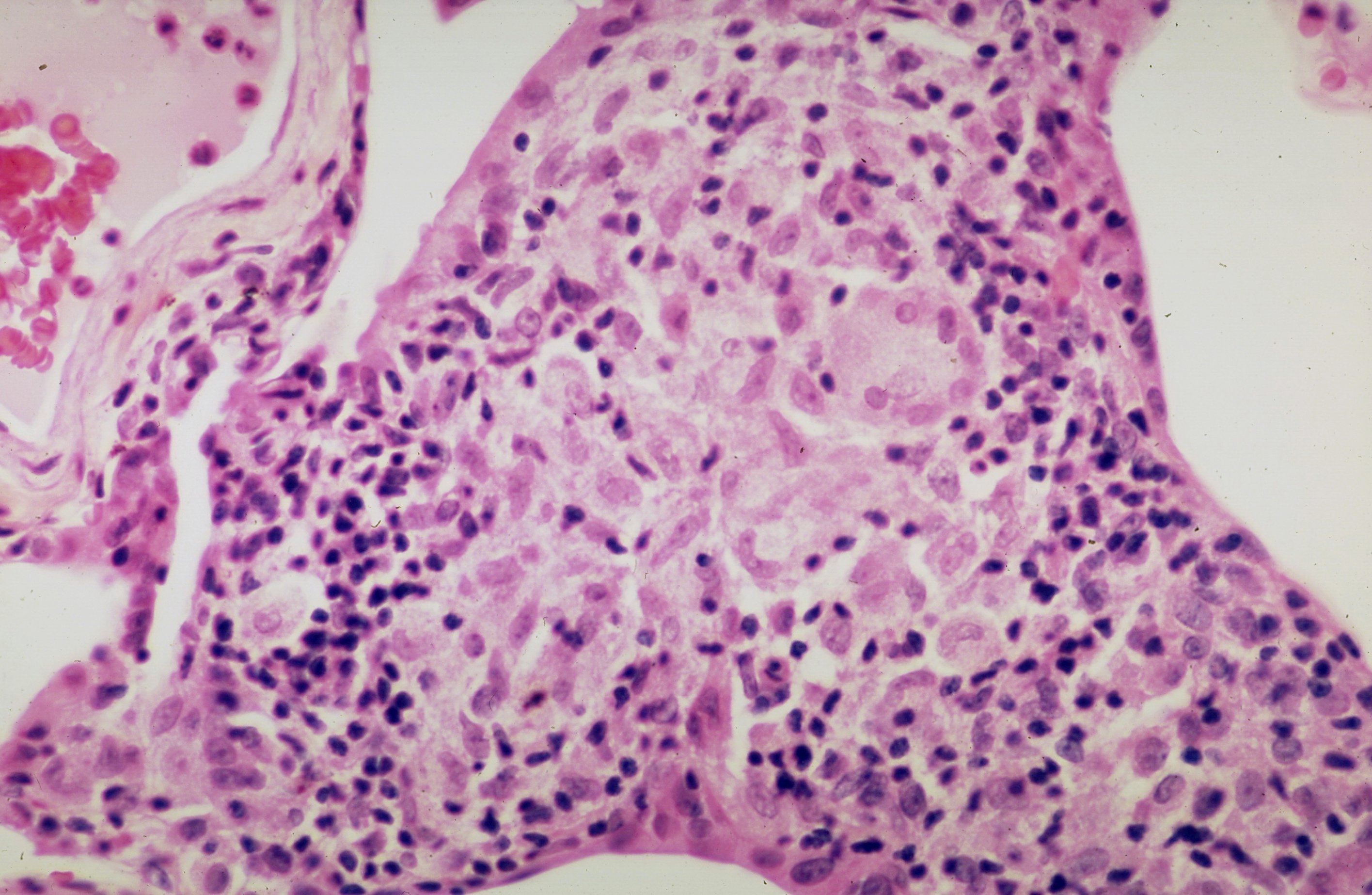
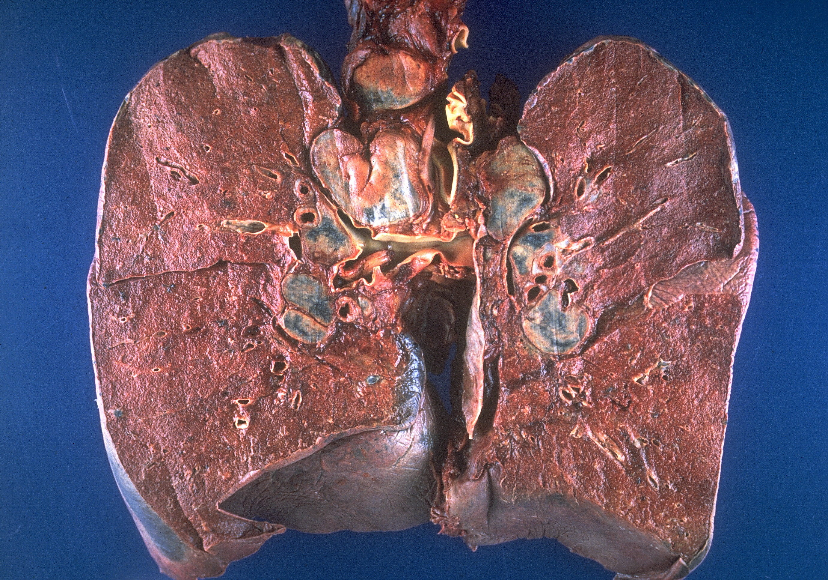
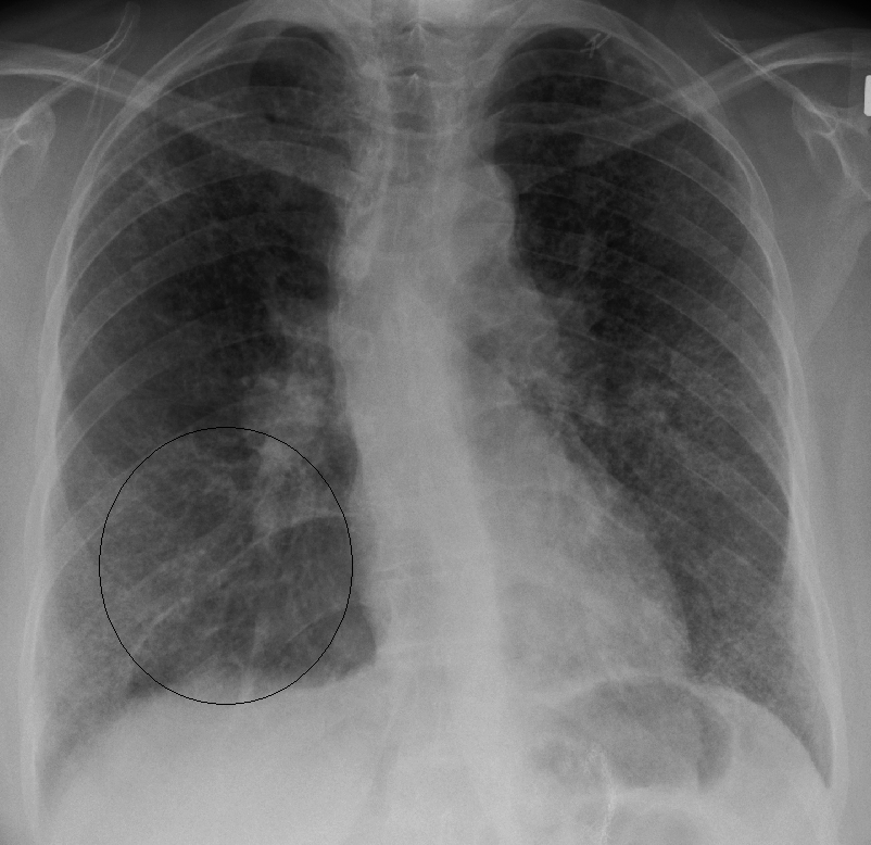
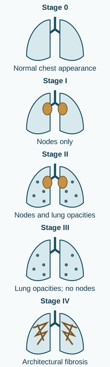
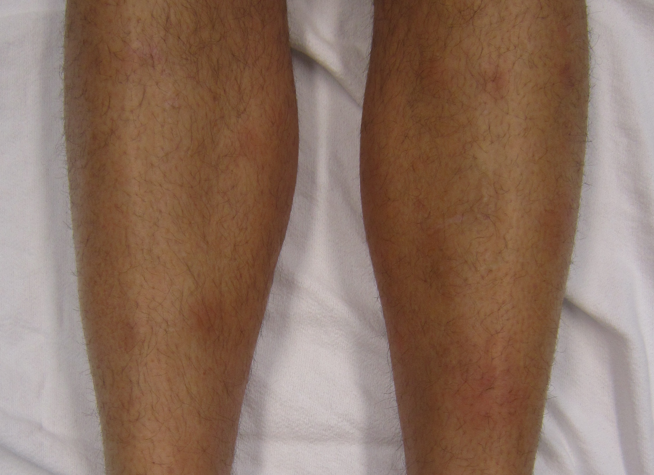
.jpg)
.jpg)

