Follow ureteral arteries and pelvic crossings, explore wall perfusion versus urine flow, and apply the anatomy to surgical injury and transplantation.
A ureter can still pass urine while its wall is losing blood supply. Follow the tube, locate the surrounding arteries, then ask which connections an operation has preserved. The distinction explains why a reassuring early urine jet cannot settle every injury question.
Follow the tube before naming an artery
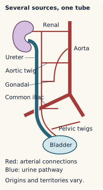
The ureter takes urine from the renal pelvis to the bladder. It travels behind the abdominal lining, over the psoas muscle, across the iliac vessels at the pelvic brim, and along the pelvic wall before entering the bladder. Proximal means nearer the kidney; distal means nearer the bladder. The ureter is anterior to the iliac vessels at the crossing, not inside their lumen. [1]
Use regions to organize possible arterial sources, not to draw three sealed territories. Renal branches commonly reach the upper ureter. Direct aortic, gonadal and common iliac branches can contribute farther down. Internal iliac branches supply the pelvis, including vesical branches and, where present, uterine and vaginal branches. Actual origins, number and dominance vary. [1][2]
A crossing is not the same as a blood supply. A gonadal artery can cross the ureter and give it a branch, but neither a crossing nor a regional label proves that this is the only feeder. When a case provides a traced branch or a perfusion finding, use that observation instead of assuming a textbook pattern.
Keep the tissue that connects the feeders
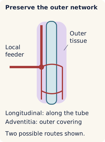
The adventitia is the outer connective-tissue covering. Small arterial connections run lengthwise within and around it. This longitudinal network links regional inflow; it does not make every segment immune to arterial loss. Separating the ureter from all surrounding tissue, called skeletonization, can interrupt both incoming branches and the connections between them. [1][2]
Imagine a local feeder has been divided but connections from nearby supplied tissue remain. An alternate route may still exist. Now also interrupt those connections: the central region loses the alternate route in this drawing. Neither scenario assigns a percentage of living tissue or establishes an operative viability threshold.
The diagram shows both routes retained. The full state table below is available without scripting.
Qualitative teaching model. Solid red lines show retained arterial connections; dashed gray lines show interruptions. No blood-flow percentage is calculated.
Original connectivity model. The red junction receives a local route and a longitudinal route from adjacent supplied tissue. [2]Open full-size diagram
Predict: Which change removes the last drawn route after the local feeder is interrupted? Try each connection separately, then reset both. Transfer: a patent neighboring artery cannot supply an isolated region across a severed connection. In a real patient, the remaining vascular anatomy still needs assessment.
Readable model states, including without scripting
Local feeder
Longitudinal route
Model consequence
Local feederPresent
Longitudinal routePresent
Model consequenceTwo drawn routes reach the region.
Local feederInterrupted
Longitudinal routePresent
Model consequenceOnly the adjacent longitudinal route remains.
Local feederPresent
Longitudinal routeInterrupted
Model consequenceOnly the local route remains.
Local feederInterrupted
Longitudinal routeInterrupted
Model consequenceNo drawn arterial route reaches the region.
In the abdomen, feeders generally approach medially; in the pelvis, they generally approach laterally. This explains the teaching preference for working away from the vascular attachment, often lateral abdominal and medial pelvic exposure. It is not permission to strip the opposite surface or to ignore an observed variant. [1]
Distal pelvic attachments are particularly complex. A cadaver study found denser fibrovascular tissue posteromedially near the distal ureter, an important limitation of a simple lateral-only rule. Preserve the actual vascular-bearing tissue and limit unnecessary dissection. [5]
Case 16
Show answer and explanations for case 16
A. Loss of all demonstrated arterial routes to the region (Best answer)
Why would the renal-side route also be affected?
The planned division is upstream of both the inferior renal artery origin and the separate aortic twig origin, so both demonstrated cranial routes lose inflow.
Why would pelvic inflow not replace these lost routes in the supplied map?
The pelvic injections stop below the scar before reaching the marked region.
Read the complete explanation
The planned division is upstream of both the inferior renal artery origin and the separate aortic twig origin, so both demonstrated cranial routes lose inflow. The pelvic injections stop below the scar before reaching the marked region.
B. Persistence of inflow through the pelvic longitudinal route (Why this does not fit)
What could make pelvic inflow a useful alternate route?
An intact longitudinal communication could connect supplied pelvic tissue to the marked region.
What did the selective injections actually show?
Pelvic contrast stops below the intervening scar.
Read the complete explanation
An intact longitudinal communication could connect supplied pelvic tissue to the marked region. Pelvic contrast stops below the intervening scar.
C. Persistence of direct inflow through the aortic twig (Why this does not fit)
Which route directly entered the marked region?
The aortic twig supplied it before the proposed resection.
Where does the proposed division lie relative to that twig?
The common trunk is divided before the twig originates.
Read the complete explanation
The aortic twig supplied it before the proposed resection. The common trunk is divided before the twig originates.
D. Persistence of inflow through the renal longitudinal route (Why this does not fit)
What supports a renal-side route before the proposed division?
Selective injection demonstrates longitudinal filling from the inferior renal artery.
What makes that route vulnerable to the proposed resection?
The shared parent trunk would be divided before the renal artery arises.
Read the complete explanation
Selective injection demonstrates longitudinal filling from the inferior renal artery. The shared parent trunk would be divided before the renal artery arises.
Takeaway: Different arterial entry points can still depend on one upstream origin; test the entire route rather than counting vessel names.
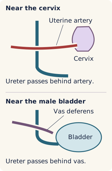
Near the cervix, the uterine artery passes in front of the ureter. The memory aid is water under the bridge: urine below the artery. In a male pelvis, the vas deferens passes in front of the distal ureter near the bladder. These are different structures and different operative settings, not interchangeable labels. [1]
During hysterectomy, an unseen ureter can be caught in a pedicle, bent by a suture, thermally injured or deprived of arterial connections. A memorized distance from the cervix is not a safe margin: anatomy varies, and a mass, adhesions or prior surgery can distort the relationship. Visual identification and careful dissection remain central. [3][5]
A ureteral catheter can help identify a lumen. It does not display every small artery or prevent all injury mechanisms. Follow the ureter through the relevant field, protect its surrounding tissue and assess a suspected injury; do not treat a catheter as a substitute for those observations. [3]
When a ureteral arterial contribution has been traced from a uterine artery, identify the uterine-directed continuation beyond the ureteral branch origin separately from the ureteral feeder itself. In the supplied branch-map examples, interruption beyond that origin can preserve the feeder only if its parent inflow and its vascular-bearing tissue remain connected. This is an anatomical interpretation of a demonstrated map, not a universal division point or a safe-distance rule. [1][2][3]
Transfer: if a mass displaces the cervix but the artery and ureter have been separately traced, the traced anatomy outranks the expected distance. Before answering a pelvic case, ask what the operator actually saw.
Case 23
Show answer and explanations for case 23
A. The separate vaginal branch near its origin (Why this does not fit)
Why could ureteral filling persist after this division?
That separate branch is not the mapped ureteral feeder.
What finding does this site fail to explain?
It does not interrupt the mapped uterine-directed path whose blush disappeared.
Read the complete explanation
That separate branch is not the mapped ureteral feeder. It does not interrupt the mapped uterine-directed path whose blush disappeared.
B. The parent uterine artery before the ureteral branch origin (Why this does not fit)
Which observation could this site explain?
A proximal uterine arterial division could remove the downstream uterine blush.
Which second observation would not fit that division?
It would also interrupt the mapped inflow to the ureteral branch that still fills.
Read the complete explanation
A proximal uterine arterial division could remove the downstream uterine blush. It would also interrupt the mapped inflow to the ureteral branch that still fills.
C. The uterine-directed continuation beyond the ureteral branch origin (Best answer)
Which path must be interrupted to remove the mapped uterine blush?
The path delivering contrast toward the uterine target must be interrupted.
What localizes that interruption beyond the ureteral takeoff?
The ureteral branch still fills from its origin through the preserved outer attachment.
Read the complete explanation
The path delivering contrast toward the uterine target must be interrupted. The ureteral branch still fills from its origin through the preserved outer attachment.
D. The ureteral branch after it leaves the uterine artery (Why this does not fit)
What territory would this division directly threaten?
It would interrupt the traced arterial contribution to the ureteral attachment.
What was observed in the ureteral branch?
The ureteral branch continues to fill from its origin.
Where did the target blush disappear?
The uterine-target blush disappeared while the ureteral branch continued to fill.
Read the complete explanation
It would interrupt the traced arterial contribution to the ureteral attachment. The ureteral branch continues to fill from its origin. The uterine-target blush disappeared while the ureteral branch continued to fill.
Takeaway: Name the uterine-directed continuation precisely; dividing it beyond a ureteral takeoff is different from dividing the ureteral feeder.
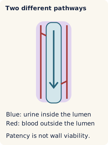
Obstruction prevents urine from passing. Ischemia is inadequate blood supply to tissue. A constricting suture can obstruct an otherwise perfused ureter; disrupted arterial connections can threaten a ureter that initially remains open. Crushing, heat and dissection can also produce mixed injuries. [3]
The diagram shows both routes retained. The full state table below is available without scripting.
Qualitative teaching model. Solid red lines show retained arterial connections; dashed gray lines show interruptions. No blood-flow percentage is calculated.
Original two-pathway schematic. A stent occupies the urine pathway; it does not reconnect the red outer vessels. [3]Open full-size diagram
Predict: What happens to urine passage if the lumen is blocked while arterial connections remain? Change each condition independently. Transfer: restoring passage does not by itself restore disrupted vessels; retained vessels do not by themselves remove a blockage. This is a qualitative model, not a diagnostic test.
Urine passage and vascular connections can differ
Lumen
Connections
Interpretation
LumenOpen
ConnectionsRetained
InterpretationNeither modeled defect is present.
LumenBlocked
ConnectionsRetained
InterpretationObstruction without modeled vascular interruption.
A urethral catheter empties the bladder. It cannot bypass an interruption between a kidney and that bladder. A ureteral stent spans the urine lumen from the collecting system toward the bladder; it needs an appropriate route across the affected ureter. A percutaneous nephrostomy reaches the collecting system through the skin and drains from above a ureteral interruption. Neither device replaces disrupted arterial connections to the wall. [3][4]
Retrograde describes travel from the bladder up the ureter. Antegrade describes travel from the collecting system down the ureter, often through nephrostomy access. These are directions, not guarantees of a suitable route. Contrast may enter a narrow recess that cannot safely accommodate an instrument; bowel or other structures can obstruct a proposed skin-to-kidney approach. The actual anatomical route determines feasibility. [3][6]
Try the distinction: a bladder catheter is draining normally, but a ureter is interrupted above it. The catheter has emptied the downstream compartment, not the disconnected kidney. A separate upper-tract route is needed to drain that kidney.
Ask what the contrast actually examined
Intravenous contrast must reach the kidney and be excreted before it outlines the collecting system and ureter. A scan ending before this happens has not meaningfully tested those urine pathways for extravasation. A negative pre-excretory image cannot reliably exclude a ureteral leak. Use actual opacification and the clinical question, rather than the clock or the word contrast alone. [3]
A bladder leak is a different imaging question. Active retrograde filling distends and opacifies the bladder for cystography. A shallow layer of passively excreted contrast in a catheterized bladder does not provide an equivalent examination. Conversely, an adequately filled bladder study does not automatically opacify the collecting systems or every ureter. A negative adequately performed study reduces concern in the examined compartment; it is not an infallible exclusion of every urinary injury. [3]
Fluid with creatinine far above simultaneously sampled serum supports a urinary collection, but does not locate its origin. Combine that chemistry with the compartments that were actually examined. With duplication, match each selective contrast study to the collecting moiety it outlines instead of assuming that one normal ureteral study clears the entire kidney. [3][4]
Dilation and obstruction are not interchangeable
Hydronephrosis describes an enlarged collecting system. A dilated reservoir can still drain. A MAG3 diuretic renogram observes renal tracer uptake and subsequent clearance during a drainage challenge; it is not simply a photograph of the enlarged pelvis. Interpretation requires attention to hydration, renal function, the actual diuretic urine response, bladder emptying and upright or postvoid images. Poor renal uptake or an inadequate urine response can make retention hard to interpret. No single washout time proves obstruction by itself. [7]
Compare two observations: a large pelvis that empties after upright positioning and voiding differs from one that continues to retain activity despite an adequate challenge. The structural image suggests a possible level; functional evidence tests whether outflow is meaningfully impaired. Read each kidney separately, including after transplantation or reconstruction. Tracer uptake is not a measurement of the exact volume of urine each kidney produced. [7]
Postoperative flank pain, a urine-containing collection or delayed leakage warrants attention even if early jets were seen. Hematuria can be absent. CT urography uses an excretory phase to look for ureteral leakage or obstruction; a routine early contrast image alone may miss the relevant urinary finding. Retrograde or antegrade contrast studies can clarify an uncertain site. [3]
Fluid creatinine substantially above a simultaneously measured serum concentration supports urine in a drain, but does not by itself distinguish ureter from bladder. Hydronephrosis describes upstream dilation, not the cause of obstruction or the health of the wall. Two obstructed ureters can produce bilateral dilation even when the bladder outlet is open. A functioning opposite kidney can obscure the effect of a unilateral injury on total urine output.
Repair planning depends on viable tissue, defect location, the gap after unhealthy tissue is removed, and whether a tension-free connection is possible. A short upper or middle defect may permit ureter-to-ureter repair. A compromised distal stump often favors reimplantation into the bladder. A psoas hitch advances and supports the bladder toward the proximal ureter to reduce the distance a repair must span. During selected elective repairs, a Boari flap made from healthy bladder tissue can bridge a gap that a psoas hitch alone cannot span; this is not a routine acute-injury maneuver. Hemodynamic instability or a delayed injury may instead require initial urinary diversion. [3]
A urinary leak and its surrounding collection occupy different compartments. Improved stented drainage can reduce new leakage while an existing collection still enlarges or compresses adjacent structures. An enlarging, painful or otherwise complicated urinary collection may require direct drainage in addition to urinary diversion. Continued assessment distinguishes a responding leak from failure of diversion that may require reconstruction. [3][8]
Reconstruction changes which ureter carries a kidney's urine. In a donor-to-native connection, the native ureter below the junction carries donor urine. In a right-to-left ureteral connection, a clip above the junction on the left limb differs from a clip on the shared distal conduit. Draw the current route before interpreting urine output or upstream pressure. When a case supplies measured urine volumes, those measurements describe that example, not universal kidney contributions. [3][4]
A stone is another cause of obstruction, not a diagnosis proved by flank pain alone. Passage depends on size, location and the clinical situation; alpha-blockers do not open every ureter to a fixed diameter. Infection with an obstructed collecting system requires urgent drainage and prompt antibiotics, not a trial of expulsive treatment while infection progresses. [6]
A transplanted ureter has different remaining connections
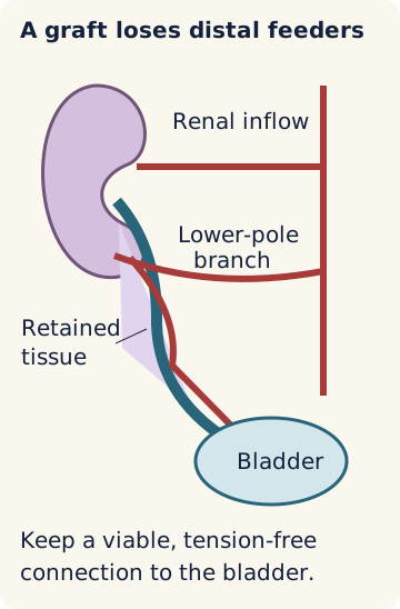
A donor ureter no longer has its original distal pelvic arterial attachments. Its retained renal-side vessels and periureteric tissue therefore matter greatly. Protect tissue around the renal pelvis and proximal ureter, including the region between the lower pole and ureter; preserve a demonstrated ureteral contribution from an accessory or lower-pole branch. The exact branch pattern is not identical in every graft. [2][4]
Original example of renal-side inflow reaching the donor ureter through preserved tissue. The recipient iliac artery supplies graft inflow through the arterial anastomosis; it does not recreate all original ureteral attachments. [2][4]Open full-size diagram
A duplicated collecting system may have two ureters. Trace each drainage pathway and preserve its surrounding tissue; one visible or stented ureter does not necessarily represent the whole kidney. When a case supplies the actual connection between a collecting region and a bladder opening, use that map instead of assuming a usual pattern. [4]
Enough length to reach the bladder without tension is useful; unnecessary length with poor perfusion is not. A working arterial anastomosis or urine in the catheter is not a complete assessment of the distal ureter. A stent helps drainage and can reduce some urinary complications, but cannot replace the retained vascular connections. [4]
After transplantation, high-creatinine drain fluid suggests urinary leakage. Persistent hydronephrosis with worsening graft function raises concern for clinically significant obstruction. The collection, lumen and graft perfusion require separate evaluation: a percutaneous nephrostomy can both decompress a blocked graft and provide an antegrade map. Management of a leak or stricture depends on its location, extent and response to initial treatment. When the donor ureter cannot provide a suitable direct bladder connection, a healthy, patent native ureter with adequate reach may provide an alternative conduit. [4]
Bring it together: locate the ureteral region, identify the observed arterial attachments, then decide whether the supplied findings concern urine passage, tissue viability or both. Do not invent an operative finding to make a familiar answer fit.
Case 10
Show answer and explanations for case 10
A. Graft biopsy (Why this does not fit)
What competing process can worsen graft function?
Intrinsic graft disease can raise creatinine and may require tissue assessment.
Why is that not the initial answer to this pattern?
The drainage study independently demonstrates persistent collecting-system outflow resistance that biopsy would not relieve.
Read the complete explanation
Intrinsic graft disease can raise creatinine and may require tissue assessment. The drainage study independently demonstrates persistent collecting-system outflow resistance that biopsy would not relieve.
B. Percutaneous nephrostomy of the graft (Best answer)
Why is the dilation clinically important?
Graft function is deteriorating while the collecting system fails to empty during an interpretable drainage study.
Which observed route reaches above the outflow resistance?
The accessible graft calyx offers drainage without crossing the narrow intravesical scar channel.
Read the complete explanation
Graft function is deteriorating while the collecting system fails to empty during an interpretable drainage study. The accessible graft calyx offers drainage without crossing the narrow intravesical scar channel.
C. Urethral catheter exchange (Why this does not fit)
How could lower-tract obstruction affect a graft?
A poorly emptied bladder can oppose upper-tract drainage.
Which observation argues against a bladder-emptying explanation?
Pelvic retention persists after bladder emptying during the functional study.
Read the complete explanation
A poorly emptied bladder can oppose upper-tract drainage. Pelvic retention persists after bladder emptying during the functional study.
D. Retrograde transplant ureteral stenting (Why this does not fit)
Why could this be a reasonable drainage option in another anatomy?
A stent can bridge a ureteral obstruction from the bladder.
What limits the available approach here?
The instruments remain separated from the transplant opening by the pinhole channel into the recess.
Read the complete explanation
A stent can bridge a ureteral obstruction from the bladder. The instruments remain separated from the transplant opening by the pinhole channel into the recess.
Takeaway: Contrast can enter a space that a drainage device cannot reach; interpret access anatomy separately from the need to decompress.
Each case asks one question. Cover the choices before committing to an answer. Open the explanation questions individually, or read a complete explanation.
Case 1
Show answer and explanations for case 1
A. Renal artery (Why this does not fit)
What could normally connect renal inflow to a lower region?
Longitudinal vessels can connect renal-side inflow to more distal tissue.
What prevents that route in this operation?
The connection above the marked region has been interrupted.
Read the complete explanation
Longitudinal vessels can connect renal-side inflow to more distal tissue. The connection above the marked region has been interrupted.
B. Gonadal artery (Why this does not fit)
Why is a gonadal branch a plausible contributor?
Gonadal branches can join the ureteral arterial network.
Where is this particular branch disconnected from the region?
It enters above the interrupted longitudinal connection.
Read the complete explanation
Gonadal branches can join the ureteral arterial network. It enters above the interrupted longitudinal connection.
C. Uterine artery (Why this does not fit)
What would make this pelvic feeder a possible route?
Its lower connection could reach the region if the feeder were open.
What happened to this feeder?
The uterine feeder has been divided.
Read the complete explanation
Its lower connection could reach the region if the feeder were open. The uterine feeder has been divided.
D. Superior vesical artery (Best answer)
Which side still has a connection to the marked region?
The connection from the lower pelvic side remains intact.
Which of the two lower feeders remains open?
The superior vesical feeder remains open after division of the uterine feeder.
Which inflow can therefore still reach the region in this map?
Superior vesical inflow has a remaining route, although the map does not quantify tissue viability.
Read the complete explanation
The connection from the lower pelvic side remains intact. The superior vesical feeder remains open after division of the uterine feeder. Superior vesical inflow has a remaining route, although the map does not quantify tissue viability.
Takeaway: An open artery matters only when an intact route connects it to the tissue.
A. Urine is obstructed in a region with a retained arterial route (Why this does not fit)
Which observation evaluates the lumen?
Sequential excretory images show passage through the region.
Which mapped route could establish arterial continuity?
Neither the divided cranial stem nor the interrupted caudal connection reaches the region.
Read the complete explanation
Sequential excretory images show passage through the region. Neither the divided cranial stem nor the interrupted caudal connection reaches the region.
B. Urine is draining, but delayed wall failure remains a concern (Best answer)
Why are the two cranial feeders not independent backups?
Both depend on the divided parent stem.
What does the sequence of excretory images establish?
Urine still traverses the marked region.
What remains unresolved by that drainage?
The isolated arterial map raises wall-injury risk despite current passage.
Read the complete explanation
Both depend on the divided parent stem. Urine still traverses the marked region. The isolated arterial map raises wall-injury risk despite current passage.
C. Urine is obstructed, and delayed wall failure remains a concern (Why this does not fit)
What supports concern about the wall?
The mapped arterial routes no longer reach the region.
What argues against current obstruction in this study?
Contrast crosses the region as the renal pelvis empties.
Read the complete explanation
The mapped arterial routes no longer reach the region. Contrast crosses the region as the renal pelvis empties.
D. Urine is draining through a region with a retained arterial route (Why this does not fit)
What supports ongoing drainage?
Excreted contrast reaches the bladder through the marked region.
Why does the iliac feeder not establish a retained route?
Its longitudinal vessels end below the region at the scar.
Read the complete explanation
Excreted contrast reaches the bladder through the marked region. Its longitudinal vessels end below the region at the scar.
Takeaway: Demonstrated urine transit cannot substitute for tracing the arteries that reach the wall.
A. Ureteral leakage or narrowing at the mobilized region (Best answer)
Which vessels were separated from their target tissue?
Small feeders and longitudinal vessels supplying the ureteral wall were interrupted.
What can follow loss of that local supply?
Delayed wall failure or scar narrowing can occur at the mobilized region.
Read the complete explanation
Small feeders and longitudinal vessels supplying the ureteral wall were interrupted. Delayed wall failure or scar narrowing can occur at the mobilized region.
B. Loss of perfusion throughout the ipsilateral kidney (Why this does not fit)
Which vessel would usually need assessment for this pattern?
The main renal arterial supply would be a concern.
What limits that explanation here?
The recording shows injury to ureteral twigs while renal organ perfusion continues.
Read the complete explanation
The main renal arterial supply would be a concern. The recording shows injury to ureteral twigs while renal organ perfusion continues.
C. Loss of perfusion throughout the ipsilateral gonad (Why this does not fit)
Why might the gonadal artery attract attention?
It gives one of the small observed ureteral branches.
What distinction matters in the recording?
The ureteral twig is divided while the parent artery continues to perfuse the gonad.
Read the complete explanation
It gives one of the small observed ureteral branches. The ureteral twig is divided while the parent artery continues to perfuse the gonad.
D. Diffuse loss of bladder-wall perfusion (Why this does not fit)
Which arterial territory supplies most of the bladder?
Pelvic vesical branches supply the bladder wall.
Where was the vascular attachment disrupted?
The disrupted cuff surrounds the abdominal ureter rather than the bladder.
Read the complete explanation
Pelvic vesical branches supply the bladder wall. The disrupted cuff surrounds the abdominal ureter rather than the bladder.
Takeaway: A small arterial branch can be dispensable to its parent organ but important to the ureter it supplies.
A. Nonobstructive enlargement of the right ureter (Why this does not fit)
Why is this a credible anatomical alternative?
A previously enlarged ureter can remain capacious without clinically important obstruction.
Which new observation weighs against it?
The collecting system fails to clear during a technically adequate drainage challenge.
Read the complete explanation
A previously enlarged ureter can remain capacious without clinically important obstruction. The collecting system fails to clear during a technically adequate drainage challenge.
B. Global renal underfunction causing slow tracer delivery (Why this does not fit)
How can reduced renal function complicate renography?
Poor tracer delivery can mimic delayed urinary clearance.
Why is that not the best explanation here?
Renal uptake is preserved and the persistent retention is unilateral despite an adequate diuretic response.
Read the complete explanation
Poor tracer delivery can mimic delayed urinary clearance. Renal uptake is preserved and the persistent retention is unilateral despite an adequate diuretic response.
C. Functionally significant obstruction of the terminal right ureter (Best answer)
Where must resistance lie to explain dilation of the full ureter?
A terminal ureteral lesion lies downstream of that entire dilated segment.
What argues that this is more than longstanding enlargement?
Angiography evaluates arterial anatomy rather than excreted urine.
What finding points to a different source?
The drain chemistry calls for localization of a urinary pathway.
Read the complete explanation
Angiography evaluates arterial anatomy rather than excreted urine. The drain chemistry calls for localization of a urinary pathway.
B. CT urography with excretory imaging (Best answer)
What does the paired creatinine measurement suggest?
The collected fluid is strongly enriched with urinary creatinine.
Which urine pathway remains unexamined?
The collecting systems and ureters were not opacified by the bladder study.
What will excretory imaging add?
It can show urine escaping from an upper urinary tract while a negative result still requires interpretation in context.
Read the complete explanation
The collected fluid is strongly enriched with urinary creatinine. The collecting systems and ureters were not opacified by the bladder study. It can show urine escaping from an upper urinary tract while a negative result still requires interpretation in context.
C. Retrograde CT cystography (Why this does not fit)
What source could this examination demonstrate?
Active bladder filling can reveal a bladder-wall leak.
Why is another compartment the priority here?
The existing filling and drainage images already evaluated a distended bladder without showing extravasation.
Read the complete explanation
Active bladder filling can reveal a bladder-wall leak. The existing filling and drainage images already evaluated a distended bladder without showing extravasation.
D. Lymphatic imaging (Why this does not fit)
Why could this test be considered after pelvic surgery?
Disrupted lymphatic channels can produce a clear pelvic collection.
Which observation makes that the weaker initial explanation?
The fluid creatinine is markedly above the simultaneous serum concentration.
Read the complete explanation
Disrupted lymphatic channels can produce a clear pelvic collection. The fluid creatinine is markedly above the simultaneous serum concentration.
Takeaway: First identify what the fluid is; then ask which relevant compartment the previous study actually tested.
A. Retrograde ureteral stent placement (Why this does not fit)
When is a retrograde stent a useful temporary diversion?
It can bridge an injury when a safe continuous route can be traversed from the bladder.
What anatomical finding defeats that route here?
The proximal and distal ureteral ends are separated by an interval without a connecting lumen.
Read the complete explanation
It can bridge an injury when a safe continuous route can be traversed from the bladder. The proximal and distal ureteral ends are separated by an interval without a connecting lumen.
B. Definitive ureteral reconstruction (Why this does not fit)
What problem could reconstruction address?
It can restore continuity between separated urinary segments.
What independently changes the immediate priority?
Persistent hypotension with hypoperfusion favors damage-control diversion before definitive repair.
Read the complete explanation
It can restore continuity between separated urinary segments. Persistent hypotension with hypoperfusion favors damage-control diversion before definitive repair.
C. Urethral catheter drainage (Why this does not fit)
Which compartment does this catheter drain?
It empties the bladder below the ureteral injury.
Why would the kidney remain undrained?
Its proximal ureter is disconnected from the catheterized bladder compartment.
Read the complete explanation
It empties the bladder below the ureteral injury. Its proximal ureter is disconnected from the catheterized bladder compartment.
D. Percutaneous nephrostomy drainage (Best answer)
What does persistent shock imply for repair timing?
A temporary urinary diversion is preferable while physiologic stabilization continues.
How can urine leave the kidney without crossing the separated ends?
A nephrostomy accesses the collecting system from above the interruption.
Read the complete explanation
A temporary urinary diversion is preferable while physiologic stabilization continues. A nephrostomy accesses the collecting system from above the interruption.
Takeaway: Temporary versus definitive treatment depends on physiology; the route of temporary drainage depends on the actual urinary connections.
A. Direct ureteral reimplantation without bladder advancement (Why this does not fit)
What benefit does bladder reimplantation provide?
It bypasses the concerning distal ureteral wall.
Why is direct reach not established here?
The resting proximal endpoint is still 4 cm from the bladder.
Read the complete explanation
It bypasses the concerning distal ureteral wall. The resting proximal endpoint is still 4 cm from the bladder.
B. Ureteral reimplantation using a Boari flap (Why this does not fit)
When does a bladder flap add useful reach?
A flap can span a distance that bladder translation alone cannot cover.
Why is that added length not required by these measurements?
Available bladder advancement already exceeds this patient-specific gap.
Read the complete explanation
A flap can span a distance that bladder translation alone cannot cover. Available bladder advancement already exceeds this patient-specific gap.
C. Ureteral reimplantation supported by a psoas hitch (Best answer)
Why avoid making the distal ureter the anastomotic partner?
Progressive slough with deficient mural enhancement raises concern that its wall will not sustain a repair.
How does the planned bladder excursion compare with the gap?
The available advancement exceeds the measured gap without requiring additional bladder-wall length.
Which reconstruction uses that geometric advantage?
A psoas hitch brings the bladder toward the viable proximal ureter to support a tension-free reimplantation.
Read the complete explanation
Progressive slough with deficient mural enhancement raises concern that its wall will not sustain a repair. The available advancement exceeds the measured gap without requiring additional bladder-wall length. A psoas hitch brings the bladder toward the viable proximal ureter to support a tension-free reimplantation.
D. Ureter-to-ureter anastomosis (Why this does not fit)
What can make this attractive for a short defect?
A healthy distal ureter could provide a nearby urinary recipient.
Which observations argue against that recipient?
The distal segment has deficient enhancement and progressive mucosal slough.
Read the complete explanation
A healthy distal ureter could provide a nearby urinary recipient. The distal segment has deficient enhancement and progressive mucosal slough.
Takeaway: Assess the recipient tissue and the predicted reach independently; a named operation is not a substitute for either assessment.
A. Delayed wall failure of the terminal ureter (Best answer)
What region lies behind the vas near bladder entry?
This relation identifies the terminal pelvic ureter.
What changes the interpretation of the early contrast passage?
Subsequent deficient mural enhancement and leakage show that initial patency did not establish durable wall integrity.
Read the complete explanation
This relation identifies the terminal pelvic ureter. Subsequent deficient mural enhancement and leakage show that initial patency did not establish durable wall integrity.
B. Mechanical angulation of the ureter at the iliac crossing (Why this does not fit)
What can mechanical angulation cause?
A bent ureter can obstruct urine passage.
What observation opposes this level?
The affected segment is near the vas rather than the iliac crossing.
What observation opposes mechanical angulation?
The aligned lumen developed progressive wall abnormalities rather than a focal bend.
Read the complete explanation
A bent ureter can obstruct urine passage. The affected segment is near the vas rather than the iliac crossing. The aligned lumen developed progressive wall abnormalities rather than a focal bend.
C. Mechanical angulation of the terminal ureter (Why this does not fit)
Why does the location fit this possibility?
Fixation near the vas can affect the terminal ureter.
Which later finding favors a different mechanism?
Progressive wall change and leakage developed while the lumen remained aligned.
Read the complete explanation
Fixation near the vas can affect the terminal ureter. Progressive wall change and leakage developed while the lumen remained aligned.
D. Delayed wall failure of the ureter at the iliac crossing (Why this does not fit)
Which findings support a wall complication?
Delayed poor mural enhancement and urinary extravasation support evolving wall injury.
Why is this level less consistent with the recording?
The operated segment was behind the vas near the bladder, not at the iliac crossing.
Read the complete explanation
Delayed poor mural enhancement and urinary extravasation support evolving wall injury. The operated segment was behind the vas near the bladder, not at the iliac crossing.
Takeaway: A pelvic crossing localizes the segment; the time course distinguishes drainage mechanics from evolving wall injury.
A. The lower-moiety collecting system and its ureter (Why this does not fit)
Why can a duplicated system complicate interpretation?
One ureteral study may not assess both collecting moieties.
What was actually demonstrated here?
The lower moiety and its continuous ureter were already opacified without extravasation.
Read the complete explanation
One ureteral study may not assess both collecting moieties. The lower moiety and its continuous ureter were already opacified without extravasation.
B. The bladder wall (Why this does not fit)
What kind of source could account for the fluid chemistry?
A bladder defect can release creatinine-rich urine into a pelvic collection.
What shifts the next localization study away from the bladder?
Active bladder filling was already performed without demonstrating extravasation.
Read the complete explanation
A bladder defect can release creatinine-rich urine into a pelvic collection. Active bladder filling was already performed without demonstrating extravasation.
C. The pelvic lymphatic channels (Why this does not fit)
What postoperative source can form a clear collection?
Pelvic lymphatic disruption can produce clear fluid.
Which independent measurement makes it less likely here?
The collection is markedly enriched in creatinine relative to serum.
Read the complete explanation
Pelvic lymphatic disruption can produce clear fluid. The collection is markedly enriched in creatinine relative to serum.
D. The upper-moiety collecting system and its ureter (Best answer)
Which moiety was demonstrated through the lateral opening?
The selective examination outlined the lower moiety and its ureter.
Why investigate the remaining upper pathway?
Urinary collection chemistry makes an unexamined urinary source relevant after the lower pathway and bladder studies.
Read the complete explanation
The selective examination outlined the lower moiety and its ureter. Urinary collection chemistry makes an unexamined urinary source relevant after the lower pathway and bladder studies.
Takeaway: One normal ureteral study does not clear an entire duplicated system; connect each study to the moiety it actually opacified.
A. Maintain the urinary diversion and add image-guided collection drainage (Best answer)
What supports retaining the current diversion?
Prompt stented drainage with much less extravasation indicates an improving urinary pathway.
What problem still requires treatment?
The enlarging collection is causing pain and venous compression despite that improvement.
Read the complete explanation
Prompt stented drainage with much less extravasation indicates an improving urinary pathway. The enlarging collection is causing pain and venous compression despite that improvement.
B. Reconstruct the ureter with interval collection imaging (Why this does not fit)
What can reconstruction address when diversion fails?
It can restore a urinary pathway that remains disrupted or inadequately drained.
What supports continuing the present urinary diversion?
The leak is already improving.
What requires a separate intervention?
The growing collection is producing symptomatic compression.
Read the complete explanation
It can restore a urinary pathway that remains disrupted or inadequately drained. The leak is already improving. The growing collection is producing symptomatic compression.
C. Maintain the urinary diversion with interval imaging (Why this does not fit)
What supports a conservative urinary approach?
The leak has decreased substantially with the stent and bladder catheter.
Which finding makes observation of the collection insufficient?
The collection is enlarging and producing symptomatic mass effect.
Read the complete explanation
The leak has decreased substantially with the stent and bladder catheter. The collection is enlarging and producing symptomatic mass effect.
D. Reconstruct the ureter and drain the collection (Why this does not fit)
Why does the collection merit drainage?
Its enlargement, pain and venous compression identify a consequential extraluminal compartment.
What does not yet support reconstruction instead of the working diversion?
Urine crosses the stent promptly and the small leak is already diminishing.
Read the complete explanation
Its enlargement, pain and venous compression identify a consequential extraluminal compartment. Urine crosses the stent promptly and the small leak is already diminishing.
Takeaway: An improving urinary leak and a worsening fluid collection can coexist because they occupy different compartments.
A. The lower-pole parenchymal branch after the division (Why this does not fit)
Which observed defect could this lesion explain?
It could account for the lower-pole cortical enhancement defect.
Which second target is outside that daughter branch?
The donor ureter is supplied by the separately branching ureteral vessel.
Read the complete explanation
It could account for the lower-pole cortical enhancement defect. The donor ureter is supplied by the separately branching ureteral vessel.
B. The ureteral branch after the division (Why this does not fit)
Which observed defect could this lesion explain?
It could account for reduced enhancement along the ureteral wall.
Which second target is outside that daughter branch?
The lower-pole cortex receives the separate parenchymal branch.
Read the complete explanation
It could account for reduced enhancement along the ureteral wall. The lower-pole cortex receives the separate parenchymal branch.
C. The inferior artery before its parenchymal and ureteral branches separate (Best answer)
Which vessel serves both affected targets in this patient?
The inferior parent artery supplies both the lower-pole cortex and the ureteral branch.
Why does a lesion before the division fit better than either daughter branch?
It can reduce inflow to both territories while sparing the main renal arterial territory.
Read the complete explanation
The inferior parent artery supplies both the lower-pole cortex and the ureteral branch. It can reduce inflow to both territories while sparing the main renal arterial territory.
D. The main renal artery serving the upper and middle cortex (Why this does not fit)
Which territory would make this lesion plausible?
Reduced upper and middle cortical enhancement would implicate the main arterial territory.
What does the observed distribution show instead?
Those regions retain enhancement while the two inferior-artery targets are affected.
Read the complete explanation
Reduced upper and middle cortical enhancement would implicate the main arterial territory. Those regions retain enhancement while the two inferior-artery targets are affected.
Takeaway: Two affected target tissues can identify a common parent vessel that neither tissue alone uniquely localizes.
A. Separation at the ureter-to-bladder anastomosis (Why this does not fit)
Why is this an important cause of postoperative leakage?
A gap at the urinary anastomosis can allow extravasation despite normal kidney perfusion.
What distribution argues against it here?
The apposed suture line is separate from multiple wall leaks along a longer proximal segment.
Read the complete explanation
A gap at the urinary anastomosis can allow extravasation despite normal kidney perfusion. The apposed suture line is separate from multiple wall leaks along a longer proximal segment.
B. Selective loss of arterial connections within periureteric tissue (Best answer)
What does maintained cortical perfusion make less likely?
Loss of the principal arterial inflow to the entire graft is less likely.
What does distributed wall deterioration above an apposed suture line suggest?
A regional problem affecting ureteral wall support is more consistent than a single anastomotic defect.
Why can these findings coexist?
The kidney and donor ureter do not require identical retained small-vessel connections.
Read the complete explanation
Loss of the principal arterial inflow to the entire graft is less likely. A regional problem affecting ureteral wall support is more consistent than a single anastomotic defect. The kidney and donor ureter do not require identical retained small-vessel connections.
C. Mechanical angulation of the donor ureter (Why this does not fit)
How could angulation produce a drainage problem?
A bend can obstruct urine transit and raise upstream pressure.
What makes it a weaker explanation for these findings?
There is no focal bend, while progressive wall deterioration occurs at several points.
Read the complete explanation
A bend can obstruct urine transit and raise upstream pressure. There is no focal bend, while progressive wall deterioration occurs at several points.
D. Thrombosis of the principal graft arterial inflow (Why this does not fit)
How could severe inflow loss affect the ureter?
It could compromise renal-side arterial support to the donor ureter.
Which contemporaneous observation opposes this mechanism?
The whole graft cortex enhances uniformly with flow through its arterial anastomosis.
Read the complete explanation
It could compromise renal-side arterial support to the donor ureter. The whole graft cortex enhances uniformly with flow through its arterial anastomosis.
Takeaway: A perfused graft does not clear the ureteral circulation; localize the wall injury rather than treating every leak as a suture-line failure.
A. Retrograde ureteral stenting (Why this does not fit)
When can a ureteral stent provide source control?
It can drain an infected obstructed system when retrograde access is feasible.
Which supplied constraint excludes that route here?
The previous reconstruction prevents retrograde ureteral access.
Read the complete explanation
It can drain an infected obstructed system when retrograde access is feasible. The previous reconstruction prevents retrograde ureteral access.
B. A trial of oral alpha-blocker therapy (Why this does not fit)
What situation can make an expulsive approach reasonable?
Selected uncomplicated ureteral stones may be managed with a trial of passage.
What changes that decision in this patient?
Infection with shock and obstruction requires urgent drainage rather than waiting for passage.
Read the complete explanation
Selected uncomplicated ureteral stones may be managed with a trial of passage. Infection with shock and obstruction requires urgent drainage rather than waiting for passage.
C. Definitive percutaneous stone fragmentation (Why this does not fit)
What problem can stone fragmentation ultimately treat?
It can treat the obstructing stone itself.
What is the initial procedural priority during active sepsis?
Urgent decompression is prioritized, with definitive stone treatment deferred until infection is controlled.
Read the complete explanation
It can treat the obstructing stone itself. Urgent decompression is prioritized, with definitive stone treatment deferred until infection is controlled.
D. Percutaneous nephrostomy drainage (Best answer)
What makes observation or stone-expulsion therapy inappropriate now?
The infected obstructed collecting system requires urgent source control.
Which drainage route is available in this anatomy?
Percutaneous access is feasible whereas retrograde ureteral access is not.
What should be performed through that route?
Nephrostomy drainage relieves obstruction while antibiotics and resuscitation continue.
Read the complete explanation
The infected obstructed collecting system requires urgent source control. Percutaneous access is feasible whereas retrograde ureteral access is not. Nephrostomy drainage relieves obstruction while antibiotics and resuscitation continue.
Takeaway: Treat infected obstruction promptly; the actual urinary anatomy determines how to drain it.
A. Left collecting-system pressure rises while most prior bladder inflow stops (Why this does not fit)
Which part of this prediction follows from the map?
The clip interrupts the left kidney route above the junction.
Which measured contribution contradicts the predicted large output loss?
The interrupted left source contributed much less urine than the still-connected right source.
Read the complete explanation
The clip interrupts the left kidney route above the junction. The interrupted left source contributed much less urine than the still-connected right source.
B. Right collecting-system pressure rises while most prior bladder inflow continues (Why this does not fit)
Why might the right kidney be confused with the left-sided conduit?
Its ureter crosses to join the left ureter before reaching the bladder.
What matters about the clip relative to that junction?
The clip is above the junction on the left limb, so it does not interrupt the right source.
Read the complete explanation
Its ureter crosses to join the left ureter before reaching the bladder. The clip is above the junction on the left limb, so it does not interrupt the right source.
C. Left collecting-system pressure rises while most prior bladder inflow continues (Best answer)
Which source lies upstream of the clip?
The left kidney is isolated because the clip lies on its limb above the right-to-left junction.
Why can most prior bladder inflow continue?
The right kidney produced most of the measured urine and retains the shared distal route.
Read the complete explanation
The left kidney is isolated because the clip lies on its limb above the right-to-left junction. The right kidney produced most of the measured urine and retains the shared distal route.
D. Right collecting-system pressure rises while most prior bladder inflow stops (Why this does not fit)
Which source produced most of the measured urine?
The right kidney produced the larger measured volume.
Why is that larger source not isolated?
Its ureter joins the common distal conduit below the left-sided clip.
Read the complete explanation
The right kidney produced the larger measured volume. Its ureter joins the common distal conduit below the left-sided clip.
Takeaway: Physical side, kidney of origin and share of urine production are different facts in a reconstructed drainage system.
A. Observation with interval urinary imaging (Why this does not fit)
What type of course could permit interval reassessment?
A clinically stable improving injury without threatened drainage may be followed under an appropriate plan.
What does this patient show instead?
Fever, hypotension and increasing pelvic dilation indicate an urgent drainage problem.
Read the complete explanation
A clinically stable improving injury without threatened drainage may be followed under an appropriate plan. Fever, hypotension and increasing pelvic dilation indicate an urgent drainage problem.
B. Percutaneous nephrostomy drainage (Why this does not fit)
Why is nephrostomy ordinarily a credible alternative?
It can decompress an infected collecting system without traversing the ureteral lesion.
What changes its suitability in this patient?
Bowel lies across the available percutaneous access corridors.
Read the complete explanation
It can decompress an infected collecting system without traversing the ureteral lesion. Bowel lies across the available percutaneous access corridors.
C. Definitive ureteral reconstruction (Why this does not fit)
When might reconstruction become necessary?
A ureteral injury that fails diversion or has unsuitable tissue may require definitive repair.
Why is decompression the immediate priority here?
The current infected obstruction is accompanied by unstable physiology.
Read the complete explanation
A ureteral injury that fails diversion or has unsuitable tissue may require definitive repair. The current infected obstruction is accompanied by unstable physiology.
D. Retrograde ureteral stent drainage (Best answer)
Why is waiting for spontaneous healing inappropriate now?
Infection with hypotension and increasing pelvic dilation requires prompt upper drainage.
Which route avoids the interposed bowel?
The visible orifice and demonstrated ureteral lumen provide a retrograde approach across the partial injury.
Read the complete explanation
Infection with hypotension and increasing pelvic dilation requires prompt upper drainage. The visible orifice and demonstrated ureteral lumen provide a retrograde approach across the partial injury.
Takeaway: Both upper drainage methods are useful, but the actual route must avoid intervening structures and reach the obstructed collecting system.
A. Reimplantation with a psoas hitch alone (Why this does not fit)
What can a psoas hitch change?
It brings the bladder closer to the proximal ureter.
Why is the psoas hitch alone insufficient in this plan?
The available bladder advancement is less than the measured gap.
Read the complete explanation
It brings the bladder closer to the proximal ureter. The available bladder advancement is less than the measured gap.
B. Reimplantation using a Boari bladder flap (Best answer)
How much of the gap does bladder advancement address?
Advancement alone does not span the 9 cm distance in this patient.
What contributes the additional urinary conduit length?
A Boari flap uses suitable bladder wall to bridge the residual distance.
Read the complete explanation
Advancement alone does not span the 9 cm distance in this patient. A Boari flap uses suitable bladder wall to bridge the residual distance.
C. Direct ureteral reimplantation (Why this does not fit)
What must be true for direct implantation?
The healthy ureter must reach the bladder without requiring harmful tension.
What does the resting geometry show?
The ureter ends well above the proposed bladder implantation point.
Read the complete explanation
The healthy ureter must reach the bladder without requiring harmful tension. The ureter ends well above the proposed bladder implantation point.
D. Ileal ureter substitution (Why this does not fit)
When can an intestinal conduit be considered?
It can bridge a defect that cannot be reconstructed using an appropriate urinary recipient strategy.
Why is it not the best fit among these choices?
The measured residual distance and available healthy bladder wall favor a bladder-derived reconstruction before substituting bowel.
Read the complete explanation
It can bridge a defect that cannot be reconstructed using an appropriate urinary recipient strategy. The measured residual distance and available healthy bladder wall favor a bladder-derived reconstruction before substituting bowel.
Takeaway: A psoas hitch translates the bladder; a Boari flap supplies additional bladder-derived conduit length.
A. The left mid-ureter at the soft-tissue transition (Best answer)
Which side fails the drainage challenge?
The left collecting system retains activity despite adequate diuresis and bladder emptying.
Which structural level matches that side?
Its dilated ureter ends at the mid-ureteral soft-tissue transition.
Read the complete explanation
The left collecting system retains activity despite adequate diuresis and bladder emptying. Its dilated ureter ends at the mid-ureteral soft-tissue transition.
B. The left ureteropelvic junction (Why this does not fit)
What makes a left upper-tract lesion plausible?
The left pelvis retains tracer during the drainage study.
Why is the junction not the best anatomical level?
The ureter is also dilated down to a mid-ureteral transition below that junction.
Read the complete explanation
The left pelvis retains tracer during the drainage study. The ureter is also dilated down to a mid-ureteral transition below that junction.
C. The shared bladder outlet (Why this does not fit)
Why can a shared outlet produce bilateral dilation?
Both ureters drain into the same downstream bladder compartment.
What pattern argues against it as the demonstrated site here?
The distal ureters are nondilated and the right tract clears after bladder emptying.
Read the complete explanation
Both ureters drain into the same downstream bladder compartment. The distal ureters are nondilated and the right tract clears after bladder emptying.
D. The right mid-ureter at the soft-tissue transition (Why this does not fit)
What makes the right transition anatomically plausible?
There is upstream right ureteral dilation at the surrounding tissue.
What weakens the inference of significant obstruction there?
The right collecting system empties on the postvoid upright images.
Read the complete explanation
There is upstream right ureteral dilation at the surrounding tissue. The right collecting system empties on the postvoid upright images.
Takeaway: Evaluate each side functionally even when both ureters look enlarged, then place the functional result on the structural map.
The kidney must excrete it into the collecting system. Longstanding parenchymal dysfunction produces negligible native excretion.
B. Diuretic renography of the native kidney (Why this does not fit)
What does this study depend on for an interpretable drainage assessment?
Renal tracer uptake and an adequate urine response are important.
Why is this a weaker way to examine the proposed conduit?
The poorly functioning native renal source may not deliver enough tracer and urine to test the ureter reliably.
Read the complete explanation
Renal tracer uptake and an adequate urine response are important. The poorly functioning native renal source may not deliver enough tracer and urine to test the ureter reliably.
C. Retrograde ureterography with observation of subsequent contrast drainage (Best answer)
Why may an excretory study fail to outline this ureter?
The native kidney produces little excreted contrast.
What does direct retrograde opacification contribute?
It examines the ureteral lumen without requiring that kidney to deliver contrast into it.
Read the complete explanation
The native kidney produces little excreted contrast. It examines the ureteral lumen without requiring that kidney to deliver contrast into it.
D. Renal arterial Doppler imaging of the native kidney (Why this does not fit)
What pathway would Doppler assess?
It assesses the native kidney arterial circulation.
What would remain unexamined?
It would not directly establish continuity and drainage through the native ureteral lumen.
Read the complete explanation
It assesses the native kidney arterial circulation. It would not directly establish continuity and drainage through the native ureteral lumen.
Takeaway: A ureter is a conduit; the function of the kidney that originally supplied its urine is a separate question.
A. Drainage remains obstructed, and the local wall requires evaluation for delayed failure (Why this does not fit)
Which half of this interpretation fits the wall observations?
Progressive thinning and poor enhancement support continued concern about wall integrity.
Which observation opposes persistent luminal obstruction?
The renal pelvis empties promptly as contrast traverses the stented segment.
Read the complete explanation
Progressive thinning and poor enhancement support continued concern about wall integrity. The renal pelvis empties promptly as contrast traverses the stented segment.
B. Drainage has improved, but the local wall still requires evaluation for delayed failure (Best answer)
What is established by pelvic emptying into the bladder?
Urine transit has improved through the stented segment.
What is not corrected simply by bridging the lumen?
The documented vascular disruption and progressive wall abnormalities remain a separate concern.
Read the complete explanation
Urine transit has improved through the stented segment. The documented vascular disruption and progressive wall abnormalities remain a separate concern.
C. Drainage has improved, with findings supporting uncomplicated local wall recovery (Why this does not fit)
Which observation supports the drainage conclusion?
Contrast reaches the bladder and the renal pelvis empties.
Which independent trend opposes uncomplicated wall recovery?
The affected wall is becoming thinner and less enhancing rather than recovering.
Read the complete explanation
Contrast reaches the bladder and the renal pelvis empties. The affected wall is becoming thinner and less enhancing rather than recovering.
D. Drainage remains obstructed, with findings supporting uncomplicated local wall recovery (Why this does not fit)
What does current excretory imaging show about drainage?
It shows prompt passage and pelvic emptying.
What does the separate wall trend show?
Progressive local abnormalities remain despite that drainage.
Read the complete explanation
It shows prompt passage and pelvic emptying. Progressive local abnormalities remain despite that drainage.
Takeaway: A successful stent changes the urine pathway; it does not establish that an injured wall has recovered its blood supply.
A. Pelvic lymphatic imaging (Why this does not fit)
What nonurinary process could make a postoperative collection?
Disrupted lymphatic drainage could do so.
Which independent observation points toward urinary localization instead?
The collection creatinine is markedly higher than serum.
Read the complete explanation
Disrupted lymphatic drainage could do so. The collection creatinine is markedly higher than serum.
B. Renal arterial CT angiography (Why this does not fit)
What can arterial imaging investigate?
It evaluates renal arterial anatomy and vascular perfusion.
Which unresolved question does that not answer?
It does not supply the missing actively filled bladder examination for the creatinine-rich collection.
Read the complete explanation
It evaluates renal arterial anatomy and vascular perfusion. It does not supply the missing actively filled bladder examination for the creatinine-rich collection.
C. Additional upper-tract excretory CT imaging (Why this does not fit)
What can an excretory phase assess?
It can demonstrate extravasation from the collecting systems and ureters when they are opacified.
What makes another compartment the next priority?
The prior serial images already outlined both complete upper pathways, whereas bladder filling was inadequate.
Read the complete explanation
It can demonstrate extravasation from the collecting systems and ureters when they are opacified. The prior serial images already outlined both complete upper pathways, whereas bladder filling was inadequate.
D. Actively filled retrograde CT cystography (Best answer)
Which urinary compartment was not adequately distended?
The bladder contained only a shallow layer of passively excreted contrast.
What does active retrograde filling add?
It evaluates the bladder wall under the distension needed for a meaningful leak study.
Read the complete explanation
The bladder contained only a shallow layer of passively excreted contrast. It evaluates the bladder wall under the distension needed for a meaningful leak study.
Takeaway: A pre-excretory scan cannot reliably exclude urinary extravasation, and even a complete excretory study is not an actively filled cystogram.
A. Early improvement in passage with continuing risk of delayed local wall failure (Best answer)
What problem can release of the fixation address?
It can relieve the externally imposed bend obstructing the lumen.
What concern is not resolved by straightening that bend?
The separately injured arterial attachments and abnormal wall still require assessment.
Read the complete explanation
It can relieve the externally imposed bend obstructing the lumen. The separately injured arterial attachments and abnormal wall still require assessment.
B. Persistent luminal hold-up with continuing risk of delayed local wall failure (Why this does not fit)
Which part of this prediction fits the separate vascular findings?
The wall remains at risk because its vascular attachments were disrupted.
What finding favors early improvement rather than persistent hold-up?
The current hold-up is at a changeable external bend with smooth adjacent lumen.
Read the complete explanation
The wall remains at risk because its vascular attachments were disrupted. The current hold-up is at a changeable external bend with smooth adjacent lumen.
C. Early improvement in passage with an otherwise uncomplicated local recovery (Why this does not fit)
What supports improvement in urine passage?
Release can remove the fixation-dependent bend.
Which independent observation prevents assuming uncomplicated recovery?
A longer wall segment already shows a vascular injury pattern.
Read the complete explanation
Release can remove the fixation-dependent bend. A longer wall segment already shows a vascular injury pattern.
D. Loss of perfusion throughout the graft kidney (Why this does not fit)
What process could threaten whole-graft perfusion?
A problem affecting the principal renal vascular inflow could do so.
What do the supplied observations identify instead?
Cortical perfusion is preserved while the demonstrated defects involve local ureteral geometry and wall support.
Read the complete explanation
A problem affecting the principal renal vascular inflow could do so. Cortical perfusion is preserved while the demonstrated defects involve local ureteral geometry and wall support.
Takeaway: Correcting one demonstrated mechanism predicts only its own effect; reassess any separately demonstrated injury.
A. Nonobstructive enlargement of the reconstructed conduit (Why this does not fit)
Why can an enlarged conduit exist without important obstruction?
A capacious urinary reservoir can retain a dilated appearance after reconstruction.
What argues against that being the full explanation here?
The collecting system retains tracer despite an adequate drainage challenge and bladder emptying.
Read the complete explanation
A capacious urinary reservoir can retain a dilated appearance after reconstruction. The collecting system retains tracer despite an adequate drainage challenge and bladder emptying.
B. Obstruction at the graft ureteropelvic junction (Why this does not fit)
Which functional finding could occur with resistance at that junction?
Graft pelvic tracer retention could occur.
What downstream anatomy does that level fail to explain?
Both the donor ureter and the native conduit below it are dilated down to a distal transition.
Read the complete explanation
Graft pelvic tracer retention could occur. Both the donor ureter and the native conduit below it are dilated down to a distal transition.
C. Functionally significant obstruction at the donor-to-native anastomosis (Why this does not fit)
Why is the anastomosis a plausible site to investigate?
A reconstructed junction can develop outflow resistance.
What places the observed transition farther downstream?
Dilation continues across the nonnarrowed junction through the native ureter toward bladder entry.
Read the complete explanation
A reconstructed junction can develop outflow resistance. Dilation continues across the nonnarrowed junction through the native ureter toward bladder entry.
D. Functionally significant obstruction in the distal native ureter (Best answer)
Which ureter now carries graft urine below the reconstruction?
The native ureter carries it from the donor-to-native junction to the bladder.
Where does the structural transition lie relative to the demonstrated functional problem?
It lies in that distal native conduit below the dilated anastomosis.
Read the complete explanation
The native ureter carries it from the donor-to-native junction to the bladder. It lies in that distal native conduit below the dilated anastomosis.
Takeaway: After reconstruction, follow the current route of graft urine rather than assuming the obstructed segment must still be donor ureter.