Explain how sodium, meal calcium and urine chemistry shape calcium-stone risk, then select measured prevention and recognize urgent obstruction.
Why can a calcium stone former benefit from less sodium but normal calcium with meals? Follow calcium through two compartments: the intestinal lumen, where it can bind oxalate, and the renal tubule, where its reabsorption determines urinary delivery. Then use a reliable urine profile to select prevention without missing an obstructed, infected kidney.
By the end, explain the sodium-calcium relationship, distinguish daily excretion from concentration, interpret competing urine risks, and choose between prevention, medication reassessment and urgent drainage. Calcium oxalate is abbreviated CaOx; calcium phosphate is abbreviated CaP. The patient examples are constructed for learning.
Why does sodium appear in a calcium-stone plan?
A patient keeps the same food calcium intake but replaces salty prepared meals. Predict whether the kidney will generally return more or less calcium to blood. The relevant change is renal handling, not the calcium content of the replacement food.
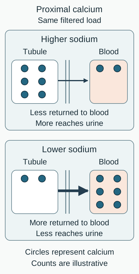
Most filtered calcium is reabsorbed before urine leaves the kidney. In the proximal tubule, much calcium transport is passive and accompanies sodium and water reabsorption. A high sodium intake promotes extracellular-volume expansion and reduces proximal reclamation of sodium, water and calcium. More calcium can remain for urinary excretion. This is a whole-nephron relationship, not a claim that sodium and calcium share one universal cotransporter. [1][2]
Trace the two exits in the sodium-calcium figure. First follow calcium back toward blood. Then follow calcium that remains in tubular fluid toward urine. Before opening the comparison, predict which exit becomes relatively more important after sodium reduction.
Follow the arrow across the tubular wall. Lower sodium favors more proximal calcium reclamation. Counts and arrow thickness show a direction, not a clinical dose-response equation. [1][2][3]Compare sodium reduction with dilution alone
Reducing sodium tends to increase calcium reclamation and lower the daily calcium amount in urine. Adding water without changing calcium excretion lowers calcium concentration but need not lower its daily amount. These are complementary effects, not interchangeable measurements.
The three-month low-sodium trial demonstrated a reduction in urinary calcium. It did not establish how many future symptomatic stone episodes sodium reduction alone prevents. Urine improvements support a mechanism; clinical recurrence remains a separate outcome. [3]
Constructed follow-up example: food calcium and urine volume remain similar
Measurement
Before
After
MeasurementUrine sodium
Before210 mmol/day
After90 mmol/day
MeasurementUrine calcium
Before330 mg/day
After240 mg/day
MeasurementUrine volume
Before2.6 L/day
After2.6 L/day
This pattern is consistent with less sodium-associated calciuria. It does not prove that sodium explains every calcium-stone disorder. Persistent hypercalciuria or high serum calcium requires further evaluation. [1]
CARI recommends less than 2,300 mg sodium daily for calcium-based stones. Sodium is not salt: 100 mmol sodium is approximately 2,300 mg sodium or 5.8 g sodium chloride. Read serving sizes and include sauces, prepared foods and supplements, not only the salt shaker. A complete collection obtained on a stable diet helps assess the response; heavy sweating or an incomplete collection limits that inference. [2]
Apply it: a patient who stops adding table salt but still has high urinary sodium needs a review of food sources and collection conditions before concluding that sodium restriction failed.
Keep calcium beside the meal, not out of the diet
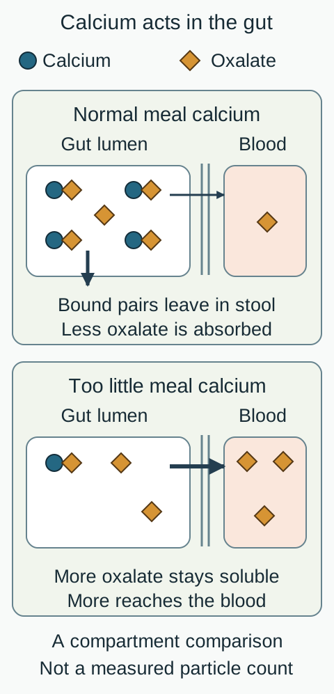
Does less calcium on the plate necessarily mean less CaOx in the kidney? Compare two otherwise similar meals in the intestinal figure. In one, calcium can bind dietary oxalate before either reaches the circulation. In the other, little calcium is available to bind it.
Compare the paired shapes that leave in stool with unbound oxalate crossing the intestinal wall. Food calcium can protect against calcium oxalate stones by acting before absorption. [1][4][5]
Meal calcium forms poorly soluble complexes with oxalate in the gut. More oxalate then leaves in stool and less is available for absorption and later urinary excretion. Severe calcium restriction can lower urinary calcium while raising urinary oxalate, defeating the intended prevention and compromising bone nutrition. The gut compartment must be considered alongside the urine compartment. [1][4]
Predict both results: after restoring an appropriate calcium intake with meals, which urine component should you especially reassess: calcium, oxalate or both?
Check the two-compartment prediction
Check both. Oxalate may fall because more is bound in the intestinal lumen. Calcium excretion can still vary with absorption, sodium intake and renal handling. Do not decide whether the plan helped by looking at calcium alone; compare the complete profile and CaOx supersaturation.
For most adults, maintain an age-appropriate food-calcium intake, commonly 1,000 to 1,200 mg/day, distributed with meals. This is normal intake, not permission for unlimited supplements. Dietary restrictions should preserve nutritional adequacy rather than eliminate whole food groups. [1][2]
A five-year trial in men with recurrent CaOx stones and hypercalciuria favored normal calcium combined with lower sodium and animal protein over low calcium alone. Because several dietary factors changed, its benefit cannot be attributed to calcium or sodium alone. [4]
When a supplement is needed for a separate nutritional indication or to bind enteric oxalate, coordinate its amount and meal timing with the clinician. In a small crossover study of healthy men, meal dosing reduced oxalate excretion whereas bedtime dosing did not, even though calcium excretion increased with both schedules. This supports timing physiology, not the study dose as a prescription or proof of long-term recurrence prevention. [5]
Apply it: a patient with low bone density and recurrent stones should not abandon prescribed calcium on their own. Review total intake, meal timing, sodium and repeat urine chemistry together. A very low-calcium diet is not the default response to the word calcium in a stone report.
Read the collection before treating the numbers
A report shows calcium falling from 320 to 160 mg/day. Is treatment working? Not necessarily: the second collection may have missed half a day. Interpret completeness, amount and concentration in that order.
For specific metabolic evaluation, EAU recommends two consecutive 24-hour samples. Obtain them when the patient has recovered from the acute episode, is free of infection and is eating and drinking normally. Follow the laboratory's collection instructions, record the collection duration and report missed voids. Creatinine excretion, prior collections, body composition and the collection history help judge plausibility; no single creatinine cutoff proves completeness in every patient. [1]
The profile commonly includes volume, calcium, sodium, oxalate, citrate, uric acid, pH and creatinine, with calculated supersaturation. Read units: mg/day or mmol/day measures total excretion, whereas mg/L measures concentration. Serum creatinine, electrolytes and calcium supply a different part of the evaluation. [1]
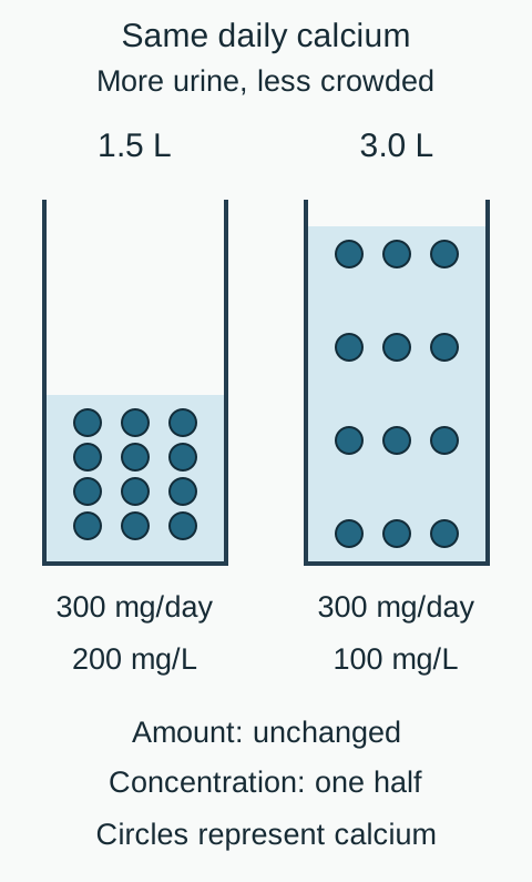
Use the dilution figure: hold daily calcium at 300 mg and increase urine volume from 1.5 to 3.0 L. Calculate concentration before reading the comparison.
Count the same number of calcium circles, then compare the water. Concentration is daily amount divided by volume; this arithmetic does not calculate supersaturation or predict a recurrence percentage. [1][2]Check the amount-versus-concentration calculation
300 mg divided by 1.5 L is 200 mg/L; 300 mg divided by 3.0 L is 100 mg/L. The daily amount is unchanged while concentration halves. This arithmetic does not calculate CaOx supersaturation, which also depends on other solutes, complex formation and chemical activity.
A practical adult target is at least 2.5 L of urine daily when medically appropriate. Drinking 2.5 L does not guarantee producing 2.5 L: sweating, diarrhea and other losses matter. Spread fluids through the day and adapt access at work. Heart failure, advanced kidney disease or risk of electrolyte imbalance requires an individual fluid plan rather than an automatic high-volume prescription. [2]
Different tests answer different questions
Information
What it establishes
InformationStone analysis
What it establishesThe material in the recovered stone
InformationSpot urine value
What it establishesA time-specific result, not a full daily profile
InformationReliable baseline 24-hour urine
What it establishesModifiable urine risks before intervention
InformationReliable follow-up collection
What it establishesBiochemical response after intervention
EAU suggests an initial follow-up collection eight to twelve weeks after starting medication, with subsequent frequency tailored to response and clinical circumstances. Do not wait for the urine collection to check medication safety. Repeat serum tests may be needed earlier. A better profile does not guarantee that no stone will recur. [1][7][8]
Apply it: if calcium, sodium and creatinine all fall by about half after an acknowledged missed collection interval, repeat a complete sample before reducing preventive therapy.
Find what concentrates, supplies or permits crystals
Two patients have identical urinary calcium but different stone risks. One has little urine and high oxalate; the other has more urine and adequate citrate. Calcium alone cannot distinguish their environments.
Low volume concentrates lithogenic solutes. Hypercalciuria supplies calcium, while hyperoxaluria supplies oxalate. Citrate binds calcium and inhibits crystal formation and growth. Low citrate therefore weakens protection even when total calcium is not high. Metabolic acidosis and potassium depletion can lower urinary citrate. Supersaturation describes a chemical tendency, not certainty that a symptomatic stone will form. [1][8]
Trace an intestinal exception: with fat malabsorption after ileal disease, resection or some bariatric procedures, unabsorbed fatty acids bind luminal calcium. More soluble oxalate remains available for intestinal uptake, particularly when the colon is in continuity. Diarrheal fluid and alkali losses may add low volume and low citrate. Treat malabsorption and use a tailored meal-calcium, fat and oxalate plan rather than labeling every high-oxalate result as an inherited disorder. [1]
Marked, persistent hyperoxaluria with early recurrent stones or nephrocalcinosis and no enteric explanation warrants specialist evaluation for primary hyperoxaluria, a disorder of endogenous oxalate production. Excessive vitamin C supplements are another potentially modifiable oxalate source. These are distinct mechanisms with different responses. [1][2]
Predict the competing effects of urine chemistry
Change
Expected concern
ChangeHigher oxalate
Expected concernMore CaOx substrate
ChangeLower citrate
Expected concernLess calcium binding and inhibition
ChangePersistently high pH
Expected concernMore favorable conditions for CaP
ChangePersistently low pH
Expected concernMore favorable conditions for uric acid
Predict before checking: citrate therapy raises citrate but also increases pH from 6.1 to 7.2. Does the citrate result alone establish success?
Check the competing-risk interpretation
No. Higher citrate can improve inhibition while greater alkalinity can increase CaP supersaturation. Compare the stone composition and the entire follow-up profile rather than maximizing one number. High pH alone does not diagnose a urinary infection.
High serum calcium prompts assessment for a systemic cause, including primary hyperparathyroidism, rather than assuming sodium explains the finding. Persistently alkaline urine with low bicarbonate and potassium suggests a renal acidification disorder after infection and medication effects are considered. Alkali may still be necessary in distal renal tubular acidosis; high urine pH is not a universal reason to withhold it. [1][8]
Apply it: distinguish a patient with fat malabsorption and high oxalate from one with high serum calcium and hypercalciuria before choosing a diet or medication.
Match medication to the residual risk and its trade-offs
A recurrent stone former reaches the fluid goal and reduces sodium, but calcium excretion remains high. What additional treatment fits, and what evidence should not be promised?
Thiazide or thiazide-like therapy can reduce urinary calcium. Continued sodium control supports that response and helps limit potassium loss. CARI conditionally suggests a thiazide for recurrent stones with persistent hypercalciuria after nutrition therapy; severe abnormalities or a high symptom burden may justify starting nutrition and medication together. Discuss expected benefit, adverse effects and the patient's priorities. [7]
Do not equate lower calciuria with proven prevention for every patient. NOSTONE randomized 416 recurrent calcium-stone formers to hydrochlorothiazide or placebo and did not find a substantial difference in its primary recurrence outcome or a dose-response relationship. Hypokalemia, gout, new diabetes and substantial creatinine increases were more frequent with hydrochlorothiazide. This does not prove that every thiazide regimen is ineffective in every selected patient. [6]
Monitor blood pressure, sodium, potassium, renal function, glucose and uric acid, as well as the follow-up urine profile. Potassium depletion can lower citrate and offset part of the desired urinary effect. Avoid responding to a poor profile by increasing the dose before assessing intake, adherence and toxicity. A loop diuretic is not an equivalent calcium-lowering substitute: furosemide can contribute to hypercalciuria. [11] Acetazolamide can produce alkaline urine and promote CaP stones rather than reproduce thiazide physiology. [1][7]
Potassium citrate is a reasonable option for persistent hypocitraturia in an appropriate recurrent stone former. It increases citrate and urine pH, so follow both and reassess CaP supersaturation. Before prescribing, check potassium, kidney function and medications that retain potassium. Hyperkalemia and significant renal impairment can make it unsafe; concomitant potassium-sparing drugs require particular caution. Sodium bicarbonate can be an alternative in selected patients unable to take potassium citrate, but is not a risk-free default because sodium load and over-alkalinisation still matter. [7][8]
Choose the residual problem: calcium remains high with adequate citrate, or citrate remains low with normal calcium. Explain which medication addresses each before reading the answer.
Compare the two treatment targets
Persistent hypercalciuria supports a shared decision about a thiazide after reviewing nutrition and safety. Persistent hypocitraturia supports consideration of potassium citrate if potassium handling and the pH profile permit. Neither drug is automatically required just because a stone contains calcium.
Allopurinol requires narrower reasoning. An older trial found benefit in recurrent CaOx stone formers with hyperuricosuria and normal urinary calcium. EAU supports allopurinol for hyperuricosuria, whereas CARI advises against routine xanthine oxidase inhibition for stones alone without a gout indication. Present this as an individualized, guideline-sensitive decision after nutrition, not a universal calcium-stone prescription. Low-pH uric acid stones also require attention to urine alkalinity, not just urate production. [1][7][10]
Apply it: when a patient taking citrate reports a much higher pH and rising CaP supersaturation, review dosing and the whole profile instead of encouraging further self-escalation. Keep a necessary acidosis treatment distinct from unnecessary excess alkali.
Separate a prevention visit from an obstructed-kidney emergency
A patient has fever, hypotension and hydronephrosis behind a ureteral stone. Would a metabolic collection change the immediate decision? The obstructed collecting system is now a potential source of sepsis.
When infection is suspected in an obstructed kidney, arrange urgent urologic decompression with a ureteral stent or percutaneous nephrostomy. Give prompt antimicrobial therapy and appropriate sepsis resuscitation, obtain cultures without delaying treatment, and tailor antibiotics to results. Antibiotics do not substitute for source control. Definitive stone fragmentation or extraction is generally deferred until sepsis has resolved. [9]
Compare urgency without infection: an afebrile patient with a solitary functioning kidney, anuria and an obstructing stone also needs urgent evaluation and relief of obstruction. Loss of renal drainage is dangerous without sepsis. Infection determines the antimicrobial indication; solitary-kidney status alone does not prove infection. [9]
Which emergency persists when the patient has no fever?
Anuria and obstructive acute kidney injury in a solitary functioning kidney remain urgent. Absence of fever does not justify waiting for passage. Conversely, pyuria or alkaline urine alone does not establish an infected system; use the overall presentation and microbiology while responding promptly to suspected infection.
For a selected uncomplicated ureteral stone, use analgesia, antiemetics when needed, and hydration matched to volume status. NSAIDs are commonly first-line analgesics when renal, gastrointestinal and cardiovascular risks permit. A passage strategy depends on size, position, pain, renal function and reliable follow-up; alpha-blocker benefit is greatest for suitable distal stones around 5 to 10 mm and use is off label. Forced fluids are not a substitute for relieving obstruction. [9]
Fever, rigors, inability to maintain intake, uncontrolled pain or reduced urine output requires reassessment. Once the acute episode has resolved, analyze available stone material and assess recurrence risk. Recurrent stones, childhood presentation, a solitary kidney or unusual composition warrants a more detailed metabolic approach, not empiric antibiotics for sterile calcium stones. [1][9]
Apply it: prioritize drainage plus infection treatment for a septic obstruction, urgent renal preservation for an anuric solitary kidney, and measured prevention for a stable recurrent stone former. These are different decisions even when all three stones contain calcium.
Apply the lesson
Use the whole clinical and urine profile to select one answer. Explain the competing alternative before reviewing the option-specific explanations. All cases remain available without a required sequence.
Case 1
Show answer and explanations for case 1
A. Reduce food calcium and maintain current fluids (Why this does not fit)
The stone contains calcium and the daily urine calcium is high. Food calcium is already appropriate, restriction can increase oxalate absorption, and unchanged fluid intake leaves the low-volume risk untreated.
Reasoning steps for option A
Why might less calcium seem attractive?
The stone contains calcium and the daily urine calcium is high.
Why does this plan fit poorly?
Food calcium is already appropriate, restriction can increase oxalate absorption, and unchanged fluid intake leaves the low-volume risk untreated.
B. Increase fluid intake and reduce dietary sodium (Best answer)
Low urine volume concentrates solutes and high sodium accompanies excessive urinary calcium. More urine dilutes the solutes while sodium reduction targets calcium excretion; his normal food calcium should be preserved.
Reasoning steps for option B
Which two risks are present?
Low urine volume concentrates solutes and high sodium accompanies excessive urinary calcium.
Why choose both interventions now?
More urine dilutes the solutes while sodium reduction targets calcium excretion; his normal food calcium should be preserved.
C. Add potassium citrate and maintain dietary sodium (Why this does not fit)
Potassium citrate helps selected patients with low citrate or an appropriate alkalinisation indication. Citrate is adequate; this plan leaves both low urine volume and excessive sodium intake uncorrected.
Reasoning steps for option C
What does potassium citrate address?
Potassium citrate helps selected patients with low citrate or an appropriate alkalinisation indication.
What does this profile show instead?
Citrate is adequate; this plan leaves both low urine volume and excessive sodium intake uncorrected.
D. Reduce dietary oxalate and maintain current fluids (Why this does not fit)
Excessive oxalate exposure or absorption can raise calcium oxalate risk. Oxalate is within range while low volume and high sodium-associated calciuria are documented.
Reasoning steps for option D
When is oxalate-directed treatment useful?
Excessive oxalate exposure or absorption can raise calcium oxalate risk.
Why is it not the best initial focus here?
Oxalate is within range while low volume and high sodium-associated calciuria are documented.
Takeaway: Treat the measured volume and sodium risks while preserving normal meal calcium.
A. Lower calcium filtration from a fall in serum calcium (Why this does not fit)
A lower plasma calcium concentration could reduce the filtered calcium load. Serum calcium and kidney function are unchanged, whereas the major measured change is sodium intake and excretion.
Reasoning steps for option A
Could less filtered calcium reduce urinary calcium?
A lower plasma calcium concentration could reduce the filtered calcium load.
Does the observed response support that route?
Serum calcium and kidney function are unchanged, whereas the major measured change is sodium intake and excretion.
B. Greater urinary citrate binding of filtered calcium (Why this does not fit)
It reduces free calcium activity and can inhibit crystal formation in urine. Complexing calcium does not itself lower the total calcium collected in mg/day; the result supports altered calcium excretion.
Reasoning steps for option B
What does urinary citrate binding change?
It reduces free calcium activity and can inhibit crystal formation in urine.
Does binding alone explain the measured daily amount?
Complexing calcium does not itself lower the total calcium collected in mg/day; the result supports altered calcium excretion.
C. Lower urine calcium concentration from water retention alone (Why this does not fit)
A larger urine volume can lower mg/L without changing total mg/day. Urine volume is unchanged and calcium fell in mg/day, requiring a change in excretion rather than dilution alone.
Reasoning steps for option C
Can more water reduce calcium concentration?
A larger urine volume can lower mg/L without changing total mg/day.
Why does dilution not explain this report?
Urine volume is unchanged and calcium fell in mg/day, requiring a change in excretion rather than dilution alone.
D. Greater proximal calcium reclamation with sodium and water (Best answer)
With reliable collections and stable conditions, it supports lower sodium intake rather than dilution alone. Less sodium-associated volume expansion favors proximal sodium, water and calcium reabsorption, leaving less calcium for urine.
Reasoning steps for option D
What does the lower urine sodium suggest in this setting?
With reliable collections and stable conditions, it supports lower sodium intake rather than dilution alone.
How does this affect calcium delivery?
Less sodium-associated volume expansion favors proximal sodium, water and calcium reabsorption, leaving less calcium for urine.
Takeaway: A fall in daily calcium with stable urine volume is not simply a dilution effect.
A. Restore normal calcium intake with meals (Best answer)
Oxalate rose substantially despite a modest reduction in urinary calcium. It binds intestinal oxalate before absorption, so restoring normal intake can reduce oxalate delivery to urine.
Reasoning steps for option A
Which urinary change undermines the intended benefit?
Oxalate rose substantially despite a modest reduction in urinary calcium.
How does meal calcium address that change?
It binds intestinal oxalate before absorption, so restoring normal intake can reduce oxalate delivery to urine.
B. Replace meal calcium with a bedtime supplement (Why this does not fit)
A supplement can provide calcium when food intake is insufficient. Calcium away from the oxalate-containing meals misses much of the luminal binding opportunity that the diet removed.
Reasoning steps for option B
Why consider a supplement?
A supplement can provide calcium when food intake is insufficient.
Why is bedtime replacement a poorer choice here?
Calcium away from the oxalate-containing meals misses much of the luminal binding opportunity that the diet removed.
C. Continue calcium restriction and increase water (Why this does not fit)
More urine can reduce the concentration of calcium and oxalate. It leaves severe food-calcium restriction and the resulting increase in oxalate absorption unaddressed.
Reasoning steps for option C
What benefit could additional water provide?
More urine can reduce the concentration of calcium and oxalate.
Why does that not correct the dietary error?
It leaves severe food-calcium restriction and the resulting increase in oxalate absorption unaddressed.
D. Maintain low calcium and reduce animal protein (Why this does not fit)
It may reduce acid and purine exposure in selected stone formers. The new abnormality followed calcium restriction without a reported protein excess; restoring gut calcium binding directly addresses it.
Reasoning steps for option D
What can reducing excessive animal protein change?
It may reduce acid and purine exposure in selected stone formers.
Why is it not the best correction of this pattern?
The new abnormality followed calcium restriction without a reported protein excess; restoring gut calcium binding directly addresses it.
Takeaway: Lower urine calcium is not a sufficient goal when oxalate rises because meal calcium was restricted.
A. Calcium binds oxalate, increasing its delivery to the colon (Why this does not fit)
It creates poorly soluble complexes that can leave in stool. Binding lowers oxalate absorption; the malabsorptive state instead diverts calcium toward fatty acids.
Reasoning steps for option A
What does luminal calcium-oxalate binding usually do?
It creates poorly soluble complexes that can leave in stool.
Why does that not explain the urine result?
Binding lowers oxalate absorption; the malabsorptive state instead diverts calcium toward fatty acids.
B. Bile salts bind oxalate, increasing fecal oxalate loss (Why this does not fit)
Ileal dysfunction alters bile salt handling and can contribute to diarrhea. Greater fecal oxalate loss would reduce, not explain, the increased oxalate reaching the urine.
Reasoning steps for option B
Why consider bile salts after ileal resection?
Ileal dysfunction alters bile salt handling and can contribute to diarrhea.
Why is increased fecal binding the wrong direction?
Greater fecal oxalate loss would reduce, not explain, the increased oxalate reaching the urine.
Unabsorbed fat remains in the intestinal lumen after malabsorptive ileal disease or surgery. Fatty acids bind calcium, leaving more oxalate soluble and available for absorption, especially with a colon in continuity.
Reasoning steps for option C
What does steatorrhea indicate?
Unabsorbed fat remains in the intestinal lumen after malabsorptive ileal disease or surgery.
How does that produce high urinary oxalate?
Fatty acids bind calcium, leaving more oxalate soluble and available for absorption, especially with a colon in continuity.
D. Dietary calcium absorption rises, increasing urinary calcium (Why this does not fit)
Greater calcium absorption can contribute to higher urinary calcium. Urinary calcium is not increased; oxalate is the abnormal substrate linked to fat malabsorption.
Reasoning steps for option D
How can absorbed calcium affect urine?
Greater calcium absorption can contribute to higher urinary calcium.
Which measured finding argues against this as the dominant process?
Urinary calcium is not increased; oxalate is the abnormal substrate linked to fat malabsorption.
Takeaway: Enteric hyperoxaluria can coexist with normal urinary calcium and low citrate.
A. Accept sodium reduction as proof of treatment success (Why this does not fit)
A lower daily urine sodium can support adherence when collection is reliable. Creatinine fell proportionally and timing is uncertain, so missing urine could account for the apparent improvement.
Reasoning steps for option A
What makes the sodium result attractive?
A lower daily urine sodium can support adherence when collection is reliable.
Why is that inference unsafe here?
Creatinine fell proportionally and timing is uncertain, so missing urine could account for the apparent improvement.
B. Repeat a carefully timed complete urine collection (Best answer)
Calcium, sodium and creatinine all fell by roughly half despite stable body composition and renal function. Uncertain timing makes incomplete collection plausible, so obtain a reliable full-day sample rather than declaring a response.
Reasoning steps for option B
Why is the broad fall in excretion concerning?
Calcium, sodium and creatinine all fell by roughly half despite stable body composition and renal function.
What is needed before changing treatment?
Uncertain timing makes incomplete collection plausible, so obtain a reliable full-day sample rather than declaring a response.
C. Estimate a full-day result by doubling every value (Why this does not fit)
All reported excretion values are about half their previous amounts. The actual collection duration and missed void composition are unknown, so multiplying cannot recover a measured 24-hour profile.
Reasoning steps for option C
Why might arithmetic correction seem reasonable?
All reported excretion values are about half their previous amounts.
Why is extrapolation unreliable?
The actual collection duration and missed void composition are unknown, so multiplying cannot recover a measured 24-hour profile.
D. Replace follow-up with a single spot calcium value (Why this does not fit)
It gives a time-specific calcium result, sometimes interpreted with creatinine. It does not resolve the uncertain daily volume and excretion profile needed to assess this intervention.
Reasoning steps for option D
What information can a spot sample provide?
It gives a time-specific calcium result, sometimes interpreted with creatinine.
Why is it not an equivalent substitute?
It does not resolve the uncertain daily volume and excretion profile needed to assess this intervention.
Takeaway: Collection validity comes before interpreting an apparent response.
A. The concentration falls by half while daily excretion stays unchanged (Best answer)
300 mg divided by 1.5 L is 200 mg/L; divided by 3.0 L it is 100 mg/L. More urine diluted the same daily calcium amount; the measurement does not show reduced total calcium excretion.
Reasoning steps for option A
What are the two calcium concentrations?
300 mg divided by 1.5 L is 200 mg/L; divided by 3.0 L it is 100 mg/L.
What changed physiologically?
More urine diluted the same daily calcium amount; the measurement does not show reduced total calcium excretion.
B. Daily excretion halves while concentration remains unchanged (Why this does not fit)
It reports the total collected calcium over the day, which remains 300 mg. The increased volume changes mg/L, not the measured daily amount.
Reasoning steps for option B
What does the report measure in mg/day?
It reports the total collected calcium over the day, which remains 300 mg.
Why is the proposed interpretation reversed?
The increased volume changes mg/L, not the measured daily amount.
C. Both concentration and daily excretion fall by one half (Why this does not fit)
A lower concentration is often described informally as less calcium in urine. Multiplying 100 mg/L by 3.0 L still gives 300 mg/day, so daily excretion did not fall.
Reasoning steps for option C
Why might both values be confused?
A lower concentration is often described informally as less calcium in urine.
Which calculation separates them?
Multiplying 100 mg/L by 3.0 L still gives 300 mg/day, so daily excretion did not fall.
D. Calcium concentration is unchanged because filtration is stable (Why this does not fit)
Filtration and tubular handling help determine excretion but do not alone determine urine concentration. Twice the urine volume contains the same calcium amount, so its average concentration must be lower.
Reasoning steps for option D
Does stable filtration require stable concentration?
Filtration and tubular handling help determine excretion but do not alone determine urine concentration.
What does the measured volume establish?
Twice the urine volume contains the same calcium amount, so its average concentration must be lower.
Takeaway: Dilution improves concentration without necessarily changing the amount excreted.
A. Escalate medication because table-salt restriction is sufficient (Why this does not fit)
Calcium excretion remains high despite existing thiazide therapy. Stopping table salt did not eliminate sodium in prepared food; the calculated intake and measured urine sodium remain high.
Reasoning steps for option A
Why might medication escalation be considered?
Calcium excretion remains high despite existing thiazide therapy.
What prevents that conclusion about nutrition?
Stopping table salt did not eliminate sodium in prepared food; the calculated intake and measured urine sodium remain high.
B. Reduce meal calcium because the food sodium is modest (Why this does not fit)
Calcium intake contributes to absorbed calcium and must be assessed in context. The food sodium is not modest at about 3,600 mg daily, and restricting normal meal calcium can increase oxalate absorption.
Reasoning steps for option B
What dietary factor can also influence urinary calcium?
Calcium intake contributes to absorbed calcium and must be assessed in context.
Why does this explanation miss the measured problem?
The food sodium is not modest at about 3,600 mg daily, and restricting normal meal calcium can increase oxalate absorption.
C. Increase water because daily sodium reflects concentration (Why this does not fit)
It reduces solute concentration by increasing urine volume. Urine sodium is reported as total mmol/day, not concentration, and the patient already produces 2.8 L daily.
Reasoning steps for option C
Why is water intake important for stone prevention?
It reduces solute concentration by increasing urine volume.
Why does extra water not correct this interpretation?
Urine sodium is reported as total mmol/day, not concentration, and the patient already produces 2.8 L daily.
D. Review the approximately 3,600 mg daily sodium exposure (Best answer)
The entree provides 1,700 mg, the soup 900 mg and other foods 1,000 mg, totaling about 3,600 mg sodium. The intake remains above the recommended calcium-stone target and the high urine sodium supports a persistent modifiable driver of calciuria.
Reasoning steps for option D
What does the food-label calculation show?
The entree provides 1,700 mg, the soup 900 mg and other foods 1,000 mg, totaling about 3,600 mg sodium.
Why address that before increasing medication?
The intake remains above the recommended calcium-stone target and the high urine sodium supports a persistent modifiable driver of calciuria.
Takeaway: Food labels and reliable urine sodium can reveal ongoing sodium exposure despite no added table salt.
A. Potassium citrate to increase the urine pH (Why this does not fit)
It increases urine citrate and alkalinity in selected patients. Citrate is already adequate and the documented residual abnormality is high urinary calcium.
Reasoning steps for option A
What does potassium citrate most directly address?
It increases urine citrate and alkalinity in selected patients.
Why is it not the strongest match here?
Citrate is already adequate and the documented residual abnormality is high urinary calcium.
B. Allopurinol to suppress uric acid production (Why this does not fit)
The historical calcium oxalate evidence concerns hyperuricosuria with normal urinary calcium. Persistent hypercalciuria, not a demonstrated urate abnormality, is the target described.
Reasoning steps for option B
Which urine pattern supports considering allopurinol?
The historical calcium oxalate evidence concerns hyperuricosuria with normal urinary calcium.
Why does that not match this patient?
Persistent hypercalciuria, not a demonstrated urate abnormality, is the target described.
C. A thiazide-like diuretic such as chlorthalidone (Best answer)
Hypercalciuria persists despite adequate urine volume and meaningful sodium control. A thiazide-like diuretic can lower urinary calcium and blood pressure, with recurrence uncertainty and safety monitoring included in the decision.
Reasoning steps for option C
What risk remains after the foundational changes?
Hypercalciuria persists despite adequate urine volume and meaningful sodium control.
Why is this a reasonable medication discussion?
A thiazide-like diuretic can lower urinary calcium and blood pressure, with recurrence uncertainty and safety monitoring included in the decision.
D. Calcium supplementation between meals each day (Why this does not fit)
Selected nutritional needs or enteric oxalate binding can justify clinician-directed supplementation. No calcium deficiency or hyperoxaluria is given, and between-meal supplementation does not target persistent renal calcium excretion.
Reasoning steps for option D
When may calcium supplementation be appropriate?
Selected nutritional needs or enteric oxalate binding can justify clinician-directed supplementation.
Why is it a poor match to these results?
No calcium deficiency or hyperoxaluria is given, and between-meal supplementation does not target persistent renal calcium excretion.
Takeaway: Persistent hypercalciuria can justify a thiazide discussion, not a guarantee of freedom from recurrence.
A. Review the diuretic and correct potassium depletion (Best answer)
Potassium depletion can lower urinary citrate even while thiazide therapy lowers urinary calcium. Symptomatic hypokalemia needs prompt assessment and correction with review of medication dosing, followed by serum and urine reassessment.
Reasoning steps for option A
What explains the new citrate abnormality?
Potassium depletion can lower urinary citrate even while thiazide therapy lowers urinary calcium.
What should the clinician prioritize?
Symptomatic hypokalemia needs prompt assessment and correction with review of medication dosing, followed by serum and urine reassessment.
B. Increase the diuretic because calcium remains measurable (Why this does not fit)
Thiazides can reduce urinary calcium, which is one treatment target. The patient has symptomatic potassium depletion and new hypocitraturia, so toxicity and an offsetting urine risk require attention first.
Reasoning steps for option B
Why might a lower calcium result encourage escalation?
Thiazides can reduce urinary calcium, which is one treatment target.
Why is escalation inappropriate now?
The patient has symptomatic potassium depletion and new hypocitraturia, so toxicity and an offsetting urine risk require attention first.
C. Restrict calcium further to lower the residual excretion (Why this does not fit)
The patient previously had excessive urinary calcium. Calcium excretion already improved; cramps, low potassium and low citrate point to a medication-associated electrolyte problem.
Reasoning steps for option C
Why does calcium excretion attract attention?
The patient previously had excessive urinary calcium.
Why does diet restriction miss the present problem?
Calcium excretion already improved; cramps, low potassium and low citrate point to a medication-associated electrolyte problem.
D. Add sodium bicarbonate without changing the current regimen (Why this does not fit)
Alkali can increase urinary citrate in selected settings. It does not adequately address symptomatic hypokalemia or the diuretic contribution, and sodium load and urine pH would also require review.
Reasoning steps for option D
What might alkali improve?
Alkali can increase urinary citrate in selected settings.
Why is this not the first response here?
It does not adequately address symptomatic hypokalemia or the diuretic contribution, and sodium load and urine pH would also require review.
Takeaway: A better calcium value does not excuse symptomatic hypokalemia or loss of urinary citrate.
A. Chlorthalidone with follow-up urinary calcium (Why this does not fit)
Persistent hypercalciuria is the principal phenotype for that discussion. Calcium is within range while citrate is markedly low, making citrate deficiency the more direct treatment target.
Reasoning steps for option A
Which residual risk would favor a thiazide?
Persistent hypercalciuria is the principal phenotype for that discussion.
What argues for a different target here?
Calcium is within range while citrate is markedly low, making citrate deficiency the more direct treatment target.
B. Allopurinol with follow-up urinary uric acid (Why this does not fit)
Allopurinol was studied in hyperuricosuric, normocalciuric calcium oxalate stone formers. The case documents low citrate rather than excessive urinary urate, so suppressing urate production misses the stated abnormality.
Reasoning steps for option B
What selected calcium-stone phenotype has historical evidence?
Allopurinol was studied in hyperuricosuric, normocalciuric calcium oxalate stone formers.
Why is that not the demonstrated problem?
The case documents low citrate rather than excessive urinary urate, so suppressing urate production misses the stated abnormality.
C. Additional calcium with each oxalate-containing meal (Why this does not fit)
It binds intestinal oxalate when normal intake is inadequate or enteric hyperoxaluria is present. The measured oxalate is within range and the persistent deficit is urinary citrate, not demonstrated intestinal calcium availability.
Reasoning steps for option C
When can meal calcium be particularly useful?
It binds intestinal oxalate when normal intake is inadequate or enteric hyperoxaluria is present.
Why is it not the most direct additional therapy?
The measured oxalate is within range and the persistent deficit is urinary citrate, not demonstrated intestinal calcium availability.
D. Potassium citrate with follow-up urine chemistry (Best answer)
Urinary citrate is persistently low despite adequate fluid and sodium measures. Potassium citrate restores a calcium-binding inhibitor but also raises urine pH, so follow both citrate and the calcium phosphate risk profile.
Reasoning steps for option D
Which protective factor is deficient?
Urinary citrate is persistently low despite adequate fluid and sodium measures.
Why choose citrate and still monitor pH?
Potassium citrate restores a calcium-binding inhibitor but also raises urine pH, so follow both citrate and the calcium phosphate risk profile.
Takeaway: Citrate replacement addresses an inhibitor deficit; pH monitoring remains part of treatment.
A. Increase alkali further to suppress the remaining crystals (Why this does not fit)
Citrate binds calcium and inhibits crystal formation. The current excess has already raised pH into a more favorable environment for calcium phosphate despite adequate citrate.
Reasoning steps for option A
Why might more citrate appear helpful?
Citrate binds calcium and inhibits crystal formation.
Why is more alkali not the best response here?
The current excess has already raised pH into a more favorable environment for calcium phosphate despite adequate citrate.
B. Reduce excess alkali and reassess the urine profile (Best answer)
Citrate increased, but the more alkaline urine coincides with higher calcium phosphate supersaturation and a changed stone composition. Review the extra dosing and reduce excessive alkalinisation while reassessing citrate, pH and supersaturation rather than maximizing pH.
Reasoning steps for option B
Which desired effect improved and which risk worsened?
Citrate increased, but the more alkaline urine coincides with higher calcium phosphate supersaturation and a changed stone composition.
What does that combination require?
Review the extra dosing and reduce excessive alkalinisation while reassessing citrate, pH and supersaturation rather than maximizing pH.
C. Replace the extra potassium citrate with sodium bicarbonate (Why this does not fit)
It can increase urine citrate and pH when an alternative to potassium citrate is needed. Excess alkalinisation remains and an additional sodium load can complicate calcium-stone prevention.
Reasoning steps for option C
Can sodium bicarbonate provide alkali?
It can increase urine citrate and pH when an alternative to potassium citrate is needed.
Why does substitution fail to solve this problem?
Excess alkalinisation remains and an additional sodium load can complicate calcium-stone prevention.
D. Treat a urease-producing infection before altering the dose (Why this does not fit)
Some organisms alkalinize urine through urease activity. There is no clinical or culture evidence of infection and the pH rise followed unprescribed alkali escalation.
Reasoning steps for option D
Why might high urine pH raise concern for infection?
Some organisms alkalinize urine through urease activity.
What makes that explanation less likely in this case?
There is no clinical or culture evidence of infection and the pH rise followed unprescribed alkali escalation.
Takeaway: Treat the combined supersaturation profile, not the citrate or pH value in isolation.
A. Acidic urine that would become less acidic with alkali (Why this does not fit)
It raises pH, which can be useful in selected stone phenotypes. The value of 5.8 does not outweigh the immediate safety problem of hyperkalemia and impaired potassium elimination.
Reasoning steps for option A
What effect does potassium citrate have on urine pH?
It raises pH, which can be useful in selected stone phenotypes.
Why is urine pH not the main contraindication here?
The value of 5.8 does not outweigh the immediate safety problem of hyperkalemia and impaired potassium elimination.
B. Low citrate indicating reduced calcium binding in urine (Why this does not fit)
It weakens calcium complexing and inhibition of crystal formation. It supplies a potential indication, but an indication cannot override the potassium-related contraindication.
Reasoning steps for option B
Why is low citrate clinically relevant?
It weakens calcium complexing and inhibition of crystal formation.
Does that finding itself argue against treatment?
It supplies a potential indication, but an indication cannot override the potassium-related contraindication.
C. Impaired renal potassium excretion with hyperkalemia and potassium-retaining medicines (Best answer)
Reduced renal clearance plus potassium-retaining drugs increases the risk of further potassium accumulation. The patient is already hyperkalemic, so adding potassium citrate can be dangerous and requires prompt review of potassium and medication safety.
Reasoning steps for option C
How do kidney disease and the listed drugs interact?
Reduced renal clearance plus potassium-retaining drugs increases the risk of further potassium accumulation.
Why does that outweigh the low citrate indication?
The patient is already hyperkalemic, so adding potassium citrate can be dangerous and requires prompt review of potassium and medication safety.
D. Normal bicarbonate indicating an absence of overt acidosis (Why this does not fit)
No; hypocitraturic calcium oxalate stones can be an indication without overt metabolic acidosis. Current potassium elevation, impaired kidney function and potassium-retaining medication exposure are the decisive safety factors.
Reasoning steps for option D
Must overt systemic acidosis be present to consider citrate?
No; hypocitraturic calcium oxalate stones can be an indication without overt metabolic acidosis.
What therefore controls this decision?
Current potassium elevation, impaired kidney function and potassium-retaining medication exposure are the decisive safety factors.
Takeaway: A stone-prevention indication does not override existing hyperkalemia.
A. The higher dose establishes protection because its point estimate is lower (Why this does not fit)
A rate ratio below one numerically favors treatment. Its confidence interval crosses one and the prespecified dose-response result is not significant, so the estimate alone cannot establish protection.
Reasoning steps for option A
Why does the point estimate appear promising?
A rate ratio below one numerically favors treatment.
Why is that not proof of benefit?
Its confidence interval crosses one and the prespecified dose-response result is not significant, so the estimate alone cannot establish protection.
B. The trial proves that all thiazide regimens have no preventive effect (Why this does not fit)
It tested specified daily hydrochlorothiazide doses in its enrolled recurrent-stone population. The result does not establish exact equivalence or rule out every different regimen and carefully selected patient group.
Reasoning steps for option B
What question did this trial directly examine?
It tested specified daily hydrochlorothiazide doses in its enrolled recurrent-stone population.
Why is the class-wide conclusion too broad?
The result does not establish exact equivalence or rule out every different regimen and carefully selected patient group.
C. Reduced urinary calcium is sufficient to outweigh the adverse effects (Why this does not fit)
It can reduce one contributor to calcium salt supersaturation. A surrogate improvement does not by itself quantify clinical benefit or justify ignoring electrolyte, metabolic and renal adverse events.
Reasoning steps for option C
Why is lower calcium a desirable urine result?
It can reduce one contributor to calcium salt supersaturation.
Why must harms and recurrence still be considered?
A surrogate improvement does not by itself quantify clinical benefit or justify ignoring electrolyte, metabolic and renal adverse events.
D. A biochemical response does not establish a recurrence benefit (Best answer)
The confidence interval includes no difference, and the dose-response test did not establish a dose-related primary-outcome benefit. Calcium lowering is a surrogate response; discuss the uncertain recurrence benefit and adverse effects rather than guaranteeing clinical prevention.
Reasoning steps for option D
What does the trial estimate allow?
The confidence interval includes no difference, and the dose-response test did not establish a dose-related primary-outcome benefit.
How should that affect counseling about lower urinary calcium?
Calcium lowering is a surrogate response; discuss the uncertain recurrence benefit and adverse effects rather than guaranteeing clinical prevention.
Takeaway: Urine calcium and future symptomatic or imaging recurrence are related but different outcomes.
A. Correct the systemic acidosis with carefully monitored alkali therapy (Best answer)
Normal-anion-gap metabolic acidosis with low potassium and persistently alkaline urine suggests impaired distal acidification after alternative causes are considered. Correcting systemic acidosis can improve citrate and calcium handling; monitor potassium and calcium phosphate risk rather than withholding necessary alkali solely because urine is alkaline.
Reasoning steps for option A
What does the acid-base and urine pattern suggest?
Normal-anion-gap metabolic acidosis with low potassium and persistently alkaline urine suggests impaired distal acidification after alternative causes are considered.
Why is alkali appropriate despite the urine pH?
Correcting systemic acidosis can improve citrate and calcium handling; monitor potassium and calcium phosphate risk rather than withholding necessary alkali solely because urine is alkaline.
B. Avoid alkali because every alkaline urine requires acidification (Why this does not fit)
Higher pH can favor calcium phosphate supersaturation. This patient has systemic acidosis and an acidification defect, which requires treatment rather than leaving the metabolic disturbance uncorrected.
Reasoning steps for option B
Why might urine alkalinity cause concern?
Higher pH can favor calcium phosphate supersaturation.
Why is a universal acidification rule inappropriate?
This patient has systemic acidosis and an acidification defect, which requires treatment rather than leaving the metabolic disturbance uncorrected.
C. Use calcium restriction as the primary correction for nephrocalcinosis (Why this does not fit)
Calcium deposition reflects an unfavorable renal mineral environment. It does not correct the systemic acidosis and low citrate and can compromise normal calcium nutrition.
Reasoning steps for option C
Why does nephrocalcinosis suggest calcium exposure matters?
Calcium deposition reflects an unfavorable renal mineral environment.
What does calcium restriction fail to address?
It does not correct the systemic acidosis and low citrate and can compromise normal calcium nutrition.
D. Treat a urease infection as the explanation for the high pH (Why this does not fit)
Urease-producing organisms generate an alkaline urinary environment. Cultures are negative and the persistent pH abnormality occurs with non-anion-gap acidosis and hypokalemia, supporting a renal acidification disorder.
Reasoning steps for option D
How can infection make urine alkaline?
Urease-producing organisms generate an alkaline urinary environment.
Why is that not the best explanation here?
Cultures are negative and the persistent pH abnormality occurs with non-anion-gap acidosis and hypokalemia, supporting a renal acidification disorder.
Takeaway: Alkaline urine does not make needed treatment of distal renal tubular acidosis inappropriate.
A. Idiopathic hypercalciuria is established by a normal-range PTH (Why this does not fit)
It falls within the laboratory reference interval. The reference interval must be interpreted with serum calcium, which is repeatedly high and should suppress PTH.
Reasoning steps for option A
Why might the PTH result look reassuring?
It falls within the laboratory reference interval.
Why does that not establish idiopathic hypercalciuria?
The reference interval must be interpreted with serum calcium, which is repeatedly high and should suppress PTH.
B. Residual sodium intake is the sufficient explanation for hypercalcemia (Why this does not fit)
Sodium intake can influence urinary calcium excretion. The urine sodium is controlled and sodium-associated calciuria does not explain repeated serum hypercalcemia with non-suppressed PTH.
Reasoning steps for option B
Why is sodium relevant to calcium stones?
Sodium intake can influence urinary calcium excretion.
Why is it insufficient to explain this pattern?
The urine sodium is controlled and sodium-associated calciuria does not explain repeated serum hypercalcemia with non-suppressed PTH.
C. Investigate a PTH-dependent cause of the recurrent hypercalcemia (Best answer)
It should be suppressed by feedback; a value inside the population reference interval can still be inappropriate during hypercalcemia. Recurrent stones, hypercalciuria and non-suppressed PTH warrant evaluation for a PTH-dependent cause such as primary hyperparathyroidism rather than diet-only attribution.
Reasoning steps for option C
What should PTH do when serum calcium is high?
It should be suppressed by feedback; a value inside the population reference interval can still be inappropriate during hypercalcemia.
How does that alter the stone assessment?
Recurrent stones, hypercalciuria and non-suppressed PTH warrant evaluation for a PTH-dependent cause such as primary hyperparathyroidism rather than diet-only attribution.
D. Intestinal oxalate absorption is the main target of the work-up (Why this does not fit)
Insufficient luminal calcium or malabsorption can increase urinary oxalate. The defining abnormality is systemic hypercalcemia with inappropriate PTH, not a reported oxalate excess.
Reasoning steps for option D
Why is oxalate absorption important in calcium oxalate disease?
Insufficient luminal calcium or malabsorption can increase urinary oxalate.
What directs evaluation elsewhere here?
The defining abnormality is systemic hypercalcemia with inappropriate PTH, not a reported oxalate excess.
Takeaway: A reference-range PTH can be inappropriate when serum calcium is high.
A. Hypercalciuria with a normal urinary citrate concentration (Why this does not fit)
Excess urinary calcium can persist even when citrate is normal. His calcium excretion is not increased; the repeated abnormality is uric acid excretion.
Reasoning steps for option A
Why is this a familiar calcium-stone phenotype?
Excess urinary calcium can persist even when citrate is normal.
Does the patient fit that phenotype?
His calcium excretion is not increased; the repeated abnormality is uric acid excretion.
B. Hyperuricosuria with normal daily urinary calcium (Best answer)
Urinary uric acid is high while urinary calcium is not high. The older calcium oxalate trial enrolled hyperuricosuric, normocalciuric patients; its finding should not be generalized to every calcium-stone phenotype.
Reasoning steps for option B
How should the two repeated excretion results be classified?
Urinary uric acid is high while urinary calcium is not high.
Why does that distinction matter for the evidence?
The older calcium oxalate trial enrolled hyperuricosuric, normocalciuric patients; its finding should not be generalized to every calcium-stone phenotype.
C. Acidic urine with uric acid rather than calcium oxalate stones (Why this does not fit)
Low pH reduces uric acid solubility and can dominate that stone phenotype. The patient has analyzed calcium oxalate stones, a pH of 6.2 and the specific high-urate, normal-calcium excretion pattern.
Reasoning steps for option C
What makes acidic urine important for uric acid stones?
Low pH reduces uric acid solubility and can dominate that stone phenotype.
Why does it not describe the trial match here?
The patient has analyzed calcium oxalate stones, a pH of 6.2 and the specific high-urate, normal-calcium excretion pattern.
D. Hypocitraturia with reduced urinary calcium excretion (Why this does not fit)
It reduces calcium binding and crystal inhibition. Citrate is within range, while repeated hyperuricosuria is the feature relevant to the historical trial.
Reasoning steps for option D
What risk does hypocitraturia represent?
It reduces calcium binding and crystal inhibition.
Does that account for the documented residual risk?
Citrate is within range, while repeated hyperuricosuria is the feature relevant to the historical trial.
Takeaway: Historical allopurinol evidence in a selected phenotype is not a universal prescription rule.
A. Direct precipitation of acetazolamide as the recovered stone (Why this does not fit)
A drug or metabolite may crystallize in urine and become part of a stone. The recovered material is calcium phosphate and the paired bicarbonate, pH and citrate changes support an altered urine environment.
Reasoning steps for option A
How can some drugs cause stones directly?
A drug or metabolite may crystallize in urine and become part of a stone.
What argues for a different mechanism here?
The recovered material is calcium phosphate and the paired bicarbonate, pH and citrate changes support an altered urine environment.
B. Urease activity causing infection-related alkaline urine (Why this does not fit)
It can explain alkaline urine in an appropriate urinary infection. Cultures are negative and the new abnormalities follow acetazolamide exposure with a fall in serum bicarbonate.
Reasoning steps for option B
What could urease activity explain?
It can explain alkaline urine in an appropriate urinary infection.
Why is it not the best link in this case?
Cultures are negative and the new abnormalities follow acetazolamide exposure with a fall in serum bicarbonate.
C. Altered urine chemistry from bicarbonate loss and reduced citrate (Best answer)
Renal bicarbonate loss can increase urine alkalinity while systemic acid-base effects contribute to lower urinary citrate. Higher pH favors calcium phosphate and low citrate reduces inhibition, creating a medication-related lithogenic environment.
Reasoning steps for option C
What effect follows carbonic anhydrase inhibition?
Renal bicarbonate loss can increase urine alkalinity while systemic acid-base effects contribute to lower urinary citrate.
How does this favor the analyzed stone?
Higher pH favors calcium phosphate and low citrate reduces inhibition, creating a medication-related lithogenic environment.
D. Enteric oxalate absorption after intestinal calcium depletion (Why this does not fit)
It raises urinary oxalate and favors calcium oxalate stones, particularly with malabsorption. There is no bowel history, the stone is calcium phosphate and the acid-base changes point to the medication.
Reasoning steps for option D
What would this intestinal process promote?
It raises urinary oxalate and favors calcium oxalate stones, particularly with malabsorption.
Why does it fit poorly here?
There is no bowel history, the stone is calcium phosphate and the acid-base changes point to the medication.
Takeaway: Medication-induced changes in urine chemistry differ from stones composed of the medication itself.
A. The normal calcium consumed with meals (Why this does not fit)
Calcium is a component of the recovered stone. Normal meal calcium binds intestinal oxalate, and the increase followed vitamin C supplementation rather than excessive calcium intake.
Reasoning steps for option A
Why might calcium attract concern after a calcium stone?
Calcium is a component of the recovered stone.
Why is reducing it a poor response to this pattern?
Normal meal calcium binds intestinal oxalate, and the increase followed vitamin C supplementation rather than excessive calcium intake.
B. The maintained fluid intake at work (Why this does not fit)
It influences urine concentration through total urine volume. Measured volume is similar while oxalate in mg/day increased, so a new oxalate source should be considered.
Reasoning steps for option B
How does fluid intake affect urinary stone risk?
It influences urine concentration through total urine volume.
Why does it not explain the new daily oxalate amount?
Measured volume is similar while oxalate in mg/day increased, so a new oxalate source should be considered.
C. The unchanged sodium content of his meals (Why this does not fit)
It can increase urinary calcium and affect other urine risks. Urine sodium and calcium are unchanged while the new high-dose supplement can supply an oxalate precursor.
Reasoning steps for option C
How can sodium alter a calcium-stone profile?
It can increase urinary calcium and affect other urine risks.
Why is it not the leading explanation here?
Urine sodium and calcium are unchanged while the new high-dose supplement can supply an oxalate precursor.
D. The high-dose vitamin C supplement (Best answer)
A high-dose ascorbic acid supplement was introduced while the major diet and urine variables remained stable. Vitamin C can contribute to oxalate production; reviewing and stopping unnecessary excess directly addresses a plausible new oxalate source.
Reasoning steps for option D
What changed alongside urinary oxalate?
A high-dose ascorbic acid supplement was introduced while the major diet and urine variables remained stable.
Why is it a relevant exposure?
Vitamin C can contribute to oxalate production; reviewing and stopping unnecessary excess directly addresses a plausible new oxalate source.
Takeaway: New supplements belong in the metabolic history when the urine profile changes.
A. An inherited disorder causing excessive endogenous oxalate production (Best answer)
Marked persistent hyperoxaluria with childhood onset and nephrocalcinosis suggests more than a minor recent dietary exposure. A primary disorder of oxalate production warrants specialist metabolic and genetic evaluation rather than diet-only attribution.
Reasoning steps for option A
Why does the degree and timing of disease matter?
Marked persistent hyperoxaluria with childhood onset and nephrocalcinosis suggests more than a minor recent dietary exposure.
What should be investigated after the history excludes common secondary sources?
A primary disorder of oxalate production warrants specialist metabolic and genetic evaluation rather than diet-only attribution.
B. Reduced proximal calcium reabsorption from sodium excess (Why this does not fit)
It can increase urinary calcium through altered renal handling. Sodium and calcium are not increased, while severe repeated hyperoxaluria and childhood disease require a different assessment.
Reasoning steps for option B
What urinary pattern does sodium excess commonly produce?
It can increase urinary calcium through altered renal handling.
Why is it not the dominant explanation here?
Sodium and calcium are not increased, while severe repeated hyperoxaluria and childhood disease require a different assessment.
C. Calcium binding by unabsorbed intestinal fatty acids (Why this does not fit)
Fat malabsorption can leave more oxalate soluble and available for absorption. There is no malabsorptive history or bowel surgery, while the early severe pattern raises concern for an endogenous disorder.
Reasoning steps for option C
What clinical setting supports this process?
Fat malabsorption can leave more oxalate soluble and available for absorption.
Why is that less supported by this history?
There is no malabsorptive history or bowel surgery, while the early severe pattern raises concern for an endogenous disorder.
D. High supplemental ascorbic acid conversion to oxalate (Why this does not fit)
Excess vitamin C can serve as an oxalate precursor. Supplement use is denied and stone disease began in childhood, so the cause should not be assumed to be a recent supplement.
Reasoning steps for option D
How can supplements contribute to hyperoxaluria?
Excess vitamin C can serve as an oxalate precursor.
Why is it not the leading exposure in this case?
Supplement use is denied and stone disease began in childhood, so the cause should not be assumed to be a recent supplement.
Takeaway: Early severe disease and marked oxalate excretion warrant investigation beyond routine diet counseling.
A. Place the entire calcium supplement at bedtime (Why this does not fit)
It can supply calcium in a single scheduled dose. Binding requires calcium and oxalate to be present together in the intestinal lumen, which bedtime-only dosing may miss.
Reasoning steps for option A
Why might bedtime dosing be convenient?
It can supply calcium in a single scheduled dose.
Why is meal timing more useful for this target?
Binding requires calcium and oxalate to be present together in the intestinal lumen, which bedtime-only dosing may miss.
B. Restore appropriate calcium intake with each oxalate-containing meal (Best answer)
Low calcium intake and unabsorbed fat leave less calcium available to bind oxalate. Appropriate calcium given with meals can bind oxalate in the lumen as part of a tailored malabsorption, fat and oxalate plan.
Reasoning steps for option B
What limits oxalate binding in this gut?
Low calcium intake and unabsorbed fat leave less calcium available to bind oxalate.
How can meal-timed calcium help?
Appropriate calcium given with meals can bind oxalate in the lumen as part of a tailored malabsorption, fat and oxalate plan.
C. Reduce calcium further and compensate with extra water (Why this does not fit)
It helps dilute urinary solutes when fluid intake is medically appropriate. Less meal calcium leaves more oxalate available for absorption, so dilution alone does not correct the gut mechanism.
Reasoning steps for option C
What benefit can extra water provide?
It helps dilute urinary solutes when fluid intake is medically appropriate.
Why does continued restriction worsen the intestinal problem?
Less meal calcium leaves more oxalate available for absorption, so dilution alone does not correct the gut mechanism.
D. Use urinary citrate alone to prevent oxalate absorption (Why this does not fit)
It can improve urinary citrate and alkalinity in a suitable patient. Its urinary effects do not replace calcium-oxalate binding within the intestinal lumen.
Reasoning steps for option D
What can prescribed potassium citrate change?
It can improve urinary citrate and alkalinity in a suitable patient.
Why is it not equivalent to meal calcium here?
Its urinary effects do not replace calcium-oxalate binding within the intestinal lumen.
Takeaway: Enteric prevention targets intestinal oxalate availability as well as the final urine.
A. Sodium restriction alone accounts for the recurrence difference (Why this does not fit)
Lower sodium can reduce urinary calcium. Calcium and animal protein also differed between groups, so the design does not attribute the outcome solely to sodium.
Reasoning steps for option A
Why is sodium a plausible contributor?
Lower sodium can reduce urinary calcium.
Why cannot the trial isolate its clinical effect?
Calcium and animal protein also differed between groups, so the design does not attribute the outcome solely to sodium.
B. Normal calcium alone explains the entire recurrence reduction (Why this does not fit)
It can reduce oxalate absorption compared with severe calcium restriction. The intervention also changed sodium and animal protein, so the trial does not measure calcium alone.
Reasoning steps for option B
Why is normal meal calcium biologically plausible?
It can reduce oxalate absorption compared with severe calcium restriction.
Why is that not a complete causal interpretation?
The intervention also changed sodium and animal protein, so the trial does not measure calcium alone.
C. The observed difference proves the same benefit in every stone type (Why this does not fit)
The trial reports a clinically relevant recurrence outcome rather than only a urine change. The enrolled patients were men with recurrent calcium oxalate stones and hypercalciuria, not every stone phenotype and patient population.
Reasoning steps for option C
Why might the result be broadly appealing?
The trial reports a clinically relevant recurrence outcome rather than only a urine change.
What limits universal application?
The enrolled patients were men with recurrent calcium oxalate stones and hypercalciuria, not every stone phenotype and patient population.
D. The combined dietary strategy outperformed calcium restriction (Best answer)
The favorable group received normal calcium together with lower sodium and animal protein, not an isolated sodium or calcium change. It supports the combined strategy in the studied population while not separating the individual contribution of each component.
Reasoning steps for option D
Which intervention was actually tested?
The favorable group received normal calcium together with lower sodium and animal protein, not an isolated sodium or calcium change.
What can the recurrence difference support?
It supports the combined strategy in the studied population while not separating the individual contribution of each component.
Takeaway: A successful combined diet does not prove that one component alone produced the observed benefit.
A. Antibiotic treatment until culture results guide the procedure (Why this does not fit)
They help tailor antimicrobial therapy to the causative organism and susceptibility. The infected obstructed kidney remains a source of sepsis, so delaying decompression leaves the underlying source untreated.
Reasoning steps for option A
Why are culture results valuable?
They help tailor antimicrobial therapy to the causative organism and susceptibility.
Why should drainage not await those results?
The infected obstructed kidney remains a source of sepsis, so delaying decompression leaves the underlying source untreated.
B. Immediate definitive ureteroscopic fragmentation of the stone (Why this does not fit)
It can treat the stone that produced obstruction. Initial management prioritizes drainage and stabilization; definitive instrumentation for stone clearance is generally deferred until infection is controlled.
Reasoning steps for option B
Why is fragmentation relevant to long-term management?
It can treat the stone that produced obstruction.
Why is it not the preferred immediate goal in septic shock?
Initial management prioritizes drainage and stabilization; definitive instrumentation for stone clearance is generally deferred until infection is controlled.
C. Urgent collecting-system drainage by ureteral stent or percutaneous nephrostomy (Best answer)
Systemic infection with shock occurs behind a mechanically obstructed collecting system. Urgent drainage is needed in addition to antibiotics and resuscitation; definitive stone treatment generally waits until sepsis is controlled.
Reasoning steps for option C
What processes coexist on this presentation?
Systemic infection with shock occurs behind a mechanically obstructed collecting system.
What provides source control?
Urgent drainage is needed in addition to antibiotics and resuscitation; definitive stone treatment generally waits until sepsis is controlled.
D. A monitored alpha-blocker trial while resuscitation continues (Why this does not fit)
Selected uncomplicated distal ureteral stones may be managed with a passage strategy. Shock and infected proximal obstruction require urgent source control rather than waiting for spontaneous passage.
Reasoning steps for option D
When can an alpha-blocker be useful?
Selected uncomplicated distal ureteral stones may be managed with a passage strategy.
Why is that insufficient here?
Shock and infected proximal obstruction require urgent source control rather than waiting for spontaneous passage.
Takeaway: Infected obstruction requires drainage as well as antimicrobial and sepsis care.
A. A home passage trial because systemic infection is absent (Why this does not fit)
An uncomplicated stone with preserved renal function, controlled symptoms and reliable follow-up may be observed. A solitary kidney with anuria and acute creatinine rise requires urgent relief of threatened renal drainage.
Reasoning steps for option A
When can outpatient observation be reasonable?
An uncomplicated stone with preserved renal function, controlled symptoms and reliable follow-up may be observed.
Why is this patient not uncomplicated?
A solitary kidney with anuria and acute creatinine rise requires urgent relief of threatened renal drainage.
B. Urgent urologic assessment and relief of obstruction (Best answer)
There is no second functioning kidney to maintain excretion, and anuria with rising creatinine indicates acute functional compromise. No; renal preservation and electrolyte assessment are urgent even without evidence of infection, while antimicrobial decisions depend on the infection evaluation.
Reasoning steps for option B
Why does this obstruction threaten total kidney function?
There is no second functioning kidney to maintain excretion, and anuria with rising creatinine indicates acute functional compromise.
Does absence of fever justify waiting?
No; renal preservation and electrolyte assessment are urgent even without evidence of infection, while antimicrobial decisions depend on the infection evaluation.
C. Observation until a urine culture determines the need for drainage (Why this does not fit)
It informs diagnosis and treatment of urinary infection. Mechanical obstruction is already compromising the only functioning kidney, regardless of whether infection is present.
Reasoning steps for option C
What does culture help decide?
It informs diagnosis and treatment of urinary infection.
Why does culture not determine the drainage urgency here?
Mechanical obstruction is already compromising the only functioning kidney, regardless of whether infection is present.
D. Metabolic urine testing before procedural evaluation (Why this does not fit)
A solitary kidney and recurrent-risk assessment warrant preventive evaluation after recovery. The patient is anuric with acute kidney injury, so protecting renal function precedes a stable outpatient urine profile.
Reasoning steps for option D
Why is metabolic testing relevant later?
A solitary kidney and recurrent-risk assessment warrant preventive evaluation after recovery.
Why is it not the immediate priority?
The patient is anuric with acute kidney injury, so protecting renal function precedes a stable outpatient urine profile.
Takeaway: An anuric solitary kidney remains an emergency without fever.
A. A time-limited passage strategy with symptom control and follow-up (Best answer)
The stone is distal and the patient has controlled symptoms, preserved renal function and no evidence of infection. Discuss an alpha-blocker as an off-label option for a suitable 5 to 10 mm distal stone, with analgesia, reassessment and clear return precautions.
Reasoning steps for option A
What permits conservative management?
The stone is distal and the patient has controlled symptoms, preserved renal function and no evidence of infection.
What can the plan include?
Discuss an alpha-blocker as an off-label option for a suitable 5 to 10 mm distal stone, with analgesia, reassessment and clear return precautions.
B. Urgent drainage before any attempt at outpatient management (Why this does not fit)
Infected obstruction, anuria or threatened renal function can require rapid intervention. Those complications are absent and a closely followed conservative option matches the stated preferences and stone characteristics.
Reasoning steps for option B
When is urgent drainage important?
Infected obstruction, anuria or threatened renal function can require rapid intervention.
Why is it not mandatory on this presentation?
Those complications are absent and a closely followed conservative option matches the stated preferences and stone characteristics.
C. Urinary alkalinisation to dissolve the likely calcium oxalate stone (Why this does not fit)
Uric acid stones can respond to an appropriate monitored alkalinisation regimen. The prior analyzed calcium oxalate stone does not support treating the current stone as one that routinely dissolves with alkali.
Reasoning steps for option C
What stone type may be dissolved by alkalinisation?
Uric acid stones can respond to an appropriate monitored alkalinisation regimen.
Why does that not fit this patient?
The prior analyzed calcium oxalate stone does not support treating the current stone as one that routinely dissolves with alkali.
D. A thiazide course to accelerate passage through the distal ureter (Why this does not fit)
It can lower urinary calcium as part of a long-term prevention decision. It does not provide the ureteral passage strategy needed for an existing symptomatic distal stone.
Reasoning steps for option D
What is a thiazide used for in selected stone formers?
It can lower urinary calcium as part of a long-term prevention decision.
Why is it not a passage treatment?
It does not provide the ureteral passage strategy needed for an existing symptomatic distal stone.
Takeaway: A passage plan requires an uncomplicated presentation, follow-up and return precautions.
A. The lower sodium result makes the higher supersaturation impossible (Why this does not fit)
It can lower calcium excretion and improve the preventive profile. A major fall in urine volume can increase solute concentrations despite less daily calcium, so the measured rise is physiologically plausible.
Reasoning steps for option A
Why is sodium reduction a favorable result?
It can lower calcium excretion and improve the preventive profile.
Why does it not determine the entire supersaturation result?
A major fall in urine volume can increase solute concentrations despite less daily calcium, so the measured rise is physiologically plausible.
B. Thiazide efficacy is disproved by any increase in supersaturation (Why this does not fit)
Daily urinary calcium decreased while the dose remained stable. Urine volume changed substantially, providing a competing explanation that must be addressed before attributing the entire result to the drug.
Reasoning steps for option B
What demonstrates a favorable calcium response?
Daily urinary calcium decreased while the dose remained stable.
Why does the higher supersaturation not isolate medication failure?
Urine volume changed substantially, providing a competing explanation that must be addressed before attributing the entire result to the drug.
C. Further food-calcium restriction is the most direct correction (Why this does not fit)
The urine still contains calcium and supersaturation increased. The dominant new factor is low volume, and restricting normal meal calcium risks increased oxalate absorption without addressing fluid access.
Reasoning steps for option C
Why might restricting calcium seem intuitive?
The urine still contains calcium and supersaturation increased.
Why is it not the best response to the documented change?
The dominant new factor is low volume, and restricting normal meal calcium risks increased oxalate absorption without addressing fluid access.
D. Lower urine volume has outweighed the lower calcium amount (Best answer)
It increased from about 111 mg/L to about 173 mg/L because volume fell more than the daily calcium amount. Workplace fluid access and the measured urine-volume target need attention while preserving the successful sodium changes; do not treat the calcium decrease as complete success.
Reasoning steps for option D
How did calcium concentration change?
It increased from about 111 mg/L to about 173 mg/L because volume fell more than the daily calcium amount.
How does that guide the next discussion?
Workplace fluid access and the measured urine-volume target need attention while preserving the successful sodium changes; do not treat the calcium decrease as complete success.
Takeaway: Several improved urine values can coexist with worse supersaturation when urine volume falls.