β-Oxidation: From Fasting Signal to Clinical Pattern
Trace β-oxidation, localize hypoketotic illness, and manage acute or recurrent fuel failure from board mechanisms to current crisis protocols.
Step 1 follows β-oxidation from fasting lipolysis through mitochondrial entry, the four-reaction spiral, and adenosine triphosphate accounting. Step 2 and Step 3 use critical samples, acylcarnitine patterns, emergency glucose, surveillance, and current ketone-crisis care; Beyond adds labeled therapy, dosing, thresholds, referral, and prevention.
Open the fasting route before counting energy
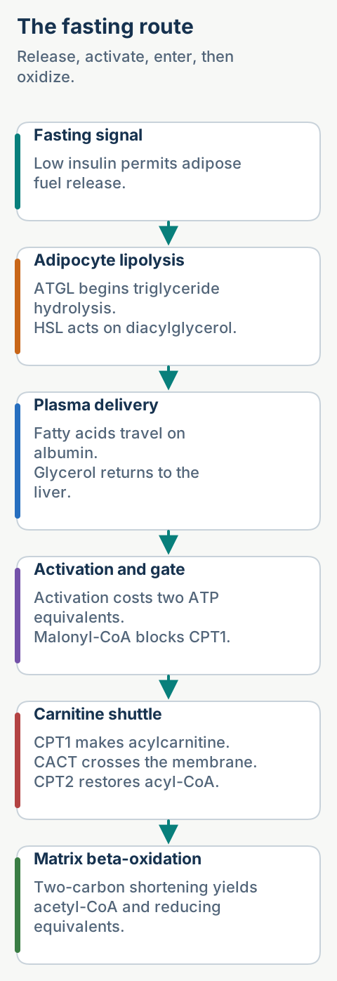
Begin outside the mitochondrion. During fasting, falling insulin removes the brake on adipose lipolysis while counterregulatory hormones favor fuel release. Adipose triglyceride lipase performs the first hydrolysis of stored triglyceride, and hormone-sensitive lipase acts mainly on diacylglycerol. Fatty acids travel on albumin; glycerol returns to the liver for gluconeogenesis. [3]
Locate the failure by following fuel from adipose release to the mitochondrial matrix. [1][2][3][4][5][6]
Adipose triglyceride ATGL, then HSL
Plasma delivery fatty acid on albumin
Activation fatty acyl-CoA
Matrix entry carnitine shuttle
β-oxidation acetyl-CoA plus reducing equivalents
Acyl-CoA synthetase activates a fatty acid by converting adenosine triphosphate (ATP) to adenosine monophosphate plus pyrophosphate. Because two high-energy phosphate bonds are consumed, activation costs two ATP equivalents. Long-chain fatty acyl-CoA then reaches the matrix through carnitine palmitoyltransferase 1 (CPT1), carnitine-acylcarnitine translocase (CACT), and carnitine palmitoyltransferase 2 (CPT2). [1][2]
Use the fed-fasted switch. After carbohydrate intake, acetyl-CoA carboxylase raises malonyl-CoA, and malonyl-CoA inhibits CPT1. During fasting, malonyl-CoA falls, so long fatty acids enter mitochondria rather than being synthesized and oxidized at the same time. Medium fatty acids rely less on the shuttle, which helps explain why transport defects and medium-chain acyl-CoA dehydrogenase deficiency produce different profiles.
Predict high malonyl-CoA
Matrix entry of long fatty acids falls. Cytosolic fatty acyl-CoA remains available for storage, while the matrix receives less substrate for β-oxidation and ketogenesis.
A defect anywhere before or within the spiral can cause catabolic fuel failure. Fasting, fever, vomiting, surgery, or prolonged exercise increases reliance on fatty acids. The clinical phenotype therefore appears when energy demand exceeds the remaining pathway capacity, not necessarily while the patient is fed and well. [2]
Try it here · Checkpoint 1 of 3
Make your prediction before reading the choices. A first attempt is just a starting point.
Case 3
Show answer and explanations for case 3
A. Carnitine-acylcarnitine translocase (Best answer)
Normal CPT1 activity forms palmitoylcarnitine, but failed inward acylcarnitine transfer together with failed outward free-carnitine exchange localizes the block to CACT.
Reasoning steps for option A
How does absent matrix delivery despite normal palmitoylcarnitine formation fit a translocase defect?
The acylcarnitine substrate is formed, but its inward transfer is blocked.
What does the simultaneous failure of free-carnitine return reveal about the transport mechanism?
The translocase couples inward acylcarnitine transport to outward free-carnitine exchange, linking the two failed fluxes.
B. CPT1 at the cytosolic face of the outer mitochondrial membrane (Why this does not fit)
CPT1 is functioning because palmitoylcarnitine is formed normally. A defect there would prevent the first measured product.
Reasoning steps for option B
Is normal palmitoylcarnitine formation consistent with defective CPT1?
No. Formation of that product demonstrates that the measured CPT1 reaction is functioning.
Where would a CPT1 block first become apparent relative to membrane exchange?
It would prevent formation of palmitoylcarnitine before that product could be supplied for exchange.
C. CPT2 (Why this does not fit)
CPT2 acts after acylcarnitine reaches the matrix side and regenerates acyl-CoA. It does not perform the reciprocal membrane exchange described.
Reasoning steps for option C
How does failure of the acyl group to reach the matrix compare with the site of CPT2 action?
CPT2 acts after acylcarnitine reaches the matrix side, whereas the observed failure precedes that step.
What biochemical conversion would a CPT2 defect directly impair rather than reciprocal carnitine exchange?
It would impair regeneration of acyl-CoA from acylcarnitine on the matrix side.
D. Medium acyl-CoA dehydrogenase (Why this does not fit)
This enzyme acts after matrix entry during oxidation. It cannot explain absent matrix delivery or failed carnitine exchange.
Reasoning steps for option D
Can a medium acyl-CoA dehydrogenase defect account for the absent matrix delivery?
Its role is in oxidation after matrix entry, not in delivering the acyl group to the matrix.
What transport behavior would distinguish an isolated defect in this oxidation enzyme from the observed block?
Carnitine exchange would not be directly blocked, unlike the observed failure of free carnitine to return outward.
Takeaway: CACT exchanges acylcarnitine into the matrix for free carnitine outward; simultaneous failure of both fluxes localizes to the exchanger.
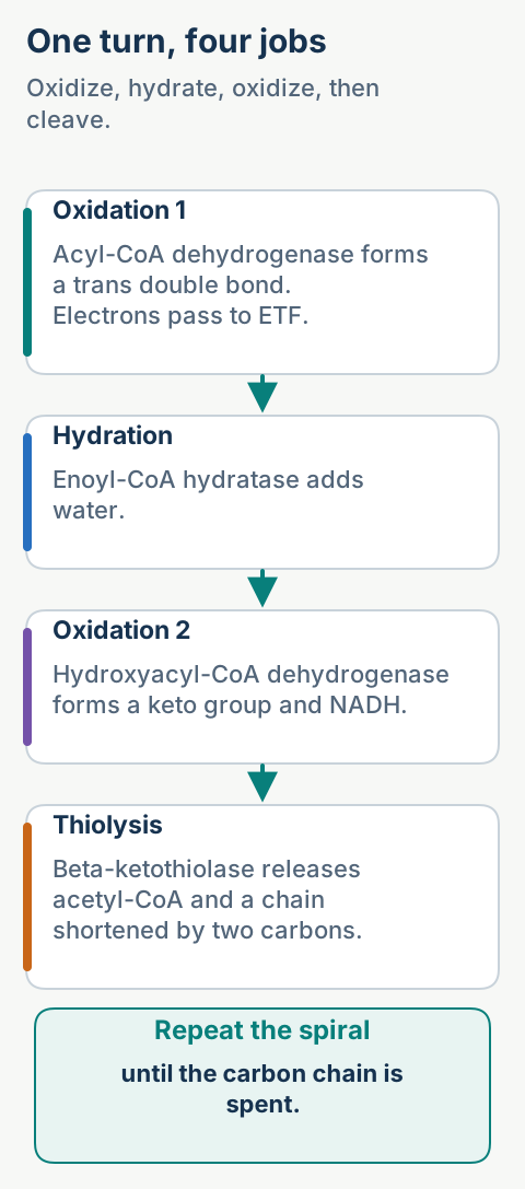
Each turn shortens an acyl-CoA by two carbons. The reusable order is oxidation, hydration, oxidation, thiolysis. The first oxidation forms a trans double bond and passes electrons from flavin adenine dinucleotide through electron transfer flavoprotein, not through succinate dehydrogenase. Hydration adds water, the second oxidation forms a keto group and nicotinamide adenine dinucleotide is reduced, and thiolysis releases acetyl-CoA. [1][2]
Name the chemistry of the accumulated intermediate before naming an enzyme. [1][2]
Think by chemistry rather than by an isolated enzyme list. Acyl-CoA dehydrogenase performs the first oxidation; enoyl-CoA hydratase adds water; hydroxyacyl-CoA dehydrogenase performs the second oxidation; β-ketothiolase uses coenzyme A to cleave the bond. A hydroxyacyl intermediate therefore points to the third reaction, whereas failed electron transfer can impair several acyl-CoA dehydrogenases at once.
Where do the electrons go?
The first oxidation uses electron transfer flavoprotein. The second oxidation produces nicotinamide adenine dinucleotide in its reduced form. Both feed respiratory energy production, but by different entry routes.
The spiral repeats until the carbon skeleton becomes acetyl-CoA units. Even-chain fatty acids end entirely as acetyl-CoA; odd-chain fatty acids finish with one propionyl-CoA. Propionyl-CoA becomes succinyl-CoA through a biotin-dependent carboxylation followed by a vitamin B12-dependent rearrangement.
Count products before assigning a net ATP yield
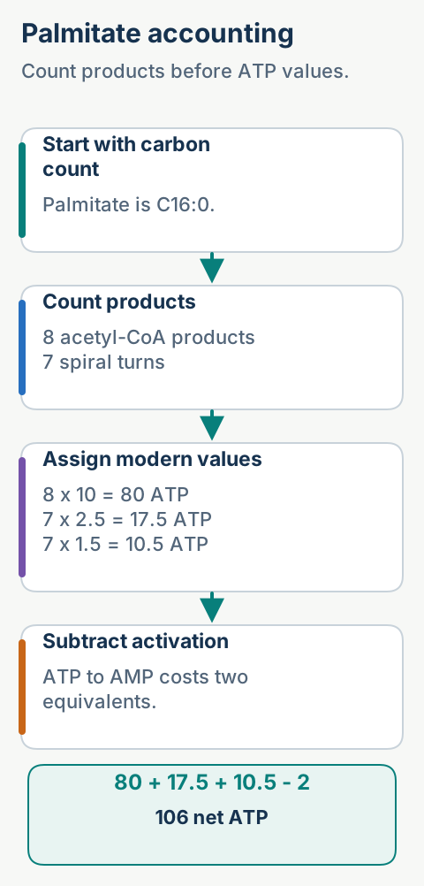
For a saturated even-chain fatty acid with n carbons, the number of turns is n/2 minus 1, and the number of acetyl-CoA products is n/2. Each turn yields one reduced nicotinamide adenine dinucleotide and one flavin-linked reducing equivalent. Modern board accounting assigns 10 ATP per acetyl-CoA, 2.5 per reduced nicotinamide adenine dinucleotide, 1.5 per flavin-linked equivalent, then subtracts two ATP equivalents for activation. [2]
Use carbon count to derive products, then apply the requested energy convention. [1][2]
Palmitate has 16 carbons, so it undergoes seven turns and produces eight acetyl-CoA, seven reduced nicotinamide adenine dinucleotide molecules, and seven flavin-linked equivalents. The result is 80 + 17.5 + 10.5 - 2 = 106 net ATP. Myristate has 14 carbons, so six turns and seven acetyl-CoA yield 92 net ATP by the same convention.
Unsaturation lowers yield because a preexisting double bond bypasses a flavin-producing first oxidation. Polyunsaturated substrates may require additional processing. Odd-chain substrates add propionyl-CoA handling, which uses biotin and vitamin B12 before carbon enters the tricarboxylic acid cycle as succinyl-CoA.
Fast calculation
Count carbons first, not reactions. For C18, expect eight turns and nine acetyl-CoA products before adjusting for double bonds or an odd final fragment.
Peroxisomes begin oxidation of very-long-chain fatty acids and shorten them for mitochondrial completion. Their first oxidation transfers electrons directly to oxygen, producing hydrogen peroxide. Peroxisomal oxidation shortens; mitochondria capture most ATP. This distinction matters in X-linked adrenoleukodystrophy, where impaired peroxisomal transport causes very-long-chain fatty acid accumulation rather than the classic hypoketotic fasting pattern of a mitochondrial spiral defect. [11]
Localize fasting hypoglycemia with simultaneous signals
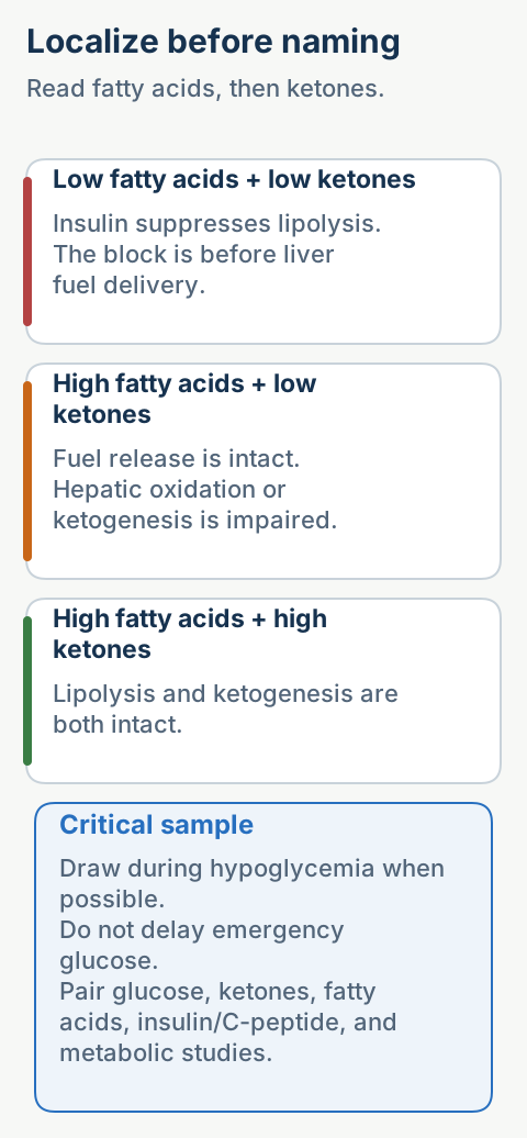
Draw the critical sample during hypoglycemia before glucose treatment when doing so does not delay emergency care. Read three signals together: insulin action, free fatty acid release, and ketone production. Suppressed free fatty acids plus suppressed ketones indicate that insulin is still blocking lipolysis. High free fatty acids plus low ketones indicate that adipose fuel was released but the liver could not oxidize it or convert it efficiently into ketones. [13]
Use simultaneous fuel signals to place the block before interpreting a disease-specific profile. [4][5][6][7][8][9][10][13]
State
Free fatty acids
Ketones
Interpretation
Hyperinsulinism
Low
Low
Lipolysis remains suppressed
Fatty acid oxidation failure
High
Low
Fuel released but not oxidized
Appropriate fasting response
High
High
Lipolysis and ketogenesis intact
A low measured insulin concentration does not exclude insulin action. Insulin may clear rapidly, assays differ, and a single value can be misleading. Suppressed fatty acids, suppressed β-hydroxybutyrate, and a brisk glucagon response are physiologic evidence of inappropriate insulin effect. [13]
For suspected fatty acid oxidation disorder, obtain plasma acylcarnitines, free and total carnitine, urine organic acids, glucose, electrolytes, liver tests, ammonia, lactate, and creatine kinase when clinically relevant. Collect first, treat immediately: a normal profile after dextrose or after the catabolic episode has resolved can be falsely reassuring.
Why are ketones low?
Hepatic β-oxidation supplies both acetyl-CoA and reducing power for ketogenesis. When oxidation fails, free fatty acids can be high while ketone output remains inappropriately low.
Newborn screening reduces mortality but does not replace bedside recognition. A child with vomiting, lethargy, seizure, hepatomegaly, cardiomyopathy, or rhabdomyolysis after fasting still needs urgent metabolic evaluation. Normal newborn screening does not exclude every phenotype, especially milder variants, unscreened births, or secondary disturbances of carnitine and flavin metabolism.
Try it here · Checkpoint 2 of 3
Make your prediction before reading the choices. A first attempt is just a starting point.
Case 11
Show answer and explanations for case 11
A. MCAD deficiency (Best answer)
High free fatty acids with low ketones localizes failure after lipolysis. The dominant C8 acylcarnitine and medium dicarboxylic acids identify MCAD deficiency.
Reasoning steps for option A
What do high free fatty acids with low β-hydroxybutyrate imply about the proposed MCAD defect?
Fatty acids are being mobilized, but ketone production is impaired, localizing the failure after lipolysis.
Which supplied findings identify the medium-chain pattern associated with MCAD deficiency?
Dominant C8 with lesser C6 and C10 acylcarnitines, together with urinary medium dicarboxylic acids.
B. VLCAD deficiency with an infant cardiac phenotype (Why this does not fit)
VLCAD deficiency can cause hypoketotic hypoglycemia, but its characteristic screening pattern centers on C14:1 and severe infant disease may include cardiomyopathy.
Reasoning steps for option B
Can VLCAD deficiency account for glucose of 31 mg/dL with low β-hydroxybutyrate?
Yes; VLCAD deficiency can cause hypoketotic hypoglycemia.
What additional findings would distinguish the proposed infant cardiac VLCAD phenotype?
A screening pattern centered on C14:1 rather than C8, with possible cardiomyopathy in severe infant disease.
C. CPT1A deficiency (Why this does not fit)
CPT1A deficiency can impair ketone production, but it more often shows high free carnitine relative to low long acylcarnitines rather than a dominant C8 species.
Reasoning steps for option C
Is low β-hydroxybutyrate during hypoglycemia compatible with CPT1A deficiency?
Yes; CPT1A deficiency can impair ketone production.
What carnitine profile would be expected from CPT1A deficiency instead of the observed C8 dominance?
High free carnitine relative to low long-chain acylcarnitines.
D. Congenital hyperinsulinism (Why this does not fit)
Excess insulin action suppresses both free fatty acids and ketones. This child has high free fatty acids and a specific C8 acylcarnitine pattern.
Reasoning steps for option D
How does the combination of high free fatty acids and low ketones compare with excess insulin action?
Excess insulin suppresses both; low ketones fit, but high free fatty acids conflict with that mechanism.
What does the dominant C8 acylcarnitine add beyond the ketone result when considering hyperinsulinism?
It provides a specific medium-chain metabolic signature that insulin-mediated suppression alone does not explain.
Takeaway: Fasting hypoketotic hypoglycemia with high free fatty acids, dominant C8 acylcarnitine, and medium dicarboxylic acids supports MCAD deficiency.
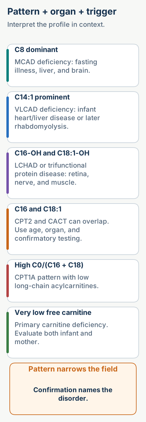
Medium-chain acyl-CoA dehydrogenase deficiency classically produces hypoketotic hypoglycemia, vomiting, lethargy, liver dysfunction, and a C8-predominant acylcarnitine pattern with C6 and C10 elevations. Urine may show medium-chain dicarboxylic acids. The first crisis often follows an overnight fast or gastrointestinal illness. [7]
Very-long-chain acyl-CoA dehydrogenase deficiency may present in infancy with cardiomyopathy, arrhythmia, hepatopathy, and hypoglycemia or later with exercise-induced myalgia and rhabdomyolysis. Long-chain C14 species, especially C14:1, support the diagnosis, but phenotype, molecular testing, and specialist interpretation determine management. [8]
Long-chain 3-hydroxyacyl-CoA dehydrogenase or mitochondrial trifunctional protein deficiency adds hydroxyacylcarnitines, neuropathy, and pigmentary retinopathy to the long-chain phenotype. A pregnancy complicated by acute fatty liver or severe hypertensive liver disease raises concern for an affected fetus, but the association is not itself diagnostic. [9]
CPT1A deficiency blocks formation of long-chain acylcarnitine in the liver, so free carnitine may be high and the C0/(C16+C18) ratio rises. CPT2 and CACT deficiencies can both produce high long-chain acylcarnitines; severe neonatal disease may include arrhythmia and hypoglycemia, while the adult myopathic CPT2 phenotype is triggered by prolonged exercise, fasting, fever, or cold. [4][5][6]
Combine pattern, age, trigger, and injured organ before confirmatory testing. [4][5][6][7][8][9][10]
Primary carnitine deficiency can cause very low free carnitine, cardiomyopathy, skeletal myopathy, and hypoketotic hypoglycemia. A low carnitine result in an infant may instead identify maternal primary carnitine deficiency, so paired infant and maternal assessment can prevent a missed adult diagnosis. [10]
Do not overcall one analyte
An acylcarnitine pattern narrows the pathway location; it rarely finishes the diagnosis alone. Confirm with molecular testing or a validated functional assay and interpret results in the clinical state in which the sample was obtained.
Acquired mitochondrial injury can imitate inherited fuel failure. Salicylate-associated Reye syndrome follows a viral prodrome with vomiting, encephalopathy, hyperammonemia, and microvesicular hepatic steatosis. Experimental and clinical literature supports impaired mitochondrial fatty acid activation and β-oxidation as part of the injury. Avoid aspirin during viral illness in children and adolescents unless a specialist-directed indication outweighs the risk. [15][18]
Stop catabolism before the laboratory pattern is complete
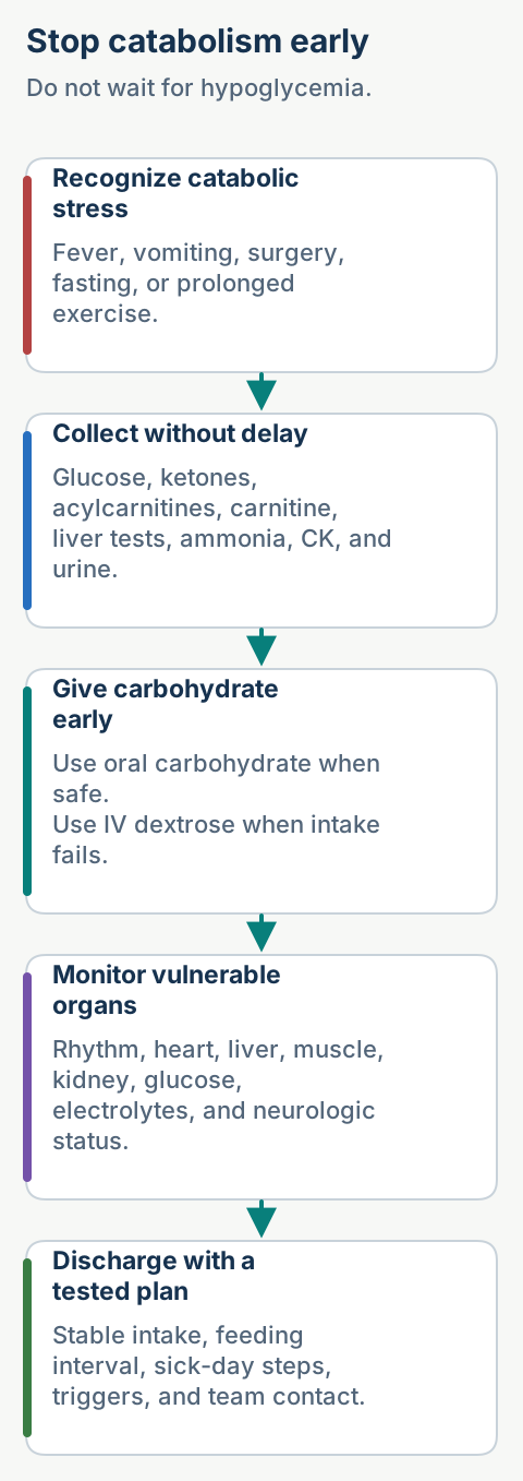
An ill patient with known or suspected fatty acid oxidation disorder cannot wait for hypoglycemia. Provide carbohydrate early, stop fasting, treat the precipitating illness, and contact a metabolic specialist. If oral carbohydrate is unsafe or not tolerated, use intravenous dextrose according to the emergency plan and clinical setting. [7][8][9]
Treat catabolism before hypoglycemia or confirmatory results appear. [7][8][9]
Initial monitoring is phenotype driven. Check glucose, electrolytes, blood gas when ill, liver tests, ammonia, creatine kinase, renal function, and urine for myoglobin when muscle injury is possible. Obtain an electrocardiogram and cardiac monitoring for long-chain disorders with arrhythmia or cardiomyopathy risk. Normal glucose does not prove metabolic stability if the patient is vomiting, weak, encephalopathic, or developing rhabdomyolysis.
Do not make lipid or protein the acute rescue fuel. Dextrose reverses catabolism and reduces fatty acid flux. Continue enough glucose to maintain euglycemia and suppress lipolysis, adding insulin only when needed for treatment-associated hyperglycemia under a protocol. Correct dehydration and electrolytes without delaying energy delivery.
A practical discharge test is whether the patient can maintain the prescribed feeding interval, hydration, glucose stability, and clinical baseline without intravenous support. Send a written sick-day plan that names fasting limits, home carbohydrate steps, emergency triggers, the nearest capable facility, and the metabolic team contact.
Emergency red flags
Escalate for altered mental status, persistent emesis, hypoglycemia, rising creatine kinase, dark urine, arrhythmia, cardiomyopathy, hyperammonemia, or liver dysfunction. These findings indicate organ energy failure or toxic metabolite accumulation.
Build prevention around the vulnerable organ
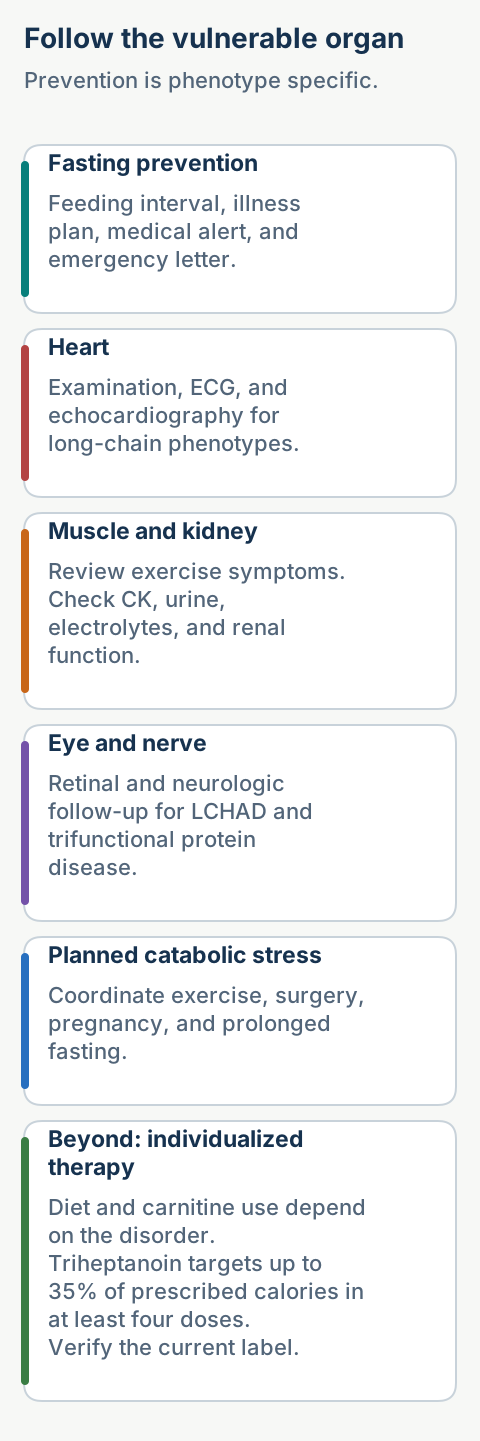
For medium-chain acyl-CoA dehydrogenase deficiency, the durable intervention is avoidance of prolonged fasting, especially during infancy, illness, surgery, and vomiting. Most well patients do not need chronic dietary fat restriction. Families need age-specific feeding intervals, rapid carbohydrate access, a medical alert, and an emergency letter that travels with the patient. [7]
Long-chain disorders require an individualized metabolic nutrition plan. Depending on genotype and phenotype, the plan may distribute carbohydrate, restrict selected long-chain fat, preserve essential fatty acids, and use medium-chain triglyceride or triheptanoin. Do not prescribe carnitine automatically; supplementation is disorder specific and should follow measured deficiency plus specialist judgment because accumulating long-chain acylcarnitines may be arrhythmogenic. [8][9]
Surveillance follows the tissue at risk. Cardiac examination, electrocardiography, and echocardiography matter in severe long-chain phenotypes. Creatine kinase and renal function help after exertional symptoms. Long-chain 3-hydroxyacyl-CoA dehydrogenase and trifunctional protein disorders also need retinal and neurologic follow-up because retinopathy and peripheral neuropathy can progress despite avoidance of acute crises. [8][9]
For exercise-sensitive disease, use a personalized plan for pre-exercise carbohydrate, hydration, temperature exposure, duration, and recovery feeding. Myalgia, weakness, or dark urine ends the session and prompts creatine kinase, electrolyte, and renal assessment. Recurrent rhabdomyolysis warrants neuromuscular or metabolic referral even when routine testing between episodes is normal.
Pregnancy and surgery are planned catabolic stress tests. Coordinate obstetrics, anesthesia, nutrition, cardiology when indicated, and metabolic genetics before fasting begins. Continuous carbohydrate during prolonged fasting may be necessary, with cardiac and biochemical monitoring tailored to the disorder. A maternal liver syndrome associated with an affected fetus should trigger direct infant testing rather than assumption. [9]
Follow the tissue at risk and rehearse the next catabolic stress before it happens. [7][8][9][16][17]
Beyond: labeled triheptanoin therapy. For molecularly confirmed long-chain fatty acid oxidation disorders, the United States label targets up to 35% of prescribed daily caloric intake, divided into at least four doses with meals or snacks. Start and titrate under a metabolic specialist, account for gastrointestinal tolerance and total calories, and verify the current label before prescribing. [16] Long-term extension data report fewer major clinical events in treated cohorts, but individual response and adverse effects still require monitoring. [17]
What belongs in follow-up?
Review interval symptoms, fasting tolerance, emergency-plan use, growth or weight, diet adequacy, cardiac status, muscle injury, liver function, retina, neuropathy, medications, pregnancy plans, and psychosocial barriers. The exact set depends on the biochemical defect and phenotype.
Connect hepatic β-oxidation to ketone physiology and crisis care
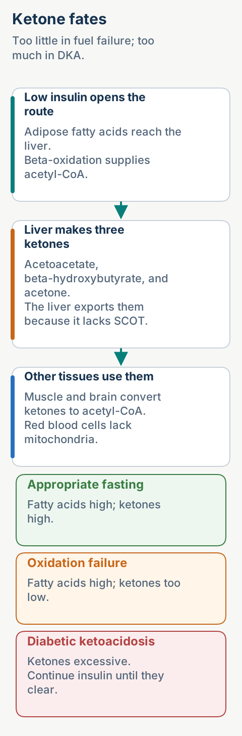
When insulin is low, adipose tissue releases fatty acids and the liver oxidizes them. Acetyl-CoA that exceeds tricarboxylic acid cycle capacity becomes acetoacetate, β-hydroxybutyrate, and acetone. Extrahepatic mitochondria convert acetoacetate and β-hydroxybutyrate back to acetyl-CoA. The liver exports ketones because it lacks succinyl-CoA:acetoacetate CoA transferase, while red blood cells cannot use ketones because they lack mitochondria. [12]
The brain can use ketones during prolonged fasting, but ketone supply still depends on hepatic fatty acid oxidation. This creates a useful contrast: appropriate fasting produces ketones, a fatty acid oxidation disorder often produces inappropriately few ketones, and uncontrolled diabetes produces excessive ketones.
Use ketone direction to distinguish normal adaptation, fuel failure, and ketone excess. [12][14]
In diabetic ketoacidosis (DKA), insulin deficiency and counterregulatory hormones drive lipolysis, hepatic β-oxidation, and ketogenesis. β-Hydroxybutyrate predominates because the hepatic redox state is reduced; acetoacetate can decarboxylate to acetone, producing fruity breath. Ketoacids release hydrogen ions, bicarbonate buffers them, and retained ketoanions widen the anion gap. Kussmaul respirations are respiratory compensation. [14]
Serum potassium can be normal or high even though total body potassium is depleted by osmotic diuresis, vomiting, and secondary hyperaldosteronism. Acidosis and insulin deficiency shift potassium outward. Insulin then shifts potassium into cells, so starting insulin during severe hypokalemia can provoke arrhythmia and respiratory muscle weakness. [14]
Sodium-glucose cotransporter 2 inhibitor exposure, pregnancy, reduced intake, and partial insulin treatment can produce DKA with modest glucose. Diagnose the crisis with diabetes or qualifying glucose, ketonemia, and metabolic acidosis, not with glucose alone. Direct blood β-hydroxybutyrate is preferred over urine ketones for diagnosis and resolution. [14]
Beyond: current adult DKA protocol anchors. In adults without cardiac or renal compromise, current consensus supports isotonic saline or balanced crystalloid at 500 to 1000 mL/h for the first 2 to 4 hours, then adjustment to hemodynamics and sodium. Intravenous regular insulin is commonly started at 0.1 units/kg/h once potassium is above 3.5 mmol/L. If potassium is below 3.5 mmol/L, give potassium, commonly 10 mmol/h, and delay insulin. When glucose falls below 250 mg/dL, add 5% to 10% dextrose and reduce insulin to 0.05 units/kg/h so ketones can continue to clear. Confirm local protocols and current labeling. [14]
Continue treatment until β-hydroxybutyrate is below 0.6 mmol/L and venous pH is at least 7.3 or bicarbonate is at least 18 mmol/L. The anion gap can remain misleadingly elevated or normalize for the wrong reason after chloride-rich fluids, so it is not the preferred resolution marker. Routine bicarbonate is not recommended; current consensus considers it when pH is below 7.0. [14]
Why add dextrose while insulin continues?
Glucose often normalizes before ketogenesis stops. Dextrose prevents treatment-induced hypoglycemia while insulin continues to suppress lipolysis and clear β-hydroxybutyrate.
Try it here · Checkpoint 3 of 3
Make your prediction before reading the choices. A first attempt is just a starting point.
Case 24
Show answer and explanations for case 24
A. Starvation ketosis that needs only oral carbohydrate (Why this does not fit)
Starvation can raise ketones, but the marked β-hydroxybutyrate level, severe bicarbonate reduction, symptoms, and SGLT2 exposure require assessment and treatment for ketoacidosis.
Reasoning steps for option A
How does β-hydroxybutyrate of 5.2 mmol/L fit a starvation mechanism?
Starvation can raise ketones, but this marked elevation cannot by itself justify interpreting the illness as uncomplicated starvation ketosis.
What do bicarbonate of 12 mmol/L and the symptoms imply about relying only on oral carbohydrate?
The severe bicarbonate reduction, nausea, abdominal pain, and deep respirations require assessment and treatment for ketoacidosis rather than oral carbohydrate alone.
B. A fatty acid oxidation disorder causing failure to make ketones (Why this does not fit)
Oxidation disorders generally cause inappropriately low ketones during hypoglycemia. This patient has excessive ketones and high-anion-gap acidosis.
Reasoning steps for option B
Does β-hydroxybutyrate of 5.2 mmol/L support a failure to make ketones?
It demonstrates substantial ketone production, contrary to the proposed failure of ketone formation.
What glucose context would typically accompany inappropriately low ketones in a fatty acid oxidation disorder?
The expected context is hypoglycemia; the supplied glucose of 176 mg/dL does not show that pattern.
C. SGLT2-associated diabetic ketoacidosis despite modest glucose (Best answer)
High β-hydroxybutyrate, high-anion-gap metabolic acidosis, compatible symptoms, and SGLT2 exposure establish a ketoacidosis pattern even though glucose is 176 mg/dL.
Reasoning steps for option C
How do the ketone and acid-base measurements fit a ketoacidosis mechanism?
β-Hydroxybutyrate of 5.2 mmol/L together with low bicarbonate and a high anion gap establishes a ketoacidosis pattern.
What does SGLT2 inhibitor exposure imply about interpreting glucose of 176 mg/dL?
SGLT2-associated diabetic ketoacidosis can occur with modest glucose, so marked hyperglycemia is not required.
D. Hyperosmolar hyperglycemic state (Why this does not fit)
Hyperosmolar hyperglycemic state usually has far higher glucose and osmolality with minimal ketonemia and less severe ketoacidosis.
Reasoning steps for option D
How does glucose of 176 mg/dL compare with the expected hyperosmolar hyperglycemic pattern?
Hyperosmolar hyperglycemic state usually involves far higher glucose and osmolality; this glucose does not show the expected degree of hyperglycemia.
What ketone and acid-base findings would distinguish a hyperosmolar state from the supplied findings?
Minimal ketonemia and less severe ketoacidosis would be expected, rather than β-hydroxybutyrate of 5.2 mmol/L with bicarbonate of 12 mmol/L.
Takeaway: SGLT2-associated diabetic ketoacidosis can occur with modest glucose; blood β-hydroxybutyrate and acid-base findings establish the diagnosis.
For each case, use one sequence every time: identify the hormonal state, confirm fuel release, locate mitochondrial entry, name the blocked reaction or carbon range, predict ketones and organ stress, then choose the immediate anti-catabolic action and the long-term prevention plan.
Case 1
Show answer and explanations for case 1
A. Hormone-sensitive lipase acting first on triglyceride (Why this does not fit)
Hormone-sensitive lipase contributes strongly to diacylglycerol hydrolysis, but the supplied sequence identifies the preceding triglyceride-to-diacylglycerol reaction.
Reasoning steps for option A
Does the early triglyceride-to-diacylglycerol conversion match the supplied role of hormone-sensitive lipase?
Hormone-sensitive lipase contributes strongly to diacylglycerol hydrolysis, whereas the early reaction produces diacylglycerol.
Which later observation would fit its role in the sequence?
The later rise in monoacylglycerol fits hydrolysis of the diacylglycerol generated first.
B. Adipose triglyceride lipase initiating triglyceride hydrolysis (Best answer)
The fasting hormone pattern permits adipose lipolysis, and the first measured product is diacylglycerol. Adipose triglyceride lipase catalyzes the initiating triglyceride hydrolysis that fits both findings.
Reasoning steps for option B
How does the first measured product test the proposed role of adipose triglyceride lipase?
Early diacylglycerol formation matches its role in initiating stored triglyceride hydrolysis.
What does the fasting hormone pattern imply about the metabolic setting for this reaction?
Low insulin and high epinephrine permit adipose lipolysis, consistent with the increased plasma free fatty acids.
C. Lipoprotein lipase hydrolyzing circulating chylomicron triglyceride (Why this does not fit)
Lipoprotein lipase acts on triglyceride in circulating lipoproteins at capillary surfaces. The experiment tracks intracellular adipocyte triglyceride stores during fasting.
Reasoning steps for option C
Does the triglyceride pool tracked in the biopsy match the proposed substrate source for lipoprotein lipase?
The biopsy tracks intracellular adipocyte stores, while this mechanism targets triglyceride in circulating lipoproteins.
Where would hydrolysis be localized if lipoprotein lipase were responsible?
It would occur at capillary surfaces rather than within the adipocyte stores being measured.
D. Pancreatic lipase hydrolyzing intestinal dietary triglyceride (Why this does not fit)
Pancreatic lipase functions in the intestinal lumen after a meal. It cannot explain a fasting adipocyte biopsy or the intracellular diacylglycerol time course.
Reasoning steps for option D
How does the overnight fast compare with the supplied setting for pancreatic lipase activity?
Pancreatic lipase functions after a meal, whereas the observed triglyceride breakdown occurs during fasting.
What compartment would need to be sampled to track the proposed pancreatic lipase mechanism?
The intestinal lumen, where dietary triglyceride is hydrolyzed, rather than an adipocyte biopsy showing intracellular diacylglycerol formation.
Takeaway: During fasting, adipose triglyceride lipase initiates stored triglyceride hydrolysis, and hormone-sensitive lipase acts mainly on the resulting diacylglycerol.
A. Inhibit CPT2 on the matrix face of the inner mitochondrial membrane (Why this does not fit)
CPT2 inhibition can impair matrix acyl-CoA regeneration, but it does not describe the normal fed-state regulatory target signaled by high malonyl-CoA.
Reasoning steps for option A
Which step would CPT2 inhibition impair in the delivery of C16 substrate for matrix oxidation?
It would impair regeneration of fatty acyl-CoA on the matrix face of the inner mitochondrial membrane, reducing the matrix substrate available for oxidation.
Why does reduced matrix oxidation alone not identify CPT2 as the fed-state regulatory target?
Although CPT2 inhibition can reduce matrix oxidation, the accompanying rise in malonyl-CoA points to inhibition of CPT1 rather than CPT2.
B. Inhibit acyl-CoA synthetase and prevent fatty acid activation (Why this does not fit)
Blocking activation would reduce acyl-CoA availability, whereas the stem states that cytosolic fatty acyl-CoA remains available.
Reasoning steps for option B
How would inhibiting acyl-CoA synthetase affect the cytosolic substrate pool described in the findings?
Preventing fatty acid activation would reduce fatty acyl-CoA availability, rather than preserve the available cytosolic fatty acyl-CoA specified in the findings.
What mechanistic distinction does available cytosolic fatty acyl-CoA establish between an activation defect and a mitochondrial delivery defect?
The activated substrate remains available outside the matrix, so reduced matrix oxidation can reflect impaired mitochondrial delivery without requiring a failure of fatty acid activation.
C. Activate CACT exchange across the inner mitochondrial membrane (Why this does not fit)
Greater CACT activity would support transport after CPT1 has formed acylcarnitine. It would not reproduce a fed-state fall in entry caused by malonyl-CoA.
Reasoning steps for option C
Which transport step would activation of CACT support?
It would support exchange across the inner mitochondrial membrane after CPT1 has formed acylcarnitine, favoring transport rather than reproducing reduced mitochondrial entry.
Why does CACT-mediated transport depend on an upstream reaction affected by malonyl-CoA?
CACT transports acylcarnitine formed by CPT1. When malonyl-CoA inhibits that formation step, increasing downstream exchange does not reproduce the upstream restriction.
D. Inhibit CPT1 formation of acylcarnitine outside the inner membrane (Best answer)
High malonyl-CoA directly inhibits CPT1. Cytosolic fatty acyl-CoA can remain present while acylcarnitine formation and mitochondrial entry of C16 substrate fall.
Reasoning steps for option D
Which biochemical reaction is directly suppressed by the rise in malonyl-CoA after the high-carbohydrate meal?
Malonyl-CoA directly inhibits CPT1-mediated formation of acylcarnitine from fatty acyl-CoA outside the inner mitochondrial membrane.
How can CPT1 inhibition reduce C16 oxidation in the matrix while cytosolic fatty acyl-CoA remains available?
CPT1 inhibition restricts conversion of the available activated fatty acid into the transport form. Mitochondrial substrate entry therefore falls without requiring depletion of cytosolic fatty acyl-CoA.
Takeaway: Malonyl-CoA inhibits CPT1 after feeding, reducing mitochondrial entry of long fatty acids while cytosolic fatty acyl-CoA remains available.
A. Hydration of trans-enoyl-CoA (Why this does not fit)
Hydration adds water after the first double bond has already been formed. Blocking electron transfer flavoprotein prevents reaching that substrate.
Reasoning steps for option A
Does arrest at the first dehydrogenation place the block at hydration of trans-enoyl-CoA?
Hydration follows formation of the first double bond, so it occurs after the observed arrest.
How would blocking electron transfer flavoprotein affect the substrate available for hydration?
It prevents formation of trans-enoyl-CoA, leaving hydration without its substrate.
B. NAD-linked oxidation of hydroxyacyl-CoA (Why this does not fit)
This later oxidation produces NADH and does not transfer electrons through electron transfer flavoprotein.
Reasoning steps for option B
Does the observed first-dehydrogenation arrest match NAD-linked oxidation of hydroxyacyl-CoA?
Hydroxyacyl-CoA oxidation is a later reaction, not the first dehydrogenation.
What electron-transfer feature distinguishes hydroxyacyl-CoA oxidation from the blocked reaction?
It produces NADH rather than passing electrons through electron transfer flavoprotein.
C. FAD-linked acyl-CoA dehydrogenation (Best answer)
Acyl-CoA dehydrogenases perform the first oxidation and pass electrons through electron transfer flavoprotein. Preserved succinate oxidation also shows that complex II itself is not the blocked enzyme.
Reasoning steps for option C
How does arrest at the first dehydrogenation fit FAD-linked acyl-CoA dehydrogenation?
Acyl-CoA dehydrogenases perform that first oxidation and pass electrons through electron transfer flavoprotein.
What does preserved succinate oxidation reveal about the location of the electron-transfer block?
Complex II remains functional, distinguishing the blocked flavoprotein-dependent pathway from succinate oxidation.
D. Thiolytic cleavage of ketoacyl-CoA (Why this does not fit)
Thiolysis uses CoA after both oxidations and hydration. It neither requires electron transfer flavoprotein nor occurs before the observed arrest.
Reasoning steps for option D
Does arrest at the first dehydrogenation match a block in thiolytic cleavage of ketoacyl-CoA?
Thiolysis occurs after both oxidations and hydration, beyond the observed arrest.
What requirement distinguishes thiolytic cleavage from an electron transfer flavoprotein-dependent reaction?
Thiolysis uses CoA and does not require electron transfer flavoprotein.
Takeaway: The first β-oxidation reaction is FAD-linked and passes electrons through electron transfer flavoprotein, independently of succinate oxidation by complex II.
Hydration must be intact because the hydroxyacyl intermediate is present. Loss of this enzyme would instead accumulate the trans-enoyl substrate.
Reasoning steps for option A
Does hydroxyacyl intermediate accumulation fit loss of enoyl-CoA hydratase?
Hydroxyacyl formation shows that hydration is intact.
Which substrate would accumulate if hydration were blocked?
The trans-enoyl substrate would accumulate instead.
B. 3-hydroxyacyl-CoA dehydrogenase using NAD+ to form a ketoacyl intermediate (Best answer)
The hydroxyacyl substrate is present, FAD-linked oxidation is intact, and NADH is absent. These findings localize the defect to the NAD-dependent third reaction that forms ketoacyl-CoA.
Reasoning steps for option B
How does absent NADH despite available hydroxyacyl substrate fit loss of 3-hydroxyacyl-CoA dehydrogenase?
The missing activity would prevent NAD+-dependent oxidation of that substrate and the accompanying NADH production.
What downstream intermediate would fail to form at this blocked reaction?
Ketoacyl-CoA would fail to form from hydroxyacyl-CoA.
C. Acyl-CoA dehydrogenase (Why this does not fit)
The first FAD-linked oxidation is reported as normal, so the initial dehydrogenase cannot explain hydroxyacyl accumulation.
Reasoning steps for option C
Does normal FAD reduction fit loss of acyl-CoA dehydrogenase?
Normal FAD reduction indicates that the initial FAD-linked oxidation is functioning.
What additional pathway progress is demonstrated by the hydroxyacyl intermediate?
The substrate has passed through the initial oxidation and subsequent hydration.
D. Thiolase (Why this does not fit)
Thiolase failure would accumulate ketoacyl-CoA after NADH had already been produced. The stem instead reports no NADH and accumulation one reaction earlier.
Reasoning steps for option D
Would thiolase loss explain the absence of NADH?
A thiolase block occurs after NADH has already been produced.
Which accumulating intermediate would distinguish a thiolase block from the observed block?
Thiolase failure would accumulate ketoacyl-CoA rather than the observed hydroxyacyl intermediate.
Takeaway: Hydroxyacyl accumulation with absent NADH localizes the block to the NAD-dependent hydroxyacyl-CoA dehydrogenase reaction.
A. 92 ATP: seven acetyl-CoA products, six spiral turns, minus one activation cost (Best answer)
A C14 substrate yields seven acetyl-CoA units and requires six cycles. Seventy plus fifteen plus nine minus two equals 92 ATP.
Reasoning steps for option A
How do seven acetyl-CoA units and six cycles fit a saturated C14 fatty acid?
Fourteen carbons yield seven two-carbon acetyl-CoA units; the final cycle releases two units, so only six cycles are needed.
What net ATP yield follows from these products and one activation?
The yield is 7 × 10 + 6 × 2.5 + 6 × 1.5 − 2 = 92 ATP.
B. 90 ATP: seven acetyl-CoA products with an extra activation penalty (Why this does not fit)
Seven cycles would overcount because the final four-carbon substrate yields two acetyl-CoA units in one last cycle. The activation cost is also not enough to rescue this count.
Reasoning steps for option B
Does producing seven acetyl-CoA units from C14 require seven cycles?
No. The final four-carbon substrate produces two acetyl-CoA units in one cycle, giving six cycles overall.
Would charging for one activation make the proposed seven-unit, seven-cycle accounting yield 90 ATP?
That accounting gives 70 + 17.5 + 10.5 = 98 ATP before activation and 96 ATP after the two-ATP cost.
C. 94 ATP: six acetyl-CoA products with seven reducing-equivalent sets (Why this does not fit)
A C14 substrate cannot yield only six acetyl-CoA units, and seven cycles would require a longer starting substrate.
Reasoning steps for option C
Can six acetyl-CoA units account for complete oxidation of the supplied C14 substrate?
Six two-carbon units account for only 12 carbons, leaving two of the supplied 14 carbons unaccounted for.
What starting chain length would the proposed seven cycles imply?
Seven cycles imply eight acetyl-CoA units and thus a C16 starting substrate, longer than the supplied C14.
D. 106 ATP: the yield expected from palmitate rather than myristate (Why this does not fit)
This is the palmitate result for C16, not the C14 result supplied here.
Reasoning steps for option D
Are eight acetyl-CoA units compatible with the carbon content of the supplied fatty acid?
Eight acetyl-CoA units contain 16 carbons, matching C16 palmitate rather than the supplied C14 substrate.
How does the proposed eight-unit, seven-cycle pattern generate 106 net ATP?
It gives 80 ATP from acetyl-CoA, 17.5 from NADH, and 10.5 from FADH2-equivalents; subtracting two for activation yields 106 ATP.
Takeaway: Myristate, C14:0, yields seven acetyl-CoA units, six cycles, and 92 net ATP under modern accounting.
A. The unsaturated substrate produces one extra FADH2-equivalent (Why this does not fit)
Bypassing the FAD-linked dehydrogenation eliminates, rather than adds, one reducing-equivalent contribution.
Reasoning steps for option A
Does bypassing one FAD-producing dehydrogenation give the unsaturated substrate an extra FADH2-equivalent?
The bypass removes one FADH2-equivalent contribution rather than adding one.
How would that lost reducing-equivalent contribution affect the ATP comparison?
It lowers the unsaturated substrate's net ATP estimate relative to the saturated substrate.
B. The saturated substrate requires both an isomerase and a reductase before standard oxidation resumes (Why this does not fit)
The preexisting cis double bond creates the need for an auxiliary isomerase. The saturated substrate follows the standard recurring sequence.
Reasoning steps for option B
Which substrate requires rearrangement of an existing double bond before usual oxidation continues?
Only the unsaturated molecule carries that structural obstacle; the saturated C18 chain can enter the recurring sequence directly.
What role is assigned to the isomerase in the stated unsaturated pathway?
It permits continued oxidation while bypassing one FAD-producing dehydrogenation; no reductase requirement is supplied.
C. The unsaturated substrate has a lower net yield because one FAD-linked oxidation is bypassed (Best answer)
The isomerase permits continued oxidation, but the bypassed dehydrogenation means one fewer FADH2-equivalent and therefore a lower net ATP estimate.
Reasoning steps for option C
How does the stated bypass support a lower net yield from the unsaturated substrate?
Skipping one FAD-linked dehydrogenation produces one fewer FADH2-equivalent, lowering the net ATP estimate.
Would that lower yield imply fewer acetyl-CoA units from the unsaturated substrate?
No. Both substrates have 18 carbons, so their predicted acetyl-CoA counts are the same.
D. Both substrates have identical yield because they contain the same number of carbons (Why this does not fit)
Carbon count predicts acetyl-CoA count, but preexisting double bonds can change the number of reducing equivalents produced during oxidation.
Reasoning steps for option D
Does the shared C18 carbon count establish identical energy yields?
It predicts the same acetyl-CoA count, but not identical reducing-equivalent production.
Which reducing-equivalent difference is predicted by the specified cis-double-bond bypass?
The unsaturated substrate produces one fewer FADH2-equivalent than the saturated substrate.
Takeaway: An unsaturated substrate can yield less ATP than a saturated substrate of the same carbon number because a preexisting double bond can bypass an FAD-linked oxidation.
Biotin is required by propionyl-CoA carboxylase. Normal activity of that enzyme and accumulation of its methylmalonyl-CoA product place the block later.
Reasoning steps for option A
How does normal propionyl-CoA carboxylase activity compare with a proposed biotin deficiency?
Biotin is required by this enzyme, so its normal activity argues against impaired biotin-dependent carboxylation.
What does accumulation of the carboxylase product reveal about the block's location?
Methylmalonyl-CoA accumulation places the block after the biotin-dependent step.
B. Vitamin B12 deficiency impairing methylmalonyl-CoA mutase (Best answer)
Normal carboxylase activity generates methylmalonyl-CoA, but low succinyl-CoA shows failure of the B12-dependent mutase reaction.
Reasoning steps for option B
Is methylmalonyl-CoA accumulation despite normal carboxylase activity consistent with impaired B12-dependent mutase activity?
Yes. The carboxylase supplies methylmalonyl-CoA, which accumulates when its subsequent conversion is impaired.
What product change is predicted by failure of the B12-dependent mutase reaction?
Succinyl-CoA production should fall, matching the supplied finding.
C. Thiamine deficiency impairing pyruvate dehydrogenase (Why this does not fit)
Thiamine affects oxidative decarboxylation pathways but does not catalyze methylmalonyl-CoA conversion to succinyl-CoA.
Reasoning steps for option C
Does thiamine-dependent pyruvate dehydrogenase impairment explain methylmalonyl-CoA accumulation with low succinyl-CoA?
Thiamine-dependent oxidative decarboxylation does not mediate the methylmalonyl-CoA-to-succinyl-CoA conversion.
How would pyruvate dehydrogenase impairment affect TCA-cycle carbon entry differently from the block described?
It would reduce conversion of pyruvate to acetyl-CoA. The propionyl-CoA produced from odd-chain fatty acids instead supplies TCA-cycle carbon through succinyl-CoA, bypassing pyruvate dehydrogenase.
D. Carnitine deficiency preventing mitochondrial entry before propionyl-CoA and methylmalonyl-CoA can form (Why this does not fit)
Carnitine deficiency can impair oxidation of long fatty acids, but the stem demonstrates downstream production of propionyl-CoA and methylmalonyl-CoA.
Reasoning steps for option D
How does propionyl-CoA production after odd-carbon fatty acid oxidation test the proposed carnitine-related entry block?
Propionyl-CoA has already formed, contradicting a block that would prevent mitochondrial entry before its formation.
What additional pathway progress is demonstrated by methylmalonyl-CoA accumulation?
Metabolism has proceeded through propionyl-CoA carboxylation, placing the observed interruption downstream of the proposed entry defect.
Takeaway: Methylmalonyl-CoA accumulation with low succinyl-CoA localizes to the vitamin B12-dependent methylmalonyl-CoA mutase reaction.
A. MCAD deficiency with failed hepatic oxidation despite intact lipolysis (Why this does not fit)
MCAD deficiency can cause low ketones, but free fatty acids should rise because fasting lipolysis is active. The low free fatty acid value argues against this localization.
Reasoning steps for option A
Can failed hepatic oxidation in MCAD deficiency account for β-hydroxybutyrate of 0.3 mmol/L during hypoglycemia?
Yes. MCAD deficiency can produce low ketones during hypoglycemia.
What plasma free fatty acid pattern would intact fasting lipolysis predict?
Free fatty acids should rise as adipose tissue releases them; the supplied low value instead indicates suppressed substrate release.
B. A ketogenesis enzyme defect with normal adipose fatty acid release (Why this does not fit)
A ketogenesis defect can produce low ketones despite hypoglycemia, but it should not suppress plasma free fatty acids. The supplied glucagon response also supports preserved hepatic glycogen under insulin action.
Reasoning steps for option B
Could a ketogenesis enzyme defect explain low β-hydroxybutyrate at a glucose of 38 mg/dL?
Yes. Impaired ketogenesis can leave ketones low despite hypoglycemia.
How do the free fatty acid level and glucagon response distinguish an isolated ketogenesis defect from insulin action?
Normal adipose release should not suppress free fatty acids, while the 45 mg/dL glucose rise supports preserved hepatic glycogen under insulin action.
C. Normal prolonged fasting adaptation (Why this does not fit)
Appropriate fasting adaptation raises both free fatty acids and ketones. This infant has suppression of both fuels during hypoglycemia.
Reasoning steps for option C
Does the low plasma free fatty acid level match normal prolonged fasting adaptation?
Normal fasting adaptation raises free fatty acids through lipolysis rather than suppressing them.
What ketone response should accompany appropriate prolonged fasting adaptation?
Ketones should rise; β-hydroxybutyrate of 0.3 mmol/L shows suppression of this alternative fuel during hypoglycemia.
D. Excess insulin action suppressing lipolysis and ketogenesis despite a low measured insulin value (Best answer)
Low free fatty acids and low β-hydroxybutyrate show that insulin action is suppressing both adipose substrate release and hepatic ketogenesis. A large glucagon response supports preserved glycogen under insulin effect, and a low measured insulin value does not exclude the diagnosis.
Reasoning steps for option D
What do low free fatty acids and β-hydroxybutyrate indicate when measured insulin is at the assay detection limit?
They indicate insulin-mediated suppression of lipolysis and ketogenesis; a low measured insulin value does not exclude excess insulin action.
What does the 45 mg/dL glucose rise after glucagon imply about hepatic fuel storage?
The marked rise supports preserved hepatic glycogen under insulin effect.
Takeaway: Low free fatty acids, low ketones, and a marked glucagon response support excess insulin action even when measured insulin is low.
A. Give a high-fat meal to provide more substrate for oxidation (Why this does not fit)
The affected pathway cannot use medium fatty acids appropriately during catabolism. Adding fat does not correct the acute glucose deficit and can increase substrate burden.
Reasoning steps for option A
With glucose at 48 mg/dL and minimal ketones, would a high-fat meal supply the fuel needed to correct the acute deficit?
Added fat does not correct the acute glucose deficit, and MCAD deficiency limits use of medium fatty acids during catabolism.
What additional metabolic burden could increasing fat intake create?
It can increase substrate burden on the affected oxidation pathway.
B. Wait for confirmatory acylcarnitine testing before treating (Why this does not fit)
The diagnosis is already known and symptomatic hypoglycemia can cause neurologic injury. Testing should not delay correction of catabolism and low glucose.
Reasoning steps for option B
What does sleepiness with glucose at 48 mg/dL imply about waiting for acylcarnitine results?
Symptomatic hypoglycemia poses a risk of neurologic injury while glucose correction is delayed.
Does confirmatory testing need to precede reversal of catabolism when MCAD deficiency is already established?
The known diagnosis allows glucose delivery and correction of catabolism to begin while evaluation continues.
C. Provide intravenous dextrose and stop fasting while continuing metabolic evaluation (Best answer)
Intravenous dextrose supplies usable fuel, suppresses catabolism, and prevents further reliance on the blocked pathway. Evaluation can continue after glucose delivery begins.
Reasoning steps for option C
How would intravenous dextrose address glucose at 48 mg/dL when oral carbohydrate cannot be tolerated?
It supplies usable fuel without requiring oral intake and addresses the acute glucose deficit.
Beyond replacing glucose, how does ending fasting affect reliance on the impaired pathway?
Glucose delivery suppresses catabolism and reduces further reliance on the blocked fatty acid oxidation pathway.
D. Start insulin to suppress free fatty acid release without pairing it with glucose replacement (Why this does not fit)
Insulin can suppress lipolysis but would worsen hypoglycemia unless paired with carefully managed glucose. It is not routine first-line treatment for an MCAD crisis.
Reasoning steps for option D
What would insulin without glucose replacement do in a child whose glucose is already 48 mg/dL?
It would worsen the existing hypoglycemia.
How does insulin's effect on lipolysis differ from supplying usable fuel?
Suppressing lipolysis reduces free fatty acid release but does not replace the missing glucose; insulin would require carefully managed glucose pairing.
Takeaway: Acute MCAD decompensation requires prompt glucose delivery and cessation of fasting; diagnostic sampling must not delay stabilization.
MCAD deficiency often presents with fasting hypoglycemia after infancy and a dominant C8 species. Cardiomyopathy with C14:1 points elsewhere.
Reasoning steps for option A
How does the dominant C14:1 species compare with the expected MCAD screening pattern?
MCAD deficiency typically produces a dominant C8 species rather than C14:1.
What presentation timing would be more typical of MCAD deficiency than this infant's course?
Fasting hypoglycemia presenting after infancy would be more typical.
B. VLCAD deficiency (Best answer)
Severe early VLCAD disease can cause cardiomyopathy, liver dysfunction, and hypoketotic hypoglycemia. A high C14:1 acylcarnitine is a characteristic screening signal.
Reasoning steps for option B
Is the elevated C14:1 acylcarnitine consistent with VLCAD deficiency?
High C14:1 is a characteristic screening signal for VLCAD deficiency.
What clinical clustering would severe early VLCAD disease predict beyond the screening signal?
Cardiomyopathy, liver dysfunction, and hypoketotic hypoglycemia can occur together, consistent with the infant's cardiac, hepatic, and metabolic findings.
C. Adult myopathic CPT2 deficiency (Why this does not fit)
The common adult myopathic CPT2 phenotype causes recurrent rhabdomyolysis after prolonged exercise, illness, or cold exposure, not infant cardiomyopathy with C14:1.
Reasoning steps for option C
Does hypoketotic hypoglycemia with infant cardiomyopathy match the adult myopathic CPT2 phenotype?
That phenotype centers on recurrent rhabdomyolysis rather than this early cardiac and metabolic presentation.
What trigger pattern would be expected for muscle episodes in adult myopathic CPT2 deficiency?
Episodes would follow prolonged exercise, illness, or cold exposure.
D. Primary carnitine deficiency (Why this does not fit)
Primary carnitine deficiency can cause cardiomyopathy, but it is marked by very low free carnitine rather than a dominant C14:1 signal.
Reasoning steps for option D
What does a dominant C14:1 peak tell you that cardiomyopathy alone cannot?
The specific accumulated chain length points toward a long-chain oxidation block, rather than a generalized shortage of the transport carrier.
Can the dilated cardiomyopathy alone distinguish primary carnitine deficiency from severe early VLCAD disease?
No; both can cause cardiomyopathy, so the cardiac finding alone does not distinguish them.
Takeaway: Infant cardiomyopathy plus hypoketotic hypoglycemia and dominant C14:1 acylcarnitine strongly supports VLCAD deficiency.
McArdle disease can cause exercise intolerance and rhabdomyolysis, often with symptoms early in intense activity and a possible second-wind phenomenon. It does not explain the C16 and C18:1 acylcarnitine pattern.
Reasoning steps for option A
Does McArdle disease explain the increased C16 and C18:1 acylcarnitines during the attack?
It can cause rhabdomyolysis but does not explain this acylcarnitine pattern.
What exercise-related history would favor McArdle disease over the prolonged-hike pattern?
Symptoms early in intense activity and a possible second-wind phenomenon would be more characteristic.
B. VLCAD deficiency with isolated C14:1 enrichment (Why this does not fit)
Later-onset VLCAD disease can cause exercise-related rhabdomyolysis, but the supplied profile centers on C16 and C18:1 rather than C14:1.
Reasoning steps for option B
How does the attack profile compare with VLCAD deficiency with isolated C14:1 enrichment?
The supplied profile centers on C16 and C18:1 rather than the proposed C14:1 enrichment.
Would exercise-related rhabdomyolysis alone distinguish later-onset VLCAD disease?
No; later-onset VLCAD disease can also cause exercise-related rhabdomyolysis, so that clinical feature overlaps.
C. Primary carnitine deficiency (Why this does not fit)
Primary carnitine deficiency may cause myopathy and cardiomyopathy, but plasma free carnitine is typically very low and broad acylcarnitine formation is limited.
Reasoning steps for option C
How does increased C16 and C18:1 compare with the expected acylcarnitine formation in primary carnitine deficiency?
Primary carnitine deficiency typically limits broad acylcarnitine formation, contrasting with the reported elevations.
What additional biochemical finding would primary carnitine deficiency predict?
Plasma free carnitine would typically be very low.
D. Myopathic CPT2 deficiency (Best answer)
Normal baseline strength, recurrent rhabdomyolysis after prolonged exercise, cold, and illness, plus C16 and C18:1 enrichment during an attack fit the common myopathic CPT2 phenotype.
Reasoning steps for option D
How does C16 and C18:1 enrichment during the attack relate to myopathic CPT2 deficiency?
Enrichment of these species during an attack fits the supplied biochemical pattern for myopathic CPT2 deficiency.
What does normal strength between episodes imply when attacks follow prolonged exercise, cold, and illness?
This combination supports an episodic, trigger-dependent muscle phenotype, as described for myopathic CPT2 deficiency.
Takeaway: Adult myopathic CPT2 deficiency causes episodic rhabdomyolysis after prolonged exercise, illness, or cold, often with C16 and C18:1 enrichment.
A. LCHAD deficiency or mitochondrial trifunctional protein deficiency with long 3-hydroxyacyl accumulation (Best answer)
The hydroxyacylcarnitine pattern localizes to long 3-hydroxyacyl processing. Retinal disease, peripheral neuropathy, myopathy, and the maternal pregnancy association form a characteristic multisystem pattern for LCHAD or trifunctional protein deficiency.
Reasoning steps for option A
What do elevated C16-OH and C18:1-OH imply about long 3-hydroxyacyl processing?
They localize the metabolic disturbance to long 3-hydroxyacyl processing, as in LCHAD or mitochondrial trifunctional protein deficiency.
How does maternal acute fatty liver connect with the child's retinal, sensory, and muscle findings?
The pregnancy association and multisystem findings form a characteristic pattern for LCHAD or trifunctional protein deficiency.
B. MCAD deficiency (Why this does not fit)
MCAD deficiency causes fasting hypoketotic hypoglycemia and C8 enrichment but does not characteristically cause hydroxyacyl accumulation, pigmentary retinal disease, or peripheral neuropathy.
Reasoning steps for option B
Does the observed hydroxyacylcarnitine pattern match the expected biochemical signal of MCAD deficiency?
MCAD deficiency typically produces C8 enrichment rather than accumulation of long hydroxyacyl species.
Which clinical findings distinguish this presentation from the fasting illness seen in MCAD deficiency?
Pigmentary retinal disease and peripheral neuropathy are not characteristic of MCAD deficiency, although fasting hypoketotic hypoglycemia can occur.
C. VLCAD deficiency (Why this does not fit)
VLCAD deficiency can cause myopathy and rhabdomyolysis, but its characteristic screening signal is C14:1 and it does not account as well for the combined hydroxyacyl, retinal, and neuropathic pattern.
Reasoning steps for option C
How do high C16-OH and C18:1-OH compare with the characteristic screening signal of VLCAD deficiency?
VLCAD deficiency is characterized by C14:1 enrichment, rather than this long hydroxyacylcarnitine pattern.
What distinction do retinal changes and distal sensory loss add to the exercise-related muscle pain?
VLCAD deficiency can cause myopathy and rhabdomyolysis, but it accounts less well for accompanying retinal disease and peripheral neuropathy.
D. CPT1A deficiency (Why this does not fit)
CPT1A deficiency can cause fasting hypoketotic hypoglycemia, but its biochemical pattern involves high free carnitine relative to low long acylcarnitines rather than high hydroxyacyl species.
Reasoning steps for option D
Are elevated long hydroxyacyl species the biochemical pattern expected in CPT1A deficiency?
CPT1A deficiency instead produces high free carnitine relative to low long acylcarnitines.
What metabolic feature could accompany fasting decompensations if CPT1A deficiency were responsible?
Fasting hypoketotic hypoglycemia could occur, so the fasting history alone does not distinguish this mechanism.
Takeaway: Hydroxyacylcarnitines with retinal disease, neuropathy, myopathy, and maternal acute fatty liver support LCHAD or trifunctional protein deficiency.
A. Diagnose maternal MCAD deficiency from the pregnancy presentation alone (Why this does not fit)
Maternal acute fatty liver is not a specific diagnosis of maternal MCAD deficiency. The newborn hydroxyacyl pattern points to a different fetal disorder.
Reasoning steps for option A
Does the maternal late-pregnancy liver illness specifically establish maternal MCAD deficiency?
No. Maternal acute fatty liver is not specific for maternal MCAD deficiency.
How do the newborn's elevated C16-OH and C18:1-OH species redirect the metabolic investigation?
The hydroxyacyl pattern points toward fetal LCHAD or trifunctional protein disease rather than establishing maternal MCAD deficiency.
B. Treat the newborn result as a transient consequence of maternal liver failure and repeat it after recovery (Why this does not fit)
Maternal illness can affect neonatal laboratory values, but a specific hydroxyacylcarnitine pattern with neonatal hypoketotic hypoglycemia requires urgent metabolic evaluation.
Reasoning steps for option B
Can the newborn's hypoketotic hypoglycemia and hydroxyacylcarnitine pattern be treated as merely transient effects of maternal liver failure?
Maternal illness can affect neonatal laboratory values, but this specific combination requires urgent metabolic evaluation.
What immediate care implication follows from suspected LCHAD or trifunctional protein disease while confirmation is pending?
Prevent fasting and catabolism rather than waiting for repeat testing after recovery.
C. Evaluate the infant for HADHA or HADHB disease and provide catabolism-prevention care (Best answer)
Maternal acute fatty liver or a HELLP-like illness can be associated with a fetus affected by LCHAD or trifunctional protein deficiency. The infant's hydroxyacyl profile makes molecular confirmation and immediate fasting prevention appropriate.
Reasoning steps for option C
How do neonatal hypoketotic hypoglycemia and elevated C16-OH and C18:1-OH support evaluation for HADHA or HADHB disease?
The hydroxyacyl profile supports suspected LCHAD or trifunctional protein deficiency and warrants molecular confirmation.
What association could link the infant's suspected disorder to the maternal pregnancy illness?
A fetus with LCHAD or trifunctional protein deficiency can be associated with maternal acute fatty liver or a HELLP-like illness.
D. Conclude that the maternal illness proves fetal disease without testing (Why this does not fit)
The pregnancy association raises suspicion but is not diagnostic. The infant needs biochemical, molecular, and clinical confirmation.
Reasoning steps for option D
Does maternal improvement after delivery prove that the fetus has LCHAD or trifunctional protein disease?
No. The pregnancy association raises suspicion but does not establish fetal disease.
How could impaired mitochondrial trifunctional protein activity explain both the infant's elevated long-chain hydroxyacylcarnitines and hypoketotic hypoglycemia?
Blocked long-chain fatty acid oxidation causes hydroxyacyl intermediates to accumulate while reducing acetyl-CoA production for ketogenesis. During fasting, the infant therefore cannot adequately substitute fat-derived fuel for glucose, producing hypoglycemia with inappropriately low ketones.
Takeaway: Maternal acute fatty liver or a HELLP-like illness can accompany fetal LCHAD or trifunctional protein disease, but the infant requires direct confirmation and immediate fasting prevention.
A. Primary carnitine deficiency (Why this does not fit)
Primary carnitine deficiency usually produces very low plasma free carnitine because cellular uptake and renal conservation are impaired. The stem instead shows high free carnitine.
Reasoning steps for option A
Does high plasma free carnitine match primary carnitine deficiency?
Primary carnitine deficiency usually causes very low plasma free carnitine, opposite to the supplied result.
Which processes would account for carnitine depletion in primary carnitine deficiency?
Impaired cellular uptake and renal conservation of carnitine.
B. CPT1A deficiency (Best answer)
CPT1A cannot efficiently convert long fatty acyl-CoA to acylcarnitine. Free carnitine therefore remains high relative to low C16 and C18 acylcarnitines, while fasting causes hypoketotic hypoglycemia.
Reasoning steps for option B
How would CPT1A deficiency explain high free carnitine with very low C16 and C18 acylcarnitines?
Inefficient conversion of long-chain fatty acyl-CoA to acylcarnitine leaves free carnitine high relative to those products.
What metabolic response to fasting would this defect predict?
Hypoketotic hypoglycemia, matching the low glucose and limited ketone production during this illness.
C. CPT2 deficiency (Why this does not fit)
CPT2 deficiency impairs reconversion of acylcarnitine to acyl-CoA and often raises C16 and C18:1 species rather than leaving them very low.
Reasoning steps for option C
Are very low long-chain acylcarnitines expected in CPT2 deficiency?
CPT2 deficiency often raises C16 and C18:1 acylcarnitines rather than leaving long-chain species very low.
Which conversion would be impaired in CPT2 deficiency?
Reconversion of acylcarnitine to acyl-CoA, placing the block after acylcarnitine formation.
D. MCAD deficiency (Why this does not fit)
MCAD deficiency produces a dominant C8 pattern after medium fatty acids reach the matrix. The stem lacks that peak and instead shows failed long acylcarnitine formation.
Reasoning steps for option D
How does the absence of a dominant C8 peak compare with MCAD deficiency?
MCAD deficiency produces a dominant C8 pattern, which is absent here.
What substrate-location distinction separates the MCAD mechanism from the observed low C16 and C18 pattern?
The MCAD pattern arises after medium-chain fatty acids reach the matrix; the supplied profile instead indicates failed long-chain acylcarnitine formation.
Takeaway: High free carnitine with low long acylcarnitines during fasting hypoketotic hypoglycemia supports CPT1A deficiency.
A. Repeat the same acylcarnitine profile after feeding (Why this does not fit)
Feeding may change concentrations but often does not separate CPT2 from CACT deficiency because both can produce the same long acylcarnitine pattern.
Reasoning steps for option A
Would repeating the elevated C16 and C18:1 profile after feeding distinguish CPT2 from CACT deficiency?
Both deficiencies can produce the same long-chain acylcarnitine pattern, so repeating it may preserve the overlap.
How could feeding lower C16 and C18:1 acylcarnitines without correcting either shuttle defect?
Feeding raises insulin, suppressing adipose lipolysis and reducing long-chain fatty acid delivery for acylcarnitine formation. Concentrations may therefore fall even though CACT-mediated transport or CPT2-mediated conversion to matrix acyl-CoA remains defective.
B. Measure urine ketones during the next fast (Why this does not fit)
Both disorders can impair ketogenesis and cause hypoketotic hypoglycemia. Deliberately waiting for another fast is unsafe and not discriminatory.
Reasoning steps for option B
Would low urine ketones during hypoglycemia distinguish the two proposed deficiencies?
Both can impair ketogenesis and cause hypoketotic hypoglycemia.
What concern does waiting for another fast raise in this neonate?
With severe hypoglycemia already present, deliberately waiting for another fast is unsafe.
C. Use the C8-to-C10 acylcarnitine ratio to distinguish the two severe neonatal shuttle disorders (Why this does not fit)
C8 is useful for MCAD deficiency but does not directly distinguish CPT2 from CACT deficiency.
Reasoning steps for option C
Does the C8-to-C10 ratio directly resolve the overlap associated with elevated C16 and C18:1?
It does not directly distinguish CPT2 from CACT deficiency.
Which alternative disorder is the supplied rationale's C8 marker useful for evaluating?
C8 is useful for evaluating MCAD deficiency rather than separating these two neonatal shuttle disorders.
D. Perform molecular or validated functional testing of CPT2 and SLC25A20 (Best answer)
CPT2 and CACT deficiency can share C16 and C18:1 enrichment, especially in severe neonatal disease. Molecular or functional testing directly identifies whether matrix-side acyl transfer or transmembrane exchange is defective.
Reasoning steps for option D
Why does the shared C16 and C18:1 enrichment support testing both CPT2 and SLC25A20?
Severe neonatal CPT2 and CACT deficiency can share this profile, leaving the defective component unresolved.
What mechanistic distinction can molecular or validated functional testing identify?
It can distinguish defective matrix-side acyl transfer in CPT2 deficiency from defective transmembrane exchange in CACT deficiency.
Takeaway: CPT2 and CACT deficiency can share a C16 and C18:1 profile; molecular or validated functional testing is needed to separate them.
A. Avoid fasting; use carbohydrate during long activity or illness (Best answer)
The common myopathic CPT2 phenotype is stressed by prolonged exercise, fasting, illness, and cold. Reducing catabolism and supplying carbohydrate during predictable stress directly addresses those triggers.
Reasoning steps for option A
How does avoiding fasting address the episodes associated with fasting for weight loss?
It reduces the catabolic stress that triggers episodes in myopathic CPT2 deficiency.
What is the metabolic purpose of carbohydrate during marathon training or febrile illness?
It supplies fuel during predictable stress rather than leaving the patient dependent on catabolic fuel use.
B. Adopt a ketogenic diet to increase dependence on fatty acid oxidation (Why this does not fit)
A ketogenic diet increases reliance on fat-derived fuel and may worsen risk in a disorder of mitochondrial fatty acid entry.
Reasoning steps for option B
What fuel dependence would a ketogenic diet create during this patient’s triggers?
It would shift energy demand toward the pathway already limited by CPT2, especially during exercise or fasting.
What change in episode risk would this fuel shift predict?
Greater reliance on fat-derived fuel may worsen rhabdomyolysis risk rather than reduce it.
C. Stop all physical activity permanently (Why this does not fit)
Avoiding all activity is unnecessarily restrictive. Management emphasizes avoiding prolonged or unplanned catabolic stress and using individualized nutrition, not complete inactivity.
Reasoning steps for option C
Does rhabdomyolysis after marathon training imply that every form of physical activity must be eliminated?
The supplied trigger is prolonged exercise; it does not establish that all activity requires permanent avoidance.
Which reported triggers would remain even with complete inactivity?
Fasting and febrile illness would still create catabolic stress, so activity cessation alone would not address them.
D. Use insulin before exercise without planned carbohydrate (Why this does not fit)
Insulin can lower glucose and suppress lipolysis, creating a risk of hypoglycemia. It is not standard prophylaxis for CPT2-related rhabdomyolysis.
Reasoning steps for option D
During prolonged exercise, what risk does insulin without planned carbohydrate introduce?
Insulin can lower glucose, creating a risk of hypoglycemia without planned carbohydrate support.
What separate effect on fuel mobilization would insulin have?
It suppresses lipolysis, but that effect does not supply carbohydrate during exercise.
Takeaway: Prevention in myopathic CPT2 deficiency centers on avoiding fasting and unplanned catabolism, with carbohydrate support during prolonged activity or illness.
A. CPT1A deficiency in the infant, confirmed by a high free-carnitine ratio (Why this does not fit)
CPT1A deficiency generally leaves free carnitine high relative to low long acylcarnitines. Both infant and mother instead have very low free carnitine.
Reasoning steps for option A
How does very low free carnitine in the infant compare with the expected CPT1A pattern?
CPT1A deficiency generally leaves free carnitine high relative to low long-chain acylcarnitines, unlike the observed depletion.
What family finding remains unexplained by an infant-only CPT1A mechanism?
The mother also has confirmed very low free carnitine, so an infant-only explanation does not account for the shared biochemical finding.
B. MCAD deficiency in the infant, confirmed by a C8 peak (Why this does not fit)
MCAD deficiency produces C8 enrichment rather than broadly low acylcarnitines and very low free carnitine in two generations.
Reasoning steps for option B
How would the metabolite profile differ if medium-chain oxidation were blocked?
A medium-chain block would tend to accumulate C8 species; the observed profile is broadly depleted instead.
What additional observation would an infant MCAD explanation need to account for?
Very low free carnitine is confirmed in both generations, not just in the infant.
C. Maternal primary carnitine deficiency affecting the newborn screen, with SLC22A5 and carnitine-transport evaluation (Best answer)
Very low free carnitine in both mother and infant, maternal cardiomyopathy, and a broadly low acylcarnitine profile support primary carnitine deficiency in the mother and possible disease or passive low carnitine in the infant. Testing both with SLC22A5 analysis and validated transport studies resolves the family pattern.
Reasoning steps for option C
How do the shared carnitine depletion and maternal cardiomyopathy fit maternal primary carnitine deficiency?
Very low free carnitine in both, broadly low infant acylcarnitines, and maternal cardiomyopathy support maternal primary carnitine deficiency affecting the newborn screen.
What distinction can SLC22A5 analysis and validated transport studies in both help resolve?
They can clarify whether the infant has primary carnitine deficiency or passive low carnitine associated with maternal deficiency.
D. CPT2 deficiency in both, confirmed only by repeating C16 and C18:1 levels (Why this does not fit)
CPT2 deficiency tends to raise long acylcarnitines rather than make the whole carnitine pool very low. Repeat screening alone would not establish the mechanism.
Reasoning steps for option D
How does the depleted carnitine profile compare with the expected CPT2 pattern?
CPT2 deficiency tends to raise long-chain acylcarnitines rather than make the whole carnitine pool very low.
What mechanistic limitation remains if C16 and C18:1 are merely repeated?
Repeat screening alone would not establish the mechanism underlying the shared carnitine depletion.
Takeaway: Very low free carnitine in an infant and mother can reveal maternal primary carnitine deficiency; both require direct SLC22A5 and functional evaluation.
A. VLCAD deficiency with a C14:1-dominant mitochondrial acylcarnitine pattern (Why this does not fit)
VLCAD deficiency can cause muscle and cardiac disease with C14:1 enrichment, but it does not explain adrenal insufficiency with high very long fatty acids and cerebral decline.
Reasoning steps for option A
How does the absent C14:1 signature compare with the proposed VLCAD defect?
VLCAD deficiency is associated with C14:1 enrichment, which the supplied acylcarnitine testing does not show.
How do the adrenal and neurologic findings alter the localization?
Their combination with very long fatty acid elevation calls for a peroxisomal explanation rather than an isolated mitochondrial C14:1 pattern.
B. ABCD1-related peroxisomal import failure (Best answer)
High very long fatty acids, adrenal insufficiency, and progressive neurologic disease support X-linked adrenoleukodystrophy from impaired peroxisomal import through ABCD1.
Reasoning steps for option B
How do high plasma very long fatty acids relate to ABCD1-related import failure?
Their elevation supports impaired peroxisomal import through ABCD1.
What additional inference follows from low morning cortisol, hyperpigmentation, and progressive school and gait changes?
The combined adrenal and progressive neurologic involvement supports the X-linked adrenoleukodystrophy phenotype associated with ABCD1 dysfunction.
C. MCAD deficiency (Why this does not fit)
MCAD deficiency typically presents with fasting hypoketotic hypoglycemia and a C8 signal, not adrenal failure or progressive cerebral disease.
Reasoning steps for option C
How does the lack of a C8 signal compare with MCAD deficiency?
MCAD deficiency typically produces a C8 signal, which is absent on the supplied testing.
What metabolic presentation would MCAD deficiency predict instead of progressive adrenal and cerebral disease?
Its typical presentation is fasting hypoketotic hypoglycemia rather than adrenal failure or progressive cerebral disease.
D. CPT2 deficiency (Why this does not fit)
CPT2 deficiency may cause neonatal multisystem disease or adult rhabdomyolysis with C16 and C18:1 enrichment. It does not produce the supplied adrenal-neurologic syndrome.
Reasoning steps for option D
Does CPT2 deficiency account for low cortisol with progressive school decline and spastic gait?
CPT2 deficiency does not produce the supplied adrenal-neurologic syndrome.
What distinct clinical and biochemical findings would CPT2 deficiency predict?
It may cause neonatal multisystem disease or adult rhabdomyolysis with C16 and C18:1 enrichment.
Takeaway: Adrenal insufficiency plus progressive neurologic disease and high very long fatty acids supports ABCD1-related peroxisomal disease.
CPT1A deficiency would limit entry of long fatty acids and reduce acetyl-CoA generation. The stem states that hepatic acetyl-CoA production is adequate.
Reasoning steps for option A
How does adequate hepatic acetyl-CoA generation compare with the expected effect of CPT1A deficiency?
CPT1A deficiency would reduce acetyl-CoA generation by limiting long-chain fatty acid entry, whereas acetyl-CoA generation is preserved here.
Would the appropriate rise in free fatty acids alone establish that CPT1A-mediated entry is intact?
No. Fatty acid release and mitochondrial entry are distinct; a rise in circulating free fatty acids does not by itself demonstrate intact entry.
B. Mitochondrial thiolase in β-oxidation (Why this does not fit)
A thiolase defect would impair acetyl-CoA production from fatty acids. The supplied acetyl-CoA generation argues against this step.
Reasoning steps for option B
What would a β-oxidation thiolase defect predict about the observed acetyl-CoA supply?
It would impair acetyl-CoA production from fatty acids, rather than preserve the adequate generation reported here.
How does failure to form mitochondrial HMG-CoA distinguish a ketogenesis block from this thiolase defect?
With acetyl-CoA generation intact, the failure lies downstream of acetyl-CoA supply rather than at its production through β-oxidation.
C. Succinyl-CoA:acetoacetate CoA transferase (Why this does not fit)
This enzyme is used for extrahepatic ketone utilization, not hepatic ketone production. Its deficiency causes recurrent ketoacidosis rather than failure to make ketones.
Reasoning steps for option C
How do very low ketones compare with the expected metabolic presentation of succinyl-CoA:acetoacetate CoA transferase deficiency?
Its deficiency causes recurrent ketoacidosis rather than failure to make ketones.
What tissue-level distinction separates this enzyme's role from the hepatic HMG-CoA formation defect?
This enzyme supports extrahepatic ketone utilization, whereas the liver finding concerns ketone production.
D. Mitochondrial HMG-CoA synthase (Best answer)
Preserved fatty acid release and acetyl-CoA generation with absent mitochondrial HMG-CoA localizes directly to HMG-CoA synthase, the committed ketogenesis step before HMG-CoA lyase.
Reasoning steps for option D
What does adequate acetyl-CoA generation with absent mitochondrial HMG-CoA imply about HMG-CoA synthase activity?
The substrate supply is preserved, but formation of the synthase product fails, localizing the block to mitochondrial HMG-CoA synthase.
Where does this synthase-dependent block fall relative to HMG-CoA lyase?
HMG-CoA synthase performs the committed ketogenesis step before HMG-CoA lyase, so failure occurs before the lyase step can receive HMG-CoA.
Takeaway: Preserved acetyl-CoA generation with failure to form mitochondrial HMG-CoA localizes hypoketosis to HMG-CoA synthase in ketogenesis.
A. The liver lacks succinyl-CoA:acetoacetate CoA transferase (Best answer)
Hepatocytes produce ketones but lack the transferase needed to activate acetoacetate for oxidation. Extrahepatic mitochondria express the enzyme and can convert ketones back to acetyl-CoA.
Reasoning steps for option A
How does hepatic ketone export fit a lack of succinyl-CoA:acetoacetate CoA transferase?
Hepatocytes can produce ketones but lack the transferase needed to activate acetoacetate for oxidation.
What does conversion of the exported ketones to acetyl-CoA imply about skeletal muscle?
Its mitochondria express the transferase needed to use ketones.
B. The liver lacks mitochondria (Why this does not fit)
Hepatocytes contain abundant mitochondria and perform both β-oxidation and ketogenesis. Red cells, not hepatocytes, lack mitochondria.
Reasoning steps for option B
Is increased hepatic ketone production compatible with hepatocytes lacking mitochondria?
No. Hepatocytes contain abundant mitochondria and carry out ketogenesis there.
Which supplied cell-type comparison distinguishes true mitochondrial absence from the liver's situation?
Red cells lack mitochondria, whereas hepatocytes have them and perform β-oxidation.
C. The liver lacks HMG-CoA lyase (Why this does not fit)
HMG-CoA lyase is required to produce acetoacetate. The observed hepatic ketone output proves this enzyme is functioning.
Reasoning steps for option C
What does the observed hepatic ketone output indicate about HMG-CoA lyase?
It indicates that the enzyme is functioning, since HMG-CoA lyase is required to produce acetoacetate.
How does the role of HMG-CoA lyase differ from the missing hepatic transferase?
HMG-CoA lyase supports acetoacetate production; the transferase activates acetoacetate for oxidation.
D. The liver cannot generate acetyl-CoA from fatty acids during fasting (Why this does not fit)
Hepatic β-oxidation supplies much of the acetyl-CoA that drives ketogenesis during fasting. The observed ketone production confirms adequate acetyl-CoA supply.
Reasoning steps for option D
Does increased ketogenesis during fasting support an inability to generate acetyl-CoA from fatty acids?
No. Hepatic β-oxidation supplies much of the acetyl-CoA driving fasting ketogenesis, and the observed output confirms an adequate supply.
How do the acetyl-CoA sources differ between the liver's production pathway and the traced muscle pathway?
The liver obtains acetyl-CoA from fatty acid β-oxidation, while skeletal muscle converts the exported ketones into acetyl-CoA.
Takeaway: The liver exports ketones because it lacks succinyl-CoA:acetoacetate CoA transferase, while extrahepatic mitochondria use that enzyme for ketone oxidation.
A. Start a full-dose insulin infusion immediately without potassium (Why this does not fit)
Insulin is required to stop ketone production, but at potassium 2.9 mmol/L it can drive potassium further into cells and provoke dangerous arrhythmia.
Reasoning steps for option A
How would immediate insulin without potassium affect a serum potassium of 2.9 mmol/L?
Insulin would drive potassium into cells, lowering serum potassium further and risking dangerous arrhythmia.
What metabolic effect makes insulin necessary despite this immediate risk?
Insulin is needed to stop the ketone production reflected by β-hydroxybutyrate of 6.0 mmol/L.
B. Replace potassium and delay insulin until potassium is above 3.5 mmol/L, with frequent reassessment (Best answer)
The potassium is below the safety threshold. Replacement must begin first because insulin will lower serum potassium further; insulin can start once potassium is above 3.5 mmol/L while electrolytes are monitored closely.
Reasoning steps for option B
How does potassium of 2.9 mmol/L guide the timing of replacement and insulin?
It is below the supplied safety threshold, so potassium replacement must precede insulin, which would lower serum potassium further.
What reassessment finding would permit insulin to begin?
Potassium above 3.5 mmol/L would permit insulin initiation, with continued close electrolyte monitoring.
C. Give bicarbonate as the only treatment until β-hydroxybutyrate normalizes (Why this does not fit)
Routine bicarbonate is not the primary treatment at this degree of acidosis and does not stop ketone production. It can also worsen potassium shifts.
Reasoning steps for option C
Would bicarbonate alone address the process producing β-hydroxybutyrate of 6.0 mmol/L?
No. Bicarbonate does not stop ketone production, even though bicarbonate is low at 10 mmol/L.
What additional concern does potassium of 2.9 mmol/L raise about bicarbonate administration?
Bicarbonate can worsen potassium shifts, adding risk in an already hypokalemic patient.
D. Add dextrose now and omit insulin because glucose is already falling (Why this does not fit)
Dextrose is added later when glucose approaches the treatment threshold so insulin can continue until ketones clear. It does not replace insulin, and severe hypokalemia still requires first priority.
Reasoning steps for option D
Does glucose of 420 mg/dL support adding dextrose now as a substitute for insulin?
No. Dextrose is added later as glucose approaches the treatment threshold; it does not replace insulin.
If glucose falls while ketones remain elevated, what role does dextrose serve?
It allows insulin to continue until ketones clear, once the severe hypokalemia has been addressed first.
Takeaway: Potassium below 3.5 mmol/L requires replacement before insulin in diabetic ketoacidosis because insulin can worsen life-threatening hypokalemia.