Same molecule. Opposite directions. They can't run at the same time.
One molecule controls the whole thing. Let's find it.
The Pattern
Your body runs fatty acid synthesis and beta-oxidation like a one-way street with a single traffic controller.
When food is plentiful, the signal goes one way. When you're starving, it flips.
The master switch is malonyl-CoA. When malonyl-CoA is high, synthesis is ON and beta-oxidation is OFF. When malonyl-CoA drops, the reverse happens. One molecule. Two pathways. Reciprocal control.
Why does the body bother? Because running both at once would be like pressing the gas and brake at the same time. You'd burn ATP for nothing. Evolution doesn't tolerate waste.
Metabolic State Selector
Insulin is high. ACC is active. Malonyl-CoA is building. Synthesis is ON. Beta-oxidation is OFF.
ANABOLIC PATHWAY
Fatty Acid Synthesis
Building palmitate from scratch. Fed state. Cytoplasm.
Synthesis (Anabolic)
Cytoplasm
↓
Citrate Shuttle
Acetyl-CoA can't cross the mitochondrial membrane. So it hitches a ride as citrate.
OAA + Acetyl-CoA = citrate, which crosses, then citrate lyase re-splits it in the cytoplasm.
↓
ACC (Rate-Limiting)
Acetyl-CoA Carboxylase adds CO₂ to acetyl-CoA using biotin as cofactor
→ Malonyl-CoA (3 carbons).
↓
FAS Complex
Adds malonyl-CoA (3C) to growing chain. CO₂ is released (net +2C per round).
2 NADPH per cycle. 7 rounds total for palmitate (16C).
↓
End Product
Palmitate (16C). The body can't go beyond 16C on its own and can't put double bonds
after C-10.
Costs: 7 ATP + 14 NADPH
Beta-Oxidation (Catabolic)
Mitochondria
↓
Carnitine Shuttle (CPT-I)
Long-chain FA can't enter mitochondria alone. CPT-I grabs it on the outer membrane,
passes it to carnitine, CPT-II releases it inside.
↓
OHOT (4 Steps)
O = oxidation (NADH), H = hydration (H₂O), O = oxidation (FADH₂), T = thiolysis (acetyl-CoA).
7 rounds for palmitate.
The Citrate Shuttle: Why Acetyl-CoA Can't Cross Alone
Acetyl-CoA is made inside the mitochondria (from pyruvate, amino acids, beta-oxidation).
Fatty acid synthesis happens in the cytoplasm. Problem: the mitochondrial membrane
is impermeable to acetyl-CoA.
The workaround: acetyl-CoA + OAA → citrateCitrate is the charged, membrane-permeable form that can cross. Think of it as the acetyl-CoA delivery van.
via citrate synthase. Citrate crosses the membrane. In the cytoplasm,
ATP-citrate lyase splits it back into acetyl-CoA + OAA. Acetyl-CoA is now
available for synthesis. The OAA gets recycled back to the mitochondria via malate
(producing 1 NADPH along the way via malic enzyme).
💡Citrate does double duty: (1) substrate for FA synthesis AND (2) allosteric activator of ACC. High citrate = fed state = make more fat. Elegant.
ACC: The Gatekeeper
🔑
ACC = Adds Carbon with Citrate (activator). When citrate is high, your cell is saying "we're fed, make fat."
Acetyl-CoA CarboxylaseAdds CO2 to acetyl-CoA to make malonyl-CoA. This ATP-dependent, biotin-dependent step is the committed step in FA synthesis.
is the rate-limiting enzyme of fatty acid synthesis.
Signal
Effect on ACC
Why
Citrate
Activates
Fed state signal. TCA is backed up, time to store.
Insulin
Activates
Postprandial hormone. "We have glucose, make fat."
Palmitoyl-CoA
Inhibits
End-product feedback. "We have enough fat already."
AMP/ADP
Inhibits
Energy crisis. Stop building, start burning.
Glucagon
Inhibits
Fasting hormone. Phosphorylates ACC = inactivates it.
Epinephrine
Inhibits
Stress. No time to build fat, burn it.
⚠Cofactor: BIOTIN. Biotin deficiency impairs both FA synthesis AND gluconeogenesis (both use biotin-dependent carboxylases). Biotin deficiency = can't build fat, can't make glucose.
The FAS Complex: Adding 2 Carbons at a Time
⚡FAS = Factories Add carbons. Not 17 times: 7 rounds. (C/2 - 1). Palmitate = 16C = 7 rounds.
Each round of elongation: malonyl-CoA (3C) attacks the growing chain. CO₂ is lost
(drives the reaction forward thermodynamically). Net addition: +2 carbons.
Each round consumes 2 NADPH (one at the ketoreductase step, one at the enoylreductase step).
For palmitate (16C from 8 acetyl-CoA): 7 rounds, 14 NADPH, 7 ATP.
FORMULAS TO MEMORIZE
Rounds to make/break an FA
(C/2) − 1
NADPH to make it
C − 2
ATP to make it
(C/2) − 1
Palmitate (16C): 7 rounds · 14 NADPH · 7 ATP
⚡Where does the NADPH come from? Primarily the Pentose Phosphate Pathway (PPP). This is why G6PD deficiency matters beyond RBCs. Also from malic enzyme (malate → pyruvate + NADPH) in the citrate shuttle recycling step.
⚠
Board Traps: Synthesis Side
FA synthesis is CYTOPLASM, not mitochondria. If the question says "synthesis occurs in the mitochondria" it's wrong. Period.
The cofactor for ACC is BIOTIN (not B12, not NAD). B12 is for methylmalonyl-CoA mutase in beta-oxidation.
NADPH fuels synthesis (reducing agent). NADH fuels the ETC (energy carrier). These are NOT the same. NADPH = building. NADH = burning.
The body makes palmitate (16C) as the primary FA. It cannot synthesize FAs beyond 16C on its own and cannot put double bonds after C-10 (hence linoleic and linolenic acids are essential FA).
Long-chain fatty acids (LCFAs) can't cross the mitochondrial double membrane on their own.
They need carnitineCarnitine is the molecular ferry. Think of it as the VIP pass that lets the FA through the mitochondrial bouncer (CPT-I)..
THE THREE STEPS
CPT-I (outer membrane): Transfers LCFA to carnitine. This is the rate-limiting step of beta-oxidation.
Carnitine: Ferries the FA between the two membranes (intermembrane space).
CPT-II (inner membrane): Releases the FA inside the mitochondria. FA is now ready for beta-oxidation.
🔗Malonyl-CoA inhibits CPT-I. This is the reciprocal link. When synthesis is running (malonyl-CoA is high), it physically blocks CPT-I so FAs can't enter the mitochondria. You can't build and burn at the same time.
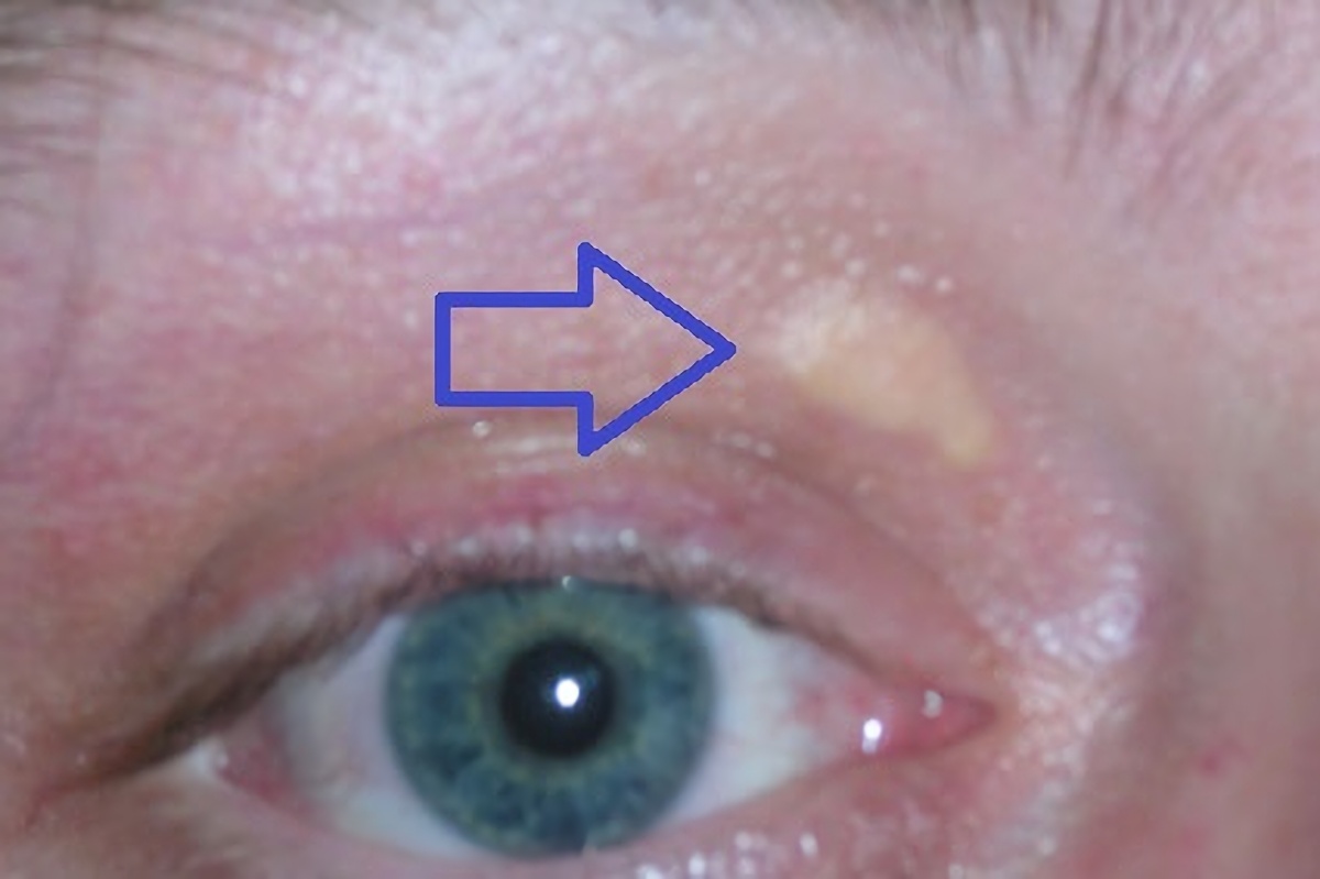
📷 Xanthelasma · lipid deposits at eyelids · tap to expand
When beta-oxidation fails or fat intake overwhelms clearance, lipids deposit in tissues.
Xanthelasma = cholesterol deposits at eyelid skin. Classic in hyperlipidemia / familial hypercholesterolemia.
The 4 Steps: OHOT
Every round of beta-oxidation removes 2 carbons as acetyl-CoA through 4 reactions.
Note: beta-oxidation uses FAD and NAD (oxidized forms).
Synthesis uses NADPH.
Board trap: confusing NADH (oxidation) with NADPH (synthesis).
Odd-Chain FA: The Propionyl-CoA Problem
Normal beta-oxidation removes 2C units cleanly. Odd-chain FAs leave a 3-carbon
propionyl-CoAPropionyl-CoA can't feed directly into TCA. It needs two extra steps involving biotin AND B12 to convert to succinyl-CoA.
at the end.
⚠B12 deficiency: methylmalonyl-CoA accumulates. Elevated methylmalonic acid in urine is the lab marker for B12 deficiency. Also causes neurological symptoms because odd-chain FA processing is needed for myelin maintenance.
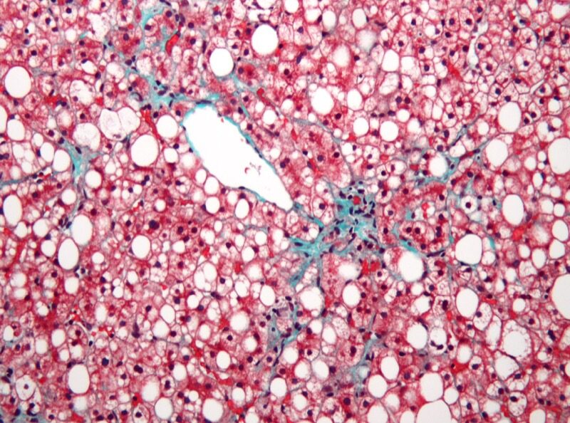
When Fat Can't Be Burned: Hepatic Steatosis
📷 NAFLD histology · lipid vacuoles in hepatocytes · tap to expand
When beta-oxidation is impaired or overwhelmed (obesity, alcohol, insulin resistance),
fatty acids accumulate as triglycerides inside hepatocytes. You see this on biopsy as
large clear vacuoles displacing the nucleus to the periphery.
This is also what happens in Adrenoleukodystrophy: defective ABCD1 (peroxisomal VLCFA transporter),
so very-long-chain FAs can't enter peroxisomes and accumulate, destroying myelin and adrenal tissue.
⚠
Board Traps: Beta-Oxidation Side
Short/medium-chain FAs cross the mitochondrial membrane freely. Carnitine is only needed for long-chain FAs. Carnitine deficiency = normal short/medium FA metabolism.
The rate-limiting step of beta-oxidation is CPT-I, NOT the OHOT cycle itself.
Beta-oxidation produces FAD and NAD forms (FADH₂, NADH). If a question asks "what NADPH-producing pathway is related to FA metabolism," the answer is the Pentose Phosphate Pathway (for synthesis), NOT beta-oxidation.
Adrenoleukodystrophy: X-linked recessive, defective ABCD1 (peroxisomal VLCFA transporter), very-long-chain FA accumulate in cytoplasm. Affects adrenal glands and white matter.
3 / 6
BOARD GOLD
The Full Comparison
Every row on this table can be a question stem. Learn them all.
Feature
Synthesis
Beta-Oxidation
Location
Cytoplasm
Mitochondria
Direction
Acetyl-CoA → Fatty Acid
Fatty Acid → Acetyl-CoA
Energy status
Requires NADPH + ATP
Produces NADH + FADH₂
Membrane shuttle
Citrate (acetyl-CoA out)
Carnitine (LCFA in)
Rate-limiting enzyme
ACC
CPT-I
Active when
Fed, insulin high
Fasted, glucagon high
Inhibited by
Palmitoyl-CoA, AMP, glucagon
Malonyl-CoA (via CPT-I)
Key cofactor
NADPH (PPP source)
FAD, NAD
Key vitamin
Biotin (ACC step)
B12 (odd-chain FA mutase)
Master switch
Malonyl-CoA (high = synthesis ON, oxidation OFF)
Malonyl-CoA: The Traffic Controller
🔑Malonyl-CoA = Master switch. High = Make fat (blocks CPT-I). Low = Liquidate fat (CPT-I opens). Fed = High. Fasted = Low.
Insulin (fed) → ACC active → Malonyl-CoA rises → Two things happen simultaneously:
(1) FAS complex uses malonyl-CoA to elongate fatty acids
(2) Malonyl-CoA physically blocks CPT-I → no FA enters mitochondria
Result: synthesis runs. Beta-oxidation is locked out. One molecule does both jobs.
Where Does the NADPH Come From?
FA synthesis is NADPH-hungry (2 per round). You need a steady supply.
Two main sources:
1. Pentose Phosphate Pathway (PPP):
G6P → Ribulose-5-P + 2 NADPH. Primary source. This is why liver cells have
high PPP activity. Also provides ribose-5-P for DNA synthesis and NADPH
for glutathione recycling (RBC antioxidant defense).
2. Malic Enzyme:
Malate → Pyruvate + CO₂ + NADPH. Part of the citrate shuttle recycling step.
Makes 1 NADPH per cycle as OAA is converted to pyruvate and then back to OAA
via pyruvate carboxylase.
🔗G6PD deficiency means less NADPH. Less NADPH means less glutathione recycling (RBC lysis) AND less FA synthesis. The board only asks about the RBC angle, but the mechanism is the same enzyme.
PHARMACOLOGY
Drug Targets in Lipid Metabolism
What targets what. Don't mix these up.
Statins
Target: HMG-CoA Reductase (cholesterol synthesis, NOT FA synthesis).
Statins do NOT affect ACC or FAS. They block mevalonate pathway in the liver.
FA synthesis is a completely separate pathway from cholesterol synthesis after acetyl-CoA.
Orlistat
Target: Intestinal lipase. Blocks dietary fat absorption in the gut.
Does NOT affect intracellular FA synthesis or oxidation. Works at the luminal level.
Fenofibrate
Target: PPAR-alpha. Activates peroxisome proliferator-activated receptor alpha
→ upregulates genes for FA oxidation, increases lipoprotein lipase, decreases triglycerides.
Used for hypertriglyceridemia.
Niacin (B3)
Target: Adipose lipolysis. Inhibits hormone-sensitive lipase in adipose tissue
→ fewer FAs released into blood → less substrate for beta-oxidation in liver →
decreased VLDL production. Also raises HDL (unknown mechanism).
Metformin
Target: Complex I / AMPK. Activates AMPK → phosphorylates ACC →
inhibits FA synthesis (same pathway as glucagon). Also reduces hepatic glucose output.
Indirectly connects glucose and fat metabolism.
4 / 6
MASTER THE ENZYMES
Flip to Unlock Each Enzyme
Tap each card. Front = the name. Back = everything clinical medicine tests about it.
⚡
ACC · Rate-Limiting Step
tap to reveal
Acetyl-CoA Carboxylase
Makes: Malonyl-CoA from Acetyl-CoA Cofactor: Biotin (B7) + ATP Location: Cytoplasm
Result
Malonyl-CoA drops. Synthesis OFF. Same downstream effect as fasting. That's why metformin is anti-obesity.
5 / 7
CLINICAL REASONING
Decision Tree: Fatty Acid Synthesis vs Oxidation
Follow the logic from hormonal state to pathway destination.
Is the cell in anabolic or catabolic state?
Fatty Acid SYNTHESIS
Cytoplasm. Acetyl-CoA carboxylase (ACC) is the rate-limiting enzyme. Insulin activates ACC. NADPH from the pentose phosphate pathway powers each reduction step. The committed product is malonyl-CoA, which simultaneously drives synthesis forward AND blocks CPT-1 to shut off beta-oxidation. End product: palmitate (C16).
VLDL synthesis. Liver packages fatty acids into triglycerides, loads them onto apoB-100, and releases VLDL into the blood. Defect: abetalipoproteinemia (no apoB produced) causes fat accumulation in enterocytes and liver, plus neurological and visual symptoms from fat-soluble vitamin deficiency.
Fatty Acid OXIDATION (beta-oxidation)
Carnitine shuttle. CPT-1 (carnitine palmitoyltransferase-1) is the rate-limiting step and the gatekeeper. Glucagon drops malonyl-CoA, which relieves CPT-1 inhibition. Long-chain fatty acids cross into the mitochondria and enter beta-oxidation. Products: NADH + FADH2 + acetyl-CoA. If oxaloacetate is depleted (prolonged fasting), acetyl-CoA cannot enter TCA and diverts to ketone bodies.
Malonyl-CoA. When synthesis is running (fed state), malonyl-CoA is high and blocks CPT-1 so oxidation cannot run simultaneously. When fasting drives malonyl-CoA down (ACC is off), CPT-1 is free and oxidation runs. The cell cannot synthesize and burn fat at the same time. This is one of the most tested reciprocal controls in clinical practice.
6 / 7
QUIZ
Test Yourself
5 random questions from a pool of 12. Shuffle on reload.
Five patients walked in. Each of them is a clue about one of these two pathways.
Don't overthink it. Pick the answer. See the cheat code. Never miss it again.
Medically reviewed by Kaitlyn Cocuzzo, MD and Fatima Ali, DO
Bone Wizardry is an independent educational resource for visual learning in the medical sciences. It is not affiliated with, endorsed by, or sponsored by any licensing or examination board, contains no real or recalled examination questions, and does not guarantee any educational or examination outcome.
 Bone WizardryMEDICAL EDUCATION