Build glucose from fasting carbon sources by crossing four bypass enzymes, then apply reciprocal regulation, redox shifts, and glycogen-storage patterns.
Central question: when stored glycogen is no longer enough, how does the body keep blood glucose available without simply reversing glycolysis? Gluconeogenesis is a carbon-routing problem: choose a usable precursor, cross three irreversible glycolytic barriers with four bypass enzymes, spend energy, and finally release free glucose. The liver carries most of that work early in fasting, while renal glucose production becomes more important as fasting continues. In healthy adults the kidney contributes only a small fraction after an overnight fast, but it can provide roughly one quarter of whole-body glucose production after about 60 hours without food. [1]
Start with the carbon budget
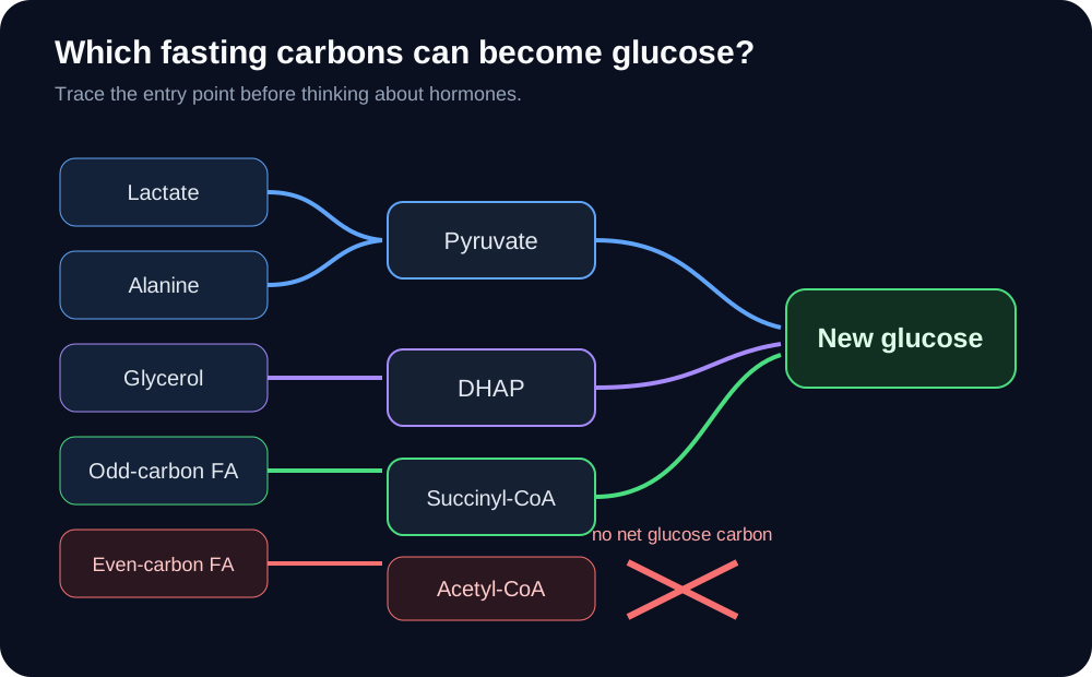
A useful first question is not "Which enzyme do I memorize?" It is which carbon can actually become glucose? Lactate returns from red blood cells and working muscle, alanine carries carbon from muscle, and glycerol arrives from triglyceride breakdown. Lactate and alanine become pyruvate, while glycerol enters higher in the pathway as dihydroxyacetone phosphate. Hepatic gluconeogenesis rises as fasting lengthens and liver glycogen contributes less. [2][3]
Trace carbon entry points before applying hormonal regulation. [2][3]
Lactate
Lactate dehydrogenase converts lactate to pyruvate. The carbon can then enter the pyruvate carboxylase step.
Alanine
Transamination converts alanine to pyruvate. Its nitrogen is handled separately, largely through hepatic urea production.
Glycerol
Glycerol becomes glycerol-3-phosphate and then dihydroxyacetone phosphate, joining gluconeogenesis above the pyruvate barrier.
The common trap is fatty acid carbon. Beta oxidation supplies abundant acetyl-CoA and ATP during fasting, but even-carbon acetyl-CoA cannot provide net glucose carbon in humans because entry into the citric acid cycle does not yield a net gain of oxaloacetate. Odd-carbon fatty acids are the exception: their propionyl-CoA end can become succinyl-CoA and contribute carbon to glucose production. The key distinction is that fat can power gluconeogenesis without most fatty-acid carbon becoming glucose. [2][3]
Building one glucose from two pyruvate requires 4 ATP, 2 GTP, and reducing power. That energy expense is sensible only when another tissue needs glucose more than the liver needs to conserve ATP. Fat oxidation helps pay the hepatic energy bill while gluconeogenic carbon is routed toward blood glucose. [3]
Try the carbon test
A tracer enters hepatocytes as palmitate-derived acetyl-CoA. Predict whether its two-carbon acetyl units can appear as a net new glucose carbon source. They cannot. Now change the tracer to propionate from an odd-carbon fatty acid: that carbon can enter through succinyl-CoA and contribute to gluconeogenesis.
Application: when a stem asks what can maintain glucose after glycogen is depleted, sort the candidate by carbon entry point before thinking about hormones. Lactate, glucogenic amino acids, glycerol, and propionyl-CoA can contribute; even-carbon acetyl-CoA cannot. [2]
Four enzymes cross three one-way barriers
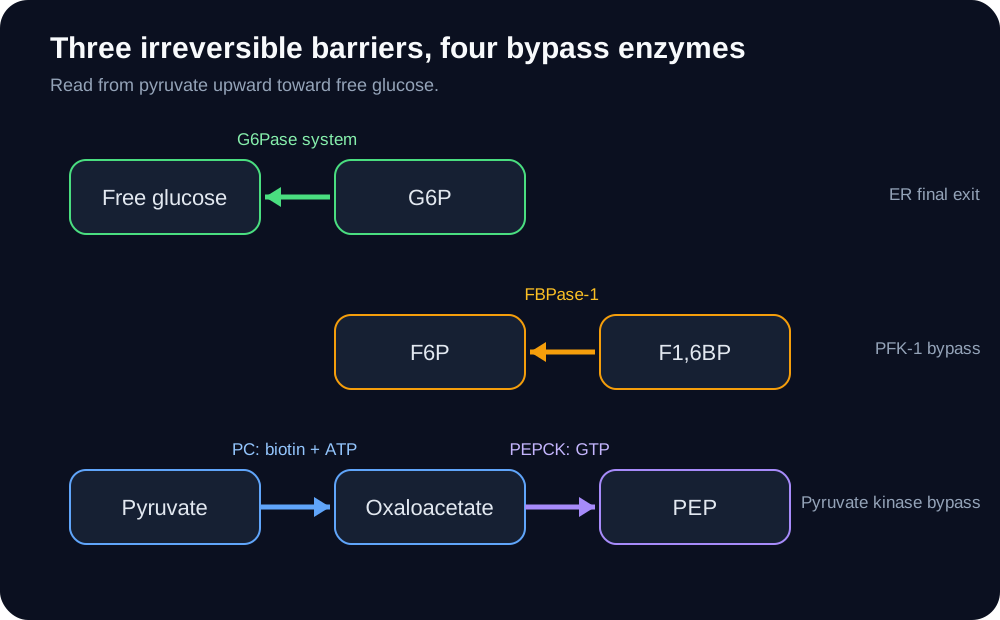
Most glycolytic reactions are near equilibrium and can run in the opposite direction. Three glycolytic reactions are strongly favorable in the forward direction, so gluconeogenesis needs bypasses. The pyruvate kinase barrier takes two enzymes; the PFK-1 and hexokinase or glucokinase barriers each take one.
Make the four bypass enzymes, energy currencies, and final ER-associated exit visible as one connected pathway. [2][3][4][8]
1. Pyruvate carboxylase: mitochondrial pyruvate + bicarbonate + ATP becomes oxaloacetate. The enzyme uses covalently bound biotin and is activated by acetyl-CoA. This reaction also replenishes oxaloacetate for other metabolic needs. [4]
2. PEP carboxykinase: oxaloacetate becomes phosphoenolpyruvate while GTP is used and CO2 is released. Mammals have cytosolic and mitochondrial PEPCK isoforms; the route used depends on substrate and redox needs. [2]
3. Fructose-1,6-bisphosphatase: fructose-1,6-bisphosphate becomes fructose-6-phosphate. AMP and fructose-2,6-bisphosphate restrain this step, linking glucose production to cellular energy and hormonal state. [2]
4. Glucose-6-phosphatase system: glucose-6-phosphate reaches the endoplasmic reticulum system and is hydrolyzed to free glucose. Liver and kidney can complete this export pathway; skeletal muscle lacks the hepatic glucose-6-phosphatase system needed to release its glycogen as circulating free glucose. [7][8]
Oxaloacetate itself does not freely cross the inner mitochondrial membrane. Carbon can be transferred through malate or aspartate routes, then returned to oxaloacetate in the cytosol when cytosolic PEPCK is used. This is why a pathway diagram that shows pyruvate simply becoming cytosolic phosphoenolpyruvate in one jump hides an important compartment problem. [2][3]
Predict a compartment consequence
If cytosolic oxaloacetate availability falls while mitochondrial pyruvate carboxylase still functions, the liver may still make oxaloacetate but cannot use that pool efficiently through the cytosolic route. A malate-based transfer can carry both carbon and reducing equivalents needed by the cytosolic pathway.
Application: biotin points toward pyruvate carboxylase, GTP points toward PEPCK, fructose-2,6-bisphosphate points toward reciprocal PFK-1 and FBPase-1 control, and an inability to release free hepatic glucose points toward the glucose-6-phosphatase system. [2][4][8]
Fed and fasting states bias carbon in opposite directions
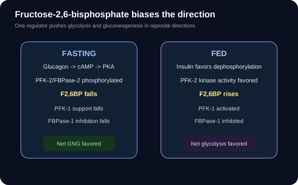
The liver avoids wasting large amounts of energy by regulating glycolysis and gluconeogenesis in opposite directions. This is reciprocal control, not an absolute claim that both pathways can never have simultaneous flux. During fasting, glucagon signaling favors hepatic glucose production; after feeding, insulin favors glycolysis, glycogen synthesis, and storage. [2][3]
Show how one reciprocal regulator changes glycolytic and gluconeogenic net flux in opposite directions. [2][3]
Fasting
Glucagon raises cAMP and activates PKA. In liver, phosphorylation of the bifunctional PFK-2/FBPase-2 protein favors its phosphatase activity, lowering fructose-2,6-bisphosphate. PFK-1 loses activation and FBPase-1 loses inhibition, so net carbon flow favors gluconeogenesis. [2]
Fed
Insulin favors dephosphorylation of the hepatic bifunctional protein, increasing PFK-2 kinase activity and fructose-2,6-bisphosphate. PFK-1 is activated while FBPase-1 is inhibited, so net carbon flow favors glycolysis. [2]
Hormones also change enzyme abundance over longer intervals. Glucagon and glucocorticoid signaling can support transcription of gluconeogenic genes, while insulin suppresses much of that program. A newborn or adult with cortisol deficiency can therefore have impaired fasting glucose defense, but it is misleading to reduce the syndrome to "PEPCK is absent." Counterregulation, substrate supply, gene expression, and glycogen metabolism all contribute. [2][3]
Metformin is another place where one-line explanations become dangerous. Its clinically important effect includes suppression of hepatic glucose production, but no single molecular route explains every setting. AMPK-dependent and AMPK-independent mechanisms, mitochondrial effects, cellular energy state, and redox-sensitive pathways have all been studied; the dominant mechanism remains debated. [5][6] Therefore, a board stem can reasonably test reduced hepatic gluconeogenesis, but a claim that one specific FBPase-1 or AMPK event is the proven universal primary mechanism overstates the evidence.
Predict the F2,6BP state
A fasting patient receives a drug that blocks hepatic glucagon receptor signaling. PKA signaling falls, the liver is less able to maintain the fasting phosphorylation state of PFK-2/FBPase-2, fructose-2,6-bisphosphate tends to rise, and net gluconeogenic drive falls.
Application: when insulin and glucagon compete in a question, translate the hormone into fructose-2,6-bisphosphate first. High fructose-2,6-bisphosphate favors PFK-1 and glycolysis; low fructose-2,6-bisphosphate releases FBPase-1 and favors gluconeogenesis. [2]
Try it here · Checkpoint 2 of 3
Make your prediction before reading the choices. A first attempt is just a starting point.
Case 14
Show answer and explanations for case 14
A. Higher PKA activity, lower fructose-2,6-bisphosphate, and greater gluconeogenesis (Why this does not fit)
Blocking glucagon lowers rather than raises cAMP and PKA signaling.
Reasoning steps for option A
How does pharmacologic loss of hepatic glucagon signaling test the option "Higher PKA activity, lower fructose-2,6-bisphosphate, and greater gluconeogenesis"?
gng-14 screen: A conflicts with clue one.
After adding the resulting PKA and fructose-2,6-bisphosphate direction, does "Higher PKA activity, lower fructose-2,6-bisphosphate, and greater gluconeogenesis" still match the required pattern?
gng-14 synthesis: choose B, not A.
B. Lower PKA activity, higher fructose-2,6-bisphosphate, and less gluconeogenesis (Best answer)
Loss of glucagon signaling shifts the hepatic bifunctional enzyme away from the fasting phosphorylation state, allowing fructose-2,6-bisphosphate to rise and restrain FBPase-1.
Reasoning steps for option B
How does pharmacologic loss of hepatic glucagon signaling test the option "Lower PKA activity, higher fructose-2,6-bisphosphate, and less gluconeogenesis"?
gng-14 screen: B remains possible.
After adding the resulting PKA and fructose-2,6-bisphosphate direction, does "Lower PKA activity, higher fructose-2,6-bisphosphate, and less gluconeogenesis" still match the required pattern?
gng-14 synthesis: both clues select B.
C. Lower PKA activity, lower fructose-2,6-bisphosphate, and greater gluconeogenesis (Why this does not fit)
Lower PKA signaling does not support the fasting low-fructose-2,6-bisphosphate state.
Reasoning steps for option C
How does pharmacologic loss of hepatic glucagon signaling test the option "Lower PKA activity, lower fructose-2,6-bisphosphate, and greater gluconeogenesis"?
gng-14 screen: C conflicts with clue one.
After adding the resulting PKA and fructose-2,6-bisphosphate direction, does "Lower PKA activity, lower fructose-2,6-bisphosphate, and greater gluconeogenesis" still match the required pattern?
gng-14 synthesis: choose B, not C.
D. Higher PKA activity, higher fructose-2,6-bisphosphate, and less gluconeogenesis (Why this does not fit)
The first step is already inconsistent: glucagon receptor blockade reduces cAMP-driven PKA activity.
Reasoning steps for option D
How does pharmacologic loss of hepatic glucagon signaling test the option "Higher PKA activity, higher fructose-2,6-bisphosphate, and less gluconeogenesis"?
gng-14 screen: D conflicts with clue one.
After adding the resulting PKA and fructose-2,6-bisphosphate direction, does "Higher PKA activity, higher fructose-2,6-bisphosphate, and less gluconeogenesis" still match the required pattern?
gng-14 synthesis: choose B, not D.
Takeaway: Glucagon receptor blockade shifts the liver away from the fasting F2,6BP state.
Recycling carbon links liver to peripheral tissues
Red blood cells have no mitochondria, so glycolysis ends in lactate. Working skeletal muscle can also send lactate to the liver when glycolytic rate exceeds mitochondrial oxidation. In the Cori cycle, hepatic lactate dehydrogenase converts lactate to pyruvate, gluconeogenesis rebuilds glucose, and glucose returns to peripheral tissue. Peripheral glycolysis gains 2 ATP per glucose, while the liver spends 6 high-energy phosphate bonds to rebuild it. The body accepts that net energy cost because the liver can use fat-derived energy while preserving glucose delivery. [3]
The alanine cycle carries both carbon and nitrogen information. Muscle transfers an amino group to pyruvate to form alanine. The liver recovers pyruvate for glucose production and disposes of the nitrogen through urea metabolism. Thus, alanine is not merely another memorized substrate; it links muscle amino-acid catabolism to hepatic glucose production. [3]
Ethanol demonstrates why redox state can block a pathway with intact enzymes. Alcohol dehydrogenase and aldehyde dehydrogenase generate NADH. A high hepatic NADH/NAD+ ratio favors pyruvate reduction to lactate and oxaloacetate reduction to malate. After prolonged poor intake, those shifts remove two important gluconeogenic substrates exactly when glycogen is scarce, producing a strong setup for hypoglycemia and high lactate. [10]
Severe illness, poor perfusion, hypoxia, liver dysfunction, and drug accumulation can all alter lactate handling, but they should not be collapsed into one mechanism. Metformin-associated lactic acidosis is most concerning when metformin accumulates or major acute illness is present; the drug is renally cleared, and its effects on mitochondrial metabolism and hepatic glucose production are mechanistically complex rather than one simple enzyme blockade. [5][6]
Try the redox perturbation
After several days without food, add a large ethanol exposure. Glycogen is already limited. High NADH now favors lactate over pyruvate and malate over oxaloacetate, so the same carbon sources become harder to route into glucose. The predicted direction is lower glucose and higher lactate.
Application: a fasting patient with alcohol exposure, hypoglycemia, and high lactate is a substrate and redox problem. A patient with an isolated hormone signal abnormality is a regulation problem. Keeping those categories separate prevents many near-miss answers. [2][10]
Try it here · Checkpoint 1 of 3
Make your prediction before reading the choices. A first attempt is just a starting point.
Case 5
Show answer and explanations for case 5
A. High hepatic NADH favors pyruvate to lactate and oxaloacetate to malate (Best answer)
Ethanol metabolism raises the hepatic NADH/NAD+ ratio, favoring reduction of pyruvate to lactate and oxaloacetate to malate. After glycogen depletion, this removes important gluconeogenic substrates.
Reasoning steps for option A
How does three days of poor intake plus active ethanol metabolism test the option "High hepatic NADH favors pyruvate to lactate and oxaloacetate to malate"?
gng-05 screen: A remains possible.
After adding the NADH-driven direction of lactate dehydrogenase and malate dehydrogenase, does "High hepatic NADH favors pyruvate to lactate and oxaloacetate to malate" still match the required pattern?
gng-05 synthesis: both clues select A.
B. Low NADH accelerates conversion of lactate to pyruvate and restores glucose output (Why this does not fit)
Ethanol metabolism raises rather than lowers NADH, and the resulting redox shift makes lactate and malate formation more favorable.
Reasoning steps for option B
How does three days of poor intake plus active ethanol metabolism test the option "Low NADH accelerates conversion of lactate to pyruvate and restores glucose output"?
gng-05 screen: B conflicts with clue one.
After adding the NADH-driven direction of lactate dehydrogenase and malate dehydrogenase, does "Low NADH accelerates conversion of lactate to pyruvate and restores glucose output" still match the required pattern?
gng-05 synthesis: choose A, not B.
C. Acetaldehyde directly inhibits glucose-6-phosphatase as the main acute lesion (Why this does not fit)
The characteristic acute fasting problem is a hepatic redox shift that diverts pyruvate and oxaloacetate, not a selective glucose-6-phosphatase block.
Reasoning steps for option C
How does three days of poor intake plus active ethanol metabolism test the option "Acetaldehyde directly inhibits glucose-6-phosphatase as the main acute lesion"?
gng-05 screen: C conflicts with clue one.
After adding the NADH-driven direction of lactate dehydrogenase and malate dehydrogenase, does "Acetaldehyde directly inhibits glucose-6-phosphatase as the main acute lesion" still match the required pattern?
gng-05 synthesis: choose A, not C.
D. Ethanol activates insulin secretion enough to stop all hepatic glucose production (Why this does not fit)
Insulin is not the central mechanism in fasting alcohol-associated hypoglycemia; substrate redox is the more direct explanation.
Reasoning steps for option D
How does three days of poor intake plus active ethanol metabolism test the option "Ethanol activates insulin secretion enough to stop all hepatic glucose production"?
gng-05 screen: D conflicts with clue one.
After adding the NADH-driven direction of lactate dehydrogenase and malate dehydrogenase, does "Ethanol activates insulin secretion enough to stop all hepatic glucose production" still match the required pattern?
gng-05 synthesis: choose A, not D.
Takeaway: Alcohol can block gluconeogenesis by changing substrate redox rather than by destroying a bypass enzyme.
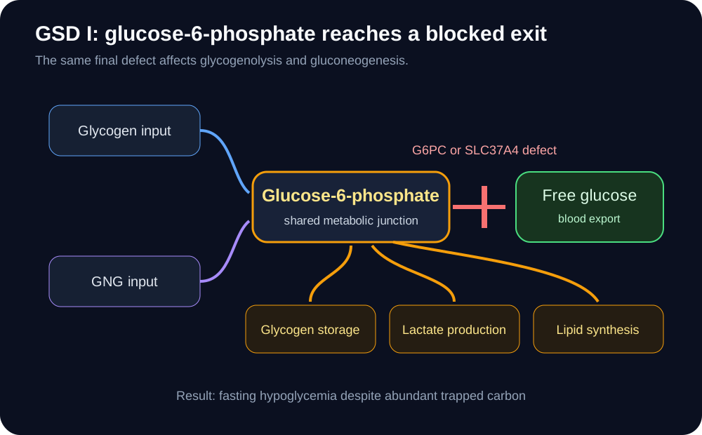
Glucose-6-phosphatase alpha and its transporter form an endoplasmic-reticulum system that allows liver, kidney, and intestine to convert glucose-6-phosphate into free glucose. In GSD Ia, G6PC activity is deficient. In GSD Ib, SLC37A4, the glucose-6-phosphate transporter, is deficient. Both defects compromise interprandial glucose homeostasis. [7][8]
Explain why GSD I impairs both glycogen-derived and gluconeogenic glucose release while upstream carbon accumulates. [7][8]
Because the block sits at the shared final step of glycogenolysis and gluconeogenesis, glucose-6-phosphate accumulates instead of becoming circulating glucose. Carbon is redirected toward glycogen storage, glycolysis and lactate, and lipid synthesis. The clinical pattern is severe fasting intolerance with hepatomegaly, hypoglycemia, lactic acidemia, hypertriglyceridemia, and hyperuricemia. GSD Ib adds chronic neutropenia and impaired neutrophil function, which can produce recurrent infections and inflammatory bowel disease. [7][8]
The useful causal sequence is: final exit blocked, glucose-6-phosphate accumulates, alternate pathways receive more carbon, and blood glucose still falls. This explains why simply increasing upstream glycogen breakdown cannot repair the defect. Modern diagnosis relies on the clinical and biochemical pattern plus molecular testing, with enzymology reserved for selected situations. Frequent carbohydrate delivery and uncooked cornstarch are central strategies for preventing fasting hypoglycemia. [7]
Older teaching sometimes treats a glucagon challenge as pathognomonic. That is not a good modern sorting rule. The more durable reasoning test is whether free glucose can be produced from glucose-6-phosphate, and current practice emphasizes molecular diagnosis and metabolic management rather than provocative fasting or glucagon testing. [7]
Predict the tracer result
Give labeled lactate to a hepatocyte with G6PC deficiency. The labeled carbon can travel through gluconeogenesis to glucose-6-phosphate, but the final hydrolysis step fails. Labeled free glucose release is therefore reduced even though upstream gluconeogenic enzymes still function.
Application: add neutropenia to the classic GSD I metabolic picture and SLC37A4 becomes more likely than G6PC. Remove neutropenia and the metabolic phenotype alone cannot by itself distinguish every subtype; genetic testing resolves the cause. [7][8]
Try it here · Checkpoint 3 of 3
Make your prediction before reading the choices. A first attempt is just a starting point.
Case 20
Show answer and explanations for case 20
A. PYGM, muscle glycogen phosphorylase (Why this does not fit)
PYGM encodes muscle glycogen phosphorylase and causes McArdle disease, which is an exercise-intolerance disorder.
Reasoning steps for option A
How does the GSD I metabolic phenotype plus neutropenia and infections test the option "PYGM, muscle glycogen phosphorylase"?
gng-20 screen: A conflicts with clue one.
After adding which glucose-6-phosphate system gene also affects neutrophil function, does "PYGM, muscle glycogen phosphorylase" still match the required pattern?
gng-20 synthesis: choose C, not A.
B. GAA, lysosomal acid alpha-glucosidase (Why this does not fit)
GAA encodes lysosomal acid alpha-glucosidase and causes Pompe disease, not the GSD I metabolic plus neutrophil phenotype.
Reasoning steps for option B
How does the GSD I metabolic phenotype plus neutropenia and infections test the option "GAA, lysosomal acid alpha-glucosidase"?
gng-20 screen: B conflicts with clue one.
After adding which glucose-6-phosphate system gene also affects neutrophil function, does "GAA, lysosomal acid alpha-glucosidase" still match the required pattern?
gng-20 synthesis: choose C, not B.
C. SLC37A4, glucose-6-phosphate transporter (Best answer)
SLC37A4 encodes the glucose-6-phosphate transporter. GSD Ib shares the GSD I metabolic phenotype and adds neutropenia and impaired neutrophil function.
Reasoning steps for option C
How does the GSD I metabolic phenotype plus neutropenia and infections test the option "SLC37A4, glucose-6-phosphate transporter"?
gng-20 screen: C remains possible.
After adding which glucose-6-phosphate system gene also affects neutrophil function, does "SLC37A4, glucose-6-phosphate transporter" still match the required pattern?
gng-20 synthesis: both clues select C.
D. G6PC1, glucose-6-phosphatase catalytic subunit (Why this does not fit)
G6PC1 deficiency causes GSD Ia and the classic metabolic phenotype, but chronic neutropenia and neutrophil dysfunction favor GSD Ib from SLC37A4 deficiency.
Reasoning steps for option D
How does the GSD I metabolic phenotype plus neutropenia and infections test the option "G6PC1, glucose-6-phosphatase catalytic subunit"?
gng-20 screen: D conflicts with clue one.
After adding which glucose-6-phosphate system gene also affects neutrophil function, does "G6PC1, glucose-6-phosphatase catalytic subunit" still match the required pattern?
gng-20 synthesis: choose C, not D.
Takeaway: Neutropenia added to GSD I physiology favors SLC37A4 and GSD Ib.
Use tissue and physiology to sort the glycogen storage diseases
The fastest comparison begins with which tissue is failing. Hepatic glycogen defects tend to present with hepatomegaly and fasting glucose problems. Muscle glycogen defects tend to present during exercise. Lysosomal glycogen disease can affect heart and skeletal muscle without behaving like a simple fasting glucose defect. [9]
High-yield patternCardiomyopathy and hypotonia in classic infantile disease; blood glucose is not usually the defining problem. [9]
TypeIII, Cori or Forbes
Core defectDebranching enzyme
High-yield patternHepatic disease with ketotic fasting hypoglycemia; some subtypes also involve skeletal and cardiac muscle. [9]
TypeIV, Andersen
Core defectBranching enzyme
High-yield patternAbnormally structured glycogen with progressive hepatic disease in the classic form. [9]
TypeV, McArdle
Core defectMuscle glycogen phosphorylase
High-yield patternExercise intolerance, cramps, rhabdomyolysis risk, and a muscle-limited glycogen problem rather than fasting hypoglycemia. [9]
TypeVI, Hers
Core defectLiver glycogen phosphorylase
High-yield patternUsually milder hepatic glycogenosis with hepatomegaly and ketotic fasting hypoglycemia. [9]
Fructose-1,6-bisphosphatase deficiency belongs in the differential even though it is not a glycogen storage disease. FBP1 deficiency causes episodic ketotic hypoglycemia with lactic acidosis, often triggered by fasting, infection, poor intake, or a large fructose load. Between episodes, affected children can be well. [11] That contrasts with GSD I, where the final glucose-6-phosphatase system is defective and hepatomegaly plus chronic metabolic abnormalities are prominent. [7]
A separate trap is biotin. Pyruvate carboxylase is biotin dependent, so deficient biotin availability can impair the first pyruvate bypass. But biotin metabolism supports several carboxylases, so severe biotin deficiency is not appropriately described as an isolated pyruvate-carboxylase syndrome. The board-level enzyme link remains valid: pyruvate carboxylase requires biotin, ATP, bicarbonate, and acetyl-CoA activation. [4]
Sort two patients
Child A has hepatomegaly, severe fasting hypoglycemia, high lactate, high triglycerides, and high uric acid. Child B has normal fasting glucose but develops painful exercise cramps and myoglobinuria. Child A points toward GSD I; Child B points toward McArdle disease. Tissue context is more useful than trying to memorize six names in isolation. [7][9]
Application: first decide whether the defect is glucose export, hepatic glycogen handling, muscle glycogen use, or a gluconeogenic bypass. Then use the laboratory pattern to identify the exact step. That hierarchy stays useful even when the vignette changes. [7][9][11]
Practice the pathway as decisions
For each case, identify the carbon source, the irreversible barrier, the organ or compartment, and the regulatory state before choosing an answer. Use the explanation to test the reasoning path, not to hunt for a memorized phrase.
Case 1
Show answer and explanations for case 1
A. Renal share of glucose production increases (Best answer)
After prolonged fasting, renal gluconeogenesis becomes a materially larger contributor to whole-body glucose production even as total glucose turnover falls.
Reasoning steps for option A
How does the change from an overnight fast to 60 hours without food test the option "Renal share of glucose production increases"?
gng-01 screen: A remains possible.
After adding the ability of liver, kidney, muscle, and brain to export free glucose, does "Renal share of glucose production increases" still match the required pattern?
gng-01 synthesis: both clues select A.
B. Skeletal muscle begins exporting free glucose from its glycogen stores (Why this does not fit)
Skeletal muscle lacks the glucose-6-phosphatase system needed to release substantial free glucose into blood, so its glycogen remains primarily a local fuel.
Reasoning steps for option B
How does the change from an overnight fast to 60 hours without food test the option "Skeletal muscle begins exporting free glucose from its glycogen stores"?
gng-01 screen: B conflicts with clue one.
After adding the ability of liver, kidney, muscle, and brain to export free glucose, does "Skeletal muscle begins exporting free glucose from its glycogen stores" still match the required pattern?
gng-01 synthesis: choose A, not B.
C. The kidney stops gluconeogenesis as liver glycogen becomes depleted (Why this does not fit)
Human studies show the opposite direction: renal glucose production contributes more after prolonged fasting.
Reasoning steps for option C
How does the change from an overnight fast to 60 hours without food test the option "The kidney stops gluconeogenesis as liver glycogen becomes depleted"?
gng-01 screen: C conflicts with clue one.
After adding the ability of liver, kidney, muscle, and brain to export free glucose, does "The kidney stops gluconeogenesis as liver glycogen becomes depleted" still match the required pattern?
gng-01 synthesis: choose A, not C.
D. Brain glycogenolysis replaces hepatic glucose production (Why this does not fit)
Brain glycogen stores are small and are not a systemic glucose source; prolonged fasting instead increases reliance on gluconeogenesis and ketones.
Reasoning steps for option D
How does the change from an overnight fast to 60 hours without food test the option "Brain glycogenolysis replaces hepatic glucose production"?
gng-01 screen: D conflicts with clue one.
After adding the ability of liver, kidney, muscle, and brain to export free glucose, does "Brain glycogenolysis replaces hepatic glucose production" still match the required pattern?
gng-01 synthesis: choose A, not D.
Takeaway: Renal glucose production gains importance as fasting is prolonged.
A. Greater hepatic conversion of acetyl-CoA carbon into glucose (Why this does not fit)
Even-carbon acetyl-CoA supplies energy and signaling during fasting but does not provide net glucose carbon in humans.
Reasoning steps for option A
How does 36 hours of fasting with depleted liver glycogen test the option "Greater hepatic conversion of acetyl-CoA carbon into glucose"?
gng-02 screen: A conflicts with clue one.
After adding the shift from glycogenolysis toward gluconeogenic carbon sources, does "Greater hepatic conversion of acetyl-CoA carbon into glucose" still match the required pattern?
gng-02 synthesis: choose B, not A.
B. Gluconeogenesis from lactate, glycerol, and amino acids (Best answer)
As fasting progresses and glycogen contribution falls, hepatic gluconeogenesis becomes the dominant source of endogenous glucose, using lactate, glycerol, and glucogenic amino acids.
Reasoning steps for option B
How does 36 hours of fasting with depleted liver glycogen test the option "Gluconeogenesis from lactate, glycerol, and amino acids"?
gng-02 screen: B remains possible.
After adding the shift from glycogenolysis toward gluconeogenic carbon sources, does "Gluconeogenesis from lactate, glycerol, and amino acids" still match the required pattern?
gng-02 synthesis: both clues select B.
C. Greater glucose export from skeletal muscle glycogen (Why this does not fit)
Muscle glycogen can support muscle glycolysis but skeletal muscle cannot efficiently release free glucose to maintain the circulation.
Reasoning steps for option C
How does 36 hours of fasting with depleted liver glycogen test the option "Greater glucose export from skeletal muscle glycogen"?
gng-02 screen: C conflicts with clue one.
After adding the shift from glycogenolysis toward gluconeogenic carbon sources, does "Greater glucose export from skeletal muscle glycogen" still match the required pattern?
gng-02 synthesis: choose B, not C.
D. Suppression of renal gluconeogenesis by glucagon (Why this does not fit)
Prolonged fasting increases, rather than suppresses, the renal contribution to glucose production.
Reasoning steps for option D
How does 36 hours of fasting with depleted liver glycogen test the option "Suppression of renal gluconeogenesis by glucagon"?
gng-02 screen: D conflicts with clue one.
After adding the shift from glycogenolysis toward gluconeogenic carbon sources, does "Suppression of renal gluconeogenesis by glucagon" still match the required pattern?
gng-02 synthesis: choose B, not D.
Takeaway: After glycogen wanes, gluconeogenesis supplies most endogenous glucose.
A. Lactate-derived pyruvate (Why this does not fit)
Lactate is oxidized to pyruvate and its carbon can enter gluconeogenesis.
Reasoning steps for option A
How does the labeled carbon source in each candidate test the option "Lactate-derived pyruvate"?
gng-03 screen: A conflicts with clue one.
After adding whether that carbon creates a net gain of gluconeogenic intermediate, does "Lactate-derived pyruvate" still match the required pattern?
gng-03 synthesis: choose C, not A.
B. Alanine-derived pyruvate (Why this does not fit)
Alanine can be transaminated to pyruvate, providing carbon for gluconeogenesis.
Reasoning steps for option B
How does the labeled carbon source in each candidate test the option "Alanine-derived pyruvate"?
gng-03 screen: B conflicts with clue one.
After adding whether that carbon creates a net gain of gluconeogenic intermediate, does "Alanine-derived pyruvate" still match the required pattern?
gng-03 synthesis: choose C, not B.
C. Palmitate acetyl-CoA (Best answer)
Even-carbon fatty acids yield acetyl-CoA, but acetyl-CoA entry into the citric acid cycle does not create net oxaloacetate and therefore does not provide net glucose carbon.
Reasoning steps for option C
How does the labeled carbon source in each candidate test the option "Palmitate acetyl-CoA"?
gng-03 screen: C remains possible.
After adding whether that carbon creates a net gain of gluconeogenic intermediate, does "Palmitate acetyl-CoA" still match the required pattern?
gng-03 synthesis: both clues select C.
D. Glycerol-derived DHAP (Why this does not fit)
Glycerol can enter the pathway at the triose phosphate level after conversion to glycerol-3-phosphate and dihydroxyacetone phosphate.
Reasoning steps for option D
How does the labeled carbon source in each candidate test the option "Glycerol-derived DHAP"?
gng-03 screen: D conflicts with clue one.
After adding whether that carbon creates a net gain of gluconeogenic intermediate, does "Glycerol-derived DHAP" still match the required pattern?
gng-03 synthesis: choose C, not D.
Takeaway: Even-carbon fatty acids power gluconeogenesis but do not supply net glucose carbon.
A. Acetyl-CoA entering citrate synthase (Why this does not fit)
Acetyl-CoA is produced from both even-carbon and odd-carbon fatty acids, but it does not create net glucose carbon.
Reasoning steps for option A
How does the terminal carbon product unique to odd-carbon fatty acids test the option "Acetyl-CoA entering citrate synthase"?
gng-04 screen: A conflicts with clue one.
After adding whether the product can enter the citric acid cycle upstream of oxaloacetate, does "Acetyl-CoA entering citrate synthase" still match the required pattern?
gng-04 synthesis: choose D, not A.
B. Malonyl-CoA entering beta oxidation (Why this does not fit)
Malonyl-CoA is a fatty acid synthesis intermediate and inhibits mitochondrial fatty acid entry rather than providing the relevant gluconeogenic carbon.
Reasoning steps for option B
How does the terminal carbon product unique to odd-carbon fatty acids test the option "Malonyl-CoA entering beta oxidation"?
gng-04 screen: B conflicts with clue one.
After adding whether the product can enter the citric acid cycle upstream of oxaloacetate, does "Malonyl-CoA entering beta oxidation" still match the required pattern?
gng-04 synthesis: choose D, not B.
C. Acetoacetate entering pyruvate carboxylase (Why this does not fit)
Ketone bodies are not converted into pyruvate for net glucose synthesis.
Reasoning steps for option C
How does the terminal carbon product unique to odd-carbon fatty acids test the option "Acetoacetate entering pyruvate carboxylase"?
gng-04 screen: C conflicts with clue one.
After adding whether the product can enter the citric acid cycle upstream of oxaloacetate, does "Acetoacetate entering pyruvate carboxylase" still match the required pattern?
gng-04 synthesis: choose D, not C.
D. Propionyl-CoA becoming succinyl-CoA (Best answer)
The terminal three-carbon fragment of an odd-carbon fatty acid becomes propionyl-CoA and then succinyl-CoA, which can contribute carbon to gluconeogenesis.
Reasoning steps for option D
How does the terminal carbon product unique to odd-carbon fatty acids test the option "Propionyl-CoA becoming succinyl-CoA"?
gng-04 screen: D remains possible.
After adding whether the product can enter the citric acid cycle upstream of oxaloacetate, does "Propionyl-CoA becoming succinyl-CoA" still match the required pattern?
gng-04 synthesis: both clues select D.
Takeaway: Odd-carbon fatty acids have a propionyl-CoA route that even-carbon fatty acids lack.
A. Acetyl-CoA inhibits pyruvate carboxylase and activates pyruvate dehydrogenase (Why this does not fit)
High acetyl-CoA has the opposite effect on pyruvate carboxylase and does not favor pyruvate entry through pyruvate dehydrogenase.
Reasoning steps for option A
How does high acetyl-CoA during beta oxidation test the option "Acetyl-CoA inhibits pyruvate carboxylase and activates pyruvate dehydrogenase"?
gng-07 screen: A conflicts with clue one.
After adding the allosteric response of pyruvate carboxylase, does "Acetyl-CoA inhibits pyruvate carboxylase and activates pyruvate dehydrogenase" still match the required pattern?
gng-07 synthesis: choose C, not A.
B. Acetyl-CoA activates fructose-1,6-bisphosphatase by covalent biotinylation (Why this does not fit)
FBPase-1 is not biotinylated; acetyl-CoA is the classic allosteric activator of pyruvate carboxylase.
Reasoning steps for option B
How does high acetyl-CoA during beta oxidation test the option "Acetyl-CoA activates fructose-1,6-bisphosphatase by covalent biotinylation"?
gng-07 screen: B conflicts with clue one.
After adding the allosteric response of pyruvate carboxylase, does "Acetyl-CoA activates fructose-1,6-bisphosphatase by covalent biotinylation" still match the required pattern?
gng-07 synthesis: choose C, not B.
C. Acetyl-CoA activates pyruvate carboxylase, increasing oxaloacetate formation (Best answer)
High acetyl-CoA signals abundant fat-derived fuel and allosterically activates pyruvate carboxylase, directing pyruvate toward oxaloacetate.
Reasoning steps for option C
How does high acetyl-CoA during beta oxidation test the option "Acetyl-CoA activates pyruvate carboxylase, increasing oxaloacetate formation"?
gng-07 screen: C remains possible.
After adding the allosteric response of pyruvate carboxylase, does "Acetyl-CoA activates pyruvate carboxylase, increasing oxaloacetate formation" still match the required pattern?
gng-07 synthesis: both clues select C.
D. Acetyl-CoA becomes phosphoenolpyruvate through PEP carboxykinase (Why this does not fit)
PEPCK uses oxaloacetate, not acetyl-CoA, and acetyl-CoA cannot supply net glucose carbon.
Reasoning steps for option D
How does high acetyl-CoA during beta oxidation test the option "Acetyl-CoA becomes phosphoenolpyruvate through PEP carboxykinase"?
gng-07 screen: D conflicts with clue one.
After adding the allosteric response of pyruvate carboxylase, does "Acetyl-CoA becomes phosphoenolpyruvate through PEP carboxykinase" still match the required pattern?
gng-07 synthesis: choose C, not D.
Takeaway: High acetyl-CoA activates pyruvate carboxylase while fat oxidation supplies energy for gluconeogenesis.
A. Conversion of cytosolic oxaloacetate to phosphoenolpyruvate after pyruvate enters gluconeogenesis (Best answer)
Mitochondrial oxaloacetate cannot freely cross the inner membrane. Transfer through malate or aspartate routes is needed when cytosolic PEPCK is used.
Reasoning steps for option A
How does intact mitochondrial PC with cytosolic PEPCK test the option "Conversion of cytosolic oxaloacetate to phosphoenolpyruvate after pyruvate enters gluconeogenesis"?
gng-08 screen: A remains possible.
After adding the inability of oxaloacetate to cross the inner mitochondrial membrane directly, does "Conversion of cytosolic oxaloacetate to phosphoenolpyruvate after pyruvate enters gluconeogenesis" still match the required pattern?
gng-08 synthesis: both clues select A.
B. Hydrolysis of glucose-6-phosphate within the ER glucose-6-phosphatase system (Why this does not fit)
The final ER hydrolysis step does not require mitochondrial oxaloacetate transfer.
Reasoning steps for option B
How does intact mitochondrial PC with cytosolic PEPCK test the option "Hydrolysis of glucose-6-phosphate within the ER glucose-6-phosphatase system"?
gng-08 screen: B conflicts with clue one.
After adding the inability of oxaloacetate to cross the inner mitochondrial membrane directly, does "Hydrolysis of glucose-6-phosphate within the ER glucose-6-phosphatase system" still match the required pattern?
gng-08 synthesis: choose A, not B.
C. Conversion of glycerol to dihydroxyacetone phosphate in the cytosol (Why this does not fit)
Glycerol enters gluconeogenesis above the pyruvate and oxaloacetate segment and does not depend on this transfer step.
Reasoning steps for option C
How does intact mitochondrial PC with cytosolic PEPCK test the option "Conversion of glycerol to dihydroxyacetone phosphate in the cytosol"?
gng-08 screen: C conflicts with clue one.
After adding the inability of oxaloacetate to cross the inner mitochondrial membrane directly, does "Conversion of glycerol to dihydroxyacetone phosphate in the cytosol" still match the required pattern?
gng-08 synthesis: choose A, not C.
D. Phosphorylation of fructose-6-phosphate by PFK-1 during glycolysis (Why this does not fit)
PFK-1 is a cytosolic glycolytic enzyme; its reaction is separate from mitochondrial carbon transfer.
Reasoning steps for option D
How does intact mitochondrial PC with cytosolic PEPCK test the option "Phosphorylation of fructose-6-phosphate by PFK-1 during glycolysis"?
gng-08 screen: D conflicts with clue one.
After adding the inability of oxaloacetate to cross the inner mitochondrial membrane directly, does "Phosphorylation of fructose-6-phosphate by PFK-1 during glycolysis" still match the required pattern?
gng-08 synthesis: choose A, not D.
Takeaway: Compartmentation matters because mitochondrial oxaloacetate cannot simply diffuse into the cytosol.
A. Pyruvate to oxaloacetate (Why this does not fit)
Pyruvate carboxylase uses ATP, bicarbonate, and biotin. With intact activity, this step continues and helps produce the accumulating oxaloacetate.
Reasoning steps for option A
How does selective cytosolic GTP depletion with preserved ATP and NADH test the option "Pyruvate to oxaloacetate"?
gng-09 screen: A conflicts with clue one.
After adding oxaloacetate accumulation with reduced phosphoenolpyruvate production, does "Pyruvate to oxaloacetate" still match the required pattern?
gng-09 synthesis: choose B, not A.
B. Oxaloacetate to phosphoenolpyruvate (Best answer)
PEP carboxykinase directly uses GTP. Limiting this reaction explains both oxaloacetate accumulation and reduced phosphoenolpyruvate production.
Reasoning steps for option B
How does selective cytosolic GTP depletion with preserved ATP and NADH test the option "Oxaloacetate to phosphoenolpyruvate"?
gng-09 screen: B remains possible.
After adding oxaloacetate accumulation with reduced phosphoenolpyruvate production, does "Oxaloacetate to phosphoenolpyruvate" still match the required pattern?
gng-09 synthesis: both clues select B.
C. 3-phosphoglycerate to 1,3-bisphosphoglycerate (Why this does not fit)
In gluconeogenesis, phosphoglycerate kinase consumes ATP rather than GTP, so a selective GTP fall does not directly block this step.
Reasoning steps for option C
How does selective cytosolic GTP depletion with preserved ATP and NADH test the option "3-phosphoglycerate to 1,3-bisphosphoglycerate"?
gng-09 screen: C conflicts with clue one.
After adding oxaloacetate accumulation with reduced phosphoenolpyruvate production, does "3-phosphoglycerate to 1,3-bisphosphoglycerate" still match the required pattern?
gng-09 synthesis: choose B, not C.
D. Fructose-1,6-bisphosphate to fructose-6-phosphate (Why this does not fit)
FBPase-1 hydrolyzes the phosphate without using GTP, so the nucleotide and metabolite pattern localize the defect upstream.
Reasoning steps for option D
How does selective cytosolic GTP depletion with preserved ATP and NADH test the option "Fructose-1,6-bisphosphate to fructose-6-phosphate"?
gng-09 screen: D conflicts with clue one.
After adding oxaloacetate accumulation with reduced phosphoenolpyruvate production, does "Fructose-1,6-bisphosphate to fructose-6-phosphate" still match the required pattern?
gng-09 synthesis: choose B, not D.
Takeaway: GTP depletion with oxaloacetate accumulation localizes a gluconeogenic block to PEP carboxykinase.
A. Pyruvate to oxaloacetate (Why this does not fit)
That reaction is catalyzed by pyruvate carboxylase. Its failure is a different gluconeogenic defect.
Reasoning steps for option A
How does episodic ketotic hypoglycemia and lactic acidosis with FBP1 variants test the option "Pyruvate to oxaloacetate"?
gng-10 screen: A conflicts with clue one.
After adding the blocked irreversible glycolytic bypass, does "Pyruvate to oxaloacetate" still match the required pattern?
gng-10 synthesis: choose B, not A.
B. Fructose-1,6-bisphosphate hydrolysis (Best answer)
FBP1 encodes fructose-1,6-bisphosphatase 1. Its deficiency blocks this gluconeogenic bypass and causes episodic ketotic hypoglycemia with lactic acidosis.
Reasoning steps for option B
How does episodic ketotic hypoglycemia and lactic acidosis with FBP1 variants test the option "Fructose-1,6-bisphosphate hydrolysis"?
gng-10 screen: B remains possible.
After adding the blocked irreversible glycolytic bypass, does "Fructose-1,6-bisphosphate hydrolysis" still match the required pattern?
gng-10 synthesis: both clues select B.
C. Oxaloacetate to phosphoenolpyruvate (Why this does not fit)
That reaction is catalyzed by PEP carboxykinase and is upstream of the FBP1 block.
Reasoning steps for option C
How does episodic ketotic hypoglycemia and lactic acidosis with FBP1 variants test the option "Oxaloacetate to phosphoenolpyruvate"?
gng-10 screen: C conflicts with clue one.
After adding the blocked irreversible glycolytic bypass, does "Oxaloacetate to phosphoenolpyruvate" still match the required pattern?
gng-10 synthesis: choose B, not C.
D. Glucose-6-phosphate transport into the ER (Why this does not fit)
That defect corresponds to the glucose-6-phosphate transporter problem in GSD Ib, which has a different chronic metabolic phenotype.
Reasoning steps for option D
How does episodic ketotic hypoglycemia and lactic acidosis with FBP1 variants test the option "Glucose-6-phosphate transport into the ER"?
gng-10 screen: D conflicts with clue one.
After adding the blocked irreversible glycolytic bypass, does "Glucose-6-phosphate transport into the ER" still match the required pattern?
gng-10 synthesis: choose B, not D.
Takeaway: FBP1 deficiency blocks the PFK-1 bypass and causes episodic ketotic hypoglycemia with lactic acidosis.
A. Fructose-2,6-bisphosphate rises; PFK-1 is activated (Why this does not fit)
That pattern favors glycolysis and is associated with the fed insulin-dominant state.
Reasoning steps for option A
How does fasting glucagon, cAMP, and PKA signaling test the option "Fructose-2,6-bisphosphate rises; PFK-1 is activated"?
gng-11 screen: A conflicts with clue one.
After adding the effect of fructose-2,6-bisphosphate on PFK-1 and FBPase-1, does "Fructose-2,6-bisphosphate rises; PFK-1 is activated" still match the required pattern?
gng-11 synthesis: choose C, not A.
B. Fructose-2,6-bisphosphate rises; FBPase-1 is activated (Why this does not fit)
Fructose-2,6-bisphosphate inhibits FBPase-1 rather than activating it.
Reasoning steps for option B
How does fasting glucagon, cAMP, and PKA signaling test the option "Fructose-2,6-bisphosphate rises; FBPase-1 is activated"?
gng-11 screen: B conflicts with clue one.
After adding the effect of fructose-2,6-bisphosphate on PFK-1 and FBPase-1, does "Fructose-2,6-bisphosphate rises; FBPase-1 is activated" still match the required pattern?
gng-11 synthesis: choose C, not B.
C. Fructose-2,6-bisphosphate falls; FBPase-1 is released from inhibition (Best answer)
Fasting PKA signaling favors the phosphatase activity of hepatic PFK-2/FBPase-2, lowering fructose-2,6-bisphosphate and favoring FBPase-1.
Reasoning steps for option C
How does fasting glucagon, cAMP, and PKA signaling test the option "Fructose-2,6-bisphosphate falls; FBPase-1 is released from inhibition"?
gng-11 screen: C remains possible.
After adding the effect of fructose-2,6-bisphosphate on PFK-1 and FBPase-1, does "Fructose-2,6-bisphosphate falls; FBPase-1 is released from inhibition" still match the required pattern?
gng-11 synthesis: both clues select C.
D. Fructose-2,6-bisphosphate falls; PFK-1 is strongly activated (Why this does not fit)
Lower fructose-2,6-bisphosphate removes an important PFK-1 activator, so glycolytic drive falls.
Reasoning steps for option D
How does fasting glucagon, cAMP, and PKA signaling test the option "Fructose-2,6-bisphosphate falls; PFK-1 is strongly activated"?
gng-11 screen: D conflicts with clue one.
After adding the effect of fructose-2,6-bisphosphate on PFK-1 and FBPase-1, does "Fructose-2,6-bisphosphate falls; PFK-1 is strongly activated" still match the required pattern?
gng-11 synthesis: choose C, not D.
Takeaway: Low hepatic fructose-2,6-bisphosphate favors FBPase-1 and gluconeogenesis.
A. Dephosphorylation favors PFK-2 kinase activity and raises fructose-2,6-bisphosphate (Best answer)
In the fed liver, insulin favors dephosphorylation of the bifunctional enzyme, increasing fructose-2,6-bisphosphate and promoting glycolysis.
Reasoning steps for option A
How does high insulin after a carbohydrate-rich meal test the option "Dephosphorylation favors PFK-2 kinase activity and raises fructose-2,6-bisphosphate"?
gng-12 screen: A remains possible.
After adding the hepatic dephosphorylated state of PFK-2/FBPase-2, does "Dephosphorylation favors PFK-2 kinase activity and raises fructose-2,6-bisphosphate" still match the required pattern?
gng-12 synthesis: both clues select A.
B. Phosphorylation favors FBPase-2 activity and raises fructose-2,6-bisphosphate (Why this does not fit)
The fasting phosphorylation state favors phosphatase activity and lowers fructose-2,6-bisphosphate.
Reasoning steps for option B
How does high insulin after a carbohydrate-rich meal test the option "Phosphorylation favors FBPase-2 activity and raises fructose-2,6-bisphosphate"?
gng-12 screen: B conflicts with clue one.
After adding the hepatic dephosphorylated state of PFK-2/FBPase-2, does "Phosphorylation favors FBPase-2 activity and raises fructose-2,6-bisphosphate" still match the required pattern?
gng-12 synthesis: choose A, not B.
C. Dephosphorylation lowers fructose-2,6-bisphosphate and activates FBPase-1 (Why this does not fit)
In liver, the fed dephosphorylated state raises fructose-2,6-bisphosphate, which inhibits FBPase-1.
Reasoning steps for option C
How does high insulin after a carbohydrate-rich meal test the option "Dephosphorylation lowers fructose-2,6-bisphosphate and activates FBPase-1"?
gng-12 screen: C conflicts with clue one.
After adding the hepatic dephosphorylated state of PFK-2/FBPase-2, does "Dephosphorylation lowers fructose-2,6-bisphosphate and activates FBPase-1" still match the required pattern?
gng-12 synthesis: choose A, not C.
D. Insulin removes PFK-1 so glucose-6-phosphate must enter gluconeogenesis (Why this does not fit)
Insulin does not remove PFK-1; it shifts allosteric and transcriptional regulation toward glucose use and storage.
Reasoning steps for option D
How does high insulin after a carbohydrate-rich meal test the option "Insulin removes PFK-1 so glucose-6-phosphate must enter gluconeogenesis"?
gng-12 screen: D conflicts with clue one.
After adding the hepatic dephosphorylated state of PFK-2/FBPase-2, does "Insulin removes PFK-1 so glucose-6-phosphate must enter gluconeogenesis" still match the required pattern?
gng-12 synthesis: choose A, not D.
Takeaway: Insulin raises hepatic fructose-2,6-bisphosphate and favors glycolysis.
A. Gluconeogenesis is more strongly suppressed than normal because AMP activates the mutant enzyme (Why this does not fit)
AMP normally inhibits FBPase-1; loss of that inhibition would make suppression weaker, not stronger.
Reasoning steps for option A
How does loss of AMP inhibition during a low-energy state test the option "Gluconeogenesis is more strongly suppressed than normal because AMP activates the mutant enzyme"?
gng-13 screen: A conflicts with clue one.
After adding whether FBPase-1 remains appropriately restrained, does "Gluconeogenesis is more strongly suppressed than normal because AMP activates the mutant enzyme" still match the required pattern?
gng-13 synthesis: choose D, not A.
B. Glycolysis stops because AMP can no longer activate PFK-1 (Why this does not fit)
The mutation affects FBPase-1, not PFK-1. AMP can still favor glycolysis through its normal effects.
Reasoning steps for option B
How does loss of AMP inhibition during a low-energy state test the option "Glycolysis stops because AMP can no longer activate PFK-1"?
gng-13 screen: B conflicts with clue one.
After adding whether FBPase-1 remains appropriately restrained, does "Glycolysis stops because AMP can no longer activate PFK-1" still match the required pattern?
gng-13 synthesis: choose D, not B.
C. Glucose-6-phosphatase becomes biotin dependent (Why this does not fit)
The mutation does not change the final ER glucose release step or create a biotin requirement.
Reasoning steps for option C
How does loss of AMP inhibition during a low-energy state test the option "Glucose-6-phosphatase becomes biotin dependent"?
gng-13 screen: C conflicts with clue one.
After adding whether FBPase-1 remains appropriately restrained, does "Glucose-6-phosphatase becomes biotin dependent" still match the required pattern?
gng-13 synthesis: choose D, not C.
D. FBPase-1 can remain inappropriately active despite a low cellular energy signal (Best answer)
If AMP can no longer inhibit FBPase-1, one important low-energy restraint on glucose synthesis is lost, allowing wasteful gluconeogenic flux when energy is scarce.
Reasoning steps for option D
How does loss of AMP inhibition during a low-energy state test the option "FBPase-1 can remain inappropriately active despite a low cellular energy signal"?
gng-13 screen: D remains possible.
After adding whether FBPase-1 remains appropriately restrained, does "FBPase-1 can remain inappropriately active despite a low cellular energy signal" still match the required pattern?
gng-13 synthesis: both clues select D.
Takeaway: AMP normally restrains FBPase-1 when cellular energy is low.
A. Metformin irreversibly blocks FBPase-1 as its single established mechanism (Why this does not fit)
Metformin suppresses hepatic glucose production through multiple proposed pathways; a single universal irreversible FBPase-1 mechanism is not established.
Reasoning steps for option A
How does metformin exposure during severe acute kidney injury test the option "Metformin irreversibly blocks FBPase-1 as its single established mechanism"?
gng-15 screen: A conflicts with clue one.
After adding the difference between a clinical net effect and a single claimed molecular mechanism, does "Metformin irreversibly blocks FBPase-1 as its single established mechanism" still match the required pattern?
gng-15 synthesis: choose C, not A.
B. Metformin lowers glucose only by increasing pancreatic insulin secretion (Why this does not fit)
Metformin does not primarily act as an insulin secretagogue. Its major glucose-lowering effect includes reduced hepatic glucose production.
Reasoning steps for option B
How does metformin exposure during severe acute kidney injury test the option "Metformin lowers glucose only by increasing pancreatic insulin secretion"?
gng-15 screen: B conflicts with clue one.
After adding the difference between a clinical net effect and a single claimed molecular mechanism, does "Metformin lowers glucose only by increasing pancreatic insulin secretion" still match the required pattern?
gng-15 synthesis: choose C, not B.
C. Renal failure raises metformin exposure; hepatic glucose output falls through multiple pathways (Best answer)
Metformin is renally cleared, so severe kidney dysfunction can increase exposure. Mechanistic reviews support several interacting pathways rather than one universally dominant molecular target.
Reasoning steps for option C
How does metformin exposure during severe acute kidney injury test the option "Renal failure raises metformin exposure; hepatic glucose output falls through multiple pathways"?
gng-15 screen: C remains possible.
After adding the difference between a clinical net effect and a single claimed molecular mechanism, does "Renal failure raises metformin exposure; hepatic glucose output falls through multiple pathways" still match the required pattern?
gng-15 synthesis: both clues select C.
D. Metformin-associated acidosis establishes an inherited loss of hepatic pyruvate carboxylase activity (Why this does not fit)
A drug-associated metabolic disturbance does not imply a genetic pyruvate carboxylase defect, and metformin's actions are broader than one enzyme lesion.
Reasoning steps for option D
How does metformin exposure during severe acute kidney injury test the option "Metformin-associated acidosis establishes an inherited loss of hepatic pyruvate carboxylase activity"?
gng-15 screen: D conflicts with clue one.
After adding the difference between a clinical net effect and a single claimed molecular mechanism, does "Metformin-associated acidosis establishes an inherited loss of hepatic pyruvate carboxylase activity" still match the required pattern?
gng-15 synthesis: choose C, not D.
Takeaway: Metformin suppresses hepatic glucose production through complex mechanisms, and severe renal dysfunction can increase exposure.
A. The body gains 8 net high-energy phosphate bonds (Why this does not fit)
The liver must spend high-energy phosphate bonds to rebuild glucose, so the cycle is not a net ATP-producing process for the body.
Reasoning steps for option A
How does 2 ATP gained in peripheral glycolysis test the option "The body gains 8 net high-energy phosphate bonds"?
gng-16 screen: A conflicts with clue one.
After adding the 4 ATP plus 2 GTP cost of hepatic glucose resynthesis, does "The body gains 8 net high-energy phosphate bonds" still match the required pattern?
gng-16 synthesis: choose C, not A.
B. The cycle is energy neutral because liver and muscle reactions exactly cancel (Why this does not fit)
Muscle gains 2 ATP, but liver gluconeogenesis spends 6 high-energy phosphate bonds, leaving a net whole-body cost.
Reasoning steps for option B
How does 2 ATP gained in peripheral glycolysis test the option "The cycle is energy neutral because liver and muscle reactions exactly cancel"?
gng-16 screen: B conflicts with clue one.
After adding the 4 ATP plus 2 GTP cost of hepatic glucose resynthesis, does "The cycle is energy neutral because liver and muscle reactions exactly cancel" still match the required pattern?
gng-16 synthesis: choose C, not B.
C. The body loses 4 high-energy phosphate bonds while transferring energy demand to the liver (Best answer)
Muscle gains 2 ATP from glycolysis while the liver spends 4 ATP plus 2 GTP to rebuild glucose, creating a net cost of 4 high-energy phosphate bonds.
Reasoning steps for option C
How does 2 ATP gained in peripheral glycolysis test the option "The body loses 4 high-energy phosphate bonds while transferring energy demand to the liver"?
gng-16 screen: C remains possible.
After adding the 4 ATP plus 2 GTP cost of hepatic glucose resynthesis, does "The body loses 4 high-energy phosphate bonds while transferring energy demand to the liver" still match the required pattern?
gng-16 synthesis: both clues select C.
D. The liver gains 2 ATP because lactate oxidation directly powers glucose synthesis (Why this does not fit)
Converting lactate back to glucose requires energy input; lactate carbon alone does not pay the ATP and GTP cost.
Reasoning steps for option D
How does 2 ATP gained in peripheral glycolysis test the option "The liver gains 2 ATP because lactate oxidation directly powers glucose synthesis"?
gng-16 screen: D conflicts with clue one.
After adding the 4 ATP plus 2 GTP cost of hepatic glucose resynthesis, does "The liver gains 2 ATP because lactate oxidation directly powers glucose synthesis" still match the required pattern?
gng-16 synthesis: choose C, not D.
Takeaway: The Cori cycle preserves glucose availability at a whole-body energy cost.
Mature erythrocytes regenerate NAD+ by reducing pyruvate to lactate. The liver can oxidize that lactate to pyruvate and return its carbon to glucose through the Cori cycle.
Reasoning steps for option A
How does mature erythrocytes lack mitochondria but maintain glycolytic ATP production test the option "Lactate through the Cori cycle"?
gng-17 screen: A remains possible.
After adding labeled erythrocyte carbon later appears in newly synthesized hepatic glucose, does "Lactate through the Cori cycle" still match the required pattern?
gng-17 synthesis: both clues select A.
B. Alanine through the glucose-alanine cycle (Why this does not fit)
The glucose-alanine cycle depends mainly on amino acid transamination in skeletal muscle; mature erythrocytes primarily export lactate from glycolysis.
Reasoning steps for option B
How does mature erythrocytes lack mitochondria but maintain glycolytic ATP production test the option "Alanine through the glucose-alanine cycle"?
gng-17 screen: B conflicts with clue one.
After adding labeled erythrocyte carbon later appears in newly synthesized hepatic glucose, does "Alanine through the glucose-alanine cycle" still match the required pattern?
gng-17 synthesis: choose A, not B.
C. Acetyl-CoA through the citrate shuttle (Why this does not fit)
Erythrocytes lack mitochondria, so they cannot generate mitochondrial acetyl-CoA or use a citrate shuttle.
Reasoning steps for option C
How does mature erythrocytes lack mitochondria but maintain glycolytic ATP production test the option "Acetyl-CoA through the citrate shuttle"?
gng-17 screen: C conflicts with clue one.
After adding labeled erythrocyte carbon later appears in newly synthesized hepatic glucose, does "Acetyl-CoA through the citrate shuttle" still match the required pattern?
gng-17 synthesis: choose A, not C.
D. Beta-hydroxybutyrate through ketone transport (Why this does not fit)
Ketone bodies are produced by hepatic mitochondria and are not the carbon carrier exported from glycolyzing erythrocytes.
Reasoning steps for option D
How does mature erythrocytes lack mitochondria but maintain glycolytic ATP production test the option "Beta-hydroxybutyrate through ketone transport"?
gng-17 screen: D conflicts with clue one.
After adding labeled erythrocyte carbon later appears in newly synthesized hepatic glucose, does "Beta-hydroxybutyrate through ketone transport" still match the required pattern?
gng-17 synthesis: choose A, not D.
Takeaway: A mitochondria-free erythrocyte returns glucose carbon to the liver as lactate through the Cori cycle.
A. Pyruvate production falls; urea formation increases (Why this does not fit)
Intact alanine aminotransferase still generates pyruvate, while a urea-cycle entry defect reduces rather than increases urea formation.
Reasoning steps for option A
How does alanine aminotransferase remains intact during fasting test the option "Pyruvate production falls; urea formation increases"?
gng-18 screen: A conflicts with clue one.
After adding mitochondrial urea-cycle entry is selectively impaired, does "Pyruvate production falls; urea formation increases" still match the required pattern?
gng-18 synthesis: choose C, not A.
B. Pyruvate still supports glucose synthesis; urea formation increases (Why this does not fit)
The carbon statement is correct because alanine can still become pyruvate, but impaired urea-cycle entry lowers nitrogen disposal.
Reasoning steps for option B
How does alanine aminotransferase remains intact during fasting test the option "Pyruvate still supports glucose synthesis; urea formation increases"?
gng-18 screen: B conflicts with clue one.
After adding mitochondrial urea-cycle entry is selectively impaired, does "Pyruvate still supports glucose synthesis; urea formation increases" still match the required pattern?
gng-18 synthesis: choose C, not B.
C. Pyruvate still supports glucose synthesis; urea formation decreases (Best answer)
Alanine transamination can still provide pyruvate for gluconeogenesis, while the separate urea-cycle defect decreases conversion of amino nitrogen to urea.
Reasoning steps for option C
How does alanine aminotransferase remains intact during fasting test the option "Pyruvate still supports glucose synthesis; urea formation decreases"?
gng-18 screen: C remains possible.
After adding mitochondrial urea-cycle entry is selectively impaired, does "Pyruvate still supports glucose synthesis; urea formation decreases" still match the required pattern?
gng-18 synthesis: both clues select C.
D. Pyruvate production falls; urea formation decreases (Why this does not fit)
Reduced urea formation fits the nitrogen defect, but intact alanine transamination preserves pyruvate production from alanine carbon.
Reasoning steps for option D
How does alanine aminotransferase remains intact during fasting test the option "Pyruvate production falls; urea formation decreases"?
gng-18 screen: D conflicts with clue one.
After adding mitochondrial urea-cycle entry is selectively impaired, does "Pyruvate production falls; urea formation decreases" still match the required pattern?
gng-18 synthesis: choose C, not D.
Takeaway: Alanine carbon can still enter gluconeogenesis when transamination is intact, even if urea-cycle nitrogen disposal is impaired.
A. Glucose-6-phosphatase alpha deficiency (Best answer)
GSD Ia blocks the final conversion of glucose-6-phosphate to free glucose, causing severe fasting intolerance and redirecting accumulated glucose-6-phosphate toward glycogen, lactate, lipid, and purine-related pathways.
Reasoning steps for option A
How does fasting hypoglycemia plus hepatomegaly, lactate, triglycerides, and uric acid test the option "Glucose-6-phosphatase alpha deficiency"?
gng-19 screen: A remains possible.
After adding the shared final step of glycogenolysis and gluconeogenesis, does "Glucose-6-phosphatase alpha deficiency" still match the required pattern?
gng-19 synthesis: both clues select A.
B. Muscle glycogen phosphorylase deficiency (Why this does not fit)
McArdle disease presents with exercise-related muscle symptoms rather than infantile hepatomegaly with severe fasting hypoglycemia and lactic acidemia.
Reasoning steps for option B
How does fasting hypoglycemia plus hepatomegaly, lactate, triglycerides, and uric acid test the option "Muscle glycogen phosphorylase deficiency"?
gng-19 screen: B conflicts with clue one.
After adding the shared final step of glycogenolysis and gluconeogenesis, does "Muscle glycogen phosphorylase deficiency" still match the required pattern?
gng-19 synthesis: choose A, not B.
C. Lysosomal acid alpha-glucosidase deficiency (Why this does not fit)
Classic infantile Pompe disease prominently causes cardiomyopathy and hypotonia rather than this GSD I metabolic pattern.
Reasoning steps for option C
How does fasting hypoglycemia plus hepatomegaly, lactate, triglycerides, and uric acid test the option "Lysosomal acid alpha-glucosidase deficiency"?
gng-19 screen: C conflicts with clue one.
After adding the shared final step of glycogenolysis and gluconeogenesis, does "Lysosomal acid alpha-glucosidase deficiency" still match the required pattern?
gng-19 synthesis: choose A, not C.
D. Hepatic branching enzyme deficiency (Why this does not fit)
Andersen disease can cause severe liver disease, but the combined hypoglycemia, lactic acidemia, hypertriglyceridemia, and hyperuricemia pattern is characteristic of GSD I.
Reasoning steps for option D
How does fasting hypoglycemia plus hepatomegaly, lactate, triglycerides, and uric acid test the option "Hepatic branching enzyme deficiency"?
gng-19 screen: D conflicts with clue one.
After adding the shared final step of glycogenolysis and gluconeogenesis, does "Hepatic branching enzyme deficiency" still match the required pattern?
gng-19 synthesis: choose A, not D.
Takeaway: Severe fasting hypoglycemia plus hepatomegaly, lactate, triglycerides, and uric acid points to GSD I.
A. Labeled carbon reaches glucose-6-phosphate, but release of labeled free glucose is reduced (Best answer)
Lactate carbon can traverse gluconeogenesis to glucose-6-phosphate, but G6PC deficiency blocks the final hydrolysis required for free glucose release.
Reasoning steps for option A
How does labeled lactate with intact upstream gluconeogenesis test the option "Labeled carbon reaches glucose-6-phosphate, but release of labeled free glucose is reduced"?
gng-21 screen: A remains possible.
After adding the final glucose-6-phosphate hydrolysis defect, does "Labeled carbon reaches glucose-6-phosphate, but release of labeled free glucose is reduced" still match the required pattern?
gng-21 synthesis: both clues select A.
B. Labeled lactate cannot become pyruvate because lactate dehydrogenase is absent (Why this does not fit)
GSD Ia does not remove lactate dehydrogenase; the metabolic block is at the final glucose-6-phosphate hydrolysis step.
Reasoning steps for option B
How does labeled lactate with intact upstream gluconeogenesis test the option "Labeled lactate cannot become pyruvate because lactate dehydrogenase is absent"?
gng-21 screen: B conflicts with clue one.
After adding the final glucose-6-phosphate hydrolysis defect, does "Labeled lactate cannot become pyruvate because lactate dehydrogenase is absent" still match the required pattern?
gng-21 synthesis: choose A, not B.
C. Labeled carbon is converted directly from acetyl-CoA to free glucose (Why this does not fit)
The labeled lactate route does not require net glucose production from acetyl-CoA, and G6PC deficiency would still block free glucose release.
Reasoning steps for option C
How does labeled lactate with intact upstream gluconeogenesis test the option "Labeled carbon is converted directly from acetyl-CoA to free glucose"?
gng-21 screen: C conflicts with clue one.
After adding the final glucose-6-phosphate hydrolysis defect, does "Labeled carbon is converted directly from acetyl-CoA to free glucose" still match the required pattern?
gng-21 synthesis: choose A, not C.
D. Labeled glucose is exported normally because glycogenolysis bypasses G6PC (Why this does not fit)
Both glycogenolysis and gluconeogenesis converge on glucose-6-phosphate, so neither route bypasses the final G6PC-dependent step.
Reasoning steps for option D
How does labeled lactate with intact upstream gluconeogenesis test the option "Labeled glucose is exported normally because glycogenolysis bypasses G6PC"?
gng-21 screen: D conflicts with clue one.
After adding the final glucose-6-phosphate hydrolysis defect, does "Labeled glucose is exported normally because glycogenolysis bypasses G6PC" still match the required pattern?
gng-21 synthesis: choose A, not D.
Takeaway: GSD Ia traps carbon at the shared final exit used by both glycogenolysis and gluconeogenesis.
A. Give glucagon nightly to force hepatic glycogen breakdown (Why this does not fit)
Upstream glycogen breakdown cannot fix the blocked glucose-6-phosphatase step, and glucagon is not a substitute for providing usable exogenous glucose in GSD I.
Reasoning steps for option A
How does a genetically confirmed final glucose-export defect test the option "Give glucagon nightly to force hepatic glycogen breakdown"?
gng-22 screen: A conflicts with clue one.
After adding whether the intervention supplies glucose or merely stimulates a blocked upstream route, does "Give glucagon nightly to force hepatic glycogen breakdown" still match the required pattern?
gng-22 synthesis: choose B, not A.
B. Provide scheduled carbohydrate feeds and age-appropriate uncooked cornstarch (Best answer)
GSD I management centers on preventing fasting through frequent carbohydrate delivery and uncooked cornstarch regimens tailored by a metabolic team.
Reasoning steps for option B
How does a genetically confirmed final glucose-export defect test the option "Provide scheduled carbohydrate feeds and age-appropriate uncooked cornstarch"?
gng-22 screen: B remains possible.
After adding whether the intervention supplies glucose or merely stimulates a blocked upstream route, does "Provide scheduled carbohydrate feeds and age-appropriate uncooked cornstarch" still match the required pattern?
gng-22 synthesis: both clues select B.
C. Restrict all carbohydrate so ketones replace glucose completely (Why this does not fit)
Some tissues continue to require glucose, and carbohydrate restriction would worsen the core fasting-intolerance problem.
Reasoning steps for option C
How does a genetically confirmed final glucose-export defect test the option "Restrict all carbohydrate so ketones replace glucose completely"?
gng-22 screen: C conflicts with clue one.
After adding whether the intervention supplies glucose or merely stimulates a blocked upstream route, does "Restrict all carbohydrate so ketones replace glucose completely" still match the required pattern?
gng-22 synthesis: choose B, not C.
D. Increase exercise to consume hepatic glucose-6-phosphate through skeletal muscle (Why this does not fit)
Exercise does not restore hepatic free-glucose export and may increase metabolic demand.
Reasoning steps for option D
How does a genetically confirmed final glucose-export defect test the option "Increase exercise to consume hepatic glucose-6-phosphate through skeletal muscle"?
gng-22 screen: D conflicts with clue one.
After adding whether the intervention supplies glucose or merely stimulates a blocked upstream route, does "Increase exercise to consume hepatic glucose-6-phosphate through skeletal muscle" still match the required pattern?
gng-22 synthesis: choose B, not D.
Takeaway: Management prevents fasting because the liver cannot complete free-glucose export.
A. Glucose-6-phosphatase alpha deficiency (Why this does not fit)
GSD Ia typically causes the broader pattern of severe fasting hypoglycemia with lactic acidemia, hypertriglyceridemia, and hyperuricemia.
Reasoning steps for option A
How does ketotic fasting hypoglycemia with short outer glycogen branches test the option "Glucose-6-phosphatase alpha deficiency"?
gng-23 screen: A conflicts with clue one.
After adding the distinction between debranching failure and the GSD I exit defect, does "Glucose-6-phosphatase alpha deficiency" still match the required pattern?
gng-23 synthesis: choose C, not A.
B. Lysosomal acid alpha-glucosidase deficiency (Why this does not fit)
Pompe disease is a lysosomal glycogen disorder dominated by muscle and cardiac manifestations rather than short outer glycogen branches.
Reasoning steps for option B
How does ketotic fasting hypoglycemia with short outer glycogen branches test the option "Lysosomal acid alpha-glucosidase deficiency"?
gng-23 screen: B conflicts with clue one.
After adding the distinction between debranching failure and the GSD I exit defect, does "Lysosomal acid alpha-glucosidase deficiency" still match the required pattern?
gng-23 synthesis: choose C, not B.
C. Debranching enzyme deficiency (Best answer)
GSD III from debranching enzyme deficiency produces limit-dextrin-like short outer branches, hepatic disease, and ketotic fasting hypoglycemia; some subtypes also affect muscle.
Reasoning steps for option C
How does ketotic fasting hypoglycemia with short outer glycogen branches test the option "Debranching enzyme deficiency"?
gng-23 screen: C remains possible.
After adding the distinction between debranching failure and the GSD I exit defect, does "Debranching enzyme deficiency" still match the required pattern?
gng-23 synthesis: both clues select C.
D. Muscle glycogen phosphorylase deficiency (Why this does not fit)
McArdle disease is a muscle-limited exercise disorder and does not typically cause hepatomegaly or fasting hypoglycemia.
Reasoning steps for option D
How does ketotic fasting hypoglycemia with short outer glycogen branches test the option "Muscle glycogen phosphorylase deficiency"?
gng-23 screen: D conflicts with clue one.
After adding the distinction between debranching failure and the GSD I exit defect, does "Muscle glycogen phosphorylase deficiency" still match the required pattern?
gng-23 synthesis: choose C, not D.
Takeaway: Short outer glycogen branches with hepatic ketotic hypoglycemia suggest GSD III.
A. Hepatic glucose-6-phosphatase deficiency (Why this does not fit)
A hepatic glucose export defect would cause fasting hypoglycemia and hepatomegaly rather than an isolated exercise syndrome.
Reasoning steps for option A
How does exercise cramps, dark urine, normal fasting glucose, and a flat lactate response test the option "Hepatic glucose-6-phosphatase deficiency"?
gng-24 screen: A conflicts with clue one.
After adding whether the defect localizes to muscle glycogen use, does "Hepatic glucose-6-phosphatase deficiency" still match the required pattern?
gng-24 synthesis: choose D, not A.
B. Debranching enzyme deficiency in liver and muscle (Why this does not fit)
GSD III can affect muscle, but the classic isolated exercise phenotype with little lactate rise points more directly to muscle phosphorylase.
Reasoning steps for option B
How does exercise cramps, dark urine, normal fasting glucose, and a flat lactate response test the option "Debranching enzyme deficiency in liver and muscle"?
gng-24 screen: B conflicts with clue one.
After adding whether the defect localizes to muscle glycogen use, does "Debranching enzyme deficiency in liver and muscle" still match the required pattern?
gng-24 synthesis: choose D, not B.
C. Lysosomal acid alpha-glucosidase deficiency (Why this does not fit)
Pompe disease produces lysosomal glycogen accumulation with myopathy and often cardiomyopathy, not this exercise-triggered glycolytic pattern.
Reasoning steps for option C
How does exercise cramps, dark urine, normal fasting glucose, and a flat lactate response test the option "Lysosomal acid alpha-glucosidase deficiency"?
gng-24 screen: C conflicts with clue one.
After adding whether the defect localizes to muscle glycogen use, does "Lysosomal acid alpha-glucosidase deficiency" still match the required pattern?
gng-24 synthesis: choose D, not C.
D. Muscle glycogen phosphorylase deficiency (Best answer)
McArdle disease blocks access to muscle glycogen during exercise, causing cramps and rhabdomyolysis risk with a blunted exercise lactate response.
Reasoning steps for option D
How does exercise cramps, dark urine, normal fasting glucose, and a flat lactate response test the option "Muscle glycogen phosphorylase deficiency"?
gng-24 screen: D remains possible.
After adding whether the defect localizes to muscle glycogen use, does "Muscle glycogen phosphorylase deficiency" still match the required pattern?
gng-24 synthesis: both clues select D.
Takeaway: Exercise intolerance with normal fasting glucose localizes the glycogen defect to muscle in McArdle disease.
A. Placental glucose delivery stops while fetal hyperinsulinism persists and suppresses endogenous glucose production (Best answer)
Chronic maternal hyperglycemia drives fetal beta-cell insulin secretion and growth. After delivery, placental glucose delivery ends abruptly while insulin can remain high, increasing glucose use and suppressing glycogenolysis and gluconeogenesis.
Reasoning steps for option A
How does macrosomia plus hypoglycemia soon after placental separation test the option "Placental glucose delivery stops while fetal hyperinsulinism persists and suppresses endogenous glucose production"?
gng-25 screen: A remains possible.
After adding the persistence of fetal insulin after maternal glucose delivery stops, does "Placental glucose delivery stops while fetal hyperinsulinism persists and suppresses endogenous glucose production" still match the required pattern?
gng-25 synthesis: both clues select A.
B. The newborn lacks all gluconeogenic enzymes until the first week of life (Why this does not fit)
Neonates possess gluconeogenic capacity; complete absence of the pathway is not normal physiology and does not explain the macrosomia.
Reasoning steps for option B
How does macrosomia plus hypoglycemia soon after placental separation test the option "The newborn lacks all gluconeogenic enzymes until the first week of life"?
gng-25 screen: B conflicts with clue one.
After adding the persistence of fetal insulin after maternal glucose delivery stops, does "The newborn lacks all gluconeogenic enzymes until the first week of life" still match the required pattern?
gng-25 synthesis: choose A, not B.
C. Maternal insulin crosses the placenta and remains in the newborn circulation for several days (Why this does not fit)
Maternal insulin does not cross the placenta in the way glucose does; fetal pancreatic insulin production is the central mechanism.
Reasoning steps for option C
How does macrosomia plus hypoglycemia soon after placental separation test the option "Maternal insulin crosses the placenta and remains in the newborn circulation for several days"?
gng-25 screen: C conflicts with clue one.
After adding the persistence of fetal insulin after maternal glucose delivery stops, does "Maternal insulin crosses the placenta and remains in the newborn circulation for several days" still match the required pattern?
gng-25 synthesis: choose A, not C.
D. High glucagon after birth converts all hepatic glycogen into lactate instead of glucose (Why this does not fit)
Glucagon supports hepatic glucose production. Persistent fetal hyperinsulinism, not excessive glucagon, explains this presentation.
Reasoning steps for option D
How does macrosomia plus hypoglycemia soon after placental separation test the option "High glucagon after birth converts all hepatic glycogen into lactate instead of glucose"?
gng-25 screen: D conflicts with clue one.
After adding the persistence of fetal insulin after maternal glucose delivery stops, does "High glucagon after birth converts all hepatic glycogen into lactate instead of glucose" still match the required pattern?
gng-25 synthesis: choose A, not D.
Takeaway: Infants of diabetic mothers can become hypoglycemic when placental glucose disappears before fetal hyperinsulinism resolves.