Congenital adrenal hyperplasia: read the blocked steroid pathway
Use pressure, potassium, androgen effects, and steroid precursors to distinguish major CAH defects, newborn crises, screening, and treatment.
Congenital adrenal hyperplasia is easiest to separate by asking where steroid synthesis is blocked, which products fall, and where the precursors are redirected. By the end, you should be able to use blood pressure, potassium, androgen effect, and the accumulated steroid to distinguish the major enzyme defects and recognize when a newborn needs urgent evaluation.
Read the pathway as products, precursors, and pressure
A common mistake is to memorize each disorder as an isolated list. Start with one relationship instead: impaired cortisol synthesis removes negative feedback, ACTH rises, the adrenal cortex is chronically stimulated, and steroid precursors accumulate before the blocked step. Those precursors can enter pathways that are still open. [2][4]
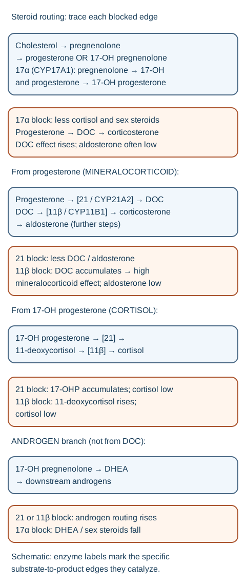
Trace each substrate through its enzyme-labeled edge; blocked steps lower downstream products and accumulate upstream precursors. [4][5][6]
The adrenal cortex is organized by steroid output. The zona glomerulosa supports mineralocorticoid production, the zona fasciculata makes cortisol, and the zona reticularis contributes adrenal androgens. A block can therefore change volume and potassium handling, glucocorticoid availability, and sexual steroid exposure at the same time. ACTH excess also explains adrenal hyperplasia when cortisol feedback is inadequate. [4]
Low mineralocorticoid effect Think salt loss, lower effective circulating volume, high renin, hyponatremia, and hyperkalemia.
High mineralocorticoid effect Think sodium retention, low renin, hypertension, and often hypokalemia.
High androgen effect Think prenatal virilization in a 46,XX fetus or peripheral androgen effects after birth.
Low sex-steroid production Think undervirilization in a 46,XY fetus or absent pubertal development.
Use the steroid-routing figure for a first pass. Cover the enzyme names and ask which direction pressure and androgen production should go if each route is interrupted. The consequence is more useful than the enzyme number: a defect before a mineralocorticoid-active precursor can cause salt loss, while a defect after that precursor can leave mineralocorticoid activity high.
Check a pathway prediction
If a child has cortisol deficiency, hypertension, hypokalemia, low renin, and androgen excess, the pattern requires both a mineralocorticoid-active precursor and an open androgen route. That combination fits 11beta-hydroxylase deficiency, where deoxycorticosterone accumulates.
Transfer the same logic to a new patient before looking at a named diagnosis. First decide whether mineralocorticoid effect is low or high. Then decide whether androgen effect is low or high. Only then use the accumulated precursor to identify the exact block.
21-hydroxylase deficiency creates the classic salt-loss pattern
21-hydroxylase deficiency accounts for about 90 to 95% of congenital adrenal hyperplasia. CYP21A2 impairment reduces cortisol synthesis and, in classic disease, can reduce aldosterone synthesis. 17-hydroxyprogesterone accumulates and adrenal androgen production increases. [1][2][4]
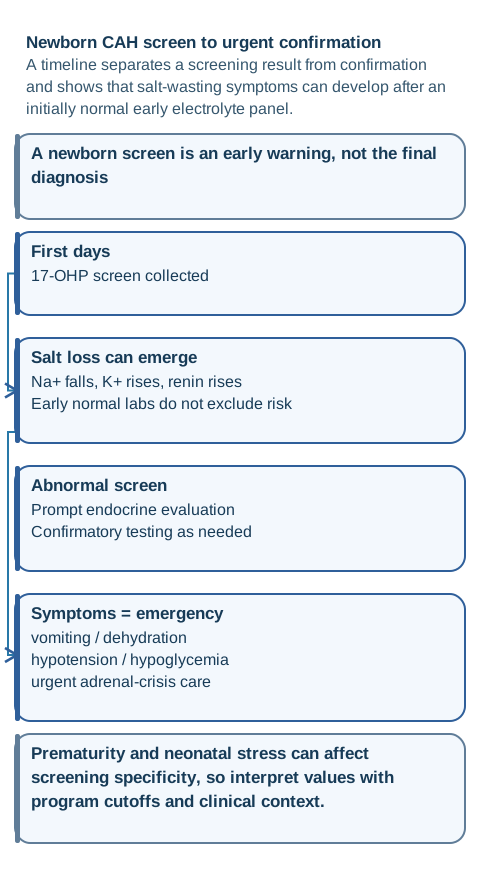
Separate screening from diagnosis while keeping neonatal crisis risk visible. [1][3]
In the salt-wasting form, an infant can develop progressive renal sodium loss, volume depletion, hyponatremia, hyperkalemia, hypoglycemia, and shock during the first days to weeks of life. A 46,XY newborn can have typical male external genitalia, so genital appearance does not exclude danger. A 46,XX fetus with classic disease can have prenatal virilization from androgen excess. The postnatal crisis reflects the infant's own mineralocorticoid deficiency, not a dependable maternal-hormone countdown. [1][4]
Classic simple-virilizing disease retains enough mineralocorticoid synthesis to avoid a salt-wasting crisis but still causes prenatal androgen excess. Nonclassic disease is milder and usually presents later with signs such as premature pubarche, acne, hirsutism, or menstrual irregularity rather than neonatal genital atypia or salt loss. [1]
Newborn screening uses 17-hydroxyprogesterone as the first-tier marker for 21-hydroxylase deficiency. Gestational age and neonatal stress affect results, especially in preterm infants, so a positive screen is not the final diagnosis. Current guidance calls for pediatric endocrine referral and confirmatory evaluation, with cosyntropin testing when needed. [1][3]
Classic disease is treated with glucocorticoid replacement, and patients with classic 21-hydroxylase deficiency benefit from mineralocorticoid replacement with fludrocortisone. Infants may also need supplemental sodium. Blood pressure, electrolytes, and renin help assess mineralocorticoid dosing; growth and adrenal steroid markers help assess glucocorticoid treatment. The goal is clinical control without chronic glucocorticoid excess. [1]
Use the newborn timeline figure and predict the next laboratory change in an untreated salt-wasting infant whose sodium is beginning to fall. Potassium and renin should rise as mineralocorticoid effect falls. A normal electrolyte panel on day 2 therefore does not make a markedly abnormal screen harmless.
Check the newborn transfer
A well-appearing newborn with a strongly positive screen still needs prompt endocrine follow-up. If vomiting, dehydration, hypotension, hypoglycemia, hyponatremia, or hyperkalemia appears, evaluate and treat suspected adrenal crisis urgently rather than waiting for elective confirmation.
Try it here · Checkpoint 1 of 3
Make your prediction before reading the choices. A first attempt is just a starting point.
Case 1
Show answer and explanations for case 1
A. 11beta-hydroxylase deficiency (Why this does not fit)
This defect can cause cortisol deficiency and androgen excess, but deoxycorticosterone produces mineralocorticoid activity. Hypertension and low potassium would fit better than salt-wasting shock.
Reasoning steps for option A
How would retained deoxycorticosterone affect sodium and potassium?
Its mineralocorticoid activity favors sodium retention and potassium excretion, unlike this infant's salt loss.
Does that fit this infant?
No. The infant has volume loss and hyperkalemia.
B. 21-hydroxylase deficiency (Best answer)
Loss of cortisol and aldosterone synthesis with high 17-hydroxyprogesterone explains the salt loss, hyperkalemia, hypoglycemia, and shock. Male-typical external genitalia do not exclude classic disease.
Reasoning steps for option B
Which product is accumulating?
17-hydroxyprogesterone is high before the 21-hydroxylase step.
What mineralocorticoid pattern is present?
Hyponatremia, hyperkalemia, and hypotension indicate deficient mineralocorticoid effect.
Which defect unifies both findings?
Classic 21-hydroxylase deficiency.
C. 17alpha-hydroxylase/17,20-lyase deficiency (Why this does not fit)
This defect reduces sex-steroid production and favors mineralocorticoid-active steroids. It more often produces hypertension and hypokalemia rather than neonatal salt loss.
Reasoning steps for option C
What happens to adrenal androgen production in a 17alpha block?
Androgen output falls because the pathway cannot reach 17-hydroxylated sex-steroid precursors.
What happens to mineralocorticoid effect?
It rises through deoxycorticosterone and corticosterone.
Why does this enzyme defect poorly explain the electrolyte crisis?
Deoxycorticosterone activity tends toward high blood pressure and potassium depletion, not shock with hyperkalemia.
D. 3beta-hydroxysteroid dehydrogenase type 2 deficiency (Why this does not fit)
Classic disease can cause salt wasting, but it typically shows a delta-5 steroid pattern and can undervirilize a 46,XY infant. The high 17-hydroxyprogesterone screen pattern instead favors 21-hydroxylase deficiency.
Reasoning steps for option D
Can 3beta-hydroxysteroid dehydrogenase deficiency cause salt loss?
Yes, both cortisol and aldosterone synthesis can be impaired by this early steroidogenic block.
What additional pattern is expected?
Delta-5 steroids predominate and 46,XY undervirilization can occur.
Which supplied marker points elsewhere?
High 17-hydroxyprogesterone favors 21-hydroxylase deficiency.
Takeaway: Salt loss with hyperkalemia plus high 17-hydroxyprogesterone is the classic 21-hydroxylase pattern.
Hypertension separates two downstream defects from salt-wasting 21-hydroxylase deficiency
When a congenital adrenal hyperplasia pattern includes hypertension and low renin, ask which mineralocorticoid-active precursor is accumulating. This is the central distinction from salt-wasting 21-hydroxylase deficiency, which has too little mineralocorticoid effect.
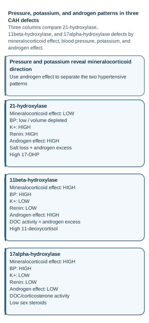
Use pressure and potassium first, then androgen effect, to distinguish the major patterns. [4][5][6]
In 11beta-hydroxylase deficiency, 11-deoxycortisol and deoxycorticosterone rise. Deoxycorticosterone has mineralocorticoid activity, so patients can have sodium retention, low renin, low aldosterone, hypertension, and hypokalemia. The androgen pathway remains open, so androgen excess can produce prenatal virilization in 46,XX patients, rapid growth, advanced bone age, or peripheral precocious puberty. Glucocorticoid replacement lowers ACTH and thereby reduces excess precursor and androgen production. [5]
In 17alpha-hydroxylase/17,20-lyase deficiency, cortisol and sex-steroid synthesis are impaired while mineralocorticoid-active steroids, including deoxycorticosterone and corticosterone, are favored. The result can be hypertension and hypokalemia with reduced androgen and estrogen production. A 46,XY patient can have undervirilized or female-typical external genitalia, and adolescents may present with absent pubertal development or primary amenorrhea. [6][4]
The old numerical memory shortcut can be kept as a final check, not as the mechanism: 11 and 17 are the hypertension-associated defects, while 21 and 11 are the classic androgen-excess defects. The pressure-potassium pair is more robust. Salt-wasting 21-hydroxylase deficiency tends toward low pressure and hyperkalemia; 11beta-hydroxylase and 17alpha-hydroxylase defects tend toward hypertension and hypokalemia.
Use the pressure-potassium figure. Cover the diagnosis labels and classify each column using only pressure, potassium, and androgen effect. Then expose the accumulated steroid. If the patient is hypertensive with androgen excess, deoxycorticosterone plus 11-deoxycortisol supports 11beta-hydroxylase deficiency. If the patient is hypertensive with low sex steroids, the pattern supports 17alpha-hydroxylase/17,20-lyase deficiency.
Check the potassium prediction
Mineralocorticoid receptor activity promotes renal sodium retention and potassium secretion. Excess deoxycorticosterone therefore pushes potassium down, while inadequate mineralocorticoid effect in salt-wasting 21-hydroxylase deficiency allows potassium to rise. Potassium direction follows mineralocorticoid effect, not the cortisol value alone.
Try it here · Checkpoint 2 of 3
Make your prediction before reading the choices. A first attempt is just a starting point.
Case 3
Show answer and explanations for case 3
A. Androgen receptor resistance (Why this does not fit)
A 46,XY patient can have female-typical external genitalia, but testosterone is usually normal or high and hypertension with low renin is not explained.
Reasoning steps for option A
Can this condition alter external sexual development?
Receptor resistance leaves circulating testosterone available, so this exceptionally low testosterone points upstream of the receptor.
What testosterone pattern is expected?
Testosterone production is preserved.
What supplied finding points to adrenal steroidogenesis?
Low-renin hypertension with high deoxycorticosterone.
B. 5alpha-reductase deficiency (Why this does not fit)
Undervirilization can occur, but testosterone synthesis is preserved and the defect does not explain deoxycorticosterone-driven hypertension or cortisol pathway impairment.
Reasoning steps for option B
Which androgen conversion is impaired by 5alpha-reductase deficiency?
Testosterone to dihydrotestosterone.
Is testosterone production absent?
Conversion to DHT is impaired, while the measured testosterone concentration should not be profoundly depleted.
Does it explain low-renin hypertension?
No. 5alpha-reductase deficiency preserves testosterone synthesis and does not cause deoxycorticosterone-mediated low-renin hypertension.
C. 11beta-hydroxylase deficiency with androgen excess (Why this does not fit)
This disorder causes low-renin hypertension, but it also preserves strong androgen production and usually causes androgen excess. Very low testosterone and sparse pubic hair point away from it.
Reasoning steps for option C
Does deoxycorticosterone fit?
Yes. This disorder causes low-renin hypertension, but it also preserves strong androgen production and usually causes androgen excess.
What androgen pattern is expected?
Androgen excess.
Does that match sparse pubic hair and very low testosterone?
The low testosterone and scant pubic hair conflict with the androgen excess predicted by an 11beta block.
D. 17alpha-hydroxylase/17,20-lyase deficiency (Best answer)
This defect reduces androgen and estrogen synthesis while favoring deoxycorticosterone and corticosterone. The combined pubertal failure, 46,XY undervirilization, hypertension, hypokalemia, and low renin fit the disorder.
Reasoning steps for option D
How does 17alpha-hydroxylase/17,20-lyase deficiency affect sex steroids in this adolescent?
They are low.
What happens to mineralocorticoid-active precursors?
They are favored, producing low-renin hypertension and potassium loss.
Which diagnosis joins both directions?
17alpha-hydroxylase/17,20-lyase deficiency.
Takeaway: Low sex steroids plus low-renin hypertension is the signature combination for 17alpha-hydroxylase/17,20-lyase deficiency.
Very early blocks reduce several steroid classes at once
Not every salt-wasting infant with genital atypia has 21-hydroxylase deficiency. A more proximal defect changes a broader part of the steroid profile, so the accumulated precursor becomes essential.
3beta-hydroxysteroid dehydrogenase type 2 deficiency impairs conversion of delta-5 steroids to delta-4 steroids. Classic disease can cause cortisol and aldosterone deficiency with salt wasting. Delta-5 steroids such as 17-hydroxypregnenolone and DHEA are high relative to downstream products. Because DHEA is a weak androgen, 46,XY infants can be undervirilized and 46,XX infants can have mild virilization. [4]
Lipoid congenital adrenal hyperplasia from severe StAR deficiency disrupts cholesterol delivery into mitochondria, reducing production of all major steroid classes. Classic disease can cause glucocorticoid and mineralocorticoid deficiency with salt loss, and severe 46,XY disease can produce female-typical external genitalia because fetal testicular androgen synthesis is markedly impaired. Lipid and cholesterol esters accumulate in steroidogenic cells as the disorder progresses. [4]
Apply the contrast to an infant with salt wasting and a 46,XY karyotype. High 17-hydroxyprogesterone with typical male external genitalia favors 21-hydroxylase deficiency. A delta-5 predominant steroid pattern with undervirilization favors 3beta-hydroxysteroid dehydrogenase deficiency. Broadly low steroid products with severe undervirilization raises concern for an early cholesterol-handling defect.
One receptor distinction from the original lesson also helps keep the feedback loop straight. ACTH signals at a cell-surface melanocortin 2 receptor through Gs and cAMP, whereas cortisol is lipophilic and acts through an intracellular glucocorticoid receptor that regulates gene transcription. The missing cortisol signal therefore increases hypothalamic-pituitary drive; cortisol itself does not require a membrane second-messenger system. [4]
Use a repeatable diagnostic sequence instead of a single laboratory value
Start with the physiology. Is the patient salt wasting or retaining sodium? Then interpret blood pressure and potassium. Next assess whether androgen effect is high, low, or appropriate for age and karyotype. Last, identify the steroid that accumulates before the suspected enzyme defect.
High-yield pattern comparison
Defect
Mineralocorticoid effect
Androgen effect
Useful steroid pattern
Defect21-hydroxylase
Mineralocorticoid effectLow in salt-wasting classic disease
Androgen effectHigh
Useful steroid patternHigh 17-hydroxyprogesterone
Defect11beta-hydroxylase
Mineralocorticoid effectHigh from deoxycorticosterone
Androgen effectHigh
Useful steroid patternHigh 11-deoxycortisol and deoxycorticosterone
Defect17alpha-hydroxylase/17,20-lyase
Mineralocorticoid effectHigh
Androgen effectLow
Useful steroid patternLow sex steroids with mineralocorticoid-active precursors
A newborn-screen result answers a narrower question. It is designed mainly to identify 21-hydroxylase deficiency and can be affected by gestational age, stress, assay method, and timing. A positive result should trigger prompt confirmation, not a claim that every form of congenital adrenal hyperplasia has high 17-hydroxyprogesterone. [1][3]
For later hyperandrogenic symptoms, nonclassic 21-hydroxylase deficiency can overlap with polycystic ovary syndrome. Baseline and stimulated 17-hydroxyprogesterone are interpreted with the assay and local laboratory standards; the guideline supports cosyntropin testing for borderline results rather than one universal number detached from method. [1]
As a transfer exercise, take any vignette and write four arrows on paper: mineralocorticoid effect, potassium, androgen effect, and accumulated steroid. If those arrows contradict the proposed enzyme, reconsider the diagnosis before using a memory shortcut.
Management must balance adrenal replacement with growth, fertility, and emergency planning
Patients who cannot produce an adequate cortisol stress response need a sick-day plan. Current guidance recommends higher glucocorticoid doses for significant febrile illness, gastroenteritis with dehydration, major surgery, or major trauma, plus education for emergency parenteral hydrocortisone and medical identification. Routine mental stress or minor exercise does not require extra dosing. [1]
Both inadequate and excessive treatment have consequences. Persistent ACTH-driven androgen excess can accelerate growth and bone age early, then reduce final adult height through premature epiphyseal maturation. Chronic glucocorticoid excess can also suppress growth and produce treatment toxicity. Monitoring therefore aims for control, not complete biochemical suppression at any cost. [1][2]
In males with classic 21-hydroxylase deficiency, chronic ACTH stimulation can contribute to testicular adrenal rest tumors, which may impair fertility. Guidance supports periodic testicular ultrasonography beginning in adolescence. Reproductive counseling also addresses adrenal androgen and progesterone control, menstrual and ovulatory function, anatomy, and partner testing when pregnancy is planned. [1]
Prenatal dexamethasone intended to reduce virilization of an affected 46,XX fetus is not routine care. The Endocrine Society regards prenatal therapy as experimental and recommends it only within appropriately approved research protocols because treatment must begin before definitive fetal diagnosis and exposes many unaffected fetuses. [1]
For a child with genital differences, elective surgery is not a one-line emergency rule. The guideline notes limited comparative evidence for early, late, or no surgery and supports experienced multidisciplinary counseling so families can make a reasoned decision that considers anatomy, function, uncertainty, and future patient participation. Urgent treatment of adrenal insufficiency remains separate from elective genital surgery decisions. [1]
Apply the same balance to medication follow-up. Hypertension, hypokalemia, and suppressed renin in a patient taking fludrocortisone point toward excessive mineralocorticoid replacement. Rising androgen markers, advanced bone age, or recurrent adrenal symptoms can point toward inadequate glucocorticoid control or adherence problems. The management question is always which physiological output is too low or too high.
Try it here · Checkpoint 3 of 3
Make your prediction before reading the choices. A first attempt is just a starting point.
Case 17
Show answer and explanations for case 17
A. Continue the usual oral dose and reassess after the fever resolves (Why this does not fit)
Significant illness with vomiting increases cortisol requirements and prevents reliable oral dosing. Continuing only the maintenance dose risks adrenal crisis.
Reasoning steps for option A
Does febrile gastroenteritis increase cortisol need?
Yes. Significant illness with vomiting increases cortisol requirements and prevents reliable oral dosing.
Can repeated vomiting make oral dosing unreliable?
Yes. Continuing only the maintenance dose risks adrenal crisis.
What emergency step is needed?
Parenteral stress-dose hydrocortisone and urgent care.
B. Increase fludrocortisone to address the sodium losses from vomiting (Why this does not fit)
Adrenal crisis during severe illness is driven by inadequate glucocorticoid stress response as well as volume and electrolyte problems. Extra mineralocorticoid alone does not replace the needed cortisol effect.
Reasoning steps for option B
Which hormone demand rises during major illness?
Cortisol. Adrenal crisis during severe illness is driven by inadequate glucocorticoid stress response as well as volume and electrolyte problems.
Does fludrocortisone replace cortisol activity?
Fludrocortisone acts at the mineralocorticoid receptor and cannot meet increased glucocorticoid demands.
What medication is urgent when oral glucocorticoid cannot be retained?
With two oral doses lost to emesis, an injectable glucocorticoid provides a reliable route of delivery.
C. Administer emergency parenteral hydrocortisone immediately and seek urgent medical evaluation (Best answer)
Vomiting prevents reliable oral stress dosing, and lethargy raises concern for evolving adrenal crisis. Families should be trained to give emergency glucocorticoid and obtain urgent care.
Reasoning steps for option C
Can the child keep oral medication down?
No. Vomiting prevents reliable oral stress dosing, and lethargy raises concern for evolving adrenal crisis.
What symptom suggests clinical deterioration?
Increasing sleepiness during febrile illness.
What replaces the missing stress response immediately?
The supplied emergency kit bypasses the child’s inability to retain oral medication.
D. Withhold steroids until electrolytes confirm adrenal crisis (Why this does not fit)
Treatment of suspected adrenal crisis should not be delayed for laboratory confirmation when the clinical setting is high risk. Steroid dependence and inability to retain medication are enough to act urgently.
Reasoning steps for option D
Is laboratory confirmation required before emergency treatment?
No. Treatment of suspected adrenal crisis should not be delayed for laboratory confirmation when the clinical setting is high risk.
What known condition raises pretest probability?
Classic adrenal steroid deficiency.
What action prevents delay?
Give emergency hydrocortisone and seek urgent care.
Takeaway: Vomiting that prevents oral stress dosing in classic CAH requires emergency parenteral glucocorticoid and urgent evaluation.
A. 21-hydroxylase deficiency (Why this does not fit)
Androgen excess can occur, but salt-wasting classic disease tends toward low mineralocorticoid effect. High deoxycorticosterone with low renin and hypertension points to a later block.
Reasoning steps for option A
Does 21-hydroxylase deficiency cause androgen excess?
Yes. Androgen excess can occur, but salt-wasting classic disease tends toward low mineralocorticoid effect.
What pressure pattern is expected in salt-wasting disease?
Low mineralocorticoid effect rather than low-renin hypertension.
Which precursor in the stem changes the answer?
High deoxycorticosterone.
B. 17alpha-hydroxylase/17,20-lyase deficiency (Why this does not fit)
Hypertension and hypokalemia fit, but sex-steroid synthesis is reduced. The patient's androgen effects and high 11-deoxycortisol fit 11beta-hydroxylase deficiency.
Reasoning steps for option B
Does the pressure pattern fit?
Yes. Hypertension and hypokalemia fit, but sex-steroid synthesis is reduced.
Does androgen excess fit?
No. Sex steroids are reduced.
Which steroid identifies the competing block?
High 11-deoxycortisol favors 11beta-hydroxylase deficiency.
C. Primary hyperaldosteronism (Why this does not fit)
Low renin hypertension and hypokalemia fit, but the patient has low cortisol, high 11-deoxycortisol, and androgen excess. Those findings indicate an adrenal steroid enzyme defect rather than autonomous aldosterone secretion.
Reasoning steps for option C
Which features resemble primary aldosteronism?
Hypertension, hypokalemia, and low renin.
Which features do not fit?
Low cortisol with high 11-deoxycortisol and androgen excess.
What diagnosis unifies the full steroid pattern?
11beta-hydroxylase deficiency.
D. 11beta-hydroxylase deficiency (Best answer)
The block raises 11-deoxycortisol and deoxycorticosterone. Deoxycorticosterone produces low-renin hypertension and potassium loss while precursor redirection supports androgen excess.
Reasoning steps for option D
Which precursors accumulate?
11-deoxycortisol and deoxycorticosterone.
What does deoxycorticosterone cause?
Sodium retention, low renin, hypertension, and potassium loss.
After an 11beta-hydroxylase block, what happens to androgen production?
It remains high because precursor flux can enter the androgen pathway.
Takeaway: Deoxycorticosterone-driven hypertension plus androgen excess identifies 11beta-hydroxylase deficiency.
A. DOC accumulation already supplies mineralocorticoid activity (Best answer)
Deoxycorticosterone accumulates before the 11beta-hydroxylase block and can cause hypertension and hypokalemia. Therapy aims to suppress ACTH-driven precursor excess, not add more mineralocorticoid effect.
Reasoning steps for option A
Which steroid accumulates in 11beta-hydroxylase deficiency?
Deoxycorticosterone. Deoxycorticosterone accumulates before the 11beta-hydroxylase block and can cause hypertension and hypokalemia.
What physiologic effect does it have?
It activates mineralocorticoid pathways.
Why avoid routine fludrocortisone?
Additional mineralocorticoid effect could worsen hypertension and potassium loss.
B. 11beta-hydroxylase deficiency causes persistent aldosterone excess (Why this does not fit)
Aldosterone can be low because renin is suppressed by deoxycorticosterone. The mineralocorticoid effect comes from the precursor, not necessarily from aldosterone.
Reasoning steps for option B
What suppresses renin?
Volume expansion from deoxycorticosterone.
What follows low renin?
Aldosterone may also be low.
What still causes hypertension?
Deoxycorticosterone itself.
C. Fludrocortisone requires intact 11beta-hydroxylase to activate the mineralocorticoid receptor (Why this does not fit)
Fludrocortisone acts at the receptor and does not require 11beta-hydroxylase for receptor activity. The problem is excess mineralocorticoid effect, not inability of the drug to act.
Reasoning steps for option C
Where does fludrocortisone act?
At the mineralocorticoid receptor.
Does that require 11beta-hydroxylase?
No. Fludrocortisone acts at the receptor and does not require 11beta-hydroxylase for receptor activity.
Why is it usually unnecessary?
The patient already has excess mineralocorticoid activity from deoxycorticosterone.
D. 11beta-hydroxylase deficiency preserves mineralocorticoid physiology despite altered androgen synthesis (Why this does not fit)
The disorder changes both cortisol and deoxycorticosterone pathways. Its characteristic hypertension is evidence that mineralocorticoid physiology is not normal.
Reasoning steps for option D
Is cortisol synthesis affected?
Yes. The disorder changes both cortisol and deoxycorticosterone pathways.
Is mineralocorticoid physiology affected?
Yes, deoxycorticosterone accumulates.
What clinical result follows?
Low-renin hypertension and often hypokalemia.
Takeaway: Deoxycorticosterone can substitute physiologically for mineralocorticoid activity in 11beta-hydroxylase deficiency.
Fludrocortisone excess causes sodium retention, volume expansion, potassium loss, hypertension, and renin suppression. That is the complete pattern in this patient.
Reasoning steps for option A
What does excess mineralocorticoid activity do to potassium?
B. Insufficient hydrocortisone causing ACTH-driven androgen excess (Why this does not fit)
Too little glucocorticoid would tend to raise ACTH and androgen markers. Stable androgen markers plus hypertension and low renin point instead to mineralocorticoid excess.
Reasoning steps for option B
What would low hydrocortisone do to ACTH?
ACTH would rise.
What would happen to androgen markers?
They would tend to rise.
Do the supplied markers show that?
No. Too little glucocorticoid would tend to raise ACTH and androgen markers.
C. Progression of the CYP21A2 defect (Why this does not fit)
A germline enzyme defect does not progressively worsen in this way. A new low-renin hypertensive pattern during replacement therapy should prompt dose assessment.
Reasoning steps for option C
Is the enzyme defect acquired over time?
No. It is genetic.
What changed clinically?
Blood pressure, potassium, and renin.
What is adjustable?
Replacement dosing.
D. New salt-wasting adrenal crisis (Why this does not fit)
Salt-wasting crisis causes volume depletion, low pressure, hyperkalemia, and high renin. The patient's findings are physiologically opposite.
Reasoning steps for option D
What pressure pattern is expected in salt wasting?
Hypotension or volume depletion.
What potassium pattern is expected?
Hyperkalemia.
How does the stem compare?
It shows hypertension, hypokalemia, and suppressed renin.
Takeaway: Suppressed renin with hypertension and hypokalemia during fludrocortisone therapy suggests mineralocorticoid over-replacement.
A. Nonclassic 21-hydroxylase deficiency (Why this does not fit)
Nonclassic disease usually has normal genital development at birth and presents later with hyperandrogenic symptoms. Prenatal virilization indicates a classic phenotype.
Reasoning steps for option A
When does nonclassic 21-hydroxylase deficiency usually become apparent?
After the neonatal period.
What is expected at birth?
External genitalia are typically not virilized.
What supplied feature argues against it?
Prenatal virilization.
B. Classic salt-wasting 21-hydroxylase deficiency (Why this does not fit)
Salt-wasting classic disease would be expected to impair mineralocorticoid physiology. Remaining untreated through 6 weeks with normal sodium, potassium, and renin argues against that phenotype.
Reasoning steps for option B
What does salt-wasting disease do to sodium and potassium?
Sodium falls and potassium can rise.
During untreated salt-wasting 21-hydroxylase deficiency, what happens to renin?
Renin rises with volume loss.
Does this untreated infant show those changes?
No. Salt-wasting classic disease would be expected to impair mineralocorticoid physiology.
C. Classic simple-virilizing form (Best answer)
Prenatal androgen excess establishes classic disease, while sustained normal mineralocorticoid physiology without treatment supports the simple-virilizing phenotype rather than the salt-wasting form.
Reasoning steps for option C
What makes the disease classic?
Prenatal virilization in a 46,XX infant.
What separates simple virilizing from salt wasting?
D. 17alpha-hydroxylase/17,20-lyase deficiency (Why this does not fit)
This disorder reduces androgen production rather than causing prenatal androgen excess. Hypertension and low potassium would also be more typical than the supplied mineralocorticoid profile.
Reasoning steps for option D
With 17alpha-hydroxylase/17,20-lyase deficiency, what happens to androgen production?
It falls.
Would that virilize a 46,XX fetus?
No. This disorder reduces androgen production rather than causing prenatal androgen excess.
What blood-pressure pattern accompanies 17alpha-hydroxylase/17,20-lyase deficiency?
Low-renin hypertension is common.
Takeaway: Prenatal virilization with preserved mineralocorticoid function supports classic simple-virilizing 21-hydroxylase deficiency.
A. Both children have deficient mineralocorticoid effect, but Child B compensates by secreting more aldosterone (Why this does not fit)
Child B's low renin and high deoxycorticosterone indicate mineralocorticoid excess, not compensation by aldosterone. Aldosterone can be suppressed.
Reasoning steps for option A
What does high renin in Child A indicate?
Low effective mineralocorticoid action.
How does the measured renin differ in the hypertensive child?
Renin suppression indicates that the kidney perceives excess effective arterial volume in the hypertensive child.
Which steroid supplies it?
The measured DOC is a source of receptor activation even when renin and aldosterone are suppressed.
B. Child A has deoxycorticosterone excess, while Child B has deficient mineralocorticoid-active steroids (Why this does not fit)
The laboratory directions are reversed. Deoxycorticosterone excess fits hypertension and hypokalemia, not salt-wasting shock.
Reasoning steps for option B
Which child is hypertensive?
Child B. The laboratory directions are reversed.
Which child has high deoxycorticosterone?
Child B has both measured DOC elevation and suppressed renin; Child A instead has a renin response to depletion.
What does that steroid do?
It supplies mineralocorticoid activity. The laboratory directions are reversed.
C. Child A has 17alpha-hydroxylase deficiency; Child B has 21-hydroxylase deficiency (Why this does not fit)
17alpha-hydroxylase deficiency usually favors mineralocorticoid-active steroids, while salt-wasting 21-hydroxylase deficiency lacks adequate mineralocorticoid effect. The proposed pairing is opposite.
Reasoning steps for option C
Which pattern fits 17alpha-hydroxylase deficiency?
Low-renin hypertension with potassium loss.
Which electrolyte and pressure findings identify the salt-wasting child?
The hypotensive hyperkalemic child has a volume-depletion signal, unlike the hypertensive child.
Does the proposed pairing match?
The proposed ordering reverses the observed directions of blood pressure and potassium for both enzyme blocks.
D. Child A has mineralocorticoid deficiency; Child B has DOC-mediated excess (Best answer)
Child A's salt loss and high renin fit classic salt-wasting 21-hydroxylase deficiency. Child B's deoxycorticosterone causes sodium retention, low renin, hypertension, and potassium loss as in 11beta-hydroxylase deficiency.
Reasoning steps for option D
What is the mineralocorticoid state in Child A?
Deficient.
What is the state in Child B?
Excess activity from deoxycorticosterone.
What explains the opposite potassium values?
Different mineralocorticoid receptor activity.
Takeaway: Salt-wasting 21-hydroxylase deficiency and deoxycorticosterone excess produce opposite pressure, renin, and potassium patterns.
A. Gonadotropin-independent peripheral precocious puberty driven by adrenal androgen excess (Best answer)
Adrenal androgen excess can produce pubic hair, penile growth, acne, rapid growth, and advanced bone age while gonadotropins remain suppressed and testes stay prepubertal in size. The family pattern and 17-hydroxyprogesterone support 21-hydroxylase deficiency.
Reasoning steps for option A
Are gonadotropins active?
No. LH and FSH are suppressed.
Where are the androgens coming from?
The adrenal gland.
Which enzyme pattern fits the family and steroid result?
21-hydroxylase deficiency. Adrenal androgen excess can produce pubic hair, penile growth, acne, rapid growth, and advanced bone age while gonadotropins remain suppressed and testes stay prepubertal in size.
B. Central precocious puberty from early hypothalamic GnRH activation (Why this does not fit)
Central puberty should activate LH and FSH and enlarge the testes. The suppressed gonadotropins and prepubertal testicular volume point to a peripheral source of androgen.
Reasoning steps for option B
What drives central puberty?
GnRH activation with gonadotropin secretion.
What happens to testicular volume?
It increases.
Does this patient show that pattern?
No. Central puberty should activate LH and FSH and enlarge the testes.
C. Androgen-secreting testicular tumor (Why this does not fit)
A testicular androgen source would more often be focal or asymmetric and does not explain the sibling's prenatal virilization or high 17-hydroxyprogesterone. The pattern is adrenal and inherited.
Reasoning steps for option C
What local finding might support a testicular tumor?
Asymmetry or a focal lesion.
What family finding argues against a sporadic tumor?
A sibling with prenatal virilization.
Which biochemical marker supports an adrenal enzyme defect?
High 17-hydroxyprogesterone.
D. 17alpha-hydroxylase/17,20-lyase deficiency (Why this does not fit)
This defect reduces androgen synthesis and causes delayed sexual development, often with hypertension. It cannot explain strong androgen effects with high adrenal androgens.
Reasoning steps for option D
What happens to sex steroids in this defect?
They fall. This defect reduces androgen synthesis and causes delayed sexual development, often with hypertension.
What pubertal pattern follows?
Delayed or absent development.
Does that match this boy?
Premature virilization with suppressed gonadotropins contradicts impaired androgen biosynthesis.
Takeaway: Adrenal androgen excess can cause peripheral precocious puberty with small testes and suppressed gonadotropins.
A. Hydrocortisone directly blocks androgen receptors in skin and bone (Why this does not fit)
Hydrocortisone does not treat the disorder by blocking androgen receptors. Its main endocrine effect is replacing cortisol and restoring negative feedback on ACTH.
Reasoning steps for option A
Does hydrocortisone antagonize the androgen receptor?
No. Hydrocortisone does not treat the disorder by blocking androgen receptors.
What hormone is being replaced?
Cortisol activity.
What upstream signal then falls?
The falling plasma ACTH is an upstream endocrine change, not a pharmacologic change in peripheral receptor occupancy.
B. Hydrocortisone restores CYP21A2 enzyme activity (Why this does not fit)
The genetic enzyme defect remains present. Treatment controls the hormonal drive around the defect rather than repairing the enzyme.
Reasoning steps for option B
Is CYP21A2 function restored by glucocorticoid?
No. The genetic enzyme defect remains present.
What remains fixed?
The inherited enzyme defect.
What adjustable signal is reduced?
ACTH stimulation.
C. Hydrocortisone raises gonadotropins, which suppress adrenal androgen synthesis (Why this does not fit)
Gonadotropins regulate the gonads, not the main ACTH-driven adrenal pathway in this disorder. The observed fall in ACTH is the relevant endocrine change.
Reasoning steps for option C
Which pituitary hormone drives adrenal steroidogenesis?
ACTH. Gonadotropins regulate the gonads, not the main ACTH-driven adrenal pathway in this disorder.
Do LH and FSH provide the main adrenal drive?
LH and FSH are gonadal signals; the measured ACTH is the adrenal trophic signal here.
Which observed laboratory change matches treatment?
The paired declines in ACTH and androstenedione track the adrenal feedback loop without invoking gonadotropins.
D. Hydrocortisone restores cortisol feedback, lowering ACTH and adrenal androgen production (Best answer)
Replacing glucocorticoid feedback lowers ACTH. With less adrenal stimulation, excess precursor and androgen production fall even though the enzyme defect remains.
A. Repeat routine testing at the 1-month well visit because normal electrolytes exclude classic disease today (Why this does not fit)
Normal electrolytes on day 2 do not exclude later salt loss. Delaying follow-up can miss a developing classic presentation.
Reasoning steps for option A
Are day-2 electrolytes definitive?
No. Normal electrolytes on day 2 do not exclude later salt loss.
Can salt loss develop after the screening sample?
Electrolytes on day 2 are a snapshot taken before the usual window for some salt-wasting presentations.
What is safer?
Prompt endocrine evaluation and confirmation.
B. Diagnose 21-hydroxylase deficiency from the screen alone and start lifelong therapy without confirmation (Why this does not fit)
A screening result identifies risk, not a final lifelong diagnosis. Confirmatory assessment is required, while any symptomatic infant is treated urgently for suspected adrenal crisis.
Reasoning steps for option B
What is the purpose of screening?
To identify infants needing prompt evaluation.
Is it a final diagnostic test by itself?
No. A screening result identifies risk, not a final lifelong diagnosis.
What separates screening from treatment decisions?
Confirmatory endocrine assessment and the infant's clinical status.
C. Prompt endocrine referral and confirmatory testing, with emergency assessment if symptoms develop (Best answer)
A markedly abnormal screen warrants prompt pediatric endocrine referral and confirmatory evaluation. Normal electrolytes at 2 days do not rule out later salt wasting; vomiting or lethargy warrants immediate emergency assessment.
Reasoning steps for option C
Does this 2-day-old boy have a diagnostic result or a screening signal?
Markedly high dried-blood-spot 17-hydroxyprogesterone is a positive screen, not a definitive diagnosis.
Do normal electrolytes and male-typical genitalia exclude future salt loss?
No. Salt wasting may appear after this initially well interval, and 46,XY appearance can be typical in classic disease.
What follow-up balances diagnostic uncertainty and danger?
Refer promptly to pediatric endocrinology for confirmation; assess urgently if poor feeding, vomiting, or lethargy develops.
D. Ignore the result because male external genitalia are typical (Why this does not fit)
A 46,XY infant with 21-hydroxylase deficiency can have typical male external genitalia. Genital appearance does not protect against salt-wasting adrenal crisis.
Reasoning steps for option D
Can a 46,XY infant look typical externally?
Yes. A 46,XY infant with 21-hydroxylase deficiency can have typical male external genitalia.
Does that exclude classic 21-hydroxylase deficiency?
An apparently normal male examination does not sample the infant’s adrenal mineralocorticoid reserve.
Which result still needs evaluation?
The markedly abnormal 17-hydroxyprogesterone screen.
Takeaway: A positive 17-hydroxyprogesterone screen needs prompt endocrine evaluation and confirmation; normal electrolytes early in life do not rule out evolving salt wasting.
A. Polycystic ovary syndrome (Why this does not fit)
PCOS is common and can produce the same symptoms, but it does not explain a diagnostic adrenal 17-hydroxyprogesterone response to cosyntropin. The adrenal test separates the competing causes in this stem.
Reasoning steps for option A
Do the symptoms alone distinguish the disorders?
Hirsutism and irregular cycles overlap with PCOS; the dynamic adrenal steroid measurement supplies the discriminator.
Which test localizes the problem to adrenal steroidogenesis?
The abnormal cosyntropin-stimulated 17-hydroxyprogesterone response.
What diagnosis does that support?
Nonclassic 21-hydroxylase deficiency.
B. Classic simple-virilizing 21-hydroxylase deficiency (Why this does not fit)
Classic disease usually produces prenatal androgen effects in a 46,XX fetus. Normal genital development at birth with later hyperandrogenic symptoms fits nonclassic disease better.
Reasoning steps for option B
When is classic virilizing disease apparent?
Prenatally or at birth in a 46,XX infant.
What does this patient report?
Normal genital development at birth.
What phenotype fits later symptoms?
Nonclassic disease.
C. Nonclassic 21-hydroxylase deficiency (Best answer)
Mild CYP21A2 impairment can present after childhood with hyperandrogenic symptoms and no salt-wasting history. A diagnostic cosyntropin-stimulated 17-hydroxyprogesterone response supports the diagnosis.
Reasoning steps for option C
What is the timing?
Symptoms began after normal neonatal development.
Is mineralocorticoid failure present?
No. Mild CYP21A2 impairment can present after childhood with hyperandrogenic symptoms and no salt-wasting history.
Which test supports partial 21-hydroxylase impairment?
The abnormal stimulated 17-hydroxyprogesterone response.
D. 11beta-hydroxylase deficiency (Why this does not fit)
Androgen excess can occur, but hypertension, low renin, and high 11-deoxycortisol would be expected in the classic form. Those features are absent.
Reasoning steps for option D
Can this disorder cause androgen excess?
The supplied blood pressure and electrolytes lack a DOC-excess phenotype.
What pressure pattern would strengthen it?
Her normal blood pressure offers no renal evidence for mineralocorticoid-active precursor accumulation.
Which steroid would be more useful?
11-deoxycortisol.
Takeaway: Nonclassic 21-hydroxylase deficiency can mimic other hyperandrogenic disorders and may require assay-appropriate cosyntropin testing.
A. Bilateral germ-cell tumors caused by hydrocortisone exposure (Why this does not fit)
Hydrocortisone does not cause bilateral germ-cell tumors. The bilateral location and chronic ACTH excess in classic CAH point to adrenal rest tissue.
Reasoning steps for option A
Is steroid replacement a known cause of bilateral germ-cell tumors?
No. Hydrocortisone does not cause bilateral germ-cell tumors.
What chronic hormone is high?
ACTH remains elevated when prescribed glucocorticoid has been taken inconsistently.
Which CAH complication fits bilateral testicular lesions?
Bilateral mediastinal lesions in a patient with prolonged ACTH elevation suggest activated adrenal rests.
B. Testicular adrenal rest tumors enlarged by chronic ACTH stimulation (Best answer)
Ectopic adrenal-like tissue in the testes can respond to ACTH and enlarge when adrenal control is poor. Large lesions can impair spermatogenesis and fertility.
Reasoning steps for option B
What stimulates adrenal rest tissue?
ACTH. Ectopic adrenal-like tissue in the testes can respond to ACTH and enlarge when adrenal control is poor.
Why is the history relevant?
Inconsistent glucocorticoid use allows chronic ACTH stimulation.
What clinical consequence can follow?
Mass effect and impaired fertility.
C. Leydig-cell hyperplasia from high LH (Why this does not fit)
LH is not the supplied abnormal driver and central gonadal stimulation is not the typical cause of bilateral adrenal-rest lesions in classic CAH.
Reasoning steps for option C
Which pituitary hormone stimulates Leydig cells?
LH.
Which hormone is actually high in the stem?
The reported endocrine driver is ACTH; no elevated LH is provided to support a gonadal trophic mechanism.
Which tissue responds to that hormone in CAH?
Adrenal rest tissue.
D. Metastatic adrenal cortical carcinoma (Why this does not fit)
Metastatic disease would require a primary malignancy and does not fit the characteristic bilateral testicular distribution with chronic ACTH excess. CAH has a specific benign adrenal-rest complication.
Reasoning steps for option D
Is a primary adrenal cancer described?
No primary adrenal malignant lesion or systemic tumor evidence is supplied.
Are the lesions bilateral in a typical CAH location?
The masses are symmetric and adjacent to the mediastinum testis, a typical adrenal-rest site.
Which benign process fits?
Chronic adrenal trophic stimulation connects the medication history to bilateral testicular lesions.
Takeaway: Chronic ACTH stimulation can enlarge testicular adrenal rest tissue and impair fertility in classic CAH.
A. Persistent androgen excess causing advanced skeletal maturation (Why this does not fit)
Androgen excess usually accelerates growth and bone age initially. This child instead has very low androgen markers, slowing growth, and weight gain.
Reasoning steps for option A
What does uncontrolled androgen excess usually do early?
It accelerates growth and bone maturation.
What are the androgen markers here?
Very low.
Which treatment effect better fits?
Glucocorticoid excess.
B. Mineralocorticoid deficiency causing chronic dehydration (Why this does not fit)
Mineralocorticoid deficiency can impair health and growth, but it would be accompanied by salt-loss physiology rather than the supplied pattern of high glucocorticoid exposure and weight gain.
Reasoning steps for option B
What laboratory pattern would mineralocorticoid deficiency suggest?
High renin, sodium loss, and often hyperkalemia.
Is that described?
A salt-loss explanation would require evidence of depleted volume or rising renin, not simply slowed height gain.
What exposure is explicit?
High hydrocortisone dosing.
C. Glucocorticoid over-treatment suppressing linear growth (Best answer)
Excess glucocorticoid exposure can impair linear growth and promote weight gain. CAH monitoring seeks hormonal control without driving adrenal markers to complete suppression at the cost of treatment toxicity.
Reasoning steps for option C
Which therapy is present at high dose?
Hydrocortisone. Excess glucocorticoid exposure can impair linear growth and promote weight gain.
What two growth findings fit glucocorticoid excess?
Slower height gain and increased weight.
What monitoring principle follows?
Control the disease without excessive glucocorticoid exposure.
D. Expected growth arrest from the CYP21A2 genotype itself (Why this does not fit)
The genotype does not require a new fall in growth velocity after years of treatment. The temporal link to aggressive dosing and low androgen markers points to treatment excess.
Reasoning steps for option D
Is a sudden growth slowdown an inevitable genotype effect?
No. The genotype does not require a new fall in growth velocity after years of treatment.
What changed over the same period?
Glucocorticoid exposure remained high.
Which modifiable factor should be reviewed?
Hydrocortisone dosing.
Takeaway: Over-treatment with glucocorticoid can suppress growth; biochemical normalization should not be pursued at the cost of chronic steroid excess.
A. Begin dexamethasone routinely because carrier pregnancies have an established fetal benefit (Why this does not fit)
Most fetuses in carrier pregnancies will not be affected 46,XX fetuses, so routine early exposure treats many fetuses who cannot benefit. Safety and outcome uncertainties remain.
Reasoning steps for option A
Can fetal status be known before the treatment window closes?
Often not with standard diagnostic timing.
Would every fetus benefit?
Only a subset of pregnancies at genetic risk would carry an affected fetus for whom virilization prevention is relevant.
Is routine use established?
No. Safety and outcome uncertainties remain.
B. Wait until genital ultrasound confirms virilization, then begin routine dexamethasone (Why this does not fit)
The intervention is intended to act before external genital differentiation, so waiting for visible virilization defeats that rationale. More importantly, the guideline still classifies prenatal therapy as experimental.
Reasoning steps for option B
When would the biologic rationale require exposure?
Before genital differentiation.
Can ultrasound confirmation provide that timing?
No. The intervention is intended to act before external genital differentiation, so waiting for visible virilization defeats that rationale.
What is the current status of therapy?
Experimental.
C. Prenatal dexamethasone is experimental and limited to approved research protocols (Best answer)
The Endocrine Society recommends against routine prenatal therapy outside research protocols because treatment must start early and exposes many unaffected fetuses while long-term benefit and risk remain uncertain.
Reasoning steps for option C
Why must therapy start early?
External genital differentiation begins before definitive routine fetal diagnosis.
What follows from that timing?
Many unaffected fetuses would receive drug exposure.
What is the guideline position?
Restrict prenatal therapy to approved research protocols.
D. Placental metabolism prevents dexamethasone from reaching the fetus, eliminating a biological rationale (Why this does not fit)
Dexamethasone can cross the placenta, which is why it has been studied. The concern is not placental inactivation but uncertain risk-benefit and unavoidable exposure of many unaffected fetuses.
Reasoning steps for option D
Does dexamethasone reach the fetus?
Yes. Dexamethasone can cross the placenta, which is why it has been studied.
Why has it been studied?
It can suppress fetal ACTH and androgen production.
Why is it not routine?
Evidence and exposure concerns keep it experimental.
Takeaway: Prenatal dexamethasone for CAH remains experimental and is not routine carrier-pregnancy care.
A. Final adult height will exceed the family target because current growth is rapid (Why this does not fit)
Rapid childhood growth can be misleading when bone maturation is also accelerated. Early epiphyseal maturation can shorten the remaining growth period.
Reasoning steps for option A
What does the tall current stature suggest?
Androgen-driven growth acceleration.
What does bone age 15 at chronological age 12 show?
Skeletal maturation is far ahead.
What happens to remaining growth time?
It decreases.
B. Earlier epiphyseal fusion reduces final adult height (Best answer)
Persistent androgen excess accelerates both linear growth and skeletal maturation. A child can look tall now yet lose future growth potential as the growth plates mature early.
Reasoning steps for option B
What is the immediate androgen effect?
Faster growth.
What is the skeletal effect?
Advanced bone maturation.
What is the long-term consequence?
Less remaining growth and potentially shorter adult height.
C. Height will be unaffected because adrenal androgens do not act on growth plates (Why this does not fit)
Androgens and their estrogenic conversion influence skeletal maturation. The markedly advanced bone age is already evidence that growth plates are affected.
Reasoning steps for option C
Is the skeleton responding to androgen exposure?
Yes. Androgens and their estrogenic conversion influence skeletal maturation.
What finding proves it?
Bone age is three years advanced.
Can adult height therefore change?
Yes. The markedly advanced bone age is already evidence that growth plates are affected.
D. The main height risk is mineralocorticoid replacement, independent of androgen control (Why this does not fit)
Mineralocorticoid dosing affects pressure and electrolytes, but the supplied advanced bone age and androgen excess directly explain the growth risk.
Reasoning steps for option D
Which abnormality is linked to growth plates in the stem?
High adrenal androgens.
What objective marker is abnormal?
Bone age.
Which treatment domain needs better control?
Glucocorticoid control of ACTH and androgen excess.
Takeaway: Uncontrolled adrenal androgen excess can produce early tall stature but reduce final height by accelerating skeletal maturation.
A. The offspring risk remains nonclassic, so no further counseling is needed (Why this does not fit)
CYP21A2 genotype combinations can produce a child with a more severe phenotype than the affected parent. Family history alone does not define carrier status.
Reasoning steps for option A
Can phenotype vary with genotype combination?
Yes. CYP21A2 genotype combinations can produce a child with a more severe phenotype than the affected parent.
Does a healthy partner necessarily lack a pathogenic variant?
An unaffected person can be a CYP21A2 carrier because the condition is recessive.
What is appropriate before pregnancy?
Genetic counseling and partner evaluation.
B. Genetic counseling and partner testing should be offered before pregnancy (Best answer)
Current guidance recommends genetic counseling for affected patients and partners planning pregnancy. Partner testing helps define the risk of an affected child and informs reproductive options.
Reasoning steps for option B
Who should receive counseling?
Affected patients and partners planning pregnancy.
What does partner testing add?
It clarifies reproductive risk.
What can follow from the result?
Informed discussion of reproductive options and fetal risk.
C. The patient's 17-hydroxyprogesterone level is sufficient because biochemical severity predicts fetal genotype (Why this does not fit)
A hormone level describes the patient's current physiology, not the partner's genotype or the exact allele combination in a fetus.
Reasoning steps for option C
What does 17-hydroxyprogesterone measure?
Current steroid physiology.
Does it reveal the partner's carrier status?
A maternal stimulation result contains no information about paternal CYP21A2 alleles.
Which tool addresses inherited risk?
Genetic counseling and testing.
D. Partner testing should be deferred until after pregnancy is established (Why this does not fit)
Preconception testing can provide information before time-sensitive reproductive decisions arise. Waiting is not required for counseling to be useful.
Reasoning steps for option D
When can reproductive risk be assessed?
Before pregnancy.
Why is preconception timing useful?
It allows more time for informed choices.
Is pregnancy required to test the partner?
No. Preconception testing can provide information before time-sensitive reproductive decisions arise.
Takeaway: Preconception genetic counseling and partner evaluation help define reproductive risk in CYP21A2-related disease.
A. 21-hydroxylase deficiency (Why this does not fit)
Classic 21-hydroxylase deficiency can cause salt wasting, but 46,XY external genitalia are usually male-typical because adrenal androgen production is high. The delta-5 predominant steroid profile points upstream.
Reasoning steps for option A
Can 21-hydroxylase deficiency cause salt wasting?
Salt loss alone could fit CYP21A2 disease, but this infant’s weak masculinization demands an additional explanation.
In 21-hydroxylase deficiency, what androgen direction is expected?
Androgen production is high.
Which steroid pattern contradicts it?
High delta-5 steroids with low androstenedione and testosterone.
B. 11beta-hydroxylase deficiency (Why this does not fit)
This disorder causes androgen excess and deoxycorticosterone-mediated hypertension. Salt wasting with undervirilization and the supplied delta-5 pattern do not fit.
Reasoning steps for option B
What blood-pressure pattern accompanies 11beta-hydroxylase deficiency?
Low-renin hypertension. This disorder causes androgen excess and deoxycorticosterone-mediated hypertension.
In 11beta-hydroxylase deficiency, what androgen direction is expected?
An 11beta block diverts steroid precursors toward androgen synthesis instead of sharply reducing testosterone.
How does this infant differ?
Salt wasting and reduced potent androgens.
C. 3beta-hydroxysteroid dehydrogenase type 2 deficiency (Best answer)
A block in delta-5 to delta-4 conversion can reduce cortisol and aldosterone, causing salt wasting, while limiting potent androgen production in a 46,XY infant. High 17-hydroxypregnenolone and DHEA relative to downstream steroids fit the block.
Reasoning steps for option C
Which steroid conversion is impaired by 3beta-HSD2 deficiency?
Delta-5 steroids to delta-4 steroids.
What adrenal consequences follow?
Cortisol and aldosterone deficiency can cause salt wasting.
What explains undervirilization?
Downstream potent androgen production is reduced.
D. 17alpha-hydroxylase/17,20-lyase deficiency (Why this does not fit)
Undervirilization fits, but this disorder favors mineralocorticoid-active steroids and tends toward hypertension and hypokalemia. The infant instead has salt loss and hyperkalemia.
Reasoning steps for option D
Does undervirilization fit?
Poor masculinization is compatible, but the infant also has strong biochemical evidence of aldosterone deficiency.
What mineralocorticoid pattern is expected?
Excess activity with low-renin hypertension.
Does the infant show that?
Dehydration with high renin and hyperkalemia is the inverse of a DOC-driven volume expansion pattern.
Takeaway: Salt wasting plus a delta-5 predominant steroid profile and 46,XY undervirilization supports 3beta-HSD2 deficiency.
StAR enables mitochondrial cholesterol delivery upstream of adrenal and gonadal steroid synthesis. Failure across cortisol, aldosterone, and androgen pathways explains high ACTH and renin, salt wasting, and severe 46,XY undervirilization; adrenal enlargement is supportive but not diagnostic alone.
Reasoning steps for option A
Which steroid classes are low in this 46,XY newborn?
Cortisol, aldosterone, and androgens are all low despite high ACTH and renin.
What upstream step can reduce all three classes?
StAR-dependent movement of cholesterol into mitochondria precedes their synthesis.
How does the combined endocrine phenotype identify the defect?
Loss of aldosterone causes salt wasting, and deficient fetal androgen production explains severe 46,XY undervirilization; enlargement alone is not specific.
B. 21-hydroxylase deficiency (Why this does not fit)
Salt wasting fits, but androgen production should be high and 17-hydroxyprogesterone should accumulate. Broadly low steroid products and severe 46,XY undervirilization indicate a more proximal defect.
Reasoning steps for option B
What androgen direction is expected in 21-hydroxylase deficiency?
High. Salt wasting fits, but androgen production should be high and 17-hydroxyprogesterone should accumulate.
What is present here?
Very low androgens.
What does that imply?
A block before multiple steroid branches.
C. 11beta-hydroxylase deficiency (Why this does not fit)
This disorder retains androgen excess and deoxycorticosterone mineralocorticoid activity. The infant instead lacks both androgen and mineralocorticoid effects.
Reasoning steps for option C
What two effects are typical in 11beta-hydroxylase deficiency?
Androgen excess and mineralocorticoid excess.
Are either present here?
No. This disorder retains androgen excess and deoxycorticosterone mineralocorticoid activity.
Which level of the pathway is more likely?
A very early shared step.
D. Nonclassic 21-hydroxylase deficiency (Why this does not fit)
Nonclassic disease does not cause severe neonatal adrenal insufficiency or broad failure of steroid synthesis. The presentation is far more severe and proximal.
Reasoning steps for option D
When would nonclassic 21-hydroxylase deficiency usually present instead?
Later with hyperandrogenic symptoms.
Does it cause neonatal salt-wasting shock?
Not typically.
Does it lower all steroid classes?
A mild late-presenting 21-hydroxylase variant cannot account for loss of both gonadal androgen production and neonatal aldosterone.
Takeaway: Classic StAR deficiency disrupts the supply of cholesterol for multiple adrenal and gonadal steroid classes, causing salt wasting and severe 46,XY undervirilization.
A. Schedule neonatal genital surgery as the standard response to prenatal virilization (Why this does not fit)
The guideline does not establish one mandatory timing for all patients. Comparative evidence for early, late, or no elective surgery is limited.
Reasoning steps for option A
Is one surgical timing proven best for all patients?
No. The guideline does not establish one mandatory timing for all patients.
Is the adrenal emergency the same as the elective surgical question?
No. Comparative evidence for early, late, or no elective surgery is limited.
What is needed instead?
Experienced multidisciplinary counseling and shared decision-making.
B. Avoid elective surgery because glucocorticoid therapy reverses established anatomy (Why this does not fit)
Glucocorticoid therapy controls future adrenal androgen excess but does not reverse established fetal anatomy. Surgery is not universally required or universally prohibited.
Reasoning steps for option B
Can glucocorticoid reverse prenatal structural anatomy?
No. Glucocorticoid therapy controls future adrenal androgen excess but does not reverse established fetal anatomy.
Does that make surgery mandatory?
No. Surgery is not universally required or universally prohibited.
How should the choice be approached?
Individualized counseling.
C. Multidisciplinary shared decision-making with acknowledgment of limited timing evidence (Best answer)
This matches the guideline's emphasis on experienced multidisciplinary care and informed decision-making. Elective genital surgery timing is distinct from urgent treatment of adrenal insufficiency.
Reasoning steps for option C
What is the quality of comparative evidence on timing?
Limited.
Who should support counseling?
An experienced multidisciplinary team.
What should the decision include?
Function, anatomy, uncertainty, family values, and future patient participation.
D. Base sex assignment on the external genital effects of prenatal androgen exposure (Why this does not fit)
External genital appearance alone does not determine gonadal, internal reproductive, chromosomal, or future identity considerations. A single anatomic feature is not a sufficient basis for such a decision.
Reasoning steps for option D
What does prenatal androgen exposure change?
External genital development.
Does it by itself define all reproductive anatomy or identity?
No. External genital appearance alone does not determine gonadal, internal reproductive, chromosomal, or future identity considerations.
What assessment is required before decisions about sex assignment?
Comprehensive multidisciplinary assessment and counseling.
Takeaway: Elective genital surgery decisions require multidisciplinary shared decision-making because comparative evidence on timing is limited.
A. Renin should rise toward the age-appropriate reference range while potassium improves (Best answer)
Reducing excessive mineralocorticoid effect decreases sodium retention and volume expansion, so renin is no longer strongly suppressed. Potassium wasting should lessen as receptor activity normalizes.
Reasoning steps for option A
Why is renin currently low?
Volume expansion suppresses renin release.
What happens when excess fludrocortisone is reduced?
Volume and mineralocorticoid effect normalize.
What laboratory response follows?
Renin rises toward normal and potassium improves.
B. Renin should fall further because less fludrocortisone directly inhibits renin secretion (Why this does not fit)
Fludrocortisone does not lower renin by direct inhibition. It suppresses renin indirectly through sodium retention and volume expansion, so lowering excess treatment should permit renin to rise.
Reasoning steps for option B
How does fludrocortisone suppress renin?
Indirectly through volume expansion.
What happens to that signal after dose reduction?
It weakens.
After reducing excess fludrocortisone, what happens to renin?
It can rise toward normal.
C. Potassium should fall further because lower mineralocorticoid activity increases renal potassium secretion (Why this does not fit)
Mineralocorticoid activity promotes potassium secretion. Reducing excessive activity should lessen, not increase, renal potassium loss.
Reasoning steps for option C
What does mineralocorticoid receptor activation do to potassium?
It promotes potassium secretion.
What does reducing activation do?
It reduces potassium loss.
Which direction should serum potassium go?
Up toward normal.
D. ACTH should rise sharply because fludrocortisone is the main glucocorticoid replacement (Why this does not fit)
Hydrocortisone supplies the principal glucocorticoid feedback in this regimen. Adjusting fludrocortisone for mineralocorticoid excess does not inherently require a sharp ACTH rise.
Reasoning steps for option D
Which drug provides glucocorticoid feedback?
Hydrocortisone. Hydrocortisone supplies the principal glucocorticoid feedback in this regimen.
Which drug is being adjusted?
Reducing a mineralocorticoid dose affects renal sodium handling rather than directly replacing cortisol feedback.
Which physiology should change most directly?
Renin, blood pressure, and potassium.
Takeaway: Renin suppression can identify excessive mineralocorticoid replacement; correcting the dose should let renin and potassium normalize.
A. Both disorders reduce mineralocorticoid receptor activity, but the adolescent has a potassium-poor diet (Why this does not fit)
The adolescent's low-renin hypertension indicates increased, not reduced, mineralocorticoid effect. Diet alone does not explain the paired pressure and potassium physiology.
Reasoning steps for option A
What does low-renin hypertension imply?
Mineralocorticoid activity is high.
What does high potassium with hypotension imply in the infant?
Hypotension plus hyperkalemia indicates less distal tubular mineralocorticoid stimulation in the infant.
Are the receptor states the same?
No. The adolescent's low-renin hypertension indicates increased, not reduced, mineralocorticoid effect.
B. Mineralocorticoid-active precursors increase renal potassium secretion; mineralocorticoid deficiency reduces it (Best answer)
Deoxycorticosterone and corticosterone can drive mineralocorticoid physiology in 17alpha-hydroxylase deficiency. Salt-wasting 21-hydroxylase deficiency has the opposite state, reducing renal potassium secretion.
Reasoning steps for option B
What happens in the adolescent?
Mineralocorticoid-active precursors are abundant.
What does that do in the distal nephron?
Promotes sodium retention and potassium secretion.
Why does the infant retain potassium despite the same cortisol deficit?
The infant excretes less potassium because aldosterone production is insufficient.
C. The adolescent has excess aldosterone from high renin, while the infant has low aldosterone from low renin (Why this does not fit)
The renin directions are opposite. 17alpha-hydroxylase deficiency usually suppresses renin through precursor-mediated mineralocorticoid activity, while salt-wasting stimulates renin.
Reasoning steps for option C
What is renin expected to do in mineralocorticoid excess?
Fall.
What is renin expected to do in salt wasting?
Rise.
Why is the proposed statement wrong?
It reverses both directions.
D. Cortisol directly controls potassium secretion, so lower cortisol predicts higher potassium (Why this does not fit)
Both disorders can impair cortisol, yet their potassium values are opposite. The discriminator is mineralocorticoid effect, not cortisol concentration alone.
Reasoning steps for option D
Is cortisol low in both disorders?
It can be.
Are potassium values the same?
No. Both disorders can impair cortisol, yet their potassium values are opposite.
Which hormonal axis explains the difference?
Mineralocorticoid receptor activity.
Takeaway: Potassium direction follows mineralocorticoid effect: excess precursor activity lowers potassium, while salt-wasting raises it.
A. The CYP21A2 variant destroys ovarian follicles, making endocrine control ineffective for fertility (Why this does not fit)
CYP21A2 disease primarily alters adrenal steroidogenesis. Reproductive difficulty is multifactorial and can improve when adrenal hormone control and other fertility factors are addressed.
Reasoning steps for option A
Where is the primary enzyme defect clinically expressed?
Adrenal steroidogenesis.
Is irreversible follicle destruction the established mechanism?
Adrenal hormone control plus a standard fertility evaluation.
B. Persistent adrenal androgen and progesterone excess disrupts reproductive physiology (Best answer)
Classic CAH can impair fertility through hormonal and anatomic factors. Abnormal adrenal androgen and progesterone patterns can interfere with ovulatory and reproductive physiology, so individualized endocrine optimization is appropriate.
Reasoning steps for option B
Which adrenal outputs remain abnormal?
Androgens and progesterone.
Can those outputs affect reproductive physiology?
Yes. Classic CAH can impair fertility through hormonal and anatomic factors.
What is the management principle?
Optimize adrenal control while avoiding excessive glucocorticoid exposure and assess other fertility factors.
C. Fludrocortisone acts as a contraceptive and should be stopped before conception (Why this does not fit)
Fludrocortisone is used to replace mineralocorticoid activity and is not a direct contraceptive. Stopping necessary replacement can destabilize blood pressure and electrolytes.
Reasoning steps for option C
Why is fludrocortisone prescribed?
Mineralocorticoid replacement.
Is it a contraceptive mechanism?
No. Fludrocortisone is used to replace mineralocorticoid activity and is not a direct contraceptive.
What risk follows abrupt withdrawal in salt-wasting disease?
Loss of mineralocorticoid control.
D. Infertility is unrelated to congenital adrenal hyperplasia once electrolytes are normal (Why this does not fit)
Electrolyte control does not guarantee normal adrenal androgen, progesterone, ovulatory, or anatomic factors. Reproductive health requires a broader assessment.
Reasoning steps for option D
What does normal electrolyte control show?
Mineralocorticoid replacement is adequate.
What remains abnormal?
Adrenal reproductive steroid patterns and ovulation.
Can these affect fertility?
Yes. Electrolyte control does not guarantee normal adrenal androgen, progesterone, ovulatory, or anatomic factors.
Takeaway: Fertility in classic CAH is multifactorial; abnormal adrenal androgen and progesterone control can contribute even when electrolytes are stable.
A. 17-hydroxyprogesterone screening is valid in term infants, so Infant A's result should be discarded (Why this does not fit)
Preterm infants are still screened, but gestational age and illness can increase false-positive results. The answer is adjusted interpretation and confirmatory follow-up, not discarding the screen.
Reasoning steps for option A
Are preterm infants screened?
Yes. Preterm infants are still screened, but gestational age and illness can increase false-positive results.
What changes test performance?
Gestational age and neonatal stress.
What follow-up is required for a preterm infant with an abnormal screen?
Appropriate cutoff interpretation and confirmation.
B. Prematurity and stress elevate screening 17-OHP, requiring context-aware cutoffs and confirmation (Best answer)
Prematurity and illness increase false-positive first-tier 17-hydroxyprogesterone results, so gestational-age-aware thresholds and second-tier or confirmatory testing matter. A repeated high result in a term newborn remains concerning but does not by itself establish the diagnosis.
Reasoning steps for option B
Why is Infant A at particular risk of a false-positive first-tier result?
Birth at 29 weeks and respiratory distress can increase measured 17-hydroxyprogesterone without proving classic CAH.
Why does Infant B still need prompt follow-up?
A markedly high repeated result in a term baby raises concern, even in the absence of illness.
What distinguishes risk classification from a diagnosis for both infants?
Apply gestational-age-aware cutoffs and indicated second-tier or confirmatory endocrine testing; neither screen alone establishes CAH.
C. Respiratory distress lowers 17-hydroxyprogesterone, making Infant A's result more specific than Infant B's (Why this does not fit)
Stress can increase, not reliably lower, screening 17-hydroxyprogesterone. The preterm stressed infant has a higher false-positive risk.
Reasoning steps for option C
What does neonatal stress do to screening accuracy?
It can increase false-positive values.
Which infant therefore has more potential screening noise?
Infant A.
What still follows?
Confirmatory evaluation rather than dismissal.
D. A repeated high result establishes congenital adrenal hyperplasia, whereas a single high result identifies 21-hydroxylase deficiency (Why this does not fit)
The screen is designed mainly for 21-hydroxylase deficiency, not all CAH forms. Repetition can strengthen concern but does not turn the marker into a universal CAH test.
Reasoning steps for option D
Which CAH form is the screening marker designed mainly to detect?
21-hydroxylase deficiency. The screen is designed mainly for 21-hydroxylase deficiency, not all CAH forms.
Is 17-hydroxyprogesterone high in every CAH form?
The assay targets a precursor of the 21-hydroxylase reaction; other blocked pathways need not accumulate that precursor.
What does repeat abnormality change?
Concern and need for confirmation, not the scope of the marker.
Takeaway: Gestational age and illness alter 17-hydroxyprogesterone screening specificity; neither an initial nor a repeated positive result is diagnostic without appropriate follow-up.
A. Anaphylaxis to hydrocortisone (Why this does not fit)
The emergency kit contains hydrocortisone rather than treating allergy to it. The major risk is inability to mount an adequate endogenous cortisol response during severe stress.
Reasoning steps for option A
What is in the emergency kit?
Hydrocortisone. The emergency kit contains hydrocortisone rather than treating allergy to it.
Would it be the treatment for allergy to itself?
The tag and injectable drug address the child’s known adrenal insufficiency during illness, not a new allergic event.
What known physiologic deficit requires it?
Inadequate cortisol stress response.
B. Adrenal crisis during major illness (Best answer)
Severe illness increases cortisol needs, and vomiting prevents reliable oral hydrocortisone absorption. In classic CAH, emergency injectable glucocorticoid and medical identification help prevent or treat adrenal crisis while urgent care is arranged.
Reasoning steps for option B
What does severe illness change for this child with classic CAH?
Physiologic cortisol requirements rise, but endogenous cortisol production cannot mount the needed response.
Why is injectable hydrocortisone relevant when the child vomits?
Vomiting makes oral stress dosing unreliable, so the taught emergency injection provides glucocorticoid before urgent evaluation.
What emergency does identification and the injection kit address?
Adrenal crisis from inadequate glucocorticoid during major illness, not a routine isolated delayed dose.
C. A hypertensive crisis caused by a single missed fludrocortisone dose (Why this does not fit)
Missing mineralocorticoid tends toward loss of sodium and volume rather than acute hypertension. The emergency identification plan is centered on glucocorticoid stress coverage.
Reasoning steps for option C
What direction does mineralocorticoid deficiency push pressure?
Down, through volume loss.
Does one missed dose explain acute hypertension?
No. Missing mineralocorticoid tends toward loss of sodium and volume rather than acute hypertension.
Which emergency is prioritized?
Cortisol-deficient adrenal crisis.
D. Acute androgen withdrawal after one delayed hydrocortisone dose (Why this does not fit)
Androgen concentrations do not create an immediate life-threatening withdrawal syndrome. The urgent danger during severe stress is glucocorticoid insufficiency and circulatory collapse.
Reasoning steps for option D
Are adrenal androgens required minute-to-minute for cardiovascular stress?
No. Androgen concentrations do not create an immediate life-threatening withdrawal syndrome.
Which steroid effect is essential during major illness?
Glucocorticoid support.
What failure can result?
Adrenal crisis.
Takeaway: Medical identification and emergency hydrocortisone prepare patients for adrenal crisis during major physiologic stress.