Confirm Cushing syndrome, use ACTH to choose the source pathway, and localize pituitary, ectopic, or adrenal disease without shortcut testing.
Cushing syndrome is a pattern of chronic glucocorticoid excess, but the diagnostic task has three separate questions: is cortisol excess truly present, is it ACTH dependent, and where is the source? After this lesson, you should be able to answer those questions without letting a single scan or suppression test substitute for the full sequence.
Recognize a cortisol pattern, not just weight gain
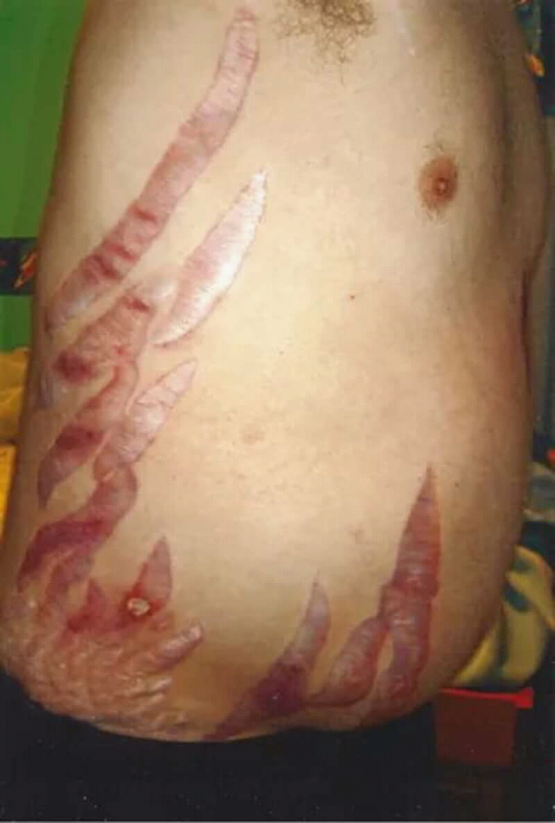
A common misconception is that central weight gain or a round face is enough to identify Cushing syndrome. Those findings overlap with common conditions. The more discriminatory cluster is wide reddish-purple striae, spontaneous bruising, facial plethora, and proximal muscle weakness, especially when the features are progressive or unusual for age. Unexplained osteoporosis, early hypertension, and diabetes can add weight to the pattern. [1]
Real clinical morphology: wide prominent striae can carry more diagnostic weight than central weight gain alone. Image: Masryyy; original source; CC BY-SA 4.0.
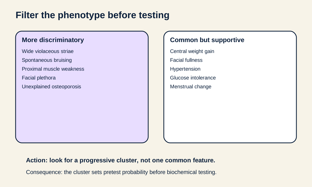
Visual task: filter the phenotype
Use the recognition figure to separate findings that are more discriminatory from findings that are common but supportive. Cover the right column first, classify each finding, then check the grouping. The consequence is practical: a patient with obesity and hypertension alone has a very different pretest probability from a patient with new bruising, two-centimeter violaceous striae, and difficulty rising from a chair. [1]
Use the whole progressive pattern to decide who needs biochemical testing. [1]
The physical findings make sense when you follow cortisol action. Protein catabolism contributes to proximal myopathy; loss of dermal support contributes to thin skin and easy bruising; insulin antagonism and hepatic glucose production worsen glycemia; and chronic glucocorticoid excess weakens bone. Severe cortisol excess can also produce mineralocorticoid effects, so hypertension and hypokalemia can become prominent, particularly in aggressive ACTH-dependent disease. [2]
Cushing syndrome is the clinical state of pathologic glucocorticoid excess from any cause. Cushing disease is one specific endogenous cause: an ACTH-secreting pituitary corticotroph tumor. Adrenal cortisol production, ectopic ACTH production, and exogenous glucocorticoid exposure cause the syndrome but are not Cushing disease. [1][2]
Trace one recognition example
A patient with weight gain, hypertension, and diabetes has common supportive findings. Add wide violaceous striae, spontaneous ecchymoses, and proximal weakness, and the pattern becomes much more discriminatory for pathologic cortisol excess. The clinical pattern tells you whom to test; it does not identify the source by itself. [1]
First prove endogenous hypercortisolism
Before biochemical testing, review every route of glucocorticoid exposure, including oral, injected, inhaled, and topical preparations. Exogenous glucocorticoids can create the phenotype while suppressing endogenous ACTH and adrenal cortisol production, so the medication history belongs before the endocrine algorithm. [1]
Initial testing samples different properties of cortisol physiology. Recommended options include at least two 24-hour urine free cortisol collections, two late-night salivary cortisol measurements, the 1-mg overnight dexamethasone suppression test, or the longer low-dose dexamethasone test. Random serum cortisol and random ACTH are not screening tests, and pituitary or adrenal imaging should not be used to establish the syndrome. [1]
Three useful windows on cortisol
Late-night salivary cortisol asks whether the normal nocturnal nadir is lost. Urine free cortisol estimates integrated free cortisol exposure. The 1-mg dexamethasone test asks whether glucocorticoid feedback can suppress morning cortisol. A post-dexamethasone serum cortisol above 1.8 micrograms/dL is abnormal in commonly used diagnostic frameworks, but assay, medication, sleep schedule, and clinical context still matter. [1][2]
Try this with each patient: name the property each test measures, then cross out any test whose assumptions are broken by sleep schedule, medication, kidney function, pregnancy, or collection quality. The consequence is fewer false labels before source localization. [1][2]
A single abnormal screen is not the end of confirmation. A second recommended test helps resolve false positives and physiologic hypercortisolism. Concordant abnormal results support proceeding to source testing; discordant results deserve clinical review rather than a forced label. In suspected cyclic Cushing syndrome, a normal sample during a quiet phase does not exclude disease, and repeated urine or late-night salivary testing during symptomatic periods can be useful. [1][2]
Apply the test to the person
A permanent night-shift worker does not have a conventional midnight sleep period, so a clock-time late-night salivary sample can be misleading. Select a test that fits the patient's schedule and confounders instead of treating every screening method as interchangeable. [1][2]
Try it here · Checkpoint 1 of 3
Make your prediction before reading the choices. A first attempt is just a starting point.
Case 3
Show answer and explanations for case 3
A. Two 24-hour urine free cortisol collections (Best answer)
Urine free cortisol integrates free cortisol exposure across a full day and does not depend on sampling at conventional bedtime. It is therefore a reasonable initial choice when an 11 PM salivary sample would not represent the patient's biologic sleep nadir.
Reasoning steps for option A
When is this nurse's biologic night?
She sleeps from 9 AM to 5 PM, so her cortisol nadir falls in the daytime rather than near 11 PM.
Which screening test does not depend on when she sleeps?
A 24-hour urine free cortisol sums free cortisol across a whole sleep-wake cycle, and two collections reduce day-to-day variation.
B. Late-night salivary cortisol collected at 11 PM (Why this does not fit)
Late-night salivary cortisol is useful when sampling reflects the physiologic nocturnal nadir. In a permanent night worker, 11 PM is not her sleep period, so the clock-time result can be misleading.
Reasoning steps for option B
Why is late-night salivary cortisol attractive in principle?
It is a simple, validated first-line screen that detects loss of the nighttime cortisol nadir.
What in her schedule undermines an 11 PM sample?
At 11 PM she is awake and mid-shift, so a high value could reflect her active phase rather than disease.
C. Random serum cortisol at the end of a shift (Why this does not fit)
Random serum cortisol is not recommended as a screening test for Cushing syndrome because cortisol varies with time and context. Ending a work shift does not create a validated diagnostic threshold.
Reasoning steps for option C
Why might an end-of-shift serum cortisol seem convenient?
It measures the hormone directly and needs no timed collection.
Why can one serum value drawn at an arbitrary time not answer the question?
Serum cortisol swings with circadian phase and stress, and no validated cutoff exists for a random draw.
D. Pituitary MRI (Why this does not fit)
Pituitary imaging is a source-localization test and should follow biochemical confirmation of endogenous hypercortisolism and ACTH classification. Imaging now could identify an incidental pituitary lesion and create diagnostic confusion.
Reasoning steps for option D
Why might pituitary MRI appeal in this nurse?
Cushing disease is the most common endogenous cause, and her weakness, bruising and hypertension are convincing.
What has not yet been shown, and why does that matter for imaging?
No test has confirmed cortisol excess, and small incidental pituitary lesions are common enough in adults to mislead.
Takeaway: Select a validated screening test that fits the patient's sleep schedule and confounders; clock-time late-night sampling can be misleading in shift workers.
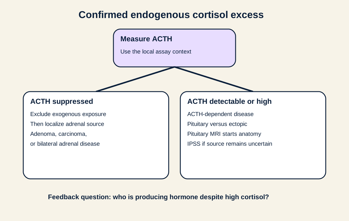
Once endogenous hypercortisolism is established, plasma ACTH separates two physiologic directions. Suppressed ACTH indicates that cortisol production is occurring without pituitary ACTH drive, which points toward an adrenal source after exogenous exposure has been excluded. Detectable or high ACTH indicates ACTH-dependent disease, where the major source question is pituitary versus ectopic secretion. [2]
Start at confirmed cortisol excess, read the ACTH branch, then choose the organ system to localize. [2]
Visual task: read ACTH before naming a tumor
On the ACTH source figure, start at confirmed cortisol excess and follow the ACTH branch. If ACTH is suppressed, ask what is making cortisol without ACTH. If ACTH remains detectable or high, ask what is producing ACTH despite cortisol feedback. This prevents a common error: sending every patient with a Cushing phenotype directly to pituitary MRI. [2]
ACTH values are interpreted with the local assay and the degree of cortisol excess rather than as universal absolute cutoffs. An indeterminate value may need repetition. The important relationship is directional: autonomous adrenal cortisol tends to suppress ACTH, whereas pituitary and ectopic ACTH sources preserve or increase the ACTH signal. [2]
Trace a low-ACTH example
Two different cortisol tests are abnormal and ACTH is repeatedly suppressed. No glucocorticoid exposure is present. The next localization question is adrenal, so dedicated adrenal imaging is appropriate. A pituitary MRI would answer the wrong branch of the physiology. [2][3]
Localize ACTH-dependent disease without overtrusting one test
In confirmed ACTH-dependent Cushing syndrome, pituitary MRI looks for a corticotroph tumor, but a normal MRI does not exclude Cushing disease and a tiny pituitary lesion can be incidental. Current consensus therefore combines imaging, biochemical behavior, and, when needed, inferior petrosal sinus sampling rather than treating the scan as a standalone answer. [2][5]
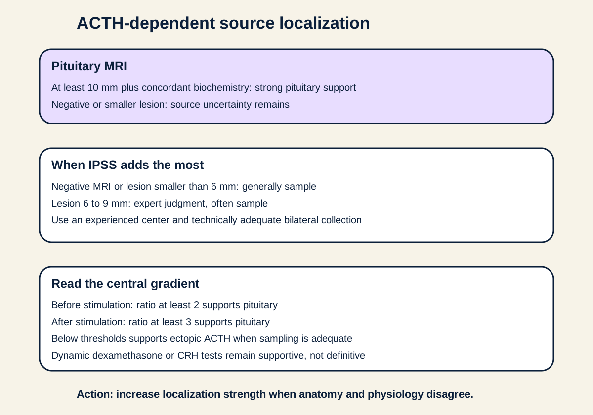
Visual task: choose the localization strength
Use the localization figure as a confidence ladder. A compatible pituitary lesion at least 10 mm with concordant biochemical findings can be sufficient in many expert algorithms. A negative MRI or a lesion smaller than 6 mm generally favors IPSS. Lesions from 6 to 9 mm remain a judgment zone in which many experts also favor IPSS. [2][5]
IPSS compares ACTH in the inferior petrosal sinuses with peripheral blood. A central-to-peripheral ACTH ratio of at least 2 before stimulation or at least 3 after stimulation supports a pituitary source when sampling is technically adequate. Ratios below those thresholds support an ectopic source, but venous sampling quality and false results must be considered. The 2026 procedural guideline emphasizes appropriate bilateral sampling, timed specimens, stimulation protocols, and internal checks rather than interpreting a ratio in isolation. [5]
Increase localization strength when MRI and biochemical evidence do not agree. [2][5][6]
High-dose dexamethasone and CRH-based tests can contribute evidence, but they are supportive, not definitive. Pituitary tumors do not always suppress with high-dose dexamethasone, and some ectopic tumors can show responses that imitate pituitary physiology. A 2026 Korean-Japanese consensus statement also shows that exact diagnostic thresholds and sequencing vary across expert frameworks, so discordant data should be reconciled rather than forced into one shortcut. [2][6]
A rapid, severe presentation with marked hypokalemia and very high ACTH can raise suspicion for ectopic ACTH production. After biochemical ACTH-dependent disease is established, imaging outside the pituitary is directed by the localization evidence and clinical context. Small-cell lung carcinoma is a classic source, but bronchial and other neuroendocrine tumors are also important. [2]
Check the high-dose dexamethasone shortcut
A 60 percent cortisol suppression with high-dose dexamethasone does not by itself prove a pituitary source. If MRI is negative or only a tiny lesion is present, IPSS may still be the stronger discriminator. [2][5]
Try it here · Checkpoint 2 of 3
Make your prediction before reading the choices. A first attempt is just a starting point.
Case 16
Show answer and explanations for case 16
A. The ratios support a pituitary source because any central value above 1 is abnormal (Why this does not fit)
A central-to-peripheral ratio above 1 simply means the sampled central concentration is somewhat higher; it does not meet validated diagnostic thresholds. The standard thresholds are at least 2 before stimulation or at least 3 after stimulation for pituitary support.
Reasoning steps for option A
Why might a 1.8 stimulated ratio seem to favor the pituitary?
Central ACTH is higher than peripheral ACTH, and stimulation raised the ratio.
How large must the gradient be to count?
Pituitary disease needs a basal ratio of 2 or more, or 3 or more after stimulation, and 1.3 and 1.8 miss both.
B. The ratios support an ectopic ACTH source, so extrapituitary localization should proceed (Best answer)
With technically adequate sampling, ratios below 2 before stimulation and below 3 after stimulation do not show a central ACTH gradient and support ectopic ACTH secretion. The next localization work should therefore focus outside the pituitary while retaining awareness of rare false-negative sampling.
Reasoning steps for option B
Was the sampling reliable enough to trust a negative result?
The study was bilateral and technically adequate, so a missed gradient is unlikely to reflect catheter position.
Do the ratios reach the pituitary cutoffs?
No; 1.3 is under the basal cutoff of 2, and 1.8 is under the stimulated cutoff of 3.
Where does the search go next?
Imaging for an ectopic ACTH tumor, starting with the chest, where most such tumors arise.
C. The study is indeterminate because only a ratio of 5 or greater is diagnostic (Why this does not fit)
A ratio of 5 is not required by current consensus. The commonly used thresholds are at least 2 before stimulation and at least 3 after stimulation when sampling is adequate.
Reasoning steps for option C
Why might a higher bar seem safer?
Demanding a larger gradient would reduce false-positive pituitary calls.
Why is a cutoff of 5 wrong here?
Standard criteria use 2 at baseline and 3 after stimulation, so this study is interpretable and negative.
D. The study proves an adrenal cortisol source because no central ACTH gradient is present (Why this does not fit)
The patient already has confirmed ACTH-dependent Cushing syndrome, so an autonomous adrenal cortisol source has been excluded physiologically. IPSS distinguishes pituitary from ectopic ACTH, not ACTH-dependent from adrenal disease.
Reasoning steps for option D
Why might an absent gradient suggest the adrenal?
If ACTH is not coming from the pituitary, the cortisol source might seem to lie outside the ACTH pathway.
What had already excluded an adrenal source?
The disease was ACTH dependent before IPSS, so the adrenals are responding rather than acting on their own.
Takeaway: With adequate IPSS, a central-to-peripheral ACTH ratio below 2 before and below 3 after stimulation supports an ectopic ACTH source.
When ACTH is suppressed, read the adrenal finding in context
Suppressed ACTH after confirmed endogenous cortisol excess directs attention to the adrenal glands. A unilateral cortisol-producing adenoma, adrenocortical carcinoma, or bilateral adrenal disease can create ACTH-independent Cushing syndrome. The imaging question is not just size; it is whether the lesion has a benign or suspicious phenotype and whether cortisol secretion is overt or mild. [3]
The 2023 European Society of Endocrinology guideline classifies an incidental adrenal mass as radiographically benign when it is homogeneous and measures 10 HU or less on unenhanced CT, without requiring follow-up imaging solely because of size. A lesion larger than 4 cm that is inhomogeneous or measures more than 20 HU carries more malignancy concern and generally merits expert multidisciplinary evaluation. [3]
For a patient with an incidental adrenal mass but no overt Cushing features, post-dexamethasone cortisol above 1.8 micrograms/dL is termed mild autonomous cortisol secretion in the 2023 guideline. ACTH independence should be established, and hypertension and type 2 diabetes should be assessed because management is individualized around comorbidity, imaging, age, health, and patient preference. [3]
Try this before naming an adrenal lesion: classify unenhanced HU, homogeneity, size, and hormone function in that order. The same diameter means something different in a homogeneous 6-HU lesion than in a heterogeneous 28-HU lesion. [3]
Compare two adrenal masses
A 3 cm homogeneous 6-HU lesion with mild autonomous cortisol secretion is not interpreted like a 6 cm heterogeneous 28-HU lesion with necrosis and overt hormone excess. The first has benign imaging features; the second has multiple features that increase concern for malignancy and needs expert evaluation. [3]
Try it here · Checkpoint 3 of 3
Make your prediction before reading the choices. A first attempt is just a starting point.
Case 10
Show answer and explanations for case 10
A. Nonfunctioning adrenal adenoma (Why this does not fit)
The CT phenotype is benign, but post-dexamethasone cortisol remains above the 1.8 micrograms/dL threshold and ACTH is low. Those findings support autonomous cortisol secretion rather than a completely nonfunctioning lesion.
Reasoning steps for option A
Why might this look like a nonfunctioning adenoma?
It was found by chance, is 8 HU and homogeneous, and he has no Cushing features.
Which result shows the adenoma is making cortisol?
Cortisol stays at 2.8 micrograms/dL after dexamethasone while ACTH is low, a sign of autonomous output.
B. Overt Cushing syndrome from an adrenal adenoma (Why this does not fit)
The biochemical cortisol autonomy is real, but the stem specifically lacks the overt discriminatory clinical findings of Cushing syndrome. Current adrenal-incidentaloma terminology separates this state from overt Cushing syndrome.
Reasoning steps for option B
Why might overt Cushing syndrome seem to fit?
The dexamethasone test is abnormal, ACTH is low, and he has hypertension and diabetes.
What separates him from overt disease?
He lacks striae, myopathy, bruising and plethora, and hypertension and diabetes are too common to define the overt syndrome.
C. Mild autonomous cortisol secretion (Best answer)
In a patient with an adrenal incidentaloma and no overt Cushing signs, post-dexamethasone cortisol above 1.8 micrograms/dL is classified as mild autonomous cortisol secretion in the 2023 European guideline. Low ACTH supports ACTH independence, and hypertension and diabetes are relevant comorbidities for management.
Reasoning steps for option C
What does the CT show about the mass itself?
A 2.4-cm, 8-HU homogeneous lesion consistent with a benign adenoma found incidentally.
Is the adenoma secreting cortisol?
Yes; incomplete suppression to 2.8 micrograms/dL with low ACTH shows autonomous cortisol output.
Why is his state not called overt Cushing syndrome?
None of the catabolic signs are present, so his hypertension and diabetes are treated as comorbidities possibly linked to the cortisol excess.
D. Pheochromocytoma (Why this does not fit)
Pheochromocytoma is a catecholamine-producing adrenal medullary tumor and is not diagnosed by failed dexamethasone suppression with low ACTH. The supplied endocrine abnormality is autonomous cortisol secretion.
Reasoning steps for option D
Why might pheochromocytoma be considered?
Every adrenal incidentaloma raises that question, and catecholamine excess can cause hypertension and diabetes.
What in this workup points to a different hormone?
The abnormal results involve cortisol and ACTH, and a lipid-rich 8-HU mass makes pheochromocytoma very unlikely.
Takeaway: An adrenal incidentaloma with no overt Cushing signs, post-dexamethasone cortisol above 1.8 micrograms/dL, and ACTH independence fits mild autonomous cortisol secretion.
Cover the procedure names and point first to the hormone source: pituitary ACTH tumor, unilateral adrenal cortisol tumor, or ectopic ACTH tumor. Then match the definitive procedure to that source. The visible consequence is that removing a causal source lowers cortisol, while the previously suppressed normal adrenal axis may need time and replacement support to recover. [4]
Definitive treatment is source directed. For Cushing disease, first-line therapy is selective transsphenoidal removal of the pituitary tumor by an experienced pituitary surgeon. A benign unilateral cortisol-producing adrenal lesion is generally treated with unilateral adrenalectomy, and an identified ectopic ACTH-producing tumor is resected when feasible. [4]
Source control does not erase the systemic effects immediately. Hypertension, diabetes, osteoporosis and fracture risk, infection susceptibility, psychiatric symptoms, and thromboembolic risk require active assessment during treatment and recovery. After successful therapy, temporary adrenal insufficiency can occur because the normal hypothalamic-pituitary-adrenal axis was chronically suppressed, so glucocorticoid replacement and recovery monitoring may be needed. [4]
The main diagnostic discipline is to keep the questions separate: confirm cortisol excess, classify ACTH dependence, then localize with the method appropriate to that branch. A correct source diagnosis makes source-directed therapy possible while avoiding unnecessary pituitary, adrenal, or whole-body procedures. [1][2][3][4][5][6]
Clinical practice
Case 1
Show answer and explanations for case 1
A. Metabolic syndrome (Why this does not fit)
Metabolic syndrome can explain central adiposity, hypertension, and hyperglycemia, but it does not explain the wide violaceous striae, spontaneous bruising, and proximal myopathy. Those catabolic findings make pathologic glucocorticoid excess more likely.
Reasoning steps for option A
Why might metabolic syndrome look like a good fit for this woman?
Central weight gain, a blood pressure of 152/94 mm Hg and a fasting glucose of 158 mg/dL are all core metabolic syndrome features.
Which of her findings does insulin resistance not produce?
Two-centimeter purple striae, bruises that appear without injury and trouble rising from a chair reflect tissue breakdown, which metabolic syndrome lacks.
B. Polycystic ovary syndrome (Why this does not fit)
PCOS can cause weight gain, insulin resistance, and androgen-related symptoms, but no androgen findings are supplied. The bruising, wide striae, and proximal weakness instead support cortisol excess.
Reasoning steps for option B
What makes PCOS tempting in a 34-year-old with weight gain and high glucose?
PCOS is common in young women and often brings central adiposity and insulin resistance with it.
What does the vignette lack for PCOS, and what does it show instead?
It gives no hirsutism, acne or irregular cycles, yet it does give dermal fragility and proximal muscle weakness.
C. Cushing syndrome (Best answer)
The combination of progressive central adiposity with wide violaceous striae, easy bruising, proximal muscle weakness, hypertension, and hyperglycemia is a classic discriminatory cluster for Cushing syndrome. The next diagnostic phase is biochemical confirmation, not immediate source imaging.
Reasoning steps for option C
Which of her features are too common to carry much diagnostic weight?
Weight gain, hypertension and hyperglycemia are frequent in the general population and only modestly raise the likelihood of cortisol excess.
Which findings raise that likelihood sharply?
Wide purple striae, bruising without trauma and proximal myopathy are the discriminating catabolic signs.
What must happen before anyone looks for a tumor?
A glucocorticoid exposure history and a validated cortisol screening test.
D. Primary hypothyroidism (Why this does not fit)
Hypothyroidism can cause weight gain and fatigue, but it does not account for wide violaceous striae, spontaneous bruising, proximal catabolic weakness, and hyperglycemia. The total pattern is more consistent with glucocorticoid excess.
Reasoning steps for option D
Why might hypothyroidism come to mind first?
Gradual weight gain and weakness are common reasons to check thyroid function, and hypothyroidism can cause a proximal myopathy.
What about her skin argues against a thyroid cause?
Myxedema thickens and dries the skin; it does not thin it into purple striae or leave spontaneous bruises.
Takeaway: Wide violaceous striae, easy bruising, and proximal weakness are more discriminatory for Cushing syndrome than obesity, hypertension, or diabetes alone.
Ectopic ACTH would usually preserve or increase ACTH despite high cortisol. The documented long-term prednisone exposure and low ACTH instead indicate exogenous glucocorticoid effect.
Reasoning steps for option A
Why might ectopic ACTH be considered in an older man with severe features?
Ectopic ACTH syndrome is more common in older adults and can cause marked weakness and bone loss.
Which laboratory result excludes an ACTH-producing tumor in his case?
His plasma ACTH is low, whereas a tumor making ACTH keeps the level normal or high.
B. Cortisol-producing adrenal adenoma (Why this does not fit)
An adrenal cortisol source can suppress ACTH, but the medication history supplies a direct and more parsimonious cause of glucocorticoid excess. Exogenous exposure should be identified before launching an endogenous source workup.
Reasoning steps for option B
Why does a cortisol-secreting adrenal adenoma match his hormone pattern?
An autonomous adenoma also shuts off pituitary ACTH, so the low ACTH alone looks identical.
What makes an adenoma an unnecessary explanation here?
Prednisone 15 mg daily for 18 months is well above physiologic replacement and accounts for both the features and the low ACTH.
C. Exogenous prednisone effect (Best answer)
Long-term prednisone can cause the full Cushing phenotype and suppress the hypothalamic-pituitary-adrenal axis, producing low ACTH. This is iatrogenic Cushing syndrome, not Cushing disease.
Reasoning steps for option C
How does his prednisone dose compare with normal cortisol output?
Fifteen milligrams of prednisone is roughly three times physiologic glucocorticoid replacement, and he has taken it for 18 months.
How does that dose produce both the phenotype and the ACTH result?
Tissues are exposed to sustained glucocorticoid excess, and feedback on the hypothalamus and pituitary shuts down ACTH release.
What does the suppressed axis mean if prednisone is reduced?
The dose must be tapered rather than stopped abruptly, because his own adrenal output will take time to recover.
D. Pituitary corticotroph tumor (Why this does not fit)
A pituitary corticotroph tumor causes Cushing disease by secreting ACTH, so ACTH would not be suppressed in the usual presentation. The long-term prednisone exposure explains the phenotype and low ACTH without a pituitary tumor.
Reasoning steps for option D
Why might a pituitary tumor come to mind?
Moon facies and a dorsocervical fat pad are the textbook picture of Cushing disease, the most common endogenous cause.
Which result is incompatible with a corticotroph tumor?
A corticotroph tumor keeps ACTH normal or high, but his ACTH is low.
Takeaway: A complete glucocorticoid exposure history comes before biochemical source localization because exogenous steroids can cause the phenotype and suppress ACTH.
A. Pituitary MRI should be obtained because the dexamethasone test is positive (Why this does not fit)
A single abnormal suppression test does not establish endogenous Cushing syndrome, especially when two other screens are normal and a known test confounder is present. Source imaging before resolving discordant biochemistry risks finding an incidental lesion.
Reasoning steps for option A
Why might imaging seem justified in this woman?
Her bruising and proximal weakness are specific signs, and her post-dexamethasone cortisol of 2.6 micrograms/dL exceeds 1.8.
What should halt the move to imaging?
Two normal late-night salivary cortisols disagree with the suppression test, so cortisol excess is not yet established.
B. Estrogen-related false-positive DST should be reassessed (Best answer)
Estrogen raises cortisol-binding globulin and can increase total serum cortisol, producing false-positive dexamethasone suppression results. Discordant testing should be reconciled with the medication context and an appropriate repeat or alternative validated test rather than treated as confirmed disease.
Reasoning steps for option B
Which of her medications distorts the dexamethasone test?
Ethinyl estradiol raises cortisol-binding globulin, which raises total serum cortisol without raising the free fraction.
Which of her tests is unaffected by that binding protein?
Salivary cortisol reflects free cortisol, and both of her samples are normal.
How can the conflict be settled?
Measure free cortisol, for example by urine free cortisol, or repeat the dexamethasone test about six weeks after stopping the estrogen.
C. Plasma ACTH should be measured immediately to distinguish pituitary from adrenal disease (Why this does not fit)
ACTH is used for source classification after endogenous hypercortisolism has been established. Here the screening tests are discordant and a medication can explain the abnormal total cortisol result.
Reasoning steps for option C
Why might ACTH seem the logical next test?
ACTH is how endogenous cortisol excess is sorted into pituitary, ectopic or adrenal branches.
Why would an ACTH result be hard to use right now?
Sorting the source only has meaning once cortisol excess is proven, and her free-cortisol tests are normal.
D. Cushing syndrome is confirmed because post-dexamethasone cortisol exceeds 1.8 micrograms/dL (Why this does not fit)
The 1.8 micrograms/dL threshold is useful for sensitivity, but a result above it is not interpreted without assay and clinical context. The estrogen exposure and normal salivary testing make automatic confirmation inappropriate.
Reasoning steps for option D
Why does confirmation look justified at first glance?
Her cortisol of 2.6 micrograms/dL after 1 mg dexamethasone is above the standard 1.8 cutoff.
Why is crossing the cutoff not decisive for her?
The 1.8 cutoff trades specificity for sensitivity, and her estrogen raises total cortisol while her free-cortisol measurements stay normal.
Takeaway: Discordant cortisol tests should be reconciled with assay and medication confounders before source testing.
A. Repeat cortisol testing during symptomatic periods (Best answer)
Cyclic Cushing syndrome can produce genuinely normal cortisol physiology during an inactive phase. Repeated urine or late-night salivary measurements timed to symptomatic periods increase the chance of documenting an active phase.
Reasoning steps for option A
What does her symptom pattern suggest?
Spells of plethora, bruising and worsening diabetes alternating with well weeks suggest cortisol excess that switches on and off.
Why do her normal results fail to exclude it?
They were all obtained in a well week, when the cortisol excess may genuinely be absent.
How can an active phase be caught?
Give her kits to collect bedtime saliva or a 24-hour urine as soon as the symptoms return, and repeat over several spells.
B. Order pituitary MRI now because cyclic disease is usually pituitary (Why this does not fit)
A pituitary lesion cannot establish the syndrome and may be incidental. The immediate problem is to document hypercortisolism during an active phase before localizing the source.
Reasoning steps for option B
Why might pituitary MRI appeal now?
Many cyclic cases are pituitary in origin, and imaging does not depend on catching an episode.
What would a pituitary lesion on MRI fail to prove?
It would not show that she has cortisol excess, which is still undocumented.
C. Exclude Cushing syndrome because two urine collections are normal (Why this does not fit)
Concordant normal tests usually argue against Cushing syndrome, but suspected cyclic disease is a specific exception. The timing of the normal collections during an asymptomatic phase matters.
Reasoning steps for option C
Why does exclusion seem reasonable?
Two normal urine free cortisols plus normal dexamethasone suppression usually make Cushing syndrome very unlikely.
What about the timing weakens that reassurance?
Every test was done in an asymptomatic week, when a cyclic disorder can be quiescent.
D. Use a random serum ACTH during the next symptomatic episode as the screening test (Why this does not fit)
ACTH classifies the source after hypercortisolism is established; it is not a recommended screening test for the syndrome. The active episode should first be captured with a validated cortisol test.
Reasoning steps for option D
Why might an ACTH drawn during an episode appeal?
It would be timed correctly, and ACTH is raised in pituitary and ectopic disease.
What can ACTH not do as a first test?
A normal-range ACTH neither proves nor excludes cortisol excess; it only classifies the source after cortisol excess is shown.
Takeaway: Suspected cyclic Cushing syndrome requires repeated validated cortisol testing across symptomatic and asymptomatic phases rather than one-time exclusion.
Concordant abnormal cortisol tests support endogenous hypercortisolism. Plasma ACTH now separates ACTH-independent adrenal cortisol production from ACTH-dependent pituitary or ectopic disease.
Reasoning steps for option A
How strong is the evidence for endogenous cortisol excess?
Two high late-night salivary values and a post-dexamethasone cortisol of 6.2 micrograms/dL agree, and she has no steroid exposure.
Which single result decides what to image next?
Plasma ACTH: a suppressed value points to the adrenal, while a normal or high value points to the pituitary or an ectopic tumor.
B. High-dose dexamethasone suppression (Why this does not fit)
High-dose dexamethasone is not the primary branch point after confirmation and has imperfect localization accuracy. ACTH should first establish whether an ACTH-dependent localization problem even exists.
Reasoning steps for option B
Why might high-dose dexamethasone seem the natural next step?
It has long been taught as the test that separates pituitary from other sources.
What must be known before that test has any meaning?
Whether ACTH is measurable; if ACTH is suppressed, the pituitary-versus-ectopic question never arises.
C. Pituitary MRI (Why this does not fit)
Pituitary MRI is appropriate only after the physiology points toward ACTH-dependent disease. Imaging now could identify an incidental lesion in a patient whose true source is adrenal.
Reasoning steps for option C
Why might pituitary MRI tempt the clinician in this young woman?
Her profile fits Cushing disease, the most common endogenous cause in women of her age.
What could make a pituitary finding misleading at this stage?
If her ACTH proves suppressed, the source is adrenal, and any pituitary spot would be incidental.
D. Adrenal CT (Why this does not fit)
Adrenal imaging is the appropriate localization branch when ACTH is suppressed, but ACTH has not yet been measured. Imaging both adrenal glands before the branch is known can create incidental findings.
Reasoning steps for option D
Why might adrenal CT be chosen?
The adrenal glands make the cortisol, and CT readily shows adrenal tumors.
What result would have to come first?
A suppressed ACTH; without it, adrenal enlargement could simply be ACTH-driven hyperplasia.
Takeaway: After endogenous hypercortisolism is established, plasma ACTH is the source pivot.
Suppressed ACTH indicates ACTH-independent cortisol production once exogenous exposure is excluded. Dedicated adrenal imaging is therefore the relevant localization study.
Reasoning steps for option A
Which cause of low ACTH has already been excluded?
He takes no glucocorticoids, so the suppression is not a drug effect.
What does endogenous cortisol excess with suppressed ACTH mean?
Cortisol is being made by adrenal tissue that no longer needs ACTH.
Which study then finds and characterizes the lesion?
Adrenal CT, which also measures the lesion's density to judge whether it looks benign.
B. Pituitary MRI (Why this does not fit)
A pituitary ACTH-secreting tumor should not produce persistently suppressed ACTH. Pituitary imaging would pursue a branch contradicted by the hormone data.
Reasoning steps for option B
Why might pituitary MRI be ordered by reflex?
Cushing disease accounts for most endogenous Cushing syndrome.
Which finding cannot be squared with a pituitary source?
Repeatedly suppressed ACTH, whereas a corticotroph tumor keeps ACTH measurable.
C. Inferior petrosal sinus sampling (Why this does not fit)
IPSS is designed to distinguish pituitary from ectopic sources in confirmed ACTH-dependent Cushing syndrome. This patient has ACTH-independent physiology.
Reasoning steps for option C
Why might IPSS appeal?
It is the most accurate localization test in ACTH-dependent Cushing syndrome.
What does IPSS need that this man lacks?
A measurable ACTH to compare between pituitary venous blood and peripheral blood; his ACTH is suppressed.
D. Chest CT for ectopic ACTH (Why this does not fit)
Ectopic ACTH secretion should keep ACTH detectable or high. Suppressed ACTH instead directs localization toward autonomous adrenal cortisol production.
Reasoning steps for option D
Why might chest CT come up in a 50-year-old man?
Ectopic ACTH from lung tumors is more common in older men.
Which lab finding points away from the chest?
An ectopic tumor would push ACTH up, and his is suppressed on repeat testing.
Takeaway: Confirmed hypercortisolism plus suppressed ACTH and no exogenous glucocorticoid exposure directs localization to the adrenal glands.
High ACTH indicates that the adrenal glands are being stimulated rather than acting autonomously. Adrenal enlargement can be secondary to ACTH and would not localize the ACTH source.
Reasoning steps for option A
Why might adrenal CT seem reasonable?
The adrenal glands are where the excess cortisol is being made.
What does an ACTH of 68 pg/mL say about her adrenals?
They are being driven from upstream, so imaging them would show at most bilateral hyperplasia, not the cause.
B. Whole-body PET as the first localization test (Why this does not fit)
Ectopic imaging may be appropriate when localization evidence points away from the pituitary, but it is not the default first anatomic study for every ACTH-dependent case. Pituitary MRI is the standard initial pituitary assessment.
Reasoning steps for option B
Why might a whole-body scan appeal?
It could find an ectopic ACTH tumor anywhere in one study.
What about this patient makes an ectopic search a poor first move?
A young woman with no known malignancy most likely has a pituitary source, so the pituitary is imaged first.
C. Pituitary MRI (Best answer)
Confirmed hypercortisolism with nonsuppressed ACTH establishes ACTH-dependent Cushing syndrome. Pituitary MRI is the initial anatomic study for Cushing disease, with further dynamic testing or IPSS determined by the imaging and biochemical context.
Reasoning steps for option C
What does an ACTH of 68 pg/mL with confirmed cortisol excess mean?
ACTH is inappropriately high for her cortisol level, so the disease is ACTH dependent.
Which ACTH source is most likely in a young woman without a known tumor?
A pituitary corticotroph adenoma, the most common cause of ACTH-dependent disease.
What decides the step after the MRI?
The size of any lesion and whether the dynamic tests agree determine whether IPSS is needed.
D. High-dose dexamethasone as a definitive source test (Why this does not fit)
High-dose dexamethasone can be supportive, but suppression or nonsuppression is not sufficiently definitive to replace imaging and, when indicated, IPSS. Treating it as a binary source test reproduces an outdated shortcut.
Reasoning steps for option D
Why might high-dose dexamethasone look like a shortcut?
Most corticotroph tumors keep partial glucocorticoid feedback and suppress, while most ectopic tumors do not.
Why can it not replace imaging and sampling?
Enough pituitary and ectopic tumors cross over that the result shifts probability rather than locating a tumor.
Takeaway: After confirmed ACTH-dependent Cushing syndrome, pituitary MRI begins anatomic localization, but small or absent lesions often require stronger localization evidence.
A. Adrenocortical carcinoma (Why this does not fit)
Adrenocortical carcinoma can produce cortisol, but the supplied imaging is homogeneous and lipid-rich at 7 HU without a suspicious phenotype. The small contralateral gland also fits chronic suppression from a unilateral cortisol-producing adenoma.
Reasoning steps for option A
Why might carcinoma be considered in this woman?
She has an adrenal mass causing overt cortisol excess, and carcinoma is the most dangerous adrenal cause.
Which imaging features argue against carcinoma?
The mass is 2.7 cm, homogeneous and 7 HU, a lipid-rich profile that carcinomas rarely show.
B. Pituitary corticotroph tumor (Why this does not fit)
Cushing disease is ACTH dependent, so a pituitary corticotroph tumor would not fit repeatedly suppressed ACTH. The unilateral adrenal lesion with benign cortical imaging characteristics fits the hormone branch better.
Reasoning steps for option B
Why might Cushing disease be the default guess?
It is the most common endogenous cause in women of her age.
Which laboratory result excludes a pituitary cause?
ACTH is suppressed on repeat testing, which a corticotroph tumor would not do.
C. Mild autonomous cortisol secretion (Why this does not fit)
Mild autonomous cortisol secretion is used for adrenal incidentaloma patients without overt clinical signs of Cushing syndrome. This patient already has overt catabolic and metabolic features of Cushing syndrome.
Reasoning steps for option C
Why might MACS seem to fit her adrenal lesion?
She has an adrenal adenoma making cortisol autonomously with suppressed ACTH.
What in her examination makes the MACS label wrong?
MACS is reserved for patients without clinical Cushing features, and she has wide purple striae and proximal weakness.
D. Functioning adrenal adenoma (Best answer)
Confirmed cortisol excess with suppressed ACTH establishes ACTH-independent physiology. A homogeneous 7-HU unilateral adrenal mass has benign adenoma characteristics, and contralateral adrenal suppression further supports a functioning unilateral adenoma.
Reasoning steps for option D
Where does suppressed ACTH place the source?
In the adrenal branch, because cortisol is being produced without pituitary drive.
What does the CT say about the left adrenal mass?
At 2.7 cm, homogeneous and 7 HU, it has the lipid-rich profile of a benign adenoma.
What does the small right adrenal add?
The opposite gland has atrophied from long-standing ACTH suppression, as expected with a one-sided autonomous tumor.
Takeaway: Overt Cushing syndrome with suppressed ACTH and a homogeneous low-attenuation unilateral adrenal lesion strongly supports a cortisol-producing adrenal adenoma.
A. Features concerning for adrenocortical carcinoma require expert evaluation (Best answer)
The mass is larger than 4 cm, heterogeneous, high attenuation, irregular, and necrotic in a patient with overt hormone excess. That combination is concerning for adrenocortical carcinoma and warrants multidisciplinary evaluation rather than routine benign-incidentaloma follow-up.
Reasoning steps for option A
Which CT features are worrying?
The mass is 6.8 cm with an unenhanced density of 31 HU, irregular margins and central necrosis.
What does the clinical picture add?
Rapid Cushing syndrome plus new hirsutism suggests a tumor making both cortisol and androgens, a pattern typical of adrenocortical carcinoma.
Who should direct the next steps?
An expert adrenal team, with staging and surgical planning rather than biopsy of the mass.
B. The mass is a benign lipid-rich adenoma because most adrenal masses are adenomas (Why this does not fit)
Most incidental adrenal masses are benign, but this lesion does not have a benign lipid-rich phenotype. A homogeneous lesion at 10 HU or less is reassuring; this mass is heterogeneous and 31 HU with necrosis.
Reasoning steps for option B
Why might a benign adenoma be assumed?
Most adrenal masses found on imaging are benign adenomas, and the source is clearly adrenal.
Which features fall outside the benign profile?
Its density is 31 HU rather than 10 HU or less, and it is 6.8 cm, heterogeneous and necrotic.
C. The lesion represents mild autonomous cortisol secretion (Why this does not fit)
MACS applies to patients without overt Cushing signs. This patient has rapidly progressive overt Cushing syndrome plus a suspicious adrenal mass, so the mild incidentaloma category does not fit.
Reasoning steps for option C
Why might MACS come up for this woman?
The mass is adrenal and ACTH is suppressed, the same physiology that MACS describes.
What separates her from MACS?
She has rapidly worsening overt Cushing syndrome, and the mass looks malignant rather than incidental.
D. Pituitary Cushing disease is most likely because hirsutism implies ACTH excess (Why this does not fit)
Hirsutism can result from adrenal androgen production and does not override the suppressed ACTH result. A large suspicious adrenal cortical mass directly explains ACTH-independent cortisol and possible androgen excess.
Reasoning steps for option D
Why might hirsutism point toward the pituitary?
In Cushing disease ACTH also drives adrenal androgens, so hirsutism can occur.
What excludes an ACTH-driven cause here?
ACTH is suppressed, so the androgens must come from the adrenal tumor itself.
Takeaway: A large adrenal mass with high unenhanced attenuation, heterogeneity, irregularity, and necrosis in overt hormone excess is concerning for adrenocortical carcinoma.
A. Repeat 1-mg dexamethasone suppression testing (Why this does not fit)
The 1-mg test addresses hypercortisolism confirmation, which is already established. Repeating it does not localize ACTH between pituitary and ectopic sources.
Reasoning steps for option A
Why might repeating the 1-mg test feel prudent?
Reconfirming the diagnosis before an invasive procedure can seem safe.
What question would a repeat 1-mg test actually answer?
Only whether cortisol excess exists, which is already proven; it says nothing about where ACTH comes from.
B. Inferior petrosal sinus sampling (Best answer)
With confirmed ACTH-dependent disease and a negative pituitary MRI, IPSS is the strongest established discriminator between pituitary and ectopic ACTH sources when performed in an expert center. The incomplete high-dose dexamethasone response does not settle the source.
Reasoning steps for option B
Why is her ACTH source still open?
MRI shows no lesion and 35% suppression leans only weakly toward an ectopic source, so neither result is decisive.
How does IPSS answer the remaining question?
It compares ACTH in blood draining the pituitary with peripheral blood, and a central gradient points to the pituitary.
C. Unenhanced adrenal CT (Why this does not fit)
ACTH-dependent disease means the adrenal glands are responding to an upstream ACTH source. Adrenal imaging cannot distinguish pituitary from ectopic ACTH production.
Reasoning steps for option C
Why might adrenal CT be considered?
The cortisol is made in the adrenals, and adrenal disease is a common cause of Cushing syndrome.
Why can adrenal images not help in her case?
Her disease is ACTH dependent, so the adrenals are responding and their images cannot show whether ACTH comes from the pituitary or elsewhere.
D. Diagnose ectopic ACTH because the pituitary MRI is normal (Why this does not fit)
A substantial fraction of corticotroph tumors are too small to be seen on MRI. Negative pituitary imaging therefore cannot by itself establish ectopic ACTH secretion.
Reasoning steps for option D
Why might ectopic ACTH seem proven?
The pituitary MRI is clean and suppression on high-dose dexamethasone is under 50%.
Why is that combination not enough?
Many corticotroph adenomas are too small for MRI, and dexamethasone responses overlap between sources.
Takeaway: Negative pituitary MRI in confirmed ACTH-dependent Cushing syndrome is a classic indication for expert IPSS rather than automatic assignment to ectopic disease.
A. Proceed directly to transsphenoidal surgery because any pituitary lesion establishes Cushing disease (Why this does not fit)
A 4-mm pituitary lesion can be incidental and falls below the size at which current expert consensus usually accepts MRI alone. Stronger localization evidence is needed before source-directed pituitary surgery.
Reasoning steps for option A
Why might surgery seem justified?
She has ACTH-dependent disease and a hypoenhancing pituitary lesion, which is how corticotroph adenomas often appear.
Why is a 4-mm lesion not enough on its own?
Below 6 mm, incidental lesions are common enough that MRI alone does not confirm the source.
B. Inferior petrosal sinus sampling (Best answer)
Current consensus generally recommends IPSS when the pituitary lesion is smaller than 6 mm in confirmed ACTH-dependent Cushing syndrome. IPSS tests whether ACTH has a central pituitary gradient before surgery.
Reasoning steps for option B
What does her workup already show?
Confirmed ACTH-dependent disease, a 4-mm pituitary lesion and no ectopic tumor on incidental body imaging.
Why does lesion size change the plan?
Under 6 mm the lesion may be incidental, so consensus asks for proof of central ACTH secretion.
What would a positive IPSS allow?
A central gradient supports pituitary surgery directed at the lesion.
C. Adrenal venous sampling (Why this does not fit)
Adrenal venous sampling is not the localization test for pituitary versus ectopic ACTH. The adrenal glands are downstream targets in an ACTH-dependent disorder.
Reasoning steps for option C
Why might adrenal venous sampling appeal?
It is a familiar venous sampling test for lateralizing adrenal hormone excess.
Why does adrenal venous sampling miss her question?
It lateralizes adrenal secretion, mainly aldosterone, while her question is whether ACTH comes from the pituitary or a tumor elsewhere.
D. Repeat pituitary MRI in one year and defer localization (Why this does not fit)
Confirmed hypercortisolism carries ongoing morbidity, and the current problem is uncertainty about source rather than insufficient time for the lesion to grow. IPSS can address the source now.
Reasoning steps for option D
Why might waiting seem reasonable?
The lesion is tiny and the other imaging is clear, so growth on a later scan might clarify things.
Why is delay harmful here?
Her cortisol excess is active and causes ongoing harm, and IPSS can answer the source question now.
Takeaway: A pituitary lesion smaller than 6 mm in confirmed ACTH-dependent Cushing syndrome generally requires IPSS before source-directed pituitary surgery.
A. Routine IPSS is mandatory before any pituitary surgery (Why this does not fit)
IPSS is valuable when localization remains uncertain, but current consensus commonly does not require it for a pituitary lesion at least 10 mm when biochemical evidence is concordant. An invasive test should not be automatic when the source evidence is already strong.
Reasoning steps for option A
Why might routine IPSS seem safer?
It is the most accurate way to confirm a pituitary source, and pituitary incidentalomas exist.
What makes IPSS unnecessary for him?
A 12-mm lesion with two concordant dynamic tests and no ectopic clues already gives high confidence.
B. The pituitary source is sufficiently supported to discuss transsphenoidal surgery without routine IPSS (Best answer)
A pituitary lesion at least 10 mm plus concordant biochemical localization commonly provides enough source confidence to proceed to pituitary surgical planning without routine IPSS. The final decision remains multidisciplinary and depends on the full clinical record.
Reasoning steps for option B
What does the MRI contribute?
A 12-mm lesion is a macroadenoma, much less likely than a tiny lesion to be an incidental finding.
Do the functional tests agree with the MRI?
CRH-based testing and high-dose dexamethasone both point to the pituitary, with no ectopic signals.
What plan follows?
Referral to an experienced pituitary surgeon, keeping IPSS for cases where findings conflict.
C. High-dose dexamethasone alone proves Cushing disease, so the MRI size is irrelevant (Why this does not fit)
High-dose dexamethasone is supportive but not definitive because pituitary and ectopic responses overlap. The confidence in this case comes from concordant anatomy and physiology, not from one suppression result.
Reasoning steps for option C
Why might his dexamethasone result seem decisive?
His high-dose test suppressed, a pattern classically attributed to pituitary tumors.
Why does the MRI size still matter?
Some ectopic tumors also suppress, so the lesion size is what makes the pituitary source convincing.
D. Search for an ectopic tumor first because ACTH-dependent disease is more often ectopic (Why this does not fit)
Pituitary Cushing disease is a major ACTH-dependent source, and this patient already has a large compatible pituitary lesion with concordant testing. Nothing in the stem creates a stronger ectopic signal.
Reasoning steps for option D
Why might an ectopic search be proposed?
Ectopic tumors can be small and hard to find, and missing one leads to failed pituitary surgery.
What in his findings points away from an ectopic source?
Pituitary disease causes most ACTH-dependent cases, and here the anatomy and both dynamic tests agree on the pituitary.
Takeaway: A pituitary lesion at least 10 mm with concordant biochemical localization can often support pituitary surgery without routine IPSS, whereas smaller or discordant cases need more source evidence.
A. Treat the 7-mm lesion as definitive Cushing disease because suppression exceeds 50% (Why this does not fit)
A high-dose dexamethasone response supports but does not prove a pituitary source. A 7-mm lesion falls in the 6- to 9-mm gray zone, and the second dynamic test is equivocal, so uncertainty remains.
Reasoning steps for option A
Why might her case seem settled?
Suppression over 50% on high-dose dexamethasone favors the pituitary, and a 7-mm lesion is visible.
What keeps the source uncertain?
The lesion sits in the 6 to 9 mm gray zone and the CRH-based test is equivocal, so the evidence is not concordant.
B. Inferior petrosal sinus sampling at an experienced center (Best answer)
For a 6- to 9-mm pituitary lesion with incomplete concordance among noninvasive tests, many experts favor IPSS. It directly tests for a central ACTH gradient and can prevent source-directed surgery based on an incidental lesion.
Reasoning steps for option B
Where does a 7-mm lesion fall in the consensus?
Between 6 and 9 mm, where experts differ on whether MRI plus dynamic testing is enough.
What tips her toward IPSS?
One dynamic test is equivocal, so the noninvasive results do not fully agree.
Why should IPSS be done at an experienced center?
The procedure is technically demanding, and its accuracy and safety depend on operator experience.
C. Adrenal CT to determine whether the cortisol source is adrenal (Why this does not fit)
ACTH-dependent hypercortisolism has already been established, so an autonomous adrenal cortisol source is not the current differential. The remaining question is pituitary versus ectopic ACTH.
Reasoning steps for option C
Why might adrenal imaging be proposed?
The adrenals are the cortisol source, and bilateral hyperplasia could be seen.
Why would adrenal images not help her?
With ACTH-dependent disease, adrenal findings are a consequence and cannot tell a pituitary from an ectopic source.
D. No further localization is needed because MRI visualized a pituitary lesion (Why this does not fit)
A visible pituitary lesion can be incidental, especially in the smaller size range. The equivocal dynamic test means the lesion cannot be assumed causal without stronger source evidence.
Reasoning steps for option D
Why might a visible lesion seem enough?
MRI found a lesion in the pituitary, the most common source of excess ACTH.
What weakens the lesion as proof?
At 7 mm it may be an unrelated incidentaloma, and the equivocal CRH-based result gives it no firm support.
Takeaway: A 6- to 9-mm pituitary lesion with discordant or equivocal dynamic testing often warrants IPSS before source-directed surgery.
Both ratios meet accepted IPSS thresholds for a central ACTH gradient: at least 2 before stimulation and at least 3 after stimulation. In the proper biochemical context, this supports Cushing disease even when MRI is equivocal.
Reasoning steps for option A
What does the basal ratio show?
A central-to-peripheral ratio of 2.7, above the basal cutoff of 2.
What happens after stimulation?
The ratio climbs to 4.6, above the stimulated cutoff of 3, so both phases agree.
How does this overcome the equivocal MRI?
The gradient places the ACTH source in the pituitary even when MRI cannot show a clear lesion.
B. Ectopic ACTH secretion (Why this does not fit)
Ectopic ACTH is supported when adequate IPSS fails to generate a central gradient. Here both basal and stimulated ratios exceed the central thresholds.
Reasoning steps for option B
Why might ectopic ACTH still be raised?
The MRI is equivocal, and ectopic tumors can be small and hidden.
Which result argues against an ectopic source?
An ectopic tumor would leave central and peripheral ACTH similar, yet central ACTH is 2.7 to 4.6 times higher here.
C. Autonomous adrenal cortisol secretion (Why this does not fit)
The patient already has confirmed ACTH-dependent disease, so an ACTH-independent adrenal source is not the remaining question. IPSS is being used specifically to distinguish pituitary from ectopic ACTH.
Reasoning steps for option C
Why might an adrenal source be listed?
Adrenal adenomas are a common cause of endogenous cortisol excess.
What had already ruled out an adrenal source?
The disease was known to be ACTH dependent, and an autonomous adrenal tumor suppresses ACTH.
D. Exogenous glucocorticoid exposure (Why this does not fit)
Exogenous glucocorticoids usually suppress endogenous ACTH and cortisol rather than create a positive central ACTH gradient. The supplied biochemical context is endogenous ACTH-dependent hypercortisolism.
Reasoning steps for option D
Why might hidden steroid use be considered?
Surreptitious or overlooked glucocorticoid use is a well-known Cushing mimic.
What is inconsistent with steroid use?
Exogenous steroids suppress ACTH, but this patient has measurable ACTH with a strong central gradient.
Takeaway: With adequate sampling, IPSS ratios at least 2 before or at least 3 after stimulation support a pituitary ACTH source.
A. The suppression proves Cushing disease and no further localization is needed (Why this does not fit)
More than 50% suppression can support a pituitary source but does not prove one because pituitary and ectopic responses overlap. With a negative MRI, stronger localization evidence is still appropriate before pituitary surgery.
Reasoning steps for option A
Why might 62% suppression seem conclusive?
It exceeds the 50% cutoff classically used for pituitary disease.
Why is it not proof in her case?
Some ectopic tumors suppress too, and with no lesion on MRI there is no anatomic confirmation.
B. The result supports a pituitary source, but IPSS is still appropriate because MRI is negative (Best answer)
The suppression pattern increases the probability of Cushing disease, but the negative MRI leaves source uncertainty. IPSS can provide central-versus-peripheral ACTH evidence before source-directed surgery.
Reasoning steps for option B
What does the suppression test add?
It raises the likelihood of a pituitary source but does not localize it.
What leaves the source unresolved?
No pituitary lesion is seen, so there is no surgical target and ectopic disease is not excluded.
What does IPSS then provide?
Direct evidence of whether ACTH comes from the pituitary before any surgery.
C. The negative MRI proves ectopic ACTH despite the suppression response (Why this does not fit)
Small corticotroph tumors can be invisible on MRI, so a negative scan does not prove ectopic ACTH. The suppression response actually points in the opposite direction, making IPSS useful for reconciliation.
Reasoning steps for option C
Why might a negative MRI suggest ectopic ACTH?
If no pituitary tumor is visible, looking elsewhere seems logical.
What undercuts that inference?
Many corticotroph microadenomas are invisible on MRI, and her 62% suppression leans toward the pituitary.
D. Adrenal CT is the next discriminator because dexamethasone acts on cortisol production (Why this does not fit)
ACTH-dependent disease has already been established, so adrenal autonomous cortisol production is not the current differential. The unresolved question is pituitary versus ectopic ACTH.
Reasoning steps for option D
Why might adrenal CT appeal after a dexamethasone test?
Dexamethasone lowers adrenal cortisol output, so the adrenal seems to be the organ being tested.
Why is adrenal imaging irrelevant here?
Dexamethasone works through pituitary ACTH feedback, and her disease is ACTH dependent, so the adrenals are not the source.
Takeaway: High-dose dexamethasone suppression is supportive, not definitive; a negative MRI in ACTH-dependent Cushing syndrome commonly keeps IPSS in the localization pathway.
A. Assume the CRH response proves a pituitary source and explore the sella surgically (Why this does not fit)
CRH responses can support pituitary disease, but some ectopic tumors can imitate that behavior. A pulmonary candidate source creates a direct conflict that should be resolved before irreversible source-directed surgery.
Reasoning steps for option A
Why might pituitary surgery seem justified?
A substantial ACTH rise after CRH is typical of corticotroph adenomas.
What prevents going straight to the sella?
MRI shows no pituitary lesion, and the lung nodule offers a competing ACTH source.
B. Assume the pulmonary nodule is the ectopic source and resect it immediately (Why this does not fit)
Small pulmonary nodules can be incidental, and the CRH response points toward pituitary physiology. The conflicting evidence should be reconciled before attributing hormone secretion to either lesion.
Reasoning steps for option B
Why might the lung nodule seem to be the answer?
A 9-mm neuroendocrine-appearing lung lesion in ACTH-dependent disease resembles a bronchial carcinoid.
What suggests the nodule could be a bystander?
Small lung nodules are common incidental findings, and the strong CRH response points to the pituitary.
C. Inferior petrosal sinus sampling (Best answer)
IPSS directly tests whether ACTH has a central pituitary gradient and is particularly useful when noninvasive findings conflict. In this case it can adjudicate a pituitary-supporting dynamic test against a plausible pulmonary source before either surgery.
Reasoning steps for option C
Why is her ACTH source unresolved?
The CRH-based test favors the pituitary, but MRI is negative and the chest holds a plausible ectopic lesion.
What does IPSS measure that neither test does?
It compares ACTH in pituitary venous drainage with peripheral blood.
How would the result guide surgery?
A central gradient supports pituitary exploration, and no gradient supports removing the lung nodule.
D. Repeat low-dose dexamethasone testing until the source becomes clear (Why this does not fit)
Low-dose dexamethasone is a confirmation test for hypercortisolism and cannot resolve pituitary versus ectopic ACTH. Repeating a test aimed at the wrong diagnostic question will not settle the conflicting localization.
Reasoning steps for option D
Why might repeating dexamethasone testing appeal?
It is noninvasive and familiar.
Why can low-dose testing not settle her question?
It only shows whether cortisol excess exists, not whether ACTH comes from the pituitary or the lung.
Takeaway: When a dynamic test favors pituitary disease but a plausible ectopic lesion is present, IPSS can adjudicate the central-versus-peripheral ACTH source before surgery.
A. Cortisol-producing adrenal adenoma (Why this does not fit)
An autonomous adrenal cortisol source would suppress ACTH rather than raise it to 190 pg/mL. The rapid severe ACTH-dependent phenotype points elsewhere.
Reasoning steps for option A
Why might an adrenal adenoma be considered in this man?
Adrenal adenomas are a common cause of endogenous cortisol excess with hypertension and diabetes.
What excludes an adrenal adenoma?
An ACTH of 190 pg/mL, whereas an autonomous adrenal source drives ACTH down.
B. Pituitary Cushing disease (Why this does not fit)
Pituitary disease remains possible in ACTH-dependent Cushing syndrome, but the very rapid severe course, profound hypokalemia, heavy smoking history, very high ACTH, and negative MRI together increase suspicion for ectopic ACTH. Further localization should target that branch.
Reasoning steps for option B
Why is Cushing disease still tempting?
It is the most common cause of ACTH-dependent disease, and many corticotroph tumors are too small for MRI.
What makes a pituitary source less likely here?
Six weeks of progression, potassium of 2.7 mEq/L and ACTH of 190 pg/mL in a heavy smoker fit ectopic secretion better.
C. Ectopic ACTH secretion from a thoracic neuroendocrine tumor (Best answer)
Rapid severe cortisol excess with profound hypokalemia, very high ACTH, heavy smoking, and negative pituitary MRI strongly supports ectopic ACTH production. Small-cell lung carcinoma is a classic possibility, although other neuroendocrine tumors can also produce ACTH.
Reasoning steps for option C
What does his ACTH level show?
ACTH is markedly high alongside cortisol excess, so the disease is ACTH dependent.
What features point outside the pituitary?
Explosive onset over 6 weeks, severe hypokalemia and very high ACTH suggest ectopic secretion.
Why focus the search on the chest?
A 55-pack-year history makes small-cell lung carcinoma or another thoracic neuroendocrine tumor likely.
D. Exogenous prednisone exposure (Why this does not fit)
Exogenous glucocorticoids typically suppress endogenous ACTH. The ACTH of 190 pg/mL indicates active endogenous ACTH secretion instead.
Reasoning steps for option D
Why might hidden prednisone use be considered?
Exogenous glucocorticoids can cause rapid Cushing features, including diabetes and weakness.
Which result rules out a drug cause?
Exogenous steroids suppress ACTH, and his is 190 pg/mL.
Takeaway: A rapidly progressive, profoundly hypokalemic, very high-ACTH Cushing syndrome in a heavy smoker strongly raises suspicion for ectopic ACTH production.
A. Extreme cortisol exposure permits cortisol to activate renal mineralocorticoid receptors (Best answer)
At very high cortisol concentrations, the renal protective capacity that normally inactivates cortisol can be overwhelmed, allowing cortisol to activate mineralocorticoid receptors. Sodium retention and potassium loss then produce low-renin, low-aldosterone hypertension with hypokalemia.
Reasoning steps for option A
What does low renin with low aldosterone imply?
A mineralocorticoid effect is present, but aldosterone is not the hormone causing it.
How can cortisol act as a mineralocorticoid?
Cortisol binds the renal mineralocorticoid receptor, which is normally shielded by 11β-HSD2 converting cortisol to cortisone.
Why does that shield fail here?
Extreme cortisol levels saturate 11β-HSD2, so cortisol reaches the receptor and drives sodium retention and potassium loss.
B. Primary aldosterone secretion from the adrenal zona glomerulosa (Why this does not fit)
Primary aldosteronism causes low renin but should have inappropriately high aldosterone. Here aldosterone is low while cortisol is extreme, so cortisol-mediated mineralocorticoid receptor activation fits better.
Reasoning steps for option B
Why might primary aldosteronism come to mind?
Hypertension with hypokalemia and low renin is its classic presentation.
Which result separates the two?
Aldosterone is low, whereas primary aldosteronism would show high aldosterone.
C. ACTH directly stimulates distal nephron potassium channels independently of steroids (Why this does not fit)
ACTH drives adrenal steroid production but is not the direct mineralocorticoid receptor agonist causing the renal sodium and potassium pattern. The relevant downstream mediator is extreme cortisol exposure.
Reasoning steps for option C
Why might ACTH seem directly responsible?
ACTH is extremely high in ectopic ACTH syndrome, and severe hypokalemia is most common in that setting.
How does ACTH actually lead to potassium loss?
Indirectly; ACTH drives massive adrenal cortisol output, and the cortisol acts on the kidney.
D. Cortisol suppresses sodium reabsorption and causes secondary aldosterone release (Why this does not fit)
The direction is reversed: the patient is sodium retaining and both renin and aldosterone are suppressed. Extreme cortisol can mimic mineralocorticoid action rather than causing salt wasting.
Reasoning steps for option D
Why might this mechanism seem plausible?
Secondary aldosteronism from volume loss is a familiar cause of hypokalemia.
Which results contradict it?
Secondary aldosteronism would raise renin and aldosterone, but both are low and the patient is hypertensive.
Takeaway: Severe cortisol excess can produce low-renin, low-aldosterone mineralocorticoid physiology with hypertension and hypokalemia.
A. Increased protein and connective-tissue catabolism from chronic glucocorticoid excess (Best answer)
Chronic glucocorticoid excess promotes protein breakdown in skeletal muscle and weakens dermal connective tissue. The result is proximal myopathy together with thin skin and easy bruising.
Reasoning steps for option A
What is the shared tissue problem in her muscle and skin?
Chronic glucocorticoid excess breaks down protein and inhibits collagen synthesis.
How does that produce each finding?
Proximal muscle loses mass, making stairs hard, and thin dermis leaves small vessels unsupported, so minor trauma causes bruises.
B. Increased skeletal-muscle glycogen storage with dermal edema (Why this does not fit)
Cushing syndrome can alter glucose metabolism, but glycogen storage and dermal edema do not explain proximal catabolic weakness plus fragile bruising skin. The dominant link is tissue protein and connective-tissue breakdown.
Reasoning steps for option B
Why might glycogen storage seem plausible?
Glucocorticoids raise blood glucose and promote glycogen storage, so a storage myopathy seems a natural link.
What does her examination show instead?
Thin skin and wasting-type proximal weakness reflect tissue breakdown, not accumulation or edema.
C. Autoimmune destruction of muscle and platelets (Why this does not fit)
An inflammatory myopathy could cause weakness and thrombocytopenia could cause bruising, but the stem provides no inflammatory or hematologic evidence and already establishes Cushing syndrome. A single glucocorticoid catabolic mechanism explains both findings.
Reasoning steps for option C
Why might autoimmunity be considered?
Inflammatory myositis causes proximal weakness, and low platelets cause easy bruising.
Why is an autoimmune explanation unnecessary?
No inflammatory or platelet abnormalities are reported, and her untreated Cushing syndrome already accounts for both findings.
D. Reduced thyroid hormone signaling causing myxedema and myopathy (Why this does not fit)
Hypothyroidism can produce weakness, but myxedematous skin is not the thin fragile skin with easy bruising described here. The established hypercortisolism provides a direct explanation for both tissues.
Reasoning steps for option D
Why might hypothyroidism be considered?
Hypothyroid myopathy causes proximal weakness and can coexist with other endocrine disease.
Which skin finding does not fit hypothyroidism?
Myxedema thickens the skin, whereas she has thin skin with large bruises.
Takeaway: Proximal myopathy and thin bruisable skin in Cushing syndrome share a catabolic glucocorticoid mechanism.
A. Cushing syndrome due to an adrenal adenoma (Why this does not fit)
The disease is ACTH dependent and IPSS demonstrates a central pituitary gradient, which conflicts with an autonomous adrenal source. An adrenal adenoma would suppress ACTH.
Reasoning steps for option A
Why might an adrenal label seem acceptable?
In every form of endogenous Cushing syndrome the adrenals are the glands actually making the cortisol.
What rules out an adrenal adenoma?
ACTH is not suppressed and IPSS shows a strong central gradient, whereas an adenoma would suppress ACTH.
B. Ectopic ACTH syndrome (Why this does not fit)
Ectopic ACTH is not supported because stimulated IPSS shows a central ratio well above the threshold of 3. The small pituitary lesion becomes causally credible when paired with central ACTH physiology.
Reasoning steps for option B
Why might ectopic ACTH be considered?
The pituitary lesion is only 5 mm, a size at which incidental lesions are common.
What resolves that doubt?
The stimulated ratio of 5.2 exceeds the cutoff of 3, showing that ACTH comes from the pituitary.
C. Cushing disease (Best answer)
Cushing disease is the specific term for Cushing syndrome caused by a pituitary ACTH-secreting corticotroph tumor. The ACTH-dependent biochemistry, pituitary lesion, and positive IPSS together establish that source.
Reasoning steps for option C
What does her evidence establish?
Nonsuppressed ACTH, a pituitary lesion and a stimulated IPSS ratio of 5.2 place the source in the pituitary.
Why is the disease term more precise than the syndrome term?
Cushing syndrome describes cortisol excess from any cause, while Cushing disease names a pituitary corticotroph tumor as the cause.
D. Mild autonomous cortisol secretion (Why this does not fit)
MACS refers to mild ACTH-independent adrenal cortisol autonomy in patients without overt Cushing signs. This case instead has ACTH-dependent pituitary physiology.
Reasoning steps for option D
Why might MACS be considered?
Her lesion is small, and some patients have only mild cortisol excess.
Why does MACS not apply to her?
MACS describes mild ACTH-independent adrenal secretion, and her ACTH is nonsuppressed with a pituitary gradient.
Takeaway: Cushing disease is the precise diagnosis when pituitary ACTH secretion is the established cause of Cushing syndrome.
A. Selective transsphenoidal pituitary surgery (Best answer)
For localized Cushing disease, selective transsphenoidal removal of the corticotroph tumor by an experienced pituitary surgeon is the preferred first-line definitive therapy. Postoperative cortisol monitoring also helps assess remission and temporary adrenal-axis suppression.
Reasoning steps for option A
Where is her ACTH source?
ACTH dependence, an 8-mm lesion and central IPSS localization place it in the pituitary.
Which treatment removes it directly?
Selective transsphenoidal adenomectomy, which aims to spare normal pituitary function.
How is remission judged afterward?
A low early postoperative cortisol suggests remission, and glucocorticoid replacement may be needed while the axis recovers.
B. Bilateral adrenalectomy as the routine first operation (Why this does not fit)
Bilateral adrenalectomy can control cortisol when other strategies fail or urgent control is needed in selected cases, but it does not treat the pituitary source and creates lifelong adrenal insufficiency. It is not routine first-line therapy for a resectable pituitary tumor.
Reasoning steps for option B
Why might bilateral adrenalectomy appeal?
It stops cortisol production immediately and reliably.
Why is it not first-line for her?
It leaves the pituitary tumor in place, commits her to lifelong replacement, and risks corticotroph tumor growth (Nelson syndrome).
C. Unilateral adrenalectomy (Why this does not fit)
Unilateral adrenalectomy treats a unilateral adrenal cortisol source, not pituitary ACTH-driven bilateral adrenal stimulation. Removing one normal target gland would leave the causal pituitary tumor intact.
Both adrenals are driven by pituitary ACTH, so the remaining gland would keep making excess cortisol.
D. Observation with annual cortisol testing (Why this does not fit)
Untreated Cushing disease carries substantial metabolic, skeletal, infectious, psychiatric, and vascular morbidity. A localized resectable pituitary source should not be observed as though it were an incidental nonfunctioning lesion.
Reasoning steps for option D
Why might observation be proposed?
She is young, and the lesion is only 8 mm.
Why is waiting inappropriate?
Untreated Cushing disease raises cardiovascular, infection, fracture and mortality risk, and her tumor is resectable.
Takeaway: Localized, resectable Cushing disease is treated first with selective transsphenoidal pituitary surgery by an experienced team.
A. Selective transsphenoidal pituitary surgery (Why this does not fit)
Pituitary surgery treats ACTH-secreting Cushing disease, but this patient has suppressed ACTH and a unilateral benign-appearing adrenal cortisol source. The pituitary is not the causal organ.
Reasoning steps for option A
Why might pituitary surgery come to mind?
Transsphenoidal surgery is the best-known definitive treatment for endogenous Cushing syndrome.
Which result shows the pituitary is not the cause?
ACTH is repeatedly suppressed, so the cortisol excess is not pituitary driven.
B. Left unilateral adrenalectomy with perioperative adrenal-axis planning (Best answer)
A unilateral benign-appearing adrenal lesion causing overt ACTH-independent Cushing syndrome is generally treated with unilateral adrenalectomy. Because the contralateral axis has been chronically suppressed, perioperative glucocorticoid coverage and postoperative recovery monitoring are important.
Reasoning steps for option B
Where is the autonomous source?
Suppressed ACTH and a 3.1-cm, 6-HU homogeneous left adrenal mass point to a benign left adenoma.
Why remove only the left gland?
The right adrenal is small from suppression, not diseased, and can recover.
What must be planned around the operation?
Steroid cover during surgery and replacement afterward, since the suppressed axis may take months to recover.
C. Bilateral adrenalectomy (Why this does not fit)
The disease is localized to one benign-appearing adrenal lesion, so removing both glands would create unnecessary permanent adrenal insufficiency. Bilateral surgery is not required for a unilateral source.
Reasoning steps for option C
Why might bilateral adrenalectomy be considered?
It guarantees an end to adrenal cortisol excess.
Why is removing both glands excessive here?
Only the left adrenal has a lesion, and removing the right would cause permanent adrenal insufficiency.
D. Observation because a 6-HU adrenal lesion is radiographically benign (Why this does not fit)
Benign imaging characteristics address malignancy risk, not whether a lesion is hormonally harmful. This patient has overt Cushing syndrome from the lesion, so endocrine function provides an indication for source-directed treatment.
Reasoning steps for option D
Why might observation seem reasonable?
A 6-HU homogeneous mass has a benign lipid-rich appearance.
What makes treatment necessary despite that appearance?
A benign appearance addresses cancer risk only; this lesion is causing overt Cushing syndrome.
Takeaway: A unilateral benign-appearing adrenal lesion causing overt ACTH-independent Cushing syndrome is generally treated with unilateral adrenalectomy, with postoperative adrenal-axis support as needed.