Use Whipple triad and a critical sample to separate insulinoma from injected insulin, secretagogues, post-bariatric, and non-insulin hypoglycemia.
Recurrent sweating, tremor, or confusion improves after carbohydrate. The central question is not simply whether glucose is low, but why insulin activity failed to switch off. By the end, you should be able to confirm a true hypoglycemic disorder, read a critical sample, separate endogenous insulin from injected insulin and secretagogues, and know when localization for insulinoma should begin.
Confirm the episode before naming the cause
A symptom report alone does not establish a hypoglycemic disorder. Whipple triad requires symptoms or signs compatible with hypoglycemia, a low plasma glucose concentration at that time, and resolution after glucose is raised. In a seemingly well adult without diabetes, that triad is the entry point to a directed evaluation. [1]
Autonomic symptoms include sweating, tremor, palpitations, hunger, and anxiety. Neuroglycopenic symptoms include difficulty concentrating, behavioral change, visual disturbance, confusion, seizure, and coma. Exercise or a prolonged interval without food can expose an insulin-dependent problem because normal insulin secretion should become very low as glucose falls. [1][7]
Symptoms during low glucose
Ask what the patient experiences and whether symptoms are autonomic, neuroglycopenic, or both. Timing with food helps reproduce the episode later but does not establish the cause by itself.
Low plasma glucose
A supervised evaluation often uses plasma glucose below 55 mg/dL as a biochemical context for interpreting insulin, C-peptide, and proinsulin. That threshold is not a substitute for the full triad. [1]
Improvement after glucose
Relief after glucose links the symptoms to the low concentration. Response to treatment completes the clinical pattern but still does not identify whether insulin, another hormone, a drug, or critical illness caused it.
Learner action: for any episode, write three facts before considering a tumor: what happened, what the laboratory plasma glucose was during it, and whether correction of glucose corrected the symptoms. Then decide whether the episode is mainly fasting, exercise-associated, or postprandial.
A patient who becomes confused before breakfast, has plasma glucose 42 mg/dL, and rapidly returns to baseline after dextrose has a documented disorder worth investigating. A patient who feels shaky with a normal plasma glucose needs a different explanation rather than an insulinoma workup.
The critical sample asks who supplied the insulin effect
During hypoglycemia, normal beta cells should nearly stop secreting insulin. The important finding is therefore not that insulin is dramatic, but that it is inappropriately present when glucose is low. When a spontaneous episode is captured, obtain plasma glucose, insulin, C-peptide, proinsulin, beta-hydroxybutyrate, and a screen for circulating oral hypoglycemic agents before carbohydrate is given when this can be done safely. Insulin antibodies are also part of the evaluation when indicated. [1]
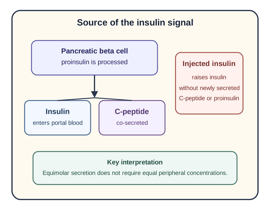
Pancreatic beta cells process proinsulin into insulin and C-peptide and secrete them in equimolar amounts. Peripheral concentrations are not expected to remain 1:1, because insulin undergoes substantial first-pass hepatic extraction while C-peptide has different clearance. Use C-peptide as evidence of endogenous secretion rather than expecting a 1:1 peripheral concentration ratio. [3]
Use the two routes to decide whether an insulin signal is accompanied by endogenous beta-cell products. Equimolar secretion does not require equal peripheral concentrations. [1][3]
At plasma glucose below 55 mg/dL during a recreated episode, the Endocrine Society guideline identifies insulin at least 3 microU/mL, C-peptide at least 0.6 ng/mL, and proinsulin at least 5 pmol/L as a pattern documenting endogenous hyperinsulinism. Individual assays and clinical context matter, and insulin alone can miss cases that C-peptide or proinsulin identify. [1][2]
Beta-hydroxybutyrate adds a physiology check. In a normal fast, falling insulin permits lipolysis and ketone production. Inappropriate insulin activity suppresses both, so beta-hydroxybutyrate at or below 2.7 mmol/L during the diagnostic episode supports insulin or insulin-like mediation. A glucose rise of at least 25 mg/dL after 1 mg IV glucagon also suggests preserved hepatic glycogen under insulin or IGF influence. [1]
Insulin high, C-peptide low The insulin signal is present without matching endogenous secretion. Think of exogenous insulin exposure.
Insulin and C-peptide inappropriately present The pancreas is secreting insulin. Now separate autonomous secretion from a secretagogue and from less common endogenous hyperinsulinemic states.
Insulin, C-peptide, and proinsulin suppressed Look outside beta-cell insulin secretion: critical illness, hormone deficiency, hepatic substrate failure, alcohol-associated impaired gluconeogenesis, or an insulin-like tumor syndrome can fit depending on the rest of the sample.
Learner action: read the source diagram from proinsulin to insulin and C-peptide, then trace a second route for injected insulin. The visible consequence is that injected insulin can raise circulating insulin while endogenous C-peptide and proinsulin remain suppressed.
Apply the same logic to a new sample: glucose 38 mg/dL, insulin 42 microU/mL, C-peptide 0.1 ng/mL, and proinsulin below the assay threshold points away from insulinoma even if the symptoms look identical.
Try it here · Checkpoint 1 of 3
Make your prediction before reading the choices. A first attempt is just a starting point.
Case 2
Show answer and explanations for case 2
A. Insulinoma with inefficient proinsulin processing (Why this does not fit)
Insulinoma is endogenous beta-cell secretion, so C-peptide and usually proinsulin should be inappropriately present during hypoglycemia. Their suppression conflicts with an endogenous source.
Reasoning steps for option A
Why might insulinoma come to mind with insulin 68 microU/mL during glucose 35 mg/dL?
The insulin value is strikingly high at a time when it should be nearly undetectable.
What would an insulinoma do to C-peptide and proinsulin?
Tumor beta cells secrete them along with insulin, so both would be inappropriately present. Here both are suppressed.
B. Exogenous insulin exposure (Best answer)
Insulin is strongly present while C-peptide and proinsulin are suppressed, showing an insulin signal without matching pancreatic secretion. Suppressed ketones confirm biologically important insulin activity.
Reasoning steps for option B
What does the combination of high insulin with suppressed C-peptide and proinsulin mean?
The circulating insulin is not coming from the patient's beta cells, because endogenous secretion always releases C-peptide too.
What does the low beta-hydroxybutyrate add?
It shows the insulin is biologically active, suppressing lipolysis and ketogenesis, as injected insulin would.
C. Sulfonylurea exposure (Why this does not fit)
A sulfonylurea stimulates the patient's beta cells, so C-peptide and proinsulin should accompany the insulin signal and the relevant drug may be detected. The present pattern instead separates insulin from endogenous secretion.
Reasoning steps for option C
Why could a sulfonylurea be suspected in unexplained hyperinsulinemic hypoglycemia?
Secretagogues are a common hidden cause, and they raise insulin during hypoglycemia.
Why does the C-peptide result exclude a secretagogue?
A sulfonylurea works by stimulating the patient's beta cells, so C-peptide and proinsulin would rise with insulin. The screen is also negative.
D. Alcohol-associated impaired gluconeogenesis (Why this does not fit)
Alcohol-related fasting hypoglycemia is a non-insulin mechanism and should permit insulin and C-peptide to suppress. The markedly present insulin with suppressed ketones points to an insulin-mediated process.
Reasoning steps for option D
Why is alcohol a common cause of fasting hypoglycemia to consider?
Alcohol impairs gluconeogenesis and can cause low glucose after poor intake.
Why do the insulin and ketone results argue against it?
Alcohol-related hypoglycemia is not insulin-mediated, so insulin should be suppressed and ketones should rise. Here insulin is high and ketones are low.
Takeaway: High insulin with suppressed C-peptide and proinsulin indicates insulin arriving without endogenous beta-cell secretion.
The major look-alikes are easiest to distinguish when all measurements come from the same hypoglycemic episode. A single insulin value obtained when glucose is normal cannot answer the source question. [1]
Critical-sample patterns during documented hypoglycemia
Cause
Insulin
C-peptide
Proinsulin
Oral agent screen
Ketones
CauseInsulinoma or another endogenous hyperinsulinemic state
InsulinInappropriately present
C-peptidePresent
ProinsulinPresent
Oral agent screenNegative
KetonesSuppressed
CauseExogenous insulin
InsulinHigh or inappropriately present
C-peptideSuppressed
ProinsulinSuppressed
Oral agent screenNegative
KetonesSuppressed
CauseSulfonylurea or glinide exposure
InsulinInappropriately present
C-peptidePresent
ProinsulinPresent
Oral agent screenPositive when detected
KetonesSuppressed
CauseNon-insulin cause
InsulinSuppressed
C-peptideSuppressed
ProinsulinSuppressed
Oral agent screenNegative
KetonesUsually rises during fasting
A secretagogue stimulates the patient's own beta cells, so its insulin, C-peptide, and proinsulin pattern can resemble insulinoma. The drug screen separates them when the relevant agent is detectable in the episode sample. A negative result should be interpreted with the timing and scope of the assay rather than as an absolute statement that exposure was impossible. [1]
Non-islet cell tumor hypoglycemia can show an insulin-like metabolic effect despite suppressed endogenous insulin markers. IGF-mediated hypoglycemia can suppress ketones and leave hepatic glycogen available for a glucagon response, so low beta-hydroxybutyrate is evidence of insulin-like action, not proof that insulin itself is high. [1]
Adrenal insufficiency, critical illness, advanced hepatic disease, or alcohol-associated impaired gluconeogenesis can cause hypoglycemia without inappropriate beta-cell secretion. During a true episode, insulin and C-peptide should be appropriately suppressed rather than simply called normal. The rest of the history and examination identifies which non-insulin pathway deserves testing. [1]
After bariatric surgery, post-bariatric hypoglycemia typically appears about 2 to 4 hours after eating and may develop well after the operation. That timing is useful, but neither postprandial timing nor normal fasting glucose alone proves the diagnosis. Document hypoglycemia and evaluate alternatives when the presentation is atypical. [6]
Classify this critical sample
During confusion, plasma glucose is 40 mg/dL, insulin is 18 microU/mL, C-peptide is 3.1 ng/mL, proinsulin is 22 pmol/L, beta-hydroxybutyrate is 0.4 mmol/L, and the oral agent screen is negative. The pattern shows endogenous hyperinsulinism with suppressed ketogenesis. Once insulin antibodies and relevant drug exposure are addressed, insulinoma becomes a leading diagnosis and localization can follow. [1]
Learner action: cover the cause column in the table and classify each row from C-peptide first, then the oral agent screen, then ketones. This ordering prevents occupation, medication access, weight change, or one dramatic symptom from overriding the biochemical pattern.
Type 2 diabetes and glucagonoma belong in the broader differential only when their physiology fits. Insulin resistance and glucagon excess ordinarily drive hyperglycemia, whereas a documented glucose of 40 mg/dL with inappropriate endogenous insulin activity points in the opposite direction.
Prove endogenous hyperinsulinism before searching for a lesion
If the episode cannot be captured spontaneously, recreate the circumstances safely. A supervised fast of up to 72 hours is used for suspected fasting hypoglycemia, while a meal-based evaluation can reproduce a postprandial syndrome. The endpoint is a diagnostic critical sample during symptoms and low plasma glucose, not simply completing the maximum time. [1][7]
Imaging does not establish biochemical insulin excess. After documented endogenous hyperinsulinemic hypoglycemia with a negative relevant oral-agent screen and no explanatory insulin antibodies, localization can begin. Cross-sectional CT or MRI is commonly followed by endoscopic ultrasonography or, where available, GLP-1 receptor PET, according to local expertise. [1][4][7]
Some insulinomas are small enough to escape first-line imaging. When biochemical evidence is strong but noninvasive localization remains negative or discordant, an expert center can consider selective pancreatic arterial calcium stimulation with hepatic venous insulin sampling. Calcium is delivered into pancreatic arterial territories and a regional insulin response helps localize active beta-cell tissue. [1][7]
1. Document Whipple triad Confirm symptoms, low plasma glucose, and relief after glucose.
2. Capture a critical sample Measure glucose, insulin, C-peptide, proinsulin, ketones and an oral-agent screen, with insulin antibodies when indicated. Classify endogenous insulin, exogenous insulin, secretagogue, IGF-like, or another non-insulin pattern.
3. Localize only after biochemistry fits Use anatomic and, when needed, specialized localization to find the source that the laboratory data have already established.
Learner action: in the workup diagram, stop at each box and ask whether the next test answers a biochemical question or an anatomic question. A CT ordered before the critical sample answers the wrong question first.
For a patient with glucose 39 mg/dL, insulin 8 microU/mL, C-peptide 1.4 ng/mL, proinsulin 10 pmol/L, suppressed beta-hydroxybutyrate, and a negative oral-agent screen, localization is appropriate after the laboratory pattern and clinical context have been confirmed. For the same CT finding with no documented hypoglycemia, biochemical confirmation still comes first.
Try it here · Checkpoint 2 of 3
Make your prediction before reading the choices. A first attempt is just a starting point.
Case 14
Show answer and explanations for case 14
A. Repeat fasting insulin while glucose is normal (Why this does not fit)
The biochemical diagnosis has already been established during hypoglycemia. Repeating insulin during euglycemia does not localize the lesion.
Reasoning steps for option A
Why might more insulin testing be proposed after a negative CT?
A negative scan can create doubt about the diagnosis.
Why does a euglycemic insulin level not help?
Endogenous hyperinsulinism has already been proven during hypoglycemia, and insulin at normal glucose neither adds to that nor localizes the tumor.
B. Whole-body MIBG imaging as the standard insulinoma study (Why this does not fit)
MIBG targets catecholamine-related tumors and is not the standard next localization study for insulinoma. The diagnostic problem is a small pancreatic neuroendocrine source.
Reasoning steps for option B
Why might a whole-body scan appeal when CT is negative?
Functional nuclear imaging can find tumors that CT misses.
Why is MIBG the wrong tracer?
MIBG targets catecholamine-secreting tumors such as pheochromocytoma, not small pancreatic insulinomas.
C. Endoscopic ultrasonography or dedicated pancreatic MRI based on local expertise (Best answer)
After biochemical confirmation, a small occult insulinoma may require another high-resolution pancreatic localization method. Endoscopic ultrasonography and MRI are established options before invasive regional stimulation testing.
Reasoning steps for option C
Why can CT miss a proven insulinoma?
Many insulinomas are under 2 cm and can be hard to see on a single CT.
What noninvasive or minimally invasive tests come next?
Endoscopic ultrasonography and dedicated pancreatic MRI give high-resolution views of the pancreas, chosen by local expertise, before invasive arterial testing.
D. Selective pancreatic arterial calcium stimulation as the first test after any negative CT (Why this does not fit)
Arterial stimulation can be valuable when noninvasive localization remains negative or discordant, but it is invasive. Additional high-resolution noninvasive or endoscopic localization is generally considered first.
Reasoning steps for option D
Why is arterial calcium stimulation a strong localization tool?
It shows which pancreatic region secretes insulin, even when imaging is negative.
Why is it not the first test after a negative CT?
It is invasive and reserved for cases where high-resolution imaging such as EUS or MRI remains negative or discordant.
Takeaway: After biochemical confirmation and a negative first scan, continue high-resolution localization before invasive regional testing.
Place insulinoma in its endocrine and treatment context
An insulinoma is a functioning pancreatic neuroendocrine tumor whose insulin secretion is inappropriate for the glucose concentration. Many are small and localized, and surgical treatment can be curative when disease is resectable. The old classroom rule of 90s is a historical memory aid, not a current diagnostic criterion or a reason to assume a specific patient's tumor is benign, solitary, or intrapancreatic. [4][7]
Insulinoma can occur with MEN1. The likelihood is especially relevant in a younger patient, multifocal pancreatic disease, a personal history of primary hyperparathyroidism or pituitary disease, or a suggestive family history. MEN1 evaluation uses the broader endocrine and genetic context rather than a single identical test set for every sporadic-appearing insulinoma. [4][5]
The familiar MEN1 triad links parathyroid, pituitary, and pancreatic neuroendocrine disease. Hypercalcemia from primary hyperparathyroidism, a prolactin-related pituitary presentation, or multiple pancreatic lesions can therefore change the pretest probability that an insulinoma belongs to a hereditary syndrome. [5]
For localized resectable insulinoma, surgery is the principal definitive treatment. Medical strategies such as diazoxide can help control hypoglycemia when surgery is delayed, unsuitable, or disease is not readily resectable, while management of advanced functioning pancreatic neuroendocrine tumors requires specialist therapy. [4][7]
Learner action: for a biochemically confirmed case, separate three decisions on paper: source of insulin effect, lesion localization, and hereditary context. The consequence is a cleaner plan in which a positive scan does not replace the laboratory diagnosis and a MEN1 history does not replace localization.
Apply this to a 24-year-old with recurrent hypoglycemia, primary hyperparathyroidism, and two pancreatic lesions: the critical sample still proves the insulin source, imaging still maps disease, and the age plus associated endocrine disease makes MEN1 evaluation particularly important.
Try it here · Checkpoint 3 of 3
Make your prediction before reading the choices. A first attempt is just a starting point.
Case 16
Show answer and explanations for case 16
A. MEN1 endocrine and genetic evaluation (Best answer)
Young age, primary hyperparathyroidism, multifocal pancreatic neuroendocrine disease, and a suggestive family history strongly support a hereditary MEN1 context. That context affects assessment of pituitary, parathyroid, and additional pancreatic disease.
Reasoning steps for option A
Which features in this woman point to a hereditary syndrome?
She is 26, has had primary hyperparathyroidism, and has two pancreatic neuroendocrine lesions.
What does the father's history add?
Recurrent kidney stones suggest hyperparathyroidism and he had a pituitary tumor, completing the parathyroid, pituitary and pancreas pattern of MEN1.
Why does this matter for her care?
MEN1 evaluation guides genetic testing, surveillance of other glands, surgical planning for multifocal disease and screening of relatives.
B. Evaluate only for MEN2 because parathyroid disease occurs in MEN2 (Why this does not fit)
Parathyroid disease can occur in MEN2, but pancreatic neuroendocrine tumors and pituitary disease fit MEN1 far better. The family pattern also points toward MEN1.
Reasoning steps for option B
Why might MEN2 be considered with parathyroid disease?
Primary hyperparathyroidism occurs in MEN2A as well as MEN1.
Why does the overall pattern not fit MEN2?
MEN2 features medullary thyroid carcinoma and pheochromocytoma, not pancreatic neuroendocrine tumors or pituitary disease.
C. Assume the pancreatic lesions are metastatic because there are two (Why this does not fit)
Multiple pancreatic lesions in a young patient with MEN1 features can reflect multifocal neuroendocrine disease. Multiplicity alone does not establish metastatic spread.
Reasoning steps for option C
Why might two pancreatic lesions raise concern for spread?
Multiple lesions in an organ can suggest metastatic disease.
What is the better explanation in this context?
MEN1 commonly causes multifocal pancreatic neuroendocrine tumors. Multiplicity alone does not establish metastases.
D. Exclude hereditary disease because insulinoma is usually sporadic (Why this does not fit)
Most insulinomas are sporadic, but that population fact does not outweigh a young patient with parathyroid disease, multifocal pancreatic lesions, and a matching family history.
Reasoning steps for option D
What fact about insulinoma supports assuming sporadic disease?
Most insulinomas are sporadic, and only a small fraction occur in MEN1.
Why should that base rate not be applied here?
Her age, parathyroid history, multifocal disease and family history make MEN1 far more likely than in a typical patient.
Takeaway: Young age plus parathyroid, pituitary, family, or multifocal pancreatic findings should raise MEN1 concern.
A. Episode-timed critical sample before glucose correction (Best answer)
The symptoms, laboratory low glucose, and response to dextrose document Whipple triad. The next diagnostic goal is to classify the source of the insulin effect with a critical sample during the low glucose episode before treatment changes the measurements.
Reasoning steps for option A
What has already been documented in this man's emergency visit?
Symptoms, a laboratory plasma glucose of 41 mg/dL during them, and recovery after dextrose complete Whipple triad.
What question remains, and how is it answered?
The cause of the low glucose is still unknown. Plasma glucose, insulin, C-peptide, proinsulin, beta-hydroxybutyrate and an oral-agent screen drawn before correction classify the source.
B. Order pancreatic CT immediately and diagnose insulinoma if a small lesion is seen (Why this does not fit)
Imaging can find incidental pancreatic lesions and cannot establish that insulin secretion caused the hypoglycemia. Biochemical classification should precede localization.
Reasoning steps for option B
Why might CT seem like the fastest route to a diagnosis?
Recurrent fasting-type episodes raise concern for insulinoma, and imaging looks for the tumor directly.
Why would a small pancreatic lesion on CT not settle the diagnosis?
Incidental pancreatic lesions are common, and imaging cannot show that insulin caused the hypoglycemia. Biochemical proof comes before localization.
C. Measure fasting insulin tomorrow while the plasma glucose is normal (Why this does not fit)
An insulin concentration obtained during euglycemia is not interpretable as evidence of inappropriate secretion during hypoglycemia. The sample must coincide with the low glucose episode.
Reasoning steps for option C
Why is a fasting insulin level a natural test to order?
Insulinoma secretes insulin, so measuring insulin sounds like a direct test.
Why is an insulin value at normal glucose uninformative?
Insulin is only inappropriate when it stays present during hypoglycemia. At normal glucose, any value can be physiological.
D. Diagnose reactive hypoglycemia because symptoms improve after carbohydrate (Why this does not fit)
Improvement after carbohydrate completes Whipple triad but does not distinguish fasting from postprandial causes. The early-morning timing and need for a critical sample argue against making a cause diagnosis from symptom relief alone.
Reasoning steps for option D
Why might relief after carbohydrate suggest reactive hypoglycemia?
Reactive symptoms also improve with eating, so the response can seem diagnostic.
Which features argue against that label?
The episodes are early-morning, a fasting pattern, and relief after glucose only completes Whipple triad. It does not identify the cause.
Takeaway: Document the critical biochemical pattern during hypoglycemia before searching for a lesion.
A. Exogenous insulin exposure (Why this does not fit)
Injected insulin can suppress ketones, but it should not produce the present C-peptide and proinsulin pattern. The drug assay also identifies an endogenous secretory stimulus.
Reasoning steps for option A
Why could injected insulin explain low glucose with low ketones?
Exogenous insulin also suppresses ketogenesis and lowers glucose.
Which results exclude it?
C-peptide 2.7 ng/mL and proinsulin 19 pmol/L show active beta-cell secretion, which injected insulin would suppress, and glipizide was detected.
B. Insulinoma (Why this does not fit)
The endogenous hyperinsulinemic pattern can resemble insulinoma, which is why the medication assay matters. Detection of glipizide on the critical specimen supplies a more direct explanation.
Reasoning steps for option B
Why does the biochemical pattern resemble insulinoma?
Insulin, C-peptide and proinsulin are all inappropriately present with suppressed ketones, the same pattern an insulinoma produces.
What separates this case from insulinoma?
Glipizide on the episode specimen explains the endogenous secretion. Insulinoma is diagnosed only after secretagogue exposure is excluded.
C. Secretagogue-induced hypoglycemia (Best answer)
Glipizide stimulates pancreatic beta cells, so insulin, C-peptide, and proinsulin remain present while glucose and ketones fall. Detection of the drug during the episode distinguishes this pattern from an insulinoma pattern.
Reasoning steps for option C
How does a sulfonylurea produce this pattern?
Glipizide closes beta-cell K-ATP channels and triggers secretion, so insulin, C-peptide and proinsulin all rise while glucose falls.
Why is detecting the drug on the same specimen decisive?
It ties the drug to the episode itself and accounts for the endogenous hyperinsulinism without a tumor.
D. Non-islet cell tumor hypoglycemia (Why this does not fit)
IGF-mediated hypoglycemia usually suppresses insulin, C-peptide, and proinsulin. The present specimen instead shows active beta-cell secretion plus a detected secretagogue.
Reasoning steps for option D
What could make an IGF-mediated tumor seem possible?
Non-islet cell tumor hypoglycemia also causes fasting hypoglycemia with low ketones.
Why does the specimen argue against it?
IGF-mediated hypoglycemia suppresses insulin, C-peptide and proinsulin. Here all three are present and a secretagogue is detected.
Takeaway: A positive relevant oral-agent assay can separate drug-stimulated endogenous insulin secretion from insulinoma.
A. Primary adrenal insufficiency (Why this does not fit)
Cortisol deficiency can cause fasting hypoglycemia, but insulin, C-peptide, and proinsulin should suppress appropriately when glucose is this low. The present pattern shows persistent endogenous insulin activity.
Reasoning steps for option A
Why might adrenal insufficiency be considered after exercise-related confusion?
Cortisol deficiency impairs gluconeogenesis and can cause fasting or exertional hypoglycemia.
What do the insulin markers show that adrenal insufficiency would not?
In adrenal insufficiency, insulin, C-peptide and proinsulin would suppress. Here all three stay present at glucose 43 mg/dL, so insulin is driving the episode.
B. Post-bariatric hypoglycemia (Why this does not fit)
Post-bariatric hypoglycemia requires an appropriate surgical history and is usually postprandial. This patient instead has fasting and exercise-associated episodes with endogenous hyperinsulinism.
Reasoning steps for option B
What makes post-bariatric hypoglycemia a common consideration?
It is a frequent cause of documented hypoglycemia with endogenous insulin secretion.
Why does it not fit this patient?
He has no bariatric surgery, and his episodes occur with fasting and exercise, not 2 to 4 hours after meals.
C. Exogenous insulin exposure (Why this does not fit)
Exogenous insulin would separate high insulin from suppressed C-peptide and proinsulin. Both endogenous products are inappropriately present here.
Reasoning steps for option C
Why should injected insulin be excluded in any hyperinsulinemic presentation?
Surreptitious or accidental insulin is an important cause of unexplained hypoglycemia with suppressed ketones.
What result rules it out here?
C-peptide 1.5 ng/mL and proinsulin 13 pmol/L confirm the pancreas is secreting, which injected insulin would suppress.
D. Pancreatic insulinoma causing fasting hyperinsulinemic hypoglycemia (Best answer)
The low glucose occurs with inappropriately present insulin, C-peptide, and proinsulin plus suppressed ketones. A negative oral-agent screen and negative insulin antibodies make an autonomous endogenous source such as insulinoma the leading consideration.
Reasoning steps for option D
What does the supervised-fast sample show at glucose 43 mg/dL?
Insulin, C-peptide and proinsulin exceed the guideline thresholds and ketones are suppressed: endogenous hyperinsulinism.
Which alternative explanations have been excluded?
The oral-agent screen and insulin antibodies are negative, removing secretagogue exposure and insulin autoimmune hypoglycemia.
What does that leave as the leading diagnosis?
Autonomous beta-cell secretion from an insulinoma, which now justifies localization.
Takeaway: Endogenous hyperinsulinemic hypoglycemia with negative drug testing and negative insulin antibodies should prompt insulinoma localization.
A. Autonomous pancreatic insulin secretion (Why this does not fit)
An insulinoma should leave endogenous insulin products inappropriately present. Here insulin, C-peptide, and proinsulin are all suppressed.
Reasoning steps for option A
What would autonomous pancreatic insulin secretion produce during glucose 38 mg/dL?
Insulin, C-peptide and proinsulin that fail to suppress.
Why does this sample exclude autonomous secretion?
Insulin is below 3 microU/mL, C-peptide below 0.2 ng/mL and proinsulin below 5 pmol/L, so beta-cell secretion is appropriately switched off.
B. Tumor-derived IGF activity (Best answer)
Suppressed beta-cell markers show that insulin itself is not driving the episode, but low ketones and a brisk glucagon response show an insulin-like metabolic effect. A large mesenchymal tumor makes an IGF-mediated syndrome the best fit.
Reasoning steps for option B
What do suppressed insulin markers with low ketones and a 32 mg/dL rise after glucagon indicate?
Something other than insulin is producing an insulin-like effect, suppressing ketogenesis and preserving hepatic glycogen.
Why does the tumor point to IGF activity?
Large mesenchymal tumors can secrete incompletely processed IGF-II, which acts on insulin and IGF-I receptors and causes non-islet cell tumor hypoglycemia.
C. Sulfonylurea-stimulated beta-cell secretion (Why this does not fit)
A secretagogue should produce measurable endogenous insulin, C-peptide, and proinsulin. Those markers are suppressed in this specimen.
Reasoning steps for option C
Why is a secretagogue worth checking in hyperinsulinemic-looking hypoglycemia?
Sulfonylureas are a common hidden cause of insulin-mediated low glucose.
Why do the markers argue against it?
A secretagogue raises endogenous insulin, C-peptide and proinsulin, and all three are suppressed here.
D. Cortisol deficiency as the sole mechanism (Why this does not fit)
Hormone deficiency can cause non-insulin hypoglycemia, but the low ketones and preserved glycogen response plus a large tumor favor an insulin-like tumor mediator. The tumor context is necessary to interpret the metabolic pattern.
Reasoning steps for option D
Why might cortisol deficiency be considered with suppressed insulin markers?
It is a non-insulin cause of fasting hypoglycemia, and insulin would appropriately suppress.
Which findings fit a tumor mediator better than cortisol deficiency?
Cortisol deficiency allows ketones to rise, and a glycogen-depleted liver responds poorly to glucagon. Low ketones and a brisk glucagon response fit IGF from the large tumor.
Takeaway: Suppressed insulin markers with suppressed ketones and a brisk glucagon response can indicate an IGF-mediated insulin-like effect.
A. Insulinoma causing persistent beta-cell secretion (Why this does not fit)
Insulinoma requires failure of endogenous insulin secretion to suppress during low glucose. All three beta-cell markers are suppressed here.
Reasoning steps for option A
Why might insulinoma be considered in a confused man with glucose 32 mg/dL?
Severe neuroglycopenia with very low glucose is a classic insulinoma presentation.
What in the sample excludes insulin excess?
Insulin, C-peptide and proinsulin are all suppressed and beta-hydroxybutyrate is high, the pattern of a normal insulin shutdown.
B. Sulfonylurea exposure causing endogenous insulin release (Why this does not fit)
A secretagogue would cause insulin and C-peptide to remain present and would suppress ketogenesis. The current sample shows the opposite metabolic state.
Reasoning steps for option B
Why is sulfonylurea exposure worth considering in an adult with heavy alcohol use?
Access to others' medications and confusion from alcohol can lead to accidental or hidden secretagogue ingestion.
Which result is opposite to secretagogue physiology?
A secretagogue keeps insulin and C-peptide present and ketones low. This sample shows suppressed markers and high ketones.
C. Alcohol-associated impaired gluconeogenesis (Best answer)
Low insulin with active ketogenesis shows an appropriate fasting hormonal response rather than hyperinsulinism. Alcohol metabolism can impair gluconeogenesis, and limited intake reduces available glycogen substrate.
Reasoning steps for option C
What does suppressed insulin with high beta-hydroxybutyrate mean?
The body has made the normal fasting switch to fat oxidation and ketone production, so insulin excess is not the cause.
How do two days of little food and heavy drinking cause hypoglycemia?
Poor intake depletes glycogen, and ethanol metabolism raises hepatic NADH, which impairs gluconeogenesis. Neither source can then maintain glucose.
D. Exogenous insulin exposure causing low C-peptide (Why this does not fit)
Exogenous insulin can lower C-peptide, but the circulating insulin itself would be inappropriately present and ketogenesis would be suppressed. Both features are absent here.
Reasoning steps for option D
Why could exogenous insulin seem to fit a low C-peptide?
Injected insulin suppresses the patient's own C-peptide.
What else would injected insulin show that is absent?
Circulating insulin would be inappropriately present and ketones would be suppressed. Both are the opposite here.
Takeaway: Suppressed insulin markers with active ketogenesis direct attention to non-insulin causes such as impaired gluconeogenesis.
A. Post-bariatric hypoglycemia after Roux-en-Y gastric bypass (Best answer)
The episodes occur in the characteristic postprandial window years after bariatric surgery and Whipple triad is documented. The history supports post-bariatric hypoglycemia while still requiring attention to atypical features that might suggest another cause.
Reasoning steps for option A
What pattern do the timing and history form?
Symptoms 2 to 3 hours after carbohydrate-rich meals, years after Roux-en-Y gastric bypass, with a documented glucose of 49 mg/dL.
Why does this fit post-bariatric hypoglycemia?
Rapid nutrient delivery after bypass causes an exaggerated incretin and insulin surge that overshoots, typically 2 to 4 hours after eating.
What would prompt looking for another cause?
Fasting or overnight episodes, or other atypical features, would raise concern for insulinoma or another disorder.
B. Insulinoma proven by postprandial timing (Why this does not fit)
Insulinoma can sometimes present after meals, so timing does not exclude it, but postprandial timing alone never proves a tumor. The bariatric history and typical 2 to 4 hour pattern favor post-bariatric hypoglycemia.
Reasoning steps for option B
Why can insulinoma not be dismissed because symptoms follow meals?
Some insulinomas cause postprandial as well as fasting episodes.
Why is postprandial timing not proof of a tumor either?
Timing alone never proves insulinoma. The bariatric history and well-tolerated overnight fasting favor post-bariatric hypoglycemia.
C. Primary adrenal insufficiency (Why this does not fit)
Adrenal insufficiency is not specifically linked to a 2 to 4 hour post-meal pattern after gastric bypass. Additional systemic features and hormone evaluation would be expected.
Reasoning steps for option C
Why is adrenal insufficiency a general cause of hypoglycemia to remember?
Cortisol deficiency reduces glucose production and can cause low glucose.
Why does it not match this pattern?
It tends to cause fasting hypoglycemia with other systemic features, not a specific 2 to 3 hour postprandial pattern while overnight fasts are tolerated.
D. Exogenous insulin exposure (Why this does not fit)
Exogenous insulin is established by a critical sample showing insulin without endogenous C-peptide, not by the bariatric history. No such biochemical source pattern is supplied.
Reasoning steps for option D
Why is exogenous insulin always part of the differential?
Hidden insulin use can cause episodic hypoglycemia at any time.
What evidence would be needed, and is it present?
A critical sample with high insulin and suppressed C-peptide. No such result is given, and the meal-related pattern has a better explanation.
Takeaway: Years after bariatric surgery, hypoglycemia typically 2 to 4 hours after meals supports post-bariatric hypoglycemia.
A. The fast is nondiagnostic because insulin is not above the usual fasting reference interval (Why this does not fit)
During hypoglycemia, insulin should become nearly completely suppressed, so a normal euglycemic reference interval is the wrong comparison. The insulin, C-peptide, and proinsulin values are inappropriately present at glucose 51 mg/dL.
It is higher than in many insulinoma samples and could look like a normal fasting rise.
How is it interpreted against the guideline threshold?
Values at or below 2.7 mmol/L during hypoglycemia support insulin action, so 1.1 still indicates suppressed ketogenesis.
D. The findings document endogenous hyperinsulinism and justify evaluation of its cause (Best answer)
At glucose below 55 mg/dL, the supplied insulin, C-peptide, and proinsulin exceed the guideline thresholds and ketones are suppressed. With a negative oral-agent screen, the next work is to separate insulinoma and other endogenous causes rather than dismiss the values as merely normal-range.
Reasoning steps for option D
How do the values at glucose 51 mg/dL compare with the guideline criteria?
Insulin at least 3 microU/mL, C-peptide at least 0.6 ng/mL and proinsulin at least 5 pmol/L are all met, and ketones are suppressed.
What does the negative oral-agent screen allow next?
Endogenous hyperinsulinism is documented without a detected secretagogue, so the next step is separating insulinoma from other endogenous causes.
Takeaway: In hypoglycemia, insulin values are judged by whether they suppress appropriately, not by a euglycemic reference interval.
A. It argues against insulin action because insulin promotes ketone production (Why this does not fit)
Insulin inhibits lipolysis and ketogenesis rather than promoting them. A very low ketone concentration during a fast is consistent with persistent insulin activity.
Reasoning steps for option A
Why might a low ketone level be thought to argue against insulin action?
It is easy to confuse insulin's effect on ketones with the ketosis of starvation.
What does insulin actually do to ketone production?
Insulin inhibits lipolysis and hepatic ketogenesis, so persistent insulin keeps beta-hydroxybutyrate low.
B. It proves the patient has diabetic ketoacidosis (Why this does not fit)
Diabetic ketoacidosis produces substantial ketonemia and usually hyperglycemia, not a low ketone concentration during hypoglycemia. The metabolic direction is opposite.
Reasoning steps for option B
Why might a ketone test suggest diabetic ketoacidosis?
Beta-hydroxybutyrate is the main ketone measured in DKA.
Why is this result the opposite of DKA?
DKA has high ketones with hyperglycemia. Here ketones are 0.2 mmol/L with glucose 37 mg/dL.
C. Suppressed ketogenesis from insulin activity (Best answer)
As glucose falls during fasting, normal insulin secretion falls and ketone production should rise. Persisting insulin blocks lipolysis and hepatic ketogenesis, explaining the low beta-hydroxybutyrate.
Reasoning steps for option C
What should happen to ketones as glucose falls during a normal fast?
Insulin falls, lipolysis increases and ketone production rises.
What does beta-hydroxybutyrate 0.2 mmol/L at glucose 37 mg/dL mean?
Insulin is still acting, blocking lipolysis and ketogenesis. It supports insulin-mediated hypoglycemia along with the present C-peptide and proinsulin.
D. It identifies a sulfonylurea as the specific cause (Why this does not fit)
Low ketones indicate insulin or insulin-like action but do not distinguish insulinoma, exogenous insulin, and secretagogue exposure. C-peptide and the oral-agent assay perform that separation.
Reasoning steps for option D
Why might low ketones be linked to a sulfonylurea?
Secretagogues raise insulin and therefore suppress ketones too.
Why can ketones not identify the specific cause?
Insulinoma, injected insulin and secretagogues all suppress ketones. C-peptide and the drug screen separate them.
Takeaway: Suppressed beta-hydroxybutyrate during fasting hypoglycemia is biological evidence of insulin or insulin-like action.
A. It excludes insulin-mediated hypoglycemia because the liver can release glucose (Why this does not fit)
A brisk rise indicates that hepatic glycogen was available despite fasting, which is compatible with ongoing insulin or insulin-like action that restrained glycogen use. It does not exclude hyperinsulinism.
Reasoning steps for option A
Why might a glucose rise after glucagon seem to exclude an insulin problem?
It shows the liver can release glucose, which could imply glucose production is normal.
What does a preserved glycogen store actually indicate during a fast?
By this point glycogen should be largely used up. Insulin or IGF action has restrained glycogen breakdown, which fits hyperinsulinism.
B. Preserved hepatic glycogen from insulin or IGF action (Best answer)
A rise of at least 25 mg/dL after glucagon during the diagnostic episode supports mediation by insulin or an IGF. The low ketones and inappropriate endogenous insulin markers make the result physiologically coherent.
Reasoning steps for option B
What does a 31 mg/dL rise after 1 mg IV glucagon show?
Hepatic glycogen was still available at the end of the fast, above the 25 mg/dL threshold in the guideline.
Why does that support insulin or IGF mediation?
Insulin and IGF inhibit glycogenolysis, so glycogen is preserved. Together with low ketones and present insulin markers, it confirms insulin action.
C. It proves a glucagonoma is causing the episode (Why this does not fit)
Administered glucagon tests hepatic glucose availability; it does not diagnose a glucagon-secreting tumor. Glucagonoma more often produces hyperglycemia and its own clinical syndrome.
Reasoning steps for option C
Why might the word glucagon bring glucagonoma to mind?
Glucagonoma is a pancreatic neuroendocrine tumor that secretes the same hormone.
Why does the glucagon test say nothing about glucagonoma?
Administered glucagon tests the liver's glycogen stores. Glucagonoma usually causes hyperglycemia with its own clinical syndrome.
D. It shows that the preceding plasma glucose was falsely low (Why this does not fit)
A metabolic response to glucagon does not invalidate the laboratory glucose measurement. The symptoms and biochemical sample already document true hypoglycemia.
Reasoning steps for option D
Why could a quick glucose rise make the initial reading seem wrong?
A brisk recovery might suggest the low value was an error.
Why is the hypoglycemia still genuine?
Symptoms occurred with a laboratory plasma glucose of 44 mg/dL. The glucagon response reflects liver physiology, not measurement error.
Takeaway: A brisk glucose rise after glucagon during a fast supports insulin or IGF mediation by demonstrating preserved hepatic glycogen.
A. Insulinoma is impossible because insulin is below 3 microU/mL (Why this does not fit)
A small number of insulinoma patients can have insulin below a single threshold during hypoglycemia. C-peptide and especially proinsulin can still demonstrate inappropriate endogenous secretion.
Reasoning steps for option A
Why might insulin 2.7 microU/mL seem to exclude insulinoma?
It is just below the 3 microU/mL insulin threshold.
Which other results show endogenous secretion?
C-peptide 1.0 ng/mL and proinsulin 17 pmol/L exceed their thresholds, and some insulinomas are detected only by C-peptide or proinsulin.
B. Exogenous insulin is most likely because insulin is low (Why this does not fit)
Exogenous insulin requires an insulin signal without matching endogenous C-peptide or proinsulin. Here the endogenous products are clearly present.
Reasoning steps for option B
What would make injected insulin a consideration?
Hidden insulin use is a key alternative in any hyperinsulinemic pattern.
Why does a low insulin with present C-peptide argue against it?
Injected insulin produces insulin without C-peptide. Here insulin is modest and the endogenous products are clearly present.
C. The episode is non-insulin mediated because the insulin value is not high (Why this does not fit)
Suppressed ketones plus inappropriately present C-peptide and proinsulin show insulin biology even though the insulin assay value is modest. Interpreting only one analyte would discard the integrated critical sample.
Reasoning steps for option C
Why might a modest insulin value suggest a non-insulin mechanism?
Non-insulin causes do present with low insulin during hypoglycemia.
What evidence of insulin action does the sample contain?
Suppressed ketones plus present C-peptide and proinsulin show ongoing beta-cell secretion and insulin effect.
D. Endogenous hyperinsulinism remains possible (Best answer)
The critical sample must be interpreted as a pattern, not by insulin alone. The low glucose, present C-peptide and proinsulin, and suppressed ketones keep an endogenous hyperinsulinemic disorder such as insulinoma in consideration.
Reasoning steps for option D
Why should the sample be read as a whole?
Insulin assays vary and a single analyte can fall just below a cutoff, while the others still show secretion.
What does the full pattern show?
Glucose 47 mg/dL with present C-peptide and proinsulin, suppressed ketones and a negative drug screen indicates endogenous hyperinsulinism, so insulinoma stays in consideration.
Takeaway: Insulin alone can miss endogenous hyperinsulinism; interpret C-peptide, proinsulin, ketones, and glucose together.
A. Beta cells release insulin without C-peptide during fasting, so C-peptide is not useful (Why this does not fit)
C-peptide is generated with insulin from proinsulin and remains useful for distinguishing endogenous secretion. Fasting normally suppresses both rather than uncoupling them.
Reasoning steps for option A
Why might someone think C-peptide is uncoupled from insulin in fasting?
Both fall during fasting, which can be misread as release of insulin without C-peptide.
How are insulin and C-peptide actually produced?
Proinsulin is cleaved into insulin and C-peptide, which are secreted together, so C-peptide remains a marker of endogenous secretion.
B. Equimolar secretion with unequal peripheral concentrations (Best answer)
Equimolar secretion occurs at the beta cell, but the bloodstream does not preserve a numeric 1:1 concentration ratio. The diagnostic value lies in whether C-peptide is appropriately suppressed or inappropriately present during hypoglycemia.
Reasoning steps for option B
Where does the 1:1 relationship between insulin and C-peptide hold?
At the beta cell, where cleavage of proinsulin releases the two in equimolar amounts.
Why do peripheral concentrations differ?
The liver extracts much of the insulin on first pass, while C-peptide is cleared mainly by the kidney with a longer half-life.
How is C-peptide actually used in the diagnosis?
The question is whether it is inappropriately present during hypoglycemia, not whether it matches insulin numerically.
C. C-peptide is secreted only by insulinomas and not by normal beta cells (Why this does not fit)
Normal beta cells secrete C-peptide whenever proinsulin is processed. Insulinoma does not create a unique C-peptide molecule.
Reasoning steps for option C
Why might C-peptide be imagined as a tumor product?
It is emphasized in insulinoma testing, which can suggest it is specific to tumors.
What do normal beta cells do with C-peptide?
They secrete it whenever they release insulin. Insulinoma secretes the same molecule, just inappropriately.
D. Peripheral insulin and C-peptide must be exactly equal unless kidney function is abnormal (Why this does not fit)
Different hepatic extraction and half-lives affect the two peptides even with normal kidney function. An exact equality rule is physiologically incorrect.
Reasoning steps for option D
Why does kidney function come up when comparing insulin and C-peptide?
The kidney clears C-peptide, so renal failure raises it.
Why is exact equality wrong even with normal kidneys?
Hepatic insulin extraction and different half-lives keep peripheral concentrations unequal regardless of kidney function.
Takeaway: Equimolar secretion from beta cells does not imply a 1:1 peripheral concentration ratio.
A. Document Whipple triad and obtain a critical sample first (Best answer)
A small incidental pancreatic finding does not establish a functioning insulinoma. First document symptoms with low plasma glucose and symptom resolution, then classify the biochemical source during an episode.
Reasoning steps for option A
What has not yet been established in this woman?
No glucose has been measured during symptoms, so a hypoglycemic disorder has not been documented.
What is the correct order of investigation?
Document Whipple triad, obtain a critical sample during an episode, and only then consider whether the cyst is relevant.
B. Repeat pancreatic MRI every 3 months until the cyst changes (Why this does not fit)
Serial anatomy cannot establish whether insulin secretion is causing the symptoms. The missing information is biochemical confirmation of hypoglycemia and its mechanism.
Reasoning steps for option B
Why might serial MRI of the cyst seem prudent?
Monitoring a pancreatic lesion for change is a common response to an incidental finding.
Why does it not answer her question?
Anatomy cannot show whether insulin causes her symptoms. The missing information is biochemical.
C. Proceed directly to endoscopic ultrasound and biopsy the cyst (Why this does not fit)
An invasive localization procedure is premature without a documented hypoglycemic disorder. A pancreatic cyst can be incidental and may not be the source of any hormone syndrome.
Reasoning steps for option C
Why could EUS with biopsy seem to give a definitive answer?
It samples tissue and can characterize pancreatic lesions.
Why is it premature?
No hypoglycemic disorder has been documented, and a 7-mm cyst may be incidental. An invasive test cannot replace biochemical proof.
D. Diagnose insulinoma because pancreatic lesions smaller than 2 cm are typical (Why this does not fit)
Size is not a biochemical diagnosis. Insulinoma requires a clinical syndrome plus evidence of inappropriate insulin secretion at low glucose.
Reasoning steps for option D
Why does lesion size make insulinoma tempting?
Most insulinomas are small, often under 2 cm.
Why is size not a diagnosis?
Many small pancreatic lesions do not secrete hormones. Insulinoma requires documented hypoglycemia with inappropriate insulin secretion.
Takeaway: Anatomic findings do not establish a functioning insulinoma; prove the hypoglycemic syndrome and biochemical source first.
A. Oral glucose tolerance testing (Why this does not fit)
An oral glucose challenge does not regionally localize pancreatic beta-cell secretion and is not the requested preoperative mapping study.
Reasoning steps for option A
Why might a glucose challenge seem informative?
Glucose tolerance tests measure insulin responses to carbohydrate.
Why does it not localize the tumor?
It measures whole-body glucose handling and cannot identify which pancreatic region is secreting insulin.
B. Repeat insulin antibody testing every hour during a fast (Why this does not fit)
Antibody testing addresses an autoimmune cause but does not localize pancreatic secretion. That alternative has already been evaluated.
Reasoning steps for option B
Why could antibody testing seem relevant to persistent hyperinsulinism?
Insulin autoimmune hypoglycemia is a recognized cause of hyperinsulinemic hypoglycemia.
Why does hourly antibody testing not help?
Antibodies have already been negative, and they address cause rather than location.
C. MIBG scanning (Why this does not fit)
MIBG is not the standard regional localization method for insulinoma. It does not sample insulin responses from pancreatic arterial territories.
Reasoning steps for option C
Why might MIBG be considered when anatomic imaging fails?
It is a functional scan used for some neuroendocrine tumors.
Why is it not used to localize insulinoma?
It targets catecholamine pathways and does not sample regional insulin output from pancreatic arteries.
D. Selective pancreatic arterial calcium stimulation with hepatic venous insulin sampling (Best answer)
Calcium stimulation can provoke insulin release from arterial territories while hepatic venous insulin is sampled. It is reserved for specialized localization when biochemical evidence is strong and standard imaging is unrevealing or discordant.
Reasoning steps for option D
Why is functional localization needed in this patient?
Endogenous hyperinsulinism is repeatedly proven, but CT, MRI and EUS have not shown a lesion, and surgery is planned.
How does selective arterial calcium stimulation localize the source?
Calcium injected into each pancreatic artery triggers insulin release from tumor tissue in that territory, detected as a rise in hepatic venous insulin.
Where is this test performed?
At expert centers when biochemical evidence is strong and standard imaging is negative or discordant.
Takeaway: Regional calcium stimulation is an expert-center localization tool for biochemically proven but occult or discordant endogenous hyperinsulinism.
A. Medullary thyroid carcinoma (Why this does not fit)
Medullary thyroid carcinoma is a hallmark MEN2 tumor and would pull the pattern toward MEN2 rather than MEN1.
Reasoning steps for option A
Why might medullary thyroid carcinoma come to mind with hyperparathyroidism?
Both occur in MEN2A, so the pairing is familiar.
What would it do to the syndrome classification?
It is the hallmark MEN2 tumor, so it would favor MEN2, not MEN1.
B. Pheochromocytoma (Why this does not fit)
Pheochromocytoma is central to MEN2 syndromes and is not the classic additional MEN1 organ pattern.
Reasoning steps for option B
Why might pheochromocytoma seem to belong in an endocrine tumor syndrome?
It is a classic component of the MEN syndromes taught alongside MEN1.
Why does it not support MEN1?
Pheochromocytoma is central to MEN2A and MEN2B, not to the MEN1 organ pattern.
C. Pituitary prolactinoma (Best answer)
The combination of parathyroid disease, a pancreatic neuroendocrine tumor, and pituitary disease is the classic MEN1 organ pattern. A prolactinoma would therefore reinforce MEN1.
Reasoning steps for option C
Which MEN1 organs are already involved in this man?
The parathyroid, shown by high calcium with inappropriately high PTH, and the pancreas, with a neuroendocrine tumor causing hypoglycemia.
Why would a prolactinoma strengthen the MEN1 diagnosis?
It completes the classic triad of parathyroid, pituitary and pancreatic neuroendocrine disease. Prolactinoma is the most common MEN1 pituitary tumor.
D. Mucosal neuromas (Why this does not fit)
Mucosal neuromas are associated with MEN2B rather than MEN1. They do not complete the pituitary-parathyroid-pancreatic pattern.
Reasoning steps for option D
Why might mucosal neuromas be linked to multiple endocrine tumors?
They are a distinctive physical finding in one MEN syndrome.
Which syndrome do they indicate?
MEN2B, along with medullary thyroid carcinoma and a marfanoid habitus. They do not complete the MEN1 pattern.
Takeaway: Pituitary, parathyroid, and pancreatic neuroendocrine disease form the classic MEN1 organ pattern.
Insulinoma is a strong explanation for this episode: endogenous insulin markers during hypoglycemia with a negative drug screen, and defensive eating explains the weight gain. But its insulin excess lowers glucose, the same direction as this episode, so it does not answer the question.
Reasoning steps for option A
How well does insulinoma explain this episode?
Very well: glucose 40 mg/dL with present insulin, C-peptide and proinsulin and a negative drug screen is the classic pattern, and defensive snacking explains the weight gain.
Which way does insulinoma push plasma glucose?
Downward, the same direction as this episode, so it is not the condition that would push glucose the opposite way.
B. Sulfonylurea exposure missed by an incomplete assay (Why this does not fit)
A sulfonylurea missed by an incomplete assay could mimic this endogenous pattern and remains a real alternative. But a secretagogue lowers glucose, the same direction as this episode, so it does not answer the question.
Reasoning steps for option B
Why does a missed sulfonylurea remain a real possibility?
Secretagogues produce the same endogenous pattern, and an assay that does not cover the drug taken can be falsely negative.
Which way does a secretagogue push plasma glucose?
It lowers glucose by stimulating beta cells, the same direction as this episode.
C. Insulin autoimmune hypoglycemia (Why this does not fit)
Insulin autoimmune hypoglycemia also produces a hyperinsulinemic episode, and the negative oral-agent screen does not exclude antibodies. But it lowers glucose, the same direction as this episode, so it does not answer the question.
Reasoning steps for option C
Why is insulin autoimmune hypoglycemia still on the list?
Insulin antibodies can cause hyperinsulinemic hypoglycemia, and a negative oral-agent screen says nothing about antibodies.
Which way does insulin autoimmune hypoglycemia push glucose?
Antibody binding can cause early postprandial hyperglycemia, but the defining episodes are hypoglycemia when bound insulin is released, the same direction as this episode.
D. Glucagonoma (Best answer)
Glucagon excess promotes hepatic glucose output and is associated with diabetes or hyperglycemia, the opposite of recurrent low glucose from endogenous insulin activity. It is the only listed explanation whose glucose direction conflicts with this episode.
Reasoning steps for option D
What does glucagon excess do to hepatic glucose output?
Glucagon stimulates glycogenolysis and gluconeogenesis, raising plasma glucose.
How does glucagonoma usually present?
With diabetes or hyperglycemia, weight loss and necrolytic migratory erythema, the opposite direction from recurrent insulin-mediated hypoglycemia.
Takeaway: Use the direction of hormone physiology: glucagon excess drives glucose upward, whereas inappropriate insulin drives it downward.
Insulinoma would prevent appropriate suppression of beta-cell markers and would tend to suppress ketogenesis. The critical sample shows a non-insulin fasting response.
Reasoning steps for option A
Why is insulinoma the reflex diagnosis for recurrent fasting hypoglycemia?
It is the classic tumor cause of fasting hypoglycemia in an otherwise well adult.
What does the critical sample show instead?
Insulin, C-peptide and proinsulin are suppressed and ketones are high, a normal fasting response rather than insulin excess.
B. Primary adrenal insufficiency causing fasting hypoglycemia (Best answer)
The insulin markers suppress appropriately and ketones rise, so beta-cell insulin excess is not the mechanism. Weight loss, salt craving, hyponatremia, and hyperkalemia point toward cortisol and mineralocorticoid deficiency.
Reasoning steps for option B
What does the critical sample tell you about mechanism?
Suppressed insulin markers with high ketones show the hypoglycemia is not insulin-mediated.
Which clinical and electrolyte findings identify the cause?
Weight loss, salt craving, sodium 128 and potassium 5.6 indicate combined cortisol and aldosterone deficiency from primary adrenal insufficiency.
How does cortisol deficiency cause fasting hypoglycemia?
Cortisol supports gluconeogenesis during fasting, so its absence limits glucose production.
C. Exogenous insulin exposure (Why this does not fit)
Exogenous insulin would leave circulating insulin inappropriately present and suppress ketone production. Both features are absent.
Reasoning steps for option C
Why must hidden insulin use be considered in unexplained hypoglycemia?
It is a recognized cause in otherwise well adults.
Which two results exclude it here?
Insulin is below 3 microU/mL and ketones are high. Injected insulin would keep insulin present and ketones low.
D. Sulfonylurea exposure (Why this does not fit)
A secretagogue would stimulate endogenous insulin and C-peptide and suppress ketogenesis. The sample instead shows appropriate beta-cell shutdown.
Reasoning steps for option D
Why would a sulfonylurea be on the differential?
Secretagogues are a common hidden cause of fasting hypoglycemia.
What does the sample show about beta-cell activity?
Beta cells are appropriately shut down, with suppressed C-peptide and high ketones. A secretagogue would do the opposite.
Takeaway: Suppressed insulin markers and active ketogenesis direct the differential toward non-insulin causes such as adrenal insufficiency.
Sepsis and severe liver injury can lower glucose production while insulin secretion suppresses appropriately. The critical sample therefore supports a non-insulin mechanism tied to organ failure.
Reasoning steps for option A
What limits glucose production in septic shock with severe liver injury?
The failing liver cannot perform gluconeogenesis or glycogenolysis well, and sepsis increases glucose use.
How does the critical sample fit that mechanism?
Insulin and C-peptide are appropriately suppressed, so the hypoglycemia reflects organ failure rather than insulin excess.
B. Insulinoma unmasked by sepsis (Why this does not fit)
Insulinoma should leave insulin and C-peptide inappropriately present during hypoglycemia. Their suppression argues against that mechanism.
Reasoning steps for option B
Why might a hidden insulinoma be imagined as unmasked by illness?
Reduced intake and stress could, in theory, expose a tumor's insulin secretion.
What result argues against it?
Insulin and C-peptide are suppressed, and an insulinoma would keep both inappropriately present.
C. Sulfonylurea exposure (Why this does not fit)
A secretagogue would stimulate endogenous insulin and C-peptide rather than permit both to suppress. The organ-failure context already supplies a coherent mechanism.
Reasoning steps for option C
Why could a secretagogue be suspected in a hospitalized older man?
Medication errors with sulfonylureas occur in hospital and older patients.
Why does the sample exclude it?
A secretagogue would keep insulin and C-peptide present. Both are suppressed, and organ failure already explains the hypoglycemia.
D. Exogenous insulin exposure (Why this does not fit)
Exogenous insulin should be detectable as inappropriate insulin activity and would generally suppress ketogenesis. The sample gives no such insulin signal.
Reasoning steps for option D
Why is insulin exposure relevant in an ICU patient?
ICU patients often receive insulin infusions, and errors can cause hypoglycemia.
What would the sample show if insulin caused this episode?
Circulating insulin would be inappropriately present. Here insulin is suppressed.
Takeaway: In critical illness, suppressed beta-cell markers plus organ failure can explain hypoglycemia without an insulinoma workup.
A. Postprandial timing excludes insulinoma (Why this does not fit)
Insulinoma often presents during fasting, but postprandial episodes do not exclude it. The biochemical pattern is more important than timing alone.
Reasoning steps for option A
Why is postprandial timing associated with a low likelihood of insulinoma?
Most insulinomas present with fasting hypoglycemia.
Why does timing not exclude it here?
A minority of insulinomas cause postprandial episodes, and this sample documents endogenous hyperinsulinism during a mixed meal.
B. Reactive symptoms without true hypoglycemia (Why this does not fit)
Plasma glucose is 48 mg/dL during symptoms and the critical sample shows inappropriate endogenous insulin activity. This is not merely a symptom syndrome with normal glucose.
Reasoning steps for option B
Why might a label of reactive symptoms be offered?
Postprandial symptoms without true hypoglycemia are common and often called reactive.
What in this test rules that out?
Glucose was 48 mg/dL during symptoms, with inappropriate insulin, C-peptide and proinsulin.
C. Endogenous hyperinsulinemia; proceed to localization (Best answer)
The mixed-meal episode shows true hypoglycemia with inappropriately present insulin, C-peptide, and proinsulin plus suppressed ketones. With drug and antibody alternatives addressed, localization for insulinoma is appropriate despite postprandial timing.
Reasoning steps for option C
What does the mixed-meal test show?
Documented hypoglycemia with insulin, C-peptide and proinsulin inappropriately present and suppressed ketones.
Which alternatives have been addressed?
The drug screen and insulin antibodies are negative, and she has had no bariatric surgery.
What is the next step?
Localization for insulinoma, since endogenous hyperinsulinism is established despite postprandial timing.
D. Post-bariatric hypoglycemia is proven (Why this does not fit)
The patient has no bariatric surgery history, so the defining clinical context for post-bariatric hypoglycemia is absent. The endogenous pattern requires another explanation.
Reasoning steps for option D
Why might post-bariatric hypoglycemia be suggested by the timing?
It is the classic cause of hypoglycemia after meals with endogenous insulin secretion.
What required element is missing?
She has never had bariatric surgery, so the defining history is absent.
Takeaway: Postprandial timing does not by itself exclude insulinoma when endogenous hyperinsulinemia is documented.
A. It proves type 2 diabetes caused the hypoglycemia (Why this does not fit)
Weight gain is common in insulin resistance, but type 2 diabetes does not explain documented fasting hypoglycemia with endogenous insulin secretion failing to suppress. The critical sample points elsewhere.
Reasoning steps for option A
Why might weight gain suggest type 2 diabetes?
Weight gain and insulin resistance often go together.
Why does type 2 diabetes not explain these episodes?
Untreated type 2 diabetes does not cause fasting hypoglycemia with endogenous insulin, C-peptide and proinsulin that fail to suppress.
B. It supports glucagonoma because glucagon increases appetite (Why this does not fit)
Glucagonoma more often produces hyperglycemia and weight loss. The supplied weight change has a direct behavioral explanation in frequent preventive eating.
Reasoning steps for option B
Why might glucagonoma be linked to a change in weight?
Glucagonoma is a pancreatic neuroendocrine tumor that alters body weight.
Why does it not fit?
Glucagonoma causes weight loss and hyperglycemia. This patient has weight gain and hypoglycemia.
C. It proves the insulinoma is malignant (Why this does not fit)
Weight gain does not establish tumor grade or metastatic behavior. It reflects the patient's adaptation to repeated low glucose episodes.
Reasoning steps for option C
Why might weight gain be read as a sign of tumor behavior?
Changes in body weight sometimes track disease activity.
What does the weight gain actually reflect?
His bedtime snacks and juice at work, eaten to prevent symptoms. Weight says nothing about whether the tumor is malignant.
D. Defensive carbohydrate intake during recurrent hypoglycemia (Best answer)
Patients with recurrent hypoglycemia may eat frequently to prevent symptoms, producing weight gain. The biochemical pattern, not the weight change, identifies endogenous hyperinsulinism.
Reasoning steps for option D
What does the patient's eating pattern show?
He snacks before bed and keeps juice at work to prevent recurrent early-morning lows.
How does that explain the weight change?
Frequent defensive carbohydrate intake adds calories. The critical sample, not the weight gain, identifies endogenous hyperinsulinism.
Takeaway: Weight gain can reflect defensive eating in recurrent hypoglycemia, but the critical sample determines the mechanism.
A. The early symptoms are neuroglycopenic and the later symptoms are purely adrenergic (Why this does not fit)
Sweating, tremor, and palpitations are autonomic warning symptoms, whereas confusion and impaired cognitive function are neuroglycopenic. The proposed order reverses the physiology.
Reasoning steps for option A
Why is the classification of symptoms easy to reverse?
Both groups appear during hypoglycemia, and patients may experience them close together.
How are these particular symptoms classified?
Sweating, tremor and palpitations are autonomic; confusion and difficulty speaking are neuroglycopenic. The option has them backward.
B. Autonomic warning followed by neuroglycopenia (Best answer)
Counter-regulatory autonomic symptoms can appear while cognition is still preserved. Further glucose decline can then impair brain function and produce confusion or seizure.
Reasoning steps for option B
What produces the early sweating, tremor and palpitations?
Sympathoadrenal activation as glucose falls, a warning response while brain function is still preserved.
Why do confusion and speech difficulty follow?
Continued decline in glucose deprives the brain of fuel, producing neuroglycopenic dysfunction.
C. The later confusion proves a focal stroke rather than worsening hypoglycemia (Why this does not fit)
A stroke remains a clinical consideration when focal findings persist, but progressive cognitive dysfunction during continuing hypoglycemia is a classic neuroglycopenic manifestation. The temporal relationship supports the glucose mechanism.
Reasoning steps for option C
Why might speech difficulty suggest a stroke?
Focal neurological deficits can occur with stroke, which must stay in mind if deficits persist.
What favors hypoglycemia as the cause here?
The dysfunction develops as glucose continues to fall in a patient with confirmed insulinoma, the classic sequence of neuroglycopenia.
D. Insulinomas produce only neuroglycopenic symptoms and no autonomic symptoms (Why this does not fit)
Insulinoma can produce both autonomic and neuroglycopenic manifestations. Which appears first depends on glucose trajectory and counter-regulatory responses.
Reasoning steps for option D
Why might insulinoma be thought to produce only neuroglycopenia?
Neuroglycopenic presentations are prominent in insulinoma and can occur without a clear warning.
Why is that overstated?
Autonomic symptoms also occur, as in this patient. Chronic recurrent hypoglycemia can blunt them, but they do not disappear entirely.
Takeaway: Hypoglycemia can progress from autonomic warning symptoms to neuroglycopenic dysfunction as cerebral glucose availability falls.
A. Appropriate fasting response without hyperinsulinism (Best answer)
Insulin, C-peptide, and proinsulin are suppressed while beta-hydroxybutyrate rises strongly. That is the expected metabolic direction during prolonged fasting and argues against pathologic insulin excess.
Reasoning steps for option A
What is the expected hormonal response to a week of severe caloric restriction?
Insulin falls, lipolysis rises and the liver produces abundant ketones.
How does this sample match that response?
Insulin 1.1 microU/mL, C-peptide 0.2 ng/mL and suppressed proinsulin with beta-hydroxybutyrate 5.8 mmol/L show appropriate insulin suppression without hyperinsulinism.
B. Insulinoma is likely because glucose is below 55 mg/dL (Why this does not fit)
Low glucose alone does not diagnose insulinoma. Insulinoma requires inappropriate insulin activity, which is absent in this sample.
Reasoning steps for option B
Why might glucose 50 mg/dL raise concern for insulinoma?
It is below the 55 mg/dL level used to interpret a critical sample.
Why does the sample argue against insulinoma?
Insulinoma requires inappropriate insulin secretion, and here every insulin marker is suppressed with high ketones.
C. Exogenous insulin exposure is likely because C-peptide is low (Why this does not fit)
Exogenous insulin lowers C-peptide but leaves insulin itself inappropriately present and suppresses ketogenesis. Here insulin is low and ketones are high.
Reasoning steps for option C
Why might low C-peptide suggest injected insulin?
Injected insulin suppresses endogenous C-peptide.
Which findings exclude it?
Insulin itself is low and ketones are very high. Injected insulin would keep insulin present and ketones suppressed.
D. Sulfonylurea exposure is likely because the oral-agent screen is negative (Why this does not fit)
Secretagogues stimulate endogenous insulin and C-peptide; a negative assay does not create that missing biochemical pattern. The present physiology is a low-insulin fast.
Reasoning steps for option D
Why would a negative oral-agent screen come up in reasoning about secretagogues?
Secretagogue assays can miss some drugs, so a negative result is sometimes doubted.
Why is a secretagogue still unlikely?
A secretagogue raises endogenous insulin and C-peptide. The problem is not the assay; the sample shows beta-cell shutdown.
Takeaway: High ketones with appropriately suppressed insulin markers support a normal fasting metabolic switch rather than hyperinsulinism.
A. Long-term carbohydrate snacking alone (Why this does not fit)
Frequent carbohydrate can reduce symptoms temporarily but does not treat a localized hormone-secreting tumor. A resectable source has already been identified.
Reasoning steps for option A
Why is frequent carbohydrate intake used in insulinoma?
It reduces symptomatic episodes while the diagnosis and treatment are being arranged.
Why is it inadequate as long-term management here?
It does not treat the tumor, and a single resectable lesion has already been localized.
B. Permanent diazoxide therapy instead of discussing surgery (Why this does not fit)
Diazoxide can control hypoglycemia when surgery is delayed or unsuitable, but a fit patient with localized resectable disease has a potential definitive surgical option.
Reasoning steps for option B
What role does diazoxide play in insulinoma?
It suppresses insulin secretion and controls hypoglycemia when surgery is delayed, unsuitable or impossible.
Why is it wrong to choose it instead of surgery here?
She has an acceptable operative risk and a localized lesion, so surgery offers a potential cure that lifelong medication does not.
C. Systemic chemotherapy as initial therapy for a small localized lesion (Why this does not fit)
Systemic therapy is not the first treatment for a small localized resectable insulinoma without metastatic disease. The disease distribution supports a local definitive approach.
Reasoning steps for option C
When is systemic therapy used for pancreatic neuroendocrine tumors?
For advanced, unresectable or metastatic disease.
Why is it not appropriate first?
This is a small localized lesion with no metastases, where local surgical treatment is the standard.
D. Pancreas-directed surgical resection (Best answer)
Surgery is the principal definitive therapy for localized resectable insulinoma and can be curative. The exact operation depends on tumor location, relation to the pancreatic duct, multiplicity, and surgical assessment.
Reasoning steps for option D
What do the biochemistry and imaging together establish?
Repeated endogenous hyperinsulinemic hypoglycemia with a single concordant lesion on MRI and EUS and no metastases: a localized insulinoma.
Why is surgical resection preferred?
It is the principal definitive therapy and can be curative for a localized resectable insulinoma.
What determines the specific operation?
Tumor location, its distance from the main pancreatic duct and whether there is more than one lesion, which decide between enucleation and partial pancreatectomy.
Takeaway: Localized resectable insulinoma is generally treated surgically, with the operation tailored to lesion anatomy.