Locate digestive tissue by region and depth, connect glands and absorptive surfaces to function, and distinguish gut electrical rhythm from coordinated propulsion.
A shallow gastric defect and a deep gastric defect may look similar from above. Their consequences differ because they interrupt different parts of the wall. Read the digestive tract on two coordinates: where along the tube and how far from the lumen.
By the end, you should be able to locate an unfamiliar specimen, explain what a damaged layer normally does, and distinguish electrical rhythm from successful propulsion. Follow the wall first, compare regional architecture next, then connect absorptive surfaces and nerve networks to function. The clinical scenarios are original educational examples, not patient records.
Which muscle belongs to the mucosa?
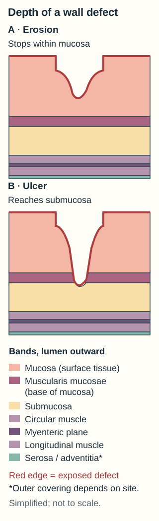
The word muscularis appears twice, but these layers are not interchangeable. The thin muscularis mucosae ends the mucosal compartment. The much thicker muscularis externa lies beyond the submucosa and provides most of the force for mixing and propulsion. A lesion can cross the first muscle sheet without reaching the second. [1]
Compare the endpoint of each defect, not its width. The outer covering depends on which surface is sampled. [1][7]Open full-size figure.
Mucosa: epithelium, lamina propria and muscularis mucosae. Epithelium provides a region-specific barrier and secretory or absorptive interface. Lamina propria contains supporting connective tissue, small vessels, lymphatics and immune cells. Muscularis mucosae supports local mucosal motion.
Submucosa: connective tissue with larger vessels and lymphatics, the submucosal plexus, and glands in selected regions. Not every gut segment has submucosal glands.
Muscularis externa: typically inner circular and outer longitudinal muscle, with the myenteric plexus between them. Circular contraction narrows the lumen; longitudinal contraction shortens a segment. Their coordination matters more than contraction alone.
Outer covering: serosa where the surface is covered by visceral peritoneum, or adventitia where connective tissue attaches the wall to surrounding structures.
Serosa includes mesothelium over connective tissue. Adventitia lacks that free mesothelial surface. One organ can have differently covered surfaces, so do not assign serosa merely because a specimen came from the abdomen. Most thoracic esophagus has adventitia. The mnemonic MSMS is useful only if its last S reminds you to check for serosa or adventitia. [1][9]
Trace it: put a finger at the luminal surface of the drawing and name each boundary before following the deeper defect. Which compartment lies immediately after muscularis mucosae?
Check the first boundary
Submucosa is next. Choosing muscularis externa skips the connective tissue compartment between the two muscle layers.
Apply the map to a new specimen: glands beneath the muscularis mucosae are submucosal, even if they resemble glands above it. Location is part of the finding, not an optional label.
How far must a defect extend to become an ulcer?
An erosion remains within the mucosa; an ulcer extends through muscularis mucosae into submucosa or deeper. An ulcer need not reach muscularis externa. Perforation requires a full-thickness communication beyond the wall. Neither a dramatic surface appearance nor bleeding alone proves perforation. [7]
The following depth comparison is a local tracing activity, not a progression requirement. For each specimen, trace the proposed endpoint on the wall map, predict the compartment, then open its drawing. Keep earlier drawings open to compare them. The complete anatomical explanation remains visible above.
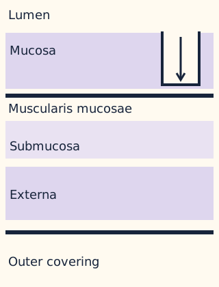
Specimen A: surface loss, muscle sheet intact
Original depth-comparison state. Same layer order and scale in each state. Open full-size figure.
The endpoint remains above muscularis mucosae: an erosion. Inflammation in lamina propria does not by itself imply submucosal extension.
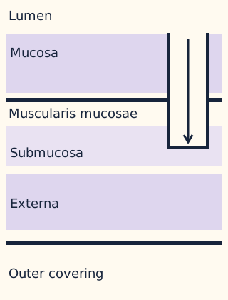
Specimen B: defect among submucosal vessels
Original depth-comparison state. Same layer order and scale in each state. Open full-size figure.
The defect has passed the thin muscle sheet but stops before externa: an ulcer. Larger submucosal vessels can be exposed even while the outer wall remains closed.
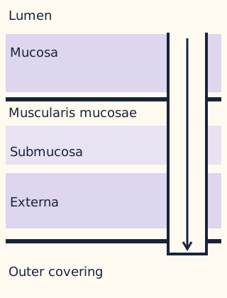
Specimen C: opening through the outer covering
Original depth-comparison state. Same layer order and scale in each state. Open full-size figure.
The defect now crosses the whole wall: perforation. At a peritoneal surface, luminal contents can enter the peritoneal cavity. Closing each drawing returns this comparison to its starting state.
Now consider a biopsy that contains only epithelium and lamina propria. It cannot establish that the deeper wall is normal because those layers were not sampled. The same sampling limit applies when describing cancer invasion or inflammation: report the deepest demonstrated involvement, distinguish absent disease from absent tissue, and use the organ-specific staging system when staging is actually required. This lesson's map is not a universal cancer-stage table.
Depth changes which structures are exposed. Specimen adequacy determines how much depth you can assess.
Can one feature locate a biopsy?
Follow the anatomical sequence: esophagus, stomach, duodenum, jejunum, ileum, then large bowel. The small intestine lies between the gastric outlet and ileocecal junction; the colon continues toward rectum and anus. The regional wall adapts to protection, secretion, absorption and stool handling. [6]
A signature feature is useful only when the specimen includes the layer where that feature lives. First identify epithelium and surface architecture. Then inspect the glands, immune tissue and depth. Failure to see Brunner glands in a mucosa-only biopsy does not exclude duodenum.
Protection and secretion before absorption
Esophagus has nonkeratinized stratified squamous epithelium, which tolerates abrasion. Its muscularis externa changes from predominantly skeletal proximally, through mixed muscle, to smooth distally. That gradient concerns the thick external muscle, not a conversion of the mucosal lining. [9]
Stomach has simple columnar mucus-secreting surface epithelium, pits and glands, but no villi. In body and fundus, parietal cells supply acid and intrinsic factor, while chief cells supply pepsinogen. Intrinsic factor supports vitamin B12 absorption in the terminal ileum; restoring acid alone does not replace intrinsic factor. [11] Acid helps activate pepsinogen to pepsin, so retained chief cells do not ensure normal protein digestion if the lumen is insufficiently acidic. Cardiac and pyloric glands have different cellular proportions; absence of abundant parietal cells does not by itself make a biopsy nongastric. Surface mucus and bicarbonate help protect the lining. The stomach also adds an inner oblique component to the usual external muscle arrangement for mixing. [2]
Compare small bowel with colon
Duodenum: villi and crypts above submucosal Brunner glands. Their alkaline mucus supports protection from incoming acidic chyme. Gastric glands, in contrast, are mucosal.
Jejunum: prominent plicae circulares and well-developed villi, without duodenal submucosal glands or ileal predominance of aggregated lymphoid patches. This is a combined pattern, not a unique named gland.
Ileum: generally shorter villi, more goblet cells and less prominent distal folds. Aggregated lymphoid follicles, Peyer patches, are especially characteristic. Normal lymphoid tissue can occur elsewhere too.
Colon: straight crypts with many goblet cells and absorptive epithelial cells, but no villi. Water and electrolyte recovery and mucus lubrication remain important despite the lack of villi. No villi does not mean no absorption.
These regional comparisons describe normal architecture. Inflammation, orientation and limited sampling can alter the appearance. The small-bowel transition is gradual, not a sharp border on every slide. [3][5]
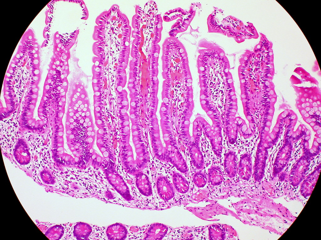
Trace the villi and crypts in this normal small-intestinal mucosa; compare their orientation with the injury patterns discussed in the lesson. Image: Ed Uthman, 2011, source photograph, CC BY 2.0.
Inspect the real tissue: follow an elongated projection from its tip toward the deeper glands. Name the projection and decide whether the image alone establishes the exact small-bowel segment.
Compare your interpretation
The projections are villi. Their architecture supports small intestine rather than normal colon or stomach. This field does not show a reliable regional submucosal landmark, so villi alone do not prove duodenum, jejunum or ileum.
Transfer the method: a separate specimen with a flat surface and densely packed straight goblet-rich crypts favors colon. A flat gastric surface instead leads into pits and region-specific glands. Always interpret an absent villus together with the structures that are present.
Do all surface folds have the same core?
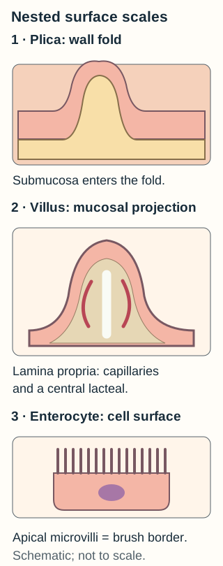
Both increase surface area, but they are made from different tissue levels. Plicae involve two wall compartments, villi project from mucosa, and microvilli extend from individual epithelial cells. A lesion can damage one level while leaving another recognizable. [3][5]
Original scale comparison. The panels are not at a common magnification. A larger drawing does not turn a cell membrane projection into a tissue fold. [3][5]Open full-size figure.
Plicae circulares are persistent folds of mucosa and submucosa that increase surface area and slow luminal transit. A villus has an epithelial covering and lamina propria core containing capillaries and a central lymphatic channel, the lacteal. Most absorbed sugars and amino acids enter the blood route. Packaged dietary lipid, particularly chylomicrons, enters the lymphatic route. [11]
Microvilli form the apical brush border of enterocytes. Their membranes contain digestive enzymes and transport machinery. Brush-border disaccharidases convert sugars such as maltose into absorbable monosaccharides. [11] Brush-border dysfunction can therefore impair digestion or uptake even when whole villi are still present. Colonic absorptive cells also have microvilli; the colon's absence of villi does not mean its epithelial surface is completely smooth at cellular scale. [3]
Predict: if a selective injury affects submucosa but spares mucosa, which pictured supporting core is directly affected?
Check the affected scale
The plica's submucosal core is affected. An individual villus contains lamina propria rather than a submucosal core. An apical microvillus contains neither compartment.
Crypts renew the surface; different cells defend it
Crypts of Lieberkuhn extend into the mucosa between villi. Stem cells near their bases replenish epithelial lineages. Enterocytes provide absorption; goblet cells supply mucus; enteroendocrine cells release regulatory signals. Paneth cells at small-intestinal crypt bases contain antimicrobial granules, including defensins and lysozyme. A crypt is not simply an inverted villus: renewal and secretion make it functionally distinct. [5]
Peyer patches organize mucosal immune surveillance, particularly in ileum. Overlying follicle-associated epithelium includes M cells, which transfer luminal antigen to underlying immune cells. They are not Paneth cells: antigen sampling differs from releasing antimicrobial substances into the crypt environment. [8]
Apply this distinction to a new problem: impaired antigen delivery with preserved crypt antimicrobial secretion points toward the follicle-associated sampling pathway, not automatically toward failed Paneth-cell defense. Conversely, loss of crypt progenitors compromises epithelial replacement even if surviving villi initially look intact.
Why does the nerve's address matter?
The two major enteric plexuses are connected networks with different emphases. The submucosal or Meissner plexus regulates local secretion, mucosal blood flow and conditions for absorption. The myenteric or Auerbach plexus lies between circular and longitudinal layers of muscularis externa and chiefly coordinates tone, motility and propulsion. These are predominant roles, not a claim that the networks never interact. [1]
Return to the wall drawing. A nerve sampled just below muscularis mucosae is not in the same plane as one sampled between the two thick external muscle layers. The mucosa can remain intact while deeper neural coordination fails. Conversely, disturbed secretion does not prove direct injury of external muscle.
Localize before predicting: in an isolated segment, direct stimulation still contracts smooth muscle, but a local lesion between its circular and longitudinal layers prevents an orderly propulsive sequence. Is the contractile apparatus absent, or is its coordination disrupted?
Check the functional distinction
Preserved response to direct stimulation argues that muscle can contract. The lesion's plane and disordered sequence point toward myenteric coordination. A submucosal lesion better fits a primary defect in local secretory regulation.
Peristalsis requires a spatial pattern: contraction behind luminal contents with relaxation ahead facilitates forward passage. Simultaneous contraction everywhere is not equivalent to effective propulsion. Segmentation mainly mixes contents rather than providing the same directional transport. [6][3]
For transfer, imagine the opposite experiment: coordinated contractions persist, but local neural stimulation no longer changes mucosal secretion. Start with the submucosal network. Neither experiment alone names a human disease; the selective lesions are teaching models that separate functions usually integrated in living bowel.
Does each wave make muscle contract?
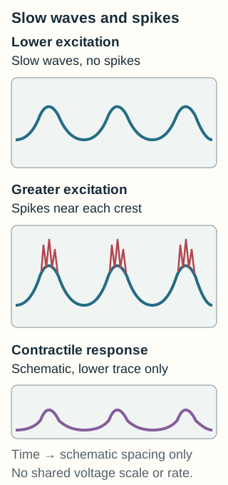
No. Interstitial cells of Cajal, electrically coupled to smooth muscle and other interstitial cells, organize slow-wave activity. ICC are not enteric neurons. Slow waves are rhythmic changes in membrane potential, not the rapid calcium-dependent spikes that may occur during depolarization. Excitatory and inhibitory inputs change the muscle network's excitability and mechanical response. [4]
Wave spacing and spike recruitment answer different questions. No voltage cutoff or diagnostic normal range is implied. [4]Open full-size figure.
Gastric slow waves are commonly about 3 cycles per minute. [12] Small-intestinal frequencies are higher and generally decline from duodenum toward distal ileum. The measured rhythm varies with region and experimental conditions; the numeric recordings in the cases are observations on the stated measurement scale; interpret them in clinical context rather than as diagnostic thresholds. [2][3][4]
For phasic contractions coupled to slow waves, the rhythm limits how often contractile opportunities recur. It does not tell you that every opportunity produces a contraction, how strong the contraction is, or whether the pattern propels contents. Neural input, hormones and local conditions influence excitation. Gastric excitation-contraction coupling is not identical to intestinal spike recruitment; the drawing deliberately models intestine rather than all gut muscle.
Count two things separately: In an ileal recording, nine slow waves occur in one minute. Initially three cycles recruit a contractile response; with greater excitation, seven do. Predict whether the pacemaker must have accelerated.
Check the two counts
No acceleration is required: the wave count remains nine, while more cycles recruit contraction. The experiment distinguishes rhythm generation from excitability; it does not establish a clinical treatment response.
Apply this to a different observation: if oscillations remain after neural blockade, that supports an intrinsic pacemaking network rather than a rhythm requiring each impulse from an external nerve. Normal rhythm alone still cannot establish normal coordinated transit. Finish by asking three separate questions: is a rhythm present, can muscle respond, and are the responses organized spatially?
Practice
Case 1
Show answer and explanations for case 1
A. B is an ulcer with access to larger submucosal vessels, but no demonstrated full-wall opening. (Best answer)
The local secretory plexus places the endpoint in submucosa. Crossing muscularis mucosae exposes that compartment and its larger vessels, while preserved externa argues against perforation.
Reasoning steps for option A
What anatomical or physiologic premise must hold for this option: B is an ulcer with access to larger submucosal vessels, but no demonstrated full-wall opening?
Submucosa can be exposed while the external muscle still separates the lumen from the outside.
Which stated finding most directly confirms or refutes this option: B is an ulcer with access to larger submucosal vessels, but no demonstrated full-wall opening?
The local secretory plexus places the endpoint in submucosa. Crossing muscularis mucosae exposes that compartment and its larger vessels, while preserved externa argues against perforation.
B. B is an ulcer with a full-wall opening, but no access to larger submucosal vessels. (Why this does not fit)
An ulcer entering submucosa can expose its vessels, but intact external muscle is inconsistent with the proposed full-wall opening.
Reasoning steps for option B
What anatomical or physiologic premise must hold for this option: B is an ulcer with a full-wall opening, but no access to larger submucosal vessels?
A full-wall defect would require crossing the intact muscle, while entry into submucosa already exposes its vessels.
Which stated finding most directly confirms or refutes this option: B is an ulcer with a full-wall opening, but no access to larger submucosal vessels?
An ulcer entering submucosa can expose its vessels, but intact external muscle is inconsistent with the proposed full-wall opening.
C. B is an ulcer with both a full-wall opening and access to larger submucosal vessels. (Why this does not fit)
Submucosal vessel exposure is supported; a full-wall opening is not, because the entire external muscle coat remains continuous.
Reasoning steps for option C
What anatomical or physiologic premise must hold for this option: B is an ulcer with both a full-wall opening and access to larger submucosal vessels?
Vessel exposure and perforation require different anatomical endpoints.
Which stated finding most directly confirms or refutes this option: B is an ulcer with both a full-wall opening and access to larger submucosal vessels?
Submucosal vessel exposure is supported; a full-wall opening is not, because the entire external muscle coat remains continuous.
D. B is an ulcer with neither a full-wall opening nor access to larger submucosal vessels. (Why this does not fit)
The absence of perforation is appropriate, but the endpoint has entered the vessel-bearing submucosa rather than remaining superficial like A.
Reasoning steps for option D
What anatomical or physiologic premise must hold for this option: B is an ulcer with neither a full-wall opening nor access to larger submucosal vessels?
An ulcer ending in submucosa would need to avoid the very compartment containing its larger vessels.
Which stated finding most directly confirms or refutes this option: B is an ulcer with neither a full-wall opening nor access to larger submucosal vessels?
The absence of perforation is appropriate, but the endpoint has entered the vessel-bearing submucosa rather than remaining superficial like A.
Takeaway: Ulcer depth can expose larger vessels without a full-thickness communication beyond the wall.
A. The lamina propria supporting the epithelium (Why this does not fit)
Preserved mucosa and secretion provide no evidence for a primary lamina propria lesion; the missing function is spatial motor coordination.
Reasoning steps for option A
What anatomical or physiologic premise must hold for this option: The lamina propria supporting the epithelium?
A mucosal support defect would need to account for the selective loss of a coordinated motor reflex.
Which stated finding most directly confirms or refutes this option: The lamina propria supporting the epithelium?
Preserved mucosa and secretion provide no evidence for a primary lamina propria lesion; the missing function is spatial motor coordination.
B. The submucosal network regulating secretion (Why this does not fit)
Submucosal circuits emphasize local mucosal regulation, which remains functional in this experiment.
Reasoning steps for option B
What anatomical or physiologic premise must hold for this option: The submucosal network regulating secretion?
A secretory-network lesion would be expected to disturb mucosal regulation more directly than an isolated spatial motor sequence.
Which stated finding most directly confirms or refutes this option: The submucosal network regulating secretion?
Submucosal circuits emphasize local mucosal regulation, which remains functional in this experiment.
C. The muscular contractile filaments throughout the segment as the source of the coordination defect (Why this does not fit)
Normal force after direct stimulation argues against generalized failure of the muscle contractile apparatus.
Reasoning steps for option C
What anatomical or physiologic premise must hold for this option: The muscular contractile filaments throughout the segment as the source of the coordination defect?
Failure of the muscle apparatus should also reduce directly evoked force.
Which stated finding most directly confirms or refutes this option: The muscular contractile filaments throughout the segment as the source of the coordination defect?
Normal force after direct stimulation argues against generalized failure of the muscle contractile apparatus.
D. The neural network between circular and longitudinal muscle (Best answer)
Preserved force with loss of the organized distension response localizes dysfunction to motor coordination, chiefly the myenteric network.
Reasoning steps for option D
What anatomical or physiologic premise must hold for this option: The neural network between circular and longitudinal muscle?
A motor control defect can preserve the force-producing cells but disrupt the sequence in which they respond.
Which stated finding most directly confirms or refutes this option: The neural network between circular and longitudinal muscle?
Preserved force with loss of the organized distension response localizes dysfunction to motor coordination, chiefly the myenteric network.
Takeaway: A bowel segment can generate force without organizing that force into propulsion.
A. At least submucosal involvement is demonstrated; involvement of externa cannot be assessed. (Best answer)
Fragment B demonstrates tissue beyond muscularis mucosae. Absence of externa from the specimens prevents assessing that compartment.
Reasoning steps for option A
What anatomical or physiologic premise must hold for this option: At least submucosal involvement is demonstrated; involvement of externa cannot be assessed?
A positive deeper fragment establishes a minimum depth, while absent external muscle limits the maximum assessment.
Which stated finding most directly confirms or refutes this option: At least submucosal involvement is demonstrated; involvement of externa cannot be assessed?
Fragment B demonstrates tissue beyond muscularis mucosae. Absence of externa from the specimens prevents assessing that compartment.
B. Disease is confined to mucosa because neither fragment shows involved external muscle, regardless of the missing deeper tissue. (Why this does not fit)
No involved external muscle is not equivalent to sampled, uninvolved external muscle. B already establishes extension beyond mucosa.
Reasoning steps for option B
What anatomical or physiologic premise must hold for this option: Disease is confined to mucosa because neither fragment shows involved external muscle, regardless of the missing deeper tissue?
Excluding external-muscle involvement requires that the relevant muscle actually be available for examination.
Which stated finding most directly confirms or refutes this option: Disease is confined to mucosa because neither fragment shows involved external muscle, regardless of the missing deeper tissue?
No involved external muscle is not equivalent to sampled, uninvolved external muscle. B already establishes extension beyond mucosa.
C. Extension to the myenteric plane is demonstrated; the outer covering is unassessed. (Why this does not fit)
The myenteric plane lies between external muscle layers, none of which were sampled.
Reasoning steps for option C
What anatomical or physiologic premise must hold for this option: Extension to the myenteric plane is demonstrated; the outer covering is unassessed?
Identifying the myenteric plane requires tissue at the interface of external muscle layers.
Which stated finding most directly confirms or refutes this option: Extension to the myenteric plane is demonstrated; the outer covering is unassessed?
The myenteric plane lies between external muscle layers, none of which were sampled.
D. Submucosal involvement is excluded because fragment A does not contain it. (Why this does not fit)
Fragment A lacks submucosa. It cannot negate the positive finding in the deeper fragment.
Reasoning steps for option D
What anatomical or physiologic premise must hold for this option: Submucosal involvement is excluded because fragment A does not contain it?
A shallow negative fragment can exclude deeper disease only if the deeper compartment was sampled.
Which stated finding most directly confirms or refutes this option: Submucosal involvement is excluded because fragment A does not contain it?
Fragment A lacks submucosa. It cannot negate the positive finding in the deeper fragment.
Takeaway: Report demonstrated depth separately from unsampled depth; an organ-specific stage requires additional context.
A. Both defects initially enter a mesothelium-lined peritoneal space. (Why this does not fit)
Thoracic esophagus is not surrounded by a peritoneal serosal surface. Its adventitia is continuous with mediastinal connective tissue.
Reasoning steps for option A
What anatomical or physiologic premise must hold for this option: Both defects initially enter a mesothelium-lined peritoneal space?
Both outer surfaces would need to face the peritoneal cavity.
Which stated finding most directly confirms or refutes this option: Both defects initially enter a mesothelium-lined peritoneal space?
Thoracic esophagus is not surrounded by a peritoneal serosal surface. Its adventitia is continuous with mediastinal connective tissue.
B. Both defects initially remain confined within the submucosa. (Why this does not fit)
The defects are specified as full-thickness, so neither stops in submucosa.
Reasoning steps for option B
What anatomical or physiologic premise must hold for this option: Both defects initially remain confined within the submucosa?
A submucosal endpoint would require the external muscle and outer covering not to have been crossed.
Which stated finding most directly confirms or refutes this option: Both defects initially remain confined within the submucosa?
The defects are specified as full-thickness, so neither stops in submucosa.
C. The esophageal defect enters adjacent mediastinal tissue; the bowel defect can enter the peritoneal cavity. (Best answer)
Adventitia attaches the thoracic esophagus to surrounding tissue, whereas mesothelial serosa faces the peritoneal cavity at the sampled bowel surface.
Reasoning steps for option C
What anatomical or physiologic premise must hold for this option: The esophageal defect enters adjacent mediastinal tissue; the bowel defect can enter the peritoneal cavity?
Different outer coverings place different tissues immediately beyond the wall.
Which stated finding most directly confirms or refutes this option: The esophageal defect enters adjacent mediastinal tissue; the bowel defect can enter the peritoneal cavity?
Adventitia attaches the thoracic esophagus to surrounding tissue, whereas mesothelial serosa faces the peritoneal cavity at the sampled bowel surface.
D. The esophageal defect enters the peritoneal cavity; the bowel defect enters an adventitial attachment first. (Why this does not fit)
This reverses the described coverings: the fibrous mediastinal attachment is adventitia and the mesothelial bowel surface is serosa.
Reasoning steps for option D
What anatomical or physiologic premise must hold for this option: The esophageal defect enters the peritoneal cavity; the bowel defect enters an adventitial attachment first?
The mesothelial and attached fibrous surfaces would need to belong to the opposite specimens.
Which stated finding most directly confirms or refutes this option: The esophageal defect enters the peritoneal cavity; the bowel defect enters an adventitial attachment first?
This reverses the described coverings: the fibrous mediastinal attachment is adventitia and the mesothelial bowel surface is serosa.
Takeaway: The consequences of full-wall extension depend on the anatomical surface actually sampled.
A. The unchanged force establishes that forward propulsion remains normal. (Why this does not fit)
Equal peak force does not establish the spatial sequence needed for propulsion. Simultaneous contraction can obstruct rather than propel.
Reasoning steps for option A
What anatomical or physiologic premise must hold for this option: The unchanged force establishes that forward propulsion remains normal?
Preserved force would have to guarantee the direction of transport regardless of spatial timing.
Which stated finding most directly confirms or refutes this option: The unchanged force establishes that forward propulsion remains normal?
Equal peak force does not establish the spatial sequence needed for propulsion. Simultaneous contraction can obstruct rather than propel.
B. Motor coordination is impaired even though contractile capacity is preserved. (Best answer)
Loss of the oral-to-anal pattern with preserved force distinguishes coordination from contractile capacity; intact secretion argues against a dominant mucosal secretory defect.
Reasoning steps for option B
What anatomical or physiologic premise must hold for this option: Motor coordination is impaired even though contractile capacity is preserved?
The cells can retain force production while losing the ordered oral-to-anal pattern.
Which stated finding most directly confirms or refutes this option: Motor coordination is impaired even though contractile capacity is preserved?
Loss of the oral-to-anal pattern with preserved force distinguishes coordination from contractile capacity; intact secretion argues against a dominant mucosal secretory defect.
C. Submucosal secretory control has failed despite the normal measured secretion. (Why this does not fit)
The measured mucosal response is preserved, not lost. The changed variable is the spatial contraction pattern.
Reasoning steps for option C
What anatomical or physiologic premise must hold for this option: Submucosal secretory control has failed despite the normal measured secretion?
A defect in secretion would need evidence of an abnormal secretory response.
Which stated finding most directly confirms or refutes this option: Submucosal secretory control has failed despite the normal measured secretion?
The measured mucosal response is preserved, not lost. The changed variable is the spatial contraction pattern.
D. The intervention has eliminated all intrinsic electrical activity of muscle. (Why this does not fit)
Mechanical measurements alone do not establish loss of slow waves; muscle still generates force after the intervention.
Reasoning steps for option D
What anatomical or physiologic premise must hold for this option: The intervention has eliminated all intrinsic electrical activity of muscle?
Inferring lost electrical rhythmicity requires an electrical observation, not only a changed motor pattern.
Which stated finding most directly confirms or refutes this option: The intervention has eliminated all intrinsic electrical activity of muscle?
Mechanical measurements alone do not establish loss of slow waves; muscle still generates force after the intervention.
Takeaway: Propulsion is a spatially organized response, not a synonym for strong contraction.
Preserved direct epithelial secretion localizes the failure upstream of the epithelial machinery. Preserved organized motor responses favor a mucosal regulatory circuit rather than myenteric motor failure.
Reasoning steps for option A
What anatomical or physiologic premise must hold for this option: The submucosal enteric circuit?
An upstream secretory circuit can fail while epithelial effectors and motor circuitry still respond.
Which stated finding most directly confirms or refutes this option: The submucosal enteric circuit?
Preserved direct epithelial secretion localizes the failure upstream of the epithelial machinery. Preserved organized motor responses favor a mucosal regulatory circuit rather than myenteric motor failure.
B. The epithelial secretory machinery (Why this does not fit)
The epithelial cells still secrete normally when directly stimulated, so their secretory machinery is functional.
Reasoning steps for option B
What anatomical or physiologic premise must hold for this option: The epithelial secretory machinery?
Damaged epithelial secretory machinery should fail during direct stimulation as well.
Which stated finding most directly confirms or refutes this option: The epithelial secretory machinery?
The epithelial cells still secrete normally when directly stimulated, so their secretory machinery is functional.
C. The myenteric motor circuit (Why this does not fit)
The organized distension response remains intact. The selective deficit is neural regulation of secretion, chiefly a submucosal function.
Reasoning steps for option C
What anatomical or physiologic premise must hold for this option: The myenteric motor circuit?
A myenteric motor lesion should threaten the organized distension response.
Which stated finding most directly confirms or refutes this option: The myenteric motor circuit?
The organized distension response remains intact. The selective deficit is neural regulation of secretion, chiefly a submucosal function.
D. The external muscle contractile apparatus (Why this does not fit)
External muscle failure would impair the motor response, not selectively interrupt secretion while preserving that response.
Reasoning steps for option D
What anatomical or physiologic premise must hold for this option: The external muscle contractile apparatus?
A muscle-apparatus lesion should impair the observed contractile motor response.
Which stated finding most directly confirms or refutes this option: The external muscle contractile apparatus?
External muscle failure would impair the motor response, not selectively interrupt secretion while preserving that response.
Takeaway: Direct and reflex responses help separate an effector defect from a regulatory-circuit defect.
A. X is more distal, and its prominent folds are cellular microvilli. (Why this does not fit)
The combined pattern favors jejunum for X and ileum for Y. Permanent folds are mucosa plus submucosa, not cellular microvilli.
Reasoning steps for option A
What anatomical or physiologic premise must hold for this option: X is more distal, and its prominent folds are cellular microvilli?
The regional fold gradient and the structural definition of a plica would both have to be reversed.
Which stated finding most directly confirms or refutes this option: X is more distal, and its prominent folds are cellular microvilli?
The combined pattern favors jejunum for X and ileum for Y. Permanent folds are mucosa plus submucosa, not cellular microvilli.
B. Y is more proximal, and its follicles replace all absorptive epithelium, leaving no remaining villus transport surface. (Why this does not fit)
Y favors ileum, which is distal to jejunum. Peyer patches do not replace all surrounding absorptive epithelium.
Reasoning steps for option B
What anatomical or physiologic premise must hold for this option: Y is more proximal, and its follicles replace all absorptive epithelium, leaving no remaining villus transport surface?
Organized immune patches would have to replace the entire remaining absorptive surface.
Which stated finding most directly confirms or refutes this option: Y is more proximal, and its follicles replace all absorptive epithelium, leaving no remaining villus transport surface?
Y favors ileum, which is distal to jejunum. Peyer patches do not replace all surrounding absorptive epithelium.
C. X is more proximal, but only Y has villus capillaries and lacteals. (Why this does not fit)
Both jejunal and ileal villi have blood and lymphatic transport routes; prominent folds do not eliminate those routes in X.
Reasoning steps for option C
What anatomical or physiologic premise must hold for this option: X is more proximal, but only Y has villus capillaries and lacteals?
The regional contrast would need to eliminate normal villus vessels from the jejunal sample.
Which stated finding most directly confirms or refutes this option: X is more proximal, but only Y has villus capillaries and lacteals?
Both jejunal and ileal villi have blood and lymphatic transport routes; prominent folds do not eliminate those routes in X.
D. X favors jejunum relative to Y; both retain villus transport routes despite different folds. (Best answer)
Prominent folds and long villi favor jejunum, whereas shorter villi with aggregated follicles favor ileum. Both remain absorptive small bowel.
Reasoning steps for option D
What anatomical or physiologic premise must hold for this option: X favors jejunum relative to Y; both retain villus transport routes despite different folds?
A change in fold prominence can occur without loss of blood and lymphatic pathways in surviving villi.
Which stated finding most directly confirms or refutes this option: X favors jejunum relative to Y; both retain villus transport routes despite different folds?
Prominent folds and long villi favor jejunum, whereas shorter villi with aggregated follicles favor ileum. Both remain absorptive small bowel.
Takeaway: Regional differences in fold prominence do not imply that one small-bowel segment lacks villus transport pathways.
A. Paneth-cell antimicrobial output is impaired, but the follicular epithelial delivery pathway is intact. (Why this does not fit)
Antimicrobial output is measured as preserved, and the luminal delivery route fails. This is not a primary Paneth-cell secretory defect.
Reasoning steps for option A
What anatomical or physiologic premise must hold for this option: Paneth-cell antimicrobial output is impaired, but the follicular epithelial delivery pathway is intact?
A Paneth-cell defect should reduce the measured crypt antimicrobial output.
Which stated finding most directly confirms or refutes this option: Paneth-cell antimicrobial output is impaired, but the follicular epithelial delivery pathway is intact?
Antimicrobial output is measured as preserved, and the luminal delivery route fails. This is not a primary Paneth-cell secretory defect.
B. Follicular epithelial antigen delivery is intact, but underlying immune-cell responsiveness is impaired. (Why this does not fit)
The bypass injection evokes a normal immune response, so the downstream immune cells respond when antigen reaches them. It is the epithelial delivery route that fails.
Reasoning steps for option B
What anatomical or physiologic premise must hold for this option: Follicular epithelial antigen delivery is intact, but underlying immune-cell responsiveness is impaired?
Downstream immune failure should remain when the epithelial barrier is bypassed.
Which stated finding most directly confirms or refutes this option: Follicular epithelial antigen delivery is intact, but underlying immune-cell responsiveness is impaired?
The bypass injection evokes a normal immune response, so the downstream immune cells respond when antigen reaches them. It is the epithelial delivery route that fails.
C. M-cell delivery and underlying immune-cell responsiveness are both impaired. (Why this does not fit)
A defect in downstream immune responsiveness should persist when antigen is injected beyond the epithelium; it does not.
Reasoning steps for option C
What anatomical or physiologic premise must hold for this option: M-cell delivery and underlying immune-cell responsiveness are both impaired?
Combined delivery and immune defects should prevent a normal response after subepithelial injection.
Which stated finding most directly confirms or refutes this option: M-cell delivery and underlying immune-cell responsiveness are both impaired?
A defect in downstream immune responsiveness should persist when antigen is injected beyond the epithelium; it does not.
D. M-cell transepithelial delivery is impaired, but underlying immune-cell responsiveness is retained. (Best answer)
Failure from the lumen but success after bypassing the follicular epithelium localizes the interruption to M-cell delivery, while demonstrating retained downstream responsiveness.
Reasoning steps for option D
What anatomical or physiologic premise must hold for this option: M-cell transepithelial delivery is impaired, but underlying immune-cell responsiveness is retained?
A barrier-specific delivery defect can fail from the lumen yet respond normally when that barrier is bypassed.
Which stated finding most directly confirms or refutes this option: M-cell transepithelial delivery is impaired, but underlying immune-cell responsiveness is retained?
Failure from the lumen but success after bypassing the follicular epithelium localizes the interruption to M-cell delivery, while demonstrating retained downstream responsiveness.
Takeaway: A bypass experiment can separate failed epithelial antigen delivery from failed underlying immune-cell activation.
A. Reduced mucus lubrication with continued capacity for water and electrolyte absorption (Best answer)
The baseline architecture is colonic. Goblet-cell depletion reduces mucus, while retained absorptive cells and microvilli preserve an absorptive route.
Reasoning steps for option A
What anatomical or physiologic premise must hold for this option: Reduced mucus lubrication with continued capacity for water and electrolyte absorption?
Mucus-producing and absorptive epithelial cells can make different contributions in the same colonic mucosa.
Which stated finding most directly confirms or refutes this option: Reduced mucus lubrication with continued capacity for water and electrolyte absorption?
The baseline architecture is colonic. Goblet-cell depletion reduces mucus, while retained absorptive cells and microvilli preserve an absorptive route.
B. Loss of villus lymphatic drainage with preserved mucus lubrication (Why this does not fit)
The described region has colonic crypts rather than villi with central lacteals; the selective loss is mucus-producing cells.
Reasoning steps for option B
What anatomical or physiologic premise must hold for this option: Loss of villus lymphatic drainage with preserved mucus lubrication?
The baseline specimen would need to contain villi and central lacteals.
Which stated finding most directly confirms or refutes this option: Loss of villus lymphatic drainage with preserved mucus lubrication?
The described region has colonic crypts rather than villi with central lacteals; the selective loss is mucus-producing cells.
C. Loss of gastric acid secretion with preserved intrinsic-factor production by the same surviving parietal cells (Why this does not fit)
The baseline architecture and targeted cells do not identify gastric parietal cells, which supply both acid and intrinsic factor.
Reasoning steps for option C
What anatomical or physiologic premise must hold for this option: Loss of gastric acid secretion with preserved intrinsic-factor production by the same surviving parietal cells?
The targeted cells would need to be gastric parietal cells rather than mucus-filled colonic cells.
Which stated finding most directly confirms or refutes this option: Loss of gastric acid secretion with preserved intrinsic-factor production by the same surviving parietal cells?
The baseline architecture and targeted cells do not identify gastric parietal cells, which supply both acid and intrinsic factor.
D. Loss of all cellular absorptive surface because the region has no villi (Why this does not fit)
Colon lacks villi but its absorptive cells have microvilli. No tissue-scale villi does not mean no cellular absorptive surface.
Reasoning steps for option D
What anatomical or physiologic premise must hold for this option: Loss of all cellular absorptive surface because the region has no villi?
Absence of villi would have to imply absence of cellular microvilli as well.
Which stated finding most directly confirms or refutes this option: Loss of all cellular absorptive surface because the region has no villi?
Colon lacks villi but its absorptive cells have microvilli. No tissue-scale villi does not mean no cellular absorptive surface.
Takeaway: Colon combines crypt-based mucus production with microvillous epithelial absorption despite lacking villi.
A. Both samples lose their response equally because their epithelium is identical. (Why this does not fit)
Shared epithelial type does not establish shared external-muscle composition. Esophageal muscle varies along its length.
Reasoning steps for option A
What anatomical or physiologic premise must hold for this option: Both samples lose their response equally because their epithelium is identical?
An identical epithelial lining would need to establish identical muscle composition.
Which stated finding most directly confirms or refutes this option: Both samples lose their response equally because their epithelium is identical?
Shared epithelial type does not establish shared external-muscle composition. Esophageal muscle varies along its length.
B. The distal sample is more affected because its muscle is predominantly skeletal. (Why this does not fit)
The skeletal-to-smooth transition runs from proximal to distal, not in the reverse direction.
Reasoning steps for option B
What anatomical or physiologic premise must hold for this option: The distal sample is more affected because its muscle is predominantly skeletal?
The distal esophagus would need to have the larger skeletal-muscle fraction.
Which stated finding most directly confirms or refutes this option: The distal sample is more affected because its muscle is predominantly skeletal?
The skeletal-to-smooth transition runs from proximal to distal, not in the reverse direction.
C. The proximal sample is more affected because its muscle is predominantly skeletal. (Best answer)
Squamous lining and the anatomical course identify esophagus. Proximal external muscle is predominantly skeletal, making it more vulnerable to the stipulated selective block.
Reasoning steps for option C
What anatomical or physiologic premise must hold for this option: The proximal sample is more affected because its muscle is predominantly skeletal?
The anatomical gradient places more skeletal muscle near the pharynx.
Which stated finding most directly confirms or refutes this option: The proximal sample is more affected because its muscle is predominantly skeletal?
Squamous lining and the anatomical course identify esophagus. Proximal external muscle is predominantly skeletal, making it more vulnerable to the stipulated selective block.
D. Neither sample is affected because squamous mucosa requires smooth external muscle. (Why this does not fit)
Epithelial type does not determine muscle type. Proximal esophagus normally contains skeletal external muscle.
Reasoning steps for option D
What anatomical or physiologic premise must hold for this option: Neither sample is affected because squamous mucosa requires smooth external muscle?
The epithelial pattern would need to prohibit skeletal muscle elsewhere in the wall.
Which stated finding most directly confirms or refutes this option: Neither sample is affected because squamous mucosa requires smooth external muscle?
Epithelial type does not determine muscle type. Proximal esophagus normally contains skeletal external muscle.
Takeaway: Esophageal epithelial continuity coexists with a proximal skeletal-to-distal smooth external-muscle transition.
A. Pepsinogen production remains absent, while vitamin B12 uptake returns because acidity replaces intrinsic factor. (Why this does not fit)
Chief cells are preserved, and acid has restored the digestive function. Acidity does not substitute for intrinsic factor in vitamin B12 absorption.
Reasoning steps for option A
What anatomical or physiologic premise must hold for this option: Pepsinogen production remains absent, while vitamin B12 uptake returns because acidity replaces intrinsic factor?
The preserved chief cells would have to stop producing precursor, and acid would need to substitute for intrinsic factor.
Which stated finding most directly confirms or refutes this option: Pepsinogen production remains absent, while vitamin B12 uptake returns because acidity replaces intrinsic factor?
Chief cells are preserved, and acid has restored the digestive function. Acidity does not substitute for intrinsic factor in vitamin B12 absorption.
B. Both pepsin activity and vitamin B12 uptake remain impaired because chief cells cannot function without parietal cells. (Why this does not fit)
The experiment directly demonstrates restored protein digestion. Chief-cell precursor production and acid-dependent activation are distinguishable functions.
Reasoning steps for option B
What anatomical or physiologic premise must hold for this option: Both pepsin activity and vitamin B12 uptake remain impaired because chief cells cannot function without parietal cells?
The demonstrated rescue of protein digestion would need to be disregarded despite retained chief cells.
Which stated finding most directly confirms or refutes this option: Both pepsin activity and vitamin B12 uptake remain impaired because chief cells cannot function without parietal cells?
The experiment directly demonstrates restored protein digestion. Chief-cell precursor production and acid-dependent activation are distinguishable functions.
C. Protein digestion improves, but vitamin B12 uptake remains impaired because intrinsic factor is still deficient. (Best answer)
Parietal loss removes acid and intrinsic factor. Acid can restore pepsinogen activation from preserved chief cells, but it does not replace the intrinsic-factor pathway needed for ileal vitamin B12 uptake.
Reasoning steps for option C
What anatomical or physiologic premise must hold for this option: Protein digestion improves, but vitamin B12 uptake remains impaired because intrinsic factor is still deficient?
Acid and intrinsic factor have distinct functions, so replacing acid can restore only one deficient pathway.
Which stated finding most directly confirms or refutes this option: Protein digestion improves, but vitamin B12 uptake remains impaired because intrinsic factor is still deficient?
Parietal loss removes acid and intrinsic factor. Acid can restore pepsinogen activation from preserved chief cells, but it does not replace the intrinsic-factor pathway needed for ileal vitamin B12 uptake.
D. Both protein digestion and vitamin B12 uptake return because both parietal-cell products are acids. (Why this does not fit)
Intrinsic factor is not acid. Replacing the luminal acidity does not replace the separate binding protein required for the vitamin B12 pathway.
Reasoning steps for option D
What anatomical or physiologic premise must hold for this option: Both protein digestion and vitamin B12 uptake return because both parietal-cell products are acids?
Intrinsic factor would need to be chemically and functionally equivalent to gastric acid.
Which stated finding most directly confirms or refutes this option: Both protein digestion and vitamin B12 uptake return because both parietal-cell products are acids?
Intrinsic factor is not acid. Replacing the luminal acidity does not replace the separate binding protein required for the vitamin B12 pathway.
Takeaway: Replacing one product of a damaged lineage does not necessarily restore the effects of its other products.
A. Loss of the supporting core of each individual villus (Why this does not fit)
Individual villus cores are lamina propria within mucosa, which is specified as preserved.
Reasoning steps for option A
What anatomical or physiologic premise must hold for this option: Loss of the supporting core of each individual villus?
An individual villus would need a submucosal rather than mucosal core.
Which stated finding most directly confirms or refutes this option: Loss of the supporting core of each individual villus?
Individual villus cores are lamina propria within mucosa, which is specified as preserved.
B. Loss of support within the broad ridges, with individual villus cores still present (Best answer)
The removed compartment is submucosa. It extends into plicae circulares, whereas individual villi retain their mucosal cores.
Reasoning steps for option B
What anatomical or physiologic premise must hold for this option: Loss of support within the broad ridges, with individual villus cores still present?
A plica can contain the injured compartment while its smaller mucosal projections do not.
Which stated finding most directly confirms or refutes this option: Loss of support within the broad ridges, with individual villus cores still present?
The removed compartment is submucosa. It extends into plicae circulares, whereas individual villi retain their mucosal cores.
C. Loss of every apical membrane projection despite preserved enterocytes (Why this does not fit)
Microvilli are part of surviving enterocyte membranes, not the removed submucosal compartment.
Reasoning steps for option C
What anatomical or physiologic premise must hold for this option: Loss of every apical membrane projection despite preserved enterocytes?
A cell membrane projection would need to depend structurally on the removed submucosal core.
Which stated finding most directly confirms or refutes this option: Loss of every apical membrane projection despite preserved enterocytes?
Microvilli are part of surviving enterocyte membranes, not the removed submucosal compartment.
D. Conversion of mucosal crypts into submucosal glands (Why this does not fit)
Removing submucosa does not relocate mucosal crypts or convert them into glands of another compartment.
Reasoning steps for option D
What anatomical or physiologic premise must hold for this option: Conversion of mucosal crypts into submucosal glands?
Changing a supporting compartment would need to relocate the epithelial gland itself.
Which stated finding most directly confirms or refutes this option: Conversion of mucosal crypts into submucosal glands?
Removing submucosa does not relocate mucosal crypts or convert them into glands of another compartment.
Takeaway: A plica includes submucosa; a villus does not.
A. Villus blood export is impaired despite preserved digestion; the defect is in the capillary bed carrying absorbed glucose toward portal blood. (Why this does not fit)
Normal portal appearance after direct glucose delivery establishes a functional route across epithelium and into blood for that substrate.
Reasoning steps for option A
What anatomical or physiologic premise must hold for this option: Villus blood export is impaired despite preserved digestion; the defect is in the capillary bed carrying absorbed glucose toward portal blood?
A failed blood-export route should impair directly supplied glucose as well as glucose derived from maltose.
Which stated finding most directly confirms or refutes this option: Villus blood export is impaired despite preserved digestion; the defect is in the capillary bed carrying absorbed glucose toward portal blood?
Normal portal appearance after direct glucose delivery establishes a functional route across epithelium and into blood for that substrate.
B. Final disaccharide digestion is impaired despite preserved glucose transport; the defect is at enterocyte brush-border membrane. (Best answer)
Providing the final monosaccharide bypasses the failed conversion of maltose to glucose. That final digestive step depends on enzymes at the apical brush border, which can fail despite preserved whole villi.
Reasoning steps for option B
What anatomical or physiologic premise must hold for this option: Final disaccharide digestion is impaired despite preserved glucose transport; the defect is at enterocyte brush-border membrane?
A digestive conversion can fail while its final product still uses intact epithelial and vascular transport.
Which stated finding most directly confirms or refutes this option: Final disaccharide digestion is impaired despite preserved glucose transport; the defect is at enterocyte brush-border membrane?
Providing the final monosaccharide bypasses the failed conversion of maltose to glucose. That final digestive step depends on enzymes at the apical brush border, which can fail despite preserved whole villi.
C. Glucose transport is impaired despite preserved digestion; the defect is at enterocyte brush-border membrane. (Why this does not fit)
If the glucose-transport step were the primary failure, directly delivered luminal glucose should also have reduced portal appearance.
Reasoning steps for option C
What anatomical or physiologic premise must hold for this option: Glucose transport is impaired despite preserved digestion; the defect is at enterocyte brush-border membrane?
A glucose transport defect should not disappear when glucose rather than maltose is supplied.
Which stated finding most directly confirms or refutes this option: Glucose transport is impaired despite preserved digestion; the defect is at enterocyte brush-border membrane?
If the glucose-transport step were the primary failure, directly delivered luminal glucose should also have reduced portal appearance.
D. Villus lipid export is impaired despite preserved digestion; the defect is in the central lacteal. (Why this does not fit)
The experiment tests carbohydrate digestion and blood export, not chylomicron transport through lacteals.
Reasoning steps for option D
What anatomical or physiologic premise must hold for this option: Villus lipid export is impaired despite preserved digestion; the defect is in the central lacteal?
A lymphatic lipid pathway would need to account for the observed carbohydrate-specific bypass result.
Which stated finding most directly confirms or refutes this option: Villus lipid export is impaired despite preserved digestion; the defect is in the central lacteal?
The experiment tests carbohydrate digestion and blood export, not chylomicron transport through lacteals.
Takeaway: Supplying a final product can distinguish failed digestion from failed transport of that product.
A. Paneth-cell function is impaired while the regenerative crypt source remains active. (Best answer)
Crypt-base antimicrobial granules identify Paneth cells. Continued proliferation and replacement demonstrate a surviving regenerative source rather than global crypt failure.
Reasoning steps for option A
What anatomical or physiologic premise must hold for this option: Paneth-cell function is impaired while the regenerative crypt source remains active?
An antimicrobial secretory lineage can fail while crypt progenitors continue supplying new cells.
Which stated finding most directly confirms or refutes this option: Paneth-cell function is impaired while the regenerative crypt source remains active?
Crypt-base antimicrobial granules identify Paneth cells. Continued proliferation and replacement demonstrate a surviving regenerative source rather than global crypt failure.
B. Crypt stem cells are absent while Paneth-cell antimicrobial function is preserved. (Why this does not fit)
The measured changes are the reverse: antimicrobial output falls while epithelial renewal persists.
Reasoning steps for option B
What anatomical or physiologic premise must hold for this option: Crypt stem cells are absent while Paneth-cell antimicrobial function is preserved?
Loss of progenitors should reduce replacement, whereas intact Paneth cells should preserve antimicrobial output.
Which stated finding most directly confirms or refutes this option: Crypt stem cells are absent while Paneth-cell antimicrobial function is preserved?
The measured changes are the reverse: antimicrobial output falls while epithelial renewal persists.
C. M-cell sampling has failed and explains the loss of crypt antimicrobial granules. (Why this does not fit)
M cells transport antigen across follicle-associated epithelium rather than supplying the described crypt-base granules.
Reasoning steps for option C
What anatomical or physiologic premise must hold for this option: M-cell sampling has failed and explains the loss of crypt antimicrobial granules?
Follicular sampling cells would need to account for crypt-base antimicrobial granules.
Which stated finding most directly confirms or refutes this option: M-cell sampling has failed and explains the loss of crypt antimicrobial granules?
M cells transport antigen across follicle-associated epithelium rather than supplying the described crypt-base granules.
D. Villus lymphatic obstruction explains both preserved renewal and reduced peptide secretion. (Why this does not fit)
Lacteals transport absorbed lipid and do not secrete the measured crypt antimicrobial peptides.
Reasoning steps for option D
What anatomical or physiologic premise must hold for this option: Villus lymphatic obstruction explains both preserved renewal and reduced peptide secretion?
A lipid transport channel would need to control crypt antimicrobial secretion directly.
Which stated finding most directly confirms or refutes this option: Villus lymphatic obstruction explains both preserved renewal and reduced peptide secretion?
Lacteals transport absorbed lipid and do not secrete the measured crypt antimicrobial peptides.
Takeaway: Loss of one crypt lineage does not necessarily imply loss of the regenerative niche.
A. X is ileum and Y is colon; both can absorb through epithelial cells despite different architecture. (Best answer)
The patterns favor ileum and colon respectively. Colonic epithelial absorption does not require tissue-scale villi, and colonic absorptive cells retain microvilli.
Reasoning steps for option A
What anatomical or physiologic premise must hold for this option: X is ileum and Y is colon; both can absorb through epithelial cells despite different architecture?
Different regional tissue architecture can coexist with epithelial absorption in both samples.
Which stated finding most directly confirms or refutes this option: X is ileum and Y is colon; both can absorb through epithelial cells despite different architecture?
The patterns favor ileum and colon respectively. Colonic epithelial absorption does not require tissue-scale villi, and colonic absorptive cells retain microvilli.
B. X is colon and Y is ileum; absorption requires a villus central lymphatic route for all absorbed products in both segments. (Why this does not fit)
This reverses the regional architecture, and water or solute uptake into blood does not require a villus lymphatic route.
Reasoning steps for option B
What anatomical or physiologic premise must hold for this option: X is colon and Y is ileum; absorption requires a villus central lymphatic route for all absorbed products in both segments?
Both the regional assignment and the need for a lymphatic route for all absorption would have to hold.
Which stated finding most directly confirms or refutes this option: X is colon and Y is ileum; absorption requires a villus central lymphatic route for all absorbed products in both segments?
This reverses the regional architecture, and water or solute uptake into blood does not require a villus lymphatic route.
C. X is ileum and Y is colon; Y lacks both villi and cellular microvilli. (Why this does not fit)
The regional assignment is reasonable, but the inference about microvilli is false: colonic absorptive cells have them.
Reasoning steps for option C
What anatomical or physiologic premise must hold for this option: X is ileum and Y is colon; Y lacks both villi and cellular microvilli?
The absence of tissue-scale villi would need to extend to cellular apical projections.
Which stated finding most directly confirms or refutes this option: X is ileum and Y is colon; Y lacks both villi and cellular microvilli?
The regional assignment is reasonable, but the inference about microvilli is false: colonic absorptive cells have them.
D. X is jejunum and Y is stomach; both require Peyer patches for absorption. (Why this does not fit)
The specimens do not show the expected jejunal versus gastric comparison, and Peyer patches provide immune surveillance rather than being necessary for all absorption.
Reasoning steps for option D
What anatomical or physiologic premise must hold for this option: X is jejunum and Y is stomach; both require Peyer patches for absorption?
The observed patterns would need to be gastric versus jejunal and require immune patches for absorption.
Which stated finding most directly confirms or refutes this option: X is jejunum and Y is stomach; both require Peyer patches for absorption?
The specimens do not show the expected jejunal versus gastric comparison, and Peyer patches provide immune surveillance rather than being necessary for all absorption.
Takeaway: Identify the region and then separate tissue architecture from cellular absorptive capacity.
A. The pacemaker accelerated fourfold, establishing faster propulsion. (Why this does not fit)
The recorded pacemaker frequency is unchanged. More contractions also do not establish forward transit, which remains absent in this preparation.
Reasoning steps for option A
What anatomical or physiologic premise must hold for this option: The pacemaker accelerated fourfold, establishing faster propulsion?
The number of slow waves would need to rise in parallel with contraction count.
Which stated finding most directly confirms or refutes this option: The pacemaker accelerated fourfold, establishing faster propulsion?
The recorded pacemaker frequency is unchanged. More contractions also do not establish forward transit, which remains absent in this preparation.
B. The muscle lost intrinsic rhythmicity, while neural firing restored transit. (Why this does not fit)
Slow waves persist, and the marker fails to advance. Neither asserted result is supported.
Reasoning steps for option B
What anatomical or physiologic premise must hold for this option: The muscle lost intrinsic rhythmicity, while neural firing restored transit?
An observed persistent rhythm would need to be absent, and an immobile marker would need to show transit.
Which stated finding most directly confirms or refutes this option: The muscle lost intrinsic rhythmicity, while neural firing restored transit?
Slow waves persist, and the marker fails to advance. Neither asserted result is supported.
C. More existing cycles recruited contraction, but the recordings do not establish effective propulsion. (Best answer)
The contraction-to-wave ratio increases without a frequency increase. Failure of forward passage shows why local recruitment must be distinguished from spatially organized propulsion.
Reasoning steps for option C
What anatomical or physiologic premise must hold for this option: More existing cycles recruited contraction, but the recordings do not establish effective propulsion?
Recruitment within a stable rhythm and successful spatial transport can vary independently.
Which stated finding most directly confirms or refutes this option: More existing cycles recruited contraction, but the recordings do not establish effective propulsion?
The contraction-to-wave ratio increases without a frequency increase. Failure of forward passage shows why local recruitment must be distinguished from spatially organized propulsion.
D. The unchanged wave count proves that neither muscle excitation nor transport changed anywhere in the preparation. (Why this does not fit)
An unchanged rhythm does not imply unchanged excitation: spike-associated contractions increased from two to eight.
Reasoning steps for option D
What anatomical or physiologic premise must hold for this option: The unchanged wave count proves that neither muscle excitation nor transport changed anywhere in the preparation?
Pacemaker frequency would need to determine every other electrical and mechanical measurement.
Which stated finding most directly confirms or refutes this option: The unchanged wave count proves that neither muscle excitation nor transport changed anywhere in the preparation?
An unchanged rhythm does not imply unchanged excitation: spike-associated contractions increased from two to eight.
Takeaway: Count rhythm, contractile recruitment and successful transit separately.
A. X favors ileum at 12 cycles per minute, and its observed electrical rate alone proves stronger forward propulsion. (Why this does not fit)
Twelve per minute is the correct conversion for X, but submucosal Brunner glands favor duodenum and frequency alone does not establish propulsion.
Reasoning steps for option A
What anatomical or physiologic premise must hold for this option: X favors ileum at 12 cycles per minute, and its observed electrical rate alone proves stronger forward propulsion?
The per-minute conversion can be right even when region and functional inference are wrong.
Which stated finding most directly confirms or refutes this option: X favors ileum at 12 cycles per minute, and its observed electrical rate alone proves stronger forward propulsion?
Twelve per minute is the correct conversion for X, but submucosal Brunner glands favor duodenum and frequency alone does not establish propulsion.
B. X favors duodenum at 12 cycles per minute, but its higher rate does not establish stronger propulsion. (Best answer)
The glands locate X to duodenum; four cycles in 20 seconds equal 12 per minute. Y favors ileum at 9 per minute. Their rhythm difference does not measure force or effective transit.
Reasoning steps for option B
What anatomical or physiologic premise must hold for this option: X favors duodenum at 12 cycles per minute, but its higher rate does not establish stronger propulsion?
A submucosal gland landmark and a timed count can establish region and rate without measuring propulsion.
Which stated finding most directly confirms or refutes this option: X favors duodenum at 12 cycles per minute, but its higher rate does not establish stronger propulsion?
The glands locate X to duodenum; four cycles in 20 seconds equal 12 per minute. Y favors ileum at 9 per minute. Their rhythm difference does not measure force or effective transit.
C. Y favors duodenum at 9 cycles per minute, and each cycle must produce a contraction. (Why this does not fit)
Y has an ileal lymphoid pattern, and the stem explicitly states incomplete cycle recruitment.
Reasoning steps for option C
What anatomical or physiologic premise must hold for this option: Y favors duodenum at 9 cycles per minute, and each cycle must produce a contraction?
Aggregated follicles would need to identify duodenum and every cycle would need to be recruited.
Which stated finding most directly confirms or refutes this option: Y favors duodenum at 9 cycles per minute, and each cycle must produce a contraction?
Y has an ileal lymphoid pattern, and the stem explicitly states incomplete cycle recruitment.
D. Y favors ileum at 3 cycles per minute, and its contractions must be weaker. (Why this does not fit)
Three cycles in 20 seconds equal 9 per minute, not 3. No contraction-force measurement is supplied.
Reasoning steps for option D
What anatomical or physiologic premise must hold for this option: Y favors ileum at 3 cycles per minute, and its contractions must be weaker?
The raw 20-second count would need to equal a full-minute rate.
Which stated finding most directly confirms or refutes this option: Y favors ileum at 3 cycles per minute, and its contractions must be weaker?
Three cycles in 20 seconds equal 9 per minute, not 3. No contraction-force measurement is supplied.
Takeaway: Regional histology, recording duration and motor recruitment must all be interpreted before drawing a physiological conclusion.
A. Local pacemaker frequencies were restored, but effective propulsion remains unestablished. (Why this does not fit)
The transit limitation is correct, but the local frequencies never changed. The repaired variable is timing between sites, not the number of local cycles per minute.
Reasoning steps for option A
What anatomical or physiologic premise must hold for this option: Local pacemaker frequencies were restored, but effective propulsion remains unestablished?
The unchanged local frequencies would need to count as a variable lost and subsequently restored.
Which stated finding most directly confirms or refutes this option: Local pacemaker frequencies were restored, but effective propulsion remains unestablished?
The transit limitation is correct, but the local frequencies never changed. The repaired variable is timing between sites, not the number of local cycles per minute.
B. Spatial electrical timing remains abnormal, and effective propulsion remains unestablished. (Why this does not fit)
The transit limitation is correct, but the second intervention explicitly restores the measured intersite timing. Persistent failure of transit does not negate the independently documented electrical improvement.
Reasoning steps for option B
What anatomical or physiologic premise must hold for this option: Spatial electrical timing remains abnormal, and effective propulsion remains unestablished?
Lack of transit would need to negate the separately measured restoration of electrical timing.
Which stated finding most directly confirms or refutes this option: Spatial electrical timing remains abnormal, and effective propulsion remains unestablished?
The transit limitation is correct, but the second intervention explicitly restores the measured intersite timing. Persistent failure of transit does not negate the independently documented electrical improvement.
C. Spatial electrical coordination improved, and effective propulsion was thereby established. (Why this does not fit)
Electrical improvement is demonstrated, but local sequential contractions do not establish effective forward transport when the luminal marker still fails to advance.
Reasoning steps for option C
What anatomical or physiologic premise must hold for this option: Spatial electrical coordination improved, and effective propulsion was thereby established?
Restoring electrical coordination would need to guarantee forward transport regardless of the independent marker observation.
Which stated finding most directly confirms or refutes this option: Spatial electrical coordination improved, and effective propulsion was thereby established?
Electrical improvement is demonstrated, but local sequential contractions do not establish effective forward transport when the luminal marker still fails to advance.
D. Spatial electrical coordination improved, but effective propulsion has not been demonstrated. (Best answer)
Restored intersite timing supports improved electrical coupling or propagation. Local sequential contractions still do not prove a sufficiently effective spatial motor pattern to propel the marker.
Reasoning steps for option D
What anatomical or physiologic premise must hold for this option: Spatial electrical coordination improved, but effective propulsion has not been demonstrated?
A successfully corrected electrical variable need not establish normal function on a separate mechanical outcome.
Which stated finding most directly confirms or refutes this option: Spatial electrical coordination improved, but effective propulsion has not been demonstrated?
Restored intersite timing supports improved electrical coupling or propagation. Local sequential contractions still do not prove a sufficiently effective spatial motor pattern to propel the marker.
Takeaway: Electrical coordination, local contraction and effective transit are related but separately measurable outcomes.
A. Reduced recruitment of muscle spikes during a preserved slow-wave rhythm (Best answer)
The rhythm persists while spike recruitment declines. Normal direct force argues that the contractile apparatus is retained, localizing the effect to excitation during existing cycles.
Reasoning steps for option A
What anatomical or physiologic premise must hold for this option: Reduced recruitment of muscle spikes during a preserved slow-wave rhythm?
A preserved electrical opportunity can be less likely to recruit muscle spikes while force capacity remains intact.
Which stated finding most directly confirms or refutes this option: Reduced recruitment of muscle spikes during a preserved slow-wave rhythm?
The rhythm persists while spike recruitment declines. Normal direct force argues that the contractile apparatus is retained, localizing the effect to excitation during existing cycles.
B. Destruction of the network responsible for local slow-wave generation (Why this does not fit)
Continued nine-per-minute oscillations argue against destruction of the local rhythm-generating network.
Reasoning steps for option B
What anatomical or physiologic premise must hold for this option: Destruction of the network responsible for local slow-wave generation?
Eliminating the local generator should eliminate its recorded slow-wave rhythm.
Which stated finding most directly confirms or refutes this option: Destruction of the network responsible for local slow-wave generation?
Continued nine-per-minute oscillations argue against destruction of the local rhythm-generating network.
C. Loss of the muscle contractile apparatus despite preserved electrical excitation (Why this does not fit)
Direct stimulation still yields normal force, and the measured electrical change is fewer spikes rather than preserved excitation.
Reasoning steps for option C
What anatomical or physiologic premise must hold for this option: Loss of the muscle contractile apparatus despite preserved electrical excitation?
Loss of contractile machinery should impair directly stimulated force.
Which stated finding most directly confirms or refutes this option: Loss of the muscle contractile apparatus despite preserved electrical excitation?
Direct stimulation still yields normal force, and the measured electrical change is fewer spikes rather than preserved excitation.
D. A reduction of intrinsic pacemaker frequency from nine to two per minute (Why this does not fit)
Two counts spike bursts and contractions, not slow waves. The slow-wave frequency remains nine.
Reasoning steps for option D
What anatomical or physiologic premise must hold for this option: A reduction of intrinsic pacemaker frequency from nine to two per minute?
Spike-burst frequency would need to be interchangeable with slow-wave frequency.
Which stated finding most directly confirms or refutes this option: A reduction of intrinsic pacemaker frequency from nine to two per minute?
Two counts spike bursts and contractions, not slow waves. The slow-wave frequency remains nine.
Takeaway: Preserved rhythm and direct force can coexist with fewer neurally recruited contractions.
A. Monosaccharides entering villus blood capillaries (Why this does not fit)
The capillary route is retained; the specifically damaged central channel is a lymphatic lacteal.
Reasoning steps for option A
What anatomical or physiologic premise must hold for this option: Monosaccharides entering villus blood capillaries?
Monosaccharide export uses the blood route within the villus.
Which stated finding most directly confirms or refutes this option: Monosaccharides entering villus blood capillaries?
The capillary route is retained; the specifically damaged central channel is a lymphatic lacteal.
B. Amino acids entering villus blood capillaries (Why this does not fit)
Amino acids predominantly use the blood route, which is structurally preserved in this selective model.
Reasoning steps for option B
What anatomical or physiologic premise must hold for this option: Amino acids entering villus blood capillaries?
Amino-acid export primarily uses the villus blood route.
Which stated finding most directly confirms or refutes this option: Amino acids entering villus blood capillaries?
Amino acids predominantly use the blood route, which is structurally preserved in this selective model.
C. Chylomicrons entering the intestinal lymphatic route (Best answer)
Central blind-ended villus channels are lacteals. They receive chylomicrons, so the postabsorptive lipid route is directly affected even if apical digestion and uptake persist.
Reasoning steps for option C
What anatomical or physiologic premise must hold for this option: Chylomicrons entering the intestinal lymphatic route?
Packaged dietary lipid uses the central lymphatic route after epithelial processing.
Which stated finding most directly confirms or refutes this option: Chylomicrons entering the intestinal lymphatic route?
Central blind-ended villus channels are lacteals. They receive chylomicrons, so the postabsorptive lipid route is directly affected even if apical digestion and uptake persist.
D. Water entering retained mucosal blood vessels (Why this does not fit)
The stipulated lesion does not directly eliminate the retained mucosal blood vessels used for water transfer.
Reasoning steps for option D
What anatomical or physiologic premise must hold for this option: Water entering retained mucosal blood vessels?
The specifically injured channels would need to be the blood route used for water transfer.
Which stated finding most directly confirms or refutes this option: Water entering retained mucosal blood vessels?
The stipulated lesion does not directly eliminate the retained mucosal blood vessels used for water transfer.
Takeaway: Apical uptake and postabsorptive export are distinct steps; villus lacteals provide a specific lipid-export route.
A. The deeper glands establish stomach, because all mucus glands are gastric. (Why this does not fit)
Gastric glands are mucosal and gastric mucosa lacks villi; the deeper submucosal glands support duodenum.
Reasoning steps for option A
What anatomical or physiologic premise must hold for this option: The deeper glands establish stomach, because all mucus glands are gastric?
Glands of different wall compartments would need to identify the same region regardless of villi.
Which stated finding most directly confirms or refutes this option: The deeper glands establish stomach, because all mucus glands are gastric?
Gastric glands are mucosal and gastric mucosa lacks villi; the deeper submucosal glands support duodenum.
B. The first specimen excludes duodenum, so the second must be from a different site. (Why this does not fit)
The original specimen did not include the compartment containing Brunner glands, so their absence there was uninformative.
Reasoning steps for option B
What anatomical or physiologic premise must hold for this option: The first specimen excludes duodenum, so the second must be from a different site?
The initial sample would need to include the expected submucosal landmark before its absence could exclude it.
Which stated finding most directly confirms or refutes this option: The first specimen excludes duodenum, so the second must be from a different site?
The original specimen did not include the compartment containing Brunner glands, so their absence there was uninformative.
C. The two samples establish ileum, because mucus glands are equivalent to lymphoid patches for regional localization. (Why this does not fit)
Mucus glands and aggregated lymphoid follicles are different structures; they cannot be used interchangeably for regional localization.
Reasoning steps for option C
What anatomical or physiologic premise must hold for this option: The two samples establish ileum, because mucus glands are equivalent to lymphoid patches for regional localization?
A mucus-secreting gland would need to be equivalent to an organized lymphoid follicle.
Which stated finding most directly confirms or refutes this option: The two samples establish ileum, because mucus glands are equivalent to lymphoid patches for regional localization?
Mucus glands and aggregated lymphoid follicles are different structures; they cannot be used interchangeably for regional localization.
D. The first specimen was insufficient to exclude duodenum; the deeper finding now supports it. (Best answer)
The second specimen samples the compartment missing from the first and demonstrates the duodenal submucosal landmark.
Reasoning steps for option D
What anatomical or physiologic premise must hold for this option: The first specimen was insufficient to exclude duodenum; the deeper finding now supports it?
New tissue from the previously unsampled compartment can legitimately change the regional interpretation.
Which stated finding most directly confirms or refutes this option: The first specimen was insufficient to exclude duodenum; the deeper finding now supports it?
The second specimen samples the compartment missing from the first and demonstrates the duodenal submucosal landmark.
Takeaway: An absent landmark is informative only when its expected compartment was adequately sampled.
A. A block containing both circular and longitudinal external muscle and their interface (Best answer)
The hypothesized network is myenteric and lies at the interface of the external muscle layers. The original mucosal sample cannot evaluate it.
Reasoning steps for option A
What anatomical or physiologic premise must hold for this option: A block containing both circular and longitudinal external muscle and their interface?
A block must span the plane where the hypothesized motor network actually lies.
Which stated finding most directly confirms or refutes this option: A block containing both circular and longitudinal external muscle and their interface?
The hypothesized network is myenteric and lies at the interface of the external muscle layers. The original mucosal sample cannot evaluate it.
B. A block containing only villus epithelium and lamina propria (Why this does not fit)
This repeats the original sampling limitation and does not include the principal motor plexus.
Reasoning steps for option B
What anatomical or physiologic premise must hold for this option: A block containing only villus epithelium and lamina propria?
Superficial epithelial support tissue would need to contain the principal external-muscle coordination plexus.
Which stated finding most directly confirms or refutes this option: A block containing only villus epithelium and lamina propria?
This repeats the original sampling limitation and does not include the principal motor plexus.
C. A block ending at muscularis mucosae without deeper connective tissue (Why this does not fit)
Muscularis mucosae ends the mucosal compartment; it is not the external muscle interface containing the myenteric network.
Reasoning steps for option C
What anatomical or physiologic premise must hold for this option: A block ending at muscularis mucosae without deeper connective tissue?
The mucosal muscle boundary would need to coincide with the external-muscle neural interface.
Which stated finding most directly confirms or refutes this option: A block ending at muscularis mucosae without deeper connective tissue?
Muscularis mucosae ends the mucosal compartment; it is not the external muscle interface containing the myenteric network.
D. A block containing submucosa but none of the external muscle layers or the interface between them (Why this does not fit)
Submucosa permits examination of the submucosal plexus but not the targeted myenteric plane.
Reasoning steps for option D
What anatomical or physiologic premise must hold for this option: A block containing submucosa but none of the external muscle layers or the interface between them?
A submucosal block would need to include the separate myenteric plane to test the stated hypothesis.
Which stated finding most directly confirms or refutes this option: A block containing submucosa but none of the external muscle layers or the interface between them?
Submucosa permits examination of the submucosal plexus but not the targeted myenteric plane.
Takeaway: Match the tissue block to the anatomical location of the proposed lesion; a normal mucosal biopsy cannot exclude a deeper enteric neuropathy.
A. The crypts prove that absorptive surface has already returned to normal. (Why this does not fit)
Proliferation indicates regenerative activity, not completed restoration of mature villus architecture or absorptive area.
Reasoning steps for option A
What anatomical or physiologic premise must hold for this option: The crypts prove that absorptive surface has already returned to normal?
Proliferation would need to prove completed maturation and restoration of surface architecture.
Which stated finding most directly confirms or refutes this option: The crypts prove that absorptive surface has already returned to normal?
Proliferation indicates regenerative activity, not completed restoration of mature villus architecture or absorptive area.
B. The intact broad folds exclude any defect in tissue-scale absorptive surface. (Why this does not fit)
Plicae and villi are different scales. A plica can remain supported while the villi on its surface are shortened.
Reasoning steps for option B
What anatomical or physiologic premise must hold for this option: The intact broad folds exclude any defect in tissue-scale absorptive surface?
A preserved plica would need to guarantee normal villi on its surface.
Which stated finding most directly confirms or refutes this option: The intact broad folds exclude any defect in tissue-scale absorptive surface?
Plicae and villi are different scales. A plica can remain supported while the villi on its surface are shortened.
C. Regeneration is active, but loss of villus surface can persist despite intact larger folds. (Best answer)
The observations separate an active regenerative compartment from an incompletely restored absorptive surface, with the larger submucosal folds preserved.
Reasoning steps for option C
What anatomical or physiologic premise must hold for this option: Regeneration is active, but loss of villus surface can persist despite intact larger folds?
Repair activity and loss of mature absorptive surface can be present at the same time.
Which stated finding most directly confirms or refutes this option: Regeneration is active, but loss of villus surface can persist despite intact larger folds?
The observations separate an active regenerative compartment from an incompletely restored absorptive surface, with the larger submucosal folds preserved.
D. The elongated crypts have become submucosal glands and establish a different gut region. (Why this does not fit)
Crypts remain mucosal epithelial structures; elongation alone does not convert them into submucosal glands or establish a new region.
Reasoning steps for option D
What anatomical or physiologic premise must hold for this option: The elongated crypts have become submucosal glands and establish a different gut region?
Increased crypt length would need to change its compartment and regional identity.
Which stated finding most directly confirms or refutes this option: The elongated crypts have become submucosal glands and establish a different gut region?
Crypts remain mucosal epithelial structures; elongation alone does not convert them into submucosal glands or establish a new region.
Takeaway: Regenerative activity and restored absorptive architecture are not interchangeable findings.