Locate gastric injury, interpret acid and gastrin together, and distinguish reactive gastropathy, infection, autoimmune atrophy and gastric malignancy.
Why can two patients have high gastrin but opposite acid output? First locate the injury, then ask which cells remain functional. Use tissue distribution, gastric pH and biopsy findings to distinguish reactive injury, infection, autoimmune gland loss, protein-losing gastropathy and gastric malignancy.
1. Can ordinary acid injure an unprotected stomach?
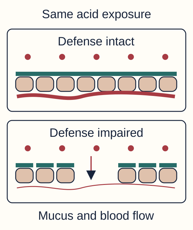
A healthy stomach tolerates acid because mucus, bicarbonate, an intact epithelium and mucosal blood flow protect its surface. Prostaglandins support these defenses. Damage does not require excessive acid production: an ordinary acid load becomes harmful when protection fails. Gastritis means inflammation; gastropathy means mucosal injury with little or no inflammation. Symptoms alone cannot establish either microscopic diagnosis. [1]
Compare the surface, not the number of acid particles.
Try it: point to the first protection impaired by an NSAID, then to the first protection impaired by shock. NSAID inhibition of cyclooxygenase reduces protective prostaglandin production; shock compromises perfusion and repair. Alcohol, bile reflux and chemical irritants can injure the surface through other routes. These insults can produce erosions and bleeding without the gland loss that defines chronic atrophic disease. [1][2]
After severe burns, hypovolemia and splanchnic hypoperfusion contribute to stress-related ulceration, traditionally called a Curling ulcer. A Cushing ulcer describes ulceration associated with intracranial disease; the classic explanation links neural disturbance to increased vagal acid drive. Neither eponym makes acid injury and defense failure mutually exclusive or establishes one mechanism in every patient. [2][15]
For a new patient in intensive care, use bleeding risk rather than an eponym to decide prevention. The 2024 SCCM/ASHP guideline identifies shock, coagulopathy and chronic liver disease as important risk factors and supports a PPI or H2-receptor antagonist for patients at risk. Enteral feeding can reduce risk but does not cancel persistent shock. Reassess and discontinue prophylaxis when its indication ends. Prevention is separate from resuscitation and urgent assessment of active hematemesis or melena. [2]
2. What has changed: the surface, the glands or the mucous cells?
Compare surface erosion with scant inflammation, neutrophils plus lymphocytes, and missing oxyntic glands. These describe different processes: reactive injury, chronic active gastritis and atrophy. Mononuclear inflammation supplies the chronic component; neutrophils supply the active component. Atrophy requires attention to its cause and distribution. [1][3]
H. pylori lives in mucus near gastric epithelium. Urease supports local acid tolerance while persistent infection provokes inflammation. Antral-predominant disease with preserved acid-producing glands differs from corpus-predominant or multifocal atrophic disease. Infection does not imply one fixed direction of acid output. [4][5]
Autoimmune gastritis preferentially damages oxyntic mucosa in the body and fundus. Parietal-cell loss reduces acid and intrinsic factor. Antibodies to the parietal H+/K+-ATPase proton pump or intrinsic factor support the assessment alongside histology. Iron deficiency can precede B12 deficiency; late intrinsic-factor deficiency can produce pernicious anemia. Autoimmune thyroid disease commonly coexists. A normal mean corpuscular volume does not exclude mixed iron and B12 deficiency. [3]
Picture giant cerebriform folds, edema and albumin 2.1 g/dL (reference 3.5-5.0), without renal protein loss or impaired liver synthesis. Menetrier disease is a hypertrophic protein-losing gastropathy with foveolar mucous-cell expansion, often in body and fundus, and usually reduced acid output. Excess EGFR signaling is implicated. It is not merely severe ordinary gastritis. [9][10]
Try it: assign erosion, absent parietal glands and enlarged mucous pits to surface defense, oxyntic tissue and foveolar expansion. Their consequences differ: bleeding, secretory deficiency and protein loss. Now consider enlarged folds with recurrent distal duodenal ulcers. Fold size alone does not establish Menetrier disease; an acid-hypersecretory disorder such as gastrinoma has a different functional pattern. Use histology and gastric pH together.
3. Does high gastrin mean high acid?
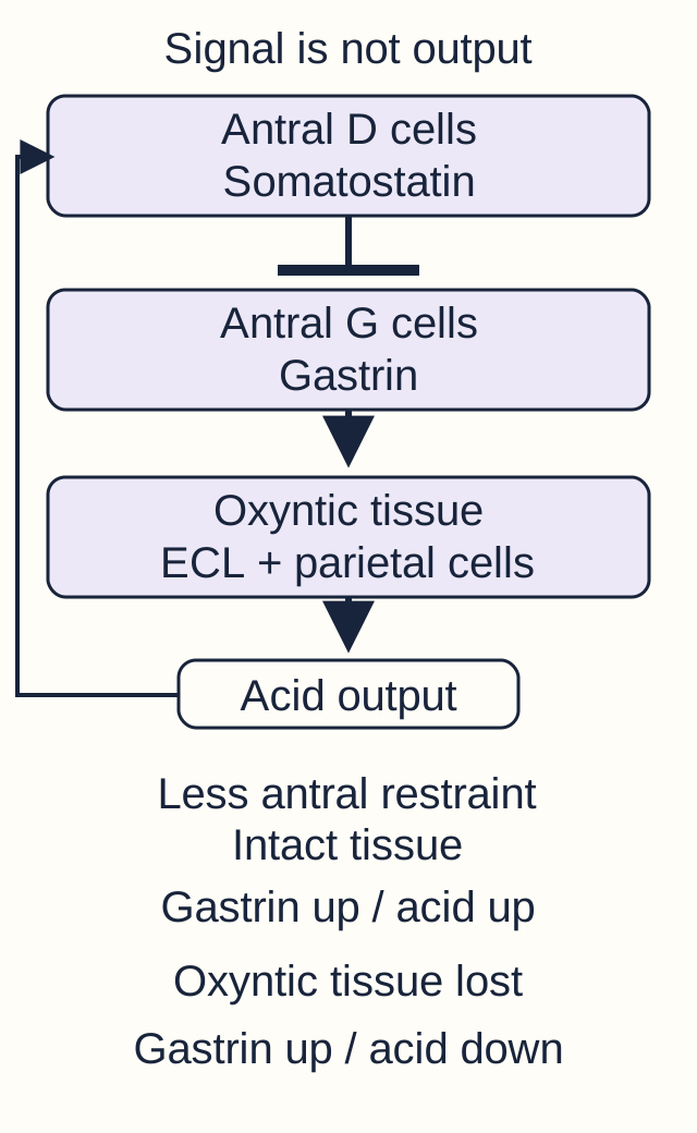
Gastrin is a signal, not an acid measurement. Antral G cells release gastrin; D-cell somatostatin restrains that release. Gastrin promotes acid secretion, importantly through histamine from enterochromaffin-like (ECL) cells in oxyntic mucosa. Functional parietal cells must remain to produce acid. Acid promotes inhibitory feedback. Chief cells supply pepsinogen, not intrinsic factor. [3][4]
The same gastrin direction can accompany opposite acid directions.
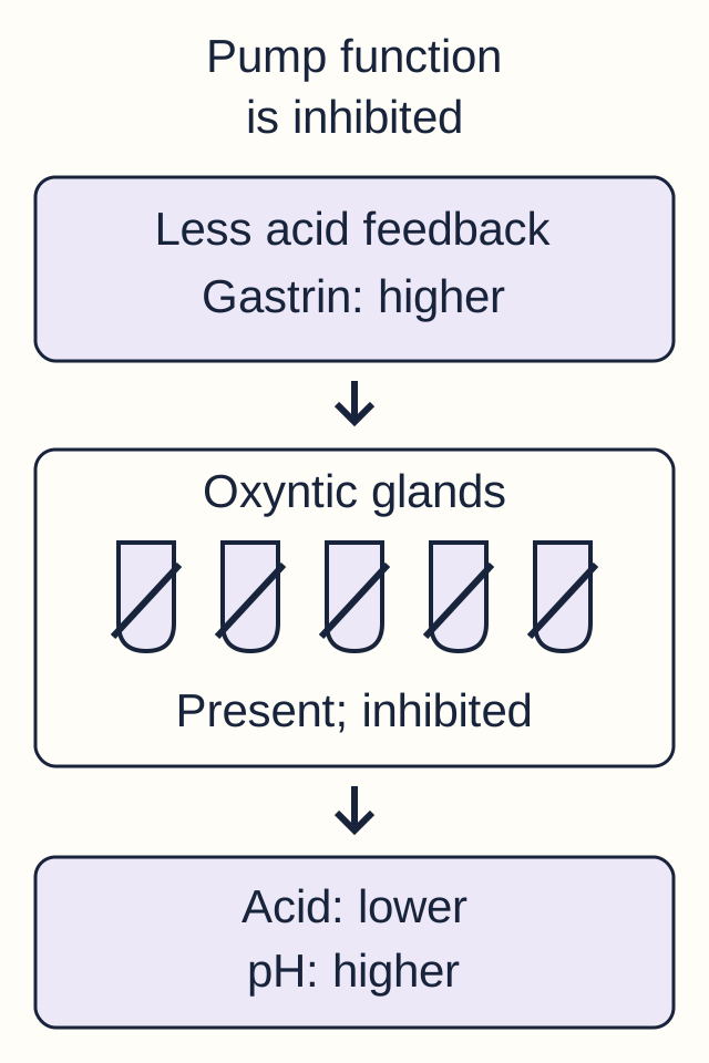
Compare one change at a time: predict acid output from the condition of the responding tissue, then inspect each worked example. The three results are also summarized in the openly readable paragraph below.
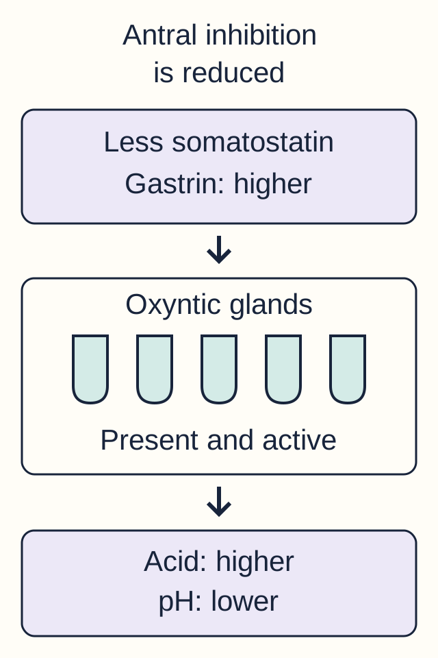
Antral inhibition is reduced
Predict the response with intact glands: less antral restraint can increase gastrin and acid together.
Reduced somatostatin permits stronger gastrin stimulation. With preserved functional corpus glands, acid output can increase and gastric pH can fall. Intrinsic-factor-producing tissue remains present. This is an antral-predominant phenotype, not a universal response to infection. [4][5]
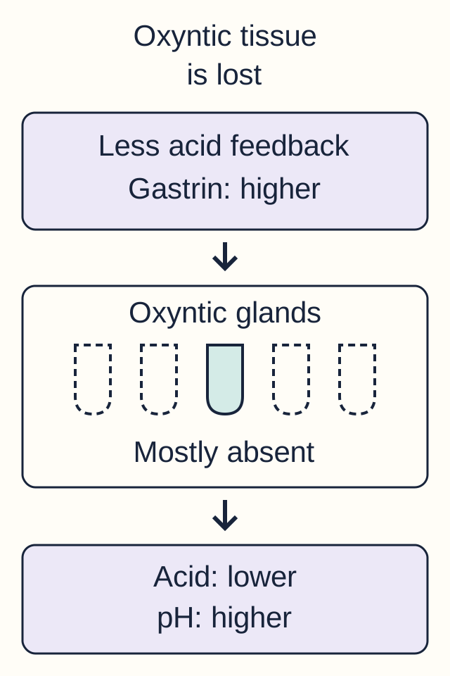
Oxyntic tissue is lost
A higher signal cannot replace lost tissue: extensive oxyntic atrophy reduces acid and raises gastrin through reduced acid feedback.
Extensive parietal-cell loss reduces acid production and weakens inhibitory feedback on gastrin. The increased gastrin signal does not restore missing glands. Intrinsic-factor capacity can also decline when parietal tissue is lost. [3][5]
Pump function is inhibited
Preserved cells can still have reduced function: pump inhibition lowers acid and can raise gastrin without destroying oxyntic glands.
A proton pump inhibitor reduces acid secretion while the glands remain present. Reduced acidity can produce compensatory hypergastrinemia. This functional inhibition does not by itself establish autoimmune intrinsic-factor loss. [3][17]
Transfer: two patients have the same high gastrin and high gastric pH. Medication exposure and topographic histology distinguish inhibited pumps from lost oxyntic tissue. These are qualitative examples, not measured cell counts, diagnostic thresholds or drug-dose models; real processes can overlap.
All results remain visible in the lesson: reduced antral somatostatin can increase gastrin and acid when the corpus is intact, favoring duodenal injury. Extensive oxyntic atrophy reduces acid, raises gastric pH and produces compensatory hypergastrinemia. Long-standing infection does not guarantee atrophy. Acid suppression can also increase pH and gastrin without destroying glands. [3][4][5]
Apply it: high gastrin with gastric pH 7 and absent oxyntic glands reflects failed acid production. High gastrin with very acidic contents and recurrent ulcers requires evaluation for inappropriate gastrin secretion, including gastrinoma. Always review medications: a result during PPI treatment is not an untreated baseline. Any diagnostic withdrawal of acid suppression requires clinical supervision. [17]
Reduced acidity can permit increased colonization by some non-H. pylori bacteria. This is not proof of symptomatic infection or an inevitable cancer pathway. [20]
Persistent hypergastrinemia also stimulates ECL-cell proliferation. This accounts for type 1 gastric neuroendocrine tumors in autoimmune atrophic gastritis, a lineage distinct from adenocarcinoma and B-cell MALT lymphoma. [3]
4. Which part of the stomach did the biopsy actually sample?
A negative antral biopsy does not describe unsampled body mucosa. The antrum is rich in G cells and D cells; the body and fundus contain acid-producing parietal cells, pepsinogen-producing chief cells and ECL cells. These are predominant distributions, not claims that every cell type exists in only one location. Distribution connects a hormone result to the tissue that can explain it. [3]
Try it: a patient has iron deficiency, autoimmune thyroid disease and a normal antral specimen. Mark the region still needed to assess oxyntic atrophy: the body and fundus. The visible consequence of an antral-only strategy is a sampling gap, not exclusion of autoimmune gastritis. Conversely, sampling only the body can miss important antral disease. Topographic assessment includes antrum/incisura and body specimens in separately identified containers, plus targeted samples from abnormalities. [3][8]
For a new patient with pernicious anemia and no recent appropriate examination, endoscopy with topographic biopsies helps confirm corpus-predominant atrophy and assess prevalent neoplasia. B12 replacement addresses a deficiency; it does not replace evaluation of the underlying gastric process. Histology, autoantibodies, iron indices and B12 results answer complementary questions. [3]
Testing for H. pylori asks a different question from mapping atrophy. Urea breath testing and stool antigen testing assess active infection when performed under suitable conditions. PPIs, PCABs, antibiotics and bismuth can suppress detection. An antibody result can remain positive after eradication and is not a test of cure. A negative result obtained under suppressive conditions may need repetition rather than reassurance. [6][7]
5. One infection, several different outcomes
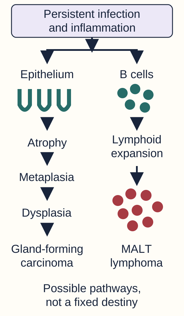
Does chronic infection have to become metaplasia, then lymphoma, then cancer? No. Acid-related ulceration, epithelial neoplasia and lymphoid neoplasia are distinct outcomes. Most infected people do not develop gastric cancer. The purpose of identifying the route is to understand the affected tissue, not to predict an inevitable timetable. [13]
Trace epithelial and lymphoid outcomes separately. No arrow means that progression occurs in every patient.
Trace the tissue: intestinal metaplasia replaces gastric-type epithelium with intestinal-type cells, including goblet cells. Dysplasia adds neoplastic cytologic and architectural abnormalities; invasion distinguishes carcinoma from a confined epithelial precursor. Chronic atrophy, metaplasia and dysplasia describe an important pathway to intestinal-type adenocarcinoma, not a required sequence for every gastric malignancy. [8][11]
In the other branch, sustained antigenic stimulation can support acquired gastric lymphoid tissue and a clonal B-cell neoplasm: gastric MALT lymphoma. Lymphoepithelial lesions involve lymphoid cells disrupting glands; they are not intestinal metaplasia. Some localized infection-associated MALT lymphomas regress after eradication, but histologic staging and specialist follow-up remain necessary. Symptom improvement is not proof that a malignancy has resolved. [13]
Apply it: after infection is treated, a patient still has extensive intestinal metaplasia. Eradication reduces future risk but does not erase the tissue finding or guarantee elimination of cancer risk. Surveillance depends on extent, histology, family history and other risk factors rather than one interval for everyone. Autoimmune atrophy and Menetrier disease also warrant appropriate specialist assessment of their neoplastic risks. [3][8][10]
For treatment-naive adults in North America with unknown susceptibility, the 2024 ACG guideline favors 14-day optimized bismuth quadruple therapy: a proton pump inhibitor, bismuth, tetracycline and metronidazole. Avoid empiric clarithromycin or levofloxacin regimens when susceptibility is unconfirmed. Confirm eradication with an appropriate active-infection test at least four weeks after antibiotics and bismuth, and after withholding PPI or PCAB therapy for two weeks under clinical guidance. [6][7]
6. Is the tumor building glands or infiltrating as separate cells?
Intestinal-type adenocarcinoma tends to form malignant glands. It is associated with chronic atrophic and metaplastic injury, often in the distal stomach; a distal or lesser-curvature location alone cannot classify a tumor. H. pylori, smoking and high-salt preserved foods contribute to gastric cancer risk. Diffuse-type adenocarcinoma instead contains poorly cohesive cells with little gland formation. Some cells contain intracellular mucin that displaces the nucleus, producing signet-ring morphology. Mixed histologic patterns also occur. [11][13]
Reduced E-cadherin function helps explain poor cell-to-cell adhesion in a subset of diffuse cancers. Germline pathogenic variants in CDH1 can predispose to hereditary diffuse gastric cancer and lobular breast cancer; somatic alterations also occur. A signet-ring cell does not by itself prove an inherited syndrome or uniquely identify the stomach as the primary site. Age, family history, tumor distribution and appropriate genetic evaluation matter. [12]
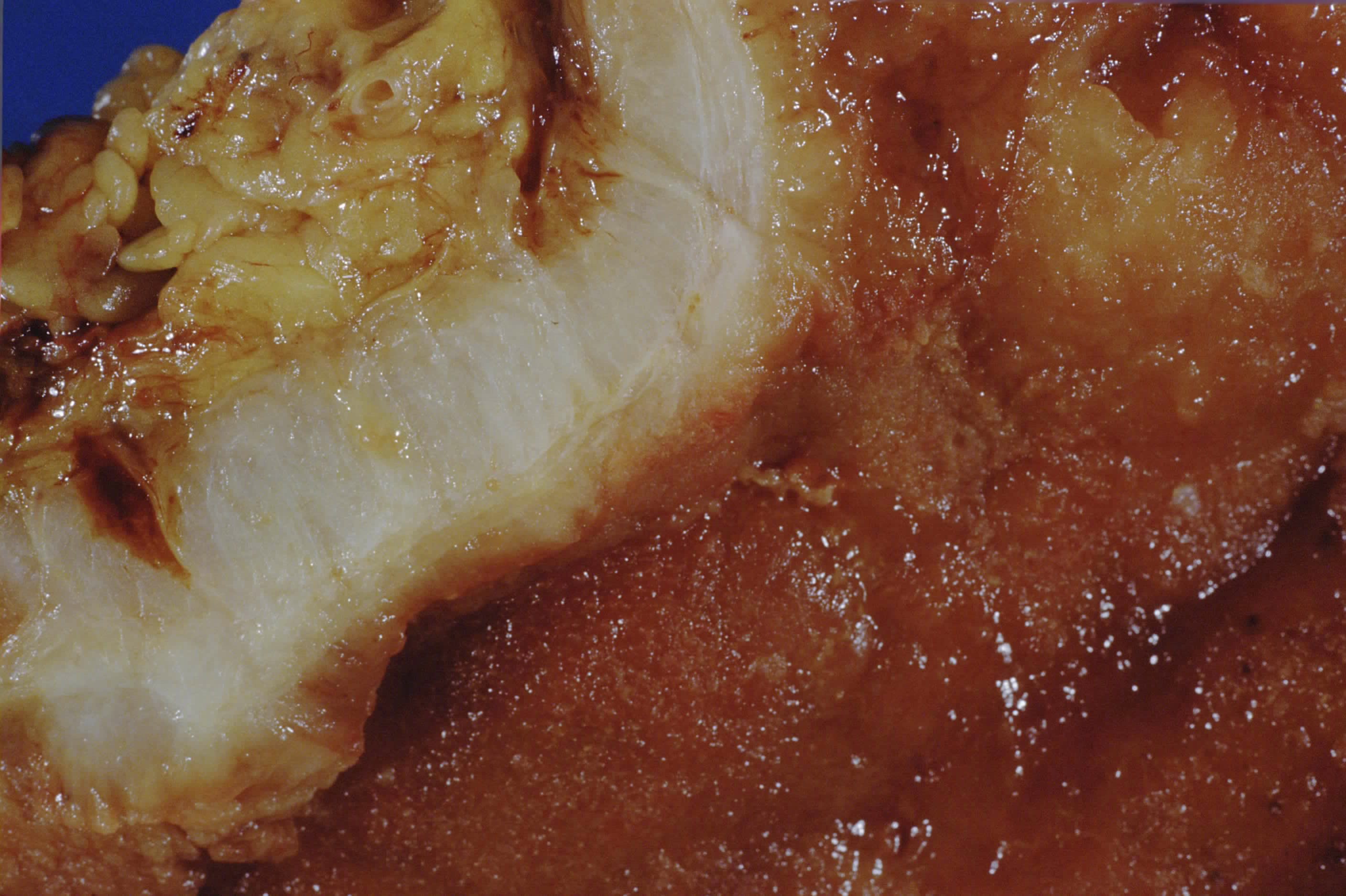
Compare the pale, thickened gastric wall with the irregular surface and predict reduced distensibility. Use histology to determine subtype and clinical genetics when CDH1 assessment is indicated. Image: Narraburra; CC0 1.0. [16]
Predict the function: if tumor and fibrosis diffusely thicken the wall, can the stomach expand normally after a meal? Reduced distensibility explains early satiety and the rigid configuration called linitis plastica. A discrete projecting mass need not be present. The next patient may have persistent alarm symptoms despite an initially limited biopsy; a negative superficial sample cannot settle a discordant infiltrative-wall assessment. Adequate repeat or deeper sampling should be planned by the treating team. [11][18]
Separate the primary tumor's architecture from the route of spread. Gastric cancer can spread through lymphatics, blood and the peritoneal cavity. The liver is an important hematogenous destination. Several named findings describe distant locations rather than a unique histologic type: Virchow node is left supraclavicular, classically connected to abdominal lymphatic drainage through the thoracic duct; a Sister Mary Joseph nodule is umbilical metastasis; a Krukenberg tumor is an ovarian metastasis containing mucin-producing signet-ring cells, often bilateral and often gastric in origin. Other primary sites can produce these findings. [11][14]
A Blumer shelf is a palpable pelvic peritoneal deposit near the rectum, in the rectovesical or rectouterine region. It can narrow the rectum from outside while the mucosal surface remains relatively preserved. Try it: locate an ovarian mass, an umbilical nodule and a rectal shelf on their actual compartments before assigning origin. Matching histology and staging determine the meaning; an eponym is not a substitute for tissue confirmation. These distant deposits are not merely regional perigastric nodes. [14]
Primary depth and distant spread are separate staging questions. Invasion into the muscularis propria is T2; extension into subserosal connective tissue without serosal or adjacent-organ invasion is T3. A separate confirmed distant deposit is M1, even when sampled regional nodes are negative. M1 gastric cancer belongs to stage IV regardless of the primary depth. [11]
Carry one sequence into a fresh vignette: identify the injured compartment, interpret acid and gastrin together, then distinguish epithelial, neuroendocrine and lymphoid outcomes. Bleeding, progressive weight loss, persistent vomiting, unexplained anemia or early satiety warrant appropriate clinical evaluation, not automatic attribution to uncomplicated gastritis. Review related physiology in gastric secretion and compare mucosal injury in peptic ulcer disease. Compare a different metaplastic setting in Barrett esophagus and esophageal cancer.
Practice with new patients
Commit to an explanation before inspecting the choices. Use the tissue distribution and functional findings together; the cases do not identify their teaching objective in advance.
Case 1
Show answer and explanations for case 1
A. Reduced antral somatostatin secretion (Why this does not fit)
Reduced D-cell restraint can increase gastrin-driven acid production. Normal acid output and the temporal NSAID exposure instead favor loss of mucosal protection.
Reasoning steps for option A
In gastric-01, given "A 72-year-old man develops melena two weeks after starting naproxen for knee pain", what mechanism or clinical process does the option "Reduced antral somatostatin secretion" propose?
Reduced D-cell restraint can increase gastrin-driven acid production.
For gastric-01, which supplied finding most directly distinguishes the option "Reduced antral somatostatin secretion" from the keyed answer?
Normal acid output and the temporal NSAID exposure instead favor loss of mucosal protection.
For gastric-01, what case-specific discriminator should be checked before selecting the option "Reduced antral somatostatin secretion" in a similar vignette?
This option is weaker because Normal acid output and the temporal NSAID exposure instead favor loss of mucosal protection. The transferable discriminator is: Reactive mucosal injury can result from impaired defense despite ordinary acid production.
B. Reduced epithelial E-cadherin expression (Why this does not fit)
Loss of cell adhesion can contribute to poorly cohesive carcinoma. Regenerative erosions without malignant cells do not support an infiltrative neoplasm.
Reasoning steps for option B
In gastric-01, given "A 72-year-old man develops melena two weeks after starting naproxen for knee pain", what mechanism or clinical process does the option "Reduced epithelial E-cadherin expression" propose?
Loss of cell adhesion can contribute to poorly cohesive carcinoma.
For gastric-01, which supplied finding most directly distinguishes the option "Reduced epithelial E-cadherin expression" from the keyed answer?
Regenerative erosions without malignant cells do not support an infiltrative neoplasm.
For gastric-01, what case-specific discriminator should be checked before selecting the option "Reduced epithelial E-cadherin expression" in a similar vignette?
This option is weaker because Regenerative erosions without malignant cells do not support an infiltrative neoplasm. The transferable discriminator is: Reactive mucosal injury can result from impaired defense despite ordinary acid production.
C. Reduced mucosal prostaglandin synthesis (Best answer)
NSAID inhibition of cyclooxygenase compromises mucus, bicarbonate and perfusion-related defenses. A normal acid load can then injure the surface, matching the preserved glands and scant inflammation.
Reasoning steps for option C
In gastric-01, given "A 72-year-old man develops melena two weeks after starting naproxen for knee pain", what mechanism or clinical process does the option "Reduced mucosal prostaglandin synthesis" propose?
NSAID inhibition of cyclooxygenase compromises mucus, bicarbonate and perfusion-related defenses.
For gastric-01, which supplied finding most directly distinguishes the option "Reduced mucosal prostaglandin synthesis" from the keyed answer?
A normal acid load can then injure the surface, matching the preserved glands and scant inflammation.
For gastric-01, what case-specific discriminator should be checked before selecting the option "Reduced mucosal prostaglandin synthesis" in a similar vignette?
This option fits because A normal acid load can then injure the surface, matching the preserved glands and scant inflammation. The transferable discriminator is: Reactive mucosal injury can result from impaired defense despite ordinary acid production.
D. Reduced intrinsic-factor binding of B12 (Why this does not fit)
Intrinsic-factor impairment reduces intestinal B12 absorption. It does not explain acute erosions after an NSAID exposure with preserved oxyntic glands.
Reasoning steps for option D
In gastric-01, given "A 72-year-old man develops melena two weeks after starting naproxen for knee pain", what mechanism or clinical process does the option "Reduced intrinsic-factor binding of B12" propose?
For gastric-01, which supplied finding most directly distinguishes the option "Reduced intrinsic-factor binding of B12" from the keyed answer?
It does not explain acute erosions after an NSAID exposure with preserved oxyntic glands.
For gastric-01, what case-specific discriminator should be checked before selecting the option "Reduced intrinsic-factor binding of B12" in a similar vignette?
This option is weaker because It does not explain acute erosions after an NSAID exposure with preserved oxyntic glands. The transferable discriminator is: Reactive mucosal injury can result from impaired defense despite ordinary acid production.
Takeaway: Reactive mucosal injury can result from impaired defense despite ordinary acid production.
A. Impaired mucosal perfusion permits acid-mediated surface injury (Best answer)
Hypoperfusion weakens mucosal defense and repair. Acid can contribute to injury even when secretion is not excessive, fitting the shock-associated erosions.
Reasoning steps for option A
In gastric-02, given "A 35-year-old woman with extensive burns has prolonged hypotension before fluid resuscitation", what mechanism or clinical process does the option "Impaired mucosal perfusion permits acid-mediated surface injury" propose?
Hypoperfusion weakens mucosal defense and repair.
For gastric-02, which supplied finding most directly distinguishes the option "Impaired mucosal perfusion permits acid-mediated surface injury" from the keyed answer?
Acid can contribute to injury even when secretion is not excessive, fitting the shock-associated erosions.
For gastric-02, what case-specific discriminator should be checked before selecting the option "Impaired mucosal perfusion permits acid-mediated surface injury" in a similar vignette?
This option fits because Acid can contribute to injury even when secretion is not excessive, fitting the shock-associated erosions. The transferable discriminator is: Burn-associated stress injury combines impaired perfusion with luminal injury; it is not proof that acid is irrelevant.
B. Gastrin secretion increases despite an intact inhibitory response (Why this does not fit)
Inappropriate gastrin secretion can increase gastric acidity and promote ulcers. No hypersecretory evidence is supplied, while prolonged hypotension directly explains compromised defense.
Reasoning steps for option B
In gastric-02, given "A 35-year-old woman with extensive burns has prolonged hypotension before fluid resuscitation", what mechanism or clinical process does the option "Gastrin secretion increases despite an intact inhibitory response" propose?
Inappropriate gastrin secretion can increase gastric acidity and promote ulcers.
For gastric-02, which supplied finding most directly distinguishes the option "Gastrin secretion increases despite an intact inhibitory response" from the keyed answer?
No hypersecretory evidence is supplied, while prolonged hypotension directly explains compromised defense.
For gastric-02, what case-specific discriminator should be checked before selecting the option "Gastrin secretion increases despite an intact inhibitory response" in a similar vignette?
This option is weaker because No hypersecretory evidence is supplied, while prolonged hypotension directly explains compromised defense. The transferable discriminator is: Burn-associated stress injury combines impaired perfusion with luminal injury; it is not proof that acid is irrelevant.
C. Oxyntic gland destruction permits compensatory gastrin secretion (Why this does not fit)
Loss of acid-producing glands can cause hypergastrinemia through reduced feedback. An acute erosive presentation with acidic contents does not establish chronic oxyntic atrophy.
Reasoning steps for option C
In gastric-02, given "A 35-year-old woman with extensive burns has prolonged hypotension before fluid resuscitation", what mechanism or clinical process does the option "Oxyntic gland destruction permits compensatory gastrin secretion" propose?
Loss of acid-producing glands can cause hypergastrinemia through reduced feedback.
For gastric-02, which supplied finding most directly distinguishes the option "Oxyntic gland destruction permits compensatory gastrin secretion" from the keyed answer?
An acute erosive presentation with acidic contents does not establish chronic oxyntic atrophy.
For gastric-02, what case-specific discriminator should be checked before selecting the option "Oxyntic gland destruction permits compensatory gastrin secretion" in a similar vignette?
This option is weaker because An acute erosive presentation with acidic contents does not establish chronic oxyntic atrophy. The transferable discriminator is: Burn-associated stress injury combines impaired perfusion with luminal injury; it is not proof that acid is irrelevant.
D. Mucous-cell expansion permits protein leakage through giant folds (Why this does not fit)
Menetrier disease can cause protein loss across enlarged mucosa. The examination shows erosions after shock, not hypertrophic folds or a protein-losing syndrome.
Reasoning steps for option D
In gastric-02, given "A 35-year-old woman with extensive burns has prolonged hypotension before fluid resuscitation", what mechanism or clinical process does the option "Mucous-cell expansion permits protein leakage through giant folds" propose?
Menetrier disease can cause protein loss across enlarged mucosa.
For gastric-02, which supplied finding most directly distinguishes the option "Mucous-cell expansion permits protein leakage through giant folds" from the keyed answer?
The examination shows erosions after shock, not hypertrophic folds or a protein-losing syndrome.
For gastric-02, what case-specific discriminator should be checked before selecting the option "Mucous-cell expansion permits protein leakage through giant folds" in a similar vignette?
This option is weaker because The examination shows erosions after shock, not hypertrophic folds or a protein-losing syndrome. The transferable discriminator is: Burn-associated stress injury combines impaired perfusion with luminal injury; it is not proof that acid is irrelevant.
Takeaway: Burn-associated stress injury combines impaired perfusion with luminal injury; it is not proof that acid is irrelevant.
A. Continue pantoprazole through the next outpatient visit (Why this does not fit)
Continued acid suppression is appropriate when a bleeding-risk or other acid-related indication persists. The supplied risk factors have resolved and no separate outpatient indication is present.
Reasoning steps for option A
In gastric-03, given "A 61-year-old woman received pantoprazole prophylaxis during septic shock", what mechanism or clinical process does the option "Continue pantoprazole through the next outpatient visit" propose?
Continued acid suppression is appropriate when a bleeding-risk or other acid-related indication persists.
For gastric-03, which supplied finding most directly distinguishes the option "Continue pantoprazole through the next outpatient visit" from the keyed answer?
The supplied risk factors have resolved and no separate outpatient indication is present.
For gastric-03, what case-specific discriminator should be checked before selecting the option "Continue pantoprazole through the next outpatient visit" in a similar vignette?
This option is weaker because The supplied risk factors have resolved and no separate outpatient indication is present. The transferable discriminator is: Select both the start and the stop of stress prophylaxis from the patient's current risk.
B. Replace pantoprazole with scheduled famotidine at discharge (Why this does not fit)
An H2-receptor antagonist is an alternative prophylactic agent in patients who remain at risk. Changing drug class does not create an indication after the risk has ended.
Reasoning steps for option B
In gastric-03, given "A 61-year-old woman received pantoprazole prophylaxis during septic shock", what mechanism or clinical process does the option "Replace pantoprazole with scheduled famotidine at discharge" propose?
An H2-receptor antagonist is an alternative prophylactic agent in patients who remain at risk.
For gastric-03, which supplied finding most directly distinguishes the option "Replace pantoprazole with scheduled famotidine at discharge" from the keyed answer?
Changing drug class does not create an indication after the risk has ended.
For gastric-03, what case-specific discriminator should be checked before selecting the option "Replace pantoprazole with scheduled famotidine at discharge" in a similar vignette?
This option is weaker because Changing drug class does not create an indication after the risk has ended. The transferable discriminator is: Select both the start and the stop of stress prophylaxis from the patient's current risk.
C. Continue pantoprazole until a negative stool antigen test (Why this does not fit)
Stool antigen can assess active H. pylori infection under suitable conditions. No infection or ulcer indication has been supplied; an unrelated test is not required to end stress prophylaxis.
Reasoning steps for option C
In gastric-03, given "A 61-year-old woman received pantoprazole prophylaxis during septic shock", what mechanism or clinical process does the option "Continue pantoprazole until a negative stool antigen test" propose?
Stool antigen can assess active H.
For gastric-03, which supplied finding most directly distinguishes the option "Continue pantoprazole until a negative stool antigen test" from the keyed answer?
pylori infection under suitable conditions. No infection or ulcer indication has been supplied; an unrelated test is not required to end stress prophylaxis.
For gastric-03, what case-specific discriminator should be checked before selecting the option "Continue pantoprazole until a negative stool antigen test" in a similar vignette?
This option is weaker because pylori infection under suitable conditions. No infection or ulcer indication has been supplied; an unrelated test is not required to end stress prophylaxis. The transferable discriminator is: Select both the start and the stop of stress prophylaxis from the patient's current risk.
D. Discontinue pantoprazole before intensive-care transfer (Best answer)
Stress-ulcer prophylaxis should be reassessed when its risk factors resolve. Normal coagulation, restored perfusion, oral intake and absence of another indication support discontinuation.
Reasoning steps for option D
In gastric-03, given "A 61-year-old woman received pantoprazole prophylaxis during septic shock", what mechanism or clinical process does the option "Discontinue pantoprazole before intensive-care transfer" propose?
Stress-ulcer prophylaxis should be reassessed when its risk factors resolve.
For gastric-03, which supplied finding most directly distinguishes the option "Discontinue pantoprazole before intensive-care transfer" from the keyed answer?
Normal coagulation, restored perfusion, oral intake and absence of another indication support discontinuation.
For gastric-03, what case-specific discriminator should be checked before selecting the option "Discontinue pantoprazole before intensive-care transfer" in a similar vignette?
This option fits because Normal coagulation, restored perfusion, oral intake and absence of another indication support discontinuation. The transferable discriminator is: Select both the start and the stop of stress prophylaxis from the patient's current risk.
Takeaway: Select both the start and the stop of stress prophylaxis from the patient's current risk.
A. Loss of oxyntic glands reduces feedback inhibition of gastrin (Why this does not fit)
Atrophic gland loss can increase gastrin when acid production falls. Here acid production increases and the number of parietal cells is preserved.
Reasoning steps for option A
In gastric-04, given "A patient undergoing evaluation after intracranial surgery develops a duodenal ulcer", what mechanism or clinical process does the option "Loss of oxyntic glands reduces feedback inhibition of gastrin" propose?
Atrophic gland loss can increase gastrin when acid production falls.
For gastric-04, which supplied finding most directly distinguishes the option "Loss of oxyntic glands reduces feedback inhibition of gastrin" from the keyed answer?
Here acid production increases and the number of parietal cells is preserved.
For gastric-04, what case-specific discriminator should be checked before selecting the option "Loss of oxyntic glands reduces feedback inhibition of gastrin" in a similar vignette?
This option is weaker because Here acid production increases and the number of parietal cells is preserved. The transferable discriminator is: The classic neurogenic ulcer explanation concerns increased neural acid drive, but an eponym alone would not prove that mechanism.
B. Increased neural drive stimulates existing parietal cells (Best answer)
Cholinergic stimulation can increase secretion by functioning acid-producing tissue. Preserved cell number and the response to neural blockade support increased drive rather than tissue expansion or loss.
Reasoning steps for option B
In gastric-04, given "A patient undergoing evaluation after intracranial surgery develops a duodenal ulcer", what mechanism or clinical process does the option "Increased neural drive stimulates existing parietal cells" propose?
Cholinergic stimulation can increase secretion by functioning acid-producing tissue.
For gastric-04, which supplied finding most directly distinguishes the option "Increased neural drive stimulates existing parietal cells" from the keyed answer?
Preserved cell number and the response to neural blockade support increased drive rather than tissue expansion or loss.
For gastric-04, what case-specific discriminator should be checked before selecting the option "Increased neural drive stimulates existing parietal cells" in a similar vignette?
This option fits because Preserved cell number and the response to neural blockade support increased drive rather than tissue expansion or loss. The transferable discriminator is: The classic neurogenic ulcer explanation concerns increased neural acid drive, but an eponym alone would not prove that mechanism.
C. Foveolar proliferation replaces acid-producing glandular tissue (Why this does not fit)
Foveolar expansion in Menetrier disease commonly accompanies reduced acid output. Reduced acid output and gland replacement would not explain increased secretion with preserved parietal-cell number.
Reasoning steps for option C
In gastric-04, given "A patient undergoing evaluation after intracranial surgery develops a duodenal ulcer", what mechanism or clinical process does the option "Foveolar proliferation replaces acid-producing glandular tissue" propose?
Foveolar expansion in Menetrier disease commonly accompanies reduced acid output.
For gastric-04, which supplied finding most directly distinguishes the option "Foveolar proliferation replaces acid-producing glandular tissue" from the keyed answer?
Reduced acid output and gland replacement would not explain increased secretion with preserved parietal-cell number.
For gastric-04, what case-specific discriminator should be checked before selecting the option "Foveolar proliferation replaces acid-producing glandular tissue" in a similar vignette?
This option is weaker because Reduced acid output and gland replacement would not explain increased secretion with preserved parietal-cell number. The transferable discriminator is: The classic neurogenic ulcer explanation concerns increased neural acid drive, but an eponym alone would not prove that mechanism.
D. Reduced mucosal blood flow limits bicarbonate delivery and repair (Why this does not fit)
Hypoperfusion is important in many stress-related gastric injuries. The measured perfusion is stable and the experimentally demonstrated abnormality is cholinergic secretion.
Reasoning steps for option D
In gastric-04, given "A patient undergoing evaluation after intracranial surgery develops a duodenal ulcer", what mechanism or clinical process does the option "Reduced mucosal blood flow limits bicarbonate delivery and repair" propose?
Hypoperfusion is important in many stress-related gastric injuries.
For gastric-04, which supplied finding most directly distinguishes the option "Reduced mucosal blood flow limits bicarbonate delivery and repair" from the keyed answer?
The measured perfusion is stable and the experimentally demonstrated abnormality is cholinergic secretion.
For gastric-04, what case-specific discriminator should be checked before selecting the option "Reduced mucosal blood flow limits bicarbonate delivery and repair" in a similar vignette?
This option is weaker because The measured perfusion is stable and the experimentally demonstrated abnormality is cholinergic secretion. The transferable discriminator is: The classic neurogenic ulcer explanation concerns increased neural acid drive, but an eponym alone would not prove that mechanism.
Takeaway: The classic neurogenic ulcer explanation concerns increased neural acid drive, but an eponym alone would not prove that mechanism.
A. Gastrin rises; stimulated acid output rises (Why this does not fit)
This pairing can follow increased stimulation of intact oxyntic tissue. Restoration of inhibitory somatostatin instead reduces gastrin drive.
Reasoning steps for option A
In gastric-05, given "A 39-year-old man has recurrent duodenal ulcers and active H", what mechanism or clinical process does the option "Gastrin rises; stimulated acid output rises" propose?
This pairing can follow increased stimulation of intact oxyntic tissue.
For gastric-05, which supplied finding most directly distinguishes the option "Gastrin rises; stimulated acid output rises" from the keyed answer?
Restoration of inhibitory somatostatin instead reduces gastrin drive.
For gastric-05, what case-specific discriminator should be checked before selecting the option "Gastrin rises; stimulated acid output rises" in a similar vignette?
This option is weaker because Restoration of inhibitory somatostatin instead reduces gastrin drive. The transferable discriminator is: Predict antral feedback effects only after establishing the condition of the acid-producing compartment.
B. Gastrin falls; stimulated acid output falls (Best answer)
Somatostatin restrains G-cell secretion and reduces stimulation of intact acid-producing tissue. Antral recovery with preserved corpus glands supports this response in the stated ulcer phenotype.
Reasoning steps for option B
In gastric-05, given "A 39-year-old man has recurrent duodenal ulcers and active H", what mechanism or clinical process does the option "Gastrin falls; stimulated acid output falls" propose?
Somatostatin restrains G-cell secretion and reduces stimulation of intact acid-producing tissue.
For gastric-05, which supplied finding most directly distinguishes the option "Gastrin falls; stimulated acid output falls" from the keyed answer?
Antral recovery with preserved corpus glands supports this response in the stated ulcer phenotype.
For gastric-05, what case-specific discriminator should be checked before selecting the option "Gastrin falls; stimulated acid output falls" in a similar vignette?
This option fits because Antral recovery with preserved corpus glands supports this response in the stated ulcer phenotype. The transferable discriminator is: Predict antral feedback effects only after establishing the condition of the acid-producing compartment.
C. Gastrin rises; stimulated acid output falls (Why this does not fit)
This pairing can result from oxyntic tissue loss with impaired acid feedback. The corpus is preserved and the specified change is restoration of antral inhibition.
Reasoning steps for option C
In gastric-05, given "A 39-year-old man has recurrent duodenal ulcers and active H", what mechanism or clinical process does the option "Gastrin rises; stimulated acid output falls" propose?
This pairing can result from oxyntic tissue loss with impaired acid feedback.
For gastric-05, which supplied finding most directly distinguishes the option "Gastrin rises; stimulated acid output falls" from the keyed answer?
The corpus is preserved and the specified change is restoration of antral inhibition.
For gastric-05, what case-specific discriminator should be checked before selecting the option "Gastrin rises; stimulated acid output falls" in a similar vignette?
This option is weaker because The corpus is preserved and the specified change is restoration of antral inhibition. The transferable discriminator is: Predict antral feedback effects only after establishing the condition of the acid-producing compartment.
D. Gastrin falls; stimulated acid output rises (Why this does not fit)
Acid can recover when corpus inflammation resolves. This scenario isolates antral restraint in a preserved-corpus hypersecretory phenotype, not recovery from corpus suppression.
Reasoning steps for option D
In gastric-05, given "A 39-year-old man has recurrent duodenal ulcers and active H", what mechanism or clinical process does the option "Gastrin falls; stimulated acid output rises" propose?
Acid can recover when corpus inflammation resolves.
For gastric-05, which supplied finding most directly distinguishes the option "Gastrin falls; stimulated acid output rises" from the keyed answer?
This scenario isolates antral restraint in a preserved-corpus hypersecretory phenotype, not recovery from corpus suppression.
For gastric-05, what case-specific discriminator should be checked before selecting the option "Gastrin falls; stimulated acid output rises" in a similar vignette?
This option is weaker because This scenario isolates antral restraint in a preserved-corpus hypersecretory phenotype, not recovery from corpus suppression. The transferable discriminator is: Predict antral feedback effects only after establishing the condition of the acid-producing compartment.
Takeaway: Predict antral feedback effects only after establishing the condition of the acid-producing compartment.
A. Inappropriate secretion causing acid excess (Why this does not fit)
Autonomous gastrin secretion can drive acid excess when oxyntic tissue functions. The high gastric pH and extensive gland loss instead demonstrate poor acid production.
Reasoning steps for option A
In gastric-06, given "A 66-year-old woman with persistent H", what mechanism or clinical process does the option "Inappropriate secretion causing acid excess" propose?
Autonomous gastrin secretion can drive acid excess when oxyntic tissue functions.
For gastric-06, which supplied finding most directly distinguishes the option "Inappropriate secretion causing acid excess" from the keyed answer?
The high gastric pH and extensive gland loss instead demonstrate poor acid production.
For gastric-06, what case-specific discriminator should be checked before selecting the option "Inappropriate secretion causing acid excess" in a similar vignette?
This option is weaker because The high gastric pH and extensive gland loss instead demonstrate poor acid production. The transferable discriminator is: Interpret high gastrin with gastric pH and gland integrity, not as a direct acid measurement.
B. Reduced somatostatin restraint with intact oxyntic tissue (Why this does not fit)
Loss of antral inhibition can increase gastrin-driven acid output when the corpus is intact. Extensive oxyntic gland loss and high pH instead support reduced acid feedback as the dominant explanation.
Reasoning steps for option B
In gastric-06, given "A 66-year-old woman with persistent H", what mechanism or clinical process does the option "Reduced somatostatin restraint with intact oxyntic tissue" propose?
Loss of antral inhibition can increase gastrin-driven acid output when the corpus is intact.
For gastric-06, which supplied finding most directly distinguishes the option "Reduced somatostatin restraint with intact oxyntic tissue" from the keyed answer?
Extensive oxyntic gland loss and high pH instead support reduced acid feedback as the dominant explanation.
For gastric-06, what case-specific discriminator should be checked before selecting the option "Reduced somatostatin restraint with intact oxyntic tissue" in a similar vignette?
This option is weaker because Extensive oxyntic gland loss and high pH instead support reduced acid feedback as the dominant explanation. The transferable discriminator is: Interpret high gastrin with gastric pH and gland integrity, not as a direct acid measurement.
C. Reduced renal clearance causing gastrin accumulation (Why this does not fit)
Impaired renal function can contribute to increased circulating gastrin. Normal renal function and demonstrated gastric gland loss favor the gastric feedback explanation.
Reasoning steps for option C
In gastric-06, given "A 66-year-old woman with persistent H", what mechanism or clinical process does the option "Reduced renal clearance causing gastrin accumulation" propose?
Impaired renal function can contribute to increased circulating gastrin.
For gastric-06, which supplied finding most directly distinguishes the option "Reduced renal clearance causing gastrin accumulation" from the keyed answer?
Normal renal function and demonstrated gastric gland loss favor the gastric feedback explanation.
For gastric-06, what case-specific discriminator should be checked before selecting the option "Reduced renal clearance causing gastrin accumulation" in a similar vignette?
This option is weaker because Normal renal function and demonstrated gastric gland loss favor the gastric feedback explanation. The transferable discriminator is: Interpret high gastrin with gastric pH and gland integrity, not as a direct acid measurement.
D. Reduced acidity causing compensatory secretion (Best answer)
Loss of functional oxyntic glands weakens acidity-dependent inhibition of gastrin. Compensatory secretion fits both the high pH and the documented atrophy.
Reasoning steps for option D
In gastric-06, given "A 66-year-old woman with persistent H", what mechanism or clinical process does the option "Reduced acidity causing compensatory secretion" propose?
Loss of functional oxyntic glands weakens acidity-dependent inhibition of gastrin.
For gastric-06, which supplied finding most directly distinguishes the option "Reduced acidity causing compensatory secretion" from the keyed answer?
Compensatory secretion fits both the high pH and the documented atrophy.
For gastric-06, what case-specific discriminator should be checked before selecting the option "Reduced acidity causing compensatory secretion" in a similar vignette?
This option fits because Compensatory secretion fits both the high pH and the documented atrophy. The transferable discriminator is: Interpret high gastrin with gastric pH and gland integrity, not as a direct acid measurement.
Takeaway: Interpret high gastrin with gastric pH and gland integrity, not as a direct acid measurement.
A. Reduced acid secretion and reduced intrinsic-factor function (Best answer)
Oxyntic autoimmune injury can impair iron handling through hypochlorhydria and impair B12 absorption through intrinsic-factor deficiency. Concurrent iron and B12 deficits explain why a normal MCV does not exclude B12-related neurologic disease.
Reasoning steps for option A
In gastric-07, given "A 54-year-old woman with autoimmune thyroid disease has fatigue and impaired vibration sense", what mechanism or clinical process does the option "Reduced acid secretion and reduced intrinsic-factor function" propose?
Oxyntic autoimmune injury can impair iron handling through hypochlorhydria and impair B12 absorption through intrinsic-factor deficiency.
For gastric-07, which supplied finding most directly distinguishes the option "Reduced acid secretion and reduced intrinsic-factor function" from the keyed answer?
Concurrent iron and B12 deficits explain why a normal MCV does not exclude B12-related neurologic disease.
For gastric-07, what case-specific discriminator should be checked before selecting the option "Reduced acid secretion and reduced intrinsic-factor function" in a similar vignette?
This option fits because Concurrent iron and B12 deficits explain why a normal MCV does not exclude B12-related neurologic disease. The transferable discriminator is: Mixed deficiencies can obscure macrocytosis; use biochemical and neurologic findings to identify gastric secretory loss.
B. Increased acid secretion and reduced somatostatin release (Why this does not fit)
Antral disinhibition can increase gastrin-dependent acidity. It does not account for intrinsic-factor antibodies and combined iron and B12 deficiency.
Reasoning steps for option B
In gastric-07, given "A 54-year-old woman with autoimmune thyroid disease has fatigue and impaired vibration sense", what mechanism or clinical process does the option "Increased acid secretion and reduced somatostatin release" propose?
Antral disinhibition can increase gastrin-dependent acidity.
For gastric-07, which supplied finding most directly distinguishes the option "Increased acid secretion and reduced somatostatin release" from the keyed answer?
It does not account for intrinsic-factor antibodies and combined iron and B12 deficiency.
For gastric-07, what case-specific discriminator should be checked before selecting the option "Increased acid secretion and reduced somatostatin release" in a similar vignette?
This option is weaker because It does not account for intrinsic-factor antibodies and combined iron and B12 deficiency. The transferable discriminator is: Mixed deficiencies can obscure macrocytosis; use biochemical and neurologic findings to identify gastric secretory loss.
C. Reduced mucus secretion and increased pepsinogen release (Why this does not fit)
Surface protection and peptic activity influence mucosal injury. This pair does not explain intrinsic-factor autoimmunity or the specific deficiency pattern.
Reasoning steps for option C
In gastric-07, given "A 54-year-old woman with autoimmune thyroid disease has fatigue and impaired vibration sense", what mechanism or clinical process does the option "Reduced mucus secretion and increased pepsinogen release" propose?
Surface protection and peptic activity influence mucosal injury.
For gastric-07, which supplied finding most directly distinguishes the option "Reduced mucus secretion and increased pepsinogen release" from the keyed answer?
This pair does not explain intrinsic-factor autoimmunity or the specific deficiency pattern.
For gastric-07, what case-specific discriminator should be checked before selecting the option "Reduced mucus secretion and increased pepsinogen release" in a similar vignette?
This option is weaker because This pair does not explain intrinsic-factor autoimmunity or the specific deficiency pattern. The transferable discriminator is: Mixed deficiencies can obscure macrocytosis; use biochemical and neurologic findings to identify gastric secretory loss.
D. Increased gastrin secretion and increased intrinsic-factor function (Why this does not fit)
Gastrin may rise in autoimmune atrophy. Intrinsic-factor function falls rather than rises when parietal tissue and intrinsic factor are targeted.
Reasoning steps for option D
In gastric-07, given "A 54-year-old woman with autoimmune thyroid disease has fatigue and impaired vibration sense", what mechanism or clinical process does the option "Increased gastrin secretion and increased intrinsic-factor function" propose?
Gastrin may rise in autoimmune atrophy.
For gastric-07, which supplied finding most directly distinguishes the option "Increased gastrin secretion and increased intrinsic-factor function" from the keyed answer?
Intrinsic-factor function falls rather than rises when parietal tissue and intrinsic factor are targeted.
For gastric-07, what case-specific discriminator should be checked before selecting the option "Increased gastrin secretion and increased intrinsic-factor function" in a similar vignette?
This option is weaker because Intrinsic-factor function falls rather than rises when parietal tissue and intrinsic factor are targeted. The transferable discriminator is: Mixed deficiencies can obscure macrocytosis; use biochemical and neurologic findings to identify gastric secretory loss.
Takeaway: Mixed deficiencies can obscure macrocytosis; use biochemical and neurologic findings to identify gastric secretory loss.
A. Colonoscopy with terminal ileal biopsies (Why this does not fit)
The terminal ileum absorbs the intrinsic-factor and B12 complex. An ileal absorption site does not locate this antibody-associated defect; gastric intrinsic-factor failure remains the process requiring assessment.
Reasoning steps for option A
In gastric-08, given "A 62-year-old man with autoimmune thyroid disease develops paresthesias and fatigue", what mechanism or clinical process does the option "Colonoscopy with terminal ileal biopsies" propose?
The terminal ileum absorbs the intrinsic-factor and B12 complex.
For gastric-08, which supplied finding most directly distinguishes the option "Colonoscopy with terminal ileal biopsies" from the keyed answer?
An ileal absorption site does not locate this antibody-associated defect; gastric intrinsic-factor failure remains the process requiring assessment.
For gastric-08, what case-specific discriminator should be checked before selecting the option "Colonoscopy with terminal ileal biopsies" in a similar vignette?
This option is weaker because An ileal absorption site does not locate this antibody-associated defect; gastric intrinsic-factor failure remains the process requiring assessment. The transferable discriminator is: Correcting a deficiency and evaluating the gastric process that caused it are separate clinical tasks.
B. Capsule endoscopy of the small intestine (Why this does not fit)
Small-intestinal disease can impair nutrient absorption. The intrinsic-factor antibodies and autoimmune background instead indicate a gastric cause that capsule examination cannot adequately map or biopsy.
Reasoning steps for option B
In gastric-08, given "A 62-year-old man with autoimmune thyroid disease develops paresthesias and fatigue", what mechanism or clinical process does the option "Capsule endoscopy of the small intestine" propose?
Small-intestinal disease can impair nutrient absorption.
For gastric-08, which supplied finding most directly distinguishes the option "Capsule endoscopy of the small intestine" from the keyed answer?
The intrinsic-factor antibodies and autoimmune background instead indicate a gastric cause that capsule examination cannot adequately map or biopsy.
For gastric-08, what case-specific discriminator should be checked before selecting the option "Capsule endoscopy of the small intestine" in a similar vignette?
This option is weaker because The intrinsic-factor antibodies and autoimmune background instead indicate a gastric cause that capsule examination cannot adequately map or biopsy. The transferable discriminator is: Correcting a deficiency and evaluating the gastric process that caused it are separate clinical tasks.
C. Upper endoscopy with gastric mapping biopsies (Best answer)
Intrinsic-factor autoimmunity with B12 deficiency supports pernicious anemia from autoimmune gastric disease. The response to replacement does not assess corpus atrophy or prevalent neoplasia; topographic and lesion-directed gastric biopsies address both.
Reasoning steps for option C
In gastric-08, given "A 62-year-old man with autoimmune thyroid disease develops paresthesias and fatigue", what mechanism or clinical process does the option "Upper endoscopy with gastric mapping biopsies" propose?
Intrinsic-factor autoimmunity with B12 deficiency supports pernicious anemia from autoimmune gastric disease.
For gastric-08, which supplied finding most directly distinguishes the option "Upper endoscopy with gastric mapping biopsies" from the keyed answer?
The response to replacement does not assess corpus atrophy or prevalent neoplasia; topographic and lesion-directed gastric biopsies address both.
For gastric-08, what case-specific discriminator should be checked before selecting the option "Upper endoscopy with gastric mapping biopsies" in a similar vignette?
This option fits because The response to replacement does not assess corpus atrophy or prevalent neoplasia; topographic and lesion-directed gastric biopsies address both. The transferable discriminator is: Correcting a deficiency and evaluating the gastric process that caused it are separate clinical tasks.
D. CT enterography with small-bowel assessment (Why this does not fit)
Enterography evaluates structural small-bowel causes of malabsorption. It does not characterize gastric gland loss or small mucosal neoplasms associated with this antibody-supported gastric process.
Reasoning steps for option D
In gastric-08, given "A 62-year-old man with autoimmune thyroid disease develops paresthesias and fatigue", what mechanism or clinical process does the option "CT enterography with small-bowel assessment" propose?
Enterography evaluates structural small-bowel causes of malabsorption.
For gastric-08, which supplied finding most directly distinguishes the option "CT enterography with small-bowel assessment" from the keyed answer?
It does not characterize gastric gland loss or small mucosal neoplasms associated with this antibody-supported gastric process.
For gastric-08, what case-specific discriminator should be checked before selecting the option "CT enterography with small-bowel assessment" in a similar vignette?
This option is weaker because It does not characterize gastric gland loss or small mucosal neoplasms associated with this antibody-supported gastric process. The transferable discriminator is: Correcting a deficiency and evaluating the gastric process that caused it are separate clinical tasks.
Takeaway: Correcting a deficiency and evaluating the gastric process that caused it are separate clinical tasks.
A. Gastrin-driven proliferation of antral D cells (Why this does not fit)
D cells participate in somatostatin-mediated inhibition. They are not the characteristic cell population producing type 1 gastric neuroendocrine tumors in oxyntic atrophy.
Reasoning steps for option A
In gastric-09, given "A 68-year-old woman has corpus-predominant atrophy, gastric pH 7", what mechanism or clinical process does the option "Gastrin-driven proliferation of antral D cells" propose?
D cells participate in somatostatin-mediated inhibition.
For gastric-09, which supplied finding most directly distinguishes the option "Gastrin-driven proliferation of antral D cells" from the keyed answer?
They are not the characteristic cell population producing type 1 gastric neuroendocrine tumors in oxyntic atrophy.
For gastric-09, what case-specific discriminator should be checked before selecting the option "Gastrin-driven proliferation of antral D cells" in a similar vignette?
This option is weaker because They are not the characteristic cell population producing type 1 gastric neuroendocrine tumors in oxyntic atrophy. The transferable discriminator is: Autoimmune oxyntic atrophy can produce a gastrin-driven ECL tumor, distinct from MALT lymphoma and adenocarcinoma.
B. Gastrin-driven proliferation of oxyntic ECL cells (Best answer)
Persistent hypergastrinemia has trophic effects on ECL cells. Compensatory gastrin secretion after acid-producing gland loss links the atrophy to type 1 gastric neuroendocrine neoplasia.
Reasoning steps for option B
In gastric-09, given "A 68-year-old woman has corpus-predominant atrophy, gastric pH 7", what mechanism or clinical process does the option "Gastrin-driven proliferation of oxyntic ECL cells" propose?
Persistent hypergastrinemia has trophic effects on ECL cells.
For gastric-09, which supplied finding most directly distinguishes the option "Gastrin-driven proliferation of oxyntic ECL cells" from the keyed answer?
Compensatory gastrin secretion after acid-producing gland loss links the atrophy to type 1 gastric neuroendocrine neoplasia.
For gastric-09, what case-specific discriminator should be checked before selecting the option "Gastrin-driven proliferation of oxyntic ECL cells" in a similar vignette?
This option fits because Compensatory gastrin secretion after acid-producing gland loss links the atrophy to type 1 gastric neuroendocrine neoplasia. The transferable discriminator is: Autoimmune oxyntic atrophy can produce a gastrin-driven ECL tumor, distinct from MALT lymphoma and adenocarcinoma.
C. Antigen-driven proliferation of gastric B cells (Why this does not fit)
Chronic infection can support B-cell MALT lymphoma. A neuroendocrine biopsy lineage and marked hypochlorhydric hypergastrinemia favor an ECL process instead.
Reasoning steps for option C
In gastric-09, given "A 68-year-old woman has corpus-predominant atrophy, gastric pH 7", what mechanism or clinical process does the option "Antigen-driven proliferation of gastric B cells" propose?
Chronic infection can support B-cell MALT lymphoma.
For gastric-09, which supplied finding most directly distinguishes the option "Antigen-driven proliferation of gastric B cells" from the keyed answer?
A neuroendocrine biopsy lineage and marked hypochlorhydric hypergastrinemia favor an ECL process instead.
For gastric-09, what case-specific discriminator should be checked before selecting the option "Antigen-driven proliferation of gastric B cells" in a similar vignette?
This option is weaker because A neuroendocrine biopsy lineage and marked hypochlorhydric hypergastrinemia favor an ECL process instead. The transferable discriminator is: Autoimmune oxyntic atrophy can produce a gastrin-driven ECL tumor, distinct from MALT lymphoma and adenocarcinoma.
D. Adhesion-deficient proliferation of mucinous epithelial cells (Why this does not fit)
Poorly cohesive mucin-containing cells can occur in diffuse adenocarcinoma. The biopsy identifies neuroendocrine tissue rather than a poorly cohesive epithelial cancer.
Reasoning steps for option D
In gastric-09, given "A 68-year-old woman has corpus-predominant atrophy, gastric pH 7", what mechanism or clinical process does the option "Adhesion-deficient proliferation of mucinous epithelial cells" propose?
Poorly cohesive mucin-containing cells can occur in diffuse adenocarcinoma.
For gastric-09, which supplied finding most directly distinguishes the option "Adhesion-deficient proliferation of mucinous epithelial cells" from the keyed answer?
The biopsy identifies neuroendocrine tissue rather than a poorly cohesive epithelial cancer.
For gastric-09, what case-specific discriminator should be checked before selecting the option "Adhesion-deficient proliferation of mucinous epithelial cells" in a similar vignette?
This option is weaker because The biopsy identifies neuroendocrine tissue rather than a poorly cohesive epithelial cancer. The transferable discriminator is: Autoimmune oxyntic atrophy can produce a gastrin-driven ECL tumor, distinct from MALT lymphoma and adenocarcinoma.
Takeaway: Autoimmune oxyntic atrophy can produce a gastrin-driven ECL tumor, distinct from MALT lymphoma and adenocarcinoma.
A. Malignant glands invading a desmoplastic stroma (Why this does not fit)
Invasive gland-forming cancer can cause weight loss and a gastric lesion. The combined giant-fold, low-acid, protein-losing pattern is more directly explained by a hypertrophic mucous-cell gastropathy.
Reasoning steps for option A
In gastric-10, given "A 49-year-old man has weight loss and ankle edema", what mechanism or clinical process does the option "Malignant glands invading a desmoplastic stroma" propose?
Invasive gland-forming cancer can cause weight loss and a gastric lesion.
For gastric-10, which supplied finding most directly distinguishes the option "Malignant glands invading a desmoplastic stroma" from the keyed answer?
The combined giant-fold, low-acid, protein-losing pattern is more directly explained by a hypertrophic mucous-cell gastropathy.
For gastric-10, what case-specific discriminator should be checked before selecting the option "Malignant glands invading a desmoplastic stroma" in a similar vignette?
This option is weaker because The combined giant-fold, low-acid, protein-losing pattern is more directly explained by a hypertrophic mucous-cell gastropathy. The transferable discriminator is: Giant folds require a functional and histologic explanation; protein loss with low acidity favors Menetrier disease.
B. Parietal-cell hyperplasia in elongated oxyntic glands (Why this does not fit)
Gastrin-driven acid hypersecretion can produce thick folds and parietal-cell expansion. Reduced acidity and protein leakage favor a different functional and cellular process.
Reasoning steps for option B
In gastric-10, given "A 49-year-old man has weight loss and ankle edema", what mechanism or clinical process does the option "Parietal-cell hyperplasia in elongated oxyntic glands" propose?
Gastrin-driven acid hypersecretion can produce thick folds and parietal-cell expansion.
For gastric-10, which supplied finding most directly distinguishes the option "Parietal-cell hyperplasia in elongated oxyntic glands" from the keyed answer?
Reduced acidity and protein leakage favor a different functional and cellular process.
For gastric-10, what case-specific discriminator should be checked before selecting the option "Parietal-cell hyperplasia in elongated oxyntic glands" in a similar vignette?
This option is weaker because Reduced acidity and protein leakage favor a different functional and cellular process. The transferable discriminator is: Giant folds require a functional and histologic explanation; protein loss with low acidity favors Menetrier disease.
C. Monomorphic B cells disrupting gastric gland epithelium (Why this does not fit)
Lymphoepithelial lesions support gastric MALT lymphoma. They do not best account for the characteristic combination of giant mucus-producing folds and protein loss.
Reasoning steps for option C
In gastric-10, given "A 49-year-old man has weight loss and ankle edema", what mechanism or clinical process does the option "Monomorphic B cells disrupting gastric gland epithelium" propose?
Lymphoepithelial lesions support gastric MALT lymphoma.
For gastric-10, which supplied finding most directly distinguishes the option "Monomorphic B cells disrupting gastric gland epithelium" from the keyed answer?
They do not best account for the characteristic combination of giant mucus-producing folds and protein loss.
For gastric-10, what case-specific discriminator should be checked before selecting the option "Monomorphic B cells disrupting gastric gland epithelium" in a similar vignette?
This option is weaker because They do not best account for the characteristic combination of giant mucus-producing folds and protein loss. The transferable discriminator is: Giant folds require a functional and histologic explanation; protein loss with low acidity favors Menetrier disease.
D. Foveolar hyperplasia with reduced oxyntic tissue (Best answer)
Menetrier disease expands foveolar mucous cells while reducing the acid-producing compartment. Protein leakage through this abnormal mucosa explains hypoalbuminemia and edema after renal and hepatic causes are excluded.
Reasoning steps for option D
In gastric-10, given "A 49-year-old man has weight loss and ankle edema", what mechanism or clinical process does the option "Foveolar hyperplasia with reduced oxyntic tissue" propose?
Menetrier disease expands foveolar mucous cells while reducing the acid-producing compartment.
For gastric-10, which supplied finding most directly distinguishes the option "Foveolar hyperplasia with reduced oxyntic tissue" from the keyed answer?
Protein leakage through this abnormal mucosa explains hypoalbuminemia and edema after renal and hepatic causes are excluded.
For gastric-10, what case-specific discriminator should be checked before selecting the option "Foveolar hyperplasia with reduced oxyntic tissue" in a similar vignette?
This option fits because Protein leakage through this abnormal mucosa explains hypoalbuminemia and edema after renal and hepatic causes are excluded. The transferable discriminator is: Giant folds require a functional and histologic explanation; protein loss with low acidity favors Menetrier disease.
Takeaway: Giant folds require a functional and histologic explanation; protein loss with low acidity favors Menetrier disease.
A. Inappropriate gastrin secretion with acid hypersecretion (Best answer)
Gastrinoma can produce hypergastrinemia despite strongly acidic gastric contents. The acidic pH, recurrent distal ulcers and diarrhea favor Zollinger-Ellison physiology over low-acid hypertrophic disease.
Reasoning steps for option A
In gastric-11, given "A 43-year-old woman has enlarged gastric folds, watery diarrhea and recurrent ulcers extending beyond the duodenal bulb", what mechanism or clinical process does the option "Inappropriate gastrin secretion with acid hypersecretion" propose?
Gastrinoma can produce hypergastrinemia despite strongly acidic gastric contents.
For gastric-11, which supplied finding most directly distinguishes the option "Inappropriate gastrin secretion with acid hypersecretion" from the keyed answer?
The acidic pH, recurrent distal ulcers and diarrhea favor Zollinger-Ellison physiology over low-acid hypertrophic disease.
For gastric-11, what case-specific discriminator should be checked before selecting the option "Inappropriate gastrin secretion with acid hypersecretion" in a similar vignette?
This option fits because The acidic pH, recurrent distal ulcers and diarrhea favor Zollinger-Ellison physiology over low-acid hypertrophic disease. The transferable discriminator is: Enlarged folds plus high gastrin require gastric acidity to distinguish compensatory secretion from a hypersecretory state.
B. Compensatory gastrin secretion after oxyntic gland loss (Why this does not fit)
Atrophic gland loss can produce marked hypergastrinemia. It would reduce acid output, conflicting with pH 1.2 and the ulcer distribution.
Reasoning steps for option B
In gastric-11, given "A 43-year-old woman has enlarged gastric folds, watery diarrhea and recurrent ulcers extending beyond the duodenal bulb", what mechanism or clinical process does the option "Compensatory gastrin secretion after oxyntic gland loss" propose?
Atrophic gland loss can produce marked hypergastrinemia.
For gastric-11, which supplied finding most directly distinguishes the option "Compensatory gastrin secretion after oxyntic gland loss" from the keyed answer?
It would reduce acid output, conflicting with pH 1.2 and the ulcer distribution.
For gastric-11, what case-specific discriminator should be checked before selecting the option "Compensatory gastrin secretion after oxyntic gland loss" in a similar vignette?
This option is weaker because It would reduce acid output, conflicting with pH 1.2 and the ulcer distribution. The transferable discriminator is: Enlarged folds plus high gastrin require gastric acidity to distinguish compensatory secretion from a hypersecretory state.
C. Foveolar expansion with protein-losing gastropathy (Why this does not fit)
Menetrier disease can produce enlarged folds. Normal albumin and acid excess oppose its usual protein-losing, low-acid pattern.
Reasoning steps for option C
In gastric-11, given "A 43-year-old woman has enlarged gastric folds, watery diarrhea and recurrent ulcers extending beyond the duodenal bulb", what mechanism or clinical process does the option "Foveolar expansion with protein-losing gastropathy" propose?
Menetrier disease can produce enlarged folds.
For gastric-11, which supplied finding most directly distinguishes the option "Foveolar expansion with protein-losing gastropathy" from the keyed answer?
Normal albumin and acid excess oppose its usual protein-losing, low-acid pattern.
For gastric-11, what case-specific discriminator should be checked before selecting the option "Foveolar expansion with protein-losing gastropathy" in a similar vignette?
This option is weaker because Normal albumin and acid excess oppose its usual protein-losing, low-acid pattern. The transferable discriminator is: Enlarged folds plus high gastrin require gastric acidity to distinguish compensatory secretion from a hypersecretory state.
D. Drug-induced pump inhibition with feedback hypergastrinemia (Why this does not fit)
PPIs can reduce acidity and increase gastrin through feedback. The supervised off-treatment assessment remains strongly acidic, so pump inhibition does not explain the result.
Reasoning steps for option D
In gastric-11, given "A 43-year-old woman has enlarged gastric folds, watery diarrhea and recurrent ulcers extending beyond the duodenal bulb", what mechanism or clinical process does the option "Drug-induced pump inhibition with feedback hypergastrinemia" propose?
PPIs can reduce acidity and increase gastrin through feedback.
For gastric-11, which supplied finding most directly distinguishes the option "Drug-induced pump inhibition with feedback hypergastrinemia" from the keyed answer?
The supervised off-treatment assessment remains strongly acidic, so pump inhibition does not explain the result.
For gastric-11, what case-specific discriminator should be checked before selecting the option "Drug-induced pump inhibition with feedback hypergastrinemia" in a similar vignette?
This option is weaker because The supervised off-treatment assessment remains strongly acidic, so pump inhibition does not explain the result. The transferable discriminator is: Enlarged folds plus high gastrin require gastric acidity to distinguish compensatory secretion from a hypersecretory state.
Takeaway: Enlarged folds plus high gastrin require gastric acidity to distinguish compensatory secretion from a hypersecretory state.
A. Autonomous gastrin release with increased acid secretion (Why this does not fit)
Autonomous gastrin secretion stimulates functioning oxyntic tissue. Strongly acidic contents would support that process; the measured high pH during pump inhibition instead fits compensatory secretion.
Reasoning steps for option A
In gastric-12, given "A 46-year-old man taking omeprazole twice daily has fasting gastrin 480 pg/mL (reference less than 100)", what mechanism or clinical process does the option "Autonomous gastrin release with increased acid secretion" propose?
For gastric-12, which supplied finding most directly distinguishes the option "Autonomous gastrin release with increased acid secretion" from the keyed answer?
Strongly acidic contents would support that process; the measured high pH during pump inhibition instead fits compensatory secretion.
For gastric-12, what case-specific discriminator should be checked before selecting the option "Autonomous gastrin release with increased acid secretion" in a similar vignette?
This option is weaker because Strongly acidic contents would support that process; the measured high pH during pump inhibition instead fits compensatory secretion. The transferable discriminator is: Medication exposure can change function without destroying tissue; interpret gastrin in that context.
B. Parietal-cell depletion with reduced acid production (Why this does not fit)
Loss of parietal cells can reduce acid and increase gastrin. The sampled oxyntic glands are preserved, so tissue depletion does not explain these findings as well as the documented medication exposure.
Reasoning steps for option B
In gastric-12, given "A 46-year-old man taking omeprazole twice daily has fasting gastrin 480 pg/mL (reference less than 100)", what mechanism or clinical process does the option "Parietal-cell depletion with reduced acid production" propose?
Loss of parietal cells can reduce acid and increase gastrin.
For gastric-12, which supplied finding most directly distinguishes the option "Parietal-cell depletion with reduced acid production" from the keyed answer?
The sampled oxyntic glands are preserved, so tissue depletion does not explain these findings as well as the documented medication exposure.
For gastric-12, what case-specific discriminator should be checked before selecting the option "Parietal-cell depletion with reduced acid production" in a similar vignette?
This option is weaker because The sampled oxyntic glands are preserved, so tissue depletion does not explain these findings as well as the documented medication exposure. The transferable discriminator is: Medication exposure can change function without destroying tissue; interpret gastrin in that context.
C. Proton-pump blockade with reduced acid feedback (Best answer)
Omeprazole inhibits acid secretion by existing parietal cells. Reduced acidity weakens inhibitory feedback, accounting for high pH and increased gastrin without histologic gland destruction.
Reasoning steps for option C
In gastric-12, given "A 46-year-old man taking omeprazole twice daily has fasting gastrin 480 pg/mL (reference less than 100)", what mechanism or clinical process does the option "Proton-pump blockade with reduced acid feedback" propose?
Omeprazole inhibits acid secretion by existing parietal cells.
For gastric-12, which supplied finding most directly distinguishes the option "Proton-pump blockade with reduced acid feedback" from the keyed answer?
Reduced acidity weakens inhibitory feedback, accounting for high pH and increased gastrin without histologic gland destruction.
For gastric-12, what case-specific discriminator should be checked before selecting the option "Proton-pump blockade with reduced acid feedback" in a similar vignette?
This option fits because Reduced acidity weakens inhibitory feedback, accounting for high pH and increased gastrin without histologic gland destruction. The transferable discriminator is: Medication exposure can change function without destroying tissue; interpret gastrin in that context.
D. Antral disinhibition with increased acid stimulation (Why this does not fit)
Reduced antral somatostatin can increase gastrin-driven secretion when oxyntic glands remain intact. That mechanism favors increased acidity, whereas these preserved glands are pharmacologically inhibited and gastric pH is high.
Reasoning steps for option D
In gastric-12, given "A 46-year-old man taking omeprazole twice daily has fasting gastrin 480 pg/mL (reference less than 100)", what mechanism or clinical process does the option "Antral disinhibition with increased acid stimulation" propose?
Reduced antral somatostatin can increase gastrin-driven secretion when oxyntic glands remain intact.
For gastric-12, which supplied finding most directly distinguishes the option "Antral disinhibition with increased acid stimulation" from the keyed answer?
That mechanism favors increased acidity, whereas these preserved glands are pharmacologically inhibited and gastric pH is high.
For gastric-12, what case-specific discriminator should be checked before selecting the option "Antral disinhibition with increased acid stimulation" in a similar vignette?
This option is weaker because That mechanism favors increased acidity, whereas these preserved glands are pharmacologically inhibited and gastric pH is high. The transferable discriminator is: Medication exposure can change function without destroying tissue; interpret gastrin in that context.
Takeaway: Medication exposure can change function without destroying tissue; interpret gastrin in that context.
A. Body biopsies plus separately identified antrum/incisura samples (Best answer)
Oxyntic autoimmune disease may affect the body despite relatively preserved antral tissue. Topographic sampling assesses the omitted compartment while allowing comparison with distal mucosa.
Reasoning steps for option A
In gastric-13, given "A 57-year-old woman has persistent iron deficiency and autoimmune thyroid disease", what mechanism or clinical process does the option "Body biopsies plus separately identified antrum/incisura samples" propose?
Oxyntic autoimmune disease may affect the body despite relatively preserved antral tissue.
For gastric-13, which supplied finding most directly distinguishes the option "Body biopsies plus separately identified antrum/incisura samples" from the keyed answer?
Topographic sampling assesses the omitted compartment while allowing comparison with distal mucosa.
For gastric-13, what case-specific discriminator should be checked before selecting the option "Body biopsies plus separately identified antrum/incisura samples" in a similar vignette?
This option fits because Topographic sampling assesses the omitted compartment while allowing comparison with distal mucosa. The transferable discriminator is: A normal specimen excludes neither disease nor injury in an unsampled gastric compartment.
B. Additional antral biopsies placed with the original site designation (Why this does not fit)
Additional antral samples improve assessment of antral disease. They do not resolve the missing information about the acid-producing body.
Reasoning steps for option B
In gastric-13, given "A 57-year-old woman has persistent iron deficiency and autoimmune thyroid disease", what mechanism or clinical process does the option "Additional antral biopsies placed with the original site designation" propose?
Additional antral samples improve assessment of antral disease.
For gastric-13, which supplied finding most directly distinguishes the option "Additional antral biopsies placed with the original site designation" from the keyed answer?
They do not resolve the missing information about the acid-producing body.
For gastric-13, what case-specific discriminator should be checked before selecting the option "Additional antral biopsies placed with the original site designation" in a similar vignette?
This option is weaker because They do not resolve the missing information about the acid-producing body. The transferable discriminator is: A normal specimen excludes neither disease nor injury in an unsampled gastric compartment.
C. Duodenal biopsies replacing gastric topographic assessment (Why this does not fit)
Duodenal disease can be relevant in iron deficiency. It does not answer whether the unsampled oxyntic mucosa has atrophy.
Reasoning steps for option C
In gastric-13, given "A 57-year-old woman has persistent iron deficiency and autoimmune thyroid disease", what mechanism or clinical process does the option "Duodenal biopsies replacing gastric topographic assessment" propose?
Duodenal disease can be relevant in iron deficiency.
For gastric-13, which supplied finding most directly distinguishes the option "Duodenal biopsies replacing gastric topographic assessment" from the keyed answer?
It does not answer whether the unsampled oxyntic mucosa has atrophy.
For gastric-13, what case-specific discriminator should be checked before selecting the option "Duodenal biopsies replacing gastric topographic assessment" in a similar vignette?
This option is weaker because It does not answer whether the unsampled oxyntic mucosa has atrophy. The transferable discriminator is: A normal specimen excludes neither disease nor injury in an unsampled gastric compartment.
D. Cardia biopsies replacing antral and body compartment sampling (Why this does not fit)
Cardia sampling describes the proximal junctional region. It is not a substitute for assessing the body and comparing it with antrum/incisura.
Reasoning steps for option D
In gastric-13, given "A 57-year-old woman has persistent iron deficiency and autoimmune thyroid disease", what mechanism or clinical process does the option "Cardia biopsies replacing antral and body compartment sampling" propose?
Cardia sampling describes the proximal junctional region.
For gastric-13, which supplied finding most directly distinguishes the option "Cardia biopsies replacing antral and body compartment sampling" from the keyed answer?
It is not a substitute for assessing the body and comparing it with antrum/incisura.
For gastric-13, what case-specific discriminator should be checked before selecting the option "Cardia biopsies replacing antral and body compartment sampling" in a similar vignette?
This option is weaker because It is not a substitute for assessing the body and comparing it with antrum/incisura. The transferable discriminator is: A normal specimen excludes neither disease nor injury in an unsampled gastric compartment.
Takeaway: A normal specimen excludes neither disease nor injury in an unsampled gastric compartment.
A. At least 2 weeks after antibiotics; 1 week off PPI (Why this does not fit)
Allowing a drug-free interval can improve detection of active infection. Both intervals remain shorter than the recommended four weeks after antibiotics and two weeks off PPI.
Reasoning steps for option A
In gastric-14, given "A 41-year-old woman has a gastric ulcer and a negative urea breath test", what mechanism or clinical process does the option "At least 2 weeks after antibiotics; 1 week off PPI" propose?
Allowing a drug-free interval can improve detection of active infection.
For gastric-14, which supplied finding most directly distinguishes the option "At least 2 weeks after antibiotics; 1 week off PPI" from the keyed answer?
Both intervals remain shorter than the recommended four weeks after antibiotics and two weeks off PPI.
For gastric-14, what case-specific discriminator should be checked before selecting the option "At least 2 weeks after antibiotics; 1 week off PPI" in a similar vignette?
This option is weaker because Both intervals remain shorter than the recommended four weeks after antibiotics and two weeks off PPI. The transferable discriminator is: A negative active-infection test is only as useful as the conditions under which it was obtained.
B. At least 4 weeks after antibiotics; 2 weeks off PPI (Best answer)
Recent antibiotics can suppress bacterial detection, while PPI use separately reduces test sensitivity. Waiting at least four weeks after antibiotics and withholding PPI for two weeks under supervision addresses both confounders.
Reasoning steps for option B
In gastric-14, given "A 41-year-old woman has a gastric ulcer and a negative urea breath test", what mechanism or clinical process does the option "At least 4 weeks after antibiotics; 2 weeks off PPI" propose?
Recent antibiotics can suppress bacterial detection, while PPI use separately reduces test sensitivity.
For gastric-14, which supplied finding most directly distinguishes the option "At least 4 weeks after antibiotics; 2 weeks off PPI" from the keyed answer?
Waiting at least four weeks after antibiotics and withholding PPI for two weeks under supervision addresses both confounders.
For gastric-14, what case-specific discriminator should be checked before selecting the option "At least 4 weeks after antibiotics; 2 weeks off PPI" in a similar vignette?
This option fits because Waiting at least four weeks after antibiotics and withholding PPI for two weeks under supervision addresses both confounders. The transferable discriminator is: A negative active-infection test is only as useful as the conditions under which it was obtained.
C. At least 4 weeks after antibiotics; 1 week off PPI (Why this does not fit)
Four weeks after antibiotics addresses the antibiotic-related suppression. A one-week PPI interruption leaves the acid-suppressant interval shorter than recommended.
Reasoning steps for option C
In gastric-14, given "A 41-year-old woman has a gastric ulcer and a negative urea breath test", what mechanism or clinical process does the option "At least 4 weeks after antibiotics; 1 week off PPI" propose?
Four weeks after antibiotics addresses the antibiotic-related suppression.
For gastric-14, which supplied finding most directly distinguishes the option "At least 4 weeks after antibiotics; 1 week off PPI" from the keyed answer?
A one-week PPI interruption leaves the acid-suppressant interval shorter than recommended.
For gastric-14, what case-specific discriminator should be checked before selecting the option "At least 4 weeks after antibiotics; 1 week off PPI" in a similar vignette?
This option is weaker because A one-week PPI interruption leaves the acid-suppressant interval shorter than recommended. The transferable discriminator is: A negative active-infection test is only as useful as the conditions under which it was obtained.
D. At least 2 weeks after antibiotics; 2 weeks off PPI (Why this does not fit)
Two weeks off PPI addresses the acid-suppressant effect. Two weeks after antibiotics is insufficient for the recommended antibiotic-free interval.
Reasoning steps for option D
In gastric-14, given "A 41-year-old woman has a gastric ulcer and a negative urea breath test", what mechanism or clinical process does the option "At least 2 weeks after antibiotics; 2 weeks off PPI" propose?
Two weeks off PPI addresses the acid-suppressant effect.
For gastric-14, which supplied finding most directly distinguishes the option "At least 2 weeks after antibiotics; 2 weeks off PPI" from the keyed answer?
Two weeks after antibiotics is insufficient for the recommended antibiotic-free interval.
For gastric-14, what case-specific discriminator should be checked before selecting the option "At least 2 weeks after antibiotics; 2 weeks off PPI" in a similar vignette?
This option is weaker because Two weeks after antibiotics is insufficient for the recommended antibiotic-free interval. The transferable discriminator is: A negative active-infection test is only as useful as the conditions under which it was obtained.
Takeaway: A negative active-infection test is only as useful as the conditions under which it was obtained.
A. Begin another antibiotic course for persistent IgG (Why this does not fit)
Retreatment is appropriate when eradication failure is established. Persistent IgG alone does not establish viable bacteria, while the properly timed negative breath test supports eradication.
Reasoning steps for option A
In gastric-15, given "A 52-year-old man completed H", what mechanism or clinical process does the option "Begin another antibiotic course for persistent IgG" propose?
Retreatment is appropriate when eradication failure is established.
For gastric-15, which supplied finding most directly distinguishes the option "Begin another antibiotic course for persistent IgG" from the keyed answer?
Persistent IgG alone does not establish viable bacteria, while the properly timed negative breath test supports eradication.
For gastric-15, what case-specific discriminator should be checked before selecting the option "Begin another antibiotic course for persistent IgG" in a similar vignette?
This option is weaker because Persistent IgG alone does not establish viable bacteria, while the properly timed negative breath test supports eradication. The transferable discriminator is: Use a properly timed active-infection test, not persistent antibodies or symptoms, to assess eradication.
B. Repeat the breath test after a longer drug-free interval (Why this does not fit)
A longer interval is useful when medications have suppressed an initial test. Eight weeks after antibiotics and bismuth and three weeks off PPI already satisfy the required intervals; the positive antibody result does not invalidate the breath test.
Reasoning steps for option B
In gastric-15, given "A 52-year-old man completed H", what mechanism or clinical process does the option "Repeat the breath test after a longer drug-free interval" propose?
A longer interval is useful when medications have suppressed an initial test.
For gastric-15, which supplied finding most directly distinguishes the option "Repeat the breath test after a longer drug-free interval" from the keyed answer?
Eight weeks after antibiotics and bismuth and three weeks off PPI already satisfy the required intervals; the positive antibody result does not invalidate the breath test.
For gastric-15, what case-specific discriminator should be checked before selecting the option "Repeat the breath test after a longer drug-free interval" in a similar vignette?
This option is weaker because Eight weeks after antibiotics and bismuth and three weeks off PPI already satisfy the required intervals; the positive antibody result does not invalidate the breath test. The transferable discriminator is: Use a properly timed active-infection test, not persistent antibodies or symptoms, to assess eradication.
C. Monitor serial IgG titers before declaring eradication (Why this does not fit)
Antibody titers may decline after eradication. Their persistence and variable decline make them unsuitable for confirming cure when an appropriately timed active-infection test is already available.
Reasoning steps for option C
In gastric-15, given "A 52-year-old man completed H", what mechanism or clinical process does the option "Monitor serial IgG titers before declaring eradication" propose?
Antibody titers may decline after eradication.
For gastric-15, which supplied finding most directly distinguishes the option "Monitor serial IgG titers before declaring eradication" from the keyed answer?
Their persistence and variable decline make them unsuitable for confirming cure when an appropriately timed active-infection test is already available.
For gastric-15, what case-specific discriminator should be checked before selecting the option "Monitor serial IgG titers before declaring eradication" in a similar vignette?
This option is weaker because Their persistence and variable decline make them unsuitable for confirming cure when an appropriately timed active-infection test is already available. The transferable discriminator is: Use a properly timed active-infection test, not persistent antibodies or symptoms, to assess eradication.
D. Document eradication without antibody-directed retreatment (Best answer)
The negative breath result assesses active infection after adequate medication-free intervals. Persistent IgG records an immune response to prior exposure and is not a reason for further antibiotics in this setting.
Reasoning steps for option D
In gastric-15, given "A 52-year-old man completed H", what mechanism or clinical process does the option "Document eradication without antibody-directed retreatment" propose?
The negative breath result assesses active infection after adequate medication-free intervals.
For gastric-15, which supplied finding most directly distinguishes the option "Document eradication without antibody-directed retreatment" from the keyed answer?
Persistent IgG records an immune response to prior exposure and is not a reason for further antibiotics in this setting.
For gastric-15, what case-specific discriminator should be checked before selecting the option "Document eradication without antibody-directed retreatment" in a similar vignette?
This option fits because Persistent IgG records an immune response to prior exposure and is not a reason for further antibiotics in this setting. The transferable discriminator is: Use a properly timed active-infection test, not persistent antibodies or symptoms, to assess eradication.
Takeaway: Use a properly timed active-infection test, not persistent antibodies or symptoms, to assess eradication.
A. Clarithromycin-based triple therapy (Why this does not fit)
This triple regimen targets H. pylori but depends on clarithromycin susceptibility. Prior macrolide exposure and unavailable susceptibility results make it an unsuitable empiric first choice here.
Reasoning steps for option A
In gastric-16, given "A 45-year-old woman in the United States has a duodenal ulcer", what mechanism or clinical process does the option "Clarithromycin-based triple therapy" propose?
This triple regimen targets H.
For gastric-16, which supplied finding most directly distinguishes the option "Clarithromycin-based triple therapy" from the keyed answer?
pylori but depends on clarithromycin susceptibility. Prior macrolide exposure and unavailable susceptibility results make it an unsuitable empiric first choice here.
For gastric-16, what case-specific discriminator should be checked before selecting the option "Clarithromycin-based triple therapy" in a similar vignette?
This option is weaker because pylori but depends on clarithromycin susceptibility. Prior macrolide exposure and unavailable susceptibility results make it an unsuitable empiric first choice here. The transferable discriminator is: Choose eradication therapy from prior exposure, susceptibility and patient suitability, then verify cure.
B. Levofloxacin-based triple therapy (Why this does not fit)
A levofloxacin regimen is an option when susceptibility supports its use. Unknown susceptibility does not justify replacing one resistance-sensitive empiric regimen with another.
Reasoning steps for option B
In gastric-16, given "A 45-year-old woman in the United States has a duodenal ulcer", what mechanism or clinical process does the option "Levofloxacin-based triple therapy" propose?
A levofloxacin regimen is an option when susceptibility supports its use.
For gastric-16, which supplied finding most directly distinguishes the option "Levofloxacin-based triple therapy" from the keyed answer?
Unknown susceptibility does not justify replacing one resistance-sensitive empiric regimen with another.
For gastric-16, what case-specific discriminator should be checked before selecting the option "Levofloxacin-based triple therapy" in a similar vignette?
This option is weaker because Unknown susceptibility does not justify replacing one resistance-sensitive empiric regimen with another. The transferable discriminator is: Choose eradication therapy from prior exposure, susceptibility and patient suitability, then verify cure.
C. Optimized bismuth quadruple therapy (Best answer)
The tissue findings identify H. pylori infection associated with ulcer disease. For an untreated North American adult with unknown susceptibility and no relevant contraindication, optimized bismuth quadruple therapy combines a PPI, bismuth, tetracycline and metronidazole.
Reasoning steps for option C
In gastric-16, given "A 45-year-old woman in the United States has a duodenal ulcer", what mechanism or clinical process does the option "Optimized bismuth quadruple therapy" propose?
The tissue findings identify H.
For gastric-16, which supplied finding most directly distinguishes the option "Optimized bismuth quadruple therapy" from the keyed answer?
pylori infection associated with ulcer disease. For an untreated North American adult with unknown susceptibility and no relevant contraindication, optimized bismuth quadruple therapy combines a PPI, bismuth, tetracycline and metronidazole.
For gastric-16, what case-specific discriminator should be checked before selecting the option "Optimized bismuth quadruple therapy" in a similar vignette?
This option fits because pylori infection associated with ulcer disease. For an untreated North American adult with unknown susceptibility and no relevant contraindication, optimized bismuth quadruple therapy combines a PPI, bismuth, tetracycline and metronidazole. The transferable discriminator is: Choose eradication therapy from prior exposure, susceptibility and patient suitability, then verify cure.
D. Clarithromycin-based concomitant therapy (Why this does not fit)
Concomitant therapy combines three antibiotics with acid suppression. It still includes clarithromycin, and the guideline favors optimized bismuth quadruple therapy over this empiric alternative.
Reasoning steps for option D
In gastric-16, given "A 45-year-old woman in the United States has a duodenal ulcer", what mechanism or clinical process does the option "Clarithromycin-based concomitant therapy" propose?
Concomitant therapy combines three antibiotics with acid suppression.
For gastric-16, which supplied finding most directly distinguishes the option "Clarithromycin-based concomitant therapy" from the keyed answer?
It still includes clarithromycin, and the guideline favors optimized bismuth quadruple therapy over this empiric alternative.
For gastric-16, what case-specific discriminator should be checked before selecting the option "Clarithromycin-based concomitant therapy" in a similar vignette?
This option is weaker because It still includes clarithromycin, and the guideline favors optimized bismuth quadruple therapy over this empiric alternative. The transferable discriminator is: Choose eradication therapy from prior exposure, susceptibility and patient suitability, then verify cure.
Takeaway: Choose eradication therapy from prior exposure, susceptibility and patient suitability, then verify cure.
A. Risk-based surveillance with high-quality endoscopy (Best answer)
Extensive metaplasia and a first-degree family history remain relevant after eradication. Endoscopic surveillance planning addresses the persistent premalignant tissue rather than assuming that bacterial clearance normalizes risk.
Reasoning steps for option A
In gastric-17, given "A 64-year-old man has documented H", what mechanism or clinical process does the option "Risk-based surveillance with high-quality endoscopy" propose?
Extensive metaplasia and a first-degree family history remain relevant after eradication.
For gastric-17, which supplied finding most directly distinguishes the option "Risk-based surveillance with high-quality endoscopy" from the keyed answer?
Endoscopic surveillance planning addresses the persistent premalignant tissue rather than assuming that bacterial clearance normalizes risk.
For gastric-17, what case-specific discriminator should be checked before selecting the option "Risk-based surveillance with high-quality endoscopy" in a similar vignette?
This option fits because Endoscopic surveillance planning addresses the persistent premalignant tissue rather than assuming that bacterial clearance normalizes risk. The transferable discriminator is: A successful infection treatment does not eliminate the need to assess residual gastric tissue risk.
B. Repeat eradication therapy for persistent metaplasia (Why this does not fit)
Eradication reduces infection-related future cancer risk. Bacterial eradication is already documented, and persistent metaplasia does not demonstrate that another antibiotic course is indicated.
Reasoning steps for option B
In gastric-17, given "A 64-year-old man has documented H", what mechanism or clinical process does the option "Repeat eradication therapy for persistent metaplasia" propose?
Eradication reduces infection-related future cancer risk.
For gastric-17, which supplied finding most directly distinguishes the option "Repeat eradication therapy for persistent metaplasia" from the keyed answer?
Bacterial eradication is already documented, and persistent metaplasia does not demonstrate that another antibiotic course is indicated.
For gastric-17, what case-specific discriminator should be checked before selecting the option "Repeat eradication therapy for persistent metaplasia" in a similar vignette?
This option is weaker because Bacterial eradication is already documented, and persistent metaplasia does not demonstrate that another antibiotic course is indicated. The transferable discriminator is: A successful infection treatment does not eliminate the need to assess residual gastric tissue risk.
C. Annual antibody testing for recurrent H. pylori infection (Why this does not fit)
Monitoring infection may be relevant in selected patients. Serology does not reliably establish active infection and does not assess the persistent metaplastic field that drives this risk discussion.
Reasoning steps for option C
In gastric-17, given "A 64-year-old man has documented H", what mechanism or clinical process does the option "Annual antibody testing for recurrent H. pylori infection" propose?
Monitoring infection may be relevant in selected patients.
For gastric-17, which supplied finding most directly distinguishes the option "Annual antibody testing for recurrent H. pylori infection" from the keyed answer?
Serology does not reliably establish active infection and does not assess the persistent metaplastic field that drives this risk discussion.
For gastric-17, what case-specific discriminator should be checked before selecting the option "Annual antibody testing for recurrent H. pylori infection" in a similar vignette?
This option is weaker because Serology does not reliably establish active infection and does not assess the persistent metaplastic field that drives this risk discussion. The transferable discriminator is: A successful infection treatment does not eliminate the need to assess residual gastric tissue risk.
D. Symptom-triggered reassessment without planned surveillance (Why this does not fit)
Symptoms can prompt diagnostic endoscopy. Premalignant progression need not produce early symptoms; extensive metaplasia plus family history supports planned risk-based surveillance instead.
Reasoning steps for option D
In gastric-17, given "A 64-year-old man has documented H", what mechanism or clinical process does the option "Symptom-triggered reassessment without planned surveillance" propose?
Symptoms can prompt diagnostic endoscopy.
For gastric-17, which supplied finding most directly distinguishes the option "Symptom-triggered reassessment without planned surveillance" from the keyed answer?
Premalignant progression need not produce early symptoms; extensive metaplasia plus family history supports planned risk-based surveillance instead.
For gastric-17, what case-specific discriminator should be checked before selecting the option "Symptom-triggered reassessment without planned surveillance" in a similar vignette?
This option is weaker because Premalignant progression need not produce early symptoms; extensive metaplasia plus family history supports planned risk-based surveillance instead. The transferable discriminator is: A successful infection treatment does not eliminate the need to assess residual gastric tissue risk.
Takeaway: A successful infection treatment does not eliminate the need to assess residual gastric tissue risk.
A. Reversal of epithelial intestinal metaplasia by acid restoration (Why this does not fit)
Metaplasia is an epithelial alteration associated with chronic injury. The lesion is a clonal B-cell proliferation, not an epithelial precursor.
Reasoning steps for option A
In gastric-18, given "A 59-year-old man has active H", what mechanism or clinical process does the option "Reversal of epithelial intestinal metaplasia by acid restoration" propose?
Metaplasia is an epithelial alteration associated with chronic injury.
For gastric-18, which supplied finding most directly distinguishes the option "Reversal of epithelial intestinal metaplasia by acid restoration" from the keyed answer?
The lesion is a clonal B-cell proliferation, not an epithelial precursor.
For gastric-18, what case-specific discriminator should be checked before selecting the option "Reversal of epithelial intestinal metaplasia by acid restoration" in a similar vignette?
This option is weaker because The lesion is a clonal B-cell proliferation, not an epithelial precursor. The transferable discriminator is: An infection-associated B-cell tumor is a separate pathway from metaplasia-associated epithelial cancer.
B. Withdrawal of infection-dependent lymphoid stimulation (Best answer)
Some localized gastric MALT lymphomas depend on chronic antigenic stimulation associated with H. pylori. B-cell lineage, lymphoepithelial lesions and regression after eradication support this pathway.
Reasoning steps for option B
In gastric-18, given "A 59-year-old man has active H", what mechanism or clinical process does the option "Withdrawal of infection-dependent lymphoid stimulation" propose?
Some localized gastric MALT lymphomas depend on chronic antigenic stimulation associated with H.
For gastric-18, which supplied finding most directly distinguishes the option "Withdrawal of infection-dependent lymphoid stimulation" from the keyed answer?
pylori. B-cell lineage, lymphoepithelial lesions and regression after eradication support this pathway.
For gastric-18, what case-specific discriminator should be checked before selecting the option "Withdrawal of infection-dependent lymphoid stimulation" in a similar vignette?
This option fits because pylori. B-cell lineage, lymphoepithelial lesions and regression after eradication support this pathway. The transferable discriminator is: An infection-associated B-cell tumor is a separate pathway from metaplasia-associated epithelial cancer.
C. Suppression of gastrin-dependent ECL-cell proliferation (Why this does not fit)
Hypergastrinemia can stimulate ECL-cell growth in atrophic gastritis. CD20-positive B cells identify a lymphoid rather than neuroendocrine lesion.
Reasoning steps for option C
In gastric-18, given "A 59-year-old man has active H", what mechanism or clinical process does the option "Suppression of gastrin-dependent ECL-cell proliferation" propose?
Hypergastrinemia can stimulate ECL-cell growth in atrophic gastritis.
For gastric-18, which supplied finding most directly distinguishes the option "Suppression of gastrin-dependent ECL-cell proliferation" from the keyed answer?
CD20-positive B cells identify a lymphoid rather than neuroendocrine lesion.
For gastric-18, what case-specific discriminator should be checked before selecting the option "Suppression of gastrin-dependent ECL-cell proliferation" in a similar vignette?
This option is weaker because CD20-positive B cells identify a lymphoid rather than neuroendocrine lesion. The transferable discriminator is: An infection-associated B-cell tumor is a separate pathway from metaplasia-associated epithelial cancer.
D. Restoration of E-cadherin adhesion in invasive signet-ring cells (Why this does not fit)
E-cadherin dysfunction is relevant to poorly cohesive epithelial cancer. The biopsy lacks that epithelial lineage, and eradication does not establish reversal of an adhesion defect.
Reasoning steps for option D
In gastric-18, given "A 59-year-old man has active H", what mechanism or clinical process does the option "Restoration of E-cadherin adhesion in invasive signet-ring cells" propose?
E-cadherin dysfunction is relevant to poorly cohesive epithelial cancer.
For gastric-18, which supplied finding most directly distinguishes the option "Restoration of E-cadherin adhesion in invasive signet-ring cells" from the keyed answer?
The biopsy lacks that epithelial lineage, and eradication does not establish reversal of an adhesion defect.
For gastric-18, what case-specific discriminator should be checked before selecting the option "Restoration of E-cadherin adhesion in invasive signet-ring cells" in a similar vignette?
This option is weaker because The biopsy lacks that epithelial lineage, and eradication does not establish reversal of an adhesion defect. The transferable discriminator is: An infection-associated B-cell tumor is a separate pathway from metaplasia-associated epithelial cancer.
Takeaway: An infection-associated B-cell tumor is a separate pathway from metaplasia-associated epithelial cancer.
A. Lymphoid recruitment followed by clonal B-cell expansion (Why this does not fit)
This sequence can support gastric MALT lymphoma. The current lesion forms invasive epithelial glands rather than a B-cell infiltrate.
Reasoning steps for option A
In gastric-19, given "A 71-year-old man has a distal gastric mass", what mechanism or clinical process does the option "Lymphoid recruitment followed by clonal B-cell expansion" propose?
This sequence can support gastric MALT lymphoma.
For gastric-19, which supplied finding most directly distinguishes the option "Lymphoid recruitment followed by clonal B-cell expansion" from the keyed answer?
The current lesion forms invasive epithelial glands rather than a B-cell infiltrate.
For gastric-19, what case-specific discriminator should be checked before selecting the option "Lymphoid recruitment followed by clonal B-cell expansion" in a similar vignette?
This option is weaker because The current lesion forms invasive epithelial glands rather than a B-cell infiltrate. The transferable discriminator is: Use the lineage and architecture of both tissue samples to identify a plausible disease pathway.
B. Parietal-cell loss followed by ECL-cell nodular proliferation (Why this does not fit)
Oxyntic atrophy with hypergastrinemia can produce type 1 neuroendocrine tumors. The current biopsy identifies malignant glands, not neuroendocrine nodules.
Reasoning steps for option B
In gastric-19, given "A 71-year-old man has a distal gastric mass", what mechanism or clinical process does the option "Parietal-cell loss followed by ECL-cell nodular proliferation" propose?
Oxyntic atrophy with hypergastrinemia can produce type 1 neuroendocrine tumors.
For gastric-19, which supplied finding most directly distinguishes the option "Parietal-cell loss followed by ECL-cell nodular proliferation" from the keyed answer?
The current biopsy identifies malignant glands, not neuroendocrine nodules.
For gastric-19, what case-specific discriminator should be checked before selecting the option "Parietal-cell loss followed by ECL-cell nodular proliferation" in a similar vignette?
This option is weaker because The current biopsy identifies malignant glands, not neuroendocrine nodules. The transferable discriminator is: Use the lineage and architecture of both tissue samples to identify a plausible disease pathway.
C. Foveolar expansion followed by gastric protein leakage (Why this does not fit)
Hypertrophic mucous-cell disease can cause a protein-losing gastropathy. This does not account for the archived intestinal phenotype and current invasive malignant glands.
Reasoning steps for option C
In gastric-19, given "A 71-year-old man has a distal gastric mass", what mechanism or clinical process does the option "Foveolar expansion followed by gastric protein leakage" propose?
Hypertrophic mucous-cell disease can cause a protein-losing gastropathy.
For gastric-19, which supplied finding most directly distinguishes the option "Foveolar expansion followed by gastric protein leakage" from the keyed answer?
This does not account for the archived intestinal phenotype and current invasive malignant glands.
For gastric-19, what case-specific discriminator should be checked before selecting the option "Foveolar expansion followed by gastric protein leakage" in a similar vignette?
This option is weaker because This does not account for the archived intestinal phenotype and current invasive malignant glands. The transferable discriminator is: Use the lineage and architecture of both tissue samples to identify a plausible disease pathway.
D. Atrophy and metaplasia followed by epithelial neoplasia (Best answer)
Atrophy and intestinal metaplasia can precede dysplasia and intestinal-type adenocarcinoma. The earlier goblet-cell epithelium and later invasive gland-forming lesion fit this epithelial pathway without implying inevitable progression in others.
Reasoning steps for option D
In gastric-19, given "A 71-year-old man has a distal gastric mass", what mechanism or clinical process does the option "Atrophy and metaplasia followed by epithelial neoplasia" propose?
Atrophy and intestinal metaplasia can precede dysplasia and intestinal-type adenocarcinoma.
For gastric-19, which supplied finding most directly distinguishes the option "Atrophy and metaplasia followed by epithelial neoplasia" from the keyed answer?
The earlier goblet-cell epithelium and later invasive gland-forming lesion fit this epithelial pathway without implying inevitable progression in others.
For gastric-19, what case-specific discriminator should be checked before selecting the option "Atrophy and metaplasia followed by epithelial neoplasia" in a similar vignette?
This option fits because The earlier goblet-cell epithelium and later invasive gland-forming lesion fit this epithelial pathway without implying inevitable progression in others. The transferable discriminator is: Use the lineage and architecture of both tissue samples to identify a plausible disease pathway.
Takeaway: Use the lineage and architecture of both tissue samples to identify a plausible disease pathway.
A. Impaired adhesion between gastric epithelial cells (Best answer)
Reduced intercellular adhesion permits poorly cohesive malignant cells to infiltrate the wall. Diffuse infiltration and associated fibrosis can impair distensibility, matching the signet-ring pattern and early satiety.
Reasoning steps for option A
In gastric-20, given "A 48-year-old woman develops progressive early satiety", what mechanism or clinical process does the option "Impaired adhesion between gastric epithelial cells" propose?
Reduced intercellular adhesion permits poorly cohesive malignant cells to infiltrate the wall.
For gastric-20, which supplied finding most directly distinguishes the option "Impaired adhesion between gastric epithelial cells" from the keyed answer?
Diffuse infiltration and associated fibrosis can impair distensibility, matching the signet-ring pattern and early satiety.
For gastric-20, what case-specific discriminator should be checked before selecting the option "Impaired adhesion between gastric epithelial cells" in a similar vignette?
This option fits because Diffuse infiltration and associated fibrosis can impair distensibility, matching the signet-ring pattern and early satiety. The transferable discriminator is: Poor cohesion explains a diffusely infiltrative wall pattern, but morphology alone does not establish a germline defect.
B. Increased secretion by antral gastrin-producing cells (Why this does not fit)
Gastrin excess can increase acid secretion and cause mucosal thickening. It does not explain isolated mucin-containing malignant epithelial cells.
Reasoning steps for option B
In gastric-20, given "A 48-year-old woman develops progressive early satiety", what mechanism or clinical process does the option "Increased secretion by antral gastrin-producing cells" propose?
Gastrin excess can increase acid secretion and cause mucosal thickening.
For gastric-20, which supplied finding most directly distinguishes the option "Increased secretion by antral gastrin-producing cells" from the keyed answer?
It does not explain isolated mucin-containing malignant epithelial cells.
For gastric-20, what case-specific discriminator should be checked before selecting the option "Increased secretion by antral gastrin-producing cells" in a similar vignette?
This option is weaker because It does not explain isolated mucin-containing malignant epithelial cells. The transferable discriminator is: Poor cohesion explains a diffusely infiltrative wall pattern, but morphology alone does not establish a germline defect.
C. Clonal expansion of mucosal marginal-zone B cells (Why this does not fit)
MALT lymphoma can infiltrate gastric mucosa. The described mucin-containing epithelial cells differ from a clonal lymphoid population.
Reasoning steps for option C
In gastric-20, given "A 48-year-old woman develops progressive early satiety", what mechanism or clinical process does the option "Clonal expansion of mucosal marginal-zone B cells" propose?
MALT lymphoma can infiltrate gastric mucosa.
For gastric-20, which supplied finding most directly distinguishes the option "Clonal expansion of mucosal marginal-zone B cells" from the keyed answer?
The described mucin-containing epithelial cells differ from a clonal lymphoid population.
For gastric-20, what case-specific discriminator should be checked before selecting the option "Clonal expansion of mucosal marginal-zone B cells" in a similar vignette?
This option is weaker because The described mucin-containing epithelial cells differ from a clonal lymphoid population. The transferable discriminator is: Poor cohesion explains a diffusely infiltrative wall pattern, but morphology alone does not establish a germline defect.
D. Hyperplasia of surface mucus-producing foveolar cells (Why this does not fit)
Foveolar hyperplasia can produce large mucosal folds. Isolated malignant cells and diffuse wall infiltration are not the architecture of noninvasive foveolar expansion.
Reasoning steps for option D
In gastric-20, given "A 48-year-old woman develops progressive early satiety", what mechanism or clinical process does the option "Hyperplasia of surface mucus-producing foveolar cells" propose?
Foveolar hyperplasia can produce large mucosal folds.
For gastric-20, which supplied finding most directly distinguishes the option "Hyperplasia of surface mucus-producing foveolar cells" from the keyed answer?
Isolated malignant cells and diffuse wall infiltration are not the architecture of noninvasive foveolar expansion.
For gastric-20, what case-specific discriminator should be checked before selecting the option "Hyperplasia of surface mucus-producing foveolar cells" in a similar vignette?
This option is weaker because Isolated malignant cells and diffuse wall infiltration are not the architecture of noninvasive foveolar expansion. The transferable discriminator is: Poor cohesion explains a diffusely infiltrative wall pattern, but morphology alone does not establish a germline defect.
Takeaway: Poor cohesion explains a diffusely infiltrative wall pattern, but morphology alone does not establish a germline defect.
A. Repeat tumor sequencing from a second tissue block (Why this does not fit)
A second tumor sample can confirm that the variant is present in malignant tissue. It does not determine whether the variant also exists in the germline, which is the question relevant to inherited risk.
Reasoning steps for option A
In gastric-21, given "A 34-year-old woman has confirmed diffuse gastric adenocarcinoma", what mechanism or clinical process does the option "Repeat tumor sequencing from a second tissue block" propose?
A second tumor sample can confirm that the variant is present in malignant tissue.
For gastric-21, which supplied finding most directly distinguishes the option "Repeat tumor sequencing from a second tissue block" from the keyed answer?
It does not determine whether the variant also exists in the germline, which is the question relevant to inherited risk.
For gastric-21, what case-specific discriminator should be checked before selecting the option "Repeat tumor sequencing from a second tissue block" in a similar vignette?
This option is weaker because It does not determine whether the variant also exists in the germline, which is the question relevant to inherited risk. The transferable discriminator is: A tumor mutation, suggestive morphology and a germline pathogenic variant are different kinds of evidence.
B. Assess E-cadherin staining in the gastric tumor (Why this does not fit)
Immunohistochemistry can assess tumor protein expression. Loss of staining neither distinguishes somatic from germline alteration nor defines relatives' inherited status.
Reasoning steps for option B
In gastric-21, given "A 34-year-old woman has confirmed diffuse gastric adenocarcinoma", what mechanism or clinical process does the option "Assess E-cadherin staining in the gastric tumor" propose?
Immunohistochemistry can assess tumor protein expression.
For gastric-21, which supplied finding most directly distinguishes the option "Assess E-cadherin staining in the gastric tumor" from the keyed answer?
Loss of staining neither distinguishes somatic from germline alteration nor defines relatives' inherited status.
For gastric-21, what case-specific discriminator should be checked before selecting the option "Assess E-cadherin staining in the gastric tumor" in a similar vignette?
This option is weaker because Loss of staining neither distinguishes somatic from germline alteration nor defines relatives' inherited status. The transferable discriminator is: A tumor mutation, suggestive morphology and a germline pathogenic variant are different kinds of evidence.
C. Perform germline testing after genetic counseling (Best answer)
The diffuse gastric and lobular breast family pattern supports evaluation for inherited CDH1 disease. Testing an appropriate non-tumor sample with consent and counseling determines whether the tumor finding reflects a germline variant.
Reasoning steps for option C
In gastric-21, given "A 34-year-old woman has confirmed diffuse gastric adenocarcinoma", what mechanism or clinical process does the option "Perform germline testing after genetic counseling" propose?
The diffuse gastric and lobular breast family pattern supports evaluation for inherited CDH1 disease.
For gastric-21, which supplied finding most directly distinguishes the option "Perform germline testing after genetic counseling" from the keyed answer?
Testing an appropriate non-tumor sample with consent and counseling determines whether the tumor finding reflects a germline variant.
For gastric-21, what case-specific discriminator should be checked before selecting the option "Perform germline testing after genetic counseling" in a similar vignette?
This option fits because Testing an appropriate non-tumor sample with consent and counseling determines whether the tumor finding reflects a germline variant. The transferable discriminator is: A tumor mutation, suggestive morphology and a germline pathogenic variant are different kinds of evidence.
D. Compare sequencing results from two tumor metastases (Why this does not fit)
Concordant variants can support a common tumor lineage. Metastatic cells still represent the tumor genome and cannot establish the variant's presence in non-tumor tissue.
Reasoning steps for option D
In gastric-21, given "A 34-year-old woman has confirmed diffuse gastric adenocarcinoma", what mechanism or clinical process does the option "Compare sequencing results from two tumor metastases" propose?
Concordant variants can support a common tumor lineage.
For gastric-21, which supplied finding most directly distinguishes the option "Compare sequencing results from two tumor metastases" from the keyed answer?
Metastatic cells still represent the tumor genome and cannot establish the variant's presence in non-tumor tissue.
For gastric-21, what case-specific discriminator should be checked before selecting the option "Compare sequencing results from two tumor metastases" in a similar vignette?
This option is weaker because Metastatic cells still represent the tumor genome and cannot establish the variant's presence in non-tumor tissue. The transferable discriminator is: A tumor mutation, suggestive morphology and a germline pathogenic variant are different kinds of evidence.
Takeaway: A tumor mutation, suggestive morphology and a germline pathogenic variant are different kinds of evidence.
A. Repeat breath testing after reviewing acid suppression (Why this does not fit)
A properly timed breath test can assess active H. pylori infection. It cannot sample the thickened wall or explain away persistent structural findings after a limited negative biopsy.
Reasoning steps for option A
In gastric-22, given "A 63-year-old man has progressive early satiety and an 8-kg unintentional weight loss", what mechanism or clinical process does the option "Repeat breath testing after reviewing acid suppression" propose?
A properly timed breath test can assess active H.
For gastric-22, which supplied finding most directly distinguishes the option "Repeat breath testing after reviewing acid suppression" from the keyed answer?
pylori infection. It cannot sample the thickened wall or explain away persistent structural findings after a limited negative biopsy.
For gastric-22, what case-specific discriminator should be checked before selecting the option "Repeat breath testing after reviewing acid suppression" in a similar vignette?
This option is weaker because pylori infection. It cannot sample the thickened wall or explain away persistent structural findings after a limited negative biopsy. The transferable discriminator is: Resolve discordance between suspicious wall behavior and limited tissue sampling rather than treating an incomplete negative sample as definitive.
B. Repeat superficial forceps sampling of gastric mucosa (Why this does not fit)
Repeat mucosal biopsies sometimes improve diagnostic yield. The discordance specifically raises concern about infiltration deeper than the compartment already sampled; wall-directed acquisition addresses that limitation more directly.
Reasoning steps for option B
In gastric-22, given "A 63-year-old man has progressive early satiety and an 8-kg unintentional weight loss", what mechanism or clinical process does the option "Repeat superficial forceps sampling of gastric mucosa" propose?
Repeat mucosal biopsies sometimes improve diagnostic yield.
For gastric-22, which supplied finding most directly distinguishes the option "Repeat superficial forceps sampling of gastric mucosa" from the keyed answer?
The discordance specifically raises concern about infiltration deeper than the compartment already sampled; wall-directed acquisition addresses that limitation more directly.
For gastric-22, what case-specific discriminator should be checked before selecting the option "Repeat superficial forceps sampling of gastric mucosa" in a similar vignette?
This option is weaker because The discordance specifically raises concern about infiltration deeper than the compartment already sampled; wall-directed acquisition addresses that limitation more directly. The transferable discriminator is: Resolve discordance between suspicious wall behavior and limited tissue sampling rather than treating an incomplete negative sample as definitive.
C. Repeat CT after an empiric acid-suppression course (Why this does not fit)
Follow-up imaging can track structural change. It does not provide the missing tissue diagnosis and would defer evaluation of progressive alarm symptoms and poor distensibility.
Reasoning steps for option C
In gastric-22, given "A 63-year-old man has progressive early satiety and an 8-kg unintentional weight loss", what mechanism or clinical process does the option "Repeat CT after an empiric acid-suppression course" propose?
Follow-up imaging can track structural change.
For gastric-22, which supplied finding most directly distinguishes the option "Repeat CT after an empiric acid-suppression course" from the keyed answer?
It does not provide the missing tissue diagnosis and would defer evaluation of progressive alarm symptoms and poor distensibility.
For gastric-22, what case-specific discriminator should be checked before selecting the option "Repeat CT after an empiric acid-suppression course" in a similar vignette?
This option is weaker because It does not provide the missing tissue diagnosis and would defer evaluation of progressive alarm symptoms and poor distensibility. The transferable discriminator is: Resolve discordance between suspicious wall behavior and limited tissue sampling rather than treating an incomplete negative sample as definitive.
D. Obtain EUS-guided tissue from the thickened gastric wall (Best answer)
Endoscopic ultrasound can localize abnormal wall layers and guide tissue acquisition. Sampling the abnormal deeper compartment addresses suspected infiltrative disease that limited superficial biopsies can miss.
Reasoning steps for option D
In gastric-22, given "A 63-year-old man has progressive early satiety and an 8-kg unintentional weight loss", what mechanism or clinical process does the option "Obtain EUS-guided tissue from the thickened gastric wall" propose?
Endoscopic ultrasound can localize abnormal wall layers and guide tissue acquisition.
For gastric-22, which supplied finding most directly distinguishes the option "Obtain EUS-guided tissue from the thickened gastric wall" from the keyed answer?
Sampling the abnormal deeper compartment addresses suspected infiltrative disease that limited superficial biopsies can miss.
For gastric-22, what case-specific discriminator should be checked before selecting the option "Obtain EUS-guided tissue from the thickened gastric wall" in a similar vignette?
This option fits because Sampling the abnormal deeper compartment addresses suspected infiltrative disease that limited superficial biopsies can miss. The transferable discriminator is: Resolve discordance between suspicious wall behavior and limited tissue sampling rather than treating an incomplete negative sample as definitive.
Takeaway: Resolve discordance between suspicious wall behavior and limited tissue sampling rather than treating an incomplete negative sample as definitive.
A. Expanded foveolar mucosa with gastric protein loss (Why this does not fit)
Foveolar expansion can produce thick folds and a protein-losing gastropathy. It does not generate the malignant mucin-filled epithelial cells present in both ovarian masses.
Reasoning steps for option A
In gastric-23, given "A 47-year-old woman develops early satiety and bilateral solid ovarian masses", what mechanism or clinical process does the option "Expanded foveolar mucosa with gastric protein loss" propose?
Foveolar expansion can produce thick folds and a protein-losing gastropathy.
For gastric-23, which supplied finding most directly distinguishes the option "Expanded foveolar mucosa with gastric protein loss" from the keyed answer?
It does not generate the malignant mucin-filled epithelial cells present in both ovarian masses.
For gastric-23, what case-specific discriminator should be checked before selecting the option "Expanded foveolar mucosa with gastric protein loss" in a similar vignette?
This option is weaker because It does not generate the malignant mucin-filled epithelial cells present in both ovarian masses. The transferable discriminator is: Bilateral ovarian signet-ring carcinoma raises concern for Krukenberg metastases; gastric confirmation and wall architecture determine the origin and functional interpretation.
B. Poorly cohesive infiltration with reduced wall compliance (Best answer)
Bilateral ovarian deposits of mucin-filled malignant epithelial cells raise concern for Krukenberg metastases, often from a gastric primary. A diffuse gastric cancer can spread as poorly cohesive cells; infiltration and fibrosis explain rigidity and early satiety. Gastric tissue confirmation is still required.
Reasoning steps for option B
In gastric-23, given "A 47-year-old woman develops early satiety and bilateral solid ovarian masses", what mechanism or clinical process does the option "Poorly cohesive infiltration with reduced wall compliance" propose?
Bilateral ovarian deposits of mucin-filled malignant epithelial cells raise concern for Krukenberg metastases, often from a gastric primary.
For gastric-23, which supplied finding most directly distinguishes the option "Poorly cohesive infiltration with reduced wall compliance" from the keyed answer?
A diffuse gastric cancer can spread as poorly cohesive cells; infiltration and fibrosis explain rigidity and early satiety. Gastric tissue confirmation is still required.
For gastric-23, what case-specific discriminator should be checked before selecting the option "Poorly cohesive infiltration with reduced wall compliance" in a similar vignette?
This option fits because A diffuse gastric cancer can spread as poorly cohesive cells; infiltration and fibrosis explain rigidity and early satiety. Gastric tissue confirmation is still required. The transferable discriminator is: Bilateral ovarian signet-ring carcinoma raises concern for Krukenberg metastases; gastric confirmation and wall architecture determine the origin and functional interpretation.
C. Clonal B-cell infiltration with lymphoepithelial lesions (Why this does not fit)
Gastric MALT lymphoma can infiltrate and disrupt glands. The ovarian cells contain mucin and express an epithelial marker, favoring carcinoma rather than a shared lymphoid process.
Reasoning steps for option C
In gastric-23, given "A 47-year-old woman develops early satiety and bilateral solid ovarian masses", what mechanism or clinical process does the option "Clonal B-cell infiltration with lymphoepithelial lesions" propose?
Gastric MALT lymphoma can infiltrate and disrupt glands.
For gastric-23, which supplied finding most directly distinguishes the option "Clonal B-cell infiltration with lymphoepithelial lesions" from the keyed answer?
The ovarian cells contain mucin and express an epithelial marker, favoring carcinoma rather than a shared lymphoid process.
For gastric-23, what case-specific discriminator should be checked before selecting the option "Clonal B-cell infiltration with lymphoepithelial lesions" in a similar vignette?
This option is weaker because The ovarian cells contain mucin and express an epithelial marker, favoring carcinoma rather than a shared lymphoid process. The transferable discriminator is: Bilateral ovarian signet-ring carcinoma raises concern for Krukenberg metastases; gastric confirmation and wall architecture determine the origin and functional interpretation.
D. Parietal-gland expansion with increased acid secretion (Why this does not fit)
Gastrin excess can enlarge oxyntic glands and increase acid secretion. It does not account for mucin-filled malignant ovarian cells or the diffuse rigid-wall pattern.
Reasoning steps for option D
In gastric-23, given "A 47-year-old woman develops early satiety and bilateral solid ovarian masses", what mechanism or clinical process does the option "Parietal-gland expansion with increased acid secretion" propose?
Gastrin excess can enlarge oxyntic glands and increase acid secretion.
For gastric-23, which supplied finding most directly distinguishes the option "Parietal-gland expansion with increased acid secretion" from the keyed answer?
It does not account for mucin-filled malignant ovarian cells or the diffuse rigid-wall pattern.
For gastric-23, what case-specific discriminator should be checked before selecting the option "Parietal-gland expansion with increased acid secretion" in a similar vignette?
This option is weaker because It does not account for mucin-filled malignant ovarian cells or the diffuse rigid-wall pattern. The transferable discriminator is: Bilateral ovarian signet-ring carcinoma raises concern for Krukenberg metastases; gastric confirmation and wall architecture determine the origin and functional interpretation.
Takeaway: Bilateral ovarian signet-ring carcinoma raises concern for Krukenberg metastases; gastric confirmation and wall architecture determine the origin and functional interpretation.
A. Thoracic-duct lymphatics; portal venous blood (Best answer)
Abdominal lymph drains toward the left venous angle through the thoracic duct, while gastric venous blood enters the portal circulation. These separate routes account for a left supraclavicular node and noncontiguous hepatic parenchymal metastases.
Reasoning steps for option A
In gastric-24, given "A 69-year-old man has gastric adenocarcinoma, a firm node just above the left clavicle and several separate lesions within the liver parenchyma", what mechanism or clinical process does the option "Thoracic-duct lymphatics; portal venous blood" propose?
Abdominal lymph drains toward the left venous angle through the thoracic duct, while gastric venous blood enters the portal circulation.
For gastric-24, which supplied finding most directly distinguishes the option "Thoracic-duct lymphatics; portal venous blood" from the keyed answer?
These separate routes account for a left supraclavicular node and noncontiguous hepatic parenchymal metastases.
For gastric-24, what case-specific discriminator should be checked before selecting the option "Thoracic-duct lymphatics; portal venous blood" in a similar vignette?
This option fits because These separate routes account for a left supraclavicular node and noncontiguous hepatic parenchymal metastases. The transferable discriminator is: The same gastric primary can reach a left supraclavicular node through lymphatics and the liver through portal blood; identify each compartment separately.
B. Right lymphatic duct; portal venous blood (Why this does not fit)
Portal venous spread is a plausible route to hepatic parenchymal deposits. The right lymphatic duct drains the right upper quadrant, not the abdominal lymphatic route associated with a left supraclavicular metastasis.
Reasoning steps for option B
In gastric-24, given "A 69-year-old man has gastric adenocarcinoma, a firm node just above the left clavicle and several separate lesions within the liver parenchyma", what mechanism or clinical process does the option "Right lymphatic duct; portal venous blood" propose?
Portal venous spread is a plausible route to hepatic parenchymal deposits.
For gastric-24, which supplied finding most directly distinguishes the option "Right lymphatic duct; portal venous blood" from the keyed answer?
The right lymphatic duct drains the right upper quadrant, not the abdominal lymphatic route associated with a left supraclavicular metastasis.
For gastric-24, what case-specific discriminator should be checked before selecting the option "Right lymphatic duct; portal venous blood" in a similar vignette?
This option is weaker because The right lymphatic duct drains the right upper quadrant, not the abdominal lymphatic route associated with a left supraclavicular metastasis. The transferable discriminator is: The same gastric primary can reach a left supraclavicular node through lymphatics and the liver through portal blood; identify each compartment separately.
C. Thoracic-duct lymphatics; peritoneal seeding (Why this does not fit)
Thoracic-duct drainage explains the left supraclavicular location. Peritoneal seeding favors surface deposits; the separate intrahepatic parenchymal lesions are better explained by blood-borne spread.
Reasoning steps for option C
In gastric-24, given "A 69-year-old man has gastric adenocarcinoma, a firm node just above the left clavicle and several separate lesions within the liver parenchyma", what mechanism or clinical process does the option "Thoracic-duct lymphatics; peritoneal seeding" propose?
Thoracic-duct drainage explains the left supraclavicular location.
For gastric-24, which supplied finding most directly distinguishes the option "Thoracic-duct lymphatics; peritoneal seeding" from the keyed answer?
Peritoneal seeding favors surface deposits; the separate intrahepatic parenchymal lesions are better explained by blood-borne spread.
For gastric-24, what case-specific discriminator should be checked before selecting the option "Thoracic-duct lymphatics; peritoneal seeding" in a similar vignette?
This option is weaker because Peritoneal seeding favors surface deposits; the separate intrahepatic parenchymal lesions are better explained by blood-borne spread. The transferable discriminator is: The same gastric primary can reach a left supraclavicular node through lymphatics and the liver through portal blood; identify each compartment separately.
D. Right lymphatic duct; peritoneal seeding (Why this does not fit)
Lymphatic and peritoneal spread both occur in malignancy. This combination mismatches both the left-sided abdominal drainage pattern and the noncontiguous hepatic parenchymal deposits.
Reasoning steps for option D
In gastric-24, given "A 69-year-old man has gastric adenocarcinoma, a firm node just above the left clavicle and several separate lesions within the liver parenchyma", what mechanism or clinical process does the option "Right lymphatic duct; peritoneal seeding" propose?
Lymphatic and peritoneal spread both occur in malignancy.
For gastric-24, which supplied finding most directly distinguishes the option "Right lymphatic duct; peritoneal seeding" from the keyed answer?
This combination mismatches both the left-sided abdominal drainage pattern and the noncontiguous hepatic parenchymal deposits.
For gastric-24, what case-specific discriminator should be checked before selecting the option "Right lymphatic duct; peritoneal seeding" in a similar vignette?
This option is weaker because This combination mismatches both the left-sided abdominal drainage pattern and the noncontiguous hepatic parenchymal deposits. The transferable discriminator is: The same gastric primary can reach a left supraclavicular node through lymphatics and the liver through portal blood; identify each compartment separately.
Takeaway: The same gastric primary can reach a left supraclavicular node through lymphatics and the liver through portal blood; identify each compartment separately.
A. T2 primary tumor; M0 distant-spread category (Why this does not fit)
Invasion confined to the muscularis propria is T2. M0 conflicts with the separate, histologically confirmed umbilical deposit, even though sampled regional nodes are negative.
Reasoning steps for option A
In gastric-25, given "A 58-year-old woman undergoes gastric cancer resection", what mechanism or clinical process does the option "T2 primary tumor; M0 distant-spread category" propose?
Invasion confined to the muscularis propria is T2.
For gastric-25, which supplied finding most directly distinguishes the option "T2 primary tumor; M0 distant-spread category" from the keyed answer?
M0 conflicts with the separate, histologically confirmed umbilical deposit, even though sampled regional nodes are negative.
For gastric-25, what case-specific discriminator should be checked before selecting the option "T2 primary tumor; M0 distant-spread category" in a similar vignette?
This option is weaker because M0 conflicts with the separate, histologically confirmed umbilical deposit, even though sampled regional nodes are negative. The transferable discriminator is: Primary depth and distant spread are separate: muscularis propria invasion is T2, while a proven umbilical metastasis is M1 even with negative regional nodes.
B. T3 primary tumor; M0 distant-spread category (Why this does not fit)
T3 requires invasion into subserosal connective tissue. That layer is uninvolved, and the separate umbilical metastasis also contradicts M0.
Reasoning steps for option B
In gastric-25, given "A 58-year-old woman undergoes gastric cancer resection", what mechanism or clinical process does the option "T3 primary tumor; M0 distant-spread category" propose?
T3 requires invasion into subserosal connective tissue.
For gastric-25, which supplied finding most directly distinguishes the option "T3 primary tumor; M0 distant-spread category" from the keyed answer?
That layer is uninvolved, and the separate umbilical metastasis also contradicts M0.
For gastric-25, what case-specific discriminator should be checked before selecting the option "T3 primary tumor; M0 distant-spread category" in a similar vignette?
This option is weaker because That layer is uninvolved, and the separate umbilical metastasis also contradicts M0. The transferable discriminator is: Primary depth and distant spread are separate: muscularis propria invasion is T2, while a proven umbilical metastasis is M1 even with negative regional nodes.
C. T2 primary tumor; M1 distant-spread category (Best answer)
Muscularis propria invasion without subserosal extension identifies T2. The noncontiguous umbilical tumor is a Sister Mary Joseph metastasis and establishes M1 independently of regional-node findings.
Reasoning steps for option C
In gastric-25, given "A 58-year-old woman undergoes gastric cancer resection", what mechanism or clinical process does the option "T2 primary tumor; M1 distant-spread category" propose?
Muscularis propria invasion without subserosal extension identifies T2.
For gastric-25, which supplied finding most directly distinguishes the option "T2 primary tumor; M1 distant-spread category" from the keyed answer?
The noncontiguous umbilical tumor is a Sister Mary Joseph metastasis and establishes M1 independently of regional-node findings.
For gastric-25, what case-specific discriminator should be checked before selecting the option "T2 primary tumor; M1 distant-spread category" in a similar vignette?
This option fits because The noncontiguous umbilical tumor is a Sister Mary Joseph metastasis and establishes M1 independently of regional-node findings. The transferable discriminator is: Primary depth and distant spread are separate: muscularis propria invasion is T2, while a proven umbilical metastasis is M1 even with negative regional nodes.
D. T3 primary tumor; M1 distant-spread category (Why this does not fit)
M1 correctly recognizes the confirmed umbilical metastasis. T3 overstates local invasion because the tumor has not reached subserosal connective tissue.
Reasoning steps for option D
In gastric-25, given "A 58-year-old woman undergoes gastric cancer resection", what mechanism or clinical process does the option "T3 primary tumor; M1 distant-spread category" propose?
M1 correctly recognizes the confirmed umbilical metastasis.
For gastric-25, which supplied finding most directly distinguishes the option "T3 primary tumor; M1 distant-spread category" from the keyed answer?
T3 overstates local invasion because the tumor has not reached subserosal connective tissue.
For gastric-25, what case-specific discriminator should be checked before selecting the option "T3 primary tumor; M1 distant-spread category" in a similar vignette?
This option is weaker because T3 overstates local invasion because the tumor has not reached subserosal connective tissue. The transferable discriminator is: Primary depth and distant spread are separate: muscularis propria invasion is T2, while a proven umbilical metastasis is M1 even with negative regional nodes.
Takeaway: Primary depth and distant spread are separate: muscularis propria invasion is T2, while a proven umbilical metastasis is M1 even with negative regional nodes.
A. A primary rectal epithelial cancer confined to mucosa (Why this does not fit)
A primary rectal cancer can cause altered bowel habits and narrowing. External compression with preserved mucosa and a peritoneal-recess abnormality localizes the dominant process outside the rectal epithelium.
Reasoning steps for option A
In gastric-26, given "A 70-year-old man previously treated for gastric adenocarcinoma develops constipation", what mechanism or clinical process does the option "A primary rectal epithelial cancer confined to mucosa" propose?
A primary rectal cancer can cause altered bowel habits and narrowing.
For gastric-26, which supplied finding most directly distinguishes the option "A primary rectal epithelial cancer confined to mucosa" from the keyed answer?
External compression with preserved mucosa and a peritoneal-recess abnormality localizes the dominant process outside the rectal epithelium.
For gastric-26, what case-specific discriminator should be checked before selecting the option "A primary rectal epithelial cancer confined to mucosa" in a similar vignette?
This option is weaker because External compression with preserved mucosa and a peritoneal-recess abnormality localizes the dominant process outside the rectal epithelium. The transferable discriminator is: Identify the abnormal compartment before interpreting a negative mucosal biopsy.
B. Pelvic peritoneal metastasis producing a Blumer shelf (Best answer)
A pelvic peritoneal deposit can be palpable through the rectum and compress it from outside. The rectovesical location, preserved mucosa and gastric cancer history fit peritoneal recurrence rather than a primary mucosal tumor.
Reasoning steps for option B
In gastric-26, given "A 70-year-old man previously treated for gastric adenocarcinoma develops constipation", what mechanism or clinical process does the option "Pelvic peritoneal metastasis producing a Blumer shelf" propose?
A pelvic peritoneal deposit can be palpable through the rectum and compress it from outside.
For gastric-26, which supplied finding most directly distinguishes the option "Pelvic peritoneal metastasis producing a Blumer shelf" from the keyed answer?
The rectovesical location, preserved mucosa and gastric cancer history fit peritoneal recurrence rather than a primary mucosal tumor.
For gastric-26, what case-specific discriminator should be checked before selecting the option "Pelvic peritoneal metastasis producing a Blumer shelf" in a similar vignette?
This option fits because The rectovesical location, preserved mucosa and gastric cancer history fit peritoneal recurrence rather than a primary mucosal tumor. The transferable discriminator is: Identify the abnormal compartment before interpreting a negative mucosal biopsy.
C. A regional perigastric lymph node pressing on the rectum (Why this does not fit)
Gastric cancer can spread to regional lymph nodes. Perigastric nodes are not located in the rectovesical recess; the demonstrated compartment is pelvic peritoneum.
Reasoning steps for option C
In gastric-26, given "A 70-year-old man previously treated for gastric adenocarcinoma develops constipation", what mechanism or clinical process does the option "A regional perigastric lymph node pressing on the rectum" propose?
Gastric cancer can spread to regional lymph nodes.
For gastric-26, which supplied finding most directly distinguishes the option "A regional perigastric lymph node pressing on the rectum" from the keyed answer?
Perigastric nodes are not located in the rectovesical recess; the demonstrated compartment is pelvic peritoneum.
For gastric-26, what case-specific discriminator should be checked before selecting the option "A regional perigastric lymph node pressing on the rectum" in a similar vignette?
This option is weaker because Perigastric nodes are not located in the rectovesical recess; the demonstrated compartment is pelvic peritoneum. The transferable discriminator is: Identify the abnormal compartment before interpreting a negative mucosal biopsy.
D. An intrinsic rectal scar following healed mucosal inflammation (Why this does not fit)
A fibrotic bowel stricture may cause luminal narrowing. It does not account for the external nodular peritoneal tissue and palpable extrinsic shelf.
Reasoning steps for option D
In gastric-26, given "A 70-year-old man previously treated for gastric adenocarcinoma develops constipation", what mechanism or clinical process does the option "An intrinsic rectal scar following healed mucosal inflammation" propose?
A fibrotic bowel stricture may cause luminal narrowing.
For gastric-26, which supplied finding most directly distinguishes the option "An intrinsic rectal scar following healed mucosal inflammation" from the keyed answer?
It does not account for the external nodular peritoneal tissue and palpable extrinsic shelf.
For gastric-26, what case-specific discriminator should be checked before selecting the option "An intrinsic rectal scar following healed mucosal inflammation" in a similar vignette?
This option is weaker because It does not account for the external nodular peritoneal tissue and palpable extrinsic shelf. The transferable discriminator is: Identify the abnormal compartment before interpreting a negative mucosal biopsy.
Takeaway: Identify the abnormal compartment before interpreting a negative mucosal biopsy.
A. Increased local ammonia formation with preserved acid tolerance (Why this does not fit)
Urease normally generates ammonia from urea and supports acid adaptation. Loss of enzyme activity reduces that product rather than increasing it.
Reasoning steps for option A
In gastric-27, given "Researchers compare a wild-type H", what mechanism or clinical process does the option "Increased local ammonia formation with preserved acid tolerance" propose?
Urease normally generates ammonia from urea and supports acid adaptation.
For gastric-27, which supplied finding most directly distinguishes the option "Increased local ammonia formation with preserved acid tolerance" from the keyed answer?
Loss of enzyme activity reduces that product rather than increasing it.
For gastric-27, what case-specific discriminator should be checked before selecting the option "Increased local ammonia formation with preserved acid tolerance" in a similar vignette?
This option is weaker because Loss of enzyme activity reduces that product rather than increasing it. The transferable discriminator is: A local bacterial acid-resistance mechanism does not imply neutralization of the entire gastric lumen.
B. Increased local ammonia formation with impaired acid tolerance (Why this does not fit)
Acid intolerance could reflect a failure of an acid-resistance system. The predicted product direction is wrong: urease loss decreases ammonia generation from urea.
Reasoning steps for option B
In gastric-27, given "Researchers compare a wild-type H", what mechanism or clinical process does the option "Increased local ammonia formation with impaired acid tolerance" propose?
Acid intolerance could reflect a failure of an acid-resistance system.
For gastric-27, which supplied finding most directly distinguishes the option "Increased local ammonia formation with impaired acid tolerance" from the keyed answer?
The predicted product direction is wrong: urease loss decreases ammonia generation from urea.
For gastric-27, what case-specific discriminator should be checked before selecting the option "Increased local ammonia formation with impaired acid tolerance" in a similar vignette?
This option is weaker because The predicted product direction is wrong: urease loss decreases ammonia generation from urea. The transferable discriminator is: A local bacterial acid-resistance mechanism does not imply neutralization of the entire gastric lumen.
C. Reduced local ammonia formation with preserved acid tolerance (Why this does not fit)
Urease loss reduces ammonia generation from urea. Preserved growth at neutral pH does not imply preserved acid tolerance; survival specifically falls in acidic medium.
Reasoning steps for option C
In gastric-27, given "Researchers compare a wild-type H", what mechanism or clinical process does the option "Reduced local ammonia formation with preserved acid tolerance" propose?
Urease loss reduces ammonia generation from urea.
For gastric-27, which supplied finding most directly distinguishes the option "Reduced local ammonia formation with preserved acid tolerance" from the keyed answer?
Preserved growth at neutral pH does not imply preserved acid tolerance; survival specifically falls in acidic medium.
For gastric-27, what case-specific discriminator should be checked before selecting the option "Reduced local ammonia formation with preserved acid tolerance" in a similar vignette?
This option is weaker because Preserved growth at neutral pH does not imply preserved acid tolerance; survival specifically falls in acidic medium. The transferable discriminator is: A local bacterial acid-resistance mechanism does not imply neutralization of the entire gastric lumen.
D. Reduced local ammonia formation with impaired acid tolerance (Best answer)
Urease-dependent ammonia production contributes to the organism's local acid resistance. A defect appearing in acidic urea-containing medium despite preserved neutral growth predicts impaired gastric survival without requiring a globally neutral stomach.
Reasoning steps for option D
In gastric-27, given "Researchers compare a wild-type H", what mechanism or clinical process does the option "Reduced local ammonia formation with impaired acid tolerance" propose?
Urease-dependent ammonia production contributes to the organism's local acid resistance.
For gastric-27, which supplied finding most directly distinguishes the option "Reduced local ammonia formation with impaired acid tolerance" from the keyed answer?
A defect appearing in acidic urea-containing medium despite preserved neutral growth predicts impaired gastric survival without requiring a globally neutral stomach.
For gastric-27, what case-specific discriminator should be checked before selecting the option "Reduced local ammonia formation with impaired acid tolerance" in a similar vignette?
This option fits because A defect appearing in acidic urea-containing medium despite preserved neutral growth predicts impaired gastric survival without requiring a globally neutral stomach. The transferable discriminator is: A local bacterial acid-resistance mechanism does not imply neutralization of the entire gastric lumen.
Takeaway: A local bacterial acid-resistance mechanism does not imply neutralization of the entire gastric lumen.
A. A: dysplasia; B: invasive adenocarcinoma (Why this does not fit)
Dysplasia requires neoplastic epithelial atypia, and invasive carcinoma requires invasion. Sample A lacks atypia, while sample B does not demonstrate stromal invasion.
Reasoning steps for option A
In gastric-28, given "Two gastric biopsy samples are reviewed", what mechanism or clinical process does the option "A: dysplasia; B: invasive adenocarcinoma" propose?
Dysplasia requires neoplastic epithelial atypia, and invasive carcinoma requires invasion.
For gastric-28, which supplied finding most directly distinguishes the option "A: dysplasia; B: invasive adenocarcinoma" from the keyed answer?
Sample A lacks atypia, while sample B does not demonstrate stromal invasion.
For gastric-28, what case-specific discriminator should be checked before selecting the option "A: dysplasia; B: invasive adenocarcinoma" in a similar vignette?
This option is weaker because Sample A lacks atypia, while sample B does not demonstrate stromal invasion. The transferable discriminator is: Separate altered epithelial identity, neoplastic atypia and demonstrated invasion when interpreting gastric tissue.
B. A: reactive gastropathy; B: intestinal metaplasia (Why this does not fit)
Reactive injury and intestinal metaplasia are distinct noninvasive epithelial findings. Goblet-cell replacement identifies metaplasia in A, while the atypia and loss of polarity in B go beyond simple metaplasia.
Reasoning steps for option B
In gastric-28, given "Two gastric biopsy samples are reviewed", what mechanism or clinical process does the option "A: reactive gastropathy; B: intestinal metaplasia" propose?
Reactive injury and intestinal metaplasia are distinct noninvasive epithelial findings.
For gastric-28, which supplied finding most directly distinguishes the option "A: reactive gastropathy; B: intestinal metaplasia" from the keyed answer?
Goblet-cell replacement identifies metaplasia in A, while the atypia and loss of polarity in B go beyond simple metaplasia.
For gastric-28, what case-specific discriminator should be checked before selecting the option "A: reactive gastropathy; B: intestinal metaplasia" in a similar vignette?
This option is weaker because Goblet-cell replacement identifies metaplasia in A, while the atypia and loss of polarity in B go beyond simple metaplasia. The transferable discriminator is: Separate altered epithelial identity, neoplastic atypia and demonstrated invasion when interpreting gastric tissue.
C. A: intestinal metaplasia; B: epithelial dysplasia (Best answer)
Intestinal-type replacement without atypia is metaplasia; neoplastic atypia without demonstrated invasion supports dysplasia. The distinction follows from both cell identity and architectural behavior rather than treating every intestinal-type cell as malignant.
Reasoning steps for option C
In gastric-28, given "Two gastric biopsy samples are reviewed", what mechanism or clinical process does the option "A: intestinal metaplasia; B: epithelial dysplasia" propose?
Intestinal-type replacement without atypia is metaplasia; neoplastic atypia without demonstrated invasion supports dysplasia.
For gastric-28, which supplied finding most directly distinguishes the option "A: intestinal metaplasia; B: epithelial dysplasia" from the keyed answer?
The distinction follows from both cell identity and architectural behavior rather than treating every intestinal-type cell as malignant.
For gastric-28, what case-specific discriminator should be checked before selecting the option "A: intestinal metaplasia; B: epithelial dysplasia" in a similar vignette?
This option fits because The distinction follows from both cell identity and architectural behavior rather than treating every intestinal-type cell as malignant. The transferable discriminator is: Separate altered epithelial identity, neoplastic atypia and demonstrated invasion when interpreting gastric tissue.
D. A: intestinal metaplasia; B: invasive adenocarcinoma (Why this does not fit)
Intestinal metaplasia is correctly recognized in sample A. Sample B demonstrates epithelial atypia without stromal invasion; calling it invasive carcinoma exceeds the supplied evidence.
Reasoning steps for option D
In gastric-28, given "Two gastric biopsy samples are reviewed", what mechanism or clinical process does the option "A: intestinal metaplasia; B: invasive adenocarcinoma" propose?
Intestinal metaplasia is correctly recognized in sample A.
For gastric-28, which supplied finding most directly distinguishes the option "A: intestinal metaplasia; B: invasive adenocarcinoma" from the keyed answer?
Sample B demonstrates epithelial atypia without stromal invasion; calling it invasive carcinoma exceeds the supplied evidence.
For gastric-28, what case-specific discriminator should be checked before selecting the option "A: intestinal metaplasia; B: invasive adenocarcinoma" in a similar vignette?
This option is weaker because Sample B demonstrates epithelial atypia without stromal invasion; calling it invasive carcinoma exceeds the supplied evidence. The transferable discriminator is: Separate altered epithelial identity, neoplastic atypia and demonstrated invasion when interpreting gastric tissue.
Takeaway: Separate altered epithelial identity, neoplastic atypia and demonstrated invasion when interpreting gastric tissue.