Follow a gallstone into the bowel, interpret incomplete imaging, and choose treatment from the obstruction site, bowel viability, and patient risk.
A gallstone can leave the gallbladder, travel through the intestine, and become a physical plug. The central question is not simply whether gallstones exist: where is the obstruction, what caused it, and is the bowel still viable? Those answers determine both the investigation and the operation. [1][3]
Read the route first, then compare the dilation pattern with CT, and finally separate urgent bowel treatment from later biliary decisions. By the end, you should be able to explain the fistula, distinguish proximal from distal impaction, interpret an incomplete imaging pattern, and select the least burdensome operation that still treats the emergency. The short prediction activities are optional; the explanation remains visible throughout.
How does a large stone reach the bowel?
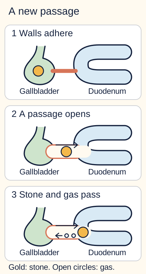
A 3 cm stone inside the distal intestine did not usually squeeze through an otherwise normal common bile duct. Repeated gallbladder inflammation can make the gallbladder adhere to adjacent bowel, most often the duodenum. Sustained pressure and inflammation damage the shared tissue until a cholecystoenteric fistula, an abnormal connection between gallbladder and bowel, forms. The duodenal connection is called a cholecystoduodenal fistula. [1][4]
Trace the large gold stone toward the bowel and the small outlined gas bubbles toward the gallbladder. [1]
Try the route: follow the stone with a finger or cursor. Which wall must fail before the stone can reach the duodenum without using the bile duct? Now trace the gas in the reverse direction.
The shared gallbladder-bowel barrier fails. The resulting opening can admit a stone into the intestinal lumen and allow intestinal gas into the gallbladder or biliary system. Gas within the bile ducts is pneumobilia. The two findings have a common anatomic explanation, but neither proves that the stone is currently obstructing the bowel. [1][4]
A useful distinction is entry site versus stopping site. A duodenal fistula describes the entrance, not necessarily the location of obstruction. Once inside, a stone can travel distally before lodging. Changing position or transient release of an impaction can produce alternating periods of pain, vomiting and partial relief. This intermittent course should not be mistaken for reliable resolution. [4]
Older adults are prominent in clinical series, women are more commonly affected than men, and substantial comorbidity is frequent. However, neither female sex nor a remembered history of biliary colic is required. A convincing anatomic pattern matters more than a demographic stereotype. [1][2]
Apply it elsewhere: a patient has a gallbladder-duodenal connection, but the obstructing stone is in the ileum. Name the entrance and the stopping site separately: duodenal fistula, ileal impaction. This prevents an erroneous diagnosis of gastric outlet obstruction based only on the fistula location. [4]
Where should the bowel dilate?
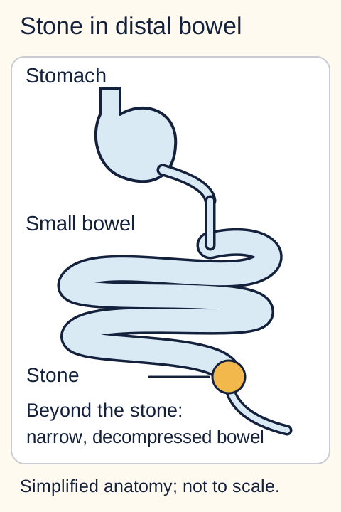
Imagine pouring fluid into a tube with a plug. The reservoir enlarges before the plug, not after it. In ordinary distal gallstone ileus, the stone commonly lodges in the terminal ileum or near the ileocecal valve. The narrower distal lumen and relatively less vigorous distal propulsion favor impaction; pre-existing narrowing and stone burden can change the stopping point. There is no single stone-size cutoff that safely excludes obstruction. [2][10]
Distal impaction produces proximal small-bowel dilation and downstream decompression. [1]
Change the stopping point
Keep the fistula the same. Before opening each comparison, predict which reservoir enlarges when the stone stops sooner, or when there is no focal plug. The diagrams show the consequence of each choice; they do not control access to the lesson.
What changes if the stone stops in the duodenal bulb?
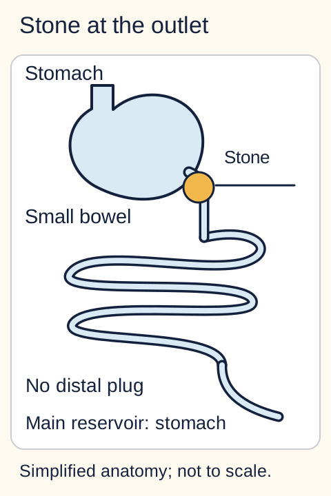
Earlier impaction: the stomach becomes the main reservoir. This pattern is gastric outlet obstruction, called Bouveret syndrome when caused by the gallstone. [4]What changes if propulsion fails without a focal plug?
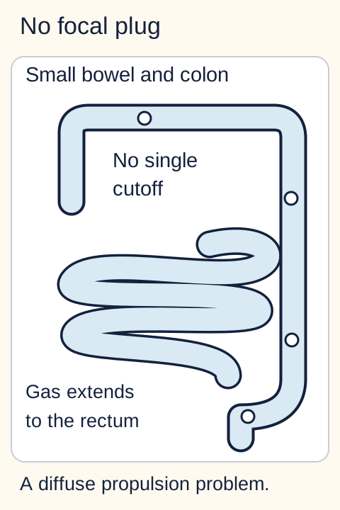
No focal plug: a diffuse small- and large-bowel pattern favors impaired propulsion rather than an obstructing ectopic stone. Interpret the full clinical and CT assessment. [7]
The complete comparison is this: duodenal bulb or pyloric impaction produces stomach-predominant distension, early satiety and vomiting; distal small-bowel impaction produces small-bowel dilation upstream of the stone; diffuse impaired propulsion lacks a discrete obstructing lesion. The proximal gallstone presentation is Bouveret syndrome. Nonbilious vomiting can support a high obstruction but is not a substitute for localization. [4][6]
Less often, a stone obstructs the colon. A diverticular stricture in the sigmoid colon provides a plausible narrow segment even though the normal colon is wider than the ileum. The entry tract and colonic anatomy must be assessed rather than assuming every ectopic stone passed through the same route. [4]
Transfer: CT shows a greatly enlarged stomach, an obstructing stone in the duodenal bulb and no distal small-bowel transition. Predict the syndrome from the blocked reservoir: gastric outlet obstruction, not distal small-bowel obstruction. The biliary origin is shared; the site changes the physiology and procedural access. [6][9]
Does the word ileus describe the mechanism?
In this name, it does not. Gallstone ileus is a mechanical obstruction: an intraluminal stone physically blocks passage. A transition from enlarged upstream bowel to decompressed downstream bowel supports a focal mechanical process. Paralytic ileus means impaired propulsion without a physical plug; diffuse dilation, often involving both small bowel and colon, is more compatible with that mechanism. Imaging and clinical context must agree. [1][7]
Find the obstruction's compartment
Inside the lumen: an ectopic stone or bezoar occupies the passage. In the wall: a tumor or stricture narrows it. Outside the wall: an adhesion or hernia can kink or constrict bowel. No focal blocker: medications, electrolyte abnormalities or recent surgery can impair propulsion. A history of gallstones does not decide among these compartments. [1][7]
Compare two reports: one describes dilated jejunum, a rim-calcified intraluminal object at the ileal cutoff and collapsed distal bowel. The other describes diffuse small- and large-bowel dilation without a cutoff after surgery. Assign a mechanism before naming a disease.
The first report identifies a focal intraluminal obstruction. Biliary findings can then establish why the object is likely an escaped gallstone. The second favors impaired propulsion; a coexisting gallbladder stone would not explain that distribution. Conversely, an abrupt cutoff at a hernia neck should not be relabeled gallstone ileus merely because pneumobilia is also present. [1][7]
A partial obstruction can permit some stool or gas passage, and temporary symptom improvement does not establish durable patency. Follow the current examination and imaging rather than letting a brief comfortable interval outweigh a demonstrated obstructing lesion. [4][7]
Transfer: change the location of the abnormality while leaving the gallstone history unchanged. If the current cutoff is caused by an enhancing mural mass rather than an intraluminal calculus, the immediate etiologic assessment follows the mass. An incidental gallbladder stone is not the cause of every obstruction in that patient. [1]
Different passage, different problem: in Mirizzi syndrome, a stone retained at the gallbladder neck or cystic duct compresses the adjacent common hepatic duct. This produces a biliary obstruction pattern, often with jaundice, rather than an ectopic intestinal plug. Localize ductal dilation versus bowel dilation before using the gallstone history to name the mechanism. [11]
What if the imaging triad is incomplete?
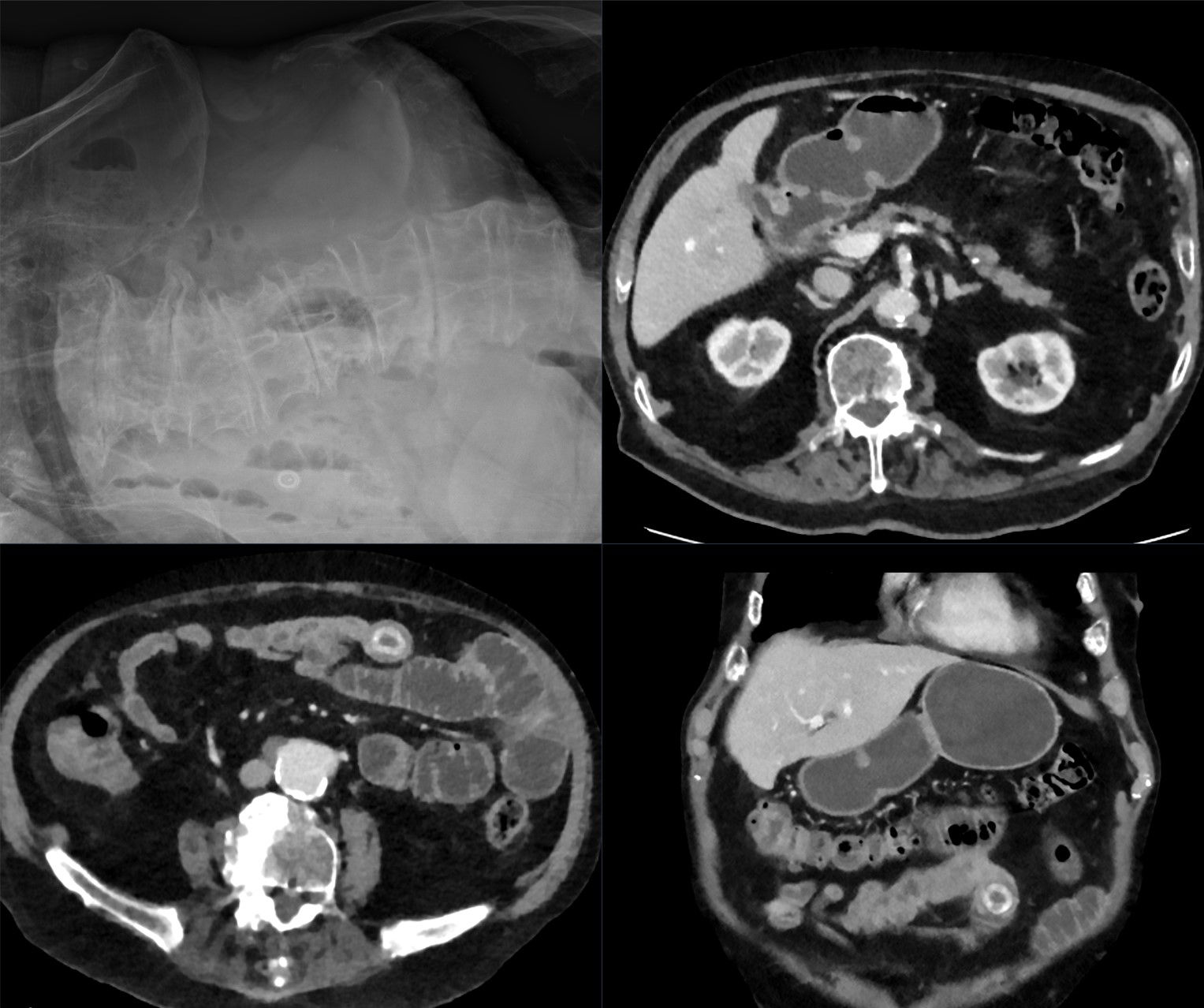
Rigler's triad is bowel obstruction, an ectopic gallstone and pneumobilia. It is a useful combination, not a requirement that every component appear on the first radiograph. Many stones are poorly visible on plain films, and pneumobilia can be absent even with a fistula. A negative or incomplete radiograph should not end the assessment of persistent obstruction. [1]
The source describes a gallbladder-duodenal fistula and an impacted small-bowel stone, but specifically reports no pneumobilia. The lower CT panels show the ectopic calculus and downstream decompression. Image: Hellerhoff, 2023; original source; CC BY-SA 4.0. [8].
Inspect before reading on: find the ring-calcified object in the lower CT panels. Compare the bowel around the object with the decompressed segment beyond it. Would a missing gas sign outweigh this focal obstruction pattern?
No. The teaching point is the combination of an ectopic object, its position at the cutoff, and the obstructed bowel pattern. Absence of pneumobilia does not erase those observations. Do not expect one displayed CT slice to establish every feature of a whole examination. [1][8]
For a stable patient needing definitive evaluation of suspected obstruction, CT of the abdomen and pelvis, generally with intravenous contrast when appropriate, assesses the transition, possible ectopic stone, additional stones, gallbladder region, and bowel perfusion or perforation. The fistula may be directly visible, but failure to display the tract does not exclude the diagnosis. Contrast selection requires clinical and radiology judgment; severe vomiting or high-grade obstruction is not a reason to delay urgent CT for a large oral contrast load. [1][7]
A minimally calcified stone can resemble fluid or adjacent bowel wall. Ask for careful review of the transition and comparison with prior imaging when the initial interpretation and the clinical picture disagree. CT is more informative than a plain-film checklist, but it is not infallible. [1]
Transfer: plain films show small-bowel dilation but neither an obvious stone nor biliary gas. Persistent vomiting and distension still warrant investigation of a mechanical cause. In a stable patient, obtain CT rather than waiting for all three signs to appear. In an unstable patient with peritonitis, urgent surgical treatment must not wait for an ideal image set. [1][7]
Which findings actually identify the cause?
Pneumobilia means gas in the biliary system. It does not, by itself, mean an obstructing intestinal gallstone. After ERCP with sphincterotomy, air can enter through the treated papilla and persist. Biliary surgery, other fistulas and some infections also provide alternative explanations. Interpret new pain, fever or instability on their own merits rather than declaring all post-procedure findings harmless. [12][4]
Rank the evidence: cover the interpretations below and assign each report to one of four categories: no demonstrated obstruction, obstruction of uncertain cause, suspicious biliary association, or an obstructing ectopic stone. Which report actually connects the proposed cause to the transition point?
Four reports, four different conclusions
Air after recent sphincterotomy, with normal-caliber bowel: no demonstrated bowel obstruction. Pneumobilia has an alternative explanation.
Small-bowel dilation on radiographs, with no identified lesion: investigate obstruction, but the cause remains uncertain.
Small-bowel obstruction and unexplained pneumobilia: a biliary-enteric cause becomes more suspicious. Look for an ectopic calculus and competing causes rather than declaring the mechanism proved.
An ectopic calculus at the transition, with proximal dilation and supportive biliary findings: the object is anatomically linked to the obstruction. This is the strongest of these four patterns. [1][12]
These categories are qualitative judgments, not a validated numerical score. A stone lying freely in nondilated bowel may have passed without producing current obstruction. Conversely, an incomplete triad can still accompany a clearly obstructing stone. Localization prevents both overdiagnosis from incidental findings and underdiagnosis from an absent classic sign. [1]
Transfer: a patient has biliary gas after sphincterotomy, but CT places the bowel cutoff at a hernia neck with no intestinal calculus. The hernia explains the obstruction; the gas does not relocate the culprit. Always connect the proposed cause to the observed cutoff. [7][12]
What must be treated now, and what can wait?
Start by treating the patient, not completing an imaging checklist. Keep the patient without oral intake, restore perfusion with appropriately titrated intravenous fluid, correct electrolyte losses, and use nasogastric decompression when indicated by vomiting or distension. Obtain prompt surgical assessment. These measures support the patient; they do not reliably dislodge an impacted stone. General small-bowel obstruction guidance supports resuscitation and urgent intervention for threatened bowel, but an observation interval designed for uncomplicated adhesive obstruction is not automatically appropriate for stone impaction. [7][2]
Peritonitis, perforation, persistent shock or concern for ischemic bowel requires urgent surgical action with resuscitation in parallel. A normal isolated lactate cannot overrule a worsening examination or concerning bowel-wall enhancement. [14] Definitive viability assessment may occur during exploration. Do not postpone treatment solely to demonstrate the fistula or obtain a complete Rigler triad. [7]
Choose the operative target: an older patient has an ileal stone, viable bowel and no active gallbladder infection. First decide what ends the obstruction. Then change only the bowel finding to an irreversibly ischemic, perforated segment. Does the smaller operation still treat the emergency?
Separate three decisions
Extract the stone: enterolithotomy opens the bowel to extract the obstructing calculus. When bowel is viable, this can relieve the emergency without dissecting the inflamed biliary region. Inspect the remaining bowel for additional stones; treating one cutoff does not prove that no other calculi remain. [2][5]
Resect damaged bowel: irreversibly ischemic or perforated bowel requires resection rather than stone extraction alone. Frailty increases operative risk, but it does not make necrotic bowel safe to leave behind. [3][7]
Address the biliary source: cholecystectomy and fistula repair add a separate surgical task. The decision depends on physiology, inflammation, anatomy and remaining biliary disease, not simply the presence of a tract. [3][13]
In a frail patient with viable bowel and no active biliary sepsis, enterolithotomy alone is often favored because it limits operative burden. A one-stage operation combines intestinal stone extraction, cholecystectomy and fistula repair. It can be considered in selected stable, lower-risk patients with suitable anatomy, particularly when residual biliary disease creates a reason to address that region. Stability is necessary context, not a universal instruction to perform the larger operation. [2][13]
The comparative evidence is largely observational. Worse outcomes in a resection group do not prove that necessary resection should be avoided; those patients may have more severe bowel injury. Likewise, differences between limited and combined procedures do not establish one operation as best for every patient. Interpret study design before turning an association into a surgical rule. [3][13]
Proximal access changes the options. In selected Bouveret syndrome, an experienced team may attempt endoscopic extraction or fragmentation, with surgical backup when endoscopy fails or complications occur. A large or firmly impacted stone can be technically difficult. Retained fragments can travel distally and obstruct later, so symptom recurrence after apparently successful proximal treatment deserves reassessment. ERCP directed at the bile duct is not the same as extracting a stone from the intestinal lumen. [6][9]
Recovery is not an automatic appointment for another operation. After successful limited surgery, a fistula may close spontaneously, but closure is not guaranteed. In an asymptomatic high-risk patient, observation with individualized follow-up may be preferable to obligatory interval cholecystectomy and fistula repair. Persistent biliary symptoms, residual gallbladder stones, recurrent infection or another obstruction should prompt reassessment of definitive treatment when feasible. [4][13]
Recurrence has two important routes: a stone already inside the intestine was not recognized during the first episode, or an additional gallbladder stone later enters through a persistent fistula. Recurrence alone cannot determine which happened. Compare prior imaging with the current transition and assess the remaining stone burden. Renewed vomiting, distension or obstructive pain after enterolithotomy calls for prompt evaluation, often including CT, rather than reassurance based only on the earlier operation. [5]
Locate the obstruction, assess the bowel, and relieve the immediate threat. Decide separately whether the patient benefits from additional biliary surgery. A fistula explains the route; bowel viability and operative risk determine the immediate scope.
Apply the lesson
Case 1
Show answer and explanations for case 1
A. Perforation of the gallbladder into the peritoneal cavity (Why this does not fit)
Gallbladder perforation can spill bile or stones outside the gastrointestinal tract. The calculus is demonstrably inside bowel, and the abnormal interface is with adjacent duodenum rather than a free peritoneal collection. Localize an escaped stone to the enteric or peritoneal compartment.
Reasoning steps for option A
Where does free perforation deposit material?
Gallbladder perforation can spill bile or stones outside the gastrointestinal tract.
Where is the obstructing object in this case?
The calculus is demonstrably inside bowel, and the abnormal interface is with adjacent duodenum rather than a free peritoneal collection.
Why does free gallbladder perforation not explain the distal ileal object?
The 3.1 cm calculus is intraluminal at the ileal cutoff, while the abnormal interface is gallbladder to duodenum, not a free peritoneal collection.
B. Passage through the cystic duct and major papilla (Why this does not fit)
Small biliary stones can enter the duodenum through the normal ductal route. The large calculus, abnormal gallbladder-duodenal interface and gallbladder gas instead support a direct abnormal communication. Account for the stone's size and the tissue interface, not merely its biliary origin.
Reasoning steps for option B
Which route can a small ductal stone take?
Small biliary stones can enter the duodenum through the normal ductal route.
What makes that route less persuasive here?
The large calculus, abnormal gallbladder-duodenal interface and gallbladder gas instead support a direct abnormal communication.
Why is passage through the cystic duct and papilla unlikely for this calculus?
A 3.1 cm former gallbladder stone, lost duodenal interface and gas in the contracted gallbladder favor direct fistulous passage over the narrow normal ductal route.
C. Erosion through the shared gallbladder-duodenal wall (Best answer)
A cholecystoduodenal fistula allows a large gallstone to enter bowel and intestinal gas to enter the biliary system. The lost interface, new gallbladder gas and previously documented calculus now at an ileal cutoff fit that route. Distinguish the fistula entrance from the eventual impaction site.
Reasoning steps for option C
What identifies the immediate obstruction?
The intraluminal ileal calculus sits at the transition with proximal dilation.
Which changes identify a plausible entrance?
The abnormal gallbladder-duodenal interface and gallbladder gas support an enteric communication.
How does the proposed route explain both findings?
The stone passes into bowel while intestinal gas can pass toward the gallbladder.
Must the entrance and obstruction be at the same site?
No. A duodenal entrance can lead to ileal impaction.
D. Formation of an enterolith within an ileal diverticulum (Why this does not fit)
Primary intestinal enteroliths can arise where luminal contents stagnate. Prior gallbladder imaging and the new gallbladder-duodenal changes supply a biliary origin that this alternative does not explain. Use prior imaging and the current route together when identifying a luminal object.
Reasoning steps for option D
What setting permits primary enterolith formation?
Primary intestinal enteroliths can arise where luminal contents stagnate.
Which observations instead connect this calculus to the gallbladder?
Prior gallbladder imaging and the new gallbladder-duodenal changes supply a biliary origin that this alternative does not explain.
Why is a primary ileal-diverticular enterolith less likely than a migrated gallstone?
The prior gallbladder calculus is similarly sized to the ileal object and the new gallbladder gas and duodenal interface change supply a biliary entry route.
E. Compression of the common hepatic duct by a neck calculus (Why this does not fit)
An impacted gallbladder-neck calculus can compress the common hepatic duct in Mirizzi syndrome. That extrinsic ductal process does not place a large calculus inside the ileum or explain the damaged duodenal interface. Separate bile-duct obstruction from obstruction of the intestinal lumen.
Reasoning steps for option E
Which passage does a neck calculus compress?
An impacted gallbladder-neck calculus can compress the common hepatic duct in Mirizzi syndrome.
Does that process explain an ileal calculus?
That extrinsic ductal process does not place a large calculus inside the ileum or explain the damaged duodenal interface.
Why does Mirizzi-type hepatic-duct compression fail to explain this obstruction?
A neck stone compressing the hepatic duct cannot account for a calculus inside the distal ileum at a bowel transition or the abnormal duodenal interface.
Takeaway: The entrance is a fistula; the final obstruction can lie far downstream.
A. Upper endoscopic extraction or lithotripsy with surgical backup (Best answer)
Selected Bouveret syndrome can be treated through upper endoscopic access by an experienced team. The stomach-predominant dilation localizes the obstruction to the accessible duodenal calculus, while cardiopulmonary disease favors limiting surgical burden. Plan for technical failure, retained fragments and escalation rather than promising endoscopic success.
Reasoning steps for option A
Where is the enlarged reservoir?
The stomach, upstream of the duodenal bulb.
Which syndrome follows from that location and cause?
A gallstone causing gastric outlet obstruction is Bouveret syndrome.
Which available approach can reach it without an initial abdominal operation?
Advanced upper endoscopy for extraction or fragmentation, with surgical backup.
What does choosing endoscopy not guarantee?
Complete extraction or avoidance of later fragment-related obstruction.
B. Endoscopic retrograde cholangiopancreatography with duct extraction (Why this does not fit)
ERCP can extract stones obstructing the biliary ductal system. This calculus is within the duodenal lumen and obstructs gastric emptying, not the common bile duct. A biliary origin does not make a luminal obstruction an ERCP target.
Reasoning steps for option B
What compartment does routine duct extraction treat?
ERCP can extract stones obstructing the biliary ductal system.
Which compartment contains the current obstruction?
This calculus is within the duodenal lumen and obstructs gastric emptying, not the common bile duct.
Why is duct-directed ERCP not the extraction route for this patient?
The 3.5 cm stone is lodged in the duodenal bulb and distends the stomach, whereas ERCP duct extraction targets a stone within the biliary ducts.
C. Laparoscopic gastrotomy or duodenotomy for calculus extraction (Why this does not fit)
Operative extraction can treat a proximal impacted calculus when endoscopy is unsuitable or unsuccessful. In this stable high-risk patient with advanced endoscopy available, an endoscopic attempt can reach the lesion without an initial abdominal operation. Keep operative extraction available as backup rather than treating endoscopy as guaranteed to succeed.
Reasoning steps for option C
When does operative proximal extraction become appropriate?
Operative extraction can treat a proximal impacted calculus when endoscopy is unsuitable or unsuccessful.
What makes an initial endoscopic approach attractive here?
In this stable high-risk patient with advanced endoscopy available, an endoscopic attempt can reach the lesion without an initial abdominal operation.
Why reserve operative duodenotomy instead of choosing it first here?
Severe lung disease raises operative burden, while stable proximal impaction can be approached by available advanced upper endoscopy; surgery remains backup if extraction fails.
D. Percutaneous gallbladder drainage followed by reassessment (Why this does not fit)
Gallbladder drainage can provide source control for selected patients with acute cholecystitis. Draining the gallbladder would leave the already escaped calculus blocking the gastric outlet. Treat the current luminal obstruction separately from residual gallbladder disease.
Reasoning steps for option D
What problem can gallbladder drainage address?
Gallbladder drainage can provide source control for selected patients with acute cholecystitis.
Would it extract a calculus already in the duodenum?
Draining the gallbladder would leave the already escaped calculus blocking the gastric outlet.
Why cannot gallbladder drainage relieve this gastric outlet obstruction?
The calculus has already escaped into the duodenal bulb and physically blocks gastric emptying; draining the gallbladder leaves that luminal plug intact.
Takeaway: Localize the blocked reservoir before choosing the extraction route.
A. MR cholangiopancreatography focused on the extrahepatic ducts (Why this does not fit)
MRCP can clarify ductal anatomy and obstruction within the biliary system. The supplied radiographs and symptoms require assessment of a small-bowel cutoff and threatened bowel rather than an isolated ductal map. A possible biliary origin does not narrow the investigation to the ducts alone.
Reasoning steps for option A
Which anatomy is the focus of MRCP?
MRCP can clarify ductal anatomy and obstruction within the biliary system.
Which anatomic and viability questions remain unanswered?
The supplied radiographs and symptoms require assessment of a small-bowel cutoff and threatened bowel rather than an isolated ductal map.
Why is a duct-focused MRCP insufficient after these radiographs?
Small-bowel dilation and persistent vomiting require localization of a bowel transition and assessment of threatened bowel, neither answered by mapping only extrahepatic ducts.
B. Hepatobiliary scintigraphy to assess cystic-duct patency (Why this does not fit)
Hepatobiliary scintigraphy addresses selected questions about gallbladder filling and bile flow. The unresolved problem is mechanical intestinal obstruction, not isolated failure of gallbladder filling. Choose the test that interrogates the affected compartment.
Reasoning steps for option B
What functional question does this scan answer?
Hepatobiliary scintigraphy addresses selected questions about gallbladder filling and bile flow.
Is that the key uncertainty in this presentation?
The unresolved problem is mechanical intestinal obstruction, not isolated failure of gallbladder filling.
Why does cystic-duct scintigraphy miss the urgent imaging target?
The dilated small bowel and air-fluid levels implicate intestinal transit, not gallbladder filling; a bowel-focused CT can locate the cutoff and complications.
C. Serial abdominal radiographs after nasogastric decompression (Why this does not fit)
Serial films can sometimes document changes in an obstruction pattern. Persistent symptoms and an unexplained obstruction require etiologic assessment; decompression and serial films do not provide the transition and perfusion information available from CT. Do not require all components of a classic radiographic pattern before pursuing its suspected cause.
Reasoning steps for option C
What can serial radiographs show?
Serial films can sometimes document changes in an obstruction pattern.
What important etiologic and viability questions would serial films leave unresolved?
Persistent symptoms and an unexplained obstruction require etiologic assessment; decompression and serial films do not provide the transition and perfusion information available from CT.
Why not wait for a visible stone and pneumobilia on repeat films?
The radiographs support small-bowel obstruction without showing a calculus or biliary gas; CT can seek a poorly visible ectopic calculus, localize the transition and assess bowel perfusion now.
D. CT of the abdomen and pelvis with intravenous contrast (Best answer)
CT can evaluate the transition, an ectopic calculus and bowel-wall perfusion in a stable patient with suspected mechanical obstruction. The radiographs establish concern for obstruction without identifying its cause, and no contrast limitation is supplied. An incomplete plain-film triad warrants a better anatomic study rather than exclusion of gallstone ileus.
Reasoning steps for option D
What do the films support without establishing an etiology?
Small-bowel obstruction remains a concern, but the obstructing lesion is not identified.
Does absence of a radiopaque calculus or biliary gas exclude gallstone ileus?
No. These findings may be absent or difficult to see.
Which investigation best addresses the remaining questions in this stable patient?
CT of the abdomen and pelvis with appropriate intravenous contrast.
What would change the sequence?
Instability or peritonitis could require urgent treatment rather than waiting for a complete imaging workup.
E. Right upper quadrant ultrasonography as the sole next study (Why this does not fit)
Ultrasound is useful for gallbladder stones and inflammatory biliary findings. It does not comprehensively map this patient's suspected small-bowel transition or assess the entire abdomen as CT can. Investigate the current obstruction rather than only its possible biliary source.
Reasoning steps for option E
What can biliary ultrasound characterize well?
Ultrasound is useful for gallbladder stones and inflammatory biliary findings.
What additional unresolved problem must be mapped here?
It does not comprehensively map this patient's suspected small-bowel transition or assess the entire abdomen as CT can.
Why is right upper quadrant ultrasound alone inadequate here?
It may depict gallbladder disease but cannot comprehensively localize the small-bowel cutoff or evaluate the full abdomen for ischemic complications.
Takeaway: Stable suspected obstruction with an incomplete radiograph needs an anatomic explanation, not a perfect triad.
A. Diffuse dysmotility with gas from prior instrumentation (Why this does not fit)
Prior instrumentation can explain pneumobilia, and illness can impair intestinal propulsion. An abrupt transition at an irreducible hernia with downstream collapse establishes a focal mechanical problem. A plausible explanation for the gas does not erase the mechanical obstruction.
Reasoning steps for option A
Which part of this explanation fits the history?
Prior instrumentation can explain pneumobilia, and illness can impair intestinal propulsion.
What finding argues against diffuse dysmotility?
An abrupt transition at an irreducible hernia with downstream collapse establishes a focal mechanical problem.
Why does prior sphincterotomy not make this a diffuse motility disorder?
Sphincterotomy explains pneumobilia, but the abrupt cutoff at an irreducible groin hernia and distal collapse identify mechanical obstruction.
B. A hernia-related transition with gas from sphincterotomy (Best answer)
A hernia can obstruct bowel, while sphincterotomy can allow persistent biliary gas independently. CT links the cutoff to the symptomatic groin defect and shows no ectopic calculus. Use one explanation for each finding when the anatomy does not support a single unifying lesion.
Reasoning steps for option B
What makes the intestinal process mechanical?
There is an abrupt hernia-associated transition with downstream decompression.
Is an ectopic calculus needed to explain that cutoff?
No. The bowel is trapped at the demonstrated defect.
What independently permits biliary gas?
Prior sphincterotomy can allow gas to enter the ducts.
Which diagnostic error is avoided?
Assigning all findings to gallstone ileus despite a different proven cause of obstruction.
C. An adhesive transition with gas from an enteric fistula (Why this does not fit)
Adhesions can cause a mechanical small-bowel transition, and fistulas can introduce biliary gas. The demonstrated cutoff is at the hernia defect, while prior sphincterotomy already explains a route for the gas. Prefer a demonstrated focal cause over an assumed one.
Reasoning steps for option C
What would support an adhesive transition?
Adhesions can cause a mechanical small-bowel transition, and fistulas can introduce biliary gas.
Where is the actual transition shown?
The demonstrated cutoff is at the hernia defect, while prior sphincterotomy already explains a route for the gas.
Why is an adhesive transition with an enteric fistula less supported than the observed hernia?
CT localizes the cutoff to the groin defect and the earlier sphincterotomy accounts for gas without an assumed adhesion or fistula.
D. A mural small-bowel lesion with gas from biliary infection (Why this does not fit)
A mural lesion can narrow bowel, and some infections can produce gas in the biliary region. No mural mass or inflammatory biliary process is described; the symptomatic hernia provides the observed transition. Do not substitute unobserved pathology for a directly demonstrated cause.
Reasoning steps for option D
What findings would favor these two processes?
A mural lesion can narrow bowel, and some infections can produce gas in the biliary region.
Which demonstrated lesion better explains the current obstruction?
No mural mass or inflammatory biliary process is described; the symptomatic hernia provides the observed transition.
Why not invoke a mural tumor and biliary infection here?
Neither a mural mass nor biliary inflammatory process is described; the irreducible tender hernia coincides with the transition.
E. An ectopic calculus with gas from an enteric fistula (Why this does not fit)
An obstructing ectopic stone and pneumobilia can accompany gallstone ileus. The only calculus is still in the gallbladder, and the bowel is trapped at the groin defect. Gallbladder calculi plus biliary gas are not equivalent to an obstructing intestinal calculus.
Reasoning steps for option E
Which stone location would support this explanation?
An obstructing ectopic stone and pneumobilia can accompany gallstone ileus.
Is a stone identified at the intestinal cutoff?
The only calculus is still in the gallbladder, and the bowel is trapped at the groin defect.
Why do gallbladder calculus and pneumobilia not establish gallstone ileus here?
The only calculus remains in the gallbladder and no intestinal stone is identified; the bowel cutoff instead occurs at the groin hernia.
Takeaway: Pneumobilia does not make every coexisting bowel obstruction gallstone ileus.
A. Urgent decompressive colonoscopy for isolated colonic pseudo-obstruction (Why this does not fit)
Selected acute colonic pseudo-obstruction may require directed colonic decompression. The supplied pattern involves both small and large bowel with major reversible motility contributors, not a stated refractory colon-predominant emergency. Distinguish diffuse postoperative ileus from a separate colon-focused syndrome before escalating.
Reasoning steps for option A
When might colon-focused decompression be useful?
Selected acute colonic pseudo-obstruction may require directed colonic decompression.
What makes an initial reversible-cause strategy more appropriate here?
The supplied pattern involves both small and large bowel with major reversible motility contributors, not a stated refractory colon-predominant emergency.
Why is immediate colonoscopic decompression not the best match for this dilation pattern?
Both small bowel and colon contain gas to the rectum after hip surgery, with opioids and potassium 2.7 mmol/L; this is diffuse motility impairment, not an isolated refractory colonic process.
B. Urgent enterolithotomy with examination for further stones (Why this does not fit)
Enterolithotomy treats a stone physically obstructing the intestinal lumen. The calculus remains in the gallbladder and the bowel pattern is diffuse without a stone-centered cutoff. Confirm an obstructing enteric object before selecting intestinal stone surgery.
Reasoning steps for option B
What lesion does enterolithotomy treat?
Enterolithotomy treats a stone physically obstructing the intestinal lumen.
Is that lesion present in this examination?
The calculus remains in the gallbladder and the bowel pattern is diffuse without a stone-centered cutoff.
Why would enterolithotomy lack a target in this patient?
The known calculus remains inside the gallbladder and CT shows no intraluminal stone or abrupt bowel transition.
C. ERCP with extraction of a presumed ductal calculus (Why this does not fit)
ERCP can treat a demonstrated bile-duct obstruction. The known stone is in the gallbladder; the presentation and imaging do not establish a ductal obstruction causing these diffuse bowel findings. Treat the abnormal physiology demonstrated rather than an incidental stone history.
Reasoning steps for option C
What problem would duct extraction address?
ERCP can treat a demonstrated bile-duct obstruction.
Which abnormality explains this bowel distribution better?
The known stone is in the gallbladder; the presentation and imaging do not establish a ductal obstruction causing these diffuse bowel findings.
Why does the gallbladder calculus not justify ERCP for the diffuse distension?
No ductal calculus or ductal obstruction is demonstrated; opioid use and marked hypokalemia better explain diffuse bowel dilation.
D. Adhesiolysis at the presumed distal small-bowel transition (Why this does not fit)
Adhesiolysis can treat a mechanical obstruction caused by adhesions. No distal transition is identified, and gas extends through the colon in a diffuse pattern after nonabdominal surgery. A mechanical procedure needs evidence of a focal mechanical target.
Reasoning steps for option D
What imaging pattern would make a focal adhesion more likely?
Adhesiolysis can treat a mechanical obstruction caused by adhesions.
What distribution is actually described?
No distal transition is identified, and gas extends through the colon in a diffuse pattern after nonabdominal surgery.
Why does presumed adhesiolysis not follow from this CT?
There is no distal small-bowel cutoff, gas extends through the colon and hip surgery was nonabdominal; no adhesive transition is identified.
E. Potassium replacement and reduction of opioid burden (Best answer)
Electrolyte abnormalities and opioids can contribute to impaired intestinal propulsion. Hypokalemia and a diffuse small- and large-bowel pattern support this mechanism rather than a focal stone obstruction. Correct reversible contributors while providing supportive care and reassessing the abdomen.
Reasoning steps for option E
Does the CT describe a discrete obstructing lesion?
No. Gas extends throughout small bowel and colon without a focal transition.
Which supplied factors support impaired propulsion?
Marked hypokalemia, recent surgery and opioid exposure.
What treatment direction follows initially?
Correct potassium, reduce avoidable opioid burden and provide supportive care with reassessment.
Why does the gallbladder calculus not determine management?
It has not entered bowel or produced the observed diffuse pattern.
Takeaway: Diffuse dilation plus reversible motility factors is not explained by a stone that remains in the gallbladder.
A. Post-inflammatory dysmotility persisting after completed stone passage (Why this does not fit)
Dysmotility can persist after an acute abdominal illness. The stone is still inside the bowel at a new abrupt cutoff, so passage through the obstructed tract is not complete. Symptom relief does not establish clearance when a focal lesion remains.
Reasoning steps for option A
What finding would support completed passage?
Dysmotility can persist after an acute abdominal illness.
Where is the calculus on the repeat study?
The stone is still inside the bowel at a new abrupt cutoff, so passage through the obstructed tract is not complete.
Why does several hours of relief not prove completed calculus passage?
Repeat CT still places the same-appearing stone at a new ileal cutoff with upstream dilation and downstream collapse.
B. Temporary release of impaction followed by a new mechanical blockage (Best answer)
A migrating calculus can intermittently lodge and then travel before impacting again. The changing stone location and renewed dilation at its current position fit temporary relief followed by persistent obstruction. Transient improvement cannot substitute for current evidence of intestinal patency.
Reasoning steps for option B
Does the calculus stay in one fixed location?
No. It is farther distally on the repeat examination.
What does the later transition demonstrate?
The calculus is again producing a focal mechanical obstruction.
How can the comfortable interval be explained?
Temporary release of impaction allowed some transit before the stone lodged again.
Does the earlier relief justify dismissing current symptoms?
No. Current anatomy and physiology show an ongoing obstruction.
C. A new adhesive obstruction with an incidental enteric calculus (Why this does not fit)
Adhesions can cause obstruction and an enteric object can occasionally be incidental. Both examinations link the transition to the calculus, and the current downstream collapse begins at its new position. Assess whether the object actually coincides with the obstruction before calling it incidental.
Reasoning steps for option C
When could an enteric calculus be incidental?
Adhesions can cause obstruction and an enteric object can occasionally be incidental.
How is the calculus related to the cutoff here?
Both examinations link the transition to the calculus, and the current downstream collapse begins at its new position.
Why is the migrated calculus not incidental to an adhesive obstruction?
The transition tracked the calculus from jejunum to ileum on serial CT, and distal collapse starts at its current position.
D. Resolution of obstruction with a residual nonobstructing distal stone (Why this does not fit)
A calculus may remain in bowel after obstruction resolves if it no longer blocks transit. Current upstream dilation and distal collapse at the stone contradict a nonobstructing state. Judge current patency from the entire transition pattern, not the direction of stone travel alone.
Reasoning steps for option D
Can a stone travel distally without remaining obstructive?
A calculus may remain in bowel after obstruction resolves if it no longer blocks transit.
What makes this later stone position clinically significant?
Current upstream dilation and distal collapse at the stone contradict a nonobstructing state.
What current findings refute resolution despite distal stone migration?
The new ileal stone remains at an abrupt transition with proximal dilation and collapsed downstream bowel, so transit is still obstructed.
E. Two unrelated fixed strictures producing alternating episodes (Why this does not fit)
Separate strictures can produce recurrent or multifocal obstruction. No fixed mural lesions are described, while one recognizable calculus changes location with the obstructing pattern. Choose the mechanism supported by the serial anatomic change.
Reasoning steps for option E
What would support separate fixed strictures?
Separate strictures can produce recurrent or multifocal obstruction.
Which changing structure instead tracks the symptoms?
No fixed mural lesions are described, while one recognizable calculus changes location with the obstructing pattern.
Why are two fixed strictures less plausible than intermittent stone impaction?
The same recognizable calculus moved from jejunum to ileum as the transition moved; neither CT describes fixed mural narrowing.
Takeaway: A migrating stone can cause intermittent relief and still become a persistent mechanical obstruction.
A. Enterolithotomy alone with inspection for additional calculi (Best answer)
Limited stone extraction can relieve gallstone ileus while avoiding extensive biliary dissection in a high-risk patient. The bowel is viable and the gallbladder has no active septic process, while cardiopulmonary reserve is poor. Limit the operation without overlooking additional intestinal stones.
Reasoning steps for option A
What is causing the immediate threat?
An impacted calculus obstructs the ileal lumen.
Does bowel viability require segmental excision?
No. The bowel is viable in the supplied operative assessment.
What is the cost of adding biliary dissection?
It increases operative burden in a patient with poor cardiopulmonary reserve and difficult adhesions.
Which scope fits these findings?
Enterolithotomy alone, while inspecting the remaining bowel for other calculi.
B. Gallbladder drainage with delayed intestinal stone extraction (Why this does not fit)
Gallbladder drainage can help control infection when cholecystitis is the urgent problem. There is no active biliary sepsis, while the escaped ileal calculus remains the immediate mechanical obstruction. Address the present intestinal threat rather than a quiet upstream organ.
Reasoning steps for option B
When would gallbladder drainage provide useful source control?
Gallbladder drainage can help control infection when cholecystitis is the urgent problem.
What is the active threat at this operation?
There is no active biliary sepsis, while the escaped ileal calculus remains the immediate mechanical obstruction.
Why would gallbladder drainage leave this patient obstructed?
There is no active biliary sepsis, whereas a 3.2 cm ileal calculus remains at the mechanical transition and requires intestinal relief.
C. Bypass of the obstructed segment with preservation of the calculus (Why this does not fit)
Bypass can be useful for selected unresectable or technically inaccessible obstructing lesions. An accessible intraluminal calculus in viable bowel can be extracted directly without leaving the obstructing material behind. Match surgical complexity to the actual lesion and its accessibility.
Reasoning steps for option C
When might bypass be considered for an obstruction?
Bypass can be useful for selected unresectable or technically inaccessible obstructing lesions.
What simpler definitive target is available here?
An accessible intraluminal calculus in viable bowel can be extracted directly without leaving the obstructing material behind.
Why is bypass rather than extraction unnecessary in this operation?
The stone is accessible inside viable ileum, so enterolithotomy removes the plug directly instead of leaving it behind.
D. Enterolithotomy with cholecystectomy and fistula closure (Why this does not fit)
Combined surgery treats the obstruction and residual biliary communication in one procedure. Severe cardiopulmonary disease, dense adhesions and no active biliary sepsis make the extra dissection less attractive during this emergency. Separate relief of the obstruction from the benefit and burden of biliary repair.
Reasoning steps for option D
What additional problem does combined surgery address?
Combined surgery treats the obstruction and residual biliary communication in one procedure.
What makes that added task less favorable in this patient?
Severe cardiopulmonary disease, dense adhesions and no active biliary sepsis make the extra dissection less attractive during this emergency.
Why avoid simultaneous fistula repair and cholecystectomy here?
Severe COPD, ejection fraction 25%, dense gallbladder-duodenal adhesions and no biliary sepsis make additional biliary dissection disproportionate to the immediate ileal obstruction.
E. Segmental ileal resection with inspection of remaining bowel (Why this does not fit)
Resection is appropriate when the affected bowel is nonviable, perforated or otherwise cannot be safely preserved. The supplied operative assessment identifies viable bowel rather than a segment requiring excision. Do not add bowel resection solely because a calculus caused the obstruction.
Reasoning steps for option E
Which bowel findings justify resection?
Resection is appropriate when the affected bowel is nonviable, perforated or otherwise cannot be safely preserved.
Are those findings present at exploration?
The supplied operative assessment identifies viable bowel rather than a segment requiring excision.
What finding rules out routine segmental ileal resection?
Exploration confirms viable bowel around the calculus; obstruction alone does not require removal of an otherwise salvageable ileal segment.
Takeaway: In viable bowel, a limited operation can treat the obstruction without obligating emergency fistula repair.
A. Enterolithotomy with closure through the affected segment (Why this does not fit)
Enterolithotomy can preserve viable bowel when the wall can be safely closed. The affected segment is necrotic and perforated, so extracting the calculus does not eliminate the nonviable tissue. Stone extraction is insufficient when the bowel itself has become an ongoing source of contamination.
Reasoning steps for option A
What condition permits a simple enterolithotomy?
Enterolithotomy can preserve viable bowel when the wall can be safely closed.
What prevents safe reliance on that approach here?
The affected segment is necrotic and perforated, so extracting the calculus does not eliminate the nonviable tissue.
Why does enterolithotomy and closure fail to control the source?
The affected ileum stays black and friable after warming and leaks through a full-thickness defect; stone removal would leave necrotic contaminated bowel.
B. Extraction of the stone with repair of the perforation alone (Why this does not fit)
A localized defect in otherwise healthy tissue may sometimes be repaired. Here the segment remains frankly necrotic after reassessment; closing one hole would leave dead tissue behind. Distinguish a repairable defect from perforation within nonviable bowel.
Reasoning steps for option B
When can local perforation repair be reasonable?
A localized defect in otherwise healthy tissue may sometimes be repaired.
How does the surrounding tissue change the decision?
Here the segment remains frankly necrotic after reassessment; closing one hole would leave dead tissue behind.
Why is simple perforation repair insufficient after reassessment?
The defect lies in a persistently necrotic segment, so closure of the hole leaves nonviable bowel behind.
C. Gallbladder and fistula surgery before reassessing the ileum later (Why this does not fit)
Biliary surgery may address residual stones or an active gallbladder source in selected patients. The immediate contaminated source is the visibly necrotic, perforated intestinal segment. Prioritize the demonstrated threatened tissue rather than the historical entry route.
Reasoning steps for option C
What residual problem can fistula surgery address?
Biliary surgery may address residual stones or an active gallbladder source in selected patients.
Which source requires treatment during the present operation?
The immediate contaminated source is the visibly necrotic, perforated intestinal segment.
Why must the ileum be treated before the adherent gallbladder region?
The necrotic perforated ileum leaks enteric contents now, whereas the gallbladder region has no purulence.
D. Bypass of the segment with the perforation left excluded (Why this does not fit)
Bypass may restore continuity around selected lesions when direct treatment is unsuitable. An excluded necrotic, perforated segment remains a source of contamination and does not meet the need for source control. Restoring transit and treating devitalized tissue are separate requirements.
Reasoning steps for option D
What can a bypass accomplish?
Bypass may restore continuity around selected lesions when direct treatment is unsuitable.
Which ongoing hazard would remain in this case?
An excluded necrotic, perforated segment remains a source of contamination and does not meet the need for source control.
Why does bypass fail to provide source control in this case?
Bypass may restore transit but the excluded black, perforated segment would still leak and contaminate the abdomen.
E. Resection of the nonviable segment with the obstructing stone (Best answer)
Necrotic or perforated bowel requires treatment of the diseased segment rather than extraction alone. Persistent black, friable bowel and a full-thickness enteric leak after resuscitation indicate that stone extraction alone would leave the source of contamination. Physiological risk changes planning but does not make retained necrotic bowel safe.
Reasoning steps for option E
What do the persistent wall changes and enteric leak establish?
The affected segment cannot safely be preserved by extracting the stone or closing the defect alone.
Which two intestinal hazards must be treated?
The obstructing calculus and the nonviable, perforated bowel wall.
What scope addresses both hazards?
Resection of the affected segment with the stone, with reconstruction tailored to physiology.
Does the quiet adherent gallbladder require a larger emergency operation?
Not solely because a fistula exists; frailty and difficult anatomy favor avoiding unnecessary biliary dissection.
Takeaway: Nonviable or perforated bowel requires treatment beyond extraction of the stone.
A. Repeat contrast CT primarily to demonstrate the fistula (Why this does not fit)
CT can help characterize an uncertain mechanism in a sufficiently stable patient. The obstructing calculus is already established, while the patient now has shock and generalized peritoneal findings. Avoid delaying urgent source control for an anatomic detail that will not remove the emergency indication.
Reasoning steps for option A
When could repeat imaging add useful information?
CT can help characterize an uncertain mechanism in a sufficiently stable patient.
Which current findings make delay hazardous?
The obstructing calculus is already established, while the patient now has shock and generalized peritoneal findings.
Why should a repeat scan to show the fistula not delay treatment?
CT already confirms a stone-centered small-bowel obstruction, and persistent shock plus generalized rigidity mandate urgent assessment regardless of fistula visibility.
B. A monitored decompression trial until the next day (Why this does not fit)
Nonoperative observation is appropriate for selected uncomplicated obstruction patterns. Persistent hypotension and peritonitis are not features of an uncomplicated stable observation candidate. Do not transfer observation intervals from adhesive obstruction to a deteriorating stone obstruction.
Reasoning steps for option B
What physiological conditions make observation more reasonable?
Nonoperative observation is appropriate for selected uncomplicated obstruction patterns.
Does this patient meet those conditions now?
Persistent hypotension and peritonitis are not features of an uncomplicated stable observation candidate.
Why is overnight decompression unsafe in this course?
Persistent hypotension and generalized guarding indicate possible ischemia or perforation, unlike an uncomplicated obstruction suitable for observation.
C. Urgent operative assessment and source control with ongoing resuscitation (Best answer)
Instability and peritonitis in an obstructed patient raise concern for compromised or perforated bowel. Her worsening examination and shock create a treatment priority that does not depend on directly displaying the fistula. Escalate for threatened bowel instead of completing a nonessential diagnostic sign.
Reasoning steps for option C
Is there already an established mechanical cause?
Yes. CT has located an ectopic calculus at the obstruction.
What does the subsequent deterioration add?
Concern for threatened or perforated bowel with systemic instability.
Which missing finding is no longer a prerequisite?
Direct imaging of the fistula is not required before urgent surgical assessment and treatment.
How should resuscitation relate to source control?
Continue resuscitation while urgent operative care is arranged rather than waiting for complete normalization first.
D. Upper endoscopy to determine whether the stone entered through the duodenum (Why this does not fit)
Upper endoscopy may assess selected proximal luminal lesions or fistulous anatomy. The current obstruction is in small bowel and systemic deterioration requires assessment of bowel viability, not just the original entrance. Distinguish the route of entry from the organ currently in danger.
Reasoning steps for option D
What question could an upper examination answer?
Upper endoscopy may assess selected proximal luminal lesions or fistulous anatomy.
Would answering it address the current peritoneal emergency?
The current obstruction is in small bowel and systemic deterioration requires assessment of bowel viability, not just the original entrance.
Why is upper endoscopy of the duodenal entrance the wrong immediate priority?
The current obstruction is in small bowel, and shock with peritonitis calls for bowel viability and source assessment rather than tracing entry.
E. Percutaneous gallbladder drainage before intestinal reassessment (Why this does not fit)
Drainage may control a gallbladder septic source in a patient unsuitable for immediate cholecystectomy. The established intestinal obstruction and new generalized peritoneal signs require urgent intestinal source assessment. Choose source control for the demonstrated source rather than the organ of origin alone.
Reasoning steps for option E
What source is treated by gallbladder drainage?
Drainage may control a gallbladder septic source in a patient unsuitable for immediate cholecystectomy.
Which source is most urgent to assess in this case?
The established intestinal obstruction and new generalized peritoneal signs require urgent intestinal source assessment.
Why would gallbladder drainage miss the threatened source?
The established small-bowel obstruction now accompanies generalized peritoneal signs; gallbladder drainage does not address compromised or perforated intestine.
Takeaway: A missing fistula image must not delay treatment of shock and peritonitis in an obstructed patient.
A. A bile-duct calculus has obstructed intestinal propulsion indirectly (Why this does not fit)
Ductal stones can produce biliary obstruction or inflammatory complications. The described calculus is inside the ileum at its cutoff, not within a bile duct. Localize the lesion before assigning its physiological consequences.
Reasoning steps for option A
Which compartment contains a ductal calculus?
Ductal stones can produce biliary obstruction or inflammatory complications.
Where is this obstructing object located?
The described calculus is inside the ileum at its cutoff, not within a bile duct.
Why is a bile-duct stone not the immediate cause of this cutoff?
The rim-calcified object is within the distal ileum exactly at the caliber change, not in the bile duct.
B. Diffuse dysmotility accounts for the upstream and downstream difference (Why this does not fit)
Dysmotility can dilate bowel without a physical obstructing object. The abrupt stone-centered cutoff and decompressed downstream segment favor a focal mechanical process. Differentiate a diffuse propulsion problem from a localized plug.
Reasoning steps for option B
What distribution is more typical of diffuse dysmotility?
Dysmotility can dilate bowel without a physical obstructing object.
What focal relationship is present in this scan?
The abrupt stone-centered cutoff and decompressed downstream segment favor a focal mechanical process.
Why does diffuse dysmotility not explain the caliber pattern?
An intraluminal stone marks an abrupt transition with proximal dilation and decompressed distal bowel, a focal mechanical plug.
C. An adhesive transition is established by the contracted gallbladder (Why this does not fit)
Adhesions can produce a focal obstruction and often are inferred from the transition pattern. A contracted gallbladder does not establish an adhesive ileal obstruction when an intraluminal calculus occupies the cutoff. Prefer a demonstrated luminal cause over an unrelated inferred extrinsic cause.
Reasoning steps for option C
How is an adhesive obstruction usually supported?
Adhesions can produce a focal obstruction and often are inferred from the transition pattern.
What directly observed lesion explains this transition?
A contracted gallbladder does not establish an adhesive ileal obstruction when an intraluminal calculus occupies the cutoff.
Why not infer an adhesion from the contracted gallbladder?
A contracted gallbladder does not establish an adhesive ileal cutoff; CT directly shows an intraluminal calculus at that point.
D. An impacted ectopic gallstone remains the leading explanation (Best answer)
Gallstone ileus can occur without visible pneumobilia or a directly demonstrated fistula. A rim-calcified intestinal calculus at the obstruction plus an abnormal gallbladder-duodenal relationship supports the diagnosis. Use the positive anatomic evidence while acknowledging what imaging has not shown.
Reasoning steps for option D
What makes the object more than an incidental calculus?
It is located at the abrupt boundary between dilated and decompressed bowel.
What supports a biliary origin?
The rim-calcified object and abnormal gallbladder-duodenal relationship fit an escaped gallstone.
What cannot be concluded from absent pneumobilia?
Its absence does not exclude gallstone ileus.
What is still uncertain?
The precise fistulous tract has not been directly demonstrated on this examination.
E. An ileal calculus is incidental because biliary gas is absent (Why this does not fit)
An enteric calculus can be incidental when there is no associated obstruction. This object lies precisely at the transition between dilated and collapsed bowel; absence of biliary gas does not make it nonobstructing. Judge causation from the object's relationship to the cutoff rather than requiring every classic sign.
Reasoning steps for option E
When can an intestinal calculus be incidental?
An enteric calculus can be incidental when there is no associated obstruction.
What makes this object's location causally important?
This object lies precisely at the transition between dilated and collapsed bowel; absence of biliary gas does not make it nonobstructing.
Why is this ileal calculus not incidental despite absent pneumobilia?
Its position at the transition between dilated proximal and collapsed distal bowel demonstrates obstruction; biliary gas is not required.
Takeaway: An incomplete triad can coexist with a convincing obstructing ectopic calculus.
A. Repeat radiographs to look for a fully calcified stone (Why this does not fit)
Radiographs may display a sufficiently radiopaque ectopic calculus. The suspected object is minimally calcified, so another radiograph is less likely to resolve its relationship to the CT transition. Choose a method suited to the reason an object is difficult to see.
Reasoning steps for option A
Which stones are readily seen on radiographs?
Radiographs may display a sufficiently radiopaque ectopic calculus.
What limitation is explicitly present here?
The suspected object is minimally calcified, so another radiograph is less likely to resolve its relationship to the CT transition.
Why are repeat plain films unlikely to settle the disputed object?
The prior stone was minimally calcified and the current rounded object is near fluid attenuation, so targeted CT comparison is more informative than seeking radiopacity.
B. Review the CT transition in multiple planes and compare the prior study (Best answer)
Subtly calcified or near-isodense stones may require targeted CT review and comparison with prior imaging. A rounded transition-point object, prior gallbladder calculus and new biliary changes are specific reasons to revisit the initial interpretation. Resolve imaging-clinical disagreement without equating an uncertain report with exclusion of a cause.
Reasoning steps for option B
What question did the initial report leave unresolved?
The cause of a definite small-bowel transition.
Why could a calculus be difficult to recognize?
Minimal calcification can make its attenuation resemble fluid or adjacent bowel.
Which comparison could clarify the object's identity?
Targeted multiplanar review of the transition against the prior gallbladder stone.
Does this replace surgical reassessment of ongoing obstruction?
No. It clarifies the cause while the obstruction continues to receive clinical and surgical attention.
C. Perform HIDA scanning to establish gallbladder emptying (Why this does not fit)
Hepatobiliary scintigraphy can assess selected gallbladder and bile-flow questions. It would not directly characterize the suspected intraluminal object at the small-bowel transition. Do not replace an anatomic obstruction question with an unrelated functional test.
Reasoning steps for option C
What type of information does HIDA provide?
Hepatobiliary scintigraphy can assess selected gallbladder and bile-flow questions.
Would that resolve the transition-point object's identity?
It would not directly characterize the suspected intraluminal object at the small-bowel transition.
Why does HIDA not characterize the suspected cause?
Gallbladder emptying is not the disputed finding; a rounded object at the ileal transition needs anatomic CT review.
D. Classify the obstruction as adhesive from the negative cause statement (Why this does not fit)
Adhesive obstruction may be inferred when appropriate imaging finds no other explanation. The team has identified a suspicious transition-point object and new biliary changes that need reconciliation before accepting that inference. A preliminary absence of a named cause is not equivalent to a completed negative etiologic assessment.
Reasoning steps for option D
When is an adhesive explanation reasonably inferred?
Adhesive obstruction may be inferred when appropriate imaging finds no other explanation.
Which observations prevent that shortcut here?
The team has identified a suspicious transition-point object and new biliary changes that need reconciliation before accepting that inference.
Why is an adhesive label premature after the initial CT report?
A near-fluid-attenuation transition-point object plus a prior gallbladder stone and new gallbladder gas conflict with an unexplained adhesive attribution.
E. Obtain MRCP primarily to exclude common bile duct obstruction (Why this does not fit)
MRCP can characterize ductal obstruction when the ducts are the unresolved target. The discordance concerns a rounded object at an intestinal transition, not a demonstrated ductal abnormality. Direct the next investigation toward the unexplained lesion actually causing the symptoms.
Reasoning steps for option E
What problem is MRCP designed to characterize?
MRCP can characterize ductal obstruction when the ducts are the unresolved target.
Where is the unresolved lesion in this case?
The discordance concerns a rounded object at an intestinal transition, not a demonstrated ductal abnormality.
Why is duct-focused MRCP not directed at the discordance?
The unexplained rounded lesion is in the intestinal transition, and no common bile duct abnormality is supplied.
Takeaway: A subtle CT object deserves targeted review when the transition and biliary findings remain unexplained.
A. Diffuse paralytic ileus with an unrelated sigmoid calculus (Why this does not fit)
Paralytic ileus can impair transit without a focal plug. Normal bowel caliber and restored stool passage provide no current diffuse ileus pattern. Neither a mechanical nor a motility obstruction should be diagnosed without supporting physiology.
Reasoning steps for option A
What findings would support diffuse ileus?
Paralytic ileus can impair transit without a focal plug.
What does the current bowel caliber show?
Normal bowel caliber and restored stool passage provide no current diffuse ileus pattern.
Why does this scan not support diffuse paralytic ileus?
Bowel remains normal in caliber above and below the sigmoid stone, while stool passage and liquid tolerance have returned.
B. A sigmoid stricture causing a compensated large-bowel obstruction (Why this does not fit)
A sigmoid stricture can trap a stone and cause proximal colonic dilation. No stricture, caliber change or proximal dilation is demonstrated around this freely situated calculus. Do not infer narrowing merely because the colon is a possible impaction site.
Reasoning steps for option B
How would a stricture change the surrounding bowel pattern?
A sigmoid stricture can trap a stone and cause proximal colonic dilation.
Is that narrowing pattern described here?
No stricture, caliber change or proximal dilation is demonstrated around this freely situated calculus.
Why does a sigmoid stone not imply a stricture in this case?
CT shows no stricture, proximal colonic dilation or caliber change at the stone; colonic location alone does not prove narrowing.
C. Biliary-enteric passage without demonstrated current bowel obstruction (Best answer)
A fistula can permit passage of a stone without that stone currently obstructing transit. The patient has resumed bowel function and CT shows neither proximal dilation nor a transition at the calculus. Distinguish an ectopic stone from an obstructing ectopic stone.
Reasoning steps for option C
What has the fistula allowed?
A gallstone has entered the intestinal lumen.
What would connect that stone to current obstruction?
A transition with upstream dilation and compatible ongoing symptoms.
Are those obstructing features present?
No. The scan and current function do not demonstrate an active obstruction.
What does this conclusion leave open?
Individualized evaluation of the remaining biliary disease and fistula; it does not declare them irrelevant.
D. Bouveret syndrome after partial migration of the calculus (Why this does not fit)
Bouveret syndrome is gastric outlet obstruction caused by a proximal ectopic gallstone. The only supplied calculus lies in the sigmoid colon, with no stomach-predominant obstruction. Name the syndrome from the current impaction site and its physiological effect.
Reasoning steps for option D
Where is the obstruction in Bouveret syndrome?
Bouveret syndrome is gastric outlet obstruction caused by a proximal ectopic gallstone.
Where is this stone and what reservoir is dilated?
The only supplied calculus lies in the sigmoid colon, with no stomach-predominant obstruction.
Why is this sigmoid stone not Bouveret syndrome?
Bouveret syndrome requires a proximal stone obstructing gastric emptying, whereas this stone is in normal-caliber sigmoid colon.
E. Persistent gallstone ileus requiring immediate intestinal extraction (Why this does not fit)
An impacted ectopic stone with obstruction commonly requires prompt treatment. Here the calculus is not associated with a cutoff or dilation and the obstructive symptoms have resolved. Do not infer an ongoing mechanical emergency from location alone.
Reasoning steps for option E
Which combination establishes current gallstone obstruction?
An impacted ectopic stone with obstruction commonly requires prompt treatment.
Is that combination present at this assessment?
Here the calculus is not associated with a cutoff or dilation and the obstructive symptoms have resolved.
Why does ectopic position alone not mandate immediate extraction?
The sigmoid stone has no associated transition, dilation, injury or free air, and the brief vomiting has resolved.
Takeaway: A stone outside the gallbladder is not synonymous with a stone currently obstructing the bowel.
A. An ampullary calculus obstructing the distal common bile duct (Why this does not fit)
An ampullary calculus can obstruct bile drainage and produce jaundice. The focal narrowing is adjacent to the gallbladder neck, and the distal common bile duct is not dilated. Localize the ductal obstruction using the level of narrowing and upstream dilation.
Reasoning steps for option A
Which segment would an ampullary lesion obstruct?
An ampullary calculus can obstruct bile drainage and produce jaundice.
Where is the narrowing in the supplied imaging?
The focal narrowing is adjacent to the gallbladder neck, and the distal common bile duct is not dilated.
Why does the ductal pattern not localize to an ampullary stone?
Narrowing is adjacent to the gallbladder neck with upstream hepatic-duct dilation, while the distal common bile duct is normal.
B. A duodenal calculus obstructing the gastric outlet (Why this does not fit)
A proximal ectopic gallstone can block gastric emptying in Bouveret syndrome. The stone remains at the gallbladder neck and the dominant findings are conjugated jaundice and proximal ductal dilation. Do not confuse biliary obstruction with gastric outlet obstruction.
Reasoning steps for option B
What symptoms and reservoir support a gastric outlet obstruction?
A proximal ectopic gallstone can block gastric emptying in Bouveret syndrome.
What findings identify a different compartment in this patient?
The stone remains at the gallbladder neck and the dominant findings are conjugated jaundice and proximal ductal dilation.
Why is a duodenal gastric-outlet stone inconsistent with these findings?
The calculus remains at the gallbladder neck, with conjugated jaundice and upstream duct dilation but no vomiting or gastric distension.
C. An adhesive band constricting a proximal jejunal loop (Why this does not fit)
Adhesions can create an extrinsic intestinal transition. No bowel transition or intestinal dilation is described; the abnormality is a neck calculus beside a narrowed hepatic duct. Extrinsic compression still requires naming the specific compressed structure.
Reasoning steps for option C
Which compartment would a jejunal adhesion affect?
Adhesions can create an extrinsic intestinal transition.
Does the observed dilation belong to that compartment?
No bowel transition or intestinal dilation is described; the abnormality is a neck calculus beside a narrowed hepatic duct.
Which structure is compressed rather than a jejunal loop here?
The gallbladder-neck calculus abuts and narrows the common hepatic duct; intestinal caliber is normal with no jejunal transition.
D. An intestinal calculus obstructing the terminal ileum (Why this does not fit)
Terminal ileal gallstone impaction produces a mechanical small-bowel obstruction. The intestinal lumen is normal, while the abnormal narrowing and upstream dilation are in the biliary ducts. Identify which passage has an upstream reservoir before naming the obstruction.
Reasoning steps for option D
What dilates above an ileal calculus?
Terminal ileal gallstone impaction produces a mechanical small-bowel obstruction.
Which passage is actually dilated here?
The intestinal lumen is normal, while the abnormal narrowing and upstream dilation are in the biliary ducts.
Why is terminal-ileal gallstone obstruction not the mechanism?
The intestinal lumen has normal caliber and no ectopic stone; upstream dilation instead involves ducts above the hepatic-duct narrowing.
E. Extrinsic hepatic-duct compression by the gallbladder-neck calculus (Best answer)
An impacted neck or cystic-duct calculus can compress the common hepatic duct in Mirizzi syndrome. The observed adjacent duct narrowing and proximal ductal dilation match that extrinsic effect, with normal intestinal caliber. A gallstone can obstruct a bile duct without becoming an intestinal plug.
Reasoning steps for option E
Which passage has upstream dilation?
The bile ducts above the common hepatic duct narrowing.
Where is the calculus relative to that passage?
It remains in the adjacent gallbladder neck rather than inside bowel.
What effect accounts for the distribution?
Extrinsic hepatic-duct compression, the mechanism of Mirizzi syndrome.
Why is this not intestinal gallstone obstruction?
There is no ectopic intestinal calculus or bowel dilation.
Takeaway: Mirizzi syndrome compresses a bile duct; gallstone ileus obstructs the intestinal lumen.
A. A migrated fragment causing distal mechanical obstruction (Best answer)
Fragments left after treatment of a proximal gallstone can travel and impact farther down the intestine. The new small-bowel pattern after relief of the gastric outlet points to a different downstream obstruction rather than persistence at the original site. A patent proximal outlet does not establish passage of every fragment through the entire bowel.
Reasoning steps for option A
Which obstruction was relieved initially?
The gastric outlet obstruction from the duodenal calculus.
What material remained after the procedure?
Unretrieved fragments within the gastrointestinal tract.
What mechanism connects that material to the new findings?
A fragment may have traveled distally and lodged in the small bowel.
What should subsequent CT evaluate?
A new transition, fragment location and signs of compromised bowel.
B. Acute biliary obstruction from a fragment in the common bile duct (Why this does not fit)
A ductal fragment can obstruct bile drainage and cause biliary symptoms. Fragments of the duodenal calculus are already on the intestinal side, and the new findings concern small-bowel transit rather than a described cholestatic pattern. Track the fragment's actual compartment and direction of travel.
Reasoning steps for option B
Which findings would support a new ductal obstruction?
A ductal fragment can obstruct bile drainage and cause biliary symptoms.
What compartment does the new dilation identify?
Fragments of the duodenal calculus are already on the intestinal side, and the new findings concern small-bowel transit rather than a described cholestatic pattern.
Do the new findings place an obstructing fragment in the common bile duct or in the intestinal transit pathway?
The fragments are already on the intestinal side of the duodenal calculus, and the new pattern concerns small-bowel transit. No cholestatic pattern is described, so acute common bile duct obstruction does not fit.
C. Persistent gastric atony after successful complete stone clearance (Why this does not fit)
Gastric emptying may remain impaired after a severe proximal illness. Complete clearance was not achieved, and gastric atony does not best explain the new distal small-bowel obstructive pattern. Do not treat the original reservoir as the only possible site of later dysfunction.
Reasoning steps for option C
What symptoms could delayed gastric emptying sustain?
Gastric emptying may remain impaired after a severe proximal illness.
Which new findings require assessment beyond the stomach?
Complete clearance was not achieved, and gastric atony does not best explain the new distal small-bowel obstructive pattern.
Was stone clearance complete, and can gastric atony account for the new distal dilation?
Clearance was incomplete, and the new dominant finding is distal small-bowel obstruction. Persistent gastric atony after complete clearance therefore does not fit.
D. Recurrent impaction confined to the duodenal bulb (Why this does not fit)
Residual material can maintain or recreate a proximal gastric outlet obstruction. The reported outlet is patent and the new dominant pattern is small-bowel dilation with lower abdominal colic. Use the changed distribution to investigate a new stopping site.
Reasoning steps for option D
What reservoir would a bulb obstruction predominantly enlarge?
Residual material can maintain or recreate a proximal gastric outlet obstruction.
Which reservoir is now reported as dilated?
The reported outlet is patent and the new dominant pattern is small-bowel dilation with lower abdominal colic.
Does the patent outlet and new distribution of dilation support renewed impaction in the duodenal bulb?
The outlet is reported patent, while small-bowel dilation and lower abdominal colic now predominate. Impaction confined to the duodenal bulb does not fit that distribution.
E. Diffuse postoperative ileus without a focal obstructing lesion (Why this does not fit)
Procedures and illness can be followed by impaired propulsion. Unretrieved stone fragments and a new obstructive syndrome after a proximal intervention require investigation of distal impaction rather than an assumption of dysmotility. Investigate a plausible mechanical complication before assigning a nonspecific post-procedure label.
Reasoning steps for option E
Why might dysmotility initially be considered?
Procedures and illness can be followed by impaired propulsion.
What specific procedural fact requires a mechanical search?
Unretrieved stone fragments and a new obstructive syndrome after a proximal intervention require investigation of distal impaction rather than an assumption of dysmotility.
What retained material makes a focal obstruction plausible after the proximal procedure?
Stone fragments were not retrieved, and a new obstructive syndrome followed the intervention. Distal fragment impaction must be investigated rather than assigning the findings to diffuse postoperative ileus.
Takeaway: After proximal fragmentation, new intestinal obstruction can result from a retained fragment lodging downstream.
A. Fresh migration from the gallbladder after the first operation (Why this does not fit)
A persistent fistula can permit another gallstone to enter the bowel after an initial episode. The currently obstructing calculus was already visible inside the jejunum before the first operation. Use serial imaging to distinguish newly entering stones from stones already in transit.
Reasoning steps for option A
How can a persistent fistula cause a new episode?
A persistent fistula can permit another gallstone to enter the bowel after an initial episode.
Which earlier finding identifies a different route to this recurrence?
The currently obstructing calculus was already visible inside the jejunum before the first operation.
Was the currently obstructing calculus absent from the bowel before the first operation?
No. It was already visible inside the jejunum before that operation, so fresh migration from the gallbladder afterward does not explain this calculus.
B. An adhesive obstruction with a nonobstructing incidental calculus (Why this does not fit)
Adhesions can develop after abdominal surgery and an enteric object can sometimes be incidental. The current transition is centered on the second calculus, which was already present in bowel during the first episode. Correlate the object with the cutoff before attributing postoperative obstruction to adhesions.
Reasoning steps for option B
What would make the current calculus incidental?
Adhesions can develop after abdominal surgery and an enteric object can sometimes be incidental.
How does the new CT connect it to the obstruction?
The current transition is centered on the second calculus, which was already present in bowel during the first episode.
Where is the current CT transition relative to the second calculus?
The transition is centered on the second calculus, which was present in bowel during the first episode. Its position at the cutoff argues against an incidental stone with adhesive obstruction.
C. Formation of a new enterolith at the enterotomy closure (Why this does not fit)
Stasis at altered intestinal anatomy can favor enterolith formation in some settings. The object is documented before the operation and now lies at a distinct downstream transition. A pre-existing object is not explained by a process proposed to begin after surgery.
Reasoning steps for option C
What chronology would support formation after surgery?
Stasis at altered intestinal anatomy can favor enterolith formation in some settings.
What does the earlier scan establish about this calculus?
The object is documented before the operation and now lies at a distinct downstream transition.
Could a calculus seen on the preoperative scan have formed at the later enterotomy closure?
No. The calculus was documented before surgery and now lies at a distinct downstream transition, so formation at the closure does not fit its chronology or location.
D. Distal impaction of an additional calculus already in the intestine (Best answer)
A second intestinal calculus missed during the first episode can travel and cause recurrent obstruction. The prior CT directly documents the same-appearing calculus in the jejunum before it reaches the current ileal cutoff. Recurrence does not prove that a stone crossed a persistent fistula after the first operation.
Reasoning steps for option D
Was the second stone outside the intestine during the first episode?
No. It was already in the proximal jejunum.
What makes it the cause of the recurrent symptoms?
It now occupies the distal ileal transition with an obstructive pattern.
Which mechanism therefore has direct support?
An additional previously enteric calculus traveled and impacted after the first stone was extracted.
What preventive assessment does this illustrate?
Inspection of the remaining bowel for additional calculi during the initial operation.
E. Delayed anesthetic dysmotility from the original procedure (Why this does not fit)
Transient dysmotility can follow anesthesia and surgery. A distinct calculus at a new focal transition identifies a mechanical process rather than explaining the findings by remote anesthesia. Recurrent symptoms after surgery require reassessment of mechanism, not an automatic postoperative label.
Reasoning steps for option E
What distribution would favor a motility problem?
Transient dysmotility can follow anesthesia and surgery.
What focal cause is demonstrated in this episode?
A distinct calculus at a new focal transition identifies a mechanical process rather than explaining the findings by remote anesthesia.
What finding at the new transition argues against delayed anesthetic dysmotility?
A distinct calculus sits at a new focal transition. That identifies a mechanical obstruction rather than delayed dysmotility from the original anesthetic.
Takeaway: Prior imaging can show that a recurrent obstructing stone was already inside the bowel at the first operation.
A. Ultrasound limited to whether the gallbladder still contains stones (Why this does not fit)
Residual gallbladder stones influence later biliary risk and planning. The immediate uncertainty is whether and where the bowel is obstructed now, which requires more than an upstream stone inventory. Separate the source of future calculi from the location of a current obstruction.
Reasoning steps for option A
What useful question can gallbladder ultrasound answer?
Residual gallbladder stones influence later biliary risk and planning.
Which urgent question would remain unanswered?
The immediate uncertainty is whether and where the bowel is obstructed now, which requires more than an upstream stone inventory.
Would a gallbladder stone inventory identify the site of the current bowel obstruction?
No. The urgent uncertainty is whether and where bowel is obstructed now. Ultrasound limited to residual gallbladder stones would not answer it.
B. Elective fistula repair before further intestinal imaging (Why this does not fit)
A persistent fistula may merit definitive treatment in selected recovered patients. The current symptomatic obstruction must first be localized and assessed for threatened bowel; repairing the entrance alone may not extract an enteric calculus. Prioritize the immediate mechanical problem before planning the residual biliary operation.
Reasoning steps for option B
What can fistula repair prevent or address?
A persistent fistula may merit definitive treatment in selected recovered patients.
What may already be inside the bowel now?
The current symptomatic obstruction must first be localized and assessed for threatened bowel; repairing the entrance alone may not extract an enteric calculus.
Could repairing the fistula alone remove a calculus that has already entered bowel?
No. A calculus may already be inside the bowel. The symptomatic obstruction and any threatened bowel need localization before elective repair of the entrance route.
C. A prolonged motility-recovery trial based on the recent operation (Why this does not fit)
Transient dysmotility can follow abdominal surgery. New recurrent symptoms five weeks later with small-bowel dilation warrant investigation rather than attribution to uncomplicated recovery. Use the new trajectory and imaging, not elapsed postoperative status alone.
Reasoning steps for option C
When is postoperative dysmotility a reasonable initial consideration?
Transient dysmotility can follow abdominal surgery.
What makes this episode require renewed etiologic assessment?
New recurrent symptoms five weeks later with small-bowel dilation warrant investigation rather than attribution to uncomplicated recovery.
Does uncomplicated postoperative motility recovery adequately explain new symptoms five weeks later with small-bowel dilation?
No. Recurrent symptoms five weeks later and small-bowel dilation require renewed assessment for an obstructing cause, not a prolonged recovery trial.
D. A normal-diet challenge to distinguish obstruction from biliary colic (Why this does not fit)
Tolerance of oral intake can be assessed during recovery after obstruction is relieved. Repeated vomiting and distension with dilated bowel already raise concern for active obstruction, making a diagnostic feeding challenge inappropriate. Do not use oral intake to test patency before assessing a symptomatic mechanical obstruction.
Reasoning steps for option D
When does a diet challenge belong in care?
Tolerance of oral intake can be assessed during recovery after obstruction is relieved.
Which present findings argue against using it now?
Repeated vomiting and distension with dilated bowel already raise concern for active obstruction, making a diagnostic feeding challenge inappropriate.
Is a normal-diet challenge appropriate with repeated vomiting, distension, and dilated bowel?
No. Those findings raise concern for active obstruction, so feeding to distinguish it from biliary colic would be inappropriate before assessing bowel patency.
E. CT localization of the transition and assessment for recurrent calculi (Best answer)
Recurrent gallstone obstruction, adhesions and other causes can produce similar symptoms after surgery. The current radiograph shows concern for obstruction but cannot determine its cause, and a radiolucent calculus remains possible. Reinvestigate the current episode rather than assuming either recurrence or routine postoperative recovery.
Reasoning steps for option E
Does the prior operation prove the present episode has the same cause?
No. Another calculus, adhesions or another lesion may be responsible.
Does the absent radiographic stone exclude recurrence?
No. Many calculi are poorly visible on radiographs.
What information is needed now?
The current transition, cause and any threatened bowel.
Which test best supplies that information in this stable patient?
CT during prompt clinical and surgical reassessment.
Takeaway: New obstructive symptoms after enterolithotomy deserve a fresh search for the current transition and cause.
A. Cholangiography focused on the common bile duct (Why this does not fit)
Cholangiography can clarify ductal stones or anatomy when indicated. The relevant missed material may already be in the intestinal lumen and therefore will not be inventoried by imaging the bile ducts. Search the compartment where the suspected residual threat is located.
Reasoning steps for option A
Which compartment is shown by cholangiography?
Cholangiography can clarify ductal stones or anatomy when indicated.
Which compartment contains the possible additional calculi here?
The relevant missed material may already be in the intestinal lumen and therefore will not be inventoried by imaging the bile ducts.
Would common bile duct cholangiography inventory calculi that may already be in the intestinal lumen?
No. The potentially missed calculi may already be enteric, outside the bile ducts examined by cholangiography.
B. Inspection and palpation of the remaining small bowel (Best answer)
Additional enteric calculi can remain after the dominant obstructing stone is extracted. A faceted stone raises suspicion for companion stones, and the limited CT did not provide a reliable intestinal inventory. Examine the remaining bowel rather than assuming extraction of one stone completes the anatomic assessment.
Reasoning steps for option B
What can the faceted surface suggest?
It can raise suspicion that the stone formed in contact with other calculi, without proving another remains.
Has the CT reliably excluded additional enteric stones?
No. Its assessment of stone number was technically limited.
Which examination addresses that uncertainty directly?
Inspection and palpation of the remaining small bowel.
Why is gallbladder or duct assessment not a substitute?
Additional stones may already be beyond the biliary system.
C. Inspection confined to the enterotomy closure and adjacent segment (Why this does not fit)
The closure and nearby bowel require careful inspection for tissue injury or technical problems. That local check does not assess for another calculus farther along the intestine, which is the risk posed by an uncertain stone count. Local procedural integrity and a complete intraluminal inventory answer different questions.
Reasoning steps for option C
What does a local closure check evaluate?
The closure and nearby bowel require careful inspection for tissue injury or technical problems.
Which potential cause of recurrence lies beyond that field?
That local check does not assess for another calculus farther along the intestine, which is the risk posed by an uncertain stone count.
What could an inspection limited to the enterotomy closure miss?
It could miss another calculus farther along the intestine. Checking the closure and adjacent segment does not resolve an uncertain intraluminal stone count.
D. Examination limited to residual stones within the gallbladder (Why this does not fit)
Residual gallbladder stones help assess later risk through a persistent fistula. An early recurrence can arise from a calculus that has already entered bowel, regardless of what remains in the gallbladder. Distinguish already enteric stones from stones that might enter later.
Reasoning steps for option D
What future risk does gallbladder inspection address?
Residual gallbladder stones help assess later risk through a persistent fistula.
Which earlier stage of stone passage would it miss?
An early recurrence can arise from a calculus that has already entered bowel, regardless of what remains in the gallbladder.
Would finding no residual gallbladder stones exclude a calculus already in bowel?
No. Early recurrence can result from a calculus that entered bowel earlier, regardless of what remains in the gallbladder.
Takeaway: After extraction, assess the remaining intestine when additional enteric stones may have been missed.
A. Schedule interval cholecystectomy and fistula repair on a fixed timetable (Why this does not fit)
Interval biliary surgery can be appropriate for selected patients with persistent symptoms or residual disease. This patient is asymptomatic, has no demonstrated remaining calculi and faces substantial operative risk, so a fixed schedule is not supported by the tract alone. Individualize later surgery rather than treating persistence of a fistula as a universal mandate.
Reasoning steps for option A
When can interval surgery provide meaningful benefit?
Interval biliary surgery can be appropriate for selected patients with persistent symptoms or residual disease.
What reduces its immediate attractiveness in this patient?
This patient is asymptomatic, has no demonstrated remaining calculi and faces substantial operative risk, so a fixed schedule is not supported by the tract alone.
What current findings argue against a fixed timetable for cholecystectomy and fistula repair?
The patient is asymptomatic, has no demonstrated remaining calculi, and faces substantial operative risk. Persistence of the fistula alone does not support scheduled surgery.
B. Percutaneous gallbladder drainage to treat the persistent communication (Why this does not fit)
Gallbladder drainage is a source-control option for selected active gallbladder infection. No infection or collection is described, and drainage is not a routine method for eliminating an otherwise asymptomatic enteric fistula. A persistent connection is not the same as an active infected source.
Reasoning steps for option B
Which condition is addressed by gallbladder drainage?
Gallbladder drainage is a source-control option for selected active gallbladder infection.
Is that condition demonstrated now?
No infection or collection is described, and drainage is not a routine method for eliminating an otherwise asymptomatic enteric fistula.
Is infection or a collection present to justify percutaneous gallbladder drainage?
Neither is described. Drainage does not routinely eliminate an otherwise asymptomatic gallbladder-enteric communication.
C. Observation with symptom education and individualized surgical follow-up (Best answer)
A persistent fistula does not automatically require repair in an asymptomatic high-risk patient after obstruction relief. Her recovery, absent residual stones and severe cardiopulmonary risk favor a tailored follow-up approach rather than obligatory surgery. Explain return precautions and reassess if biliary symptoms, infection or obstruction develops.
Reasoning steps for option C
Has the intestinal obstruction resolved clinically?
Yes. She is eating normally without obstructive symptoms.
Does the scan show an active infected source or remaining calculus?
No. It shows a persistent communication without those additional findings.
What major cost would an elective repair carry?
Substantial risk from severe pulmonary hypertension.
Which plan fits that balance?
Observation with symptom education and individualized follow-up, with reassessment if new disease develops.
D. Repeat enterolithotomy to confirm absence of another intestinal calculus (Why this does not fit)
Enterolithotomy treats an obstructing calculus within bowel. She has normal bowel function and no demonstrated intestinal stone or obstruction requiring another intestinal operation. Use the current clinical and imaging findings to establish a surgical target.
Reasoning steps for option D
What finding justifies an enterolithotomy?
Enterolithotomy treats an obstructing calculus within bowel.
Is that target present in this follow-up?
She has normal bowel function and no demonstrated intestinal stone or obstruction requiring another intestinal operation.
What present intestinal target would a repeat enterolithotomy treat?
None is demonstrated. Bowel function is normal, with no identified intestinal stone or obstruction requiring another extraction.
Takeaway: An asymptomatic persistent fistula is a reason for individualized follow-up, not an automatic second operation.
A. Elective surgical assessment for cholecystectomy and fistula management (Best answer)
Recurrent biliary symptoms and residual calculi can justify reassessment for definitive biliary treatment after recovery. He has symptomatic residual gallbladder disease, a persistent route into bowel and acceptable elective risk, without a current intestinal emergency. Use recovery to reconsider the residual source rather than assuming the initial limited operation settled every future decision.
Reasoning steps for option A
Is a current intestinal obstruction demonstrated?
No. Bowel function and imaging do not show an intestinal transition.
Which residual problem has become clinically important?
Recurrent symptomatic gallbladder disease with remaining stones and a persistent communication.
What permits reconsideration of definitive treatment?
He has recovered and now has acceptable elective operative risk.
What is the appropriate direction?
Elective surgical assessment for cholecystectomy and individualized fistula management.
B. Repeat intestinal stone extraction during the present admission (Why this does not fit)
Intestinal extraction treats an ectopic calculus that is currently obstructing bowel. Normal bowel function and the absence of an intestinal transition do not identify a present intestinal extraction target. Do not repeat the previous operation when the current disease is in a different compartment.
Reasoning steps for option B
What current finding would justify another enterolithotomy?
Intestinal extraction treats an ectopic calculus that is currently obstructing bowel.
Which compartment contains the residual disease now?
Normal bowel function and the absence of an intestinal transition do not identify a present intestinal extraction target.
Is there a current intestinal transition or impaired bowel function calling for repeat stone extraction?
No. Bowel function is normal and no intestinal transition is identified, so repeat extraction has no demonstrated intestinal target.
C. ERCP to extract the residual gallbladder calculi through the papilla (Why this does not fit)
ERCP is primarily directed toward the biliary ducts rather than routine extraction of stones remaining inside the gallbladder. No ductal obstruction is described, while the symptomatic residual disease is in the gallbladder and its fistula. Match the planned procedure to the actual location of residual stones.
Reasoning steps for option C
Which stone compartment is routinely treated by ERCP?
ERCP is primarily directed toward the biliary ducts rather than routine extraction of stones remaining inside the gallbladder.
Where do the supplied images place the residual calculi?
No ductal obstruction is described, while the symptomatic residual disease is in the gallbladder and its fistula.
Are the symptomatic residual calculi shown in the common bile duct or the gallbladder?
They are in the gallbladder, with disease involving its fistula; no ductal obstruction is described. ERCP extraction through the papilla does not match their location.
D. Continued observation based solely on the absence of recurrent ileus (Why this does not fit)
Observation can be reasonable for selected asymptomatic patients at high operative risk. This patient has recurrent biliary symptoms, a treated inflammatory episode and acceptable operative risk despite no recurrent intestinal obstruction. Assess biliary outcomes as well as recurrent obstruction when deciding about definitive treatment.
Reasoning steps for option D
When does observation have a particularly favorable balance?
Observation can be reasonable for selected asymptomatic patients at high operative risk.
Which features have changed that balance in this patient?
This patient has recurrent biliary symptoms, a treated inflammatory episode and acceptable operative risk despite no recurrent intestinal obstruction.
What active problems remain despite the absence of recurrent ileus?
The patient has recurrent biliary symptoms and a treated inflammatory episode, with acceptable operative risk. Absence of another intestinal obstruction alone does not support continued observation.
Takeaway: After recovery, recurrent biliary disease can change the balance toward elective source treatment even without another bowel obstruction.
A. The patient is younger than the typical gallstone ileus cohort (Why this does not fit)
Age can contribute to assessment of physiological reserve and operative risk. Age alone does not identify a source requiring treatment; the active infected gallbladder and operative anatomy are more direct determinants. Use physiology and current disease rather than an age stereotype to decide operative scope.
Reasoning steps for option A
How can age inform but not determine surgical planning?
Age can contribute to assessment of physiological reserve and operative risk.
What supplied finding is more directly relevant than age?
Age alone does not identify a source requiring treatment; the active infected gallbladder and operative anatomy are more direct determinants.
Does being younger than the usual gallstone ileus cohort identify the reason to expand this operation?
No. Age alone does not identify a source needing treatment. The active infected gallbladder and operative anatomy are more directly relevant.
B. The bowel wall recovered after extraction of the calculus (Why this does not fit)
Viable bowel may allow preservation without segmental resection. That favorable intestinal finding does not itself justify or preclude treatment of a separate infected biliary source. Make bowel-preservation and biliary-source decisions independently.
Reasoning steps for option B
What does recovery of the bowel wall permit?
Viable bowel may allow preservation without segmental resection.
Which separate finding must determine biliary source control?
That favorable intestinal finding does not itself justify or preclude treatment of a separate infected biliary source.
Does recovery of the bowel wall after extraction resolve the separate biliary source question?
No. Bowel recovery supports preservation of that segment, but it neither justifies nor rules out treating the separately infected gallbladder.
C. The gallbladder contains an active infected, nonviable source (Best answer)
Purulence and gangrenous gallbladder tissue identify residual active disease requiring source-control consideration. The patient is stable with identifiable anatomy, so leaving this active source is different from deferring an asymptomatic fistula in a frail patient. Treat active source disease while tailoring the extent of cholecystectomy and fistula management to operative safety.
Reasoning steps for option C
What immediate intestinal problem has been addressed?
The calculus has been extracted and the intestinal wall is viable.
What active hazard remains?
Purulent contents and gangrenous gallbladder wall identify an infected, nonviable biliary source.
Why is routine deferral less appropriate than in an asymptomatic fistula?
The region contains active source disease rather than only a persistent passage.
Does this establish a universal one-stage rule?
No. The actual source-control approach and fistula management still depend on anatomy and operative safety.
D. The distal ileum was the original site of impaction (Why this does not fit)
The impaction site determines where the intestinal obstruction must be relieved. It does not by itself establish a need for additional biliary surgery once viable bowel has been decompressed. Keep the location of obstruction separate from an active residual source.
Reasoning steps for option D
What surgical decision follows from the impaction site?
The impaction site determines where the intestinal obstruction must be relieved.
Does that site identify an infected gallbladder source?
It does not by itself establish a need for additional biliary surgery once viable bowel has been decompressed.
Does the original distal ileal impaction establish a need for additional biliary surgery?
No. The ileal site locates the former obstruction; after viable bowel is decompressed, it does not itself identify an active biliary source.
E. The patient has a visible gallbladder-enteric communication (Why this does not fit)
A fistula explains the passage route and can be relevant to later biliary planning. A tract alone does not require emergency repair; the supplied purulence and gangrene create a more immediate indication to address the region. Do not equate an anatomic communication with an active source-control requirement.
Reasoning steps for option E
What does a persistent tract establish?
A fistula explains the passage route and can be relevant to later biliary planning.
Which additional finding changes the urgency?
A tract alone does not require emergency repair; the supplied purulence and gangrene create a more immediate indication to address the region.
What finding makes the gallbladder region more urgent than the visible communication alone?
Purulence and gangrene indicate an active source requiring attention. A visible gallbladder-enteric tract by itself does not mandate emergency repair.
Takeaway: An active biliary source is different from a quiet fistula; operative safety and physiology still govern the scope of treatment.
A. Cholecystocolonic passage followed by diffuse impairment of colonic propulsion (Why this does not fit)
A gallbladder-colon fistula permits direct stone entry, and a separate motility disorder can dilate the colon. The calculus-centered sigmoid transition and decompressed rectum establish a focal mechanical process rather than diffuse propulsion failure. An entry route and the mechanism of obstruction must each match the anatomy.
Reasoning steps for option A
What part of this explanation fits the tract?
A gallbladder-colon fistula permits direct stone entry, and a separate motility disorder can dilate the colon.
Which finding distinguishes a focal plug from diffuse dysmotility?
The calculus-centered sigmoid transition and decompressed rectum establish a focal mechanical process rather than diffuse propulsion failure.
Does the sigmoid calculus produce a focal cutoff or diffuse colonic propulsion failure?
The transition is centered on the sigmoid calculus, and the rectum is decompressed. Those findings support a focal mechanical plug, not diffuse dysmotility.
B. Cholecystocolonic passage followed by calculus impaction at the sigmoid stricture (Best answer)
Direct gallbladder-colon passage can be followed by impaction at a narrower distal segment. The hepatic-flexure tract identifies colonic entry, while the calculus at the known sigmoid narrowing explains the dilation-to-collapse boundary. Separate how a stone enters the intestine from why it stops at a later location.
Reasoning steps for option B
What route is demonstrated at the hepatic flexure?
A cholecystocolonic fistula allows direct entry from gallbladder to colon.
Where is the current mechanical transition?
At the calculus within the fixed sigmoid narrowing, not at the fistula.
How does the local anatomy explain impaction?
The stricture reduces the available lumen and traps a stone that might traverse normal colon.
What must the selected explanation account for?
Both direct colonic entry and distal stricture-related impaction; either feature alone is insufficient.
C. Cholecystoduodenal passage followed by calculus impaction at the sigmoid stricture (Why this does not fit)
A duodenal fistula is a common entry route, and a stone reaching the colon can lodge at a stricture. The distal impaction mechanism fits, but this examination directly demonstrates a gallbladder-colon tract rather than a duodenal communication. Do not substitute the most common route for the route demonstrated in the patient.
Reasoning steps for option C
Why is this entry route initially plausible?
A duodenal fistula is a common entry route, and a stone reaching the colon can lodge at a stricture.
What observation instead establishes direct colonic entry?
The distal impaction mechanism fits, but this examination directly demonstrates a gallbladder-colon tract rather than a duodenal communication.
Which demonstrated tract contradicts cholecystoduodenal entry in this case?
The examination directly shows a gallbladder-colon tract. Although sigmoid impaction fits the distal obstruction, cholecystoduodenal passage does not match the observed entry route.
D. Transpapillary passage followed by calculus impaction at the sigmoid stricture (Why this does not fit)
A small biliary calculus can enter the duodenum through the papilla and travel downstream. This route does not explain the demonstrated gas-containing gallbladder-hepatic-flexure tract; direct passage through that tract fits the supplied anatomy. A plausible distal stopping site does not establish the correct entry route.
Reasoning steps for option D
What normal route can allow small biliary calculi into bowel?
A small biliary calculus can enter the duodenum through the papilla and travel downstream.
Which abnormal communication supplies the more direct route here?
This route does not explain the demonstrated gas-containing gallbladder-hepatic-flexure tract; direct passage through that tract fits the supplied anatomy.
What anatomic finding favors direct colonic entry over transpapillary passage?
A gas-containing tract connects the gallbladder to the hepatic flexure. Direct passage through that tract explains entry more specifically than a transpapillary route.
E. Cholecystocolonic passage followed by calculus impaction at the ileocecal valve (Why this does not fit)
A colonic fistula permits direct colonic entry, and the ileocecal region is a common stopping site for stones traveling through small bowel. The actual transition is at the sigmoid calculus with rectal decompression, not at the ileocecal valve; no small-bowel transition is described. Locate the current transition rather than assuming the usual site for a different route.
Reasoning steps for option E
Which component matches the demonstrated communication?
A colonic fistula permits direct colonic entry, and the ileocecal region is a common stopping site for stones traveling through small bowel.
Where does the dilation-to-collapse boundary actually lie?
The actual transition is at the sigmoid calculus with rectal decompression, not at the ileocecal valve; no small-bowel transition is described.
Is the dilation-to-collapse boundary at the ileocecal valve or the sigmoid calculus?
It is at the sigmoid calculus, with rectal decompression; no small-bowel transition is described. Ileocecal valve impaction does not fit.
Takeaway: A direct colonic fistula explains entry; a separate distal stricture explains where the stone becomes obstructive.
A. Nasogastric decompression and urine monitoring with deferred intravenous replacement (Why this does not fit)
Decompression can reduce vomiting and distension in an obstructed patient. It does not replace the already lost extracellular fluid or potassium, and ongoing tube losses require monitoring. Treat both the obstructed reservoir and the systemic consequences of fluid loss.
Reasoning steps for option A
What immediate problem can decompression relieve?
Decompression can reduce vomiting and distension in an obstructed patient.
Which demonstrated deficits still require replacement?
It does not replace the already lost extracellular fluid or potassium, and ongoing tube losses require monitoring.
What losses would remain untreated by nasogastric decompression and urine monitoring alone?
The patient has already lost extracellular fluid and potassium, and tube output can add further losses. Deferring intravenous replacement leaves those deficits untreated.
B. Intravenous bicarbonate treatment and decompression with deferred potassium replacement (Why this does not fit)
Bicarbonate may be considered for selected severe metabolic acidemia. Her bicarbonate is already increased in the setting of chloride and potassium loss from vomiting. Identify the acid-base direction before giving a buffer.
Reasoning steps for option B
What acid-base problem can bicarbonate treatment address?
Bicarbonate may be considered for selected severe metabolic acidemia.
What direction of bicarbonate change is present here?
Her bicarbonate is already increased in the setting of chloride and potassium loss from vomiting.
Does the measured bicarbonate call for intravenous bicarbonate?
No. Bicarbonate is already increased in the setting of vomiting-related chloride and potassium loss. Giving bicarbonate while deferring potassium does not fit the disturbance.
C. Oral rehydration and prokinetic treatment with serial abdominal examinations (Why this does not fit)
Oral replacement can be useful when gastrointestinal intake and passage are functioning. Repeated vomiting and a documented mechanical plug limit safe and effective oral replacement, and a prokinetic does not extract the obstructing calculus. Resuscitate through an appropriate route while addressing the physical obstruction.
Reasoning steps for option C
What must be functioning for oral rehydration to be effective?
Oral replacement can be useful when gastrointestinal intake and passage are functioning.
Which current lesion makes a motility-directed approach insufficient?
Repeated vomiting and a documented mechanical plug limit safe and effective oral replacement, and a prokinetic does not extract the obstructing calculus.
Can oral rehydration and a prokinetic address the documented mechanical plug?
No. Repeated vomiting limits oral replacement, and a prokinetic cannot extract the obstructing calculus.
D. Intravenous loop diuresis and electrolyte monitoring with fluid restriction (Why this does not fit)
Diuresis and fluid restriction may be useful in selected patients with congestion. This patient instead has vomiting-related volume loss, dry mucosa, reduced urine output and no pulmonary congestion. Choose fluid treatment from current perfusion and volume findings rather than abdominal distension alone.
Reasoning steps for option D
Which findings would favor decongestion?
Diuresis and fluid restriction may be useful in selected patients with congestion.
Which supplied findings favor extracellular volume depletion?
This patient instead has vomiting-related volume loss, dry mucosa, reduced urine output and no pulmonary congestion.
Do the volume findings support loop diuresis and fluid restriction?
No. Vomiting, dry mucosa, and reduced urine output favor extracellular volume depletion, and no pulmonary congestion is described.
E. Intravenous saline and potassium replacement with gastric decompression (Best answer)
Vomiting can cause volume, chloride and potassium depletion with increased bicarbonate and prerenal dysfunction. Hypotension, tachycardia, oliguria and the electrolyte pattern support intravenous resuscitation and replacement while obstruction care proceeds. Reassess perfusion, renal function and electrolytes rather than assuming decompression alone corrects the losses.
Reasoning steps for option E
What do blood pressure, urine output and dry mucosa suggest?
Inadequate circulating volume and renal perfusion in the context of ongoing losses.
What does the electrolyte pattern add?
Vomiting-associated chloride and potassium depletion with increased bicarbonate.
Which supportive measures address both the reservoir and the losses?
Nasogastric decompression when indicated, titrated intravenous isotonic fluid and monitored potassium replacement.
Does successful resuscitation by itself treat the physical plug?
No. Definitive management of the impacted calculus still needs to proceed.
Takeaway: Decompression supports obstruction care but does not replace lost volume, chloride and potassium.
A. A mortality estimate favoring tissue preservation despite established intestinal necrosis (Why this does not fit)
Bowel resection can be associated with higher risk because it often accompanies more severe disease. Necrotic intestine is a separate source-control indication; an observational risk association does not justify retaining nonviable tissue. Treatment-selection risk must not be mistaken for evidence against a necessary life-preserving procedure.
Reasoning steps for option A
Why can a resection group have worse outcomes?
Bowel resection can be associated with higher risk because it often accompanies more severe disease.
Does that remove the indication to treat necrotic bowel?
Necrotic intestine is a separate source-control indication; an observational risk association does not justify retaining nonviable tissue.
Can a mortality association justify leaving established necrotic intestine in place?
No. Necrotic bowel independently requires source control. An observational mortality estimate does not support preserving nonviable tissue.
B. A causal mortality estimate independent of anatomical treatment-selection differences (Why this does not fit)
An adjusted comparison can reduce imbalance in measured factors. Nonrandom treatment selection and incompletely measured severity can still contribute to the observed difference. Statistical adjustment does not turn a registry comparison into a randomized causal experiment.
Reasoning steps for option B
What can adjustment address?
An adjusted comparison can reduce imbalance in measured factors.
What sources of bias remain in this study?
Nonrandom treatment selection and incompletely measured severity can still contribute to the observed difference.
What prevents the registry comparison from establishing a causal mortality difference between procedures?
Treatment was not randomized, and anatomical selection differences and incompletely measured severity may remain despite adjustment.
C. An estimate of reduced late biliary complications after limited surgery (Why this does not fit)
A limited operation may reduce immediate procedural burden in selected patients. The described comparison concerns mortality and does not establish that leaving residual biliary disease prevents future complications. Do not convert one measured outcome into an unmeasured long-term benefit.
Reasoning steps for option C
Which outcome was compared?
A limited operation may reduce immediate procedural burden in selected patients.
Which separate outcome is this conclusion claiming?
The described comparison concerns mortality and does not establish that leaving residual biliary disease prevents future complications.
Did the reported mortality comparison show that limited surgery reduces later biliary complications?
No. It compared mortality and did not establish a long-term benefit from leaving residual biliary disease untreated.
D. An observational association requiring patient-specific risk and source assessment (Best answer)
Observational outcome differences can inform surgical caution without establishing one procedure as best for every patient. Residual confounding and the patient's active biliary disease mean the study must be applied alongside physiology, anatomy and source-control needs. Distinguish population associations from an individual patient's indication for treatment.
Reasoning steps for option D
Was the treatment assignment randomized?
No. Clinical and anatomical factors influenced which operation patients received.
Can measured-variable adjustment address every such factor?
No. Incompletely measured severity and anatomy can leave residual confounding.
What conclusion remains useful?
The observed association supports caution and attention to operative burden.
How should it affect the stable patient with active biliary disease?
Inform an individualized discussion rather than categorically prohibit source-directed combined treatment.
E. An equivalence estimate for procedures performed in clinically stable patients (Why this does not fit)
Clinical stability affects suitability for more extensive surgery. The study did not randomize stable patients or establish equivalence in that subgroup. Do not infer subgroup equivalence from an overall adjusted comparison.
Reasoning steps for option E
What would be needed to establish equivalence in stable patients?
Clinical stability affects suitability for more extensive surgery.
Does the described study provide that design?
The study did not randomize stable patients or establish equivalence in that subgroup.
Did the study establish procedural equivalence specifically among clinically stable patients?
No. It did not randomize stable patients or establish equivalence in that subgroup; the overall adjusted comparison cannot supply that conclusion.
Takeaway: An observational comparison informs risk but does not establish a universal operative rule.
A. Escalate urgent surgical assessment for compromised bowel (Best answer)
Worsening continuous pain, guarding and reduced enhancement raise concern for ischemic bowel during mechanical obstruction. These findings warrant urgent reassessment even though the single lactate value remains within its reference interval. Use the integrated clinical and imaging assessment; tissue viability may require direct operative evaluation.
Reasoning steps for option A
How has the clinical pattern changed?
Pain is increasingly continuous with guarding rather than uncomplicated intermittent colic.
What does reduced bowel-wall enhancement add?
Concern that the affected segment is not being adequately perfused.
Does a normal isolated lactate eliminate that concern?
No. No single laboratory result should override these convergent findings.
What action follows?
Urgent surgical reassessment for compromised bowel while supportive care continues.
B. Plan interval fistula repair after the obstructive pain settles (Why this does not fit)
Fistula management may be considered after recovery in selected patients. The current concern is compromised intestine at the impaction, not merely the future risk of another stone crossing the tract. Separate a present tissue threat from elective treatment of the entry route.
Reasoning steps for option B
What future problem can interval fistula planning address?
Fistula management may be considered after recovery in selected patients.
Which present problem requires a more immediate response?
The current concern is compromised intestine at the impaction, not merely the future risk of another stone crossing the tract.
What present threat takes priority over interval fistula repair?
The concern is compromised intestine at the current impaction. Planning later fistula repair does not address that immediate tissue threat.
C. Reduce decompression to assess whether pain improves after oral intake (Why this does not fit)
Return of oral tolerance is assessed after a resolving obstruction. Persistent focal obstruction with guarding and reduced enhancement is not a suitable setting for a diagnostic feeding trial. Protect against aspiration and delay while evaluating threatened bowel.
Reasoning steps for option C
When is oral tolerance assessed during recovery?
Return of oral tolerance is assessed after a resolving obstruction.
Which current findings make that recovery test inappropriate?
Persistent focal obstruction with guarding and reduced enhancement is not a suitable setting for a diagnostic feeding trial.
Do guarding and reduced bowel-wall enhancement permit an oral-intake test after reducing decompression?
No. They accompany persistent focal obstruction and raise concern for threatened bowel, making a diagnostic feeding trial inappropriate.
D. Continue observation because the lactate is within its reference interval (Why this does not fit)
Lactate can be useful as one part of perfusion assessment. A single normal value does not negate new guarding and reduced bowel-wall enhancement in an obstructed patient. Do not use a normal laboratory value to overrule convergent clinical and imaging evidence of threatened tissue.
Reasoning steps for option D
What does a normal lactate measure at this moment?
Lactate can be useful as one part of perfusion assessment.
Which separate findings remain concerning despite it?
A single normal value does not negate new guarding and reduced bowel-wall enhancement in an obstructed patient.
Does a normal lactate negate the new guarding and reduced bowel-wall enhancement?
No. A single lactate within the reference interval does not override those clinical and contrast-imaging signs of threatened bowel.
E. Repeat lactate until it rises before requesting operative reassessment (Why this does not fit)
Serial laboratory trends can contribute to reassessment. Waiting for a biomarker threshold would delay evaluation of a bowel segment already concerning on examination and contrast imaging. Urgent decisions may be justified before a laboratory marker becomes abnormal.
Reasoning steps for option E
When can a serial trend be helpful?
Serial laboratory trends can contribute to reassessment.
Why is waiting for an abnormal trend insufficient here?
Waiting for a biomarker threshold would delay evaluation of a bowel segment already concerning on examination and contrast imaging.
Must lactate rise before requesting operative reassessment of this obstructed segment?
No. Guarding and reduced enhancement already warrant urgent assessment; waiting for an abnormal lactate would delay evaluation.
Takeaway: A normal isolated lactate does not neutralize a concerning examination and reduced bowel-wall enhancement.
A. The small diameter makes the calculus incidental to a motility disorder (Why this does not fit)
An enteric calculus may be incidental if it does not interrupt passage. The focal transition and downstream collapse occur exactly at this calculus within a narrowed segment. A size generalization cannot overrule a directly observed obstructing relationship.
Reasoning steps for option A
When can a small calculus be nonobstructing?
An enteric calculus may be incidental if it does not interrupt passage.
Which findings demonstrate its significance here?
The focal transition and downstream collapse occur exactly at this calculus within a narrowed segment.
Does the calculus's small diameter make it incidental despite its position at the cutoff?
No. The focal transition and downstream collapse occur exactly at the calculus within a narrowed segment, directly identifying its obstructing role.
B. The prior anastomosis establishes adhesions as the cause of the cutoff (Why this does not fit)
Prior surgery increases the possibility of adhesions causing a mechanical obstruction. Here the scan directly identifies an intraluminal object lodged at the narrowed anastomosis rather than merely an unexplained postoperative transition. Use a demonstrated lesion rather than defaulting to the most familiar postoperative cause.
Reasoning steps for option B
Why are adhesions a plausible initial consideration?
Prior surgery increases the possibility of adhesions causing a mechanical obstruction.
What more direct cause is visible on this scan?
Here the scan directly identifies an intraluminal object lodged at the narrowed anastomosis rather than merely an unexplained postoperative transition.
What visible cause at the narrowed anastomosis is more specific than presumed adhesions?
The scan shows an intraluminal object lodged at the narrowed anastomosis. That demonstrated lesion explains the cutoff more directly than adhesions inferred from prior surgery.
C. The local anastomotic narrowing permits a smaller calculus to obstruct (Best answer)
Impaction depends on the relationship between the object and the available lumen, not a universal diameter cutoff. The 2.0 cm object sits at a known narrowed anastomosis and coincides with the transition, while the biliary changes support its origin. Assess the actual caliber change before excluding an obstructing calculus on size grounds.
Reasoning steps for option C
What determines whether an object can pass this segment?
Its size relative to the locally narrowed lumen.
What shows that this object is obstructing rather than incidental?
The transition and upstream dilation are centered on it, with distal collapse.
What supports the origin despite an atypical age and diameter?
The prior matching gallbladder calculus and new biliary-enteric changes.
What rule should replace a rigid diameter exclusion?
Evaluate the actual local anatomy and the object's relationship to the obstruction.
D. The patient's age makes a biliary origin less likely than a new enterolith (Why this does not fit)
Primary enteroliths can develop in areas of stasis, and gallstone ileus is more frequent in older cohorts. The prior gallbladder calculus, new biliary gas and matching intestinal object provide stronger anatomic evidence than age alone. Demographic tendencies do not outweigh longitudinal evidence of a stone's origin.
Reasoning steps for option D
What could permit a primary intestinal enterolith?
Primary enteroliths can develop in areas of stasis, and gallstone ileus is more frequent in older cohorts.
Which serial findings support a biliary origin instead?
The prior gallbladder calculus, new biliary gas and matching intestinal object provide stronger anatomic evidence than age alone.
What longitudinal findings outweigh age as evidence of this calculus's origin?
A prior gallbladder calculus, new biliary gas, and a matching intestinal object support biliary origin. Age alone does not favor a newly formed enterolith over that evidence.
Takeaway: Local narrowing can make a smaller calculus obstructive; no size cutoff replaces the observed transition.
A. The absence of a cutoff at the calculus supports diffuse paralytic ileus (Why this does not fit)
Failure to identify any focal transition can support a motility explanation in the appropriate context. A definite focal cutoff is present at the ileal wall lesion, even though none occurs at the calculus. A negative observation at one candidate site is not absence of a transition anywhere.
Reasoning steps for option A
What would favor a diffuse rather than focal process?
Failure to identify any focal transition can support a motility explanation in the appropriate context.
Where is the focal transition actually demonstrated?
A definite focal cutoff is present at the ileal wall lesion, even though none occurs at the calculus.
Does the lack of a cutoff at the jejunal calculus mean there is no focal transition?
No. A definite focal cutoff is present at the ileal wall lesion, so diffuse paralytic ileus does not fit.
B. The bile-duct gas establishes the jejunal calculus as the obstructing lesion (Why this does not fit)
Pneumobilia can support a biliary-enteric communication in the appropriate setting. It does not show which lesion obstructs the bowel, and no transition is present at the jejunal calculus. Do not use a supportive biliary sign as a substitute for locating the mechanical cutoff.
Reasoning steps for option B
What does pneumobilia contribute to the assessment?
Pneumobilia can support a biliary-enteric communication in the appropriate setting.
What does it fail to identify by itself?
It does not show which lesion obstructs the bowel, and no transition is present at the jejunal calculus.
Does bile-duct gas place the mechanical cutoff at the jejunal calculus?
No. Bile-duct gas supports a biliary passage route but does not locate the bowel obstruction, and there is no transition at the jejunal calculus.
C. The presence of a jejunal calculus excludes an independent mural cause (Why this does not fit)
An ectopic stone is an important candidate cause when found in an obstructed patient. The scan explicitly demonstrates a different lesion at the true transition, so coexisting stone passage does not exclude another cause. Consider concurrent pathology when the anatomic relationships do not support a single lesion.
Reasoning steps for option C
When is an ectopic stone strongly causal?
An ectopic stone is an important candidate cause when found in an obstructed patient.
What shows that another lesion deserves priority here?
The scan explicitly demonstrates a different lesion at the true transition, so coexisting stone passage does not exclude another cause.
Can the jejunal calculus exclude an independent lesion at the true transition?
No. The scan demonstrates a separate ileal wall lesion at the focal cutoff. Coexisting stone passage does not exclude that mural cause.
D. The enhancing ileal wall lesion coincides with the actual transition (Best answer)
A mural lesion can cause obstruction where it narrows the bowel lumen. The dilation-to-collapse boundary is at the ileal wall lesion, while the enteric calculus has no local cutoff. Even when classic biliary findings coexist, assign causation to the lesion anatomically linked to the obstruction.
Reasoning steps for option D
What shows that a gallstone has entered bowel?
The communication, biliary gas and ectopic jejunal calculus support that route.
What shows that this calculus is not the demonstrated cutoff?
There is no local caliber change at its position.
Which lesion explains the current mechanical transition?
The ileal wall lesion is at the boundary between dilated and decompressed bowel.
Can its precise histology be assigned from this information?
No. The mural obstructing lesion needs further evaluation; its anatomic role is established more directly than its histology.
E. The gallbladder-duodenal communication directly accounts for the distal cutoff (Why this does not fit)
The communication explains how an ectopic stone may enter the intestine. It does not by itself explain an ileal mural narrowing or establish that the freely situated jejunal calculus is obstructing. Distinguish a passage route from a separate lesion that currently interrupts transit.
Reasoning steps for option E
What does the communication explain?
The communication explains how an ectopic stone may enter the intestine.
Does that explanation locate the actual obstruction?
It does not by itself explain an ileal mural narrowing or establish that the freely situated jejunal calculus is obstructing.
Does the gallbladder-duodenal communication explain the ileal mural cutoff?
No. It explains a route for stone passage, but the calculus lies freely in the jejunum without a transition there, while the actual narrowing is in the ileal wall.
Takeaway: Coexisting biliary gas and an ectopic stone do not override a different lesion at the actual transition point.