Trace pancreatic enzyme delivery, activation, and sugar uptake to distinguish secretion defects, maldigestion, and intestinal malabsorption.
Two patients can lose nutrients in stool for entirely different reasons: food may not be digested, or its digestion products may not cross the intestinal lining. Where did the process fail? Follow pancreatic secretion into the duodenum, then follow a dietary sugar across an enterocyte. By the end, you should be able to predict what changes when one enzyme, signal, or membrane transporter fails.
Use four questions: Was the pancreatic product delivered? Was the necessary enzyme activated? Was the nutrient reduced to an absorbable form? Could that product enter and leave the intestinal cell? The same sequence explains a protein-digestion defect, milk-related diarrhea, and the usefulness of glucose in oral rehydration.
Two pancreatic compartments supply different necessities
Does more pancreatic fluid necessarily mean more digestive enzyme? No. Acinar cells supply digestive proteins; the duct system supplies much of the bicarbonate-rich fluid that carries those proteins. Centroacinar cells are the beginning of that duct system, not endocrine islet cells. Digestion needs both the chemical tools and a suitable luminal environment. [1][2]
Follow the stimulus to its dominant pancreatic response.
Input
Dominant response
Purpose
InputProtein and fat digestion products; CCK and cholinergic signaling
Dominant responseAcinar enzyme secretion
PurposeDeliver amylase, lipase, nucleases, and protease precursors
InputAcid in the proximal small intestine; secretin from S cells
Dominant responseDuctal and centroacinar bicarbonate-rich water secretion
PurposeBuffer acid and carry enzymes into the duodenum
CCK means cholecystokinin. It links intestinal nutrients to pancreatic enzyme release through coordinated hormonal and neural pathways. Acetylcholine from cholinergic nerves also stimulates acini. Secretin activates a cAMP-dependent ductal response. These are dominant associations, not isolated circuits: neural signals and hormones potentiate one another, and human CCK responses include neural mediation. [1][2]
Bicarbonate neutralizes acid arriving from the stomach. The resulting environment supports pancreatic enzyme activity. Producing an enzyme in an acinar cell is therefore not equivalent to delivering an effective enzyme into the intestinal lumen. Obstruction can prevent delivery; defective ductal secretion can impair fluid transport and buffering. Neither problem is repaired merely by making additional intracellular enzyme. [1][2]
Compare two inputs. A research preparation receives acid in the duodenum without added protein or fat. Point to the response in the table that should become especially prominent, then identify the intestinal messenger.
Which response follows the acid stimulus?
Bicarbonate-rich pancreatic fluid increases. Acid stimulates intestinal S cells to release secretin, which acts prominently on the duct system.
Transfer: With a protein-containing meal, enzyme release and buffering usually rise together. A meal response is coordinated, whereas a selective stimulus helps identify the contribution of one compartment. Do not infer that an enzyme-rich secretion lacks water or that duct cells manufacture trypsinogen. [1][2]
Composition and delivery rate answer different questions
Does bicarbonate-rich pancreatic juice become hypertonic? In the usual physiological relationship, increased stimulated flow raises bicarbonate concentration while chloride concentration falls. Sodium and potassium concentrations change comparatively little, and pancreatic juice remains approximately isotonic with plasma. The principal change is the balance of the major anions, not a large change in total osmolality. [2]
Ductal epithelial transport produces this pattern. Apical CFTR and chloride-bicarbonate exchange participate in bicarbonate secretion; CFTR can also conduct bicarbonate. Sodium and water accompany net ion secretion. This is an actively regulated epithelial process, not merely a dilute enzyme solution passing more quickly through an inert tube. Cystic fibrosis can impair both ductal ion transport and fluid secretion. [2]
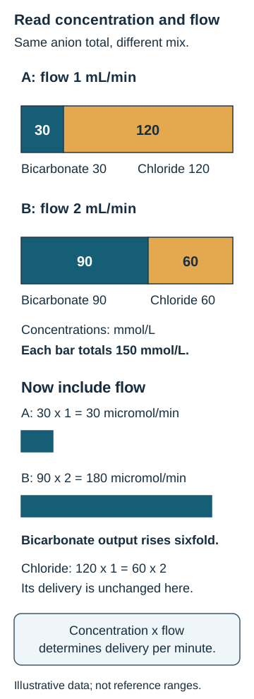
The paired anion diagram compares low-flow and stimulated samples. Each stacked bar has the same combined chloride-plus-bicarbonate concentration. Trace the part that grows, then the part that shrinks. A change in their proportions does not imply that the sum must increase.
The upper bars compare anion proportions; the lower bars include fluid flow. In this numerical example bicarbonate delivery rises from 30 to 180 micromol/min, while chloride delivery remains 120 micromol/min. [2]
Compare bicarbonate and chloride concentrations across the two flow states.
Measurement
Sample A
Sample B
MeasurementFluid flow
Sample A1 mL/min
Sample B2 mL/min
MeasurementBicarbonate
Sample A30 mmol/L
Sample B90 mmol/L
MeasurementChloride
Sample A120 mmol/L
Sample B60 mmol/L
Work the relationship: delivery rate equals concentration multiplied by volume per time. Because 1 mmol/L equals 1 micromol/mL, bicarbonate delivery is 30 micromol/min in A and 180 micromol/min in B. Chloride delivery is 120 micromol/min in both. Its concentration fell by half, but the fluid flow doubled. These numbers illustrate arithmetic, not a required chloride-output response in every person.
Cover the worked results and calculate the bicarbonate delivery ratio. Use the diagram to check whether a falling chloride concentration must mean less chloride delivered per minute.
What ratio should the calculation produce?
Bicarbonate delivery increases sixfold: 180 divided by 30. Chloride delivery is unchanged in this example because its concentration and flow change reciprocally.
Transfer: When a pancreatic-function report describes a low bicarbonate concentration, ask separately how much fluid was collected and under what stimulus. Concentration, total output, and clinical digestive adequacy are related but are not interchangeable measurements. [2][6]
Protease delivery is not protease activation
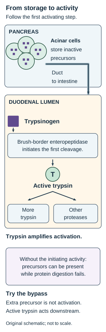
Why can pancreatic fluid contain abundant protein-digesting precursors yet digest protein poorly? A precursor can be present without being active. Acinar cells package trypsinogen, chymotrypsinogen, proelastase, and procarboxypeptidases as zymogens. Their inactive form limits premature digestion of pancreatic tissue. Ductal fluid delivers these precursors to the duodenum. [3]
At the duodenal brush border, enteropeptidase converts trypsinogen to trypsin. Trypsin then activates additional trypsinogen and the other pancreatic protease precursors. This amplification supplies chymotrypsin, elastase, and carboxypeptidases for luminal protein digestion. Brush-border and intracellular peptidases finish smaller peptide products. Amino acids and small peptides have absorption pathways distinct from the carbohydrate transporters below. [3]
Read the activation diagram from acinar storage through the duct to the intestinal surface. The location of the first physiological activating cleavage matters. Enteropeptidase does not need to enter the pancreas, and secretin is not the enzyme that performs this cleavage.
Trace the location of the initiating cleavage, then compare extra precursor with a downstream supply of active trypsin. [3][10]
Pancreatic amylase and pancreatic lipase are secreted in active forms, so not every pancreatic digestive enzyme requires this activation sequence. The reverse statement, that every precursor must be a protease, is also incorrect: pancreatic phospholipase A2 is a lipid-digesting enzyme secreted as a precursor. Keep the enzyme's substrate separate from its secreted activation state. [3][10]
Trace a bypass. In an educational sample, pancreatic precursors are present but brush-border enteropeptidase is absent. Predict whether adding more trypsinogen or adding active trypsin would restore downstream protease activation. This is a laboratory comparison, not a treatment instruction.
What does extra trypsinogen accomplish?
It adds more precursor without supplying the missing initiating activity. The downstream activation defect remains.
What does a small amount of active trypsin accomplish?
It supplies the activating protease downstream of enteropeptidase. With functional substrates present, additional trypsinogen and other protease precursors can now be activated.
Transfer: An infant with defective enteropeptidase can have severe protein maldigestion despite pancreatic precursor synthesis. Conversely, inappropriate trypsin activity within the pancreas threatens tissue rather than improving digestion. Inactive storage is one protective feature, not a guarantee against all premature activation. [3]
Make the sugar small enough before testing its transport
Would intact sucrose use the same transporter as glucose? No. The usual absorptive substrates from dietary carbohydrates are monosaccharides. Luminal digestion and brush-border hydrolysis must first create glucose, galactose, or fructose. An intact disaccharide is not an alternative substrate for SGLT1 or GLUT5. [3][4]
Separate the digestive substrate from the final transport substrate.
Starting carbohydrate
Processing
Products available for uptake
Starting carbohydrateStarch
ProcessingSalivary and pancreatic amylase produce shorter fragments; brush-border maltase-glucoamylase and sucrase-isomaltase finish digestion
Products available for uptakeGlucose
Starting carbohydrateLactose
ProcessingBrush-border lactase
Products available for uptakeGlucose and galactose
Starting carbohydrateSucrose
ProcessingBrush-border sucrase
Products available for uptakeGlucose and fructose
Pancreatic amylase cleaves internal alpha-1,4 linkages in starch, producing maltose, maltotriose, and limit dextrins rather than completing every step to absorbable glucose. Isomaltase helps process alpha-1,6 branch points. Lactase acts on lactose, not on starch. Those substrate distinctions explain why normal pancreatic amylase cannot compensate for deficient lactase. [3][4]
Undigested lactose retains water in the lumen and reaches bacteria that ferment it, producing gas and other metabolites. Bloating and loose stools after milk can therefore reflect a hydrolysis problem without a primary defect in the glucose transporter. Intestinal injury can produce secondary lactase deficiency; not every case is an inherited lifelong deficiency. [9]
Predict the effect of predigestion. Compare ordinary milk with the same milk after its lactose has been hydrolyzed. Assume the patient lacks lactase but has functioning monosaccharide transport. Trace the two products to their absorptive route before revealing the result.
Why should lactose hydrolysis help in this comparison?
It supplies free glucose and galactose upstream of their intact uptake pathway. The intestinal cell no longer needs to perform the deficient lactase step.
Transfer: Predigestion would not repair a transporter that cannot absorb glucose or galactose. A lactose-free preparation can still contain glucose-producing carbohydrate. Distinguish the ingredient that was excluded from the monosaccharides that remain after digestion. [4][5]
Every transporter has a substrate and a membrane side
Getting into an enterocyte is not the same as reaching blood. The apical membrane faces the intestinal lumen. The basolateral membrane faces interstitial fluid and capillaries. A dietary monosaccharide must cross both interfaces to reach the portal circulation. Brush-border digestive enzymes and apical transporters are neighbors, but hydrolysis and membrane transport remain different jobs. [3][4]
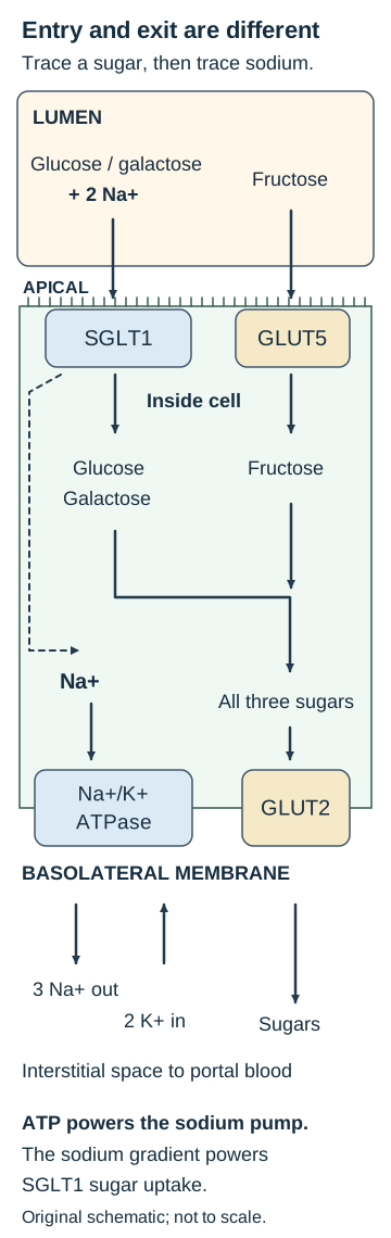
Trace glucose through the enterocyte diagram. Apical SGLT1 couples one glucose or galactose molecule to two sodium ions entering the cell. Sodium flows down its electrochemical gradient and can drive sugar uptake against a sugar gradient. SGLT1 is therefore secondary active transport; it does not itself hydrolyze ATP. The basolateral sodium-potassium ATPase uses ATP to export three sodium ions and import two potassium ions, maintaining low intracellular sodium. [4]
Trace glucose and fructose separately from lumen to blood-facing space. The sodium pump consumes ATP; SGLT1 uses the resulting sodium gradient. The drawing shows dominant absorptive routes and is not an exhaustive map of regulated transport. [3][4][5]
Now trace fructose. Apical GLUT5 provides facilitated diffusion without direct sodium coupling. In the standard absorptive map, basolateral GLUT2 provides a common exit for glucose, galactose, and fructose. These assignments describe the dominant teaching pathways, not a claim that intestinal transport has no regulated or alternative routes. [4]
Predict the first affected step in a selective short-term experiment.
Perturbation
Immediate consequence
PerturbationLoss of SGLT1 function
Immediate consequenceImpaired apical sodium-coupled glucose and galactose uptake; the GLUT5 route remains available
PerturbationLoss of GLUT5 function
Immediate consequenceImpaired apical fructose uptake through that route; SGLT1 is not the direct target
PerturbationLoss of the sodium gradient
Immediate consequenceReduced driving force for SGLT1 despite the transporter being present
PerturbationSelective impairment of basolateral sugar exit
Immediate consequenceEntry into the cell need not produce normal transfer toward blood
Trace, interrupt, predict. Choose glucose in the diagram. First interrupt the sodium pump rather than the apical sugar carrier. Predict intracellular sodium, then glucose entry. Keep fructose transport separate from generalized cell injury.
Which gradient changes after sodium-pump inhibition?
Intracellular sodium rises as its export falls. The inward sodium electrochemical gradient becomes less favorable.
What follows for SGLT1-mediated glucose entry?
Its driving force falls even though SGLT1 is still present. GLUT5 is not directly sodium-coupled, although prolonged metabolic injury can impair the cell more broadly.
This relationship explains oral rehydration. In many secretory diarrheas, sodium-glucose cotransport remains functional despite continued fluid secretion. An appropriately prepared oral rehydration solution provides both substrates, promoting sodium and water absorption. It need not stop stool production to improve hydration. Severe dehydration, shock, or inability to drink requires urgent clinical care and may require intravenous fluids; ordinary sweet drinks are not equivalent to a correctly prepared rehydration solution. [4][8]
Transfer: Congenital glucose-galactose malabsorption is an important exception to assuming an intact glucose uptake pathway. SLC5A1 dysfunction impairs SGLT1 and can cause severe diarrhea soon after feeding begins. Fructose uses a different apical route. Such infants need specialist-directed feeding and dehydration management, not repeated unsupervised sugar challenges. [5]
Localize nutrient loss without overreading a test
Does fat in stool prove that the intestinal lining is damaged? No. Exocrine pancreatic insufficiency, or EPI, can prevent adequate intraluminal digestion even when the mucosa can absorb a ready-made monosaccharide. Chronic pancreatitis, cystic fibrosis, pancreatic surgery, and pancreatic malignancy increase suspicion. Steatorrhea, weight loss, and fat-soluble vitamin deficiencies are important consequences, but symptoms alone do not establish the cause. [6]
Fat digestion is particularly vulnerable when pancreatic enzyme delivery is inadequate and the luminal environment is unfavorable. Do not memorize a universal sequence in which one nutrient must fail first. EPI can affect several nutrients, and its functional severity is not determined by a single symptom or image. A structural pancreatic abnormality can support the search for a cause without measuring digestive adequacy by itself. [1][6]
Fecal elastase needs a suitable sample. On a solid or semisolid specimen, a value below 100 micrograms/g provides good evidence of EPI; 100 to 200 micrograms/g is indeterminate. A watery sample can dilute the concentration and mislead interpretation. Interpret the result with symptoms and risk factors, and obtain an appropriate specimen when dilution is a concern. Testing can be performed while a patient takes pancreatic enzyme replacement. [6][7]
D-xylose is a historical way to separate digestion from absorption because this monosaccharide does not require pancreatic hydrolysis before intestinal uptake. In the classic comparison, preserved absorption is compatible with pancreatic maldigestion, whereas reduced recovery can accompany proximal small-bowel disease. Normal recovery does not diagnose EPI, and low recovery does not uniquely identify mucosal disease. Bacterial overgrowth, altered transit, dehydration, renal dysfunction, and collection problems can affect interpretation. The 2025 European consensus does not support routine D-xylose testing in the general evaluation of malabsorption; selected specialist uses are different. [7]
Use the pattern to choose the failed step, not just a disease label.
Supplied findings
Interpretation
Supplied findingsPancreatic disease, steatorrhea, convincingly low elastase on a formed sample
InterpretationInsufficient pancreatic enzyme delivery is supported
InterpretationLoss of mucosal surface and brush-border function is supported
Supplied findingsMilk-specific symptoms with preserved free glucose and galactose absorption
InterpretationLactose hydrolysis can fail despite functioning monosaccharide transport
Supplied findingsSevere feeding-related diarrhea with impaired glucose and galactose uptake but preserved fructose uptake
InterpretationA selective apical transport defect is supported
Test the interpretation. A historical urine D-xylose result is low, but the patient has poor renal function and evidence of preserved intestinal uptake. Name the additional step between intestinal absorption and the measured urine result.
Why does the urine result not localize the problem by itself?
Absorbed D-xylose still requires renal handling and an adequate urine collection to appear in the measured sample. Low urinary recovery can occur without a primary intestinal uptake defect.
Once EPI is established, pancreatic enzyme replacement is taken during meals so enzymes accompany food. Treatment includes assessment of nutrition and fat-soluble vitamins and attention to the underlying disorder. Improvement during an empirical enzyme trial is not a reliable stand-alone diagnostic test. Persistent symptoms require reassessing administration, adequacy, and competing causes rather than declaring every loose stool pancreatic. [6]
Transfer: Return to the four questions from the beginning. A bypass that supplies absorbable sugar may overcome failed hydrolysis but cannot restore a missing transporter. Enzyme replacement addresses deficient luminal digestion but does not rebuild an injured villus. Use the supplied evidence to choose the step before choosing the intervention.
Apply the physiology to new patients and experiments
Case 1
Show answer and explanations for case 1
A. Reduced acinar trypsinogen synthesis (Why this does not fit)
A synthesis defect would reduce the available pancreatic trypsinogen substrate. Trypsinogen output is preserved, and supplying healthy brush-border material restores its activation rather than replacing missing pancreatic protein.
Reasoning steps for option A
What would a synthesis defect reduce?
A synthesis defect would reduce the available pancreatic trypsinogen substrate.
Is that substrate missing in this infant?
Trypsinogen output is preserved, and supplying healthy brush-border material restores its activation rather than replacing missing pancreatic protein.
B. Deficient brush-border enteropeptidase activity (Best answer)
It cleaves trypsinogen at the intestinal surface to produce active trypsin. The healthy brush-border preparation supplies the missing initiating activity while the infant's pancreatic precursors provide usable substrates.
Reasoning steps for option B
What does enteropeptidase normally produce?
It cleaves trypsinogen at the intestinal surface to produce active trypsin.
Why is the rescue experiment informative?
The healthy brush-border preparation supplies the missing initiating activity while the infant's pancreatic precursors provide usable substrates.
C. Reduced ductal bicarbonate secretion (Why this does not fit)
Inadequate buffering can leave a luminal environment unfavorable for pancreatic enzyme function. The measured pH is appropriate, and changing brush-border material restores activation without a change in buffering.
Reasoning steps for option C
How can low ductal bicarbonate impair digestion?
Inadequate buffering can leave a luminal environment unfavorable for pancreatic enzyme function.
Which observations favor another explanation?
The measured pH is appropriate, and changing brush-border material restores activation without a change in buffering.
D. Deficient enterocyte amino-acid transport (Why this does not fit)
It would impair uptake after luminal proteins had been digested into suitable substrates. Trypsin activity fails to develop before amino-acid uptake is tested; the rescue concerns enzyme activation, not membrane absorption.
Reasoning steps for option D
Which step would an amino-acid transporter defect impair?
It would impair uptake after luminal proteins had been digested into suitable substrates.
Where does the demonstrated failure occur?
Trypsin activity fails to develop before amino-acid uptake is tested; the rescue concerns enzyme activation, not membrane absorption.
Takeaway: Preserved pancreatic precursor output does not establish normal protease activation at the intestinal surface.
A. Preserved chymotrypsin activation; reduced amylase activity (Why this does not fit)
Pancreatic amylase is secreted in an active form rather than as a trypsin-dependent zymogen. Its activation is impaired when newly formed trypsin cannot catalyze downstream reactions, so the proposed pattern reverses the expected effects.
Reasoning steps for option A
Does pancreatic amylase require trypsin activation?
Pancreatic amylase is secreted in an active form rather than as a trypsin-dependent zymogen.
What happens to chymotrypsinogen in this sample?
Its activation is impaired when newly formed trypsin cannot catalyze downstream reactions, so the proposed pattern reverses the expected effects.
B. Preserved chymotrypsin activation; preserved amylase activity (Why this does not fit)
The inhibitor is selective for trypsin and the amylase environment remains suitable. The supplied chymotrypsinogen still requires a functional activating protease; the experiment disables that trypsin activity.
Reasoning steps for option B
Why is preserved amylase activity plausible?
The inhibitor is selective for trypsin and the amylase environment remains suitable.
Why should chymotrypsin activation not also be preserved?
The supplied chymotrypsinogen still requires a functional activating protease; the experiment disables that trypsin activity.
C. Reduced chymotrypsin activation; reduced amylase activity (Why this does not fit)
Trypsin inhibition limits conversion of chymotrypsinogen to active chymotrypsin. It does not: active amylase was delivered, and neither pH nor amylase itself was altered by the selective intervention.
Reasoning steps for option C
Why would chymotrypsin activity fall?
Trypsin inhibition limits conversion of chymotrypsinogen to active chymotrypsin.
Does the same requirement apply to amylase?
It does not: active amylase was delivered, and neither pH nor amylase itself was altered by the selective intervention.
D. Reduced chymotrypsin activation; preserved amylase activity (Best answer)
Activation of chymotrypsinogen depends on a functional activating protease, normally trypsin. Amylase-mediated starch digestion uses an enzyme secreted in active form and remains available in the stated controlled sample.
Reasoning steps for option D
Which tested process depends on active trypsin?
Activation of chymotrypsinogen depends on a functional activating protease, normally trypsin.
Which tested process bypasses this dependency?
Amylase-mediated starch digestion uses an enzyme secreted in active form and remains available in the stated controlled sample.
Takeaway: A selective activation defect affects dependent precursors, not every enzyme secreted by the pancreas.
A. Delivery of digestive enzymes to luminal food (Best answer)
The pancreatic duct carries exocrine products from the pancreas to the intestine. Inadequate enzyme delivery impairs intraluminal digestion, while a supplied monosaccharide can still cross intact intestinal cells.
Reasoning steps for option A
What physical route has become narrowed?
The pancreatic duct carries exocrine products from the pancreas to the intestine.
Why can fat loss coexist with normal sugar uptake?
Inadequate enzyme delivery impairs intraluminal digestion, while a supplied monosaccharide can still cross intact intestinal cells.
B. Production of digestive proteins within acini (Why this does not fit)
Insufficient synthesis can reduce the amount of enzyme ultimately reaching intestinal contents. Upstream production is preserved; the demonstrated loss occurs between production and delivery to the duodenum.
Reasoning steps for option B
Could reduced acinar synthesis cause maldigestion?
Insufficient synthesis can reduce the amount of enzyme ultimately reaching intestinal contents.
Which finding argues against it as the direct stricture effect?
Upstream production is preserved; the demonstrated loss occurs between production and delivery to the duodenum.
C. Cleavage of lactose at the intestinal surface (Why this does not fit)
Lactase hydrolyzes lactose at the intestinal brush border rather than inside the pancreatic duct. It does not account for a demonstrated obstruction of pancreatic output and greasy stools from impaired fat digestion.
Reasoning steps for option C
Where is lactose normally hydrolyzed?
Lactase hydrolyzes lactose at the intestinal brush border rather than inside the pancreatic duct.
Does a lactose-specific defect explain this pattern?
It does not account for a demonstrated obstruction of pancreatic output and greasy stools from impaired fat digestion.
D. Export of absorbed sugars across the basolateral membrane (Why this does not fit)
It completes transfer of absorbed monosaccharides toward interstitial fluid and blood. The sampled intestinal cells transfer supplied monosaccharides normally, whereas pancreatic delivery to the lumen is demonstrably reduced.
Reasoning steps for option D
What does basolateral sugar export accomplish?
It completes transfer of absorbed monosaccharides toward interstitial fluid and blood.
What information argues against that being the stricture's direct target?
The sampled intestinal cells transfer supplied monosaccharides normally, whereas pancreatic delivery to the lumen is demonstrably reduced.
Takeaway: An enzyme must reach food to digest it; production upstream of an obstruction is insufficient.
A. Bicarbonate rises fivefold; chloride delivery falls (Why this does not fit)
Bicarbonate concentration rises from 20 to 100 mmol/L, a fivefold concentration change. Flow also triples, so bicarbonate output rises fifteenfold; chloride output is 150 rather than 130 micromol/min despite its lower concentration.
Reasoning steps for option A
Which change is fivefold?
Bicarbonate concentration rises from 20 to 100 mmol/L, a fivefold concentration change.
Why is that not the delivery ratio?
Flow also triples, so bicarbonate output rises fifteenfold; chloride output is 150 rather than 130 micromol/min despite its lower concentration.
B. Bicarbonate rises threefold; chloride delivery is unchanged (Why this does not fit)
Fluid flow increases from 1 to 3 mL/min. Ion concentrations change too: bicarbonate delivery becomes 300 versus 20 micromol/min, and chloride delivery becomes 150 versus 130 micromol/min.
Reasoning steps for option B
Which measured variable triples?
Fluid flow increases from 1 to 3 mL/min.
What must also enter the calculation?
Ion concentrations change too: bicarbonate delivery becomes 300 versus 20 micromol/min, and chloride delivery becomes 150 versus 130 micromol/min.
C. Bicarbonate rises fifteenfold; chloride delivery rises (Best answer)
Using 1 mmol/L = 1 micromol/mL gives 20 times 1 = 20 and 100 times 3 = 300 micromol/min. They are 130 times 1 = 130 and 50 times 3 = 150 micromol/min; bicarbonate rises fifteenfold and chloride delivery also rises.
Reasoning steps for option C
What are the bicarbonate outputs?
Using 1 mmol/L = 1 micromol/mL gives 20 times 1 = 20 and 100 times 3 = 300 micromol/min.
How do the chloride outputs compare?
They are 130 times 1 = 130 and 50 times 3 = 150 micromol/min; bicarbonate rises fifteenfold and chloride delivery also rises.
D. Bicarbonate rises fifteenfold; chloride delivery falls (Why this does not fit)
The fifteenfold bicarbonate increase correctly incorporates the fivefold concentration increase and tripled flow. The new chloride delivery is 50 times 3 = 150 micromol/min, greater than the initial 130 micromol/min.
Reasoning steps for option D
Which part correctly incorporates both measurements?
The fifteenfold bicarbonate increase correctly incorporates the fivefold concentration increase and tripled flow.
What defeats the chloride prediction?
The new chloride delivery is 50 times 3 = 150 micromol/min, greater than the initial 130 micromol/min.
Takeaway: Concentration multiplied by flow determines delivery; a falling concentration does not necessarily mean falling output.
A. Higher chloride concentration with higher juice osmolality (Why this does not fit)
Without compensating changes, increasing both major anions would increase the dissolved solute load. No. Bicarbonate largely replaces chloride in the anion mixture while water accompanies secretion, preserving approximate isotonicity.
Reasoning steps for option A
What would parallel large increases in both anions imply?
Without compensating changes, increasing both major anions would increase the dissolved solute load.
Is that the usual stimulated pancreatic pattern?
No. Bicarbonate largely replaces chloride in the anion mixture while water accompanies secretion, preserving approximate isotonicity.
B. Lower chloride concentration with similar juice osmolality (Best answer)
Chloride concentration falls in the usual stimulated pancreatic secretion relationship. The anion proportions change while sodium and potassium concentrations remain relatively stable and water accompanies ion secretion.
Reasoning steps for option B
Which anion tends to decrease as bicarbonate concentration rises?
Chloride concentration falls in the usual stimulated pancreatic secretion relationship.
Why need juice osmolality not rise markedly?
The anion proportions change while sodium and potassium concentrations remain relatively stable and water accompanies ion secretion.
C. Lower chloride concentration with markedly lower juice osmolality (Why this does not fit)
Stimulated bicarbonate-rich fluid generally has a lower chloride concentration than low-flow fluid. Bicarbonate replaces much of the chloride contribution, and pancreatic fluid remains approximately isotonic rather than becoming markedly hypotonic.
Reasoning steps for option C
Why is the chloride direction plausible?
Stimulated bicarbonate-rich fluid generally has a lower chloride concentration than low-flow fluid.
Why does that not imply major dilution of total solute?
Bicarbonate replaces much of the chloride contribution, and pancreatic fluid remains approximately isotonic rather than becoming markedly hypotonic.
D. Similar chloride concentration with higher juice osmolality (Why this does not fit)
It treats the bicarbonate rise as added anions without a reciprocal change in the major anion mixture. The usual chloride-bicarbonate reciprocal concentration change allows increased bicarbonate-rich secretion without a large osmolality increase.
Reasoning steps for option D
What does this option assume about the added bicarbonate?
It treats the bicarbonate rise as added anions without a reciprocal change in the major anion mixture.
Which physiological relationship was omitted?
The usual chloride-bicarbonate reciprocal concentration change allows increased bicarbonate-rich secretion without a large osmolality increase.
Takeaway: Stimulated pancreatic secretion changes the major anion proportions while remaining approximately isotonic.
A. Impaired apical anion transport by pancreatic duct cells (Best answer)
Pancreatic duct cells secrete bicarbonate-rich fluid through apical anion transport involving CFTR and associated transporters. cAMP signaling occurs, but anions do not enter the duct lumen normally, localizing the demonstrated defect to the downstream ductal secretory machinery.
Reasoning steps for option A
Where does secretin-driven anion secretion occur?
Pancreatic duct cells secrete bicarbonate-rich fluid through apical anion transport involving CFTR and associated transporters.
Where do the patient's measurements place the failure?
cAMP signaling occurs, but anions do not enter the duct lumen normally, localizing the demonstrated defect to the downstream ductal secretory machinery.
B. Impaired protease-precursor packaging by pancreatic acini (Why this does not fit)
It affects the production, storage, or delivery of pancreatic digestive proteins. Acinar enzyme production is relatively preserved, while duct-cell anion transport remains poor despite an appropriate intracellular signal.
Reasoning steps for option B
What does defective precursor packaging primarily affect?
It affects the production, storage, or delivery of pancreatic digestive proteins.
Which measured dissociation argues against that being the immediate defect?
Acinar enzyme production is relatively preserved, while duct-cell anion transport remains poor despite an appropriate intracellular signal.
C. Impaired acid sensing by intestinal secretin-producing cells (Why this does not fit)
It could reduce endogenous secretin release before the hormone reaches the pancreatic duct. Externally supplied secretin already produces a normal cAMP response, yet anion secretion remains defective beyond that signaling step.
Reasoning steps for option C
How could defective acid sensing reduce bicarbonate secretion?
It could reduce endogenous secretin release before the hormone reaches the pancreatic duct.
Does it explain the duct-cell experiment?
Externally supplied secretin already produces a normal cAMP response, yet anion secretion remains defective beyond that signaling step.
D. Impaired peptide hydrolysis at the intestinal brush border (Why this does not fit)
They act at the intestinal epithelial surface after pancreatic products enter the intestine. No. The measured defect occurs within isolated pancreatic duct cells, upstream of intestinal peptide processing.
Reasoning steps for option D
Which compartment contains brush-border peptidases?
They act at the intestinal epithelial surface after pancreatic products enter the intestine.
Can that localization explain defective duct-cell anion transport?
No. The measured defect occurs within isolated pancreatic duct cells, upstream of intestinal peptide processing.
Takeaway: An intact hormone signal does not guarantee an intact epithelial transport response.
A. Acid-dependent secretin release from intestinal S cells (Why this does not fit)
It would limit the hormone signal sent from the intestine after acid exposure. The circulating secretin response is normal, and added secretin also fails at the duct cell, placing the demonstrated problem after release.
Reasoning steps for option A
What would deficient S-cell secretion prevent?
It would limit the hormone signal sent from the intestine after acid exposure.
Which observation shows that signal is available?
The circulating secretin response is normal, and added secretin also fails at the duct cell, placing the demonstrated problem after release.
B. Bicarbonate passage through the ductal apical membrane (Why this does not fit)
It would prevent normal secretion even when the intracellular stimulatory signal is supplied. Bicarbonate secretion can occur when cAMP is increased directly, so an obligatory failure of the final transport pathway does not fit.
Reasoning steps for option B
What would a nonfunctional final bicarbonate transport pathway prevent?
It would prevent normal secretion even when the intracellular stimulatory signal is supplied.
What does the direct cAMP rescue establish?
Bicarbonate secretion can occur when cAMP is increased directly, so an obligatory failure of the final transport pathway does not fit.
C. Pancreatic enzyme exocytosis from acinar granules (Why this does not fit)
Acinar exocytosis delivers digestive proteins rather than providing the principal ductal bicarbonate response. It was demonstrated in duct cells between secretin application and cAMP generation, not in acinar enzyme release.
Reasoning steps for option C
Which pancreatic product is released from acinar granules?
Acinar exocytosis delivers digestive proteins rather than providing the principal ductal bicarbonate response.
Where was the stimulus-response failure demonstrated?
It was demonstrated in duct cells between secretin application and cAMP generation, not in acinar enzyme release.
D. Secretin-receptor signaling to intracellular cAMP production (Best answer)
Directly increasing cAMP bypasses the steps linking secretin reception to cAMP generation. The upstream hormone is available and the downstream secretory machinery works when supplied with cAMP, leaving the intervening signaling interval as the supported site.
Reasoning steps for option D
Which signaling interval is bypassed by the reagent?
Directly increasing cAMP bypasses the steps linking secretin reception to cAMP generation.
Why does restoration of secretion localize the problem there?
The upstream hormone is available and the downstream secretory machinery works when supplied with cAMP, leaving the intervening signaling interval as the supported site.
Takeaway: A successful downstream bypass localizes a defect only within the interval actually bypassed.
Why is this not the best explanation for the protein-output change?
The experiment specifically interrupts cholinergic signaling and measures diminished enzyme secretion, while a ductal response to administered secretin remains available.
B. Reduced bicarbonate conductance at the ductal apical surface (Why this does not fit)
It would impair secretion of bicarbonate-rich fluid in response to an appropriate ductal stimulus. The ductal secretory response remains demonstrable; diminished cholinergic enzyme stimulation is a closer explanation for the protein-output change.
Reasoning steps for option B
What would severe loss of ductal bicarbonate transport diminish?
It would impair secretion of bicarbonate-rich fluid in response to an appropriate ductal stimulus.
What does the preserved secretin response suggest?
The ductal secretory response remains demonstrable; diminished cholinergic enzyme stimulation is a closer explanation for the protein-output change.
C. Reduced cholinergic stimulation of acinar enzyme exocytosis (Best answer)
Acinar cells release them by secretion of enzyme-containing granules. Blocking muscarinic signaling reduces cholinergic support of acinar secretion, including the neural contribution to coordinated meal-stimulated enzyme output.
Reasoning steps for option C
Which compartment releases pancreatic digestive proteins?
Acinar cells release them by secretion of enzyme-containing granules.
How does atropine alter the meal response?
Blocking muscarinic signaling reduces cholinergic support of acinar secretion, including the neural contribution to coordinated meal-stimulated enzyme output.
D. Reduced enteropeptidase cleavage of luminal trypsinogen (Why this does not fit)
It acts after secretion by initiating protease activation at the intestinal surface. The study reports reduced pancreatic digestive protein output, not merely a reduction in the activity of proteases already delivered to the lumen.
Reasoning steps for option D
Does enteropeptidase control enzyme secretion from acini?
It acts after secretion by initiating protease activation at the intestinal surface.
Which measured endpoint distinguishes secretion from activation?
The study reports reduced pancreatic digestive protein output, not merely a reduction in the activity of proteases already delivered to the lumen.
Takeaway: Acinar secretion, ductal buffering, and intestinal activation are distinct endpoints even though a meal coordinates them.
A. Less fermentable carbohydrate remains unabsorbed in the lumen (Best answer)
It produces free glucose and galactose that the patient's intact uptake pathways can absorb. Less unabsorbed carbohydrate remains to retain luminal water and undergo bacterial fermentation, reducing loose stools and gas.
Reasoning steps for option A
What does lactose hydrolysis supply?
It produces free glucose and galactose that the patient's intact uptake pathways can absorb.
What happens to water retention and bacterial substrate?
Less unabsorbed carbohydrate remains to retain luminal water and undergo bacterial fermentation, reducing loose stools and gas.
B. Less epithelial chloride secretion in response to bacterial enterotoxins (Why this does not fit)
It can cause watery diarrhea even without an unabsorbed dietary sugar. Only lactose hydrolysis changes between exposures, and the products can be absorbed; reduced osmotic and fermentable substrate explains the result without an identified toxin process.
Reasoning steps for option B
What can toxin-driven chloride secretion cause?
It can cause watery diarrhea even without an unabsorbed dietary sugar.
What makes it a poorer explanation for this controlled milk comparison?
Only lactose hydrolysis changes between exposures, and the products can be absorbed; reduced osmotic and fermentable substrate explains the result without an identified toxin process.
C. Less immune-mediated inflammation in response to milk proteins (Why this does not fit)
Immune-mediated mucosal inflammation can cause symptoms after the triggering food. The comparison uses the same milk with its sugar predigested, so the improved tolerance supports altered carbohydrate handling rather than removal of a protein antigen.
Reasoning steps for option C
How can a food-protein reaction cause gastrointestinal symptoms?
Immune-mediated mucosal inflammation can cause symptoms after the triggering food.
Did hydrolysis of lactose remove the milk proteins?
The comparison uses the same milk with its sugar predigested, so the improved tolerance supports altered carbohydrate handling rather than removal of a protein antigen.
D. Less bile-acid delivery to the colon after fat malabsorption (Why this does not fit)
They can promote colonic fluid secretion and loose stools. Lactose hydrolysis was changed without a supplied alteration in fat or bile-acid handling, favoring a carbohydrate-specific explanation.
Reasoning steps for option D
How can excess colonic bile acids affect stool?
They can promote colonic fluid secretion and loose stools.
Which component was selectively changed in this comparison?
Lactose hydrolysis was changed without a supplied alteration in fat or bile-acid handling, favoring a carbohydrate-specific explanation.
Takeaway: Predigestion can bypass an enzyme deficit when the resulting monosaccharide transport pathways work.
A. Preserved galactose uptake with impaired fructose uptake (Why this does not fit)
Galactose shares sodium-coupled apical uptake with glucose. Glucose-producing feeds fail while fructose is tolerated, supporting the opposite substrate pattern.
Reasoning steps for option A
Which glucose-related substrate shares SGLT1?
Galactose shares sodium-coupled apical uptake with glucose.
How do the feeding observations compare with this prediction?
Glucose-producing feeds fail while fructose is tolerated, supporting the opposite substrate pattern.
B. Impaired galactose uptake with preserved fructose uptake (Best answer)
Glucose can be produced but cannot be absorbed adequately through its normal dominant apical route. Galactose also uses SGLT1, whereas fructose can enter through GLUT5, matching the tolerated fructose regimen.
Reasoning steps for option B
What does failure of a glucose-polymer formula imply after normal hydrolysis?
Glucose can be produced but cannot be absorbed adequately through its normal dominant apical route.
Which additional substrate should be affected?
Galactose also uses SGLT1, whereas fructose can enter through GLUT5, matching the tolerated fructose regimen.
C. Preserved galactose uptake with preserved glucose uptake (Why this does not fit)
The lactose-free glucose-polymer formula supplies glucose after digestion. Diarrhea persists with that feed despite preserved disaccharidase activity, while a different monosaccharide route is tolerated.
Reasoning steps for option C
Which clinical observation tests glucose availability for absorption?
The lactose-free glucose-polymer formula supplies glucose after digestion.
Why is normal glucose uptake inconsistent with the supplied pattern?
Diarrhea persists with that feed despite preserved disaccharidase activity, while a different monosaccharide route is tolerated.
D. Impaired galactose uptake with impaired fructose uptake (Why this does not fit)
It could impair multiple monosaccharide pathways rather than selectively sparing one apical substrate. The specialist-directed fructose regimen is tolerated, favoring a selective glucose-galactose uptake defect rather than the proposed combined impairment.
Reasoning steps for option D
What would a shared exit or widespread absorptive failure tend to affect?
It could impair multiple monosaccharide pathways rather than selectively sparing one apical substrate.
Which observation demonstrates useful fructose absorption?
The specialist-directed fructose regimen is tolerated, favoring a selective glucose-galactose uptake defect rather than the proposed combined impairment.
Takeaway: Lactose-free is not glucose-free; SGLT1 dysfunction impairs glucose and galactose uptake while the GLUT5 route can remain available.
A. Failure of ATP hydrolysis at the SGLT1 transport site (Why this does not fit)
SGLT1 is a sodium-coupled secondary active transporter, not an ATP-hydrolyzing glucose pump. Intracellular sodium rises after pump inhibition, reducing the inward sodium gradient that normally drives SGLT1.
Reasoning steps for option A
Does SGLT1 directly hydrolyze ATP to transport glucose?
SGLT1 is a sodium-coupled secondary active transporter, not an ATP-hydrolyzing glucose pump.
What supplied change explains the reduced transport instead?
Intracellular sodium rises after pump inhibition, reducing the inward sodium gradient that normally drives SGLT1.
B. Loss of the glucose gradient across the basolateral membrane (Why this does not fit)
It affects facilitated sugar exit from the enterocyte toward the interstitial compartment. The demonstrated perturbation is intracellular sodium accumulation, which weakens sodium-coupled apical uptake rather than establishing a new basolateral glucose defect.
Reasoning steps for option B
What does a basolateral glucose gradient primarily affect?
It affects facilitated sugar exit from the enterocyte toward the interstitial compartment.
Which driving force is directly altered in this experiment?
The demonstrated perturbation is intracellular sodium accumulation, which weakens sodium-coupled apical uptake rather than establishing a new basolateral glucose defect.
C. Loss of the inward sodium electrochemical driving force (Best answer)
The basolateral sodium-potassium ATPase exports sodium and thereby maintains the gradient used by apical SGLT1. The inward sodium driving force falls, so SGLT1-mediated glucose uptake can decline even with unchanged transporter abundance and available ATP.
Reasoning steps for option C
Why is intracellular sodium normally kept low?
The basolateral sodium-potassium ATPase exports sodium and thereby maintains the gradient used by apical SGLT1.
What follows when sodium accumulates inside?
The inward sodium driving force falls, so SGLT1-mediated glucose uptake can decline even with unchanged transporter abundance and available ATP.
D. Reduced glucose availability after impaired starch hydrolysis (Why this does not fit)
If the lumen lacked free glucose, inadequate hydrolysis could limit the substrate available to apical transporters. No. Luminal glucose is maintained, so the altered sodium gradient rather than failed substrate generation explains the uptake change.
Reasoning steps for option D
When could failed starch digestion limit glucose uptake?
If the lumen lacked free glucose, inadequate hydrolysis could limit the substrate available to apical transporters.
Was free glucose reduced in this experiment?
No. Luminal glucose is maintained, so the altered sodium gradient rather than failed substrate generation explains the uptake change.
Takeaway: ATP powers the sodium pump; the resulting sodium gradient powers SGLT1.
Glucose and galactose use its sodium-coupled entry pathway. Their initial entry is preserved, fructose is also retained, and the demonstrated failure is transfer out of the cell toward the basolateral chamber.
Reasoning steps for option A
Which sugars principally use apical SGLT1?
Glucose and galactose use its sodium-coupled entry pathway.
Why does the observed pattern require another site?
Their initial entry is preserved, fructose is also retained, and the demonstrated failure is transfer out of the cell toward the basolateral chamber.
B. Apical GLUT5 (Why this does not fit)
It provides facilitated fructose entry from the lumen. Initial fructose uptake is preserved, and glucose and galactose also fail at the shared downstream exit step.
Reasoning steps for option B
Which principal absorptive step is mediated by apical GLUT5?
It provides facilitated fructose entry from the lumen.
Which findings do not fit a selective GLUT5-entry defect?
Initial fructose uptake is preserved, and glucose and galactose also fail at the shared downstream exit step.
C. Basolateral sodium-potassium ATPase (Why this does not fit)
It could weaken the sodium gradient used for SGLT1-mediated glucose and galactose uptake. The sodium gradient and initial entry are preserved; the reported defect is shared sugar exit after uptake.
Reasoning steps for option C
How could pump dysfunction affect sugar entry?
It could weaken the sodium gradient used for SGLT1-mediated glucose and galactose uptake.
What measurements argue against that mechanism here?
The sodium gradient and initial entry are preserved; the reported defect is shared sugar exit after uptake.
D. Basolateral GLUT2 (Best answer)
GLUT2 provides the standard shared exit route for glucose, galactose, and fructose. Sugars reach the cell but fail to leave normally through the blood-facing interface, matching a common basolateral exit impairment.
Reasoning steps for option D
Which basolateral carrier handles the three principal dietary monosaccharides?
GLUT2 provides the standard shared exit route for glucose, galactose, and fructose.
Why does intracellular accumulation support this localization?
Sugars reach the cell but fail to leave normally through the blood-facing interface, matching a common basolateral exit impairment.
Takeaway: Normal entry into an enterocyte does not establish normal transfer across the whole epithelium.
A. Inhibition of toxin-driven chloride secretion by absorbed glucose (Why this does not fit)
No. It can improve fluid balance while the diarrhea-producing secretory process continues. Stools remain watery while perfusion and urine output improve, supporting enhanced absorption rather than demonstrated toxin-pathway inhibition.
Reasoning steps for option A
Must oral rehydration terminate secretory signaling to be useful?
No. It can improve fluid balance while the diarrhea-producing secretory process continues.
Which finding illustrates that distinction?
Stools remain watery while perfusion and urine output improve, supporting enhanced absorption rather than demonstrated toxin-pathway inhibition.
B. Preserved sodium-glucose uptake with accompanying water absorption (Best answer)
SGLT1-mediated sodium-glucose cotransport can remain functional despite ongoing secretory diarrhea. The solution supplies substrates for sodium uptake and associated water absorption, allowing hydration to improve even before stool output stops.
Reasoning steps for option B
Which apical route can remain useful during cholera?
SGLT1-mediated sodium-glucose cotransport can remain functional despite ongoing secretory diarrhea.
How can that improve the patient's condition?
The solution supplies substrates for sodium uptake and associated water absorption, allowing hydration to improve even before stool output stops.
C. Water absorption driven by retained glucose in the intestinal lumen (Why this does not fit)
An unabsorbed osmotic solute tends to retain water in the lumen rather than draw it into the circulation. Glucose is transported with sodium through an intact absorptive pathway, allowing sodium and water uptake despite continued diarrheal losses.
Reasoning steps for option C
How does unabsorbed glucose affect water in the intestinal lumen?
An unabsorbed osmotic solute tends to retain water in the lumen rather than draw it into the circulation.
What use of glucose supports rehydration instead?
Glucose is transported with sodium through an intact absorptive pathway, allowing sodium and water uptake despite continued diarrheal losses.
D. Restoration of water absorption after complete cessation of epithelial secretion (Why this does not fit)
It would reduce ongoing luminal fluid losses and could improve fluid balance. Watery stools continue during improved perfusion and urine output, so effective absorption can improve hydration without first stopping the secretory diarrhea.
Reasoning steps for option D
What would stopping secretion accomplish?
It would reduce ongoing luminal fluid losses and could improve fluid balance.
What observation shows that cessation is not required here?
Watery stools continue during improved perfusion and urine output, so effective absorption can improve hydration without first stopping the secretory diarrhea.
Takeaway: Oral rehydration supports absorption; persistent diarrhea does not by itself mean that rehydration is failing.
A. Glucose entry is impaired; fructose entry is preserved (Best answer)
Sucrase produces glucose and fructose. Glucose loses its SGLT1 route, whereas fructose can still enter through the preserved GLUT5 route.
Reasoning steps for option A
Which monosaccharides come from sucrose?
Sucrase produces glucose and fructose.
Which of their dominant apical routes remains functional?
Glucose loses its SGLT1 route, whereas fructose can still enter through the preserved GLUT5 route.
B. Glucose entry is preserved; galactose entry is impaired (Why this does not fit)
No. Galactose is produced from lactose, whereas sucrose yields glucose and fructose. Loss of SGLT1 impairs the dominant sodium-coupled glucose entry route rather than preserving it.
Reasoning steps for option B
Does sucrose hydrolysis produce galactose?
No. Galactose is produced from lactose, whereas sucrose yields glucose and fructose.
What additional part conflicts with the defined defect?
Loss of SGLT1 impairs the dominant sodium-coupled glucose entry route rather than preserving it.
C. Glucose entry is impaired; fructose entry is impaired (Why this does not fit)
No. Glucose and fructose have distinct dominant apical pathways. The preparation has functional GLUT5-mediated entry, so a selective SGLT1 loss does not directly block both products.
Reasoning steps for option C
Do both sucrose products share the same apical carrier?
No. Glucose and fructose have distinct dominant apical pathways.
Which supplied result preserves fructose absorption?
The preparation has functional GLUT5-mediated entry, so a selective SGLT1 loss does not directly block both products.
D. Glucose entry is preserved; fructose entry is preserved (Why this does not fit)
No. Producing monosaccharides completes digestion but does not repair their transporters. Free glucose still requires its sodium-coupled apical route, which is impaired by SGLT1 loss.
Reasoning steps for option D
Does successful sucrose hydrolysis ensure uptake of both products?
No. Producing monosaccharides completes digestion but does not repair their transporters.
Which product still encounters the defined defect?
Free glucose still requires its sodium-coupled apical route, which is impaired by SGLT1 loss.
Takeaway: Predict digestion products first, then test each product's transporter separately.
A. Sucrase-dependent generation of free fructose (Why this does not fit)
It releases fructose from sucrose before that product can be absorbed. The vesicles receive free fructose directly, so reduced entry persists even after bypassing sucrose digestion.
Reasoning steps for option A
When is sucrase needed to provide fructose?
It releases fructose from sucrose before that product can be absorbed.
Why is hydrolysis not the measured failure here?
The vesicles receive free fructose directly, so reduced entry persists even after bypassing sucrose digestion.
B. Sodium-dependent uptake of fructose by SGLT1 (Why this does not fit)
Glucose and galactose use its sodium-coupled pathway, not fructose. Preserved sodium-coupled glucose entry shows that this measured pathway works while the separate fructose-entry pathway is impaired.
Reasoning steps for option B
Which monosaccharides use SGLT1 in the standard map?
Glucose and galactose use its sodium-coupled pathway, not fructose.
How does the glucose measurement help localize the defect?
Preserved sodium-coupled glucose entry shows that this measured pathway works while the separate fructose-entry pathway is impaired.
C. Facilitated entry of fructose at the apical membrane (Best answer)
GLUT5 provides facilitated fructose transport without direct sodium coupling. Free fructose entry is impaired, glucose entry is preserved, and fructose exit after preloading still works, localizing the observed bottleneck to apical fructose uptake.
Reasoning steps for option C
Which dominant carrier provides apical fructose entry?
GLUT5 provides facilitated fructose transport without direct sodium coupling.
Why does the full set of measurements favor that step?
Free fructose entry is impaired, glucose entry is preserved, and fructose exit after preloading still works, localizing the observed bottleneck to apical fructose uptake.
D. Shared basolateral export of dietary monosaccharides (Why this does not fit)
Sugar could enter or be preloaded into the cell but would fail to leave normally toward the blood-facing side. Preloaded fructose exits across the basolateral membrane normally, whereas its initial apical entry is reduced.
Reasoning steps for option D
How would a basolateral exit defect differ from an entry defect?
Sugar could enter or be preloaded into the cell but would fail to leave normally toward the blood-facing side.
Which experiment tests and preserves that step?
Preloaded fructose exits across the basolateral membrane normally, whereas its initial apical entry is reduced.
Takeaway: Symptoms alone do not identify a transporter defect; sided substrate measurements can localize the specific failed step.
A. Pancreatic amylase cleavage of internal alpha-1,4 bonds (Why this does not fit)
It produces shorter carbohydrates, including maltose, maltotriose, and limit dextrins. Amylase output is preserved, while the selective failure occurs when brush-border enzymes must process branched residual material.
Reasoning steps for option A
What does pancreatic amylase normally produce from starch?
It produces shorter carbohydrates, including maltose, maltotriose, and limit dextrins.
Why does the supplied pattern point beyond that initial reaction?
Amylase output is preserved, while the selective failure occurs when brush-border enzymes must process branched residual material.
B. Brush-border lactase hydrolysis of lactose (Why this does not fit)
Lactase acts on lactose to yield glucose and galactose. The abnormal assay involves branched starch-derived limit dextrins, not lactose, and free glucose uptake is preserved.
Reasoning steps for option B
Which disaccharide is the lactase substrate?
Lactase acts on lactose to yield glucose and galactose.
Which tested substrates instead identify the affected process?
The abnormal assay involves branched starch-derived limit dextrins, not lactose, and free glucose uptake is preserved.
C. Brush-border maltase cleavage of unbranched maltose (Why this does not fit)
It provides free glucose through cleavage of the unbranched disaccharide. Glucose release from maltose is preserved, while the deficit appears with branched limit dextrins that need additional branch-point processing.
Reasoning steps for option C
What product does maltose hydrolysis provide?
It provides free glucose through cleavage of the unbranched disaccharide.
What assay shows this particular activity is retained?
Glucose release from maltose is preserved, while the deficit appears with branched limit dextrins that need additional branch-point processing.
D. Brush-border isomaltase cleavage of alpha-1,6 bonds (Best answer)
Limit dextrins retain branch-point alpha-1,6 bonds that pancreatic amylase does not resolve. Isomaltase contributes alpha-1,6 hydrolysis, so its deficiency fits poor branched-substrate digestion despite preserved maltose processing and glucose uptake.
Reasoning steps for option D
What feature distinguishes limit dextrins from unbranched maltose?
Limit dextrins retain branch-point alpha-1,6 bonds that pancreatic amylase does not resolve.
Which brush-border activity addresses that remaining structure?
Isomaltase contributes alpha-1,6 hydrolysis, so its deficiency fits poor branched-substrate digestion despite preserved maltose processing and glucose uptake.
Takeaway: A normal pancreatic amylase result does not prove that brush-border starch digestion is complete.
A. Replace the sucrose with its free glucose and fructose products (Best answer)
Sucrase normally hydrolyzes sucrose into glucose and fructose. Their absorption has been shown to work, so the comparison avoids the impaired hydrolysis step without relying on a new transport pathway.
Reasoning steps for option A
Which digestive reaction is missing?
Sucrase normally hydrolyzes sucrose into glucose and fructose.
Why does supplying those products bypass this particular defect?
Their absorption has been shown to work, so the comparison avoids the impaired hydrolysis step without relying on a new transport pathway.
B. Add pancreatic amylase while keeping the sucrose substrate (Why this does not fit)
No. Its principal carbohydrate substrate is starch rather than sucrose. The sucrose hydrolysis requirement would remain, so adding this different enzyme would not directly correct the demonstrated defect.
Reasoning steps for option B
Does pancreatic amylase perform sucrase's reaction?
No. Its principal carbohydrate substrate is starch rather than sucrose.
Would more amylase bypass the documented abnormality?
The sucrose hydrolysis requirement would remain, so adding this different enzyme would not directly correct the demonstrated defect.
C. Add lactase while keeping the sucrose substrate (Why this does not fit)
Lactase hydrolyzes lactose rather than sucrose. It is consistent with the measured preserved lactase activity and shows that replacing this already functioning, substrate-specific activity does not address the sucrase defect.
Reasoning steps for option C
What does lactase hydrolyze?
Lactase hydrolyzes lactose rather than sucrose.
Why does preserved milk tolerance matter?
It is consistent with the measured preserved lactase activity and shows that replacing this already functioning, substrate-specific activity does not address the sucrase defect.
D. Increase luminal sodium while keeping the sucrose intact (Why this does not fit)
Free glucose and galactose can use it; intact sucrose is not its substrate. More sodium does not generate glucose and fructose from intact sucrose, so the missing digestive reaction still precedes transport.
Reasoning steps for option D
Which substrates can use the sodium-coupled SGLT1 route?
Free glucose and galactose can use it; intact sucrose is not its substrate.
Why can sodium not substitute for hydrolysis here?
More sodium does not generate glucose and fructose from intact sucrose, so the missing digestive reaction still precedes transport.
Takeaway: A useful bypass supplies products that the intact downstream pathway can actually use.
A. Pancreatic lipase itself requires trypsin cleavage before lipid digestion (Why this does not fit)
Pancreatic lipase activity is already present before the added trypsin exposure. Phospholipid hydrolysis increases, so the result concerns a distinct enzyme rather than a requirement to activate all pancreatic lipase first.
Reasoning steps for option A
What does the baseline triglyceride result establish?
Pancreatic lipase activity is already present before the added trypsin exposure.
Which activity changes after exposure?
Phospholipid hydrolysis increases, so the result concerns a distinct enzyme rather than a requirement to activate all pancreatic lipase first.
B. More phospholipase was newly secreted by acini during incubation (Why this does not fit)
A greater amount of active enzyme could increase the measured hydrolytic activity. The experiment uses an already collected sample exposed to trypsin, so precursor activation explains the change without postulating additional acinar secretion.
Reasoning steps for option B
How could increased enzyme delivery change hydrolysis?
A greater amount of active enzyme could increase the measured hydrolytic activity.
Was new cellular secretion supplied in this comparison?
The experiment uses an already collected sample exposed to trypsin, so precursor activation explains the change without postulating additional acinar secretion.
C. Trypsin-dependent activation of a lipid-digesting precursor (Best answer)
Pancreatic phospholipase A2 can be secreted as a precursor activated by trypsin. Pancreatic lipase and phospholipase A2 have different substrates and activation states, so active triglyceride digestion can coexist with an unactivated phospholipase precursor.
Reasoning steps for option C
Which pancreatic lipid enzyme provides this example?
Pancreatic phospholipase A2 can be secreted as a precursor activated by trypsin.
Why does the baseline lipase result not contradict this?
Pancreatic lipase and phospholipase A2 have different substrates and activation states, so active triglyceride digestion can coexist with an unactivated phospholipase precursor.
D. Additional bile salts corrected an unfavorable lipid-water interface (Why this does not fit)
The physical presentation of lipid and bile-salt interactions can affect digestive enzyme access. Trypsin exposure, rather than bile-salt delivery or substrate presentation, changed phospholipid hydrolysis, supporting activation of the enzyme precursor.
Reasoning steps for option D
Why can the intestinal lipid interface matter for digestion?
The physical presentation of lipid and bile-salt interactions can affect digestive enzyme access.
What was actually changed under the stated controls?
Trypsin exposure, rather than bile-salt delivery or substrate presentation, changed phospholipid hydrolysis, supporting activation of the enzyme precursor.
Takeaway: Enzyme substrate and secreted activation state are separate properties; not every zymogen is a protease.
A. Assign EPI from the numerical threshold without further sampling (Why this does not fit)
The recommended interpretation requires a solid or semisolid stool specimen in the relevant clinical setting. The sample was liquid, so dilution can make the concentration misleadingly low and the number alone should not assign chronic EPI.
Reasoning steps for option A
When does an elastase value below 100 provide good evidence of EPI?
The recommended interpretation requires a solid or semisolid stool specimen in the relevant clinical setting.
Which requirement was not met?
The sample was liquid, so dilution can make the concentration misleadingly low and the number alone should not assign chronic EPI.
B. Repeat elastase using a solid or semisolid stool specimen (Best answer)
Excess water can dilute fecal elastase in a liquid specimen. Testing an appropriate solid or semisolid specimen improves interpretability before attributing the low value to chronic pancreatic enzyme failure.
Reasoning steps for option B
What sample property can lower the measured concentration?
Excess water can dilute fecal elastase in a liquid specimen.
What repeat measurement addresses that concern directly?
Testing an appropriate solid or semisolid specimen improves interpretability before attributing the low value to chronic pancreatic enzyme failure.
C. Use improvement on an enzyme trial as the definitive diagnostic test (Why this does not fit)
They can, but symptom changes during such a trial are not specific enough to establish EPI reliably. The original liquid-stool measurement would still be vulnerable to dilution, and clinical improvement would not validate that sample.
Reasoning steps for option C
Can symptoms improve during an empirical enzyme trial?
They can, but symptom changes during such a trial are not specific enough to establish EPI reliably.
What unresolved issue would remain after improvement?
The original liquid-stool measurement would still be vulnerable to dilution, and clinical improvement would not validate that sample.
D. Use pancreatic imaging alone to confirm exocrine functional failure (Why this does not fit)
Imaging can identify structural causes such as pancreatic inflammation, obstruction, or malignancy. No. Imaging does not itself establish EPI and cannot correct the preanalytic limitation of the watery stool result.
Reasoning steps for option D
What is the principal contribution of pancreatic imaging?
Imaging can identify structural causes such as pancreatic inflammation, obstruction, or malignancy.
Does structure alone validate a functional elastase interpretation?
No. Imaging does not itself establish EPI and cannot correct the preanalytic limitation of the watery stool result.
Takeaway: Interpret fecal elastase only after checking whether the specimen is suitable.
A. Loss of absorptive surface from diffuse proximal villous injury (Why this does not fit)
Reduced mucosal surface and brush-border function can impair absorption of multiple nutrients. Chronic pancreatic disease and a convincingly low elastase result on formed stool provide direct support for pancreatic insufficiency; historical preserved D-xylose is compatible with that localization but is not independently diagnostic.
Reasoning steps for option A
How could villous injury cause nutritional loss?
Reduced mucosal surface and brush-border function can impair absorption of multiple nutrients.
Why is pancreatic maldigestion better supported as the dominant process here?
Chronic pancreatic disease and a convincingly low elastase result on formed stool provide direct support for pancreatic insufficiency; historical preserved D-xylose is compatible with that localization but is not independently diagnostic.
B. Insufficient bile-salt availability for luminal micelle formation (Why this does not fit)
Inadequate micelle formation can impair absorption of lipid digestion products and cause fat-related nutrient loss. The formed-stool elastase is markedly low in chronic pancreatitis, whereas no primary bile-salt loss or biliary obstruction is supplied.
Reasoning steps for option B
How can bile-salt deficiency affect fat absorption?
Inadequate micelle formation can impair absorption of lipid digestion products and cause fat-related nutrient loss.
What identifies pancreatic enzyme deficiency more directly in this patient?
The formed-stool elastase is markedly low in chronic pancreatitis, whereas no primary bile-salt loss or biliary obstruction is supplied.
C. Obstruction of intestinal lymphatic transport of chylomicrons (Why this does not fit)
It can impair transport of absorbed dietary lipid packaged into chylomicrons. Markedly low formed-stool elastase with chronic pancreatic disease supports insufficient luminal pancreatic digestion rather than a demonstrated lymphatic transport obstruction.
Reasoning steps for option C
How can lymphatic obstruction affect dietary fat handling?
It can impair transport of absorbed dietary lipid packaged into chylomicrons.
Which supplied finding localizes a different failure earlier in digestion?
Markedly low formed-stool elastase with chronic pancreatic disease supports insufficient luminal pancreatic digestion rather than a demonstrated lymphatic transport obstruction.
D. Insufficient pancreatic enzyme-mediated intraluminal digestion (Best answer)
Chronic pancreatitis plus elastase below 100 micrograms/g on formed stool provides strong support for EPI in this symptomatic patient. D-xylose does not need pancreatic hydrolysis before uptake, so its absorption can coexist with inadequate intraluminal digestion of other nutrients.
Reasoning steps for option D
What do the risk condition and valid elastase result jointly support?
Chronic pancreatitis plus elastase below 100 micrograms/g on formed stool provides strong support for EPI in this symptomatic patient.
Why can D-xylose absorption have been preserved?
D-xylose does not need pancreatic hydrolysis before uptake, so its absorption can coexist with inadequate intraluminal digestion of other nutrients.
Takeaway: Preserved absorption of a ready-made sugar can coexist with pancreatic maldigestion; diagnose EPI from the full clinical and testing context.
A. Low urinary recovery establishes diffuse proximal villous injury (Why this does not fit)
Yes, reduced intestinal uptake can lower subsequent recovery in this historical test. Renal dysfunction can also alter recovery, and the serum and biopsy findings do not corroborate the proposed diffuse mucosal lesion.
Reasoning steps for option A
Can proximal mucosal injury reduce D-xylose recovery?
Yes, reduced intestinal uptake can lower subsequent recovery in this historical test.
Why is that not established by this patient's urine result?
Renal dysfunction can also alter recovery, and the serum and biopsy findings do not corroborate the proposed diffuse mucosal lesion.
B. Normal serum testing proves that all intestinal absorption is intact (Why this does not fit)
It examines handling of that administered substrate under a particular protocol, not every nutrient or absorptive process. Renal handling also affects the interpretation, so one serum result cannot establish normal absorption of all nutrients or exclude every intestinal disorder.
Reasoning steps for option B
What does a single sugar test actually examine?
It examines handling of that administered substrate under a particular protocol, not every nutrient or absorptive process.
Why is an absolute exclusion unsupported here?
Renal handling also affects the interpretation, so one serum result cannot establish normal absorption of all nutrients or exclude every intestinal disorder.
C. Renal dysfunction limits localization from the urine result (Best answer)
It must undergo renal handling and be recovered in an adequately collected urine sample to contribute to the urinary measurement. Low recovery with substantial renal dysfunction does not uniquely localize failure to intestinal uptake; the historical result needs its clinical and testing context.
Reasoning steps for option C
What must happen after D-xylose enters the circulation?
It must undergo renal handling and be recovered in an adequately collected urine sample to contribute to the urinary measurement.
What does this mean for the discordant result?
Low recovery with substantial renal dysfunction does not uniquely localize failure to intestinal uptake; the historical result needs its clinical and testing context.
D. Low urinary recovery establishes absent pancreatic amylase secretion (Why this does not fit)
No. It is supplied as a monosaccharide rather than a starch substrate needing pancreatic hydrolysis. The markedly reduced renal filtration estimate provides a relevant confounder, whereas the urine result does not establish absent pancreatic amylase.
Reasoning steps for option D
Does D-xylose require pancreatic amylase before absorption?
No. It is supplied as a monosaccharide rather than a starch substrate needing pancreatic hydrolysis.
Which nonintestinal variable is actually abnormal?
The markedly reduced renal filtration estimate provides a relevant confounder, whereas the urine result does not establish absent pancreatic amylase.
Takeaway: A urinary absorption-test result includes both intestinal uptake and later renal recovery.
A. The elastase value confirms EPI independently of the clinical context (Why this does not fit)
A value below 100 micrograms/g provides good evidence; 100 to 200 micrograms/g is indeterminate. It falls in the indeterminate interval, so it should not be treated as a stand-alone definitive confirmation.
Reasoning steps for option A
Which elastase interval provides good evidence of EPI on a suitable sample?
A value below 100 micrograms/g provides good evidence; 100 to 200 micrograms/g is indeterminate.
Where does 155 fall?
It falls in the indeterminate interval, so it should not be treated as a stand-alone definitive confirmation.
B. The elastase value is indeterminate and the clinical suspicion remains important (Best answer)
It is in the indeterminate 100 to 200 micrograms/g interval despite appropriate specimen consistency. They increase concern for a pancreatic cause and justify further clinical evaluation, but do not convert the test into a definitive functional result.
Reasoning steps for option B
How is 155 micrograms/g classified?
It is in the indeterminate 100 to 200 micrograms/g interval despite appropriate specimen consistency.
What do calcifications and symptoms contribute?
They increase concern for a pancreatic cause and justify further clinical evaluation, but do not convert the test into a definitive functional result.
C. The elastase value excludes EPI despite the pancreatic risk condition (Why this does not fit)
No. Values from 100 to 200 remain indeterminate rather than excluding EPI. Weight loss, greasy stools, and chronic pancreatic structural disease maintain meaningful suspicion even though this single test is not confirmatory.
Reasoning steps for option C
Does being above 100 make this result normal?
No. Values from 100 to 200 remain indeterminate rather than excluding EPI.
Why does the clinical context still matter?
Weight loss, greasy stools, and chronic pancreatic structural disease maintain meaningful suspicion even though this single test is not confirmatory.
D. Pancreatic calcifications establish EPI regardless of the functional result (Why this does not fit)
It can demonstrate pancreatic disease and help identify the cause of suspected dysfunction. No. Imaging does not independently establish EPI, so the indeterminate functional result still requires contextual assessment.
Reasoning steps for option D
What does structural imaging demonstrate?
It can demonstrate pancreatic disease and help identify the cause of suspected dysfunction.
Does structural disease by itself measure digestive adequacy?
No. Imaging does not independently establish EPI, so the indeterminate functional result still requires contextual assessment.
Takeaway: An indeterminate function test remains indeterminate even when imaging identifies a plausible cause.
A. Increase the enzyme dose while retaining the two-hour delay (Why this does not fit)
An inadequate enzyme dose can contribute to persistent maldigestion despite treatment. The enzymes are separated from food by two hours; correcting administration targets the identified mixing problem before judging the response to the prescribed amount.
Reasoning steps for option A
When can dose adjustment be useful?
An inadequate enzyme dose can contribute to persistent maldigestion despite treatment.
Which problem is already demonstrated before assuming dose failure?
The enzymes are separated from food by two hours; correcting administration targets the identified mixing problem before judging the response to the prescribed amount.
B. Add acid suppression while retaining the delayed enzyme schedule (Why this does not fit)
Luminal acid and formulation can affect enzyme effectiveness; non-enteric-coated preparations require appropriate acid suppression. The observed defect is a two-hour separation from meals, and correcting that timing is necessary regardless of whether formulation-specific acid management is also appropriate.
Reasoning steps for option B
When might acid control matter during enzyme replacement?
Luminal acid and formulation can affect enzyme effectiveness; non-enteric-coated preparations require appropriate acid suppression.
Which supplied defect is more directly addressed first?
The observed defect is a two-hour separation from meals, and correcting that timing is necessary regardless of whether formulation-specific acid management is also appropriate.
C. Investigate a second malabsorptive disorder before changing administration (Why this does not fit)
Yes. Persistent symptoms can warrant investigation of competing or coexisting causes. The current treatment has not been used with meals, so the known mismatch should be corrected before its response is used to infer an additional cause.
Reasoning steps for option C
Can another disorder contribute to persistent symptoms?
Yes. Persistent symptoms can warrant investigation of competing or coexisting causes.
Why should the documented administration error be addressed first?
The current treatment has not been used with meals, so the known mismatch should be corrected before its response is used to infer an additional cause.
D. Take the prescribed enzyme replacement during the meals (Best answer)
They must encounter food in the intestinal lumen during digestion. Taking enzymes during meals places them with the food they must digest; symptoms and nutritional response can then be reassessed.
Reasoning steps for option D
Where must replacement enzymes encounter their substrates?
They must encounter food in the intestinal lumen during digestion.
Which adjustment targets the identified mismatch?
Taking enzymes during meals places them with the food they must digest; symptoms and nutritional response can then be reassessed.
Takeaway: Pancreatic enzyme replacement must accompany food; correct administration is part of assessing treatment response.
A. Reduced lactase activity associated with injured small-bowel mucosa (Best answer)
Lactase acts at the intestinal brush border, so mucosal injury can reduce its functional activity. Predigestion reduces the need for local lactose hydrolysis, fitting new secondary lactose maldigestion in the documented mucosal disease.
Reasoning steps for option A
Where is lactase situated?
Lactase acts at the intestinal brush border, so mucosal injury can reduce its functional activity.
How does the milk comparison support that explanation?
Predigestion reduces the need for local lactose hydrolysis, fitting new secondary lactose maldigestion in the documented mucosal disease.
B. Reduced pancreatic enzyme delivery during milk digestion (Why this does not fit)
Yes. Inadequate pancreatic enzyme delivery can cause maldigestion, particularly of fat. Lactose is hydrolyzed by intestinal lactase, the biopsy shows mucosal injury, and predigestion of that sugar improves tolerance without changing pancreatic delivery.
Reasoning steps for option B
Can pancreatic insufficiency cause nutrient loss and loose stools?
Yes. Inadequate pancreatic enzyme delivery can cause maldigestion, particularly of fat.
Why is it less explanatory for the new lactose-specific pattern?
Lactose is hydrolyzed by intestinal lactase, the biopsy shows mucosal injury, and predigestion of that sugar improves tolerance without changing pancreatic delivery.
C. Reduced glucose-galactose uptake after successful lactose hydrolysis (Why this does not fit)
Free glucose and galactose could remain poorly absorbed even after lactose had been hydrolyzed. It shows that bypassing hydrolysis helps, favoring reduced brush-border lactase rather than a primary failure to absorb both products.
Reasoning steps for option C
What products would accumulate with impaired uptake of lactose products?
Free glucose and galactose could remain poorly absorbed even after lactose had been hydrolyzed.
What does the improved response to predigestion favor instead?
It shows that bypassing hydrolysis helps, favoring reduced brush-border lactase rather than a primary failure to absorb both products.
D. Immune-mediated injury triggered by the proteins in milk (Why this does not fit)
A food-protein reaction can produce symptoms that are associated with milk consumption. The protein-containing milk is better tolerated when its lactose is hydrolyzed, fitting a brush-border sugar-digestion problem in the documented mucosal injury.
Reasoning steps for option D
Why can milk proteins be considered in gastrointestinal symptoms?
A food-protein reaction can produce symptoms that are associated with milk consumption.
What specifically favors the carbohydrate explanation here?
The protein-containing milk is better tolerated when its lactose is hydrolyzed, fitting a brush-border sugar-digestion problem in the documented mucosal injury.
Takeaway: Mucosal injury can impair brush-border digestion as well as absorptive surface; new lactose symptoms need not indicate pancreatic failure.
A. Brush-border hydrolysis followed by free amino-acid uptake (Why this does not fit)
It would supply free amino acids for uptake at the luminal surface. Intact dipeptides enter the cells, and inhibiting intracellular hydrolysis reduces subsequent amino-acid appearance without reducing that entry.
Reasoning steps for option A
What would hydrolysis before entry supply?
It would supply free amino acids for uptake at the luminal surface.
Which measurements require a different sequence here?
Intact dipeptides enter the cells, and inhibiting intracellular hydrolysis reduces subsequent amino-acid appearance without reducing that entry.
B. Dipeptide uptake followed by hydrolysis inside enterocytes (Best answer)
Intact labeled dipeptides enter the enterocytes rather than requiring complete luminal conversion to free amino acids. It separates preserved entry from reduced free amino-acid output, supporting intracellular peptide hydrolysis before basolateral amino-acid export.
Reasoning steps for option B
What has already been demonstrated at the apical interface?
Intact labeled dipeptides enter the enterocytes rather than requiring complete luminal conversion to free amino acids.
What does selective intracellular inhibition show?
It separates preserved entry from reduced free amino-acid output, supporting intracellular peptide hydrolysis before basolateral amino-acid export.
C. Paracellular dipeptide passage followed by extracellular hydrolysis (Why this does not fit)
Dipeptides would pass between cells and be hydrolyzed outside the enterocyte before free amino acids were measured. The experiment measures intact intracellular dipeptides, supplies no basolateral peptidase, and links amino-acid appearance to intracellular hydrolysis.
Reasoning steps for option C
Which sequence would this alternative require?
Dipeptides would pass between cells and be hydrolyzed outside the enterocyte before free amino acids were measured.
Which controls argue against this as the demonstrated route?
The experiment measures intact intracellular dipeptides, supplies no basolateral peptidase, and links amino-acid appearance to intracellular hydrolysis.
D. Luminal proteolysis followed by direct amino-acid absorption (Why this does not fit)
Yes. Dietary proteins can be digested into free amino acids before epithelial uptake. Initial uptake is of intact labeled dipeptides, and subsequent output depends on intracellular peptide hydrolysis rather than a newly supplied luminal protease reaction.
Reasoning steps for option D
Could complete luminal digestion normally provide amino acids?
Yes. Dietary proteins can be digested into free amino acids before epithelial uptake.
Why does it not explain this experimental sequence?
Initial uptake is of intact labeled dipeptides, and subsequent output depends on intracellular peptide hydrolysis rather than a newly supplied luminal protease reaction.
Takeaway: Small peptides can be absorbed and hydrolyzed inside enterocytes; their pathway should not be confused with monosaccharide transport.