Spontaneous bacterial peritonitis: sample, treat and reassess
Interpret ascitic neutrophils and cultures, detect a secondary source, select antibiotics and albumin, and plan prevention after spontaneous infection.
A patient with cirrhosis becomes confused and stops making much urine, yet has no fever and little abdominal tenderness. Can an infection inside ascitic fluid explain both changes? Spontaneous bacterial peritonitis can be clinically quiet while threatening the circulation. The central decision is when to sample and treat, and when an apparent fluid infection instead requires source control.
By the end, you should be able to calculate an absolute neutrophil count, interpret it alongside cultures and anatomy, choose initial treatment, assess response, and distinguish three prevention settings. Follow the path from early sampling to fluid interpretation, source assessment, treatment, and prevention.
Why sample an abdomen that barely hurts?
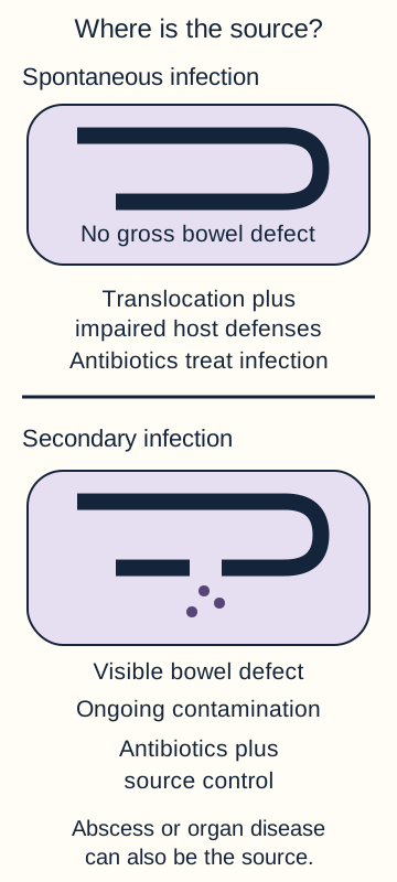
Spontaneous bacterial peritonitis (SBP) is bacterial infection of ascites without an intra-abdominal source requiring drainage or surgery. Cirrhosis increases intestinal permeability, bacterial translocation and susceptibility to infection. Impaired host defenses and low ascitic opsonic activity make a small bacterial inoculum consequential. Translocation is not the same as a bowel perforation: organisms can reach extraintestinal sites without a gross hole in the bowel. [1][4]
The usual infection is monomicrobial. Enteric gram-negative organisms such as Escherichia coli and Klebsiella are important, but streptococci, staphylococci and enterococci also occur. Healthcare exposure and antibiotic use alter the organism distribution and resistance risk. Do not infer susceptibility merely from the word spontaneous. [1][2]
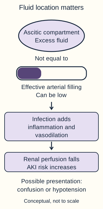
Inflammation can intensify the splanchnic vasodilation already present in cirrhosis. Effective arterial filling falls even though total body fluid is excessive. Renal perfusion may deteriorate, encephalopathy may appear, and hypotension may progress. Kidney injury during infection is not automatically hepatorenal syndrome: hypovolemia, tubular injury and other causes also need assessment. [1]
Trace why infection in ascites can first present as AKI or encephalopathy. [1][3]
Consider two patients with similar tense ascites. One is at a stable outpatient visit. The other is newly hospitalized with confusion and a creatinine increase from 0.8 to 1.5 mg/dL. Predict which needs urgent infection testing even if both temperatures are normal.
Compare the two situations
The newly hospitalized patient needs prompt diagnostic paracentesis. Acute brain and kidney changes can be the presentation of infection rather than isolated progression of cirrhosis.
The broader rule remains visible: perform diagnostic paracentesis promptly in hospitalized patients with cirrhotic ascites, including those without abdominal symptoms. Fever, abdominal pain, gastrointestinal symptoms, bleeding, shock, worsening jaundice, encephalopathy or declining kidney function strengthen the urgency. New-onset ascites also warrants diagnostic evaluation. In the stable outpatient, new symptoms or deterioration should trigger sampling rather than waiting for the next scheduled visit. [1][2]
Collect information without delaying treatment
Obtain ascitic cell count with differential and culture. At the bedside, inoculate aerobic and anaerobic blood-culture bottles according to local specimen-volume instructions; send a separate appropriate sample for cell count. Obtain blood cultures too. Sample before the first antibiotic when feasible, but do not delay antibiotics and resuscitation in an unstable patient to achieve an ideal sampling order. [1]
Ascitic albumin and total protein, with paired serum albumin, characterize new ascites. The serum-ascites albumin gradient addresses portal hypertension, not whether fluid is infected. Add glucose and LDH when a secondary source is suspected. Ultrasound helps identify a safe pocket. Cirrhosis-related INR prolongation or thrombocytopenia alone generally does not justify prophylactic plasma or platelet transfusion and delay; active bleeding, disseminated intravascular coagulation or other exceptional risks require individualized procedural assessment. [1][2]
Transfer this to a bleeding admission: a low initial neutrophil count does not eliminate the need for short-course infection prophylaxis during upper gastrointestinal hemorrhage. Testing for an existing infection and preventing a new infection are different decisions.
Which number triggers treatment?
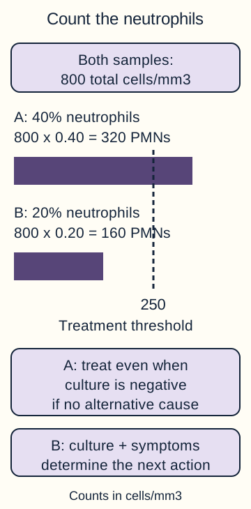
The relevant number is the absolute polymorphonuclear neutrophil (PMN) count, not total nucleated cells and not the percentage alone. Multiply total nucleated cells by the neutrophil fraction. A PMN count at least 250 cells/mm3 meets the treatment threshold for presumed SBP when a secondary source is not evident. Cells/mm3 and cells/microliter are equivalent units. Culture identifies organisms and susceptibility; it is not a prerequisite for starting treatment. [1][4]
Compare identical total counts with different PMN fractions, then apply culture and symptoms. [1][4]
Trace a sample through the decision
Keep the following paired samples in view. First calculate each PMN count, then change only the culture result and predict whether the initial antibiotic decision changes.
Same total cell count, different neutrophil fractions
Sample
Total cells/mm3
Neutrophils
Absolute PMNs/mm3
SampleA
Total cells/mm3800
Neutrophils40%
Absolute PMNs/mm3320
SampleB
Total cells/mm3800
Neutrophils20%
Absolute PMNs/mm3160
Sample A crosses the threshold: 800 x 0.40 = 320. Sample B does not: 800 x 0.20 = 160. The total count cannot distinguish these decisions. For sample A, changing culture from pending to negative does not cancel treatment. For sample B, a positive culture demands assessment of symptoms and repeat sampling, not automatic dismissal.
Predict the consequence of fever in sample B
Fever with culture-positive fluid and PMNs below 250 supports treatment of symptomatic bacterascites. The threshold is not a reason to withhold antibiotics from a clinically infected patient.
Interpret count and culture together
Pattern
Interpretation and action
PatternPMNs at least 250; culture positive
Interpretation and actionTreat. Determine whether infection is spontaneous or secondary.
PatternPMNs at least 250; culture negative
Interpretation and actionCulture-negative neutrocytic ascites: treat as SBP after considering alternative causes.
PatternPMNs below 250; culture positive; symptoms
Interpretation and actionSymptomatic bacterascites: give antibiotics and evaluate the patient.
PatternPMNs below 250; culture positive; well patient
Interpretation and actionPrompt repeat paracentesis and clinical reassessment; treatment is not automatic.
A positive culture with few neutrophils can represent transient colonization, contamination or early infection. In an asymptomatic patient, a repeat count and culture help distinguish these possibilities. New symptoms, persistent positive cultures or progression to neutrocytic ascites change management. Negative cultures are common in true SBP, especially after antibiotics; malignancy, pancreatitis, tuberculosis and secondary inflammation can also complicate interpretation. [1][2][4]
For substantially bloody fluid, a conventional correction subtracts one PMN for every 250 red cells/mm3. For example, 420 measured PMNs with 30,000 red cells gives 420 - 120 = 300 corrected PMNs/mm3. Treatment remains indicated. This approximation does not establish why bleeding occurred and must not overrule clinical sepsis. Apply the calculation to a different sample: 380 PMNs with 20,000 red cells also corrects to 300, despite a different measured count. [4]
When are antibiotics alone not enough?
Neutrocytic ascites tells you inflammation is present; it does not prove the bowel wall is intact. Secondary bacterial peritonitis can arise from perforation, abscess, ischemic bowel or another inflamed abdominal organ. Infection may continue until that source is drained, repaired or otherwise controlled. A biliary source requires the same attention to anatomy. [1][2][4]
Distinguish infection without a gross perforation from secondary contamination requiring source control. [1][2][4]
Compare diffuse mild tenderness with rapidly increasing focal pain and guarding. A single enteric isolate fits SBP, but polymicrobial growth, particularly with anaerobes, raises concern for a structural source. Free intraperitoneal air or a drainable collection should redirect care even before cultures finalize. Conversely, one organism does not exclude a secondary source.
The classic Runyon chemistry pattern is at least two of: ascitic protein above 1 g/dL, glucose below 50 mg/dL, and LDH above the serum laboratory upper limit of normal. These findings support a secondary source; they neither prove perforation nor safely exclude it when absent. The original algorithm had limited specificity, and the clinical course remains essential. [4][5]
Try a comparison: protein 1.7 g/dL, glucose 38 mg/dL and LDH 310 U/L with serum LDH upper limit 220 U/L meet all three features. Before reading further, choose what information is still missing: the bacterial species or the location of the source.
Check the next diagnostic priority
The location of the source is the urgent missing information. Cross-sectional abdominal imaging and early surgical or procedural consultation are needed alongside broader antimicrobial treatment.
Obtain urgent abdominal imaging, broaden antibiotics to cover the suspected abdominal organisms including anaerobes, and arrange source-control assessment. Do not wait for a second tap when perforation is already suspected. In a new patient whose chemistry does not meet the pattern, persistent focal tenderness still warrants imaging. A checklist cannot replace anatomy. [1][2][4]
Treat the infection and protect organ perfusion
For community-acquired SBP without substantial resistant-organism risk, IV ceftriaxone or cefotaxime is a usual initial choice when local susceptibility supports it. Recent hospitalization, broad antibiotic exposure, prior resistant isolates, nosocomial onset or septic shock require broader empiric therapy tailored to local microbiology. Piperacillin-tazobactam or a carbapenem may be appropriate in selected settings; resistant gram-positive coverage depends on specific risks. These are not interchangeable universal regimens. Narrow treatment once cultures and susceptibility permit. Uncomplicated responsive infection is commonly treated for 5 to 7 days. [1][2]
Compare two patients with 900 PMNs/mm3. One presents from home without recent antibiotics. The other develops infection on hospital day 6 after recent piperacillin-tazobactam and has a prior ESBL-producing isolate. Predict whether the identical count should lead to identical antibiotics.
Explain the antibiotic difference
The count establishes the need for treatment, not the required spectrum. Recent exposure and the resistant isolate favor broader initial coverage in the second patient, followed by culture-directed narrowing.
Albumin is an adjunct, not an antibiotic
Albumin supports effective circulation and has additional biologic effects. In the 126-patient trial, cefotaxime plus albumin reduced renal impairment from 33% to 10% and in-hospital mortality from 29% to 10%, compared with cefotaxime alone. These are results from that trial, not guaranteed individual outcomes. The studied schedule was 1.5 g/kg on day 1 and 1.0 g/kg on day 3. [3]
AASLD 2021 recommends albumin with antibiotics for SBP and emphasizes greater expected benefit with AKI or jaundice. BSG/BASL particularly recommends it with increased or rising creatinine. Classic risk markers include creatinine above 1 mg/dL, BUN above 30 mg/dL or marked hyperbilirubinemia; the commonly taught bilirubin cutoff above 4 mg/dL appears in KASL guidance. These markers identify likely benefit, not a universal prohibition below them. Assess volume status and monitor respiratory status because fluid overload can occur. [1][2][4]
For a patient whose estimated dry weight is 60 kg, the schedule gives 90 g on day 1 and 60 g on day 3. This dose is not calculated from the diagnostic sample volume. Albumin replacement after large-volume paracentesis is a separate indication. Antibiotics still treat the infection; albumin does not sterilize fluid. Reassess diuretics and nephrotoxins during AKI or hypotension, and do not diagnose hepatorenal syndrome from a single creatinine result. [1][2]
Read the trajectory, not just the latest value
Review mental status, perfusion, urine output, creatinine and microbiology throughout treatment. Repeat paracentesis near 48 hours is especially important with inadequate clinical response, resistant-organism concern or possible secondary infection. A fall in PMNs of less than 25% from baseline suggests inadequate response and should prompt antibiotic reassessment and a search for a secondary source. An improving patient with a susceptible isolate may not need routine repeat sampling. [1][2]
Calculate the percentage fall as (initial PMNs - repeat PMNs) / initial PMNs x 100. A decrease from 1,200 to 960 is only 20%; from 1,200 to 480 is 60%. The second count remains above 250 but shows a substantial early response. It does not justify stopping after just two days. Transfer the rule: a falling count with new focal guarding is still a reason to investigate anatomy, not reassurance that source control is unnecessary. [1][5]
Which prevention problem is present?
The misconception is that any low-protein ascites requires indefinite antibiotics. Instead, identify the setting before choosing a preventive course. Compare a patient recovering from SBP, a patient with acute variceal hemorrhage and a stable patient with low-protein ascites but no previous infection. Their indications and time horizons differ. [1][2]
Three prevention settings
Setting
Approach
SettingRecovery after SBP
ApproachLong-term secondary prophylaxis after treatment; select the agent for susceptibility, tolerance and availability.
SettingCirrhosis with upper GI bleeding
ApproachShort-course prophylaxis, commonly IV ceftriaxone 1 g daily, for a maximum of 7 days under the bleeding protocol.
SettingNo SBP or bleeding; low ascitic protein
ApproachIndividualized primary prophylaxis for selected high-risk patients, not an automatic prescription.
Before reading the criteria, decide which of the three settings changes if kidney function normalizes after treatment of a first SBP episode.
Check the effect of kidney recovery
Kidney recovery does not erase the prior infection. The indication remains secondary prevention, rather than becoming primary prevention or disappearing.
AASLD-based selection for primary prevention combines ascitic protein below 1.5 g/dL with renal dysfunction or advanced liver failure. Commonly used criteria are creatinine at least 1.2 mg/dL, BUN at least 25 mg/dL or sodium at most 130 mmol/L; alternatively, Child-Pugh score at least 9 with bilirubin at least 3 mg/dL. Protocol wording varies at boundaries, so use the local criteria rather than treating a rounded threshold as a biological switch. Low protein alone is not an automatic indication under this approach. BSG/BASL uses a broader low-protein high-risk definition but explicitly acknowledges uncertainty. [1][2][4]
Secondary prophylaxis options include daily ciprofloxacin or trimethoprim-sulfamethoxazole under local guidance; norfloxacin availability varies. Review prior cultures, drug interactions, kidney function, allergy and adverse effects. Resistance, C. difficile infection and fluoroquinolone toxicities matter, especially for primary prevention where benefit is less certain. Do not substitute an encephalopathy regimen or intermittent symptom-triggered antibiotics for a deliberate SBP prevention plan. [1][2]
An SBP episode is also a sign of serious hepatic decompensation. Arrange hepatology follow-up and transplant evaluation when appropriate, alongside recurrence prevention and medication review. Return to the opening patient: confusion and AKI led to a tap, the PMN result led to prompt treatment, the trajectory tested that treatment, and the discharge plan addresses the next infection rather than only the last one. [1]
Apply the decisions independently
Case 1
Show answer and explanations for case 1
A. Give plasma to normalize INR, then obtain ascitic studies (Why this does not fit)
New encephalopathy and kidney injury can reflect occult ascitic infection despite a normal temperature. Cirrhosis-related INR prolongation alone does not justify routine plasma before this low-risk procedure; transfusion would delay the needed sample.
Reasoning steps for option A
Why investigate the ascitic fluid?
New encephalopathy and kidney injury can reflect occult ascitic infection despite a normal temperature.
Does this INR require plasma first?
Cirrhosis-related INR prolongation alone does not justify routine plasma before this low-risk procedure; transfusion would delay the needed sample.
B. Obtain ascitic studies now without prophylactic transfusion (Best answer)
Infection can precipitate both encephalopathy and AKI in a hospitalized patient with cirrhotic ascites. A safe ultrasound pocket and absence of active bleeding or disseminated intravascular coagulation support prompt paracentesis without routine correction of the cirrhotic INR or platelet count.
Reasoning steps for option B
What links the brain and kidney changes?
Infection can precipitate both encephalopathy and AKI in a hospitalized patient with cirrhotic ascites.
Which supplied findings permit prompt sampling?
A safe ultrasound pocket and absence of active bleeding or disseminated intravascular coagulation support prompt paracentesis without routine correction of the cirrhotic INR or platelet count.
C. Give platelets to reach 100,000/microliter, then obtain ascitic studies (Why this does not fit)
Thrombocytopenia raises a procedural bleeding concern that requires clinical assessment. The supplied count and absence of active bleeding do not require a routine 100,000/microliter target before diagnostic paracentesis; this plan postpones infection testing.
Reasoning steps for option C
Why does the platelet count attract attention?
Thrombocytopenia raises a procedural bleeding concern that requires clinical assessment.
Is this transfusion target needed here?
The supplied count and absence of active bleeding do not require a routine 100,000/microliter target before diagnostic paracentesis; this plan postpones infection testing.
D. Obtain abdominal CT, then sample only if inflammation appears (Why this does not fit)
CT helps locate a suspected perforation, abscess or other structural source. No focal source is suggested here, and ascitic infection may not produce a diagnostic CT abnormality; direct cell count and culture are still needed.
Reasoning steps for option D
When does CT help?
CT helps locate a suspected perforation, abscess or other structural source.
Can an unrevealing CT replace the tap?
No focal source is suggested here, and ascitic infection may not produce a diagnostic CT abnormality; direct cell count and culture are still needed.
Takeaway: Recognize occult infection and assess procedural risk without delaying the diagnostic tap.
A. PMNs 396/mm3; start treatment before culture results (Why this does not fit)
Multiplying 720 by the non-neutrophil fraction, 0.55, gives 396. Treatment is warranted, but the correct PMN count uses 0.45 and equals 324/mm3.
Reasoning steps for option A
What calculation produces the option's reported count of 396 cells/mm3 from 720 nucleated cells/mm3?
Multiplying 720 by the non-neutrophil fraction, 0.55, gives 396.
Why is immediate treatment appropriate even though this option reports the wrong PMN count?
Treatment is warranted, but the correct PMN count uses 0.45 and equals 324/mm3.
B. PMNs 324/mm3; defer treatment until culture results (Why this does not fit)
The absolute count is correctly calculated as 720 x 0.45. At 324/mm3, it meets the treatment threshold, so culture confirmation should not delay therapy.
Reasoning steps for option B
How is the absolute PMN count calculated from 720 nucleated cells/mm3 with 45% neutrophils?
The absolute count is correctly calculated as 720 x 0.45.
Should therapy wait for the pending ascitic fluid culture when the PMN count is 324/mm3?
At 324/mm3, it meets the treatment threshold, so culture confirmation should not delay therapy.
C. PMNs 396/mm3; defer treatment until culture results (Why this does not fit)
The non-neutrophil count is 396/mm3, not the PMN count. The actual 324 PMNs/mm3 warrants prompt treatment regardless of pending culture.
Reasoning steps for option C
What does the option's figure of 396 cells/mm3 actually count?
The non-neutrophil count is 396/mm3, not the PMN count.
What does the true PMN count imply about treatment while the culture is pending?
The actual 324 PMNs/mm3 warrants prompt treatment regardless of pending culture.
D. PMNs 324/mm3; start treatment before culture results (Best answer)
The absolute count is 720 x 0.45 = 324/mm3. This meets the treatment threshold and requires empiric therapy without waiting for culture.
Reasoning steps for option D
What is the absolute PMN count for ascitic fluid with 720 nucleated cells/mm3 and 45% neutrophils?
The absolute count is 720 x 0.45 = 324/mm3.
What treatment timing follows from that PMN count despite the pending culture?
This meets the treatment threshold and requires empiric therapy without waiting for culture.
Takeaway: Calculate the absolute count and then decide whether culture can wait.
A. Complete five days of therapeutic ceftriaxone (Best answer)
The initial 460 PMNs/mm3 supports culture-negative neutrocytic ascites when no alternative abdominal source is found. Clinical improvement supports completion of a short therapeutic course, commonly 5 to 7 days, rather than stopping after two days.
Reasoning steps for option A
Does negative culture erase the initial count?
The initial 460 PMNs/mm3 supports culture-negative neutrocytic ascites when no alternative abdominal source is found.
How does improvement at 48 hours affect duration?
Clinical improvement supports completion of a short therapeutic course, commonly 5 to 7 days, rather than stopping after two days.
B. Replace ceftriaxone with daily antibiotic prophylaxis (Why this does not fit)
Secondary prophylaxis addresses recurrence after treatment of an SBP episode. A negative culture at 48 hours does not establish that the active episode has received an adequate therapeutic course.
Reasoning steps for option B
When does a prevention regimen belong?
Secondary prophylaxis addresses recurrence after treatment of an SBP episode.
Has treatment finished at this reassessment?
A negative culture at 48 hours does not establish that the active episode has received an adequate therapeutic course.
C. Change ceftriaxone to therapeutic IV meropenem (Why this does not fit)
Clinical failure or resistant-organism evidence would prompt reassessment of the initial spectrum. Improvement on ceftriaxone without a structural source supports its activity; culture negativity alone is not evidence of resistance.
Reasoning steps for option C
What would support broader coverage?
Clinical failure or resistant-organism evidence would prompt reassessment of the initial spectrum.
What does this course show instead?
Improvement on ceftriaxone without a structural source supports its activity; culture negativity alone is not evidence of resistance.
D. Extend ceftriaxone to fourteen treatment days (Why this does not fit)
A complicated focus or another documented infection can require a different treatment duration. The supplied imaging, negative blood cultures and improving course do not support a prolonged course for this uncomplicated episode.
Reasoning steps for option D
When might a longer course be needed?
A complicated focus or another documented infection can require a different treatment duration.
Are those conditions established here?
The supplied imaging, negative blood cultures and improving course do not support a prolonged course for this uncomplicated episode.
Takeaway: Treat the neutrocytic syndrome, then use clinical response to choose a therapeutic duration.
A. Withhold antibiotics pending a repeat ascitic PMN result (Why this does not fit)
An asymptomatic patient with low-PMN bacterascites can undergo prompt repeat sampling without automatic antibiotics. New fever and pain indicate symptomatic infection; obtain repeat fluid when feasible without making a higher PMN count a prerequisite for treatment.
Reasoning steps for option A
When can repeat testing precede a treatment decision?
An asymptomatic patient with low-PMN bacterascites can undergo prompt repeat sampling without automatic antibiotics.
Does that observation pathway fit now?
New fever and pain indicate symptomatic infection; obtain repeat fluid when feasible without making a higher PMN count a prerequisite for treatment.
B. Begin daily prophylaxis while repeating the ascitic culture (Why this does not fit)
A preventive regimen is intended to reduce future infection risk. The positive culture together with new fever and abdominal pain requires therapeutic antibiotics rather than a prevention dose.
Reasoning steps for option B
What does a prophylactic dose address?
A preventive regimen is intended to reduce future infection risk.
What needs treatment in this patient?
The positive culture together with new fever and abdominal pain requires therapeutic antibiotics rather than a prevention dose.
C. Begin therapeutic antibiotics now and repeat the fluid assessment (Best answer)
E. coli growth with 120 PMNs/mm3 is bacterascites rather than established neutrocytic ascites at that earlier time. Fever and abdominal pain favor clinically important infection, so start therapeutic antibiotics and reassess rather than waiting for the count to rise.
Reasoning steps for option C
How should the earlier specimen be classified?
E. coli growth with 120 PMNs/mm3 is bacterascites rather than established neutrocytic ascites at that earlier time.
How does the new clinical state change care?
Fever and abdominal pain favor clinically important infection, so start therapeutic antibiotics and reassess rather than waiting for the count to rise.
D. Withhold antibiotics pending final susceptibility test results (Why this does not fit)
They allow subsequent selection or narrowing of an active regimen. Symptomatic culture-positive ascites warrants empiric therapeutic treatment before final susceptibility results become available.
Reasoning steps for option D
Why obtain susceptibility results?
They allow subsequent selection or narrowing of an active regimen.
Should that result determine treatment timing?
Symptomatic culture-positive ascites warrants empiric therapeutic treatment before final susceptibility results become available.
Takeaway: The symptoms at reassessment determine whether low-PMN bacterascites needs treatment.
A. Start a full empiric course solely for the positive culture (Why this does not fit)
Culture growth can represent early infection. In a well patient below the PMN threshold, immediate treatment is not automatic; reassessment and repeat fluid testing distinguish persistence or progression.
Reasoning steps for option A
What might E. coli growth in ascites signal despite an initial PMN count of 90/mm3?
Culture growth can represent early infection.
In this well patient with PMNs below the SBP threshold, why is an immediate antibiotic course not automatic, and what would distinguish persistence from progression?
In a well patient below the PMN threshold, immediate treatment is not automatic; reassessment and repeat fluid testing distinguish persistence or progression.
B. Repeat paracentesis promptly with count and culture (Best answer)
Low-PMN culture-positive fluid may clear or progress. Prompt repeat sampling in this asymptomatic patient tests that trajectory while avoiding automatic antibiotic exposure.
Reasoning steps for option B
What two outcomes are possible for culture-positive ascitic fluid with only 90 PMNs/mm3?
Low-PMN culture-positive fluid may clear or progress.
What does prompt repeat paracentesis assess in this asymptomatic patient, and what exposure can it avoid?
Prompt repeat sampling in this asymptomatic patient tests that trajectory while avoiding automatic antibiotic exposure.
C. Begin indefinite secondary prophylaxis immediately (Why this does not fit)
Secondary prevention follows a treated SBP episode. This single low-PMN culture in a well patient has not established such an episode.
Reasoning steps for option C
What prior event is required before secondary SBP prevention is appropriate?
Secondary prevention follows a treated SBP episode.
Why does one E. coli-positive ascitic sample with 90 PMNs/mm3 in a well patient not yet meet that requirement?
This single low-PMN culture in a well patient has not established such an episode.
D. Discard the result and return to routine monthly follow-up (Why this does not fit)
Contamination is one explanation for a positive culture. The possibility of evolving infection requires prompt reassessment rather than ignoring the result.
Reasoning steps for option D
What could explain a positive ascitic culture without true infection?
Contamination is one explanation for a positive culture.
Why should the E. coli result prompt reassessment instead of routine monthly follow-up?
The possibility of evolving infection requires prompt reassessment rather than ignoring the result.
Takeaway: Well patients with bacterascites need prompt reassessment, not dismissal.
A. Corrected PMNs 320/mm3; start empiric antibiotics (Best answer)
The blood contribution is 50,000/250 = 200 PMNs/mm3. Subtraction leaves 320 PMNs/mm3, still meeting the threshold for treatment.
Reasoning steps for option A
With 50,000 red cells/mm3 in the traumatic ascitic tap and a correction of one PMN per 250 red cells, how many measured PMNs are attributed to blood contamination?
The blood contribution is 50,000/250 = 200 PMNs/mm3.
After correcting the measured 520 PMNs/mm3 for blood contamination, what PMN count remains, and does it warrant empiric antibiotics?
Subtraction leaves 320 PMNs/mm3, still meeting the threshold for treatment.
B. Corrected PMNs 320/mm3; await culture before treatment (Why this does not fit)
The blood correction is numerically correct. A corrected count of 320 remains treatment-level, so a pending culture should not postpone antibiotics.
Reasoning steps for option B
For the option reporting 320 corrected PMNs/mm3 from 520 measured PMNs/mm3, is the blood correction accurate?
Subtracting 50,000/250 = 200 blood-derived PMNs/mm3 from the measured 520 leaves 320 PMNs/mm3, so the correction is accurate.
Should antibiotics be delayed for culture results when the corrected ascitic PMN count is 320/mm3?
A corrected count of 320 remains treatment-level, so a pending culture should not postpone antibiotics.
C. Corrected PMNs 200/mm3; await culture before treatment (Why this does not fit)
The 200 PMNs/mm3 is the estimated blood-derived contribution. It must be subtracted from 520, not used as the final corrected count; the remaining 320 supports treatment.
Reasoning steps for option C
In this tap, what does the value of 200 PMNs/mm3 calculated from 50,000 red cells/mm3 represent?
The 200 PMNs/mm3 is the estimated blood-derived contribution.
Why is 200 PMNs/mm3 not the final corrected count, and what does the remaining count indicate about treatment?
It must be subtracted from 520, not used as the final corrected count; the remaining 320 supports treatment.
D. Corrected PMNs 520/mm3; start empiric antibiotics (Why this does not fit)
The measured count would support treatment but includes blood-derived cells. The specified correction yields 320, not 520 PMNs/mm3, although treatment is still required.
Reasoning steps for option D
Why is the uncorrected count of 520 PMNs/mm3 insufficient as the reported ascitic PMN count, even though it supports treatment?
The measured count would support treatment but includes blood-derived cells.
Using the specified red cell correction, what count replaces 520 PMNs/mm3, and is treatment still indicated?
The specified correction yields 320, not 520 PMNs/mm3, although treatment is still required.
Takeaway: Correction changes the count without necessarily changing treatment.
A. Give ceftriaxone now; obtain ascitic fluid when the operator arrives (Why this does not fit)
Hypotension and acute confusion with suspected infection make a 90-minute antimicrobial delay unsafe. The recent ESBL-producing isolate was resistant to ceftriaxone, so prompt timing does not compensate for likely inadequate empiric coverage.
Reasoning steps for option A
What does immediate treatment address?
Hypotension and acute confusion with suspected infection make a 90-minute antimicrobial delay unsafe.
Does ceftriaxone fit the resistance information?
The recent ESBL-producing isolate was resistant to ceftriaxone, so prompt timing does not compensate for likely inadequate empiric coverage.
B. Obtain ascitic fluid in 90 minutes; then give IV meropenem (Why this does not fit)
It covers the documented susceptible ESBL-producing isolate and fits the supplied severe-infection protocol. The patient is unstable; the benefit of pre-antibiotic culture yield does not justify postponing active therapy while waiting for an operator.
Reasoning steps for option B
Why does meropenem fit the history?
It covers the documented susceptible ESBL-producing isolate and fits the supplied severe-infection protocol.
Is waiting for the specimen appropriate?
The patient is unstable; the benefit of pre-antibiotic culture yield does not justify postponing active therapy while waiting for an operator.
C. Obtain ascitic fluid in 90 minutes; then give IV ceftriaxone (Why this does not fit)
Pre-antibiotic fluid collection can improve culture recovery when it is promptly feasible. Severe hypotension makes the delay unsafe, and the recent resistant isolate makes ceftriaxone an inadequate empiric choice.
Reasoning steps for option C
What advantage does sampling first have?
Pre-antibiotic fluid collection can improve culture recovery when it is promptly feasible.
Which two supplied findings defeat this sequence?
Severe hypotension makes the delay unsafe, and the recent resistant isolate makes ceftriaxone an inadequate empiric choice.
D. Give meropenem now; obtain ascitic fluid when the operator arrives (Best answer)
The recent ceftriaxone-resistant ESBL isolate and severe hospital-onset illness favor an active regimen such as meropenem under the supplied protocol. Unstable circulation requires treatment now while resuscitation continues; paracentesis remains necessary as soon as it can be performed.
Reasoning steps for option D
Which findings determine the initial spectrum?
The recent ceftriaxone-resistant ESBL isolate and severe hospital-onset illness favor an active regimen such as meropenem under the supplied protocol.
Which findings determine the sequence?
Unstable circulation requires treatment now while resuscitation continues; paracentesis remains necessary as soon as it can be performed.
Takeaway: Choose both active empiric coverage and a sampling sequence that does not delay unstable-patient treatment.
A. Give prophylactic plasma before sampling; treat after the cell result (Why this does not fit)
A treatment-level count would justify antibiotics. Routine prophylactic plasma for this cirrhotic INR would unnecessarily delay a low-risk diagnostic procedure.
Reasoning steps for option A
Which part of the plasma-first plan is supported by 680 nucleated cells/mm3 with 45% neutrophils?
Starting antibiotics after the cell result is appropriate: 680 x 0.45 = 306 PMNs/mm3 meets the threshold of at least 250/mm3. This does not justify the preceding plasma transfusion.
Why should an INR of 2.1 in this patient with cirrhosis not prompt prophylactic plasma before paracentesis?
Routine prophylactic plasma for this cirrhotic INR would unnecessarily delay a low-risk diagnostic procedure.
B. Sample without prophylactic plasma; await a positive culture to treat (Why this does not fit)
Prompt sampling without routine plasma is appropriate in this procedural context. The count is 680 x 0.45 = 306 PMNs/mm3, so treatment should not wait for culture.
Reasoning steps for option B
With a safe ultrasound pocket and no active bleeding or disseminated intravascular coagulation, how should ascitic fluid be obtained?
Prompt sampling without routine plasma is appropriate in this procedural context.
What does 45% neutrophils among 680 nucleated cells/mm3 imply about waiting for culture before treatment?
The count is 680 x 0.45 = 306 PMNs/mm3, so treatment should not wait for culture.
C. Sample without prophylactic plasma; treat after the cell result (Best answer)
The supplied coagulation findings alone do not require routine blood products before paracentesis. The subsequent 306 PMNs/mm3 meets the threshold for immediate empiric treatment.
Reasoning steps for option C
Do an INR of 2.1 and platelets of 58,000/microliter, without active bleeding, require blood products before paracentesis?
The supplied coagulation findings alone do not require routine blood products before paracentesis.
What does the calculated ascitic PMN count indicate about the timing of empiric treatment?
The subsequent 306 PMNs/mm3 meets the threshold for immediate empiric treatment.
D. Give prophylactic plasma before sampling; await a positive culture to treat (Why this does not fit)
Both procedural bleeding and unnecessary antibiotics deserve consideration. These findings do not justify plasma, and the treatment-level PMN result does not require culture confirmation.
Reasoning steps for option D
Do the INR of 2.1 and platelets of 58,000/microliter justify the plasma-first component of this plan?
No. With a safe ultrasound pocket and no active bleeding or disseminated intravascular coagulation, these cirrhotic coagulation findings do not require routine prophylactic plasma.
Why is the proposed sequence of plasma before sampling and antibiotics only after a positive culture inappropriate?
Routine plasma would delay sampling without a supplied indication, and 680 x 0.45 = 306 PMNs/mm3 warrants empiric treatment without waiting for a positive culture.
Takeaway: Procedural safety and fluid interpretation require separate decisions.
A. IV piperacillin-tazobactam without IV albumin (Why this does not fit)
Healthcare-associated infection or a suspected mixed abdominal source can require broader empiric coverage. Stable community acquisition with local cephalosporin susceptibility favors ceftriaxone, while the creatinine increase supports adjunctive albumin; this plan is unnecessarily broad and omits the indicated adjunct.
Reasoning steps for option A
Which setting might justify the broader agent?
Healthcare-associated infection or a suspected mixed abdominal source can require broader empiric coverage.
How do this setting and the kidney findings differ?
Stable community acquisition with local cephalosporin susceptibility favors ceftriaxone, while the creatinine increase supports adjunctive albumin; this plan is unnecessarily broad and omits the indicated adjunct.
B. IV ceftriaxone with adjunctive IV albumin (Best answer)
The symptomatic treatment-level PMN count requires therapy, and stable community acquisition without resistance risks supports ceftriaxone under the supplied antibiogram. Creatinine rising from 0.7 to 1.6 mg/dL supports adjunctive albumin for SBP-related renal risk, with reassessment for fluid intolerance during administration.
Reasoning steps for option B
What selects the antibiotic spectrum?
The symptomatic treatment-level PMN count requires therapy, and stable community acquisition without resistance risks supports ceftriaxone under the supplied antibiogram.
What additional treatment does the kidney change support?
Creatinine rising from 0.7 to 1.6 mg/dL supports adjunctive albumin for SBP-related renal risk, with reassessment for fluid intolerance during administration.
C. IV ceftriaxone without adjunctive IV albumin (Why this does not fit)
Ceftriaxone fits the stable community presentation and supplied local susceptibility. The creatinine increase establishes a renal-risk indication for adjunctive albumin; a normal blood pressure does not remove that indication.
Reasoning steps for option C
Does the antibiotic match the acquisition setting?
Ceftriaxone fits the stable community presentation and supplied local susceptibility.
Which independent finding makes the plan incomplete?
The creatinine increase establishes a renal-risk indication for adjunctive albumin; a normal blood pressure does not remove that indication.
D. IV piperacillin-tazobactam with IV albumin (Why this does not fit)
Albumin is appropriate because the patient has SBP with a creatinine increase. No recent exposure, resistant isolate, instability or focal source is supplied; ceftriaxone is the narrower appropriate empiric choice in this setting.
Reasoning steps for option D
Does the adjunct fit the organ-risk findings?
Albumin is appropriate because the patient has SBP with a creatinine increase.
Is the additional antibiotic breadth needed?
No recent exposure, resistant isolate, instability or focal source is supplied; ceftriaxone is the narrower appropriate empiric choice in this setting.
Takeaway: Choose antibiotic breadth from acquisition risk and adjunctive albumin from the kidney findings.
The supplied susceptibility report identifies meropenem and gentamicin as active, unlike ceftriaxone and piperacillin-tazobactam. Rising creatinine and reduced urine output favor an active beta-lactam over aminoglycoside monotherapy; meropenem dosing still requires renal assessment.
Reasoning steps for option A
Which listed agents cover the recent isolate?
The supplied susceptibility report identifies meropenem and gentamicin as active, unlike ceftriaxone and piperacillin-tazobactam.
Which active agent better fits the kidney findings?
Rising creatinine and reduced urine output favor an active beta-lactam over aminoglycoside monotherapy; meropenem dosing still requires renal assessment.
B. IV ceftriaxone (Why this does not fit)
Ceftriaxone is an established initial drug for susceptible community-acquired SBP. The recent isolate is ceftriaxone-resistant and this infection developed in hospital with hypotension; avoiding nephrotoxicity does not justify inactive coverage.
Reasoning steps for option B
Why might this be selected in another patient?
Ceftriaxone is an established initial drug for susceptible community-acquired SBP.
What makes it a poor match here?
The recent isolate is ceftriaxone-resistant and this infection developed in hospital with hypotension; avoiding nephrotoxicity does not justify inactive coverage.
C. IV piperacillin-tazobactam (Why this does not fit)
Some hospital-acquired infections require broader coverage than a third-generation cephalosporin. The recent isolate is explicitly resistant to piperacillin-tazobactam, so the supplied susceptible beta-lactam is the more appropriate empiric choice.
Reasoning steps for option C
Why consider a broad beta-lactam?
Some hospital-acquired infections require broader coverage than a third-generation cephalosporin.
Does nominal breadth establish activity?
The recent isolate is explicitly resistant to piperacillin-tazobactam, so the supplied susceptible beta-lactam is the more appropriate empiric choice.
D. IV gentamicin (Why this does not fit)
The recent isolate is susceptible to gentamicin, so its exclusion cannot be based on an assumed resistance result. The patient already has acute kidney injury, and aminoglycoside nephrotoxicity makes this a poorer initial choice when an active beta-lactam is available.
Reasoning steps for option D
Why is this a real competitor?
The recent isolate is susceptible to gentamicin, so its exclusion cannot be based on an assumed resistance result.
Which finding changes the choice between active agents?
The patient already has acute kidney injury, and aminoglycoside nephrotoxicity makes this a poorer initial choice when an active beta-lactam is available.
Takeaway: Distinguish antimicrobial activity from suitability in a patient with acute kidney injury.
A. Continue meropenem for a five-day therapeutic course (Why this does not fit)
Severe undifferentiated infection can justify broad empiric therapy. A ceftriaxone-susceptible isolate, substantial PMN decline and clinical recovery support narrowing rather than maintaining unnecessary carbapenem exposure.
Reasoning steps for option A
Why was meropenem initially reasonable?
Severe undifferentiated infection can justify broad empiric therapy.
What has changed after culture and reassessment?
A ceftriaxone-susceptible isolate, substantial PMN decline and clinical recovery support narrowing rather than maintaining unnecessary carbapenem exposure.
B. Stop therapeutic antibiotics and begin daily prophylaxis (Why this does not fit)
A treated SBP episode carries recurrence risk that warrants a prevention plan. Improvement after 48 hours does not establish completion of the current therapeutic course; prophylaxis should not replace its remaining treatment.
Reasoning steps for option B
Why plan secondary prevention?
A treated SBP episode carries recurrence risk that warrants a prevention plan.
Why is this switch premature?
Improvement after 48 hours does not establish completion of the current therapeutic course; prophylaxis should not replace its remaining treatment.
C. Use ceftriaxone to complete five treatment days (Best answer)
The isolated E. coli is susceptible to ceftriaxone, and imaging identifies no structural source. Improved blood pressure and mentation with PMNs falling from 1,100 to 300/mm3 support de-escalation while completing a short therapeutic course, commonly 5 to 7 days.
Reasoning steps for option C
Which result permits narrower therapy?
The isolated E. coli is susceptible to ceftriaxone, and imaging identifies no structural source.
Does the clinical trajectory support the change?
Improved blood pressure and mentation with PMNs falling from 1,100 to 300/mm3 support de-escalation while completing a short therapeutic course, commonly 5 to 7 days.
D. Use ceftriaxone plus metronidazole for five days (Why this does not fit)
Secondary abdominal contamination can require anaerobic coverage as well as source control. The monomicrobial susceptible isolate, negative source evaluation and improving course do not support routinely adding metronidazole.
Reasoning steps for option D
Why consider adding anaerobic coverage?
Secondary abdominal contamination can require anaerobic coverage as well as source control.
Is that indication supplied here?
The monomicrobial susceptible isolate, negative source evaluation and improving course do not support routinely adding metronidazole.
Takeaway: Use susceptibility to narrow treatment and the clinical course to confirm that narrowing is appropriate.
A. No albumin, because drainage was less than 5 liters (Why this does not fit)
Large-volume drainage is one indication for albumin. This patient has treatment-level neutrocytic ascites and AKI, creating a separate SBP-related indication despite the small sample.
Reasoning steps for option A
In cirrhosis, what albumin indication depends on removing a large volume of ascites?
Large-volume drainage is one indication for albumin.
Why can albumin be indicated when only 40 mL of ascites was sampled, the absolute PMN count is 350/mm3, and creatinine has risen to 1.8 mg/dL?
This patient has treatment-level neutrocytic ascites and AKI, creating a separate SBP-related indication despite the small sample.
B. Albumin 60 g on day 1 and 60 g on day 3 (Why this does not fit)
The day-3 amount is consistent with 1 g/kg. After identifying SBP with AKI, the studied day-1 factor is 1.5 g/kg, requiring 90 g rather than 60 g.
Reasoning steps for option B
How does the proposed 60 g albumin dose on day 3 compare with weight-based dosing for a 60 kg patient?
The day-3 amount is consistent with 1 g/kg.
With SBP and AKI in a 60 kg patient, what studied day-1 albumin factor and dose distinguish the appropriate regimen from 60 g on day 1?
After identifying SBP with AKI, the studied day-1 factor is 1.5 g/kg, requiring 90 g rather than 60 g.
C. Albumin 0.32 g once, based on fluid sampled (Why this does not fit)
Eight grams per liter times 0.04 L equals 0.32 g. That calculation misapplies large-volume paracentesis replacement to an infection-related indication.
Reasoning steps for option C
What dose results from applying 8 g of albumin per liter to the 0.04 L ascitic sample?
Eight grams per liter times 0.04 L equals 0.32 g.
Why is a dose calculated from the 40 mL sample inappropriate when neutrocytic ascites and AKI are present?
That calculation misapplies large-volume paracentesis replacement to an infection-related indication.
D. Albumin 90 g on day 1 and 60 g on day 3 (Best answer)
The absolute PMN count is 700 x 0.50 = 350/mm3, supporting SBP treatment in this setting. AKI supports adjunctive albumin; 1.5 x 60 = 90 g initially and 1 x 60 = 60 g on day 3, with volume monitoring.
Reasoning steps for option D
With 700 ascitic nucleated cells/mm3 and 50% neutrophils, what is the absolute PMN count and what does it support?
The absolute PMN count is 700 x 0.50 = 350/mm3, supporting SBP treatment in this setting.
For a 60 kg patient with SBP and AKI, what albumin doses are indicated on days 1 and 3, and what monitoring is needed?
AKI supports adjunctive albumin; 1.5 x 60 = 90 g initially and 1 x 60 = 60 g on day 3, with volume monitoring.
Takeaway: Identify the indication before choosing the albumin dosing rule.
A. 23 percentage points; improved effective arterial filling (Best answer)
Absolute risk reduction is 33% - 10% = 23 percentage points. Similar infection resolution with less renal injury is consistent with circulatory support, although the trial does not isolate every biologic effect of albumin.
Reasoning steps for option A
In patients with cirrhosis and ascitic infection, renal impairment occurred in 33% given cefotaxime alone and 10% given cefotaxime plus albumin. What was the absolute risk reduction?
How can albumin's renal benefit be interpreted when infection resolution was similar in both groups, and what mechanistic limitation remains?
Similar infection resolution with less renal injury is consistent with circulatory support, although the trial does not isolate every biologic effect of albumin.
B. 23 percentage points; direct bacterial killing in ascites (Why this does not fit)
The absolute risk reduction is correctly calculated. Albumin is not an antibiotic; the similar infection-resolution rates favor organ support rather than direct sterilization as the explanation.
Reasoning steps for option B
Does the option's 23-percentage-point reduction correctly reflect the difference between renal impairment rates of 33% and 10%?
Subtracting the albumin group's 10% renal-impairment rate from the control group's 33% gives an absolute reduction of 23 percentage points.
Why does similar infection resolution with and without albumin argue against direct bacterial killing as its renal-protective mechanism?
Albumin is not an antibiotic; the similar infection-resolution rates favor organ support rather than direct sterilization as the explanation.
C. About 70 percentage points; improved effective arterial filling (Why this does not fit)
About 70% describes the relative reduction, 23/33, rather than an absolute difference. Effective arterial support is plausible, but the absolute reduction is 23 percentage points.
Reasoning steps for option C
What does the calculation 23/33 represent when renal impairment falls from 33% to 10%?
About 70% describes the relative reduction, 23/33, rather than an absolute difference.
Which part of the proposed explanation remains plausible, and what is the correct absolute renal-risk reduction?
Effective arterial support is plausible, but the absolute reduction is 23 percentage points.
D. About 70 percentage points; direct bacterial killing in ascites (Why this does not fit)
The relative risk reduction is about 70%, not 70 percentage points of absolute risk. Albumin does not directly replace antimicrobial killing, so both components of this interpretation are incorrect.
Reasoning steps for option D
How should a claim of a 70-percentage-point absolute reduction be corrected for renal impairment rates of 33% and 10%?
The relative risk reduction is about 70%, not 70 percentage points of absolute risk.
Why are both the proposed 70-percentage-point absolute reduction and direct bacterial killing by albumin incorrect?
The absolute reduction is 33% - 10% = 23 percentage points; about 70% is the relative reduction. Albumin supports organ perfusion rather than directly killing bacteria, so antibiotics remain necessary.
Takeaway: Keep absolute benefit separate from relative benefit and from mechanism.
A. A 15% decrease; continue the regimen without additional assessment (Why this does not fit)
The decline is correctly calculated as 150/1,000 = 15%. A fall below 25% is an inadequate early PMN response and warrants reassessment rather than reassurance from stability alone.
Reasoning steps for option A
After 48 hours of ceftriaxone, ascitic PMNs have changed from 1,000 to 850/mm3. How is the percentage decline calculated?
The decline is correctly calculated as 150/1,000 = 15%.
What does an ascitic PMN fall of less than 25% at 48 hours imply, despite stable blood pressure?
A fall below 25% is an inadequate early PMN response and warrants reassessment rather than reassurance from stability alone.
B. An 85% decrease; continue the regimen without additional assessment (Why this does not fit)
Eighty-five percent is the fraction of the original count that remains. The reduction is only 15%, so this is not a robust response.
Reasoning steps for option B
When ascitic PMNs fall from 1,000 to 850/mm3, what does the proposed 85% figure represent?
Eighty-five percent is the fraction of the original count that remains.
What is the actual percentage reduction from 1,000 to 850 ascitic PMNs/mm3, and is it a robust response?
The reduction is only 15%, so this is not a robust response.
C. A 15% decrease; reassess spectrum and promptly investigate a secondary source (Best answer)
The count fell by 150/1,000 = 15%. That is below the expected response threshold and warrants antibiotic review and anatomic investigation despite stable blood pressure.
Reasoning steps for option C
Using ascitic PMN counts of 1,000/mm3 initially and 850/mm3 after 48 hours, how is the decline expressed?
The count fell by 150/1,000 = 15%.
What do the 48-hour PMN result and stable blood pressure indicate about antibiotic review and investigation for a secondary source?
That is below the expected response threshold and warrants antibiotic review and anatomic investigation despite stable blood pressure.
D. An 85% decrease; reassess spectrum and investigate a secondary source (Why this does not fit)
Further assessment is appropriate, but not because of an 85% decline. Dividing the final by initial value gives the remaining fraction; the actual 15% fall indicates inadequate response.
Reasoning steps for option D
Is reassessment after 48 hours of ceftriaxone justified by a claimed 85% decline in ascitic PMNs from 1,000 to 850/mm3?
Further assessment is appropriate, but not because of an 85% decline.
Why does dividing the final ascitic PMN count by the initial count misstate the decline, and what does the actual fall indicate?
Dividing the final by initial value gives the remaining fraction; the actual 15% fall indicates inadequate response.
Takeaway: Calculate the change rather than the fraction remaining.
A. Change to meropenem for persistent neutrocytic ascites (Why this does not fit)
The repeat count of 420 PMNs/mm3 remains above the threshold used to begin treatment. The count has fallen by 780/1,200 = 65% with clinical recovery and a susceptible isolate, so residual neutrophilia alone does not establish antibiotic failure.
Reasoning steps for option A
Why might the repeat count appear concerning?
The repeat count of 420 PMNs/mm3 remains above the threshold used to begin treatment.
What does the actual trajectory show?
The count has fallen by 780/1,200 = 65% with clinical recovery and a susceptible isolate, so residual neutrophilia alone does not establish antibiotic failure.
B. Continue ceftriaxone for the planned therapeutic course (Best answer)
The fall from 1,200 to 420/mm3 is 65%, exceeding the 25% early-response benchmark. A substantial decline, symptom resolution and ceftriaxone susceptibility support completion of the active regimen rather than escalation or early cessation.
Reasoning steps for option B
How large is the PMN reduction?
The fall from 1,200 to 420/mm3 is 65%, exceeding the 25% early-response benchmark.
How should that calculation be used clinically?
A substantial decline, symptom resolution and ceftriaxone susceptibility support completion of the active regimen rather than escalation or early cessation.
C. Replace ceftriaxone with daily secondary prophylaxis today (Why this does not fit)
Recovery from an SBP episode should lead to a secondary prevention plan. No; falling PMNs and symptom improvement do not substitute for completing an appropriate therapeutic course before prevention begins.
Reasoning steps for option C
Why does improvement make prevention relevant?
Recovery from an SBP episode should lead to a secondary prevention plan.
Does a 48-hour response complete treatment?
No; falling PMNs and symptom improvement do not substitute for completing an appropriate therapeutic course before prevention begins.
D. Continue ceftriaxone until daily fluid samples normalize (Why this does not fit)
Repeat PMNs can help evaluate an inadequate or uncertain treatment response. This substantial decline and clinical improvement do not require daily paracentesis or normalization-based dosing duration; complete an appropriate therapeutic course.
Reasoning steps for option D
Why follow the cell-count trajectory?
Repeat PMNs can help evaluate an inadequate or uncertain treatment response.
Should repeated counts determine the stopping date here?
This substantial decline and clinical improvement do not require daily paracentesis or normalization-based dosing duration; complete an appropriate therapeutic course.
Takeaway: Judge early response from the proportional decline and the clinical course, not a residual diagnostic-level count alone.
A. Two supportive chemistry findings; defer imaging until culture grows (Why this does not fit)
Protein above 1 and glucose below 50 meet two classic features. These support prompt investigation of a secondary source rather than making imaging contingent on positive culture.
Reasoning steps for option A
Which two ascitic chemistry results meet classic criteria for a secondary source of peritonitis?
Protein above 1 and glucose below 50 meet two classic features.
With those two findings present and cultures still pending, should investigation of a secondary source wait for culture growth?
These support prompt investigation of a secondary source rather than making imaging contingent on positive culture.
B. One supportive chemistry finding; repeat chemistry before imaging (Why this does not fit)
Protein and glucose both meet the specified pattern, whereas LDH does not. Counting only one misses a reason to investigate a structural source now.
Reasoning steps for option B
How do the ascitic protein of 1.9 g/dL, glucose of 31 mg/dL and LDH of 180 U/L compare with the specified pattern?
Protein and glucose both meet the specified pattern, whereas LDH does not.
Why is counting only one supportive finding and repeating chemistry before imaging inappropriate here?
Protein above 1 g/dL and glucose below 50 mg/dL provide two supportive findings despite LDH below the serum upper limit; prompt imaging for a structural source should not await repeated chemistry.
C. One supportive chemistry finding; obtain abdominal CT now (Why this does not fit)
CT is appropriate, but the stated count of supportive findings is incorrect. Both protein 1.9 and glucose 31 meet the pattern, even though LDH is below the serum upper limit.
Reasoning steps for option C
In the option proposing abdominal CT now, what is correct about the investigation and incorrect about the stated count?
CT is appropriate, but the stated count of supportive findings is incorrect.
Which values explain the undercount when ascitic LDH is below the serum upper limit of 240 U/L?
Both protein 1.9 and glucose 31 meet the pattern, even though LDH is below the serum upper limit.
D. Two supportive chemistry findings; obtain abdominal CT now (Best answer)
Protein above 1 g/dL and glucose below 50 mg/dL give two supportive findings. The pattern warrants anatomic assessment for a secondary source; it does not itself prove perforation.
Reasoning steps for option D
Which protein and glucose thresholds produce two supportive findings in this patient's ascitic fluid?
Protein above 1 g/dL and glucose below 50 mg/dL give two supportive findings.
What investigation does this pattern warrant, and what diagnosis does it not establish by itself?
The pattern warrants anatomic assessment for a secondary source; it does not itself prove perforation.
Takeaway: Supportive chemistry leads to anatomic investigation, not a definitive source label.
A. Repeat ascitic count and chemistry after another 48 hours (Why this does not fit)
The PMN decline is 65%, and none of the three classic chemistry features is present. New focal guarding and mixed enteric growth suggest a secondary source despite the apparent cell-count response; imaging should not wait for another tap.
Reasoning steps for option A
Which findings might encourage serial observation?
The PMN decline is 65%, and none of the three classic chemistry features is present.
Why should those findings not postpone investigation?
New focal guarding and mixed enteric growth suggest a secondary source despite the apparent cell-count response; imaging should not wait for another tap.
B. Obtain urgent abdominal CT now to define an intra-abdominal source (Best answer)
Mixed enteric organisms including an anaerobe together with focal guarding raise concern for ongoing secondary contamination. Neither an improving count nor absence of the classic chemistry pattern excludes a structural source, so urgent CT and source-control assessment are warranted.
Reasoning steps for option B
What do the culture and new examination suggest?
Mixed enteric organisms including an anaerobe together with focal guarding raise concern for ongoing secondary contamination.
Do the falling PMNs and chemistry rule it out?
Neither an improving count nor absence of the classic chemistry pattern excludes a structural source, so urgent CT and source-control assessment are warranted.
C. Obtain an upright abdominal radiograph as the final source test (Why this does not fit)
It can show free intraperitoneal air from some perforations. A contained perforation or abscess can lack visible free air; the focal examination and mixed culture require a more complete anatomic assessment.
Reasoning steps for option C
What can an upright radiograph detect?
It can show free intraperitoneal air from some perforations.
Why is it insufficient as the final test?
A contained perforation or abscess can lack visible free air; the focal examination and mixed culture require a more complete anatomic assessment.
D. Repeat blood cultures before deciding whether to obtain imaging (Why this does not fit)
They could identify an associated bloodstream infection and guide antimicrobial decisions. They do not localize a perforation or collection; suspected secondary peritonitis requires prompt anatomic investigation rather than waiting for further microbiology.
Reasoning steps for option D
What could repeat cultures contribute?
They could identify an associated bloodstream infection and guide antimicrobial decisions.
Do they answer the urgent question?
They do not localize a perforation or collection; suspected secondary peritonitis requires prompt anatomic investigation rather than waiting for further microbiology.
Takeaway: Reassuring chemistry and a falling cell count cannot exclude a secondary source when focal signs and mixed growth emerge.
A. Daily ciprofloxacin as long-term secondary prophylaxis (Best answer)
The prior SBP episode and persistent ascites support secondary prevention despite kidney recovery. Ciprofloxacin fits the supplied susceptibility and safety information, unlike the agent that caused anaphylaxis.
Reasoning steps for option A
After treatment of a first SBP episode, why is prophylaxis still indicated when ascites persists but creatinine has returned to baseline?
The prior SBP episode and persistent ascites support secondary prevention despite kidney recovery.
Given ciprofloxacin-susceptible E. coli, no fluoroquinolone contraindication, and prior trimethoprim-sulfamethoxazole anaphylaxis, why choose ciprofloxacin?
Ciprofloxacin fits the supplied susceptibility and safety information, unlike the agent that caused anaphylaxis.
B. Daily trimethoprim-sulfamethoxazole as secondary prophylaxis (Why this does not fit)
Secondary prevention is the appropriate category. The documented anaphylaxis excludes this otherwise protocol-listed option for this patient.
Reasoning steps for option B
For a patient discharged after treatment of SBP with persistent ascites, what prevention category applies?
Secondary prevention is the appropriate category.
Why should trimethoprim-sulfamethoxazole not be prescribed despite its inclusion in the local secondary prevention protocol?
The documented anaphylaxis excludes this otherwise protocol-listed option for this patient.
C. Ciprofloxacin for seven days as bleeding-associated prophylaxis (Why this does not fit)
Ciprofloxacin is the tolerable listed agent in this scenario. No bleeding episode defines the indication; prior SBP calls for a long-term secondary plan rather than that short-course category.
Reasoning steps for option C
With norfloxacin unavailable and trimethoprim-sulfamethoxazole ruled out by anaphylaxis, which listed agent can this patient receive?
Ciprofloxacin is the tolerable listed agent in this scenario.
Why is a seven-day bleeding-associated ciprofloxacin course the wrong duration and indication after this SBP episode?
No bleeding episode defines the indication; prior SBP calls for a long-term secondary plan rather than that short-course category.
D. No antibiotic prevention after normalization of creatinine (Why this does not fit)
Recovery reduces immediate renal concerns. It does not erase recurrence risk after SBP or eliminate the available preventive option.
Reasoning steps for option D
What immediate concern is lessened when creatinine returns to baseline after SBP treatment?
Recovery reduces immediate renal concerns.
Why does normalized creatinine not justify discharge without antibiotic prevention while ascites persists?
It does not erase recurrence risk after SBP or eliminate the available preventive option.
Takeaway: Choose both the prevention category and a tolerable agent.
A. Withhold antibiotics unless ascitic PMNs reach 250/mm3 (Why this does not fit)
The low count argues against established neutrocytic ascites. It does not eliminate the separate indication for prophylaxis during acute upper GI bleeding.
Reasoning steps for option A
What does an ascitic PMN count of 70/mm3 indicate about established neutrocytic ascites?
The low count argues against established neutrocytic ascites.
Why does a PMN count below 250/mm3 not justify withholding antibiotics during this variceal hemorrhage?
It does not eliminate the separate indication for prophylaxis during acute upper GI bleeding.
B. Begin indefinite secondary prophylaxis for documented SBP (Why this does not fit)
Secondary prophylaxis follows a confirmed prior episode. The stem establishes bleeding risk, not a documented SBP episode.
Reasoning steps for option B
What history is required before starting indefinite secondary SBP prophylaxis?
Secondary prophylaxis follows a confirmed prior episode.
Does this patient's acute variceal hemorrhage establish a prior SBP episode?
The stem establishes bleeding risk, not a documented SBP episode.
C. Give short-course antibiotic prophylaxis under the bleeding protocol (Best answer)
Acute upper GI hemorrhage in cirrhosis increases infection risk. Ceftriaxone prophylaxis for a limited course, no more than seven days under AASLD guidance, is appropriate despite the low PMN count.
Reasoning steps for option C
Why does acute variceal hemorrhage create an indication for infection prophylaxis in this patient with cirrhosis?
Acute upper GI hemorrhage in cirrhosis increases infection risk.
What antibiotic regimen and maximum duration are appropriate despite an ascitic PMN count of 70/mm3?
Ceftriaxone prophylaxis for a limited course, no more than seven days under AASLD guidance, is appropriate despite the low PMN count.
D. Wait for ascitic culture before deciding about prophylaxis (Why this does not fit)
Cultures help detect an existing infection. Prophylaxis is time-sensitive prevention during hemorrhage and does not depend on culture positivity.
Reasoning steps for option D
What is the role of ascitic cultures when evaluating this patient for infection?
Cultures help detect an existing infection.
Why should prophylaxis during acute hemorrhage begin without waiting for a positive ascitic culture?
Prophylaxis is time-sensitive prevention during hemorrhage and does not depend on culture positivity.
Takeaway: A negative initial tap does not cancel bleeding-associated prophylaxis.
A. Give IV ceftriaxone to treat an active ascitic infection (Why this does not fit)
Low ascitic protein is a susceptibility factor rather than a measure of current neutrophilic inflammation. The stable clinical state and 40 PMNs/mm3 do not establish active SBP requiring therapeutic ceftriaxone.
Reasoning steps for option A
Does low protein diagnose an infection?
Low ascitic protein is a susceptibility factor rather than a measure of current neutrophilic inflammation.
Do the current findings justify treatment?
The stable clinical state and 40 PMNs/mm3 do not establish active SBP requiring therapeutic ceftriaxone.
B. Begin daily antibiotics for secondary infection prevention (Why this does not fit)
Secondary prophylaxis follows a previous SBP episode. The patient has no prior SBP, so ascites alone does not place this patient in the secondary prevention group.
Reasoning steps for option B
What defines secondary prevention?
Secondary prophylaxis follows a previous SBP episode.
Is that defining history present?
The patient has no prior SBP, so ascites alone does not place this patient in the secondary prevention group.
C. Begin daily antibiotics for primary infection prevention (Why this does not fit)
Ascitic protein of 1.1 g/dL is below the 1.5 g/dL risk threshold. The supplied renal function, sodium, bilirubin and Child-Pugh score lack the additional organ-risk features used by the AASLD-based approach, so low protein alone does not justify routine initiation.
Reasoning steps for option C
Which finding supports considering primary prevention?
Ascitic protein of 1.1 g/dL is below the 1.5 g/dL risk threshold.
What does the stated framework additionally require?
The supplied renal function, sodium, bilirubin and Child-Pugh score lack the additional organ-risk features used by the AASLD-based approach, so low protein alone does not justify routine initiation.
D. Continue clinical follow-up without starting antibiotic prophylaxis (Best answer)
There is no previous SBP or current gastrointestinal hemorrhage, leaving only a possible primary prevention indication. Despite low ascitic protein, the supplied renal and advanced liver-risk criteria are absent under the stated framework; continue follow-up and reassess if the risk profile changes.
Reasoning steps for option D
Which prevention categories have been excluded?
There is no previous SBP or current gastrointestinal hemorrhage, leaving only a possible primary prevention indication.
How do the additional risk data affect that indication?
Despite low ascitic protein, the supplied renal and advanced liver-risk criteria are absent under the stated framework; continue follow-up and reassess if the risk profile changes.
Takeaway: Apply the full specified primary prevention criteria rather than the protein result in isolation.
A. Therapeutic antibiotics for culture-negative ascitic infection (Why this does not fit)
Yes, when the clinical syndrome and treatment-level neutrocytic ascites support infection. The patient is asymptomatic with 60 PMNs/mm3; hyponatremia and renal dysfunction indicate risk but do not establish active SBP.
Reasoning steps for option A
Can sterile cultures coexist with SBP?
Yes, when the clinical syndrome and treatment-level neutrocytic ascites support infection.
Do those conditions apply here?
The patient is asymptomatic with 60 PMNs/mm3; hyponatremia and renal dysfunction indicate risk but do not establish active SBP.
B. Primary antibiotic prophylaxis before a first infection (Best answer)
No previous SBP or current GI hemorrhage is present, so any long-term antibiotic prevention would be primary prophylaxis. Low ascitic protein accompanies renal dysfunction and hyponatremia, meeting a high-risk pattern; discuss potential benefit, resistance, adverse effects and patient preferences before choosing a regimen.
Reasoning steps for option B
Which category fits the history?
No previous SBP or current GI hemorrhage is present, so any long-term antibiotic prevention would be primary prophylaxis.
Which data justify a prevention discussion?
Low ascitic protein accompanies renal dysfunction and hyponatremia, meeting a high-risk pattern; discuss potential benefit, resistance, adverse effects and patient preferences before choosing a regimen.
C. Secondary antibiotic prophylaxis to prevent recurrent infection (Why this does not fit)
Secondary prophylaxis reduces recurrence risk after an SBP episode. This patient has never had SBP; renal dysfunction increases first-episode risk rather than converting primary prevention into secondary prevention.
Reasoning steps for option C
Why is recurrence prevention relevant in cirrhosis?
Secondary prophylaxis reduces recurrence risk after an SBP episode.
What history is missing?
This patient has never had SBP; renal dysfunction increases first-episode risk rather than converting primary prevention into secondary prevention.
D. Clinical surveillance without discussing antibiotic prophylaxis (Why this does not fit)
Low protein without added organ-risk features does not automatically justify long-term prophylaxis under the stated framework. Creatinine 1.5 mg/dL, BUN 32 mg/dL and sodium 128 mmol/L accompany low protein, warranting an individualized prevention discussion before an infection occurs.
Reasoning steps for option D
Why is surveillance reasonable for some low-protein ascites?
Low protein without added organ-risk features does not automatically justify long-term prophylaxis under the stated framework.
How is this patient different?
Creatinine 1.5 mg/dL, BUN 32 mg/dL and sodium 128 mmol/L accompany low protein, warranting an individualized prevention discussion before an infection occurs.
Takeaway: Identify the prevention category and the high-risk criteria before discussing the benefits and harms of prophylaxis.
Low-protein cirrhotic ascites has reduced opsonic activity, including complement-associated defense, which contributes to susceptibility without itself proving infection. Infection can intensify vasodilation and reduce effective arterial filling despite excess total body fluid; this supports a circulatory contribution to AKI without establishing hepatorenal syndrome.
Reasoning steps for option A
How does the low protein relate to local defense?
Low-protein cirrhotic ascites has reduced opsonic activity, including complement-associated defense, which contributes to susceptibility without itself proving infection.
Why can kidney function worsen without loss of ascitic fluid?
Infection can intensify vasodilation and reduce effective arterial filling despite excess total body fluid; this supports a circulatory contribution to AKI without establishing hepatorenal syndrome.
B. Lower ascitic opsonic activity; higher effective arterial filling (Why this does not fit)
Reduced opsonic activity is consistent with the lower-protein cirrhotic ascitic fluid. The new hypotension and renal dysfunction during infection favor reduced, not increased, effective arterial filling; unchanged ascites volume does not establish adequate arterial perfusion.
Reasoning steps for option B
Is the local defense interpretation appropriate?
Reduced opsonic activity is consistent with the lower-protein cirrhotic ascitic fluid.
What is wrong with the circulatory prediction?
The new hypotension and renal dysfunction during infection favor reduced, not increased, effective arterial filling; unchanged ascites volume does not establish adequate arterial perfusion.
C. Higher ascitic opsonic activity; lower effective arterial filling (Why this does not fit)
Inflammation-related vasodilation can reduce effective arterial filling and impair renal perfusion during infection. Higher opsonic activity would favor bacterial clearance, whereas low-protein cirrhotic ascites is associated with weaker opsonic defense.
Reasoning steps for option C
Does the circulatory component fit?
Inflammation-related vasodilation can reduce effective arterial filling and impair renal perfusion during infection.
Does the proposed local defense explain greater susceptibility?
Higher opsonic activity would favor bacterial clearance, whereas low-protein cirrhotic ascites is associated with weaker opsonic defense.
D. Higher ascitic opsonic activity; higher effective arterial filling (Why this does not fit)
More effective opsonization would tend to improve local bacterial clearance rather than explain the greater risk associated with low-protein ascites. No; the new hypotension and kidney injury can reflect arterial underfilling during infection even when ascites volume is unchanged.
Reasoning steps for option D
Would stronger opsonic defense favor this susceptibility?
More effective opsonization would tend to improve local bacterial clearance rather than explain the greater risk associated with low-protein ascites.
Does persistent ascites imply improved circulation?
No; the new hypotension and kidney injury can reflect arterial underfilling during infection even when ascites volume is unchanged.
Takeaway: Distinguish local fluid defenses from effective arterial filling when infection threatens kidney function.
A. Low albumin gradient; PMNs below the treatment threshold (Why this does not fit)
The gradient is calculated by subtracting ascitic albumin from serum albumin. Here it is 1.9 g/dL and PMNs are 315/mm3, so both classifications are incorrect.
Reasoning steps for option A
When assessing the proposed low gradient in this patient, how is the serum ascites albumin gradient calculated?
The gradient is calculated by subtracting ascitic albumin from serum albumin.
Given serum albumin of 2.8 g/dL, ascitic albumin of 0.9 g/dL, and 900 nucleated cells/mm3 with 35% neutrophils, what values refute both parts of this option?
Here it is 1.9 g/dL and PMNs are 315/mm3, so both classifications are incorrect.
B. High albumin gradient; PMNs below the treatment threshold (Why this does not fit)
A gradient of 1.9 g/dL supports portal hypertensive physiology. PMNs equal 900 x 0.35 = 315/mm3, which is not below the treatment threshold.
Reasoning steps for option B
What does the 1.9 g/dL albumin gradient indicate about the cause of this patient's ascites?
A gradient of 1.9 g/dL supports portal hypertensive physiology.
What neutrophil calculation shows that the proposed count below the treatment threshold is incorrect?
PMNs equal 900 x 0.35 = 315/mm3, which is not below the treatment threshold.
C. Low albumin gradient; PMNs meet the treatment threshold (Why this does not fit)
The 315 PMNs/mm3 does meet the treatment threshold. Serum albumin minus ascitic albumin is 1.9 g/dL, a high rather than low gradient.
Reasoning steps for option C
Does the neutrophil count of 315/mm3 warrant treatment despite the pending ascitic fluid culture?
The 315 PMNs/mm3 does meet the treatment threshold.
What albumin calculation contradicts this option's classification of the gradient as low?
Serum albumin minus ascitic albumin is 1.9 g/dL, a high rather than low gradient.
D. High albumin gradient; PMNs meet the treatment threshold (Best answer)
The gradient is 2.8 - 0.9 = 1.9 g/dL, supporting portal hypertension. The PMN count is 900 x 0.35 = 315/mm3, independently meeting the infection treatment threshold.
Reasoning steps for option D
What gradient results from this patient's serum and ascitic albumin values, and what physiology does it support?
The gradient is 2.8 - 0.9 = 1.9 g/dL, supporting portal hypertension.
What calculation establishes that this ascitic fluid meets the infection treatment threshold independently of the gradient?
The PMN count is 900 x 0.35 = 315/mm3, independently meeting the infection treatment threshold.
Takeaway: Ascites cause and ascitic infection are separate interpretive questions.
A. No secondary prophylaxis; defer transplant assessment (Why this does not fit)
The episode meets the clinical pattern of treated SBP. Persistent decompensation warrants both recurrence prevention and consideration of transplant evaluation, so neither deferral fits.
Reasoning steps for option A
What diagnosis is supported by 390 ascitic PMNs/mm3, a single enteric organism, no secondary abdominal source, and resolution after antibiotics?
The episode meets the clinical pattern of treated SBP.
Why is the combination of no secondary prophylaxis and deferred transplant assessment unsuitable when ascites persists?
Persistent decompensation warrants both recurrence prevention and consideration of transplant evaluation, so neither deferral fits.
B. No secondary prophylaxis; arrange transplant assessment (Why this does not fit)
Transplant assessment is appropriate for this decompensated patient. It does not replace infection prevention after a first SBP episode while ascites persists.
Reasoning steps for option B
How should the lack of prior transplant assessment be addressed in this patient with persistent ascites and no known transplant contraindication?
Transplant assessment is appropriate for this decompensated patient.
Does arranging transplant assessment remove the need for infection prevention after this first SBP episode?
It does not replace infection prevention after a first SBP episode while ascites persists.
C. Begin secondary prophylaxis; arrange transplant assessment (Best answer)
Treatment-level PMNs with a single organism and no secondary source establish the relevant SBP history. Recurrence prevention and evaluation of the underlying decompensated liver disease address different ongoing risks.
Reasoning steps for option C
Which ascitic fluid and abdominal evaluation findings establish the SBP history relevant to secondary prophylaxis?
Treatment-level PMNs with a single organism and no secondary source establish the relevant SBP history.
Why should secondary prophylaxis and transplant assessment both be arranged after the therapeutic antibiotic course?
Recurrence prevention and evaluation of the underlying decompensated liver disease address different ongoing risks.
D. Begin secondary prophylaxis; defer transplant assessment (Why this does not fit)
Secondary prevention addresses recurrence risk after the episode. Clinical recovery from infection does not erase the prognostic importance of hepatic decompensation or justify deferring evaluation.
Reasoning steps for option D
What risk does secondary prophylaxis address after this treated SBP episode?
Secondary prevention addresses recurrence risk after the episode.
Does resolution of infection symptoms justify deferring transplant assessment while hepatic decompensation persists?
Clinical recovery from infection does not erase the prognostic importance of hepatic decompensation or justify deferring evaluation.
Takeaway: Infer the prior episode before planning prevention and liver evaluation.
A. Complete therapeutic treatment; inoculate culture bottles at bedside (Best answer)
The absolute PMN count was 750 x 0.40 = 300/mm3, supporting treatment despite negative microbiology. In a future episode, bedside inoculation before antibiotics when feasible improves organism recovery without making culture positivity a treatment prerequisite.
Reasoning steps for option A
With 750 ascitic nucleated cells/mm3 and 40% neutrophils, what PMN count supports treatment despite negative Gram stain and culture?
The absolute PMN count was 750 x 0.40 = 300/mm3, supporting treatment despite negative microbiology.
In a future suspected episode, how should ascitic fluid be collected to improve organism recovery, and must the culture be positive before treatment?
In a future episode, bedside inoculation before antibiotics when feasible improves organism recovery without making culture positivity a treatment prerequisite.
B. Stop therapeutic treatment; inoculate culture bottles at bedside (Why this does not fit)
Bedside bottle inoculation would improve future recovery. The current culture-negative result does not overrule the original treatment-level count and justify stopping after 48 hours.
Reasoning steps for option B
What benefit would bedside culture-bottle inoculation offer in a future episode?
Bedside bottle inoculation would improve future recovery.
Does a negative culture justify stopping ceftriaxone after 48 hours when the initial ascitic PMN count met the treatment threshold?
The current culture-negative result does not overrule the original treatment-level count and justify stopping after 48 hours.
C. Complete therapeutic treatment; send a sterile tube for delayed inoculation (Why this does not fit)
Completion of treatment fits the PMN count and clinical response. Delayed laboratory inoculation does not provide the same yield advantage as bedside blood-culture bottle inoculation.
Reasoning steps for option C
Why is completing treatment appropriate after the patient's symptoms improve on ceftriaxone?
Completion of treatment fits the PMN count and clinical response.
How does delayed laboratory inoculation compare with bedside blood-culture bottle inoculation for organism recovery?
Delayed laboratory inoculation does not provide the same yield advantage as bedside blood-culture bottle inoculation.
D. Stop therapeutic treatment; send a sterile tube for delayed inoculation (Why this does not fit)
Conventional tube transport may produce a culture result. It is less effective for recovery, and sterile results do not cancel treatment of the 300-PMN episode.
Reasoning steps for option D
Can transporting ascitic fluid in a conventional sterile tube still yield a culture result?
Conventional tube transport may produce a culture result.
Why do sterile culture results and conventional tube transport not justify ending treatment for this 300-PMN episode?
It is less effective for recovery, and sterile results do not cancel treatment of the 300-PMN episode.
Takeaway: Interpret the current count and improve future specimen handling independently.