Trace swallowed liquid and airway gas to distinguish esophageal atresia from H-type fistula, interpret imaging, protect breathing, and plan follow-up.
A newborn can have an air-filled stomach yet be unable to swallow into it. Solve that apparent contradiction by tracing two routes: liquid from the mouth and air from the trachea. This lesson connects the anatomy to prenatal findings, tube studies, feeding symptoms, safe stabilization, and follow-up. [1][2][3]
Why can the same defect appear before birth and at the first feed?
A fetus swallows amniotic fluid. A newborn swallows saliva before taking milk. Follow those liquids through the same passage, and the apparently separate prenatal and postnatal findings start to fit together. The esophagus and respiratory tract develop from the early foregut; abnormal development can interrupt the swallowing passage, create an unwanted connection to the airway, or do both. The developmental description is useful, but it does not identify one proven cause in an individual infant. [2]
Esophageal atresia means interrupted continuity. Swallowed fluid cannot travel through an intact esophageal lumen to the stomach. Reduced gastrointestinal clearance of swallowed amniotic fluid can contribute to polyhydramnios. A small or persistently absent fetal stomach bubble adds concern. Neither finding is diagnostic: excess amniotic fluid has other causes, and a stomach bubble can still be visible in an affected fetus. A normal prenatal scan therefore does not exclude the condition. [1][3]
Try the route in words: mouth, upper esophagus, blind end. Before the first feed, saliva accumulates in that upper pouch and returns to the mouth as copious secretions. Adding milk fills the same reservoir rather than repairing the interruption. Regurgitation, coughing, choking, or cyanosis during attempted feeding may follow. Material that returns to the pharynx can enter the larynx and lungs; aspiration does not require a fistula. [1][2][3]
Consider two infants with the same upper-pouch obstruction. One has no airway connection. The other has a separate connection from the trachea to the lower esophagus. Both can drool and both can aspirate pooled saliva. Their abdominal gas patterns can differ because the second infant has an additional route into the stomach. That distinction concerns anatomy below the obstruction, not the severity of drooling.
Do not offer a trial feed to reproduce symptoms when atresia is suspected. Stop oral intake and obtain neonatal and surgical assessment. A choking or cyanotic newborn needs immediate clinical attention, not another feeding experiment. [3]
Apply the sequence: why can secretions be excessive before milk is given?
The baby is already producing and swallowing saliva. An interrupted esophagus prevents normal clearance, so the upper pouch fills even without feeding. Milk is an added load, not the origin of the obstruction.
Transfer: when prenatal imaging was reassuring but a newborn cannot clear saliva, reassess the postnatal swallowing route. An earlier negative screen cannot substitute for the current examination and tube study. [1][3]
Trace the routes before naming a subtype
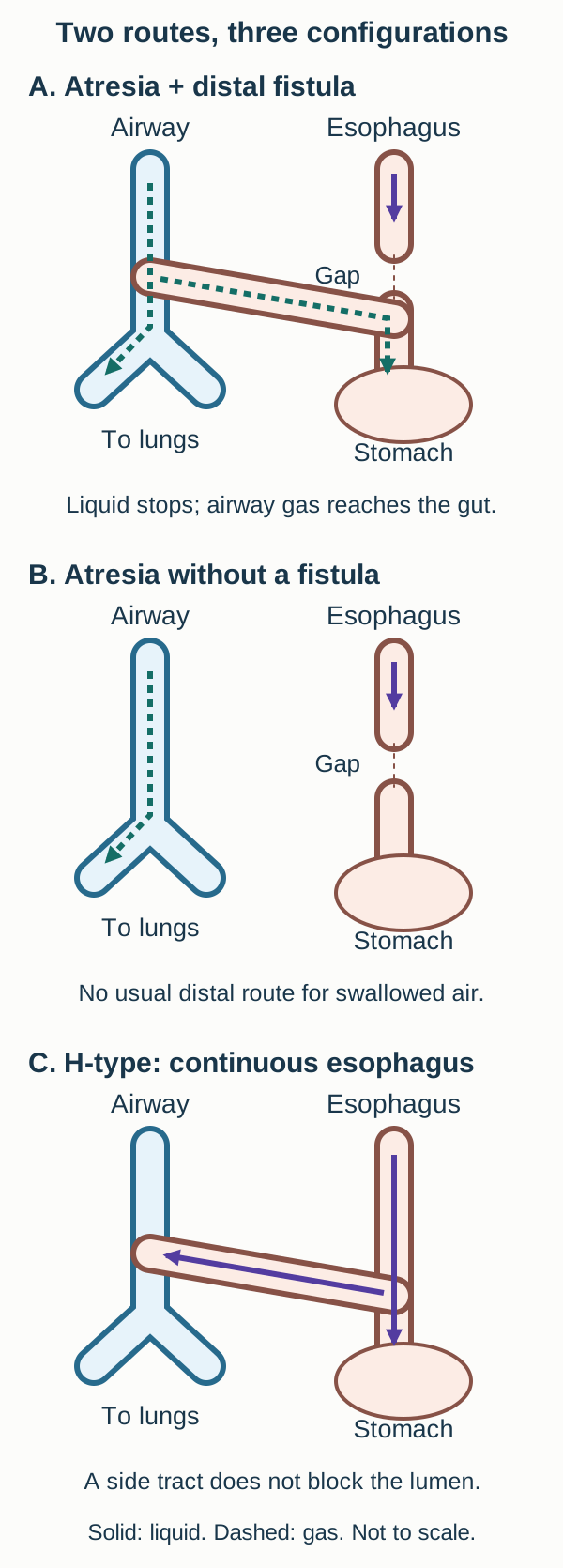
Can liquid reach the stomach, and can airway air reach it by a different path? These are separate questions. Atresia interrupts a passage. A tracheoesophageal fistula, or TEF, is an abnormal connection between the trachea and esophagus. Either can exist without the other. In the route diagram, first trace from the mouth downward, then start again in the trachea. Do not jump across a gap just because the two esophageal segments are drawn close together. [1][2]
Atresia with a distal fistula
The common configuration has a blind upper esophageal pouch and a lower esophageal segment that joins the trachea. Swallowed liquid stops above the gap. Air can enter the lower segment from the trachea and continue into the stomach. This explains how a baby can have an air-filled stomach despite being unable to swallow into it. The distal fistula does not reconnect the upper pouch to the lower esophagus. [1][2]
Atresia without a fistula
In isolated, or pure, atresia, the esophageal segments do not join each other or the airway. There is no usual route for swallowed air or airway air to enter the stomach. A gasless abdomen is therefore expected. The upper pouch still fills with saliva, and overflow into the pharynx can still cause aspiration. Absence of a fistula is not protection against all aspiration. [1][3]
A fistula with a continuous esophagus
In an H-type fistula, the esophageal lumen remains continuous. A correctly positioned feeding tube can reach the stomach, and much of a feed can follow the normal route. Some liquid can cross into the airway through the abnormal tract. The name H-type is a classification shorthand; the tract is often oblique rather than a perfectly horizontal bar. Tube passage tests continuity, not separation from the airway. [2][4][5]
Keep the uncommon configurations in view. A fistula can connect the trachea to the upper pouch alone, or there can be both upper and lower connections. An upper-pouch fistula can contaminate the airway without providing a route into the stomach. Thus, a gasless abdomen does not establish that every possible fistula is absent. Definitive assessment must describe which segment communicates with the airway. [1][2]
Trace-and-predict exercise: use the common configuration in the diagram. Imagine closing only the distal fistula while leaving the esophageal gap unchanged. Predict separately what happens to airway gas entering the stomach and to saliva entering the stomach.
Trace solid liquid arrows and dashed gas arrows separately. A distal fistula provides an alternate gas route, not a bridge across the esophageal gap. H-type anatomy preserves the lumen but adds a side connection. [1][2][1][2]
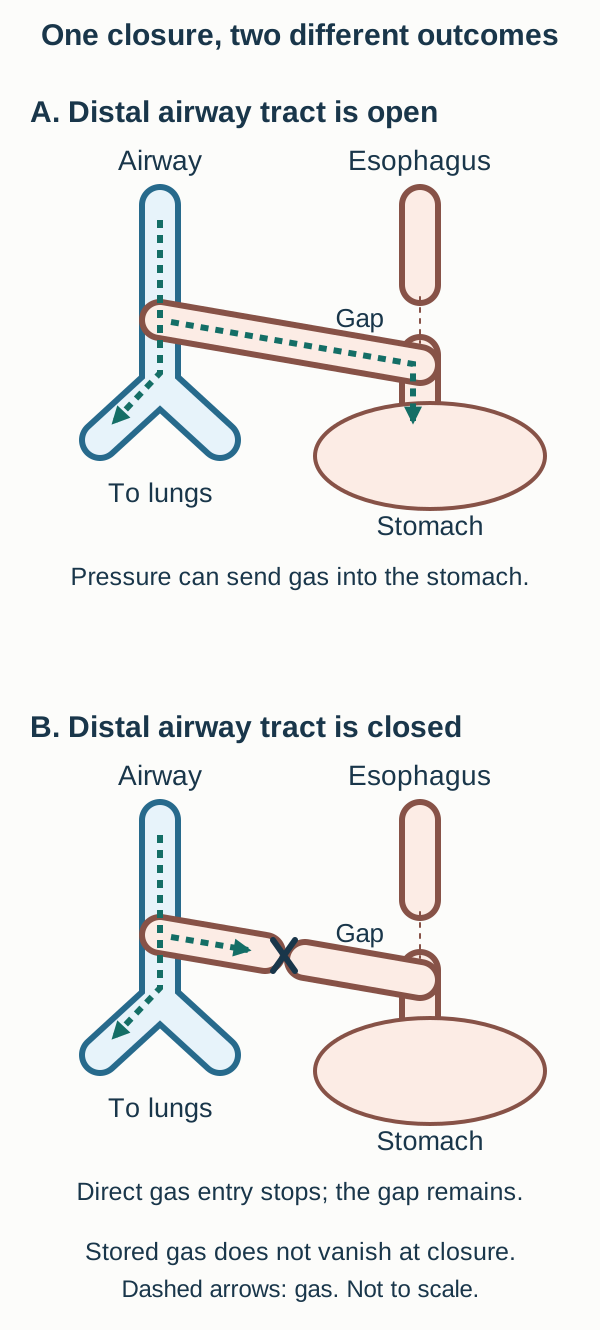
Worked comparison: closing the distal tract stops that direct gas route, but the esophageal gap still blocks swallowed saliva. Track these two outcomes independently rather than treating repair as a single switch.
Compare the two predictions after distal fistula closure
The direct trachea-to-stomach gas route is interrupted. Swallowed saliva still cannot cross the esophageal gap. Closing a fistula and restoring esophageal continuity solve different problems.
Transfer: a quieter abdomen after fistula closure does not by itself establish readiness for oral feeding. The swallowing route must also be restored and assessed by the treating team. [3][8]
Use tube position and abdominal gas as different tests
Start with a practical question: where is the tube, and how could air reach the stomach? Clinicians gently attempt passage of an appropriate radiopaque tube and obtain imaging when atresia is suspected. Resistance alone is not the diagnosis. A tube can bend in the mouth or pharynx, enter the airway, or be incorrectly positioned. Do not force it. A radiograph that actually localizes the tube in an upper esophageal pouch supports atresia; include the abdomen when assessing gas. [1][3]
Interpret the findings together, not as interchangeable tests
Observed route
Gas finding
Supported interpretation
Observed routeTube ends in an upper esophageal pouch
Gas findingGas in stomach or bowel
Supported interpretationAtresia with a functioning distal airway connection is strongly supported.
Observed routeTube ends in an upper esophageal pouch
Gas findingGasless abdomen
Supported interpretationNo distal gas route is demonstrated; pure atresia is typical, but a proximal-only fistula remains possible.
Observed routeTube follows the esophagus into the stomach
Gas findingGas present or absent
Supported interpretationComplete atresia is not supported by this route; a fistula is not excluded.
The gas inference is conditional. First establish atresia. In a continuous esophagus, swallowed air can normally reach the stomach, so gastric gas alone does not diagnose TEF. In established atresia, gastric or intestinal gas strongly suggests an alternative airway route to the distal esophagus. Interpret timing, image coverage, previous instrumentation, and the functional patency of a tract before making an absolute statement about a gasless film. A radiograph is not a complete fistula map. [1][2][3]
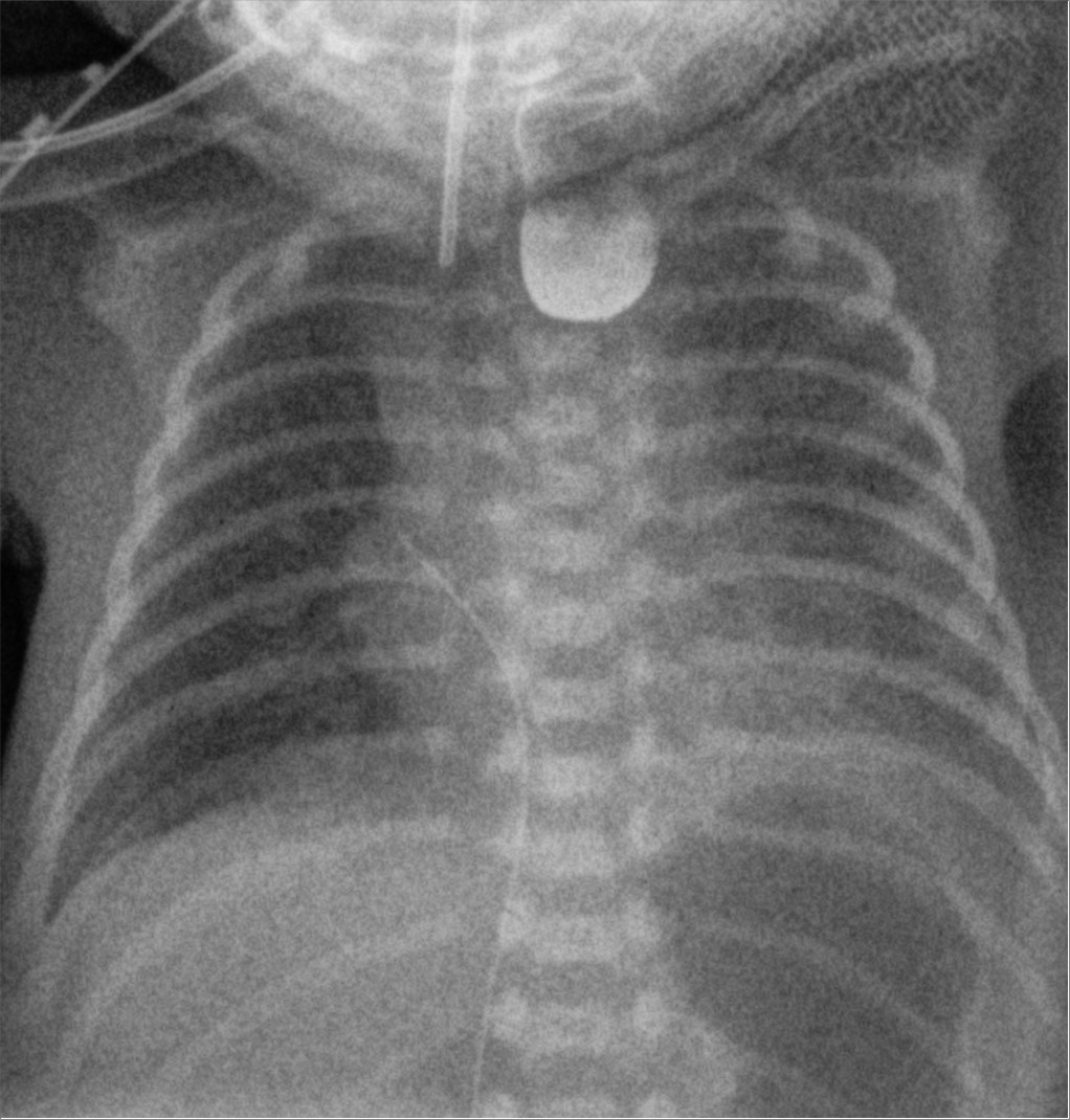
Read the clinical radiograph in two regions. Identify the rounded, contrast-filled upper pouch in the upper chest. Then find the dark gastric gas below the left hemidiaphragm. The combination illustrates interruption of the swallowed route with gas reaching the distal gut. The connection itself is not directly outlined in this still image, so do not claim its exact airway level from these findings alone. This published image used contrast; it is not an instruction to give routine oral contrast to a newborn whose tube film already establishes atresia. Such administration adds aspiration risk and is generally unnecessary for that initial diagnosis. [3][9]
A real radiograph pairs an opacified blind upper pouch with gastric gas. The lower gas supports an alternate distal route in the setting of atresia, but the tract is inferred rather than directly seen. This historical contrast image teaches recognition; routine oral contrast is not required after diagnostic tube imaging and can create aspiration risk. Image: Hellerhoff, 2021; CC BY-SA 4.0; [3][9] Hellerhoff; original source; CC BY-SA 4.0. [3][9].Compare two infants: why is stomach gas more informative in the one with a blind upper pouch?
That infant cannot deliver swallowed air through a continuous esophagus, so gas below the interruption needs another explanation, usually a distal fistula. In the infant whose tube correctly reaches the stomach, normal swallowing already provides a gas route.
Transfer: document the evidence as tube location plus gas distribution plus clinical context. Replace the shortcut “gas means fistula” with an explanation of which route is blocked and which alternative route is supported.
When the tube passes, ask where swallowed liquid goes
A feeding tube reaches the stomach, but the infant repeatedly coughs and becomes blue during feeds. Which question did the tube actually answer? It established a continuous route to the stomach. It did not test whether an abnormal side connection also reaches the airway. That is why H-type fistulas can be missed, including in infants who gain weight or have reassuring earlier imaging. [2][4][5]
Use the H-type flow diagram to follow a swallow. Most liquid may continue down the esophagus, while a smaller amount enters the connecting tract and reaches the trachea. Repeated contamination can produce respiratory illness even when enough nutrition reaches the stomach for growth. The amount crossing can vary, so one uneventful feed or one negative study is not a dependable exclusion. Air may also cross from the airway into the esophagus when the pressure relationship favors that direction. The tract is not a one-way valve. [4][5]
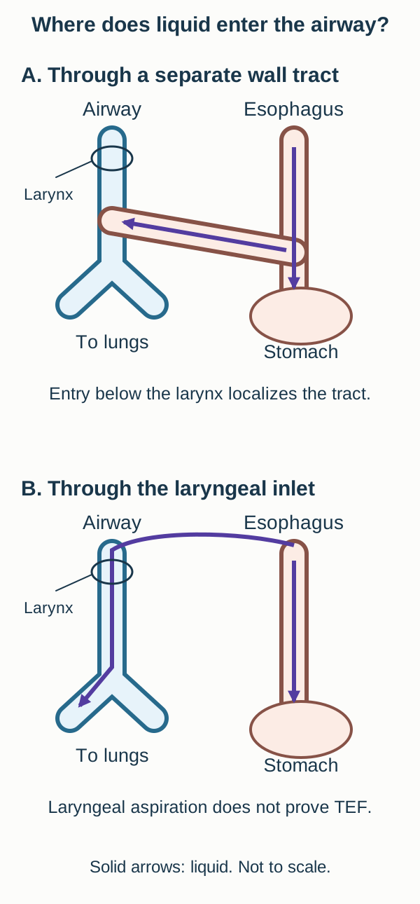
The top panel depicts direct fistulous entry below the larynx. The lower panel depicts laryngeal aspiration without a fistula. Cough during feeds alone cannot distinguish these routes; targeted studies must establish the actual entry site. These are conceptual paths, not patient fluoroscopy. [2][4][5][2][4][5]
Compare the location of entry. Liquid passing into the airway at the larynx during swallowing supports impaired airway protection during the swallow. A directly seen tract or jet entering through the tracheal wall below the larynx supports a fistula. Cough, cyanosis, or recurrent pneumonia alone cannot make that distinction. Swallowing dysfunction, a laryngeal cleft, reflux-associated aspiration, and an abnormal airway connection can produce overlapping symptoms and sometimes coexist. The specialist assessment must investigate the observed route rather than treating every feeding cough as H-type TEF. [4][5][6]
When suspicion remains, specialist investigations may include a carefully performed dynamic contrast esophagogram, sometimes using a prone tube-withdrawal technique, and airway endoscopy. Bronchoscopy can help identify and localize a tracheal opening for operative planning. The choice and sequence depend on the infant's stability and local expertise. Retrospective H-type series include patients whose fistula was identified only after repeat studies; therefore a negative initial examination does not necessarily end the investigation. Those series do not provide a universal negative predictive value for every child with a cough. [4][5]
Predict before revealing: an infant has a correctly positioned gastric tube, good weight gain, and recurrent feed-related respiratory episodes. Which observation would establish more than the symptom pattern: another stomach gas bubble, or a demonstrable communication through the tracheal wall?
Check the route that needs to be demonstrated
A tracheal-wall communication supplies direct anatomic evidence. A gastric gas bubble is expected with a patent esophagus and cannot distinguish normal swallowing from an H-type tract. Good growth does not exclude intermittent airway contamination.
Transfer: after an initially negative study, persistent compelling symptoms warrant reassessment of study technique and alternative causes, not repeated unmonitored feeding to provoke cyanosis. [4][5]
Protect breathing before completing the anatomic map
What can still enter the lungs after oral feeding stops? Saliva continues to collect in the upper pouch, and gastric contents can reach the airway through a distal fistula. Keeping an infant without oral intake is necessary, but it does not by itself drain the pouch or eliminate every aspiration route. Initial care requires a neonatal team and prompt pediatric surgical involvement. [3]
In suspected or established atresia, stop oral feeds and oral medicines. Trained staff drain the upper pouch, commonly using a double-lumen sump catheter with controlled low-pressure suction according to the unit's protocol. This catheter drains the proximal reservoir; it is not a feeding tube into the stomach. Check that drainage remains effective when secretions recur. A monitored head-up position can reduce overflow while awaiting repair. This hospital positioning is not a home infant sleep recommendation. Provide appropriate intravenous fluid and glucose support, temperature control, and cardiorespiratory monitoring while arranging definitive care. [3]
Now trace a breath in the pressure diagram. Before a distal fistula is closed, positive airway pressure can send some gas down the lower esophagus. The stomach can distend, restrict diaphragmatic excursion, and make ventilation more difficult. A greater delivered pressure does not guarantee that the added gas reaches the lungs. An infant with an air leak into the gut may therefore have a growing abdomen and inadequate lung ventilation at the same time. [3]
Predict two outcomes after isolated distal fistula closure: direct airway-to-gut gas entry stops, but saliva still cannot cross the gap. Existing stomach gas need not disappear immediately. Distension can restrict breathing; required ventilation must not be withheld while experts manage the airway. [3][8][3][8]
Avoid unnecessary or excessive positive-pressure support in a stable infant, and obtain experienced neonatal anesthesia and airway assistance when support is needed. Do not withhold effective ventilation from an apneic, severely hypoxemic, or bradycardic newborn. Oxygenation and ventilation take priority while experts limit flow through the fistula. When anatomy permits, endoscopic guidance may help position an endotracheal tube beyond the fistula but above the carina; other strategies are needed for different fistula locations. Blindly advancing a tube or simply stopping necessary respiratory support is not an acceptable substitute for airway assessment. [3]
Imaging and evaluation for associated anomalies should proceed in parallel with stabilization. A stable infant needs organized urgent specialist planning. Deteriorating ventilation, severe distension, or other instability can require more immediate intervention. Do not assign every patient a fixed operation time or delay resuscitation until a complete diagnostic checklist is finished. The surgical consensus literature itself acknowledges important limits and variation in the evidence. [3][8]
Predict the consequence: pouch suction works, but the abdomen enlarges during ventilation. Why?
The suction catheter addresses pooled saliva above the esophageal interruption. It does not close a distal connection from the pressurized airway into the stomach. New distension requires reassessment of ventilation and fistula anatomy, as well as other causes of deterioration.
Transfer: match each action to its target: no oral intake limits added swallowed liquid, pouch drainage limits pooled secretions, and an expert airway plan limits gas diversion while maintaining breathing. None alone completes definitive repair. [3]
Repair the anatomy, then reassess feeding and breathing
Does identifying one foregut defect finish the evaluation? No. Assess for associated vertebral, anorectal, cardiac, renal, and limb abnormalities. These are the systems represented in VACTERL association, alongside the tracheoesophageal component. Esophageal atresia and TEF are not counted as two separate VACTERL systems. The pattern usually involves at least three component features, but a label does not replace assessment for other congenital or genetic diagnoses. Many affected infants have no identified single cause. [1][2]
Examine the spine, limbs, and perineum, and arrange appropriate cardiac and renal assessment. A normal murmur examination does not establish normal cardiac or aortic arch anatomy. Echocardiography informs both assessment of congenital heart disease and the relationship of the arch to thoracic operative planning. Stabilization still comes first; looking for associated conditions must not delay treatment of a breathing emergency. [2][3]
Apply the two-route model to repair. Closing the fistula separates the airway from the esophagus. Joining suitable esophageal ends restores the swallowed route. These are distinct goals. Gap length, prematurity, associated disease, and current physiology can affect whether reconstruction is immediate or staged. A long gap is not solved by assuming that a gastrostomy or fistula closure also reconnects the upper pouch. Nutrition and secretion management must be planned for the actual postoperative anatomy. [3][8]
A repaired passage is not necessarily a normally functioning passage
Compare three follow-up observations. A focal narrowing at the anastomosis with material held above it supports a structural restriction. A patent repair with poorly coordinated contractions and slow clearance supports impaired esophageal transport. Retrograde passage of gastric contents raises a different issue, reflux. These processes can overlap, and symptoms alone do not identify one of them. Evaluation of dysphagia, feeding difficulty, reflux, and anastomotic narrowing is part of long-term care, not evidence that every operation failed. Changing acidity alone would not correct a demonstrated failure of peristaltic clearance. [1][6]
Respiratory symptoms also need localization. New feed-related cough after an interval of improvement can warrant evaluation for a recurrent fistula as well as swallowing and reflux problems. After cervical H-type repair, a weak cry, stridor, or respiratory difficulty should prompt assessment of vocal cord function; recurrent laryngeal nerve injury with cord paresis is a documented complication. Do not attribute a new postoperative airway problem to reflux without evaluating the airway. [7]
Compare two postoperative studies: a narrow anastomosis versus an open anastomosis with poor clearance
The first identifies a focal structural restriction. The second directs attention to transport rather than a fixed blockage. Both may impair feeding, but the same symptom does not justify the same treatment without defining the cause.
Transfer: when following a repaired child, ask whether the passage is open, whether it clears a swallow, and whether it remains separate from the airway. Persistent symptoms warrant coordinated surgical, gastroenterology, feeding, and airway assessment according to the presentation. [6][7]
Clinical practice
Use the findings in each case to decide which route is interrupted, which route is abnormal, and what consequence follows. Select an answer before opening the explanation.
Case 1
Show answer and explanations for case 1
A. Increased fetal urination from hyperglycemia (Why this does not fit)
Fetal osmotic diuresis can increase the urinary contribution. Maternal glucose is normal and the newborn has a demonstrated interruption of swallowing. Prefer the observed clearance defect to an unsupported excess-production hypothesis.
Reasoning steps for option A
How can hyperglycemia increase amniotic fluid?
Fetal osmotic diuresis can increase the urinary contribution.
Which findings favor a different process here?
Maternal glucose is normal and the newborn has a demonstrated interruption of swallowing.
B. Reduced clearance through fetal swallowing (Best answer)
Swallowed fluid cannot follow a continuous esophageal route to the stomach. Less swallowed fluid reaches the gastrointestinal tract for clearance, favoring polyhydramnios. Connect the prenatal fluid balance to the postnatal obstruction.
Reasoning steps for option B
What does the confirmed upper pouch imply?
Swallowed fluid cannot follow a continuous esophageal route to the stomach.
How does that affect amniotic fluid balance?
Less swallowed fluid reaches the gastrointestinal tract for clearance, favoring polyhydramnios.
Why do normal kidneys not exclude this explanation?
Urine production can remain intact while swallowing-related clearance is impaired.
C. Reduced fetal urination from renal agenesis (Why this does not fit)
It markedly reduces urine production and tends to reduce amniotic fluid. Normal kidneys and bladder filling oppose agenesis, and reduced urine does not explain excess fluid. Check both the direction of the fluid change and the organ findings.
Reasoning steps for option C
What would bilateral renal agenesis do to fetal urine?
It markedly reduces urine production and tends to reduce amniotic fluid.
Does that explain this scan and examination?
Normal kidneys and bladder filling oppose agenesis, and reduced urine does not explain excess fluid.
D. Retained lung fluid from complete airway obstruction (Why this does not fit)
It blocks the respiratory outlet rather than the esophageal swallowing passage. The tube documents an upper esophageal pouch, with no evidence supplied for complete airway obstruction. Localize the demonstrated interruption before assigning the fluid abnormality.
Reasoning steps for option D
What anatomy does complete fetal airway obstruction involve?
It blocks the respiratory outlet rather than the esophageal swallowing passage.
Which passage is actually interrupted here?
The tube documents an upper esophageal pouch, with no evidence supplied for complete airway obstruction.
Takeaway: Reduced swallowing-related clearance can explain polyhydramnios, but prenatal findings alone are not diagnostic. [1][2][3]
A. Normal transit through a continuous esophageal lumen (Why this does not fit)
An appropriately positioned tube could reach the stomach through the esophagus. The tube is confirmed within a blind upper esophageal pouch. Do not discard current anatomy because a prenatal screening feature was reassuring.
Reasoning steps for option A
What would a continuous lumen permit?
An appropriately positioned tube could reach the stomach through the esophagus.
What contradicts that explanation?
The tube is confirmed within a blind upper esophageal pouch.
B. Normal swallowed air retained before the postnatal tube study (Why this does not fit)
It can pass down a continuous esophageal lumen. A localized blind upper pouch establishes an interruption, so normal swallowed transit does not account for the gas. Prenatal stomach visibility is not evidence of postnatal esophageal continuity.
Reasoning steps for option B
When can swallowed air normally enter the stomach?
It can pass down a continuous esophageal lumen.
What prevents that route in this infant?
A localized blind upper pouch establishes an interruption, so normal swallowed transit does not account for the gas.
C. Stomach filling through a proximal-pouch airway connection (Why this does not fit)
It communicates with the upper pouch above the esophageal interruption. No; the isolated distal segment remains separated from the upper pouch. Distinguish upper-pouch contamination from a distal route into the gut.
Reasoning steps for option C
Where does a proximal-only fistula deliver airway gas?
It communicates with the upper pouch above the esophageal interruption.
Can that route alone reach this stomach?
No; the isolated distal segment remains separated from the upper pouch.
D. Postnatal air entry through a distal airway-esophageal tract (Best answer)
The normal esophageal swallowing route is interrupted. A distal connection carries tracheal air into the lower esophagus and stomach without joining the upper pouch to it. A visible fetal stomach does not exclude atresia; fetal stomach fluid and postnatal gas are different observations.
Reasoning steps for option D
What is established by the localized upper pouch?
The normal esophageal swallowing route is interrupted.
What alternate route explains postnatal gastric gas?
A distal connection carries tracheal air into the lower esophagus and stomach without joining the upper pouch to it.
What does prenatal stomach visibility establish by itself?
It does not establish normal esophageal continuity and should not override the diagnostic postnatal findings.
Takeaway: A stomach bubble can coexist with an interrupted esophagus when an alternate distal route is present. [1][2][3]
A. Upper-pouch overflow into the pharynx and larynx (Best answer)
Swallowed saliva continues to accumulate. It can return to the pharynx and enter the laryngeal inlet. No fistula does not mean no aspiration risk.
Reasoning steps for option A
What continues to enter the blind upper pouch without feeding?
Swallowed saliva continues to accumulate.
How can that fluid reach the lungs without a fistula?
It can return to the pharynx and enter the laryngeal inlet.
Which reservoir needs drainage?
The proximal esophageal pouch, not an inaccessible stomach.
B. Gastric reflux through an intact esophageal lumen (Why this does not fit)
It requires a continuous passage from stomach to upper esophagus. The two blind segments are separated, so this proposed esophageal route is interrupted. Trace the entire proposed fluid route rather than assuming normal continuity.
Reasoning steps for option B
What does reflux reaching the pharynx require through the esophagus?
It requires a continuous passage from stomach to upper esophagus.
Is that passage present?
The two blind segments are separated, so this proposed esophageal route is interrupted.
C. Distal-pouch contents through a lower airway tract (Why this does not fit)
A distal fistula would connect the lower esophageal segment to the airway. Airway assessment found no fistula, while saliva is visibly accumulating above the gap. Use the demonstrated proximal reservoir when the proposed distal connection is absent.
Reasoning steps for option C
What anatomy would permit this route?
A distal fistula would connect the lower esophageal segment to the airway.
What finding opposes that explanation?
Airway assessment found no fistula, while saliva is visibly accumulating above the gap.
D. Milk crossing an isolated tract in a patent esophagus (Why this does not fit)
An H-type fistula occurs with esophageal continuity. The esophagus has separated blind segments, and no milk has been given. Match both the fluid source and the anatomic passage.
Reasoning steps for option D
Which configuration permits milk transit and side leakage?
An H-type fistula occurs with esophageal continuity.
Which two details contradict that route?
The esophagus has separated blind segments, and no milk has been given.
Takeaway: Pooled saliva can be aspirated from above the obstruction even in pure atresia. [1][2][3]
A. Close a proximal tract and connect the esophageal segments (Why this does not fit)
A fistula between the upper pouch and trachea would require separation. Bronchoscopy identifies a distal tract and excludes a proximal one. Use the identified segment rather than the generic word fistula.
Reasoning steps for option A
When would proximal tract closure be appropriate?
A fistula between the upper pouch and trachea would require separation.
Which tract is actually present here?
Bronchoscopy identifies a distal tract and excludes a proximal one.
B. Close both airway tracts and leave esophageal continuity unchanged (Why this does not fit)
Both upper and lower esophageal segments would have airway connections. Only one distal tract is present, and leaving the gap would preserve swallowing obstruction. Count the demonstrated connections and separately assess continuity.
Reasoning steps for option B
What anatomy would require two fistula closures?
Both upper and lower esophageal segments would have airway connections.
Why is this not the complete plan here?
Only one distal tract is present, and leaving the gap would preserve swallowing obstruction.
C. Close the distal tract and connect the esophageal segments (Best answer)
The confirmed distal fistula connects the airway to the lower esophagus. The upper and lower esophageal segments must be joined. Fistula closure and esophageal continuity solve separate problems.
Reasoning steps for option C
Which connection supplies gas below the interruption?
The confirmed distal fistula connects the airway to the lower esophagus.
What additional step restores swallowed transit?
The upper and lower esophageal segments must be joined.
Why are both goals necessary?
Closing the tract alone leaves atresia; joining the esophagus alone leaves airway contamination.
D. Divide an isolated tract and preserve the continuous esophageal lumen (Why this does not fit)
An isolated H-type fistula has no complete esophageal atresia. The tube ends in a confirmed blind upper pouch. A side connection does not erase an accompanying interruption.
Reasoning steps for option D
Which subtype has a continuous esophagus to preserve?
An isolated H-type fistula has no complete esophageal atresia.
What rules out that starting anatomy?
The tube ends in a confirmed blind upper pouch.
E. Connect the esophageal segments without closing an airway tract (Why this does not fit)
In pure atresia there is no fistula to separate. The confirmed distal airway connection would remain open. Do not repair continuity while ignoring a demonstrated airway communication.
Reasoning steps for option E
When could joining segments alone address the defect?
In pure atresia there is no fistula to separate.
What would remain untreated here?
The confirmed distal airway connection would remain open.
Takeaway: Restoring transit and separating the airway are distinct operative goals. [1][2][3][8]
A. The lower pouch drains into the airway, but the upper pouch reaches the stomach (Why this does not fit)
It requires both a distal fistula and restored upper-to-lower continuity. The lower segment has no airway opening and the upper segment is blind. Do not reverse the documented locations of the two segments.
Reasoning steps for option A
What would this arrangement require?
It requires both a distal fistula and restored upper-to-lower continuity.
Do the observed findings supply either feature?
The lower segment has no airway opening and the upper segment is blind.
B. Both pouches connect to the airway, but neither can admit air from the trachea (Why this does not fit)
Separate airway connections to both esophageal segments. Inspection excludes a distal connection; a communication is not intrinsically restricted to liquid flow. Specify the segment and consider pressure-dependent flow.
Reasoning steps for option B
What identifies a double-fistula configuration?
Separate airway connections to both esophageal segments.
What contradicts that configuration here?
Inspection excludes a distal connection; a communication is not intrinsically restricted to liquid flow.
C. The lower pouch is isolated, but the upper pouch can contaminate the airway (Best answer)
The lower segment is isolated from both the upper pouch and trachea. The upper pouch communicates with the airway and can transmit pooled secretions. A gasless abdomen does not exclude a proximal-only fistula.
Reasoning steps for option C
Which esophageal segment has no route for airway gas?
The lower segment is isolated from both the upper pouch and trachea.
Why can aspiration still occur?
The upper pouch communicates with the airway and can transmit pooled secretions.
Which part of the usual gas inference needs qualification?
Gas assesses a functioning distal route, not every possible airway connection.
D. The esophagus is continuous, but liquid bypasses the stomach through a side tract (Why this does not fit)
An H-type fistula has a patent esophagus. The upper pouch ends blindly and is separate from the lower esophagus. An upper fistula can coexist with atresia; it does not restore continuity.
Reasoning steps for option D
Which configuration has this continuous swallowed route?
An H-type fistula has a patent esophagus.
What makes this infant different?
The upper pouch ends blindly and is separate from the lower esophagus.
Takeaway: A proximal-only fistula can transmit secretions while leaving the abdomen gasless. [1][2][3]
A. Preserved gastric feeding transit with intermittent direct airway contamination (Best answer)
The esophageal lumen is continuous to the stomach. It provides a route for esophageal liquid to enter the airway despite that continuity. A patent main passage can coexist with an abnormal side connection.
Reasoning steps for option A
What does the correctly positioned gastric tube establish?
The esophageal lumen is continuous to the stomach.
What does the separately visualized tract add?
It provides a route for esophageal liquid to enter the airway despite that continuity.
Why can nutrition and aspiration occur together?
Part of a swallow can reach the stomach while another part crosses the tract.
B. Complete loss of gastric feeding transit with isolated upper-pouch storage (Why this does not fit)
Complete esophageal atresia interrupts the route to the stomach. The tube follows the esophageal lumen into the stomach. Do not treat every tracheoesophageal defect as atresia.
Reasoning steps for option B
What anatomy creates complete upper-pouch storage?
Complete esophageal atresia interrupts the route to the stomach.
Which direct observation contradicts complete atresia?
The tube follows the esophageal lumen into the stomach.
C. Preserved gastric feeding transit with aspiration restricted to laryngeal entry (Why this does not fit)
Liquid would cross the laryngeal inlet during the swallow. The tract enters through the tracheal wall below the larynx. Localize the point of airway entry rather than relying on cough alone.
Reasoning steps for option C
What observation would identify laryngeal aspiration?
Liquid would cross the laryngeal inlet during the swallow.
Where is entry actually demonstrated?
The tract enters through the tracheal wall below the larynx.
D. Complete loss of gastric feeding transit with distal airway gas entry (Why this does not fit)
Atresia with a distal fistula blocks swallowing while admitting airway gas below the gap. The documented continuous esophageal tube route excludes complete atresia along that route. Use tube continuity and airway communication as separate observations.
Reasoning steps for option D
Which common subtype combines these features?
Atresia with a distal fistula blocks swallowing while admitting airway gas below the gap.
Which finding opposes that interruption here?
The documented continuous esophageal tube route excludes complete atresia along that route.
Takeaway: Direct tract visualization establishes abnormal communication even when the esophagus remains patent. [2][4][5]
A. More swallowed saliva passes through the upper pouch into the stomach (Why this does not fit)
Continuity would have to be restored across the esophageal gap. No; it affects gas flow through a fistula, not the disconnected upper segment. Separate pressure-driven gas flow from reconstruction of swallowing.
Reasoning steps for option A
What would permit saliva to pass from the upper pouch?
Continuity would have to be restored across the esophageal gap.
Does raising tracheal pressure provide that continuity?
No; it affects gas flow through a fistula, not the disconnected upper segment.
B. Less gas reaches the stomach because the esophageal interruption seals the airway (Why this does not fit)
The normal upper-to-lower esophageal route is interrupted. The distal airway connection already explains the gas below the gap. An esophageal interruption does not seal a separate tracheal opening.
Reasoning steps for option B
Which route is blocked by atresia?
The normal upper-to-lower esophageal route is interrupted.
Which existing route bypasses that interruption?
The distal airway connection already explains the gas below the gap.
C. More air leaves through the upper pouch while the distal gut stays isolated (Why this does not fit)
Atresia with a proximal-only fistula has no direct lower gas route. Stomach and bowel gas appeared before gastrointestinal instrumentation. Let the distribution below the obstruction inform distal connectivity.
Reasoning steps for option C
What subtype would leave the distal gut isolated despite an upper connection?
Atresia with a proximal-only fistula has no direct lower gas route.
What argues against isolated distal gut in this infant?
Stomach and bowel gas appeared before gastrointestinal instrumentation.
D. More tracheal gas enters the distal esophagus, producing progressive stomach distension (Best answer)
A functioning distal airway connection is strongly supported. It can direct additional gas into the lower esophagus and stomach. More airway pressure does not guarantee more useful lung ventilation.
Reasoning steps for option D
How did gas reach the gut despite the blind upper pouch?
A functioning distal airway connection is strongly supported.
What can a higher tracheal pressure do through that route?
It can direct additional gas into the lower esophagus and stomach.
How can the expanding stomach affect breathing?
Distension can restrict diaphragmatic excursion and further complicate ventilation.
Takeaway: In established atresia, distal gas predicts a route through which positive airway pressure can inflate the gut. [1][2][3]
A. Obtain a routine oral contrast swallow immediately (Why this does not fit)
Specialist contrast studies can investigate selected unresolved anatomy, particularly a suspected H-type tract. The initial problem is pharyngeal tube coiling; repeat safe localization can clarify continuity without unnecessary oral contrast exposure. Correct the inadequate observation before escalating to an aspiration-prone test.
Reasoning steps for option A
When can a targeted contrast examination be useful?
Specialist contrast studies can investigate selected unresolved anatomy, particularly a suspected H-type tract.
Why is it not the first correction to this incomplete test?
The initial problem is pharyngeal tube coiling; repeat safe localization can clarify continuity without unnecessary oral contrast exposure.
B. Repeat gentle tube placement and confirm radiographic localization (Best answer)
No; it does not show where a correctly positioned esophageal tube would end. A careful repeat placement and radiographic localization can establish the actual route without forcing the tube. Treat an inadequately localized tube study as incomplete.
Reasoning steps for option B
Does a loop in the pharynx establish an esophageal blind pouch?
No; it does not show where a correctly positioned esophageal tube would end.
What observation is needed next?
A careful repeat placement and radiographic localization can establish the actual route without forcing the tube.
Why does gastric gas not settle the diagnosis?
It can come from normal swallowing or from a distal fistula when atresia exists.
C. Schedule esophageal reconstruction from the current film (Why this does not fit)
The interruption and relevant airway anatomy need to be established. No tube has been localized within an upper esophageal pouch. Pharyngeal coiling is not the same as confirmed atresia.
Reasoning steps for option C
What evidence should precede reconstruction planning?
The interruption and relevant airway anatomy need to be established.
What essential finding is missing?
No tube has been localized within an upper esophageal pouch.
D. Observe another oral feed before repeating the tube test (Why this does not fit)
It might reproduce symptoms without defining the anatomic route. The infant already has concerning secretions and an unresolved obstruction assessment, so feeding adds aspiration risk. Do not use provoked choking to compensate for incomplete localization.
Reasoning steps for option D
What would another feed add to this assessment?
It might reproduce symptoms without defining the anatomic route.
Why is that a poor next diagnostic step?
The infant already has concerning secretions and an unresolved obstruction assessment, so feeding adds aspiration risk.
Takeaway: Tube resistance is not diagnostic until position is adequately established; never force a suspected obstruction. [1][3]
A. Arrange specialist assessment using the existing diagnostic tube film (Best answer)
It provides the expected evidence of esophageal atresia. It is generally unnecessary for this initial diagnosis and adds aspiration exposure. Proceed with specialist assessment rather than repeating an already answered question.
Reasoning steps for option A
What has the localized tube film already established?
It provides the expected evidence of esophageal atresia.
What does another oral contrast challenge add to the immediate plan?
It is generally unnecessary for this initial diagnosis and adds aspiration exposure.
What still needs specialist definition?
The exact airway anatomy, associated anomalies, and suitable repair strategy.
B. Perform the oral contrast study before requesting surgical assessment (Why this does not fit)
Selected unresolved anatomy can require a specialist contrast study. No; the upper pouch is already documented, so routine oral contrast is not required to establish the initial diagnosis. Do not delay consultation for redundant confirmation.
Reasoning steps for option B
When might contrast imaging contribute?
Selected unresolved anatomy can require a specialist contrast study.
Is failure of the esophageal tube route unresolved here?
No; the upper pouch is already documented, so routine oral contrast is not required to establish the initial diagnosis.
C. Repeat feeding under observation before accepting the tube result (Why this does not fit)
It could reproduce regurgitation or respiratory symptoms. It adds aspiration risk without improving the already localized structural finding. Symptoms need not be deliberately reproduced after an anatomic obstruction is shown.
Reasoning steps for option C
What could observed feeding demonstrate?
It could reproduce regurgitation or respiratory symptoms.
Why would that be less useful than the current evidence?
It adds aspiration risk without improving the already localized structural finding.
D. Repeat prenatal ultrasound interpretation before arranging further care (Why this does not fit)
It provided screening information before birth. A direct postnatal tube-and-radiograph assessment establishes the current obstruction. A prenatal screen should not delay care for confirmed postnatal findings.
Reasoning steps for option D
What role did prenatal imaging have?
It provided screening information before birth.
Which evidence now takes priority?
A direct postnatal tube-and-radiograph assessment establishes the current obstruction.
Takeaway: Typical localized tube findings support prompt specialist care without routine oral contrast confirmation. [1][3][8]
A. Escalate acid suppression and defer further structural investigation (Why this does not fit)
Reflux can contribute to respiratory symptoms in some children. The longstanding feed-associated pattern and limited anatomic study leave a structural communication unresolved. Do not let an empirical treatment replace an indicated structural reassessment.
Reasoning steps for option A
Why might reflux treatment appear relevant?
Reflux can contribute to respiratory symptoms in some children.
What remains unexplained by that plan?
The longstanding feed-associated pattern and limited anatomic study leave a structural communication unresolved.
B. Repeat targeted esophagography and plan airway endoscopic assessment (Best answer)
It does not exclude an H-type connection beside a continuous esophageal lumen. Specialist reassessment can use targeted dynamic imaging and airway endoscopy to investigate a small or intermittently visualized tract. A negative initial examination is interpreted in light of technique and remaining suspicion.
Reasoning steps for option B
What does successful tube passage fail to exclude?
It does not exclude an H-type connection beside a continuous esophageal lumen.
How should persistent symptoms after a limited study be handled?
Specialist reassessment can use targeted dynamic imaging and airway endoscopy to investigate a small or intermittently visualized tract.
Does this plan presume a fistula is already proven?
No; it investigates the concern while retaining alternative causes of aspiration.
C. Repeat routine gastric tube passage and resume unrestricted feeding (Why this does not fit)
It would again demonstrate a continuous main esophageal passage. No; repeated tube passage does not examine an H-type tract. Repeating the wrong test does not improve exclusion of the suspected abnormality.
Reasoning steps for option C
What would another successfully passed tube establish?
It would again demonstrate a continuous main esophageal passage.
Would it evaluate the unresolved side connection?
No; repeated tube passage does not examine an H-type tract.
D. Use a repeat chest radiograph to localize the suspected tract (Why this does not fit)
It can show pulmonary consequences such as opacities. No; targeted contrast imaging and endoscopy answer a different anatomic question. Distinguish imaging of respiratory injury from demonstration of its possible route.
Reasoning steps for option D
What can chest radiography show in recurrent aspiration?
It can show pulmonary consequences such as opacities.
Can a routine chest film reliably map a small H-type tract?
No; targeted contrast imaging and endoscopy answer a different anatomic question.
Takeaway: Persistent compatible symptoms after a limited negative study warrant targeted reassessment, not exclusion based on tube passage. [2][4][5][6]
A. Join the blind upper pouch to the lower esophageal segment (Why this does not fit)
Complete esophageal atresia requires restoration of continuity when feasible. Contrast has already traversed an uninterrupted esophagus into the stomach. Choose reconstruction for demonstrated interruption, not for a fistula label alone.
Reasoning steps for option A
What defect is treated by joining separated esophageal ends?
Complete esophageal atresia requires restoration of continuity when feasible.
Why is that not the identified defect?
Contrast has already traversed an uninterrupted esophagus into the stomach.
B. Drain the blind upper pouch while awaiting growth of the gap (Why this does not fit)
It protects an infant whose upper esophageal segment remains disconnected. The swallowed route is continuous and the identified lesion is a side tract. Do not apply an atresia-specific reservoir strategy to a patent esophagus.
Reasoning steps for option B
When is prolonged upper-pouch drainage relevant?
It protects an infant whose upper esophageal segment remains disconnected.
How does the observed esophageal transit contradict a blind upper pouch?
The swallowed route is continuous and the identified lesion is a side tract.
C. Dilate a narrowed anastomosis to restore passage into the stomach (Why this does not fit)
A postoperative focal narrowing with obstructed transit would support it. No; the study shows intact transit and direct leakage through a separate airway tract. Use the demonstrated lesion rather than a different cause of feeding difficulty.
Reasoning steps for option C
What finding would support anastomotic dilation assessment?
A postoperative focal narrowing with obstructed transit would support it.
Is such a restriction supplied here?
No; the study shows intact transit and direct leakage through a separate airway tract.
D. Divide the fistula and preserve the continuous esophageal lumen (Best answer)
The esophageal lumen is continuous. The localized side tract permits direct esophageal-to-airway contamination. Treat the abnormal connection without inventing an esophageal interruption.
Reasoning steps for option D
What does contrast reaching the stomach demonstrate?
The esophageal lumen is continuous.
What abnormal structure still needs correction?
The localized side tract permits direct esophageal-to-airway contamination.
Why is joining two blind segments not the target?
No separated blind segments are demonstrated in this H-type configuration.
Takeaway: H-type repair targets the abnormal communication; there is no complete esophageal gap to reconnect. [2][4][5]
A. Entry through a distal fistula below an esophageal interruption (Why this does not fit)
Atresia with a distal airway communication would link the lower esophagus and trachea. The main esophageal route is patent and contrast enters at the larynx before esophageal descent. Use timing and entry location together.
Reasoning steps for option A
What would that route require?
Atresia with a distal airway communication would link the lower esophagus and trachea.
Which observations oppose that explanation of this event?
The main esophageal route is patent and contrast enters at the larynx before esophageal descent.
B. Entry through an isolated tract in the posterior tracheal wall (Why this does not fit)
Contrast would traverse a connection into the trachea below the laryngeal inlet. The entry is through the laryngeal inlet, not through a demonstrated tracheal-wall tract. Feeding-associated cough alone does not identify a fistula.
Reasoning steps for option B
How would an H-type tract be directly demonstrated?
Contrast would traverse a connection into the trachea below the laryngeal inlet.
Where does this study actually show entry?
The entry is through the laryngeal inlet, not through a demonstrated tracheal-wall tract.
C. Entry through the laryngeal inlet during the initial pharyngeal swallow (Best answer)
It crosses the laryngeal inlet during the pharyngeal phase. No; the observed event is laryngeal aspiration, which does not itself prove an H-type fistula. Distinguish the demonstrated route from an unconfirmed alternative.
Reasoning steps for option C
At what point does contrast enter the airway?
It crosses the laryngeal inlet during the pharyngeal phase.
Does the recording demonstrate passage through a tracheal-wall tract?
No; the observed event is laryngeal aspiration, which does not itself prove an H-type fistula.
Does one recording exclude every coexisting problem?
No; persistent concerns still require appropriate assessment rather than an absolute exclusion.
D. Entry after retrograde flow from the stomach to the pharynx (Why this does not fit)
Gastric material would travel upward before entering the larynx. Aspiration occurs during the pharyngeal swallow before the bolus descends, not after documented gastric reflux. The temporal sequence can separate different sources of aspirated fluid.
Reasoning steps for option D
What sequence would demonstrate reflux-associated aspiration?
Gastric material would travel upward before entering the larynx.
Does that sequence match this recording?
Aspiration occurs during the pharyngeal swallow before the bolus descends, not after documented gastric reflux.
Takeaway: The site and timing of contrast entry distinguish demonstrated laryngeal aspiration from an unproven fistula. [4][5][6]
A. The vertebral level where the upper pouch ends (Why this does not fit)
It helps characterize esophageal anatomy relevant to repair. No; the fistula opening and carina must be directly related to each other. Do not substitute esophageal landmarks for airway landmarks.
Reasoning steps for option A
What does the upper-pouch level help describe?
It helps characterize esophageal anatomy relevant to repair.
Does it specify a safe tracheal tube position?
No; the fistula opening and carina must be directly related to each other.
B. The total amount of gas visible within the stomach (Why this does not fit)
Increasing distension can indicate clinically important diversion into the gut. No; it provides a functional consequence, not the opening-to-carina distance. Quantifying a consequence does not supply missing geometry.
Reasoning steps for option B
What can gastric gas suggest about ventilation?
Increasing distension can indicate clinically important diversion into the gut.
Does the amount of gas locate the tracheal opening?
No; it provides a functional consequence, not the opening-to-carina distance.
C. The severity of polyhydramnios recorded during pregnancy (Why this does not fit)
It supported concern about impaired fetal swallowing. It cannot locate the fistula relative to the carina. Use current airway anatomy for an airway positioning decision.
Reasoning steps for option C
What did polyhydramnios contribute before birth?
It supported concern about impaired fetal swallowing.
Can it guide present endotracheal tube placement?
It cannot locate the fistula relative to the carina.
D. The number of bowel loops containing air on imaging (Why this does not fit)
Gas entering the stomach can pass into the intestine. No; downstream gas distribution cannot map the tracheal opening. Separate downstream passage from the location of the original connection.
Reasoning steps for option D
Why can several bowel loops contain air?
Gas entering the stomach can pass into the intestine.
Does that distribution define the fistula level?
No; downstream gas distribution cannot map the tracheal opening.
E. The fistula opening position relative to the carina (Best answer)
It supports a distal gas route but does not provide its exact tracheal level. There must be a suitable position below the fistula opening and above the carina. A functional gas pattern is not a substitute for airway localization.
Reasoning steps for option E
What does the abdominal gas pattern establish about function?
It supports a distal gas route but does not provide its exact tracheal level.
What geometry matters for the proposed tube position?
There must be a suitable position below the fistula opening and above the carina.
Why avoid blind advancement?
Advancing too far can enter a bronchus or a fistula rather than safely ventilating both lungs.
Takeaway: Endoscopic airway localization answers a different question from the initial tube-and-gas radiograph. [3]
A. Place an oral gastric tube to drain the stomach directly (Why this does not fit)
It would need a continuous esophageal route into the stomach. Atresia interrupts that route, and the observed accumulating fluid is proximal saliva. Do not force a tube across atresia or confuse pouch drainage with gastric drainage.
Reasoning steps for option A
What would a gastric tube need to traverse?
It would need a continuous esophageal route into the stomach.
Why does that not address the supplied problem?
Atresia interrupts that route, and the observed accumulating fluid is proximal saliva.
B. Check proximal drainage patency and position with the neonatal team (Best answer)
The upper-pouch reservoir is no longer being drained effectively. Saliva continues to collect and can overflow toward the airway. Match the intervention to the proximal fluid reservoir.
Reasoning steps for option B
What does the combination of less drainage and more saliva suggest?
The upper-pouch reservoir is no longer being drained effectively.
What source remains active despite fasting?
Saliva continues to collect and can overflow toward the airway.
Why is a soft abdomen relevant?
It makes new gastric distension less explanatory for this particular pattern, without excluding other problems.
C. Start acid suppression before assessing the existing suction catheter (Why this does not fit)
It reduces gastric acidity rather than draining pooled saliva. Falling catheter output with increasing oral secretions points toward ineffective proximal drainage. Address a demonstrated drainage problem before attributing it to acidity.
Reasoning steps for option C
What is acid suppression intended to change?
It reduces gastric acidity rather than draining pooled saliva.
What does the new pattern specifically point toward?
D. Wait for oral fasting to clear the esophageal secretions (Why this does not fit)
No; saliva continues to be produced and swallowed. It accumulates above the interruption and can contaminate the airway. No oral intake is necessary but is not sufficient pouch management.
Reasoning steps for option D
Does fasting stop salivary production?
No; saliva continues to be produced and swallowed.
What happens when it cannot be drained or swallowed into the stomach?
It accumulates above the interruption and can contaminate the airway.
Takeaway: Recurrent saliva pooling despite fasting calls for reassessment of proximal pouch drainage. [3]
A. Mainstem intubation with loss of ventilation to one lung (Why this does not fit)
It directs the tube into one main bronchus and excludes the other lung from normal ventilation. Bronchoscopy places the tube above the carina with both main bronchi open. Check tube location before assigning a unilateral ventilation problem.
Reasoning steps for option A
How can mainstem intubation reduce ventilated lung volume?
It directs the tube into one main bronchus and excludes the other lung from normal ventilation.
Which direct observation opposes that explanation?
Bronchoscopy places the tube above the carina with both main bronchi open.
B. Tension pneumothorax with compression of the ventilated lung (Why this does not fit)
It can impair lung expansion and produce acute respiratory or circulatory deterioration. Bilateral sliding and the direct fistula finding accompany rapidly increasing gastric distension. A dangerous differential remains important, but use the supplied pleural and airway evidence.
Reasoning steps for option B
What can tension pneumothorax do during ventilation?
It can impair lung expansion and produce acute respiratory or circulatory deterioration.
What favors gas diversion in this case?
Bilateral sliding and the direct fistula finding accompany rapidly increasing gastric distension.
C. Gas diversion into the stomach with restricted diaphragmatic excursion (Best answer)
The distal fistula provides a route from the trachea into the lower esophagus and stomach. Gas enters the gut, and gastric distension can restrict the diaphragm while some delivered gas bypasses the lungs. Interpret the abdominal and respiratory changes together.
Reasoning steps for option C
Which additional outlet is exposed to the increased airway pressure?
The distal fistula provides a route from the trachea into the lower esophagus and stomach.
How can this produce a larger abdomen and less useful lung ventilation?
Gas enters the gut, and gastric distension can restrict the diaphragm while some delivered gas bypasses the lungs.
What practical implication follows?
Experienced airway and surgical teams must limit fistula flow while maintaining needed ventilation.
D. Endotracheal tube obstruction with reduced inspiratory gas delivery (Why this does not fit)
Increased resistance can restrict inspiratory flow to the airway. Bronchoscopy shows a patent tube and a pressure-exposed fistula with new gastric inflation. A patent tube does not ensure that its gas reaches the lungs.
Reasoning steps for option D
How could a blocked tube reduce delivered ventilation?
Increased resistance can restrict inspiratory flow to the airway.
What makes it less explanatory here?
Bronchoscopy shows a patent tube and a pressure-exposed fistula with new gastric inflation.
Takeaway: A fistula can divert delivered gas into the gut, and distension can further impair ventilation. [3]
A. Provide effective assisted ventilation and obtain expert airway control (Best answer)
Absent effective ventilation threatens oxygen delivery and contributes to bradycardia. No; necessary ventilation takes priority while experienced clinicians limit fistula flow and secure the airway. An aspiration or insufflation precaution is not a prohibition on lifesaving respiratory support.
Reasoning steps for option A
What is the immediate threat in an apneic hypoxemic newborn?
Absent effective ventilation threatens oxygen delivery and contributes to bradycardia.
Does possible gastric insufflation justify withholding ventilation?
No; necessary ventilation takes priority while experienced clinicians limit fistula flow and secure the airway.
What should be avoided during support?
Unnecessary excessive pressure and blind tube advancement, while maintaining adequate oxygenation and ventilation.
B. Withhold positive pressure until the fistula is completely mapped (Why this does not fit)
It helps select a safer airway strategy and operative plan. No; delaying effective ventilation would prolong severe hypoxemia and bradycardia. Diagnostic completeness must not delay resuscitation.
Reasoning steps for option B
Why is fistula mapping useful?
It helps select a safer airway strategy and operative plan.
Can mapping precede treatment of ongoing apnea here?
No; delaying effective ventilation would prolong severe hypoxemia and bradycardia.
C. Use oxygen alone while awaiting spontaneous recovery of breathing (Why this does not fit)
Gas must reach ventilated alveoli; oxygen concentration alone cannot replace absent effective breaths. The infant is apneic with severe hypoxemia and bradycardia. Distinguish oxygen administration from ventilation.
Reasoning steps for option C
What does oxygen delivery require to support alveolar gas exchange?
Gas must reach ventilated alveoli; oxygen concentration alone cannot replace absent effective breaths.
Which finding prevents oxygen alone from being sufficient?
The infant is apneic with severe hypoxemia and bradycardia.
D. Use pouch suction instead of assisting the absent respirations (Why this does not fit)
It reduces pooled secretions that can contaminate the airway. No; it addresses a reservoir, not the missing breaths. Necessary secretion care does not substitute for respiratory resuscitation.
Reasoning steps for option D
What does upper-pouch suction accomplish?
It reduces pooled secretions that can contaminate the airway.
Does it provide alveolar ventilation during apnea?
No; it addresses a reservoir, not the missing breaths.
Takeaway: Avoid excessive pressure, but never withhold needed ventilation from an apneic or critically hypoxemic newborn. [3]
A. Complete cardiac and renal screening before starting secretion drainage (Why this does not fit)
It informs perioperative risk and identifies other congenital abnormalities. The upper-pouch reservoir continues to pose aspiration risk while screening is performed. Screening and stabilization should be coordinated rather than made competing priorities.
Reasoning steps for option A
Why is associated-anomaly screening important?
It informs perioperative risk and identifies other congenital abnormalities.
Why is it not a reason to postpone secretion control?
The upper-pouch reservoir continues to pose aspiration risk while screening is performed.
B. Proceed directly to anesthesia without addressing the pooled secretions (Why this does not fit)
It separates abnormal routes and restores continuity when feasible. Existing pooled fluid can contaminate the airway before reconstruction is complete. Definitive planning does not replace initial airway protection.
Reasoning steps for option B
What does definitive repair ultimately address?
It separates abnormal routes and restores continuity when feasible.
Why still manage secretions before induction?
Existing pooled fluid can contaminate the airway before reconstruction is complete.
C. Use small oral feeds while arranging transfer to the surgical center (Why this does not fit)
No; they still collect above the gap. Gas below the gap does not show a safe swallowed feeding route. Do not interpret an air-filled stomach as permission to feed.
Reasoning steps for option C
Would smaller feeds cross a complete esophageal interruption?
No; they still collect above the gap.
What does gastric gas fail to establish?
Gas below the gap does not show a safe swallowed feeding route.
D. Withhold oral intake, drain the pouch, and arrange specialist care (Best answer)
Saliva can continue to pool above the esophageal interruption. Avoiding oral intake and providing trained proximal drainage reduce preventable contamination while specialist care is arranged. Room-air stability does not make oral feeding safe.
Reasoning steps for option D
Which liquid reservoir remains hazardous in a stable infant?
Saliva can continue to pool above the esophageal interruption.
What immediate care targets that reservoir and its added load?
Avoiding oral intake and providing trained proximal drainage reduce preventable contamination while specialist care is arranged.
What supports hydration during the wait?
Appropriate intravenous fluid and glucose support under neonatal supervision.
Takeaway: Stable breathing does not remove the need for no oral intake and proximal secretion management in atresia. [1][3]
A. A small restrictive defect in the ventricular septum (Why this does not fit)
Its hemodynamic effects contribute to perioperative cardiac assessment. No; it is an intracardiac shunt rather than a description of arch sidedness. Distinguish operative spatial relationships from shunt physiology.
Reasoning steps for option A
Why is a ventricular septal defect relevant?
Its hemodynamic effects contribute to perioperative cardiac assessment.
Does a small septal defect itself locate the arch beside the esophagus?
No; it is an intracardiac shunt rather than a description of arch sidedness.
B. An aortic arch passing rightward around the trachea (Best answer)
It specifies which side of the trachea the arch occupies in the thorax. The arch can alter access to the esophagus and fistula, so its position informs individualized surgical planning. An absent murmur does not define the aortic arch position.
Reasoning steps for option B
Which relationship does arch sidedness describe?
It specifies which side of the trachea the arch occupies in the thorax.
Why is that relevant to esophageal repair?
The arch can alter access to the esophagus and fistula, so its position informs individualized surgical planning.
C. A patent foramen ovale with left-to-right flow (Why this does not fit)
It describes a communication between the atria and its current flow direction. No; the interatrial finding does not establish the arch relationship to the thoracic operative field. Choose the finding that answers the stated anatomic question.
Reasoning steps for option C
What does flow across the foramen ovale describe?
It describes a communication between the atria and its current flow direction.
Does that define the side of the aortic arch?
No; the interatrial finding does not establish the arch relationship to the thoracic operative field.
D. Mild tricuspid regurgitation without right ventricular dilation (Why this does not fit)
It concerns right-sided valve function and may contribute to hemodynamic assessment. It does not specify which side of the trachea contains the aortic arch. Cardiac function and operative vascular geometry are different questions.
Reasoning steps for option D
What does tricuspid regurgitation help assess?
It concerns right-sided valve function and may contribute to hemodynamic assessment.
What spatial information is still missing from that finding?
It does not specify which side of the trachea contains the aortic arch.
Takeaway: Arch sidedness informs operative anatomy even when auscultation is reassuring. [3]
A. Four component systems are involved; a single-gene cause is established (Why this does not fit)
Atresia and fistula can be mistakenly counted as separate VACTERL systems. It involves three component systems and does not establish a single-gene etiology. Distinguish a phenotypic association from a molecular diagnosis.
Reasoning steps for option A
Why might four components be counted incorrectly?
Atresia and fistula can be mistakenly counted as separate VACTERL systems.
What does the actual pattern establish?
It involves three component systems and does not establish a single-gene etiology.
B. Three component systems are involved; urine output excludes renal anomalies (Why this does not fit)
It shows that urine is being produced at the time observed. No; useful urine production can coexist with structural abnormalities. Function observed at the bedside does not replace anatomic screening.
Reasoning steps for option B
What does normal urine output show?
It shows that urine is being produced at the time observed.
Does that exclude congenital renal structural abnormalities?
No; useful urine production can coexist with structural abnormalities.
C. Three component systems are involved; assess the other associated organs (Best answer)
They belong to one tracheoesophageal component, alongside the vertebral and limb components. No; normal urine output and absent murmur do not establish normal renal or cardiac anatomy. A multisystem pattern calls for systematic assessment rather than double-counting related lesions.
Reasoning steps for option C
How should atresia and its fistula be counted in this pattern?
They belong to one tracheoesophageal component, alongside the vertebral and limb components.
Do reassuring bedside observations finish cardiac and renal assessment?
No; normal urine output and absent murmur do not establish normal renal or cardiac anatomy.
Does the pattern prove one genetic cause?
No; etiologic and genetics assessment remains individualized, including consideration of other diagnoses.
D. Two component systems are involved; an absent murmur excludes heart defects (Why this does not fit)
The tracheoesophageal, vertebral, and limb systems are affected. An absent murmur does not establish normal cardiac or arch anatomy. Count the actual components and investigate clinically important unseen anatomy.
Reasoning steps for option D
Which three systems have demonstrated findings?
The tracheoesophageal, vertebral, and limb systems are affected.
What is wrong with relying on auscultation to finish the assessment?
An absent murmur does not establish normal cardiac or arch anatomy.
Takeaway: VACTERL is a multisystem pattern, not a single-gene diagnosis or a reason to omit screening after a reassuring bedside examination. [1][2][3]
A. Focal scar-related narrowing at the esophageal repair (Best answer)
It accumulates immediately above a fixed narrowing at the anastomosis. A focal reduction in caliber can impede textured or solid material more than liquids. Localize a structural restriction before attributing dysphagia to reflux alone.
Reasoning steps for option A
Where is the contrast held up?
It accumulates immediately above a fixed narrowing at the anastomosis.
How does this fit the food pattern?
A focal reduction in caliber can impede textured or solid material more than liquids.
Does this exclude every coexisting motility problem?
No; the case identifies the dominant demonstrated lesion, not all possible contributors.
B. Diffuse failure of coordinated esophageal propulsion (Why this does not fit)
Poor clearance along a patent esophagus without a focal fixed restriction would support dysmotility. There is a short fixed anastomotic narrowing with proximal hold-up. Use the geometry of the contrast study to distinguish focal restriction from diffuse transport failure.
Reasoning steps for option B
What pattern would suggest a dominant transport problem?
Poor clearance along a patent esophagus without a focal fixed restriction would support dysmotility.
Which finding instead identifies the dominant lesion here?
There is a short fixed anastomotic narrowing with proximal hold-up.
C. Recurrent communication between esophagus and trachea (Why this does not fit)
Swallowed material could enter the airway through a reopened tract. A focal obstruction is shown, with no airway tract demonstrated. Do not substitute an airway-diversion diagnosis for a documented structural narrowing.
Reasoning steps for option C
What would a recurrent fistula directly permit?
Swallowed material could enter the airway through a reopened tract.
What is actually demonstrated in this case?
A focal obstruction is shown, with no airway tract demonstrated.
D. Retrograde gastric flow into a widely patent esophagus (Why this does not fit)
It describes retrograde flow rather than a focal reduction in luminal caliber. The short fixed narrowing at the repair site with hold-up above it. A history of repair does not make all feeding symptoms reflux.
Reasoning steps for option D
How does reflux differ from a fixed narrowing?
It describes retrograde flow rather than a focal reduction in luminal caliber.
Which finding needs a structural explanation here?
The short fixed narrowing at the repair site with hold-up above it.
Takeaway: A focal narrowing at the repair with proximal hold-up supports an anastomotic stricture as the dominant lesion. [1][6]
A. A fixed anastomotic scar is preventing passage through the repair (Why this does not fit)
A focal narrowed segment that restricts transit. The anastomosis is patent and retention occurs along the esophagus with poorly propagated contractions. A functional clearance study can identify a problem despite an open repair.
Reasoning steps for option A
What would a fixed stricture require?
A focal narrowed segment that restricts transit.
Which direct finding argues against that as the dominant explanation?
The anastomosis is patent and retention occurs along the esophagus with poorly propagated contractions.
B. A reopened airway tract is diverting each swallow away from the stomach (Why this does not fit)
A demonstrable airway communication or corresponding direct passage into the airway would support it. Retention within a patent esophagus with ineffective propulsion, not a demonstrated airway leak. Distinguish retained bolus from diverted bolus.
Reasoning steps for option B
What observation would support recurrent fistula-related diversion?
A demonstrable airway communication or corresponding direct passage into the airway would support it.
What is the supplied study actually showing?
Retention within a patent esophagus with ineffective propulsion, not a demonstrated airway leak.
C. Persistent acid reflux is producing inflammatory resistance to bolus transit (Why this does not fit)
Documented acid exposure decreased. Poor propagation and prolonged clearance remained despite the improvement in acid exposure. An improved acid measurement does not imply restored transport, and persistent symptoms are not proof of persistent acidity.
Reasoning steps for option C
What changed with acid suppression in this patient?
Documented acid exposure decreased.
What abnormality persisted independently?
Poor propagation and prolonged clearance remained despite the improvement in acid exposure.
The dominant demonstrated problem is transport rather than a fixed anastomotic blockage. Reducing acidity does not itself restore coordinated esophageal propulsion. Separate treatment of acidity from treatment and support of impaired bolus clearance.
Reasoning steps for option D
What do the patent lumen and poorly propagated contractions indicate?
The dominant demonstrated problem is transport rather than a fixed anastomotic blockage.
Why can symptoms persist after acid exposure improves?
Reducing acidity does not itself restore coordinated esophageal propulsion.
What should follow persistent symptoms?
Reassessment of feeding and esophageal function within coordinated follow-up, rather than assuming more acid suppression will normalize transport.
Takeaway: Esophageal continuity, transport, and acidity are separate dimensions of postoperative function. [1][6]
A. Esophageal manometry to quantify contractile coordination (Why this does not fit)
It assesses pressure patterns and esophageal motor function. A separate esophageal-to-tracheal tract is demonstrated. Select a test that maps the identified defect before pursuing a different explanation.
Reasoning steps for option A
What question does manometry primarily address?
It assesses pressure patterns and esophageal motor function.
What structural problem is already directly shown here?
A separate esophageal-to-tracheal tract is demonstrated.
B. Bronchoscopy to define the tracheal opening (Best answer)
An airway communication has recurred or persisted at the previous repair region despite esophageal continuity. It can localize the airway opening and inform the corrective plan. Recurrent respiratory symptoms after repair require localization, not an automatic reflux diagnosis.
Reasoning steps for option B
What do the interval and direct jet suggest?
An airway communication has recurred or persisted at the previous repair region despite esophageal continuity.
What does bronchoscopy add to the contrast finding?
It can localize the airway opening and inform the corrective plan.
Why does a patent anastomosis not settle the problem?
It establishes continuity but does not establish separation from the airway.
C. pH-impedance testing to quantify retrograde reflux (Why this does not fit)
It can assess retrograde esophageal events and their acidity. No; it does not replace direct airway localization of a demonstrated communication. Reflux and a fistula are different possible contributors to respiratory symptoms.
Reasoning steps for option C
What would reflux testing help characterize?
It can assess retrograde esophageal events and their acidity.
Would it define the tracheal opening of the shown tract?
No; it does not replace direct airway localization of a demonstrated communication.
D. Laryngeal examination to assess vocal fold motion (Why this does not fit)
Weak cry, stridor, or suspected impaired laryngeal protection can warrant it. The jet enters through a separate tracheal-wall tract below the larynx. Match the endoscopic target to the demonstrated entry site.
Reasoning steps for option D
When is vocal cord assessment particularly relevant?
Weak cry, stridor, or suspected impaired laryngeal protection can warrant it.
Where is the direct abnormal route in this case?
The jet enters through a separate tracheal-wall tract below the larynx.
Takeaway: New symptoms after repair can reflect recurrent communication even when esophageal continuity is intact. [5][6][7]
A. External superior laryngeal motor supply to the cricothyroid (Why this does not fit)
The cricothyroid contributes to vocal fold tension and pitch control. The prominent finding is a nearly immobile fold with impaired motion after surgery near the recurrent laryngeal nerve. Distinguish pitch-related tension from the broader intrinsic motor function needed for fold motion.
Reasoning steps for option A
What is the main motor target of this branch?
The cricothyroid contributes to vocal fold tension and pitch control.
Why is it less explanatory for this examination?
The prominent finding is a nearly immobile fold with impaired motion after surgery near the recurrent laryngeal nerve.
B. Internal superior laryngeal sensory supply above the vocal folds (Why this does not fit)
It conveys supraglottic sensation relevant to airway protective responses. It would not account for loss of vocal fold motor motion. Separate impaired sensation from a demonstrated motor deficit.
Reasoning steps for option B
What does this sensory branch contribute?
It conveys supraglottic sensation relevant to airway protective responses.
Would isolated sensory loss explain the directly observed fold paresis?
It would not account for loss of vocal fold motor motion.
C. Recurrent laryngeal motor supply to intrinsic laryngeal muscles (Best answer)
It identifies impaired laryngeal motor function rather than esophageal luminal obstruction. Recurrent laryngeal nerve injury can produce vocal cord paresis and postoperative respiratory difficulty. A new weak cry or stridor after repair warrants vocal cord assessment.
Reasoning steps for option C
What does a poorly mobile vocal fold localize?
It identifies impaired laryngeal motor function rather than esophageal luminal obstruction.
Which nerve function is especially relevant after cervical H-type repair?
Recurrent laryngeal nerve injury can produce vocal cord paresis and postoperative respiratory difficulty.
Why reconsider feeding protection?
Impaired vocal fold function can compromise the laryngeal contribution to airway protection.
D. Hypoglossal motor supply to the tongue muscles (Why this does not fit)
Tongue weakness affects oral handling and can impair feeding. The laryngoscope shows vocal fold paresis, not a tongue motor abnormality. Localize the observed deficit before selecting a nerve.
Reasoning steps for option D
What deficit follows hypoglossal motor injury?
Tongue weakness affects oral handling and can impair feeding.
Where is the demonstrated deficit here?
The laryngoscope shows vocal fold paresis, not a tongue motor abnormality.
Takeaway: Vocal cord dysfunction after cervical H-type repair is an airway problem that should not be attributed reflexively to reflux. [7]
A. Continue proximal drainage and the planned nonoral nutrition route (Best answer)
Closing the distal fistula interrupts direct airway gas entry into the lower esophagus. The upper pouch still has no continuous esophageal passage to the stomach, so saliva still requires drainage and oral feeding is not established. A successful fistula closure does not by itself correct atresia.
Reasoning steps for option A
Which abnormal route has been corrected?
Closing the distal fistula interrupts direct airway gas entry into the lower esophagus.
Which route remains interrupted?
The upper pouch still has no continuous esophageal passage to the stomach, so saliva still requires drainage and oral feeding is not established.
Why is less gastric inflation not a feeding test?
It reflects the gas route, not restoration of the swallowed route.
B. Start oral feeds because distal gas entry has stopped (Why this does not fit)
It is consistent with closure of the airway-to-gut gas route. No; the esophageal segments remain disconnected. Do not infer swallowing continuity from a change in ventilation-related gas.
Reasoning steps for option B
What does reduced gastric inflation indicate here?
It is consistent with closure of the airway-to-gut gas route.
Does it show that a swallowed bolus can reach the stomach?
No; the esophageal segments remain disconnected.
C. Stop proximal drainage because the airway fistula is closed (Why this does not fit)
Saliva continues to accumulate even without oral feeding. It can overflow into the pharynx and be aspirated through the larynx. Closing one route does not eliminate the proximal reservoir.
Reasoning steps for option C
What material continues to enter the upper pouch?
Saliva continues to accumulate even without oral feeding.
How can it remain hazardous after distal closure?
It can overflow into the pharynx and be aspirated through the larynx.
D. Pass an oral gastric tube through the unrepaired esophageal gap (Why this does not fit)
It requires a continuous lumen or a specifically established operative route. The esophageal gap is explicitly unrepaired. Do not force a catheter across a known interruption.
Reasoning steps for option D
What does a gastric tube passed through the esophagus require?
It requires a continuous lumen or a specifically established operative route.
What prevents ordinary passage in this infant?
The esophageal gap is explicitly unrepaired.
Takeaway: After isolated fistula closure, care must still account for any unrepaired esophageal interruption. [1][3][8]
A. The gastric gas identifies a distal fistula in both infants regardless of tube position (Why this does not fit)
It is informative after an interruption of the normal swallowed route has been established. B has a demonstrated continuous esophageal route. Do not apply a conditional rule without its prerequisite.
Reasoning steps for option A
When is gastric gas strongly informative about a distal fistula?
It is informative after an interruption of the normal swallowed route has been established.
Which infant lacks that prerequisite?
B has a demonstrated continuous esophageal route.
B. The gastric tube route in B establishes separation of the esophagus from the trachea (Why this does not fit)
It establishes continuity to the stomach. It does not prove that the esophageal wall has no side connection to the airway. Continuity and separation from the airway are different questions.
Reasoning steps for option B
What does correct esophageal tube passage establish?
It establishes continuity to the stomach.
What separate property does it not establish?
It does not prove that the esophageal wall has no side connection to the airway.
C. The upper-pouch tube in A establishes the absence of associated airway connections (Why this does not fit)
It supports esophageal atresia when localization is secure. Atresia commonly coexists with a distal fistula, which explains the gastric gas in A. An interrupted passage and an abnormal side connection can coexist.
Reasoning steps for option C
What does an upper-pouch tube establish?
It supports esophageal atresia when localization is secure.
Why does that not exclude an airway connection?
Atresia commonly coexists with a distal fistula, which explains the gastric gas in A.
D. Only A's interruption makes gastric gas evidence of a distal alternate route (Best answer)
The normal swallowed route is interrupted, yet air has reached the stomach. B has a continuous esophagus through which swallowed air can reach the stomach normally. A test finding acquires meaning from the anatomic context.
Reasoning steps for option D
Why is an alternate distal route needed to explain gas in A?
The normal swallowed route is interrupted, yet air has reached the stomach.
Why is the same gas finding not diagnostic of a fistula in B?
B has a continuous esophagus through which swallowed air can reach the stomach normally.
What remains unresolved in B?
Tube passage does not exclude an H-type connection; the feed-associated symptoms need appropriate evaluation.
Takeaway: Interpret gas only after defining continuity; a tube reaching the stomach never serves as a complete fistula exclusion test. [1][2][3][5]