Zenker and Meckel diverticula: follow the wall, pressure, and tissue
Trace Zenker pressure and food retention, then connect Meckel anatomy to bleeding, obstruction, gastric-tissue scanning, and individualized treatment.
Why can one digestive pouch send yesterday's food into the airway while another causes blood loss far from the stomach? The answer is not the word diverticulum. It is the pouch's address, wall, and contents. Follow those three features to explain symptoms, choose a useful test, and identify what treatment must correct.
First locate the pouch, then trace its wall
A diverticulum is an outpouching from a hollow organ. True means that all normal wall layers participate. False, or pseudodiverticulum, means that the lining protrudes without the full muscular wall. These terms describe construction, not whether the symptoms are real or whether the lesion is dangerous. A true congenital pouch can bleed; a false acquired pouch can cause aspiration. [6][7]
Zenker diverticulum forms in the posterior hypopharynx, immediately above the cricopharyngeus at the upper esophageal sphincter. Mucosa and submucosa protrude through Killian triangle, between the oblique thyropharyngeus fibers above and the transverse cricopharyngeus fibers below. Increased pressure during swallowing pushes the lining outward through this weak region. It is an acquired pulsion pseudodiverticulum, not a retained embryonic intestinal connection. [1][2][7]
Meckel diverticulum is a congenital remnant of the vitelline duct, also called the omphalomesenteric duct. This embryonic connection joins the developing midgut to the yolk sac. Incomplete disappearance at its intestinal end leaves a true diverticulum on the antimesenteric border of the distal ileum, opposite the mesenteric attachment. Other persistent portions can leave a band or an open connection toward the umbilicus. The two names describe the same embryonic duct. [5][6]
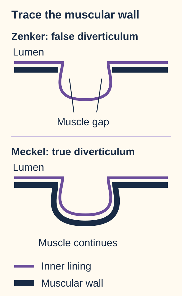
Use the wall drawing to trace the muscular boundary with a finger. In the upper drawing, the lining enters a sac through a gap in the muscle. In the lower drawing, the muscle continues around the sac. Before reading on, predict which drawing matches a congenital ileal specimen containing mucosa, submucosa, muscularis propria, and serosa.
Trace the thick muscular boundary. It ends at the gap in the false pouch but continues around the true pouch. These are original wall-construction models, not scale anatomy. [1][6][7]Check the wall prediction
The lower drawing matches a true ileal diverticulum: the muscular wall continues into its sac.
The consequence of this comparison is specific: wall layers classify the pouch; location identifies which disorder fits. An ileal full-wall pouch is not Zenker merely because it is called a diverticulum. Conversely, finding a posterior cervical pouch does not imply a congenital vitelline remnant. Apply the same method to a new specimen before using its patient's age. [6][7]
A restricted outlet creates a reservoir above it
Does a normal resting sphincter pressure exclude Zenker? No. The critical event occurs while a swallowed bolus tries to pass through the upper esophageal sphincter. Reduced opening and compliance increase resistance during that event. Pressure within the bolus rises upstream, encouraging mucosa and submucosa to protrude posteriorly. Studies distinguish restricted opening and increased residual pressure from a universally high resting pressure or a universal failure of neural relaxation. [1][2]
Imagine water entering a flexible tube whose outlet cannot widen sufficiently. More driving pressure is needed to deliver the same amount. Now give the wall above that outlet a weak point: a side reservoir can enlarge there. This is a limited mechanical analogy, not a pressure threshold for diagnosis. Normal swallowing also requires coordinated pharyngeal contraction, sphincter relaxation, and opening forces. [1][2]
The resulting sac retains food and saliva. Retention explains halitosis and cervical gurgling; delayed emptying explains regurgitation of recognizable food long after a meal. Material returning toward the pharynx can enter the airway, producing cough or aspiration. Progressive dysphagia, reduced intake, and weight loss may follow. These findings are common in older adults, but no single symptom is mandatory. Regurgitation is not necessarily vomiting from the stomach. [7]
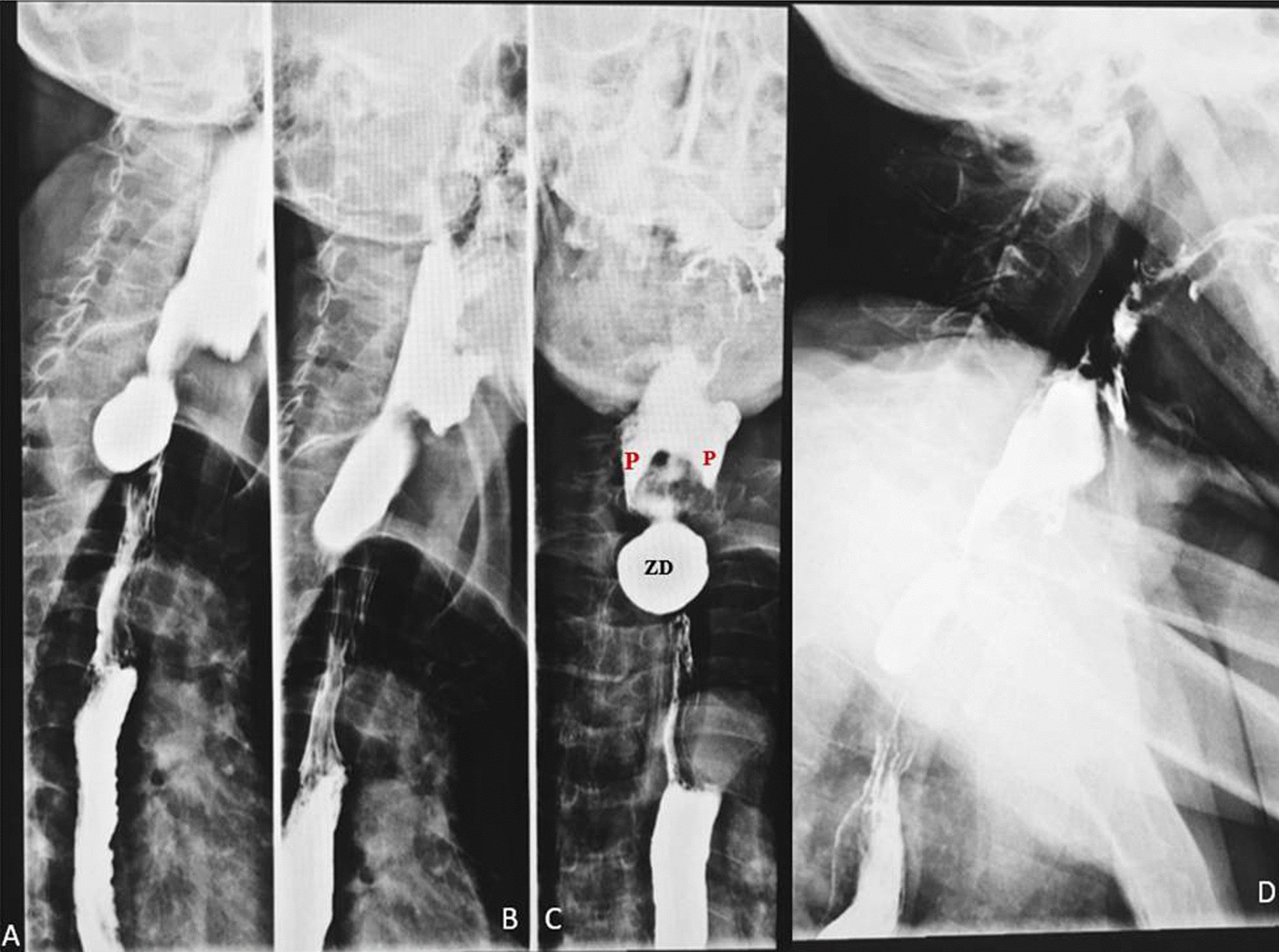
The published esophagram in this section demonstrates a posterior proximal pouch while contrast also passes down the esophagus. Trace the continuing esophageal column separately from the side reservoir. Passage of some contrast does not mean that the reservoir empties normally. Contrast swallowing studies define the pouch and its relationship to the pharyngoesophageal junction; the whole study and clinical context matter, not an isolated still frame. [7]
Compare the side pouch with the continuing contrast column. The published panels demonstrate that contrast passage and pouch retention can coexist. Image: Senai Goitom Sereke, Felix Bongomin, and Zeridah Muyinda; original source; CC BY 4.0. [7].
Now predict a change: if opening resistance is reduced but the lower esophageal sphincter is unchanged, which upstream pressure should decrease during swallowing?
Check the pressure prediction
The pressure needed to propel the bolus through the upper outlet should decrease.
This is why symptomatic treatment addresses the cricopharyngeal component, commonly through endoscopic septotomy with myotomy or an appropriately selected surgical approach. The target is not simply the visible sac. Procedure choice depends on anatomy, patient risk, and local expertise. The 2020 ESGE guideline favors flexible endoscopic treatment over open surgery as first-line therapy, but labels that recommendation weak with low-quality evidence; it is not a claim that one technique suits every patient. [3]
Transfer the pressure model to recurrence: a patient whose sac was treated but who still has restricted upper sphincter opening may retain the original driving problem. Do not assume that recurrent dysphagia proves regrowth without reassessing anatomy and swallowing. Persistent aspiration or impaired nutrition warrants clinical assessment; acute respiratory compromise takes priority over elective pouch testing. [1][2][3][7]
Location also prevents misleading comparisons. A distal esophageal pouch near the diaphragm is not a cervical Zenker pouch, and a mid-esophageal traction pouch reflects a different anatomic setting. Likewise, lower sphincter dysfunction cannot be inferred from a posterior pouch above the cricopharyngeus. Match the actual level of the abnormality before importing another esophageal explanation. [7]
Separate acid injury from a mechanical attachment
Must an ileal remnant contain gastric tissue to cause trouble? No. Meckel diverticulum has two partly independent routes to harm: tissue can secrete acid, or the pouch and its attachments can distort bowel anatomy. Keeping these routes separate explains why a patient can have obstruction without a history of bleeding. [5][6]
Ectopic gastric mucosa can secrete acid inside the ileal remnant. Nearby ileal mucosa lacks the stomach's usual protection against that acid, so ulceration often occurs in the adjacent ileum. The injured surface bleeds. Recurrent painless maroon stools or iron-deficiency anemia can therefore arise from an ileal lesion even when the stomach and colon have no bleeding source. Bleeding may be substantial; the word painless does not mean physiologically safe. Ectopic pancreatic tissue is another possible finding, but pancreatic acini are not the usual acid source or the target of the Meckel scan. [5][6]
Mechanical complications use different anatomy. An inverted diverticulum can project into the lumen as a lead point for intussusception, where one bowel segment telescopes into another. A persistent band can form an aperture for internal herniation or an axis around which bowel twists. Volvulus, entrapment, and intussusception can obstruct the lumen and threaten perfusion. A band-related obstruction does not require gastric heterotopia. [6]
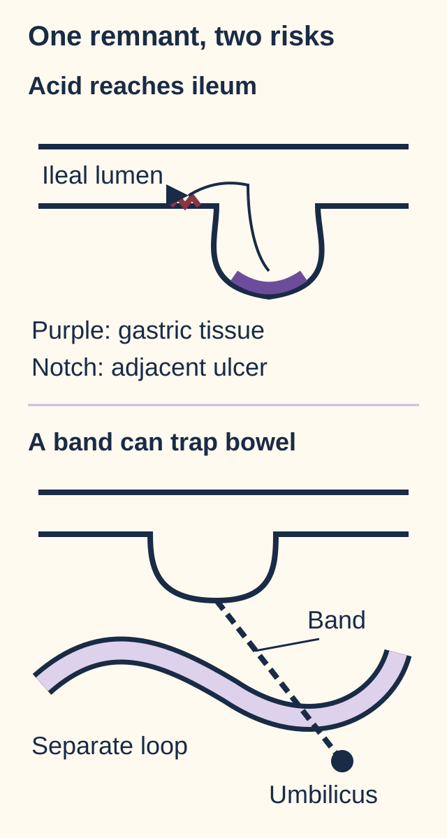
In the remnant drawing, trace one route from the gastric patch to the neighboring ulcer and a second from the tether to the bent bowel. Mentally change just one feature at a time. First suppress acid secretion while leaving the anatomy unchanged. Then restore the original acid secretion but release the tether. Predict which injury each change addresses.
Follow acid toward adjacent ileal injury, then follow the tether toward mechanical entrapment. The two simplified models isolate independent relationships; they do not imply that every remnant has gastric tissue or a band. [5][6]Check the acid-only change
Less acid should reduce acid-mediated mucosal injury; it does not release a tether or eliminate a lead point.
Check the tether-only change
Releasing a causal tether relieves that mechanical constraint; it does not eliminate ectopic gastric tissue.
The two predictions explain why acid suppression is not definitive correction of a symptomatic congenital remnant. They also prevent an error in a new patient: a negative gastric-tissue scan cannot reassure against a band causing strangulating obstruction. Clinical urgency follows obstruction, perfusion, and bleeding, not whether the mnemonic fits. [5][6]
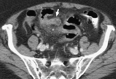
Inflammation of Meckel diverticulum may imitate appendicitis. In the published CT image, the important structure is a blind-ending bowel outpouching connected to distal ileum with adjacent inflammatory change. In a fresh case, identify that connection and assess the appendix rather than deciding from right lower quadrant pain alone. A target-shaped intussusception establishes bowel telescoping, but does not by itself identify the lead point as Meckel; additional anatomic or tissue evidence is needed. [6]
The publisher reports surgically confirmed Meckel diverticulitis. Identify the blind-ending ileal structure and the surrounding inflammation. The original arrow and asterisk are part of the published figure; no annotations were added. Image: Alexandra Platon, Pascal Gervaz, Christoph D. Becker, Philippe Morel, and Pierre-Alexandre Poletti; original source; CC BY 2.0. [6].
The tracer detects tissue, not the word Meckel
What does technetium-99m pertechnetate actually identify? Gastric-type mucosa can concentrate the tracer and secrete it into its lumen. Uptake occurs in mucin-producing gastric cells and is not dependent on acid-producing parietal cells. The same property can reveal ectopic gastric mucosa outside the stomach. The study is therefore neither a generic pouch detector nor a direct measurement of acid output. [5]
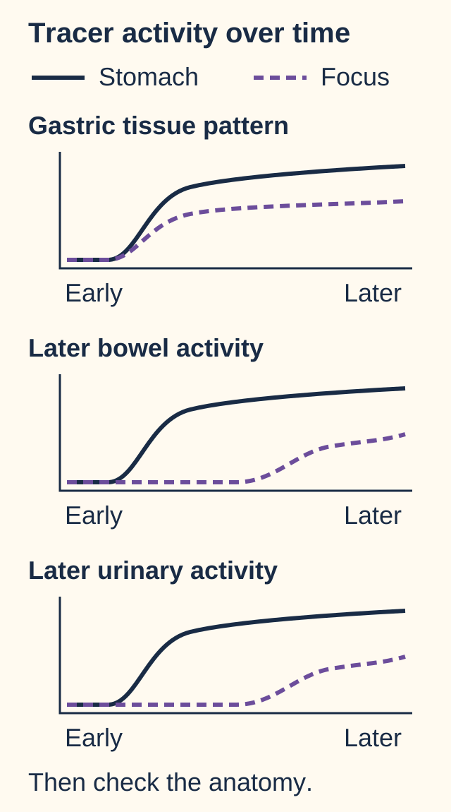
The timing diagram compares three patterns, not patient scans. In the first, a focal abdominal site becomes visible alongside the stomach and its activity increases with gastric activity. In the second, later diffuse bowel activity follows gastric secretion into the intestine. In the third, later urinary activity outlines an excretory structure. Compare onset and behavior rather than simply choosing the brightest abdominal spot. [5]
Solid curves represent stomach activity and dashed curves represent the comparison focus. These are qualitative teaching curves, not measured patient data or diagnostic intensity cutoffs. Late bowel and urinary patterns require additional anatomical information. [5]
An ectopic focus appearing in temporal agreement with gastric activity supports gastric heterotopia. It is often in the right lower abdomen, but the location is not fixed to that quadrant and a mobile ileal loop can change position. Conversely, a late focus may represent urinary activity or gastric tracer that has entered normal bowel. Additional views and the full acquisition help resolve these alternatives. The diagram simplifies this interpretation; no drawn intensity is a diagnostic cutoff. [5]
Try a tissue substitution. Keep the same ileal pouch and its band, but replace its gastric lining with ordinary ileal mucosa. What diagnostic signal disappears even though the congenital anatomy persists?
Check the tissue substitution
The ectopic gastric uptake signal is lost; the anatomic remnant and its mechanical risks remain.
A negative scan can occur when gastric mucosa is absent, sparse, obscured, or insufficiently visualized because of technical or physiological factors. It does not exclude every Meckel diverticulum. A positive scan also needs anatomic interpretation: a duplication containing ectopic gastric mucosa can produce uptake without being Meckel diverticulum. A test for a tissue property is not automatically a test unique to one diagnosis.[5]
The SNMMI/EANM guideline uses this study to investigate otherwise unexplained bleeding when the patient is not actively bleeding. A stable child with intermittent painless blood loss is a characteristic setting. A child with brisk ongoing hemorrhage and poor perfusion needs stabilization and an acute bleeding assessment rather than delay for elective scintigraphy. Radiolabeled red blood cell imaging detects bleeding by a different mechanism; the two technetium studies are not interchangeable. [5][6]
Specialist preparation can improve visualization. For example, an H2 blocker can limit secretion of accumulated tracer into the lumen and improve retention within gastric mucosa. That is a test-preparation principle, not permission to diagnose from acid suppression or to prescribe a universal preparation regimen. Apply the distinction to a new report: persistent focal uptake despite reduced acid secretion is compatible with gastric tissue because uptake is not a direct parietal-cell acid assay. [5]
Use the rule of twos to recall, not to exclude
Can a 40-year-old with a pouch 90 cm from the ileocecal valve have Meckel diverticulum? Yes. The traditional rule of twos gathers approximate associations: about 2% prevalence, about 2 feet from the valve, about 2 inches in length, presentation before age two, male predominance among symptomatic patients, and gastric or pancreatic ectopic tissue. These are memory aids, not required diagnostic criteria. Anatomy and histology can fit even when age, distance, and length do not. [4][5][6]
Compare two kinds of evidence. Age under two suggests a familiar clinical setting. A true antimesenteric ileal pouch with gastric heterotopia supplies much more direct evidence. Even a characteristic tracer focus establishes a tissue behavior rather than uniquely proving the pouch's origin. Rank the observations by what they actually demonstrate, not by how closely they resemble a remembered phrase. [4][5][6]
Symptomatic Meckel diverticulum generally requires surgical treatment, with the extent tailored to the source of bleeding, inflammation, damaged adjacent bowel, and the diverticular base. An incidental asymptomatic pouch is a different decision. The potential future complication risk must be balanced against the added risk of the current operation. Age, sex, length, abnormal tissue, the reason for surgery, and overall patient risk inform that judgment; they are not an automatic instruction to resect every incidental lesion. [4][6]
The Mayo surgical cohort found associations between symptomatic disease and younger age, male sex, greater length, and histologic abnormality. Such associations help frame an individualized discussion. They do not give a universal lifetime complication probability for every person with an incidental pouch: this was a selected surgical population, and histology may not be known before resection. [4]
For a final comparison, imagine an asymptomatic short pouch discovered during a high-risk operation, then the same pouch actively bleeding with a neighboring ulcer. In the first situation, the benefit of prophylactic treatment is uncertain and added operative risk matters. In the second, there is an identified symptomatic source to address. The changed fact is active disease, not whether a measurement equals two. [4][6]
Check the treatment target
For symptomatic Zenker, address impaired pharyngoesophageal outflow. For symptomatic Meckel, address the remnant and the complication it has produced.
Carry three questions into practice: Where is the pouch? Which wall or tissue feature explains its behavior? Which danger or structural problem must be addressed now? A location, a tissue signal, and an urgent physiological problem answer different questions.
Apply the relationships in new cases
Case 1
Show answer and explanations for case 1
A. Granulomatous scar pulling the full wall outward (Why this does not fit)
Inflammatory scarring outside the esophagus can produce a traction diverticulum. A posterior sac immediately above the upper sphincter fits the pharyngeal pulsion site rather than a mid-esophageal traction setting.
Reasoning steps for option A
How could granulomatous scarring create an esophageal outpouching with a full wall?
External inflammatory scarring can pull the full esophageal wall outward to form a traction diverticulum.
Does this sac's position immediately above the cricopharyngeus favor traction from mediastinal scar?
No. This posterior upper sphincter site is characteristic of pharyngeal pulsion, not a mid-esophageal traction setting.
B. Mucosa and submucosa protruding through a muscular gap (Best answer)
An acquired pulsion pouch can form when luminal lining protrudes through a weak muscle boundary. The sac above the cricopharyngeus fits Zenker diverticulum, whose wall lacks the complete muscular layer.
Reasoning steps for option B
What wall layers would pressure push through a weak muscular boundary in this older patient's posterior pouch?
Mucosa and submucosa protrude through the muscle gap in an acquired pulsion pouch.
Why does the sac above the cricopharyngeus support an incomplete muscular wall?
That location identifies a Zenker diverticulum, whose sac does not contain the complete muscular layer.
C. Mucosa, submucosa, and a continuous muscular wall (Why this does not fit)
A true diverticulum carries its muscular wall into the outpouching. The posterior pharyngoesophageal location and delayed food retention instead identify a pulsion pseudodiverticulum.
Reasoning steps for option C
What would a continuous muscle layer around this sac signify?
Muscle extending into the outpouching would make it a true diverticulum.
Why is a true full-wall pouch a poor fit for the delayed food return and posterior cervical sac?
The retained food and pharyngoesophageal position instead fit an acquired pulsion pseudodiverticulum.
D. A duplicated muscular tube sharing the esophageal wall (Why this does not fit)
An enteric duplication can form an additional epithelial-lined structure with smooth muscle. The communicating posterior sac at the characteristic weak zone with delayed food return is more consistent with acquired herniation.
Reasoning steps for option D
What construction would the proposed duplicated esophageal tube require?
An enteric duplication is an additional epithelial-lined structure with smooth muscle, rather than just lining pushed through a gap.
Which feature of the food-retaining posterior sac argues for herniation instead of duplication?
Its communication at the characteristic weak zone above the cricopharyngeus, with delayed food return, favors acquired herniation.
Takeaway: Locate a pouch before using its wall layers to distinguish pulsion from a true diverticulum.
A. Reduced pharyngeal contraction with a compliant outlet (Why this does not fit)
Weak pharyngeal propulsion can impair bolus clearance. Contraction is preserved, and increased intrabolus pressure accompanies restricted outlet opening rather than weak driving force.
Reasoning steps for option A
How could reduced pharyngeal contraction ordinarily impair bolus transit?
Weak pharyngeal propulsion can leave a swallowed bolus uncleared even if the outlet is compliant.
Do this patient's contraction and intrabolus pressure indicate weak propulsion?
No. Contraction is preserved, and raised intrabolus pressure accompanies restricted opening rather than a weak driving force.
B. Reduced sphincter compliance despite preserved timing (Best answer)
A poorly distensible outlet can require greater pressure even when relaxation begins at the expected time. Normal resting pressure and coordination combined with restricted opening and increased intrabolus pressure support impaired compliance.
Reasoning steps for option B
Can the upper outlet resist a bolus despite normal relaxation timing?
Yes. A poorly distensible sphincter can require greater pressure even when relaxation starts on time.
What combination on simultaneous manometry and fluoroscopy points to reduced compliance?
Normal resting pressure and relaxation timing coexist with restricted opening and elevated upstream intrabolus pressure.
C. Increased resting sphincter tone with normal distensibility (Why this does not fit)
High tonic sphincter pressure can suggest resistance at rest. Resting pressure is normal here; the demonstrated abnormality occurs during restricted opening.
Reasoning steps for option C
What would increased resting sphincter tone imply before the swallow begins?
High tonic pressure could suggest resistance at rest, even with otherwise normal distensibility.
Why does this study not attribute the outlet problem to tonic pressure?
Resting pressure is within the reference range; the observed resistance appears during restricted bolus passage.
D. Delayed neural relaxation with normal wall compliance (Why this does not fit)
A timing abnormality can hinder passage during an otherwise adequate pharyngeal contraction. The measured relaxation timing is normal, so it does not explain the restricted opening in this study.
Reasoning steps for option D
How could delayed neural relaxation obstruct an otherwise strong pharyngeal swallow?
Late relaxation can leave the sphincter closed during bolus arrival despite adequate pharyngeal contraction.
What measured timing finding argues against delayed relaxation here?
Relaxation occurs at the expected time, so timing alone cannot explain the restricted opening.
Takeaway: Zenker physiology is not equivalent to universally high resting upper sphincter pressure.
A. Persistent obstruction at the distal esophageal sphincter (Why this does not fit)
Distal outflow obstruction can cause esophageal stasis and regurgitation. The main esophageal column has cleared, while the retained material is specifically in the pharyngoesophageal sac.
Reasoning steps for option A
How could distal sphincter obstruction otherwise produce regurgitation after dinner?
A resistant distal outlet can cause material to stagnate in the esophageal lumen and return upward.
Where does fluoroscopy actually locate retained contrast after the main column clears?
It remains in the pharyngoesophageal pouch rather than in the main esophageal column, which has cleared instead of showing distal stasis.
B. Immediate failure to initiate the pharyngeal swallow (Why this does not fit)
Impaired swallow initiation can cause coughing during ingestion. The described episodes occur hours after a completed swallow and correspond to material remaining in the pouch.
Reasoning steps for option B
When would failure to initiate a pharyngeal swallow provoke cough?
Impaired swallow initiation can provoke cough during ingestion as material enters the airway.
Does coughing two hours after dinner match failure at swallow initiation?
No. The swallow has finished; material retained in the pouch can return hours later.
C. Recurrent acid secretion by gastric tissue in the pouch (Why this does not fit)
Acid-containing material can irritate the airway in reflux disease. The demonstrated source is retained swallowed contrast in a cervical pouch, not ectopic gastric tissue or continued distal acid reflux.
Reasoning steps for option C
How might acid-containing reflux material cause cough in a different patient?
Refluxed acid-containing contents can irritate the airway.
Why do improved heartburn and retained cervical contrast weaken an acid-secreting pouch explanation?
The demonstrated reservoir contains swallowed contrast despite improved acid symptoms; neither ectopic gastric tissue nor ongoing distal acid reflux is shown.
D. Delayed return of retained material into the pharynx (Best answer)
A side reservoir can retain swallowed material after the main lumen has emptied. Delayed regurgitation when reclining exposes the airway to that retained material even when acid-related heartburn improves.
Reasoning steps for option D
What happens to a swallowed bolus when the esophageal column clears but the cervical side sac does not?
Some swallowed material remains in the side reservoir after the main lumen empties.
How does reclining later expose this patient's airway despite improvement in heartburn?
Retained pouch contents can return to the pharynx hours later and enter the airway, producing cough independent of improved heartburn.
Takeaway: Treating acid symptoms does not empty a food-retaining pharyngoesophageal pouch.
A. Greater pressure needed to distend the pharyngeal outlet (Why this does not fit)
A stiffer outlet requires more distending pressure during passage. Successful treatment of the restrictive cricopharyngeal component is expected to reduce, not increase, that requirement.
Reasoning steps for option A
Under what outlet condition would more pressure be needed to distend the pharyngeal opening?
A stiffer upper outlet would demand more distending pressure during passage of the same bolus.
Should successful cricopharyngeal myotomy make that distending requirement greater?
No. Treating the restricted upper opening should reduce, not increase, the pressure requirement.
B. Greater intrabolus pressure above the upper sphincter (Why this does not fit)
Greater upstream pressure would be required if resistance increased for a comparable bolus. Myotomy targets the restrictive outlet, so increased required pressure is opposite to the expected effect.
Reasoning steps for option B
What change in resistance would raise intrabolus pressure above the upper sphincter for a comparable bolus?
Increased outlet resistance would require greater upstream pressure to propel that bolus.
Why is a pressure rise opposite to the intended septotomy and myotomy effect?
The operation relieves the restrictive cricopharyngeal outlet rather than increasing its resistance.
C. Lower intrabolus pressure above the upper sphincter (Best answer)
Reducing outlet resistance lowers the pressure needed to propel a comparable swallowed bolus. The restricted upper opening is the measured abnormality and the cricopharyngeal myotomy addresses that site.
Reasoning steps for option C
How should upstream bolus pressure respond when resistance to upper sphincter passage falls?
Less pressure is needed to propel a comparable swallowed bolus through the outlet.
Which pretreatment measurement and surgical target support that predicted decrease?
Fluoroscopy shows restricted upper opening, and cricopharyngeal myotomy directly treats that restrictive site.
D. Lower resting pressure at the lower esophageal sphincter (Why this does not fit)
Distal sphincter pressure changes can affect emptying in distal outflow disorders. The treated muscle is cricopharyngeus at the upper outlet; the operation does not directly target the lower sphincter.
Reasoning steps for option D
When would lowering lower esophageal sphincter pressure improve bolus emptying?
A pressure change at the distal sphincter may matter in a distal outflow disorder.
Does this septotomy directly treat the lower esophageal sphincter?
No. It treats the cricopharyngeus at the upper outlet, not the lower esophageal sphincter.
Takeaway: The myotomy addresses outflow resistance, not merely the visible reservoir.
A. Persistent restriction at the pharyngoesophageal outlet (Best answer)
A pouch procedure can leave the driving outlet abnormality uncorrected. Restricted opening persists at the cricopharyngeus, providing an upstream pressure mechanism for recurrent pouch formation.
Reasoning steps for option A
What mechanism could remain after excision of a posterior pouch without cricopharyngeal treatment?
Removing the reservoir alone can leave the restrictive upper outlet and its pressure-generating mechanism intact.
How does the repeat study link that omission to the recurrent sac?
Persistent restriction at the cricopharyngeus can raise upstream pressure during swallowing and favor recurrent posterior pouch formation.
B. An untreated increase in lower sphincter resistance (Why this does not fit)
A distal outflow disorder can sustain esophageal symptoms after an unrelated proximal procedure. The repeat study shows normal distal emptying and identifies persistent resistance at the upper sphincter.
Reasoning steps for option B
Could an untreated lower sphincter disorder cause persistent symptoms after proximal pouch surgery?
Yes. Distal outflow resistance can sustain esophageal symptoms despite an unrelated proximal operation.
What repeat imaging finding instead localizes resistance to the upper outlet?
Distal emptying remains normal while upper sphincter opening is restricted.
C. An untreated fibrotic stricture in the distal esophagus (Why this does not fit)
A fibrotic stricture can cause dysphagia by narrowing the main esophageal lumen. The repeat study instead demonstrates normal distal emptying and persistent restriction at the upper sphincter.
Reasoning steps for option C
How would a distal fibrotic stricture ordinarily produce dysphagia?
Fibrosis could narrow the main distal esophageal lumen and impede passage.
Does the repeat study show the distal hold-up expected from such a stricture?
No. Distal emptying is normal, while the demonstrated narrowing of opening is at the upper sphincter.
D. Persistent extrinsic traction from mediastinal scarring (Why this does not fit)
Scarring can pull the wall outward in a traction diverticulum. The recurrent sac is cervical and directly associated with restricted upper opening rather than a demonstrated mid-esophageal traction lesion.
Reasoning steps for option D
How can mediastinal scarring generate a traction pouch?
External scar can pull the esophageal wall outward to form a traction diverticulum.
Why does this recurrent cervical sac not implicate persistent mediastinal traction?
The sac is posterior and cervical, associated with restricted upper opening rather than a demonstrated mid-esophageal traction lesion.
Takeaway: Reassess both anatomy and swallowing function when symptoms recur after pouch treatment.
A. Radionuclide gastric emptying scintigraphy (Why this does not fit)
Gastric emptying studies assess delayed transit from the stomach. The requested abnormality is a pharyngoesophageal reservoir, not delayed gastric emptying.
Reasoning steps for option A
What transit problem would a radionuclide gastric emptying study assess in this woman?
It assesses delayed passage of a meal out of the stomach, not retention in the neck.
Why does late return of recognizable food with neck gurgling weaken the gastric emptying choice?
Those symptoms point to a pharyngoesophageal reservoir whose anatomy needs definition, rather than delayed gastric transit.
B. Esophageal manometry without anatomic imaging (Why this does not fit)
Manometry measures pressure and may help characterize swallowing physiology. Pressure recordings alone do not define the requested shape and communication of a structural pouch.
Reasoning steps for option B
What could manometry contribute to evaluating her cervical swallowing difficulty?
It records pressure and can characterize swallowing physiology, including upper sphincter function.
Why would manometry alone not answer whether her suspected pouch communicates with the esophagus?
Pressure traces do not depict the sac's shape, location, or communication during swallowing.
C. Ambulatory esophageal acid exposure monitoring (Why this does not fit)
Acid monitoring investigates reflux burden and its relationship to symptoms. It does not display the suspected cervical reservoir or its communication during swallowing.
Reasoning steps for option C
Which alternative process would ambulatory acid monitoring investigate in her regurgitation?
It evaluates esophageal acid exposure and the association between reflux and symptoms.
Can an acid exposure record establish the source of the neck gurgling and delayed undigested food?
No. It cannot show a cervical reservoir or its filling and communication during swallowing.
D. Videofluoroscopic contrast esophagram (Best answer)
A contrast swallow depicts bolus passage and communicating outpouchings. It directly shows the suspected pharyngoesophageal sac and how it fills and empties.
Reasoning steps for option D
What would a videofluoroscopic contrast swallow show as she moves a bolus past the neck?
It depicts bolus passage and any communicating pharyngoesophageal outpouching.
Why does this contrast study answer the structural question in a patient handling secretions comfortably?
It can define the suspected cervical sac and demonstrate how contrast enters and leaves it.
Takeaway: Choose the test for the clinical question: contrast imaging defines the pouch; pressure testing answers a different question.
A. The mid-esophageal wall adjacent to mediastinal nodes (Why this does not fit)
Inflammatory nodal scarring can be associated with a mid-esophageal traction pouch. The study places this sac at the pharyngoesophageal junction rather than in the thorax.
Reasoning steps for option A
How could mediastinal nodal disease produce a pouch in a different esophageal segment?
Inflammatory scarring beside thoracic nodes can pull the mid-esophageal wall into a traction pouch.
What location in this image rules against a node-associated mid-esophageal origin?
The sac arises at the posterior pharyngoesophageal junction, not beside mediastinal nodes in the thorax.
B. The posterior gap between the two inferior constrictor parts (Best answer)
Killian triangle lies between the oblique thyropharyngeus and transverse cricopharyngeus components. The imaged origin above the cricopharyngeus and posterior to the junction identifies this weak region.
Reasoning steps for option B
Which muscle boundaries enclose the weak region indicated by this posterior sac?
Killian triangle lies between the oblique thyropharyngeus fibers above and the transverse cricopharyngeus fibers below.
How does the neck above the cricopharyngeus pinpoint the sac's origin?
Its posterior origin above those transverse fibers and below the oblique fibers places it in Killian triangle.
C. The distal esophageal wall immediately above the diaphragm (Why this does not fit)
A distal pulsion pouch can arise in the epiphrenic region. Its site is separated from the upper sphincter landmarks supplied in this examination.
Reasoning steps for option C
Where would an epiphrenic pulsion pouch arise relative to the diaphragm?
It arises in the distal esophageal region just above the diaphragm.
Why cannot that distal location account for the imaged pouch above the transverse cricopharyngeal fibers?
The supplied landmarks are at the upper sphincter, far from the epiphrenic esophagus.
D. The anterior wall below the upper esophageal sphincter (Why this does not fit)
A different cervical outpouching can arise below the cricopharyngeal level. This sac originates posteriorly and above the transverse cricopharyngeal fibers.
Reasoning steps for option D
What cervical site would an anterior pouch below the upper sphincter require?
It would require an anterior origin below the cricopharyngeal level, a different cervical outpouching.
Which two directional findings contradict that anterior, subcricopharyngeal origin?
This sac begins posteriorly and above the transverse cricopharyngeal fibers.
Takeaway: A Zenker pouch arises above the cricopharyngeus through the posterior weak region.
A. Incomplete involution of the vitelline duct (Best answer)
Persistence of the intestinal end of the vitelline duct leaves a true antimesenteric ileal diverticulum. That anatomy and gastric heterotopia explain this lesion despite an age and distance outside the familiar mnemonic.
Reasoning steps for option A
Which embryonic remnant forms a full-wall pouch on the ileum's antimesenteric border?
Persistence of the intestinal end of the vitelline duct produces a true Meckel diverticulum at that site.
Do this woman's age and the pouch's 90 cm distance negate the gastric-lined remnant?
No. Its full-wall antimesenteric anatomy and gastric heterotopia support that origin despite deviation from the rule-of-twos mnemonic.
B. Failure of the midgut to return to the abdominal cavity (Why this does not fit)
Failure of normal return is associated with an abdominal wall defect containing herniated viscera. The finding here is an isolated full-wall ileal outpouching inside the abdomen, not persistent external herniation.
Reasoning steps for option B
What anatomical outcome would failure of the midgut to return normally predict?
It is associated with an abdominal wall defect containing externally herniated viscera.
How does the operative finding differ from persistent external midgut herniation?
The surgeon found an isolated, intra-abdominal ileal side pouch containing all bowel wall layers.
C. Incomplete rotation around the superior mesenteric artery (Why this does not fit)
Abnormal rotation can alter intestinal fixation and predispose to volvulus. It does not specifically explain the observed antimesenteric full-wall pouch with gastric heterotopia.
Reasoning steps for option C
What complication might incomplete intestinal rotation around the superior mesenteric artery predispose to?
Abnormal rotation changes fixation and can predispose to volvulus.
Why does a rotation defect fail to account for the glands in this antimesenteric 3-cm pouch?
It does not specifically produce a true antimesenteric ileal diverticulum lined partly by ectopic gastric tissue.
D. Failure of an intestinal segment to recanalize (Why this does not fit)
Failure of recanalization can produce a narrowed or obstructed lumen. The lesion is a communicating side pouch with all wall layers rather than an intraluminal occlusion.
Reasoning steps for option D
What luminal abnormality would failed intestinal recanalization suggest instead of this pouch?
Failure of recanalization can narrow or obstruct an intestinal lumen.
Which operative feature separates a recanalization defect from the bleeding lesion?
The lesion is a communicating side pouch with all bowel wall layers, not an intraluminal blockage.
Takeaway: Anatomy and tissue can establish the remnant even when the rule of twos does not fit.
A. A urachal diverticulum opening into the bladder (Why this does not fit)
A persistent urachal segment can communicate with the bladder. The demonstrated tract connects to ileum and explicitly has no bladder connection.
Reasoning steps for option A
Where would a persistent urachal connection lead from the umbilicus?
A urachal remnant can communicate with the bladder rather than the distal ileum.
What imaging result excludes the proposed bladder-linked explanation for this newborn's feculent discharge?
The patent tract joins distal ileum to the umbilicus and has no bladder communication.
B. A false ileal pouch at a mesenteric vascular entry point (Why this does not fit)
Acquired intestinal pseudodiverticula can arise where mucosa protrudes through muscle. The neonatal ductal connection is congenital, and its ileal remnant carries all wall layers on the antimesenteric side.
Reasoning steps for option B
How is the wall of a mesenteric vascular-entry pseudodiverticulum formed?
Mucosa can protrude through muscle at a vascular entry point, producing an acquired false pouch.
Why does persistence of this newborn's ileoumbilical duct predict a different wall and border?
Its congenital intestinal-end remnant is a true pouch with all wall layers on the antimesenteric ileal border.
C. A mesenteric intestinal duplication sharing a muscular wall (Why this does not fit)
An enteric duplication can create an additional bowel-associated structure. The question asks about persistence of the proven ileum-to-umbilicus duct, not formation of a duplicated bowel segment.
Reasoning steps for option C
What additional bowel structure does an enteric duplication represent?
It is an additional bowel-associated structure that can share a muscular wall on the mesenteric side.
Why does the demonstrated umbilical tract favor duct persistence over duplication?
Imaging proves a connection from ileum to umbilicus; an intestinal-end remnant of that same duct is not a duplicated bowel segment.
D. A true diverticulum on the antimesenteric ileal border (Best answer)
Persistence of the intestinal end of the vitelline connection produces a Meckel diverticulum. The fully patent ileum-to-umbilicus tract identifies the same embryonic system from which this blind-ending remnant can arise.
Reasoning steps for option D
What becomes of the vitelline connection if only its intestinal end persists?
It forms a blind-ending Meckel diverticulum, a true pouch on the antimesenteric distal ileum.
How does the patent tract in this newborn link that pouch to the same embryonic origin?
The fully patent ileum-to-umbilicus tract demonstrates the vitelline connection; retaining just its ileal end produces the related diverticulum.
Takeaway: The patent ileoumbilical tract and Meckel diverticulum represent different patterns of persistence of the vitelline connection.
A. Acid exposure of adjacent ileum lacking gastric protection (Best answer)
Ectopic gastric mucosa can deliver acid to neighboring intestinal epithelium. The relative preservation of gastric-type tissue with ulceration beside it fits injury to less protected ileal mucosa.
Reasoning steps for option A
What secretion from the pouch's oxyntic glands can injure nearby ileal lining?
Ectopic gastric mucosa can secrete acid into the neighboring ileal environment.
Why is the ulcer beside the relatively preserved gastric tissue in this child?
Adjacent ileal mucosa lacks the gastric lining's acid protection, so local acid exposure can ulcerate and bleed there.
B. Pressure necrosis at the apex of an inverted lead point (Why this does not fit)
An inverted lead point can produce mechanical injury during intussusception. The described lesion is a focal adjacent ileal ulcer next to oxyntic tissue, without a reported inverted segment or telescoping.
Reasoning steps for option B
How could an inverted diverticulum cause mechanical mucosal damage?
An inverted pouch can act as a lead point for intussusception, with pressure injury during bowel telescoping.
What feature of this specimen argues against pressure necrosis at an inverted apex?
The ulcer is in ileum adjacent to oxyntic glands; no inverted segment or telescoping is reported.
C. Diffuse immune injury targeting all ileal epithelial cells (Why this does not fit)
An inflammatory enteropathy can injure ileal mucosa and cause bleeding. The focal ulcer adjacent to acid-secreting gastric glands is better explained by a local acid exposure than a diffuse process.
Reasoning steps for option C
What injury pattern would diffuse immune-mediated enteropathy imply?
An inflammatory process could involve ileal epithelium broadly and cause bleeding.
Why does the ulcer's precise position favor another cause over diffuse immune injury?
It is focal and immediately beside acid-secreting gastric glands, supporting localized acid exposure rather than diffuse inflammation.
D. Pancreatic enzyme release from the gastric oxyntic glands (Why this does not fit)
Pancreatic heterotopia is a possible finding in a congenital ileal remnant. The identified glands are gastric oxyntic tissue, whose acid secretion explains adjacent ileal injury rather than pancreatic enzyme release.
Reasoning steps for option D
Could a congenital ileal remnant contain tissue capable of pancreatic enzyme secretion?
Pancreatic heterotopia can occur in such a remnant, but it is distinct from gastric oxyntic glands.
Which identified tissue explains this child's adjacent ulcer without invoking pancreatic enzymes?
The glands are gastric oxyntic tissue; their acid secretion can injure neighboring ileum while the gastric lining remains relatively preserved.
Takeaway: In bleeding Meckel disease, the acid source and the ulcerated surface need not be the same tissue.
A. Technetium-labeled red blood cell scintigraphy (Why this does not fit)
Labeled circulating red cells can reveal bleeding when blood escapes into the intestinal lumen. The child is not currently bleeding, and the requested target is ectopic gastric mucosa rather than active blood extravasation.
Reasoning steps for option A
When would labeled red cells reveal the source of this child's maroon stools?
They reveal blood escaping into the intestinal lumen during active bleeding, rather than the ectopic gastric tissue itself.
Does three days without visible bleeding favor a red-cell bleeding scan for the requested target?
No. There is no current bleeding, and the question seeks gastric mucosa rather than active extravasation.
B. Technetium-99m pertechnetate scintigraphy (Best answer)
Pertechnetate is concentrated by gastric mucosa, including gastric tissue in an ileal remnant. Intermittent painless bleeding without a source on upper or lower endoscopy is a fitting setting for this tissue-directed study.
Reasoning steps for option B
What can pertechnetate detect in a suspected ileal remnant despite a symptom-free interval?
Gastric mucosa in the remnant concentrates pertechnetate independently of visible bleeding at the time of imaging.
Why do painless intermittent maroon stools with negative upper and lower endoscopy favor this study?
They fit an ectopic gastric source beyond those examinations, and the stable child is not currently bleeding.
C. Contrast CT angiography during the symptom-free interval (Why this does not fit)
CT angiography can localize sufficiently active intestinal hemorrhage. No current bleeding is described, and angiographic contrast does not selectively identify gastric-type mucosa.
Reasoning steps for option C
What event must CT angiography capture to localize this child's intestinal hemorrhage?
It can localize sufficiently active bleeding through contrast extravasation, not selective gastric mucosal uptake.
Why is CT angiography during these three bleed-free days poorly matched to the question?
No current hemorrhage is described, and contrast angiography does not selectively identify gastric-type mucosa.
D. Repeat colonoscopy during the symptom-free interval (Why this does not fit)
Colonoscopy can evaluate a mucosal source within the colon. A prior examination has not found such a source, and the specified diagnostic target is ectopic gastric tissue outside the examined stomach and colon.
Reasoning steps for option D
What source could another colonoscopy investigate in this child?
A colonic mucosal lesion could be assessed directly by colonoscopy.
Why would repeating colonoscopy now miss the requested diagnostic target?
The prior examination found no colonic source, while the suspected ectopic gastric tissue lies outside the examined stomach and colon.
Takeaway: A Meckel scan and a labeled red cell bleeding scan answer different questions.
A. Retention inside red cells leaking through an ulcer (Why this does not fit)
A radiolabeled red cell study can show blood entering the intestinal lumen. This is a pertechnetate tissue study with gastric-type uptake, not a study using labeled circulating erythrocytes.
Reasoning steps for option A
How would labeled erythrocytes produce an intestinal focus in a bleeding child?
A red-cell scan can show labeled circulating cells escaping into the bowel lumen.
Were labeled red cells used to produce this focus that tracks gastric activity?
No. The study uses pertechnetate to image tissue uptake, not radiolabeled erythrocyte leakage.
B. Uptake by pancreatic acini releasing digestive enzymes (Why this does not fit)
Pancreatic heterotopia can occur in an ileal remnant. The tissue and synchronous gastric pattern identify gastric mucosa, whose tracer behavior does not require pancreatic acini.
Reasoning steps for option B
Could ectopic pancreatic tissue occur in this child's ileal pouch?
Yes. Pancreatic heterotopia can occur in an ileal remnant.
Does pancreatic enzyme release explain uptake synchronous with the stomach in confirmed gastric tissue?
No. The gastric-pattern signal and pathology identify gastric mucosa; pancreatic acini are not needed for its tracer uptake.
C. Uptake by acid-secreting parietal-cell proton pumps (Why this does not fit)
Parietal-cell proton pumps account for gastric acid secretion. Pertechnetate accumulation is not dependent on parietal-cell acid secretion, so proton-pump activity is not the required uptake mechanism.
Reasoning steps for option C
What do parietal-cell proton pumps contribute to gastric physiology here?
They mediate gastric acid secretion, a process distinct from the mucosal concentration of pertechnetate.
Why does persisting tracer uptake despite suppressed acid output weaken a proton-pump explanation?
Pertechnetate accumulation does not depend on parietal-cell acid secretion, so proton-pump activity cannot be the required uptake mechanism.
D. Concentration by mucin-producing gastric epithelial cells (Best answer)
Gastric mucin-producing cells can concentrate pertechnetate independently of parietal-cell acid output. Persistence of uptake after acid suppression therefore remains compatible with the histologically confirmed gastric mucosa.
Reasoning steps for option D
Which gastric cells can retain this tracer when acid secretion is suppressed?
A. Normal bowel has filled with tracer secreted by the stomach (Why this does not fit)
Gastric tracer can later enter normal intestine and produce bowel activity. A separate focal site appearing with initial gastric activity is more consistent with ectopic mucosa than delayed luminal transit.
Reasoning steps for option A
When might secreted gastric tracer appear in otherwise normal bowel?
After gastric secretion enters the intestinal lumen, later bowel activity can develop.
Does that delayed transit explain a separate focus appearing simultaneously with the stomach?
No. Early focal uptake that rises alongside gastric activity better supports ectopic mucosa than later luminal transit.
B. A collecting-system abnormality explains the early focus (Why this does not fit)
Urinary excretion can produce abdominal activity and mimic an abnormal site. The reported early parallel gastric pattern and separate later bladder activity support a gastric focus over this urinary explanation.
Reasoning steps for option B
How could urinary excretion mimic an abdominal focus on this study?
Excreted tracer can accumulate in the urinary collecting system and create abdominal activity.
What timing and location argue against a urinary source for this child's early focus?
The focus develops in parallel with the stomach, while bladder activity appears later at a separate site.
C. Active hemorrhage is quantified by the abdominal focus (Why this does not fit)
A tracer study of escaping blood can demonstrate active intestinal bleeding. Pertechnetate uptake with the stomach identifies a mucosal behavior and does not quantify an active hemorrhage rate.
Reasoning steps for option C
What would a study of escaping labeled blood demonstrate during hemorrhage?
Such a study can demonstrate active blood entering the intestinal lumen.
Can the gastric-pattern pertechnetate focus quantify this child's bleeding rate?
No. Concurrent uptake with the stomach describes mucosal tracer handling, not the rate of active hemorrhage.
D. Ectopic gastric mucosa is present at the abdominal focus (Best answer)
A focal site with onset and increasing activity concordant with the stomach supports gastric heterotopia. The distinct later bladder activity makes urinary excretion less explanatory for this early gastric-pattern focus.
Reasoning steps for option D
What does a lower abdominal focus that appears and intensifies alongside the stomach suggest?
Its concordant onset and progression support ectopic gastric mucosa at that focus.
Why does later, spatially distinct bladder activity reinforce that interpretation?
It makes urinary excretion a less persuasive explanation of the earlier gastric-pattern focus; the scan does not uniquely prove a particular lesion.
Takeaway: Relative onset and behavior are more informative than the mere presence of an abdominal hot spot.
A. No gastric target is present despite mechanical disease (Best answer)
A pouch without gastric heterotopia may produce no ectopic gastric signal. Its fibrous attachment can still trap bowel, so negative scintigraphy and remnant-related obstruction are compatible.
Reasoning steps for option A
Why might this surgically observed ileal pouch produce a negative pertechnetate study?
Its histology lacks gastric heterotopia, so there may be no ectopic gastric tissue to concentrate tracer.
How can the same remnant still obstruct this child's bowel?
Its fibrous umbilical attachment traps bowel mechanically, irrespective of the absent gastric signal.
B. The negative scan excludes the identified congenital remnant (Why this does not fit)
A negative tissue-targeted study can reduce support for a detectable ectopic gastric focus. It does not negate directly observed congenital anatomy when the target gastric tissue is absent.
Reasoning steps for option B
What does the prior negative scan actually weaken in this child?
It weakens evidence for a detectable ectopic gastric mucosal focus, not for every congenital remnant.
Can that scan overturn the operative finding of an antimesenteric pouch and trapping band?
No. The congenital anatomy is directly observed, and histology confirms that the gastric target is absent.
C. The obstruction requires acid injury that the scan missed (Why this does not fit)
Acid-mediated injury can explain bleeding from a remnant with gastric heterotopia. The observed bowel entrapment provides a mechanical cause, and no gastric tissue was identified.
Reasoning steps for option C
How could gastric heterotopia in an ileal remnant otherwise cause injury?
Acid from ectopic gastric mucosa can injure neighboring ileum and cause bleeding.
What instead explains the vomiting and distension with bowel trapped under a band?
The attachment produces mechanical entrapment; histology identifies no gastric tissue requiring an acid-mediated explanation.
D. The ileal lining should accumulate tracer like gastric mucosa (Why this does not fit)
The Meckel scan depends on a specific gastric mucosal uptake property. Ordinary ileal epithelium does not supply the gastric tissue target, even though it lines a true diverticulum.
Reasoning steps for option D
Which epithelial property does pertechnetate scanning seek in a suspected Meckel remnant?
It seeks gastric mucosal uptake, not simply the presence of a true ileal diverticulum.
Does the ordinary ileal lining found on histology supply that target?
No. Ordinary ileal epithelium does not concentrate tracer like gastric mucosa, even within this true pouch.
Takeaway: A negative gastric-tissue scan cannot exclude a remnant's mechanical complication.
A. An enteric duplication containing ectopic gastric mucosa (Best answer)
Enteric duplications can contain gastric heterotopia and therefore produce a gastric-pattern signal. The mesenteric location and shared bowel wall identify the duplication while its mucosa explains the positive scan.
Reasoning steps for option A
Can a bowel duplication produce the gastric-pattern focus seen in this child?
Yes. A duplication may contain gastric heterotopia that concentrates pertechnetate.
Which operative details make duplication the better anatomic diagnosis?
The cyst lies on the mesenteric side and shares the ileal muscular wall; its gastric lining explains the scan.
B. An ileal vascular malformation with active tracer leakage (Why this does not fit)
Vascular lesions can cause recurrent bleeding and may complicate scintigraphic interpretation. The demonstrated cystic bowel-wall structure with gastric lining supplies a direct anatomic and tissue explanation.
Reasoning steps for option B
Why might recurrent intestinal bleeding initially raise a vascular explanation?
Vascular lesions can bleed recurrently and complicate interpretation of scintigraphic activity.
What directly observed findings favor a different cause than active vascular tracer leakage?
Surgery shows a mesenteric cyst sharing bowel wall and lined by gastric mucosa, explaining both anatomy and gastric-pattern uptake.
C. An inverted ileal diverticulum forming an intraluminal mass (Why this does not fit)
An inverted Meckel diverticulum can project into the lumen and serve as a lead point. The lesion here is a mesenteric cyst with shared muscular wall, not an intraluminal inverted antimesenteric pouch.
Reasoning steps for option C
What lead-point configuration would an inverted ileal diverticulum create?
An inverted Meckel diverticulum can project into the lumen as a mass and lead point.
Is the child's mesenteric shared-wall cyst such an intraluminal projection?
No. It is not an inverted antimesenteric pouch projecting into the lumen.
D. A Meckel diverticulum with an unusually distal position (Why this does not fit)
A Meckel diverticulum can contain gastric mucosa and produce the reported tracer pattern. Its expected antimesenteric outpouching differs from the observed mesenteric cyst sharing an intestinal muscular wall.
Reasoning steps for option D
Why can a Meckel diverticulum also yield a positive gastric-tissue scan?
A Meckel pouch may contain ectopic gastric mucosa and show concordant gastric tracer uptake.
Which structural finding rules against Meckel despite that compatible signal?
The observed cyst is mesenteric and shares the adjacent intestinal muscular wall, rather than forming an antimesenteric outpouching.
Takeaway: The tracer can identify ectopic gastric mucosa in more than one anatomic disorder.
A. An inflammatory fibroid polyp with an edematous core (Why this does not fit)
An inflammatory fibroid polyp can create an intraluminal lead point. A stromal polyp does not account for the complete inverted bowel wall and central serosal component described here.
Reasoning steps for option A
How could an edematous inflammatory fibroid polyp cause this child’s persistent ileoileal intussusception?
It could project into the ileal lumen and serve as a lead point.
Why does the resected projection exceed what a stromal polyp would contain?
It contains inverted full bowel-wall layers and a central serosal component, not just an edematous polyp core.
B. A submucosal lipoma projecting into the ileal lumen (Why this does not fit)
A lipoma can act as an intestinal lead point and contains adipose tissue. It does not explain a projection made from inverted full bowel-wall layers with a central serosal surface and gastric glands.
Reasoning steps for option B
Why might a submucosal lipoma initially seem compatible with this fat-containing lead point?
A lipoma contains fat and can project into the ileal lumen as an intussusception lead point.
Which components of this projection cannot be attributed to a simple lipoma?
The inverted full bowel wall, central serosal surface and gastric glands are not explained by a submucosal lipoma.
C. An inverted Meckel diverticulum within the ileal lumen (Best answer)
A true ileal diverticulum can invert, carrying its wall and serosal surface into an intraluminal projection. That configuration with gastric heterotopia provides a fixed lead point for the observed ileoileal intussusception.
Reasoning steps for option C
How does inversion of a true ileal diverticulum produce this elongated intraluminal projection?
Inversion carries the diverticulum’s full wall and serosal surface inward beneath the ileal mucosal surface.
Why do the gastric glands near the tip strengthen the explanation for the recurrent intussusception?
Gastric heterotopia within that inverted diverticulum supports a fixed Meckel lead point for the ileoileal intussusception.
D. A localized lymphoid mass enlarging the ileal wall (Why this does not fit)
Lymphoid enlargement can contribute to pediatric intussusception. The organized inverted wall architecture and gastric tissue are not explained by a lymphoid mass.
Reasoning steps for option D
Why could localized ileal lymphoid enlargement be considered in this 10-year-old with intussusception?
Lymphoid enlargement can act as a pediatric intussusception lead point.
What operative tissue pattern argues against a lymphoid mass?
A lymphoid mass does not account for the organized inverted bowel wall, central serosa and gastric tissue.
Takeaway: A target sign identifies telescoping; tissue and anatomic architecture identify the lead point.
A. Diffuse mucosal inflammation causing absorptive failure (Why this does not fit)
Mucosal inflammation can alter absorption and produce pain or diarrhea. It does not explain why the twisted segment is congested and dusky while neighboring untwisted bowel appears normal.
Reasoning steps for option A
Could diffuse mucosal inflammation explain abdominal pain or impaired absorption in this child?
Mucosal inflammation can cause pain or diarrhea and alter absorption.
Why is diffuse inflammation a poor explanation for the selectively dusky twisted ileum?
The twisted segment alone is congested and dusky while adjacent untwisted bowel is normal, favoring a localized vascular insult.
B. Telescoping of the ileum around an intraluminal lead point (Why this does not fit)
Intussusception can obstruct bowel and compromise its blood supply. Exploration shows torsion around an external tether rather than one intestinal segment inside another.
Reasoning steps for option B
How could an intraluminal ileal lead point endanger blood flow if telescoping occurred?
Intussusception can obstruct bowel and compromise its blood supply.
What operative geometry rules out telescoping as the mechanism here?
The distal ileum twists around an external pouch-to-umbilicus tether; no segment is described inside another.
C. Vascular compression by torsion around a tether (Best answer)
Twisting a bowel segment can compress its mesenteric venous drainage and arterial supply. The dusky congested twisted segment, contrasted with healthy adjacent bowel, supports impaired perfusion rather than isolated luminal obstruction.
Reasoning steps for option C
What happens to mesenteric flow when distal ileum rotates around this fibrous tether?
Torsion can compress mesenteric venous drainage and arterial supply.
How does the contrast between the twisted and adjacent bowel establish the immediate threat?
The twisted segment is dusky and congested while untwisted bowel appears normal, indicating compromised perfusion rather than isolated luminal obstruction.
D. Acid secretion from heterotopic gastric cells in the pouch (Why this does not fit)
Gastric heterotopia can ulcerate neighboring ileal mucosa and cause blood loss. The focal dusky twisted bowel supplies a mechanical explanation for the immediate threat, rather than a demonstrated acid-mediated ulcer.
Reasoning steps for option D
What injury could gastric tissue in an ileal pouch cause if it were present?
Heterotopic gastric cells could secrete acid, ulcerate nearby ileal mucosa and cause bleeding.
Why does that possible acid mechanism not explain the threatened viability at exploration?
The focal dusky, congested segment is twisted around a tether; a mechanical vascular insult is evident, without a demonstrated acid-mediated ulcer.
Takeaway: A vitelline attachment can threaten bowel perfusion through mechanical torsion without requiring gastric tissue.
A. Segmental Crohn ileitis without a diverticulum (Why this does not fit)
Crohn disease can inflame ileum and produce right lower quadrant pain. The focal blind-ending side structure with adjacent inflammation is more specific here than segmental ileal wall disease.
Reasoning steps for option A
Why might Crohn disease be considered for this adult’s right lower quadrant pain?
Crohn ileitis can inflame the terminal ileum and cause right lower quadrant symptoms.
Which CT distribution argues against segmental Crohn ileitis without a diverticulum?
Inflammation centers on a blind-ending side structure, and the terminal ileum has no long-segment wall thickening.
B. An infected duplication cyst on the mesenteric ileal border (Why this does not fit)
An enteric duplication can become symptomatic and produce inflammation adjacent to bowel. The reported lesion is a communicating antimesenteric outpouching rather than a mesenteric duplication.
Reasoning steps for option B
How could an infected ileal duplication mimic this focal inflammatory presentation?
A symptomatic enteric duplication can inflame tissue adjacent to bowel.
What location distinguishes the CT structure from a mesenteric duplication cyst?
The inflamed structure arises as an antimesenteric distal ileal outpouching rather than a mesenteric duplication.
C. Meckel diverticulitis arising from the distal ileum (Best answer)
An inflamed Meckel diverticulum can appear as a thick-walled blind-ending structure attached to distal ileum. Its demonstrated ileal origin and normal appendix explain why this presentation resembles appendicitis but has a different source.
Reasoning steps for option C
What does a thick-walled blind-ending structure arising from antimesenteric distal ileum suggest?
An inflamed Meckel diverticulum can form this focal blind-ending ileal structure with surrounding fat stranding.
Why is this source more convincing than the appendix in an appendicitis-like presentation?
CT traces the inflamed pouch to distal ileum and shows a normal appendix, localizing inflammation to Meckel diverticulitis.
D. Acute appendicitis with reactive terminal ileal change (Why this does not fit)
Appendicitis commonly causes right lower quadrant inflammation. The appendix is reported normal, and the inflammatory structure communicates directly with distal ileum.
Reasoning steps for option D
Why does acute appendicitis remain an initial consideration in this febrile patient?
Appendicitis commonly causes right lower quadrant tenderness and inflammation.
What traced anatomy on CT contradicts appendicitis with reactive ileal change?
The appendix is normal, whereas the inflamed blind-ending structure arises directly from distal ileum.
Takeaway: Right lower quadrant pain is a region, not a diagnosis; identify the structure from which inflammation arises.
A. Intussusception initiated by an inverted diverticulum (Why this does not fit)
An inverted diverticulum can become an intraluminal lead point. No bowel-within-bowel configuration is present, and the obstructing structure is an external bridge.
Reasoning steps for option A
How would an inverted ileal diverticulum produce intussusception instead of bridge entrapment?
An inverted diverticulum could form an intraluminal lead point around which bowel telescopes.
What is absent when this separate loop is found beneath the fibrous bridge?
There is no bowel-within-bowel configuration; obstruction occurs at an external bridge rather than an inverted intraluminal lead point.
B. Intraluminal obstruction by a polypoid lead point (Why this does not fit)
An intraluminal mass can obstruct bowel directly or initiate intussusception. The obstruction here occurs at an external aperture surrounding a separate loop, not at a mass within its lumen.
Reasoning steps for option B
How could a polypoid mass obstruct ileum under a different operative configuration?
An intraluminal mass can block the lumen directly or initiate intussusception.
Where is this child’s loop trapped relative to the proposed intraluminal mass?
It passes beneath an external fibrous bridge into an aperture, not against a mass inside its lumen.
C. Internal herniation through a congenital aperture (Best answer)
A band can create an opening through which another loop enters and becomes entrapped. The bowel passing beneath the bridge without twisting or telescoping fits an internal hernia.
Reasoning steps for option C
What does a narrow fibrous bridge create for a separate bowel loop?
The band creates an opening through which another loop can pass and become entrapped.
Why is the observed obstruction classified as internal herniation?
A separate loop is trapped beneath the bridge, with neither axial twisting nor bowel telescoping.
D. Volvulus around a fixed axis formed by the attachment (Why this does not fit)
An attachment can act as an axis for bowel twisting. The operative description explicitly finds no axial twist; the loop is trapped beneath a bridge.
Reasoning steps for option D
How could the diverticulum-associated attachment otherwise cause volvulus?
A fixed attachment can serve as an axis around which bowel twists.
What operative observation favors entrapment over rotation around that axis?
The surgeon finds no axial twisting; the separate loop passes beneath and is trapped by the bridge.
Takeaway: A tether can cause obstruction by an aperture or by torsion; the observed configuration decides which.
A. The diverticular wall includes a complete muscular layer (Why this does not fit)
A complete muscular layer classifies the lesion as a true diverticulum. That classification does not, by itself, specify whether the bleeding source is confined to the tip or extends into adjacent bowel.
Reasoning steps for option A
What does a complete muscular layer establish about this bleeding ileal pouch?
A complete muscular layer identifies a true diverticulum.
Why does that wall classification not justify removing only its distant tip?
Wall composition does not locate the bleeding injury; the ulcer extends into neighboring ileum.
B. The pouch arises in the ileum rather than the colon (Why this does not fit)
Ileal origin supports the identity of the congenital remnant. Origin alone does not determine whether the bleeding ulcer is confined to the pouch or extends into adjacent bowel.
Reasoning steps for option B
What does ileal rather than colonic origin contribute to identification of the pouch?
Ileal origin supports identification of this congenital diverticular remnant.
Why cannot the pouch’s ileal location alone set the treatment boundary?
Location does not show whether the ulcer is confined to the pouch; here it extends into adjacent ileum.
C. The ulcer involves ileum beyond the pouch (Best answer)
A bleeding lesion may involve adjacent bowel as well as the diverticulum. Treating only the distant pouch tip would leave the observed ulcerated ileum outside the treated area.
Reasoning steps for option C
What does the ulcer extending beyond the broad diverticular base imply about the bleeding source?
Bleeding injury involves adjacent ileum as well as the diverticulum.
What would remain untreated if the surgeon addressed only the distant pouch tip?
The demonstrated ulcerated ileum beyond the pouch would remain outside the treated area.
D. The patient is a child rather than an adult (Why this does not fit)
Age influences probability and some discussions of incidental findings. It does not determine the area of tissue requiring treatment when a neighboring ulcer has been directly demonstrated.
Reasoning steps for option D
How might this patient’s childhood affect interpretation of a Meckel diverticulum?
Age can influence disease probability and discussions of incidental diverticula.
Why is age less useful than the ulcer’s extent for choosing tissue to treat?
Being a child does not define the treatment area when an ulcer involving neighboring ileum has been directly demonstrated.
Takeaway: The definitive operation must address the actual source and extent of disease, not just the visible pouch tip.
A. The distance argues against a remnant; treatment follows the distance criterion (Why this does not fit)
Distance is part of the familiar rule of twos. That approximate distance is not an exclusion criterion for a genuine antimesenteric ileal remnant.
Reasoning steps for option A
Does an ileal pouch 85 cm from the valve cease to be a congenital remnant because it misses the rule of twos?
No. Distance is an approximate mnemonic, while the antimesenteric ileal anatomy remains compatible with a true remnant.
Can the 85 cm measurement dictate whether this incidental pouch is removed?
No. That measurement neither excludes the remnant nor supplies an operative indication by itself.
B. The anatomy fits; balance future benefit against operative risk (Best answer)
A true antimesenteric ileal remnant can be incidental, and prophylactic treatment has a different benefit-risk question from symptomatic disease. The lack of prior complications and substantial comorbidity make the added operative burden important rather than automatically favoring resection.
Reasoning steps for option B
What does the short antimesenteric ileal pouch suggest despite its incidental discovery?
Its location and orientation fit a true ileal remnant, which can remain asymptomatic.
How should this woman's cardiopulmonary disease and lack of prior complications affect an added procedure?
Prophylactic benefit must be weighed against her substantial operative burden rather than assuming resection is beneficial.
C. The anatomy supports a remnant; age alone establishes a need for resection (Why this does not fit)
Age can contribute to a discussion of incidental complication risk. Age alone does not establish that additional surgery provides net benefit in an asymptomatic high-risk patient.
Reasoning steps for option C
Can being 67 influence consideration of future complications from this remnant?
Age may inform the discussion of risk for an incidentally identified ileal remnant.
Does age alone justify adding resection in this asymptomatic woman with cardiopulmonary disease?
No. Age does not establish net benefit when an additional operation has meaningful risk.
D. The absence of symptoms argues against a remnant; no further assessment is needed (Why this does not fit)
An asymptomatic finding does not establish that a pouch will produce complications. It also does not negate the congenital anatomy; the team must distinguish identity from whether treatment is worthwhile.
Reasoning steps for option D
Does never having bled or obstructed rule out the antimesenteric pouch as a remnant?
No. Congenital ileal remnants can be discovered without symptoms or prior inflammation.
Does her symptom-free history settle whether this pouch warrants no further consideration?
No. It does not prove future complications, but the team still must distinguish anatomic identity from individualized treatment benefit.
Takeaway: An incidental remnant is a benefit-risk decision, not an automatic consequence of naming it.
A. The series includes patients whose anatomy differs from the rule of twos (Why this does not fit)
Genuine Meckel diverticula can occur outside the approximate age and distance mnemonic. Mnemonic variation does not explain the central mismatch between a selected surgical proportion and a population lifetime risk.
Reasoning steps for option A
Would departures from the rule of twos make the operated ileal remnants invalid cases?
No. Genuine Meckel diverticula may fall outside approximate age or distance mnemonics.
Would restricting the series to textbook distances turn its symptomatic proportion into healthy adults' lifetime risk?
No. Variation in mnemonic anatomy is not the mismatch between selected surgical patients and an unselected lifetime cohort.
B. Operated patients do not represent an unselected lifetime cohort (Best answer)
A selected surgical series describes the people and indications that brought them to an operation. The proportion symptomatic at surgery is not a prospectively measured lifetime risk for otherwise healthy incidental findings.
Reasoning steps for option B
Who contributes to the reported proportion symptomatic at surgery?
Only people with identified remnants who entered this surgical series, often through indications for an operation.
Why can that proportion not be assigned as lifetime complication probability to a healthy adult with an incidental remnant?
A snapshot in selected operated patients is not prospective lifetime follow-up of an unselected incidental cohort.
C. The series distinguishes symptomatic from incidental findings (Why this does not fit)
Separating symptomatic and incidental findings is useful for describing the surgical cohort. The distinction itself is not the bias; the error is applying that cross-sectional surgical proportion as a lifetime risk elsewhere.
Reasoning steps for option C
Is recording which operated patients had symptoms itself a flaw in the series?
No. Distinguishing symptomatic from incidental findings usefully describes the surgical cohort.
What inference fails even if that symptom distinction is recorded accurately?
The symptomatic fraction at surgery still cannot be transported to lifetime risk in otherwise healthy incidental cases.
D. The series records the diverticular wall layers after surgery (Why this does not fit)
Histology can characterize the tissue and confirm a true diverticulum. Knowing wall composition does not convert the selected sample into longitudinal follow-up of an unselected population.
Reasoning steps for option D
What could examining the removed diverticular wall establish?
Histology could characterize the layers and confirm a true diverticulum.
Could histologic confirmation supply the missing lifetime denominator for incidental adult remnants?
No. Tissue findings do not replace longitudinal observation of an unselected population.
Takeaway: A proportion symptomatic in a surgical series is not the lifetime complication risk for every incidental remnant.
A. Increased passage of gastric tracer through normal distal bowel (Why this does not fit)
Tracer secreted by the stomach can later produce normal intestinal activity. Increasing that luminal passage would not be the intended way to distinguish an ectopic mucosal focus from normal bowel activity.
Reasoning steps for option A
Why might activity appear in normal intestine after gastric uptake on this child's pertechnetate scan?
Tracer secreted into the gastric lumen can subsequently travel through normal bowel.
Would increasing that distal luminal tracer passage clarify an ectopic gastric focus?
No. More normal bowel activity would not be the intended means of distinguishing gastric heterotopia.
B. Conversion of ordinary ileal mucosa into tracer-avid gastric tissue (Why this does not fit)
Ectopic gastric mucosa has the tissue property targeted by this scan. An H2 blocker does not create gastric heterotopia in a remnant that lacks it.
Reasoning steps for option B
Which preexisting tissue in an ileal remnant is the target of pertechnetate imaging?
Ectopic gastric mucosa, if present, can accumulate the tracer.
Can the H2 blocker make ordinary ileal lining in this child become tracer-avid gastric mucosa?
No. Preparation may improve visualization, but it cannot create gastric heterotopia where none exists.
C. Greater concentration of tracer within circulating erythrocytes (Why this does not fit)
Labeling erythrocytes is relevant to a different scintigraphic study of bleeding. This preparation aims to improve visualization of gastric mucosa, not convert the study into red-cell imaging.
Reasoning steps for option C
What type of bleeding scan would involve concentrating tracer in erythrocytes?
A labeled red-cell study uses erythrocytes to evaluate bleeding, unlike a gastric-mucosa pertechnetate study.
Does the H2 blocker change this planned repeat pertechnetate study into red-cell imaging?
No. The preparation is intended to improve gastric tissue visualization, not erythrocyte labeling.
D. Reduced release of accumulated tracer into the gastric lumen (Best answer)
Limiting luminal secretion can retain accumulated tracer in gastric mucosa longer. That retention can improve visualization of ectopic gastric tissue during the pertechnetate study.
Reasoning steps for option D
How does limiting secretion of accumulated pertechnetate affect gastric mucosal activity?
Reduced release into the lumen allows accumulated tracer to remain in gastric mucosa longer.
Why is that retention useful in this child's repeat study for bleeding?
It can make an existing ectopic gastric tissue focus easier to visualize without creating new gastric mucosa.
Takeaway: Preparation can improve visualization of an existing tissue target; it cannot create gastric tissue where none exists.
A. Physiological urinary tracer activity in the pelvis (Best answer)
Pertechnetate excretion can produce later urinary activity. Its timing, posterior location, and disappearance after voiding jointly support that explanation.
Reasoning steps for option A
Can pertechnetate reach the urinary tract after the stomach first becomes visible?
Yes. Excretion can yield a later pelvic urinary focus.
Which changes in this posterior pelvic focus identify urinary activity?
Its delayed appearance and disappearance after voiding, together with posterior localization, support activity in the bladder.
B. An active ileal bleeding site containing labeled erythrocytes (Why this does not fit)
A labeled erythrocyte study can demonstrate blood extravasation. This study uses pertechnetate for gastric mucosa, and the focus behaves as urinary activity rather than escaping blood.
Reasoning steps for option B
What would labeled erythrocytes demonstrate in a scan for active intestinal bleeding?
A separate labeled red-cell study can show blood extravasation.
Why is the vanishing pelvic focus not evidence of erythrocytes escaping from an ileal bleed?
This is a gastric-mucosa pertechnetate study, and disappearance with bladder emptying fits urinary tracer instead.
C. A focal site of ectopic gastric mucosa (Why this does not fit)
Gastric heterotopia can produce a separate abdominal focus. Later posterior activity that disappears after bladder emptying fits urinary tracer rather than a persistent gastric tissue site.
Reasoning steps for option C
When would a separate focus on pertechnetate imaging suggest ectopic gastric mucosa?
A separate focus that appears and intensifies alongside the stomach supports ectopic gastric mucosa.
Does a late posterior focus lost after voiding behave like persistent ectopic gastric tissue?
No. The timing and response to bladder emptying favor urinary tracer rather than a persistent mucosal focus.
D. Normal bowel activity after gastric tracer secretion (Why this does not fit)
Delayed activity in normal bowel can follow gastric secretion. Disappearance specifically after bladder emptying and posterior localization favor a urinary structure over intestinal transit.
Reasoning steps for option D
How could secreted gastric tracer generate later normal bowel activity?
Tracer entering the gastric lumen can pass onward and appear in normal intestine.
Why does this particular pelvic focus favor urine over intestinal transit?
Posterior localization and disappearance specifically after voiding indicate a urinary structure, not bowel passage.
Takeaway: A late abdominal focus needs anatomical and temporal interpretation before it is called ectopic gastric tissue.
A. Begin resuscitation and urgent assessment of the ongoing hemorrhage (Best answer)
Hypotension, tachycardia, cool extremities, and altered mental status indicate impaired perfusion during major blood loss. Immediate stabilization and an acute bleeding pathway take priority over waiting for a tissue-localization study.
Reasoning steps for option A
What do blood pressure 78/44, pulse 132, cool extremities, and confusion signal during repeated maroon stools?
Together they indicate impaired perfusion with major ongoing blood loss.
What must precede the planned next-morning gastric-tissue scan in this unstable patient?
Immediate resuscitation and urgent assessment through an acute bleeding pathway take priority over elective localization.
B. Advance the elective pertechnetate study before giving circulatory support (Why this does not fit)
Pertechnetate imaging can help identify an ectopic gastric source in an appropriate stable setting. This patient has ongoing hemorrhage with poor perfusion; completing that study first would delay essential stabilization.
Reasoning steps for option B
What could pertechnetate imaging establish once a patient with suspected ileal bleeding is stable?
It can help identify ectopic gastric mucosa as a potential bleeding source in an appropriate setting.
Why should the scheduled scan not come before circulatory support at 78/44 mmHg?
Ongoing hemorrhage and poor perfusion make delay for elective imaging unsafe; stabilization is essential now.
C. Start oral iron replacement while awaiting the planned tissue study (Why this does not fit)
Iron replacement can be part of treating iron deficiency after the bleeding problem is addressed. It does not correct the immediate circulatory threat from major ongoing blood loss.
Reasoning steps for option C
What role might oral iron have after the source of this patient's bleeding is addressed?
It can help treat iron deficiency later if indicated.
Can oral iron restore perfusion during repeated large maroon stools and shock signs?
No. It cannot address the immediate circulatory threat from major active blood loss.
D. Reassess after stool output stops because the bleeding is painless (Why this does not fit)
A painless presentation is compatible with an ectopic gastric source. Pain severity does not determine hemodynamic safety; this patient's vital signs and mental status already demonstrate urgency.
Reasoning steps for option D
Does a painless bleed remain compatible with an ectopic gastric source?
Yes. Lack of pain does not exclude bleeding related to ectopic gastric tissue.
Can absence of pain justify waiting despite hypotension, tachycardia, and confusion?
No. Hemodynamic status and altered mental status show urgency regardless of pain or whether stool output later stops.
Takeaway: Painless bleeding can still cause shock; stabilization takes priority over the planned scan.