⌘ KStart free
0%
Skip to lesson

Hematology

Hemophilic arthropathy: from joint bleed to target joint

Trace joint bleeding into synovitis and cartilage damage, distinguish factor defects from mucosal bleeding, and act on infection or threatened-limb signs.

A swollen knee in a person with hemophilia could reflect new bleeding, accumulated joint injury, or infection. Which finding changes the immediate plan? Follow the same joint from the first bleed through its laboratory workup, an acute decision and prevention of repeated damage. The figures beside these sections let you compare the relationships; the explanation remains readable without them.

Why one bleed can make the next more likely

Imagine a child with factor VIII deficiency who develops ankle swelling after walking. The absent coagulation activity makes it harder to stabilize a deep-tissue clot. Blood accumulates within the joint capsule: a hemarthrosis. Knees, ankles and elbows are common sites, but the site alone cannot identify which factor is deficient. Compare the neighboring joint-progression schematic with the sequence below. [1]

Compare intact, blood-filled and recurrently injured conceptual joints; upper and lower are opposing bone ends, not anatomical directions. Thin synovial lining surrounds the cavity; cartilage coats both bone ends. Not to scale.
Three conceptual opposing bone-end views show thin synovial lining around a cavity and cartilage on both ends, then blood in the cavity, then thickened lining and cartilage injury. Upper and lower indicate layout, not anatomical direction. [1] [5]

Trace blood from the joint space into the synovial lining. Red-cell breakdown releases iron; persistent blood exposure contributes to synovial inflammation and thickening. Fragile hypertrophic synovium is more prone to further bleeding, while recurrent inflammation damages cartilage and subchondral bone. A repeatedly affected joint becomes a target joint; chronic pain, reduced range of motion and deformity reflect accumulated arthropathy, not simply blood remaining from one event. [1][5]

Predict: if the same ankle stops bleeding today but bleeds again each month, does its future risk return to baseline? The visible sequence is blood, synovitis, another bleed, cartilage loss. No: repeated synovial injury makes later bleeding and disability more likely. Apply the same sequence to a patient whose ankle is stiff between acute episodes; persistent stiffness warrants evaluation for chronic joint injury rather than labeling every symptom a fresh bleed. [1]

A painful swelling may initially lack obvious external bruising: a deep bleeding compartment is different from skin hemorrhage. A muscle hematoma can also follow factor deficiency and, in a tight space, threaten nerves or vessels. When pain is disproportionate or there are new sensory changes, weakness or compromised perfusion, an urgent alternative pathway is needed rather than an ordinary joint-rest plan. [1]

Three spontaneous episodes in the same joint within six months meet the operational target-joint definition; a fall-related episode is not interchangeable with a spontaneous one when counting toward that threshold. This classification helps track a pattern, not determine whether another diagnosis is present today. Two spontaneous episodes with one traumatic episode call for prevention review even before the formal threshold is met. [1]

Try it here · Checkpoint 1 of 3

Make your prediction before reading the choices. A first attempt is just a starting point.

Case 11

A 13-year-old with severe hemophilia A has had two spontaneous bleeds in the left ankle in five months and one traumatic bleed in that ankle after a soccer collision. The right ankle has had none. Which interpretation of the left ankle is best?

Show answer and explanations for case 11
  1. A. Meets the target-joint definition because any three bleeds count (Why this does not fit)

    The trauma-related event does not supply a third spontaneous bleed.

    Reasoning steps for option A
    1. Why is counting all three ankle events misleading?

      One followed a soccer collision, whereas the usual target-joint count concerns spontaneous bleeds in the same joint over six months.

    2. If the collision had not occurred, what would a third atraumatic bleed mean?

      Three spontaneous left-ankle bleeds within six months would meet the threshold.

  2. B. Meets it because two spontaneous bleeds suffice (Why this does not fit)

    Two spontaneous events are concerning but below the usual three-in-six-month threshold.

    Reasoning steps for option B
    1. Why is two spontaneous bleeds still important even though this label is premature?

      Two in five months signal recurrence and warrant preventive review, but do not equal the conventional three-in-six-month count.

    2. Should care wait for an official target-joint designation?

      No; revise prevention and monitor this ankle now rather than waiting for additional damage.

  3. C. Not yet a target joint: only two spontaneous bleeds (Best answer)

    Only two spontaneous bleeds occurred within six months; revise prevention even before formal threshold.

    Reasoning steps for option C
    1. What is the precise count for the left ankle?

      There are two spontaneous events in five months; the third event is traumatic and the right ankle has none.

    2. How can prevention and formal classification diverge?

      A joint may warrant intervention before it crosses the operational target-joint threshold.

  4. D. Cannot be assessed until magnetic resonance imaging shows cartilage loss (Why this does not fit)

    Target-joint status is defined by recurrent spontaneous bleeding, not required magnetic resonance imaging damage.

    Reasoning steps for option D
    1. Why not demand magnetic resonance imaging before classifying this joint?

      The operational threshold is based on recurrent spontaneous bleeds, not established cartilage injury on imaging.

    2. What role could magnetic resonance imaging still play?

      It could stage joint damage or synovitis if clinically needed, without changing how bleeds are counted.

Takeaway: A target joint is defined by three or more spontaneous bleeds into the same joint over six months; count trauma separately.

Case sources: [1]

Read the bleeding surface, then the factor activity

Compare two histories: recurrent knee and muscle bleeding versus prolonged nosebleeds, heavy menstrual bleeding and dental-site oozing. Which represents a failure to form a durable deep clot? Deep joint or muscle bleeding raises suspicion for a coagulation factor problem; predominantly mucocutaneous bleeding raises a platelet or von Willebrand factor problem. von Willebrand factor supports platelet adhesion and protects factor VIII in circulation, so these patterns can overlap. Do not diagnose from the surface alone. [1]

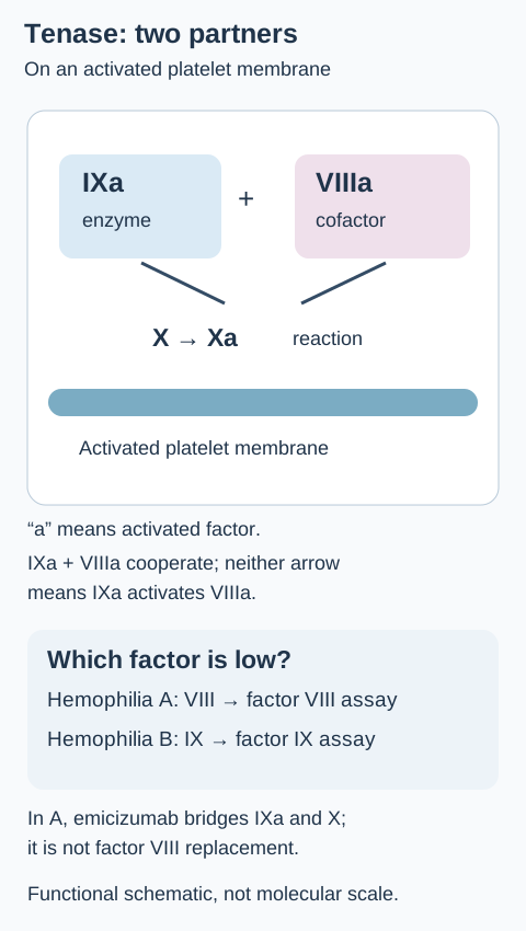
IXa enzyme and VIIIa cofactor cooperate on an activated platelet membrane to convert X to Xa; factor VIII and IX assays distinguish hemophilia A and B. Emicizumab bridges IXa and X in A.
At an activated platelet membrane, IXa enzyme plus VIIIa cofactor converge on the X to Xa reaction; A lacks VIII and B lacks IX, distinguished by specific factor assays. Activated forms carry the suffix a. Not molecular scale. [1] [3]

Use the adjacent laboratory comparison schematic as a sorting exercise: first identify whether platelet number is low, then whether prothrombin time (PT) or activated partial thromboplastin time (aPTT) is prolonged. An isolated prolonged aPTT with preserved PT and platelet count can fit factor VIII deficiency (hemophilia A) or factor IX deficiency (hemophilia B). The factor activity assay, not the joint involved, distinguishes the two. A normal aPTT cannot exclude mild hemophilia; investigate a convincing bleeding history with appropriate specific assays. [1]

A woman with factor VIII activity below 40% can have mild hemophilia A rather than merely an asymptomatic carrier label; an affected father makes his daughter an obligate genetic carrier regardless of her bleeding severity. [1][8]

Predict a change: a patient with repeated knee bleeds has low factor IX activity but normal factor VIII activity. The label changes from hemophilia A to hemophilia B, while the joint-risk mechanism remains. Deficiency means reduced functional activity; do not assume the antigen concentration must be normal in A or that the activity threshold automatically prescribes an identical treatment for everyone. [1]

A 1:1 mixing study combines patient plasma with normal plasma. Initial correction can support factor deficiency, whereas noncorrection, including after incubation when indicated, raises an inhibitor or another interfering substance. In a previously treated patient whose factor VIII activity remains unexpectedly low after replacement, test for a factor VIII inhibitor; poor response alone does not establish its presence. Lupus anticoagulant can prolong aPTT without producing the characteristic inherited deep-bleed phenotype. [1]

A final assay qualification matters with modern prevention: emicizumab functionally bridges activated factor IX and factor X to substitute for activated factor VIII in hemophilia A, but does not replace factor IX in B. It shortens standard aPTT and can distort activated partial thromboplastin time based factor VIII assays. If clinical assessment requires factor VIII activity or inhibitor testing during emicizumab use, arrange an appropriate bovine-reagent chromogenic assay through the specialist laboratory. A short aPTT in this setting is not proof of normal endogenous factor VIII. [1][3]

A supervised desmopressin challenge can show whether stored factor VIII rises enough, for long enough, to treat an appropriate mild or moderate hemophilia A bleed or minor procedure. It does not increase factor IX and is not a universal substitute for factor replacement. Assess the achieved activity, required duration and risks of water retention and hyponatremia; severe bleeding or prolonged coverage may instead need factor concentrate. [1]

Restore hemostasis without overlooking a dangerous joint

A person with known severe hemophilia develops a familiar swollen knee shortly after a minor collision; there is no fever, fracture concern, or nerve or vessel deficit. What is the first clinical priority? Follow the adjacent decision-fork diagram: promptly provide the patient-specific hemostatic treatment and protect the joint; do not delay treatment solely to obtain advanced imaging of an otherwise typical bleed. Arrange assessment and a rehabilitation plan with the hemophilia team. [1]

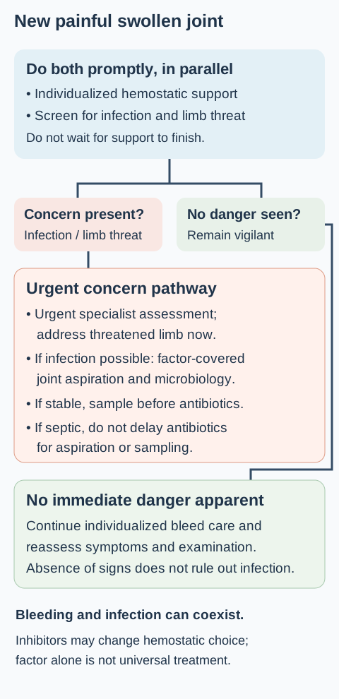
A new swollen joint calls for individualized hemostatic support and prompt parallel screening for infection and limb threat. Infection concern requires urgent assessment and factor-covered aspiration; sample before antibiotics if stable, but do not delay antibiotics in sepsis. If no danger is apparent continue bleed care and reassess: infection remains possible, and bleeding and infection can coexist.
For a new painful swollen joint, start individualized hemostatic support while screening promptly in parallel for infection and limb threat. Infection concern warrants urgent assessment and factor-covered aspiration with microbiology; sample before antibiotics if stable, but never delay antibiotics in sepsis. If danger is not apparent, continue bleed care and reassess; infection is not excluded. Inhibitors can change hemostatic choice. [1] [4]

Now change one branch: the knee is hot, the patient is febrile and cannot tolerate passive movement. Hemophilia does not prevent septic arthritis. Suspicion calls for rapid evaluation, joint-fluid sampling for microbiology and appropriate antimicrobial therapy while coordinating hemostatic coverage for the invasive procedure. In a stable patient, obtain samples before empiric antibiotics when feasible; with sepsis, do not delay antibiotics for aspiration. This is a combined infection-and-bleeding plan, not a choice between them. [1][4]

Try another change: the patient is afebrile but has a painful new effusion and a high-risk source of bacteremia. Absence of fever or a reassuring inflammatory blood test cannot reliably exclude joint infection. Likewise, crystals found in joint fluid do not rule it out. Reconsider the differential if a presumed bleed does not improve as expected, particularly with escalating pain or systemic symptoms. Aspiration is not routinely necessary to prove every typical hemarthrosis, but suspected infection warrants a protected diagnostic procedure. [1][4]

After an injury with focal bony tenderness, deformity or inability to bear weight, evaluate for fracture as well as bleeding. After marked swelling with progressive paresthesias, weakness or vascular concern, obtain immediate emergency assessment for threatened limb function. These findings alter concurrent investigation and urgency; they do not make clotting support optional in a bleeding patient. In an inhibitor patient, an ineffective usual factor dose requires a specialist alternative hemostatic strategy rather than blind repetition of the same dose. [1]

Distinguish effusion from its cause. Ultrasound readily shows joint fluid and synovial hypertrophy; magnetic resonance imaging depicts cartilage, subchondral changes and hemosiderin, while plain radiographs may remain normal early in arthropathy. Neither imaging nor a fluid level independently diagnoses uncomplicated hemophilic bleeding. After acute knee trauma, a verified intra-articular fat-fluid level raises concern for marrow fat from an intra-articular fracture; a simple fluid-fluid level may also be blood separating into layers. Seek fracture-directed imaging without postponing indicated hemostasis. [1][5][7]

An enlarging calf hematoma with severe pain on passive stretch and new sensory change can signal compartment syndrome despite palpable distal pulses. Groin pain with a flexed hip, weak knee extension and a reduced knee reflex suggests an iliopsoas bleed compressing the femoral nerve. Both scenarios need urgent specialist assessment and individualized hemostatic support, not routine rehabilitation during the acute event. [1]

When prophylaxis with emicizumab does not prevent a bleed, treatment depends on inhibitor status: factor VIII remains effective in a patient without an inhibitor, but a high-responding factor VIII inhibitor generally requires bypassing treatment. Recombinant activated factor VII is preferred over activated prothrombin complex concentrate during emicizumab prophylaxis because the combination with the latter increases thrombosis and thrombotic microangiopathy risk. Coordinate selection with the hemophilia team; an extra emicizumab dose is not an acute rescue treatment. [1]

Try it here · Checkpoint 2 of 3

Make your prediction before reading the choices. A first attempt is just a starting point.

Case 15

A stable 29-year-old with hemophilia A and an indwelling catheter presents with an acutely hot knee and marked pain on passive motion. Blood cultures were drawn; no antibiotic has been given. Which sequence best preserves diagnostic yield and addresses both hazards?

Show answer and explanations for case 15
  1. A. Observe after factor replacement; aspirate only if fever appears (Why this does not fit)

    Catheter-associated infection and hot painful joint demand evaluation even without fever.

    Reasoning steps for option A
    1. Why not label the hot knee a familiar hemarthrosis and observe?

      An indwelling catheter and marked passive-motion pain raise septic arthritis concern even without documented fever.

    2. What should happen while bleeding risk is controlled?

      Arrange prompt diagnostic aspiration with cultures, rather than waiting for fever.

  2. B. Begin several days of antibiotics, then obtain cultures (Why this does not fit)

    Delaying sampling until after antibiotics can reduce organism recovery in this stable patient.

    Reasoning steps for option B
    1. Why is antibiotic-first sampling delay avoidable in this stable patient?

      Blood cultures are already drawn, but joint fluid before antibiotics improves organism recovery when aspiration is promptly feasible.

    2. Would the same sequencing apply during septic shock?

      No; shock demands immediate antibiotics and resuscitation without waiting for transfer or aspiration.

  3. C. Factor-covered aspiration, cultures, then antibiotics (Best answer)

    Hemostatic coverage facilitates safe sampling; culture before antibiotics when feasible, then treat promptly.

    Reasoning steps for option C
    1. What does factor coverage accomplish before knee aspiration?

      It reduces procedure-related bleeding in hemophilia while allowing synovial fluid culture before antibiotics in a stable patient.

    2. After obtaining fluid, what should not be postponed?

      Prompt antimicrobial treatment based on the clinical infection concern.

  4. D. Aspirate immediately without hemostatic planning, then consider antibiotics (Why this does not fit)

    Unprotected puncture needlessly adds bleeding risk; infection treatment must not be deferred.

    Reasoning steps for option D
    1. Why does unprotected needle entry create a second hazard?

      Aspiration in hemophilia can itself bleed; coordinating hemostatic coverage protects the joint without abandoning infection diagnosis.

    2. Does arranging factor mean deferring antibiotics for days?

      No; plan expedited covered sampling and then prompt antibiotics, with urgency overriding sampling delay if unstable.

Takeaway: In a stable patient, obtain factor-covered joint fluid for culture before empiric antibiotics when promptly feasible; do not delay antibiotics in sepsis.

Case sources: [1] [4]

Prevent recurrence, then measure what remains

An ankle that swells every few weeks is not adequately managed by repeatedly treating episodes without revisiting prevention. Regular prophylaxis aims to prevent bleeding before it injures synovium. In a randomized study of 65 young boys with severe hemophilia A, 93% assigned regular factor VIII prophylaxis versus 55% assigned enhanced episodic therapy had normal index-joint structure on magnetic resonance imaging at age six. These figures describe that particular trial, not reversal of established arthropathy or efficacy in every other population. [1][2]

Emicizumab is a factor VIII function-mimicking prophylactic antibody for hemophilia A, not an acute replacement for every joint bleed. In a randomized comparison among eligible participants aged at least 12 years without inhibitors who previously received episodic treatment, weekly or every-two-week prophylaxis reduced treated bleeding relative to no prophylaxis. Its trial endpoint was treated bleeding, not restoration of already damaged cartilage. Individualize prophylaxis and breakthrough-bleed plans with the specialist team. [1][3]

Predict what happens if prophylaxis reduces new bleeding after years of ankle damage: the recurrent blood exposure may decrease, but existing cartilage loss does not instantly disappear. Follow serial pain, function, range of motion and joint evaluation; coordinated physical therapy and musculoskeletal care address mobility and protection. If a target joint still bleeds, reassess treatment adherence, exposure and potential inhibitors rather than treating persistent swelling as inevitable. [1]

One last three-part retrieval: ask where bleeding occurs, what the factor assays show, and whether a new danger signal changes the action. A person with nosebleeds and heavy menses needs a different initial diagnostic emphasis than one with repeated hemarthroses; an isolated prolonged aPTT does not pick VIII over IX; and a febrile joint needs infection workup even when hemophilia is already established. Use these distinctions again in the independent cases below. [1][4]

Test the boundary: a child with new hip pain may have a deep hematoma without a visible swollen joint. Reassess the time course, neurologic findings and response to the individualized hemostatic plan; escalating pain may instead require evaluation for infection, fracture or treatment resistance. Hemostasis and targeted investigation proceed together rather than replacing one another. [1]

For recurrent bleeding despite adequate prevention, distinguish ongoing synovitis from end-stage joint destruction. After optimizing prophylaxis and excluding inhibitors or other causes, persistent vascular synovium with preserved cartilage can prompt consideration of nonsurgical synoviorthesis under specialist care; it is not automatically an indication for joint replacement. End-stage deformity, lost joint space and disabling pain warrant coordinated orthopedic and rehabilitation evaluation with continued hemostatic planning. [1]

Try it here · Checkpoint 3 of 3

Make your prediction before reading the choices. A first attempt is just a starting point.

Case 22

A 28-year-old with hemophilia A has recurrent spontaneous knee hemarthroses despite verified adherence to optimized prophylaxis; inhibitors are excluded. magnetic resonance imaging shows hypertrophic vascular synovium but preserved articular cartilage and useful range of motion. Which joint-directed escalation best fits?

Show answer and explanations for case 22
  1. A. Immediate total knee arthroplasty (Why this does not fit)

    Arthroplasty is generally reserved for advanced destructive arthropathy, not preserved cartilage.

    Reasoning steps for option A
    1. Why is immediate arthroplasty premature?

      magnetic resonance imaging shows preserved knee cartilage and useful motion despite recurrent bleeding, so an end-stage replacement strategy sacrifices a salvageable joint.

    2. What contrasting image would make arthroplasty more relevant?

      Advanced destructive cartilage loss and disabling mechanical symptoms despite optimized conservative care.

  2. B. Consider synoviorthesis with specialist multidisciplinary care (Best answer)

    Persistent bleeding from hypertrophic synovium despite prevention may benefit from synoviorthesis.

    Reasoning steps for option B
    1. Which mechanism is synoviorthesis meant to interrupt?

      Hypertrophic vascular synovium perpetuates knee hemarthroses despite verified optimized prophylaxis and excluded inhibitors.

    2. What should continue alongside joint-directed therapy?

      Multidisciplinary hematology, imaging, rehabilitation, and individualized prophylaxis rather than isolated procedure care.

  3. C. Stop prophylaxis and rely on episodic factor alone (Why this does not fit)

    Withdrawing prevention exposes the still functional joint to more bleeding.

    Reasoning steps for option C
    1. Why not replace optimized prophylaxis with episodic factor alone?

      Recurrent spontaneous bleeds already threaten a joint whose cartilage is still preserved; abandoning prevention can accelerate injury.

    2. What does failure of one prevention layer imply?

      Add appropriate local evaluation and intervention, not withdrawal of systemic protection.

  4. D. Treat permanent fixed deformity with joint fusion (Why this does not fit)

    Fusion addresses severe structural disease, not a mobile joint with intact cartilage.

    Reasoning steps for option D
    1. Why does fusion not match the current knee state?

      Fusion addresses severe fixed destruction, while this patient retains useful range of motion and preserved cartilage.

    2. Which symptom pattern would favor a salvage-focused approach instead?

      Bleeding driven by vascular synovium rather than fixed mechanical deformity.

Takeaway: For recurrent bleeding from vascular synovitis despite optimized prevention and preserved cartilage, consider specialist synoviorthesis.

Case sources: [1] [5]

Independent cases

Case 1

A 12-year-old has repeated spontaneous elbow swelling and a prolonged activated partial thromboplastin time. A 1:1 mixture corrects immediately and after two hours at 37 C. Factor VIII activity is 2%, factor IX activity 89%; platelets and prothrombin time are normal. Which interpretation best accounts for the results?

Show answer and explanations for case 1
  1. A. Factor VIII deficiency without a demonstrable time-dependent inhibitor (Best answer)

    Sustained correction supports deficiency; selective VIII loss explains the deep bleeding.

    Reasoning steps for option A
    1. Why does a mixing study help here but not finish the diagnosis?

      Fresh normal plasma supplies the missing activity; correction persisting through incubation favors deficiency, while VIII 2% versus IX 89% identifies which factor is missing.

    2. Would this conclusion survive loss of correction after two hours?

      No. A falling incubated correction would trigger a search for a time-dependent VIII inhibitor rather than uncomplicated deficiency.

  2. B. Factor IX deficiency without a demonstrable time-dependent inhibitor (Why this does not fit)

    IX is preserved, so the same mixing pattern does not establish IX deficiency.

    Reasoning steps for option B
    1. Why might hemophilia B initially seem plausible in this elbow-bleed history?

      IX deficiency also causes deep spontaneous bleeding and a prolonged activated partial thromboplastin time that corrects with mixing; the IX activity of 89% rules it out here.

    2. What result would shift the diagnosis toward B?

      Selective low IX with preserved VIII would reverse the factor-specific interpretation.

  3. C. Time-dependent factor VIII inhibitor despite sustained mixing correction (Why this does not fit)

    An incubated inhibitor commonly abolishes correction; here correction persists.

    Reasoning steps for option C
    1. What makes an acquired VIII inhibitor worth considering at first glance?

      A prolonged activated partial thromboplastin time and low VIII can occur with an inhibitor, but this mixture remains corrected after two hours at 37 C.

    2. What laboratory reversal would support this alternative?

      Correction immediately followed by prolongation after incubation would suggest a time-dependent inhibitor and merit inhibitor quantification.

  4. D. Lupus anticoagulant causing the low factor VIII activity (Why this does not fit)

    An interfering anticoagulant may affect activated partial thromboplastin time but does not explain this correction and concordant severe VIII deficit.

    Reasoning steps for option D
    1. Can lupus anticoagulant explain any part of the screen?

      It can prolong activated partial thromboplastin time, making it tempting, but sustained mixing correction plus a profound selective VIII deficit and recurrent deep bleeds point elsewhere.

    2. If antiphospholipid interference were suspected despite these findings, what would matter?

      Use appropriate confirmatory phospholipid-dependent studies and assess assay interference rather than attributing VIII 2% automatically to lupus anticoagulant.

Takeaway: Persistent mixing correction plus selectively low VIII favors deficiency; an incubated noncorrecting mix raises concern for inhibitor.

Case sources: [1]

Case 2

A 16-year-old with documented mild hemophilia A (baseline VIII 12%) had a supervised desmopressin challenge: VIII increased to 58% and remained 42% at four hours. He needs one uncomplicated dental extraction tomorrow, with local dental hemostasis available; no cardiovascular disease or hyponatremia risk is present. Which planned hemostatic agent fits the response?

Show answer and explanations for case 2
  1. A. Factor IX concentrate (Why this does not fit)

    IX replacement does not address documented VIII deficiency.

    Reasoning steps for option A
    1. Why is IX concentrate the wrong replacement despite a bleeding-risk procedure?

      His diagnosed defect is VIII, not IX; the supervised challenge already raised VIII from 12% to 58%.

    2. When would IX replacement become relevant?

      Documented IX deficiency, rather than a responsive mild A phenotype, would require IX-directed coverage.

  2. B. Desmopressin with fluid precautions (Best answer)

    Measured and sustained VIII release supports desmopressin for this brief minor procedure, alongside individualized local measures and fluid precautions.

    Reasoning steps for option B
    1. Which part of the challenge supports a minor-procedure desmopressin plan?

      VIII remained 42% four hours after rising to 58%, demonstrating useful magnitude and duration for uncomplicated extraction.

    2. What safety detail accompanies that choice even without baseline hyponatremia risk?

      Limit free water and follow local sodium precautions because desmopressin can cause water retention and hyponatremia.

  3. C. Emicizumab as a single perioperative rescue dose (Why this does not fit)

    Emicizumab is prevention, not a one-time substitute for perioperative coverage.

    Reasoning steps for option C
    1. Why could emicizumab sound useful for a patient with A?

      It can prevent bleeds in A, but a new loading injection tomorrow will not provide tailored immediate extraction hemostasis like his proven desmopressin response.

    2. Would established emicizumab prophylaxis remove all procedural planning?

      No; procedural hemostasis still needs an individualized plan rather than an assumed rescue dose.

  4. D. Recombinant activated factor VII as first-line bypass (Why this does not fit)

    Bypass therapy is reserved for selected inhibitor scenarios, not responsive mild A.

    Reasoning steps for option D
    1. What finding makes a bypass agent disproportionate for this extraction?

      He has mild A with robust endogenous VIII release and no described inhibitor; activated factor VII bypass is not the first response to this setting.

    2. What new complication might make bypass therapy relevant?

      A clinically important inhibitor preventing effective VIII-based hemostasis would change the agent discussion.

Takeaway: In mild hemophilia A, use a documented adequate desmopressin response for suitable brief procedures with fluid precautions and local measures.

Case sources: [1]

Case 3

An 11-year-old with hemophilia B (IX activity 4%) has an uncomplicated recurrent ankle bleed. His prior factor IX infusions produced expected recovery; inhibitor screening is negative. Which factor-specific acute therapy should be selected?

Show answer and explanations for case 3
  1. A. Recombinant factor VIII (Why this does not fit)

    VIII does not replace the deficient IX in B.

    Reasoning steps for option A
    1. Why does VIII concentrate fail despite being a familiar hemophilia drug?

      This is hemophilia B with IX at 4%; raising VIII leaves the deficient IX-dependent step unresolved.

    2. Which factor result would justify switching to VIII?

      A demonstrated VIII deficit rather than the measured IX deficiency would support VIII replacement.

  2. B. Desmopressin for endogenous VIII release (Why this does not fit)

    Desmopressin releases VIII/von Willebrand factor, not clinically sufficient IX.

    Reasoning steps for option B
    1. Why is desmopressin not a substitute for IX in this ankle bleed?

      It mobilizes VIII and von Willebrand factor, whereas this child needs IX after an uncomplicated bleed.

    2. Does a previously successful IX infusion make desmopressin more attractive?

      No; expected IX recovery is evidence that direct IX replacement works.

  3. C. Factor IX concentrate (Best answer)

    IX replacement matches the measured deficiency and previously demonstrated recovery.

    Reasoning steps for option C
    1. How do the prior infusions sharpen the acute decision?

      They yielded expected IX recovery, and inhibitor screening is negative, so matched IX concentrate is likely to work again.

    2. What would poor postinfusion IX recovery prompt instead?

      Investigate an IX inhibitor and reassess hemostatic therapy rather than assuming routine replacement will suffice.

  4. D. Emicizumab loading injection (Why this does not fit)

    VIII mimetic prophylaxis cannot replace IX during a B bleed.

    Reasoning steps for option D
    1. Why not give the prophylactic VIII mimetic as acute rescue?

      Emicizumab mimics VIII cofactor action for A and cannot supply the IX absent in this B patient.

    2. Would a larger loading injection replace IX immediately?

      No; neither its target nor its preventive dosing matches acute IX replacement.

Takeaway: An acute bleed in hemophilia B without an inhibitor requires matched factor IX rather than factor VIII or desmopressin.

Case sources: [1]

Case 4

A 9-year-old with severe hemophilia A, no inhibitor on recent testing and previously normal factor VIII recovery develops a rapidly painful knee after a fall. There is no fever, bony tenderness or neurovascular deficit. Which immediate hemostatic agent best fits?

Show answer and explanations for case 4
  1. A. Factor IX concentrate (Why this does not fit)

    IX cannot correct isolated VIII deficiency.

    Reasoning steps for option A
    1. What single missing protein makes IX an apparent but incorrect factor choice?

      Both A and B can cause a swollen knee, but this child has known severe A and previously responded to VIII.

    2. What diagnostic change would favor IX concentrate?

      Established B with low IX, rather than known severe VIII deficiency, would make IX the matched factor.

  2. B. Recombinant activated factor VII (Why this does not fit)

    Bypassing VIII is unnecessary when infused VIII works and no inhibitor is present.

    Reasoning steps for option B
    1. Why avoid bypassing VIII in this fall-related bleed?

      Recent inhibitor testing is negative and prior VIII recovery normal; bypassing is unnecessary when direct factor works.

    2. When might activated factor VII become the safer acute route?

      A high-responding VIII inhibitor blocking replacement would justify an inhibitor-focused bypass strategy.

  3. C. Desmopressin alone (Why this does not fit)

    Severe A generally needs replacement for an acute substantial joint bleed.

    Reasoning steps for option C
    1. Does a severe A bleed resemble the mild A cases treated with desmopressin?

      Both involve VIII, but severe A cannot be presumed to release enough endogenous factor for this rapidly painful joint bleed.

    2. What would make desmopressin an option in another patient?

      A measured adequate and sustained challenge response in mild A for an appropriate lesser hemostatic challenge.

  4. D. Factor VIII concentrate (Best answer)

    Prompt VIII replacement limits ongoing blood exposure in this uncomplicated bleed.

    Reasoning steps for option D
    1. Why give VIII before waiting for imaging or later symptoms?

      Rapid knee pain after trauma in severe A signals active hemarthrosis; normal neurovascular findings and absent fever do not negate prompt hemostasis.

    2. What would alter this straightforward treatment pathway?

      Fever, bony tenderness, or neurovascular compromise would prompt parallel infection, fracture, or limb-threat assessment.

Takeaway: For a typical new joint bleed in severe hemophilia A without inhibitors, treat promptly with factor VIII and protect the joint.

Case sources: [1]

Case 5

A 25-year-old with severe hemophilia A on emicizumab prophylaxis has a breakthrough wrist hemarthrosis. He has never had an inhibitor, and previous factor VIII infusions showed expected recovery. Which acute hemostatic treatment is appropriate?

Show answer and explanations for case 5
  1. A. Factor VIII concentrate (Best answer)

    Emicizumab prevention does not preclude use of effective VIII for breakthrough bleeding without inhibitors.

    Reasoning steps for option A
    1. Does emicizumab prevent use of infused VIII for this breakthrough wrist bleed?

      No; no inhibitor history and previously expected VIII recovery make VIII a matched acute treatment despite preventive antibody therapy.

    2. What change would weaken reliance on infused VIII?

      New poor factor recovery or a documented high-responding inhibitor would require an inhibitor-directed approach.

  2. B. Activated prothrombin complex concentrate (Why this does not fit)

    activated prothrombin complex concentrate is not indicated here and carries thrombotic risk with emicizumab.

    Reasoning steps for option B
    1. What special harm makes activated prothrombin complex concentrate a poor shortcut on emicizumab?

      activated prothrombin complex concentrate combined with emicizumab can increase thrombosis and thrombotic microangiopathy risk, with no inhibitor-driven need here.

    2. Could activated prothrombin complex concentrate ever be considered at all in an inhibitor patient?

      Only under specialist oversight if alternatives fail, with careful risk and dose consideration; it is not preferred here.

  3. C. Recombinant activated factor VII instead of effective VIII (Why this does not fit)

    activated factor VII is useful for inhibitor bleeds but need not displace functional VIII in this patient.

    Reasoning steps for option C
    1. Why is activated factor VII not the best default simply because bleeding broke through prophylaxis?

      Breakthrough does not imply VIII neutralization; this patient historically recovers well after VIII.

    2. What specific evidence would favor activated factor VII over VIII?

      A high-responding inhibitor that makes infused VIII ineffective would favor bypassing therapy.

  4. D. An extra emicizumab dose in place of acute therapy (Why this does not fit)

    A prophylactic antibody dose is not rapid matched treatment for this joint bleed.

    Reasoning steps for option D
    1. Why does another preventive injection not settle active hemarthrosis?

      Emicizumab dosing maintains prophylaxis; the currently bleeding wrist needs prompt effective acute hemostasis.

    2. What additional observation would still require reassessment after acute treatment?

      Persistent swelling or pain despite effective VIII warrants checking recovery and considering other joint pathology.

Takeaway: Emicizumab prevents many bleeds but does not rescue an acute bleed; effective factor VIII remains appropriate when no inhibitor is present.

Case sources: [1] [3]

Case 6

A 22-year-old with severe hemophilia A receives emicizumab and has a documented high-responding factor VIII inhibitor. He develops a spontaneous knee bleed without infection or fracture signs. Which acute agent avoids dependence on neutralized VIII and avoids activated prothrombin complex concentrate-related risk with emicizumab?

Show answer and explanations for case 6
  1. A. Factor VIII concentrate at his former home dose (Why this does not fit)

    High-responding inhibitor makes conventional VIII unreliable.

    Reasoning steps for option A
    1. Why not reuse his former home VIII dose?

      A documented high-responding inhibitor neutralizes VIII, so the old effective dose cannot be trusted now.

    2. What evidence would change confidence in direct replacement?

      Demonstrated adequate VIII recovery without clinically important inhibitor would allow reconsideration.

  2. B. Recombinant activated factor VII (Best answer)

    activated factor VII bypasses VIII and is preferred for inhibitor breakthrough bleeding on emicizumab.

    Reasoning steps for option B
    1. Which two constraints select activated factor VII for this knee bleed?

      Recombinant activated factor VII bypasses neutralized VIII and avoids the particularly problematic activated prothrombin complex concentrate-emicizumab combination.

    2. Does emicizumab prophylaxis eliminate the need for acute treatment?

      No; the spontaneous knee bleed still needs hemostatic therapy despite prevention.

  3. C. Activated prothrombin complex concentrate as preferred first-line (Why this does not fit)

    Concomitant activated prothrombin complex concentrate and emicizumab increase thrombotic microangiopathy/thrombosis risk.

    Reasoning steps for option C
    1. Why is activated prothrombin complex concentrate tempting yet not preferred here?

      It bypasses VIII, but pairing activated prothrombin complex concentrate with emicizumab heightens thrombosis and thrombotic microangiopathy risk.

    2. What monitoring issue would arise if specialists had no alternative?

      They would need tightly controlled exposure and surveillance for thrombosis or microangiopathy, not routine first-line activated prothrombin complex concentrate.

  4. D. Desmopressin (Why this does not fit)

    Severe VIII loss plus inhibitor is not corrected by endogenous release.

    Reasoning steps for option D
    1. Why is endogenous VIII release unlikely to solve this case?

      Severe A leaves little reserve, and the released VIII would still face the documented inhibitor.

    2. In which contrasting A patient might desmopressin help?

      Someone with mild A and a proven adequate sustained VIII response, not severe inhibitor-positive A.

Takeaway: With a high-responding factor VIII inhibitor on emicizumab, recombinant activated factor VII is preferred for breakthrough bleeding.

Case sources: [1] [3]

Case 7

A child on emicizumab has an unexpectedly high one-stage factor VIII result despite genetically confirmed severe hemophilia A. The team needs endogenous factor VIII activity before changing treatment. Which assay should the laboratory use?

Show answer and explanations for case 7
  1. A. One-stage activated partial thromboplastin time based factor VIII activity (Why this does not fit)

    Emicizumab falsely elevates activated partial thromboplastin time based VIII estimates.

    Reasoning steps for option A
    1. Why does the surprisingly high one-stage VIII activity mislead?

      Emicizumab shortens activated partial thromboplastin time based clotting tests, creating an apparent activity inconsistent with genetically severe A.

    2. Would repeating the same one-stage method settle endogenous VIII?

      No; the same drug interference persists regardless of repeated measurement.

  2. B. Human-reagent chromogenic factor VIII activity (Why this does not fit)

    Human activated factor IX/factor X reagents respond to the antibody and overestimate native VIII.

    Reasoning steps for option B
    1. Why is human chromogenic testing still vulnerable?

      Emicizumab bridges human activated factor IX and factor X reagents, so a human-reagent chromogenic assay can read antibody activity as VIII-like activity.

    2. What reagent change isolates native VIII better?

      Use bovine-reagent chromogenic components that respond minimally to emicizumab.

  3. C. Bovine-reagent chromogenic factor VIII activity (Best answer)

    Bovine components do not react significantly with emicizumab and allow native VIII measurement.

    Reasoning steps for option C
    1. What property makes bovine chromogenic testing fit this precise question?

      The bovine activated factor IX/factor X reagent system has little response to emicizumab and can estimate endogenous VIII rather than its mimic.

    2. Would this same logic matter for inhibitor quantification on emicizumab?

      Yes; a bovine chromogenic residual-VIII method can avoid misleading antibody-derived activity.

  4. D. Routine activated partial thromboplastin time alone (Why this does not fit)

    Clotting time alone cannot quantify native factor activity on this drug.

    Reasoning steps for option D
    1. Why is a routine activated partial thromboplastin time insufficient even if it looks normal?

      Emicizumab can normalize or shorten activated partial thromboplastin time without restoring native VIII in this severe A child.

    2. Which inference must be avoided from a normal clotting time here?

      Do not infer normal endogenous VIII or absence of an anti-factor VIII inhibitor from drug-affected activated partial thromboplastin time.

Takeaway: Emicizumab invalidates routine clot-based VIII quantitation; use an appropriate bovine-reagent chromogenic VIII assay.

Case sources: [1] [3]

Case 8

A patient on emicizumab previously had a high-titer factor VIII inhibitor. The hematologist must quantify whether neutralizing anti-factor VIII activity persists. Which laboratory strategy is interpretable?

Show answer and explanations for case 8
  1. A. Conventional one-stage inhibitor quantification assay alone (Why this does not fit)

    Emicizumab confounds conventional activated partial thromboplastin time dependent residual VIII assays.

    Reasoning steps for option A
    1. Why can the conventional Nijmegen-Bethesda approach misread this inhibitor?

      Its activated partial thromboplastin time based residual VIII measurement is affected by emicizumab, which can mask true neutralization.

    2. What assay modification targets the interference?

      Measure residual VIII using a validated bovine chromogenic substrate method.

  2. B. activated partial thromboplastin time normalization after incubation (Why this does not fit)

    The antibody shortens activated partial thromboplastin time even if anti-factor VIII neutralization remains.

    Reasoning steps for option B
    1. Can a normalized activated partial thromboplastin time prove the old inhibitor has disappeared?

      No; the prophylactic antibody shortens the test even if the anti-factor VIII antibody persists.

    2. What result is needed before concluding inhibitor status changed?

      An interpretable inhibitor titer measured by an emicizumab-insensitive method.

  3. C. Human chromogenic factor VIII inhibitor assay (Why this does not fit)

    Human chromogenic reagents respond to emicizumab and can obscure residual activity.

    Reasoning steps for option C
    1. Why does a human chromogenic Bethesda readout remain problematic?

      Human activated factor IX/factor X reagents respond to emicizumab; apparent residual activity may not be genuine VIII activity.

    2. Would an inhibitor-positive history make this interference negligible?

      No; the question is persistence of neutralizing antibody, so residual VIII must be measured without the drug signal.

  4. D. Bovine chromogenic factor VIII inhibitor assay (Best answer)

    Bovine chromogenic residual VIII assessment is relatively insensitive to emicizumab and supports inhibitor quantification.

    Reasoning steps for option D
    1. What is measured by the bovine chromogenic inhibitor strategy?

      Residual functional VIII after mixing/incubation, using reagents that do not substantially register emicizumab.

    2. Why not simply discontinue inhibitor assessment because prophylaxis works?

      Inhibitor status affects future factor replacement and procedures even while breakthrough bleeds are prevented.

Takeaway: During emicizumab prophylaxis, quantify a factor VIII inhibitor with a bovine-reagent chromogenic inhibitor method.

Case sources: [1]

Case 9

A 30-year-old with severe hemophilia A receives supervised factor VIII for a knee effusion. Baseline activity is 1%; the measured peak is far below the expected postdose level, and a delayed sample falls rapidly. Which investigation directly tests the likely cause of replacement failure?

Show answer and explanations for case 9
  1. A. Quantitative factor VIII inhibitor assay (Best answer)

    Poor immediate recovery and rapid loss of VIII raise concern for neutralization, requiring inhibitor testing.

    Reasoning steps for option A
    1. What makes an inhibitor assay the immediate investigation?

      VIII starts at 1%, then recovers much less than predicted after supervised dosing and falls rapidly; neutralization is a testable explanation.

    2. Would an isolated low baseline VIII establish an inhibitor?

      No; unexpectedly poor measured postinfusion recovery provides the crucial additional clue.

  2. B. magnetic resonance imaging for chronic cartilage thinning (Why this does not fit)

    Cartilage imaging characterizes injury but not failure of circulating factor recovery.

    Reasoning steps for option B
    1. Why might magnetic resonance imaging be clinically interesting but not answer this question?

      A knee effusion can coexist with cartilage injury, yet cartilage thinning cannot explain a low circulating VIII peak after infusion.

    2. When would magnetic resonance imaging become the more fitting next study?

      Persistent mechanical symptoms after effective hemostasis would shift attention to joint structure.

  3. C. Synovial ultrasound for hypertrophy (Why this does not fit)

    Synovial thickening can explain recurring bleeds but not unexpectedly absent postinfusion VIII.

    Reasoning steps for option C
    1. Could vascular synovium explain repeated bleeding here?

      Yes, it can perpetuate hemarthroses, but it cannot account for the unexpectedly low and rapidly declining VIII activity in blood.

    2. Which test separates synovial recurrence from factor failure?

      Measured postdose recovery and a quantitative inhibitor assay address the biochemical failure.

  4. D. Plain radiograph for joint-space narrowing (Why this does not fit)

    Bony changes cannot account for markedly low immediate VIII activity.

    Reasoning steps for option D
    1. Why is a plain film too remote from the postdose finding?

      Joint-space loss documents chronic damage, not destruction or neutralization of administered VIII.

    2. If radiographs showed narrowing, would that rule out inhibitor?

      No; structural disease and inhibitor-mediated replacement failure can coexist.

Takeaway: Unexpectedly poor factor VIII recovery after an observed dose warrants specific inhibitor testing.

Case sources: [1]

Case 10

A 34-year-old with hemophilia A receives factor VIII for an acutely painful ankle. Peak and trough VIII activities match the expected recovery profile and the effusion resolves, but focal pain and restricted motion persist for months without fever. Which next evaluation best addresses the residual symptom?

Show answer and explanations for case 10
  1. A. Repeat VIII loading daily until all pain disappears (Why this does not fit)

    Adequate factor recovery and resolved effusion do not justify indefinite acute replacement.

    Reasoning steps for option A
    1. Why should factor not be loaded daily merely for months of pain?

      The expected peak and trough plus resolved effusion show acute factor treatment worked; persistent focal restriction suggests a different source.

    2. When would additional factor be newly justified?

      A fresh bleed or objective evidence of ongoing bleeding would require renewed hemostatic treatment.

  2. B. Joint imaging and functional assessment (Best answer)

    Persistent mechanical limitation despite hemostasis suggests established joint damage requiring structural and functional assessment.

    Reasoning steps for option B
    1. What changes the clinical question from hemostasis to joint structure?

      Pain and restricted ankle motion persist for months despite normal VIII recovery and effusion resolution.

    2. What should evaluation pair with imaging?

      Functional examination and rehabilitation needs, because scans alone cannot measure lived limitation.

  3. C. Diagnose inhibitor solely from persistent pain (Why this does not fit)

    An inhibitor is not inferred from pain when postdose factor recovery is appropriate.

    Reasoning steps for option C
    1. What evidence directly argues against inferring inhibitor from symptoms?

      Both measured peak and trough VIII match expectations after infusion; pain is not a replacement-recovery assay.

    2. What laboratory pattern would reopen inhibitor testing?

      An unexpectedly low immediate VIII peak or accelerated postdose decline.

  4. D. Treat presumed bacterial arthritis without sampling (Why this does not fit)

    Absent infection signals do not establish bacterial disease or warrant empirical treatment.

    Reasoning steps for option D
    1. Why are empirical antibiotics unsupported by this ankle history?

      No fever is reported, the effusion resolved, and limitation is chronic rather than a new infectious presentation.

    2. What new signs would change the infection workup?

      A newly hot joint, systemic illness, or infectious risk would warrant prompt sampling and cultures with factor cover.

Takeaway: Persistent restricted motion despite documented resolution of bleeding needs structural and functional joint assessment.

Case sources: [1] [5]

Case 12

An 8-year-old on prophylaxis has three atraumatic right-elbow bleeds over the last four months, each documented separately, and a single fall-related left-knee bleed. Which site should drive target-joint surveillance and prevention review?

Show answer and explanations for case 12
  1. A. Left knee, because trauma adds greatest diagnostic weight (Why this does not fit)

    One provoked knee bleed does not establish a recurrent spontaneous target joint.

    Reasoning steps for option A
    1. What makes the left-knee bleed less relevant to target-joint designation?

      It is a single fall-related event, unlike the three atraumatic right-elbow episodes.

    2. Could a knee become the focus later?

      Yes, if spontaneous knee bleeds recur in that same joint within the defined period.

  2. B. Both sites equally, because four total bleeds occurred (Why this does not fit)

    Thresholds apply to a single joint, not pooled events across joints.

    Reasoning steps for option B
    1. Why can the four events not be pooled into two target sites?

      The threshold is site-specific and based on spontaneous recurrence; three right elbows and one provoked left knee are not interchangeable.

    2. Which site would remain eligible if the knee episode were excluded?

      The right elbow still has three atraumatic bleeds in four months.

  3. C. Neither site, because prophylaxis prevents target-joint classification (Why this does not fit)

    Bleeds on prophylaxis still count and prompt reassessment.

    Reasoning steps for option C
    1. Does prophylaxis erase a documented spontaneous bleed?

      No; breakthrough atraumatic elbow bleeds still count and indicate that prevention should be reviewed.

    2. What practical question follows three such events on prophylaxis?

      Check adherence, regimen adequacy, and local joint disease while planning surveillance.

  4. D. Right elbow, because repeated spontaneous bleeding clusters there (Best answer)

    Three spontaneous elbow events in four months meet the standard target-joint definition.

    Reasoning steps for option D
    1. What combination establishes the right elbow as the surveillance priority?

      Three distinct atraumatic bleeds in one elbow over four months satisfy the standard recurrence threshold.

    2. Does the unrelated left-knee trauma modify that conclusion?

      No; it should be treated but does not dilute the elbow-specific count.

Takeaway: Repeated spontaneous bleeds at one site trigger target-joint surveillance and prophylaxis review even if other joints also bleed.

Case sources: [1]

Case 13

A 17-year-old with severe hemophilia B has recurrent ankle swelling and normal plain radiographs. The clinician needs to characterize early cartilage injury, subchondral changes and hemosiderin-laden synovium in one examination. Which modality best fits?

Show answer and explanations for case 13
  1. A. Magnetic resonance imaging (Best answer)

    magnetic resonance imaging assesses early soft tissue, cartilage and bone changes missed by radiographs.

    Reasoning steps for option A
    1. Why is magnetic resonance imaging the best single examination despite normal radiographs?

      magnetic resonance imaging can depict hemosiderin-rich synovium, cartilage injury, and subchondral change before plain films turn abnormal.

    2. What would a normal x-ray fail to reassure against?

      Early synovitis and cartilage damage after recurrent ankle bleeds.

  2. B. Plain radiography with comparison views (Why this does not fit)

    Radiographs are insensitive to early synovial and cartilage damage.

    Reasoning steps for option B
    1. Why not repeat radiographs for a more detailed early assessment?

      Comparison films still lack sensitivity for early soft tissue and cartilage abnormalities in this radiographically normal ankle.

    2. When are radiographs more useful?

      They can characterize later osseous deformity and joint-space loss rather than the requested early triad.

  3. C. Point-of-care ultrasound alone (Why this does not fit)

    Ultrasound can assess effusion and synovium but not comprehensive deep cartilage/subchondral injury.

    Reasoning steps for option C
    1. What makes ultrasound attractive but insufficient as the sole test?

      It readily detects fluid and synovial abnormalities, but deep cartilage and subchondral mapping are part of this request.

    2. When would bedside ultrasound alone better fit?

      A rapid acute effusion question without comprehensive structural staging.

  4. D. Bone scintigraphy (Why this does not fit)

    Bone scans are nonspecific and less anatomically informative for this question.

    Reasoning steps for option D
    1. Why is scintigraphy too nonspecific for this ankle?

      Tracer uptake does not map the synovium, cartilage and subchondral anatomy as required.

    2. What imaging feature distinguishes the selected modality?

      Direct multi-tissue anatomic visualization, rather than a nonspecific bone-turnover signal.

Takeaway: Magnetic resonance imaging characterizes early synovial, cartilage and subchondral injury when radiographs remain unrevealing.

Case sources: [1] [5]

Case 14

A child with hemophilia A has new painful knee swelling and suspected effusion; rapid bedside assessment is needed while hemostatic treatment is arranged. magnetic resonance imaging access requires transfer, and no deep cartilage mapping is requested. Which imaging approach best answers the immediate fluid question?

Show answer and explanations for case 14
  1. A. Serial plain radiographs (Why this does not fit)

    X-rays may show late bone changes but are relatively insensitive to effusion/synovial detail.

    Reasoning steps for option A
    1. What does a plain film miss in an acutely swollen knee?

      Effusion and synovial changes can be poorly shown; x-ray is more useful for fracture or established bone injury.

    2. Would focal bony tenderness change imaging priorities?

      Yes; trauma with focal bone signs could justify radiographs for fracture assessment.

  2. B. Point-of-care joint ultrasound (Best answer)

    Ultrasound rapidly characterizes joint fluid and synovial hypertrophy without delaying hemostasis.

    Reasoning steps for option B
    1. Why is bedside ultrasound matched to this encounter?

      It can answer whether joint fluid and synovial hypertrophy are present rapidly while factor treatment is arranged locally.

    2. Does ultrasound replace acute factor treatment?

      No; imaging must not delay hemostasis for a suspected active bleed.

  3. C. Delayed magnetic resonance imaging before any factor treatment (Why this does not fit)

    magnetic resonance imaging is helpful for structural staging but should not hold up acute therapy for a simple fluid question.

    Reasoning steps for option C
    1. Why does transfer for magnetic resonance imaging impose an avoidable cost?

      The question is immediate fluid detection, not deep cartilage mapping, and factor therapy should not await transfer.

    2. When would magnetic resonance imaging become more valuable later?

      When staging persistent synovial or cartilage injury after stabilization.

  4. D. computed tomography of the entire limb (Why this does not fit)

    computed tomography adds radiation and is not first choice to identify an uncomplicated effusion.

    Reasoning steps for option D
    1. Why does whole-limb computed tomography not target the bedside question?

      It adds radiation and is not the preferred first test for uncomplicated knee effusion or synovial detail.

    2. What contrasting finding could justify computed tomography or targeted fracture imaging?

      A suspected complex fracture or other structural trauma rather than isolated fluid assessment.

Takeaway: Ultrasound answers a rapid joint-fluid question without delaying necessary hemostatic therapy for advanced imaging.

Case sources: [1] [5]

Case 16

A 40-year-old with hemophilia B has a very hot swollen knee, hypotension, confusion and rigors. Aspiration requires transfer to another hospital. Which initial combined plan is best?

Show answer and explanations for case 16
  1. A. Wait for aspiration before starting any antimicrobial therapy (Why this does not fit)

    In shock, preserving joint-culture yield cannot justify delaying antibiotics.

    Reasoning steps for option A
    1. Why cannot transfer for aspiration delay antibiotics here?

      Hypotension, confusion, and rigors signal shock; culture yield is secondary to immediate life-saving sepsis therapy.

    2. How does this differ from a stable patient with prompt aspiration available?

      In stability, pre-antibiotic joint sampling is desirable; in shock, do not wait.

  2. B. Give factor IX only and observe for reversal of confusion (Why this does not fit)

    Factor IX can address bleeding risk but does not treat systemic infection.

    Reasoning steps for option B
    1. Why is IX infusion alone incomplete even if bleeding coexists?

      He has hemophilia B, so IX helps hemostasis, but cannot reverse infection-associated hypotension and confusion.

    2. What treatment must start in parallel?

      Resuscitation and empiric antibiotics while organizing covered aspiration.

  3. C. Give antibiotics only, since shock excludes joint bleeding (Why this does not fit)

    Bleeding and infection can coexist, so procedural hemostasis still matters.

    Reasoning steps for option C
    1. Why should sepsis treatment not erase the bleeding plan?

      A swollen knee can contain infection and blood simultaneously; B still requires IX coverage for invasive sampling.

    2. What is unsafe about assuming shock excludes hemarthrosis?

      Systemic instability describes infection severity, not whether the joint or procedure has bleeding risk.

  4. D. Resuscitate and give antibiotics; coordinate factor IX and aspiration (Best answer)

    Shock mandates immediate sepsis treatment; arrange hemostatic cover and sampling without holding antibiotics.

    Reasoning steps for option D
    1. Which signs set the immediate order of priorities?

      Hypotension and altered mentation require resuscitation and antibiotics now, followed by coordinated IX-supported aspiration as soon as feasible.

    2. Should blood or joint cultures delay initial antimicrobials in shock?

      Obtain rapidly if possible, but never postpone antibiotics for transfer-dependent joint sampling.

Takeaway: Suspected septic shock requires immediate resuscitation and antibiotics while coordinating matched hemostasis and prompt aspiration.

Case sources: [1] [4]

Case 17

A 36-year-old with hemophilia A has a warm swollen knee after transient Staphylococcus aureus bacteremia. Factor VIII controls associated bleeding. Aspirate shows 25,000 leukocytes/µL, predominantly neutrophils; the patient is afebrile. Which inference is safest?

Show answer and explanations for case 17
  1. A. Infection still possible: culture the aspirate (Best answer)

    Recent bacteremia and neutrophilic fluid warrant microbiologic evaluation; intermediate counts do not exclude infection.

    Reasoning steps for option A
    1. Why culture despite a 25,000-cell aspirate and no fever?

      Recent S aureus bacteremia plus neutrophil-predominant joint fluid keeps septic arthritis plausible below a simplistic cell cutoff.

    2. Does successful VIII treatment eliminate this concern?

      No; bleeding may improve while infection remains in the joint.

  2. B. The count excludes infection because it is below 50,000/µL (Why this does not fit)

    A 50,000 threshold is not sufficiently sensitive to rule out native-joint infection.

    Reasoning steps for option B
    1. What is wrong with treating 50,000 leukocytes/µL as an exclusion threshold?

      Septic native joints can have lower counts; 25,000 predominantly neutrophilic cells are not a sterile certificate.

    2. What test directly seeks the organism?

      Gram stain and culture of properly obtained synovial fluid, interpreted alongside clinical findings.

  3. C. Afebrile status rules out bacterial invasion of the joint (Why this does not fit)

    Fever can be absent in septic arthritis.

    Reasoning steps for option C
    1. How can an afebrile patient still have a seeded joint?

      Absence of fever does not negate recent bloodstream S aureus exposure and an inflammatory knee aspirate.

    2. Which history item keeps suspicion high despite normal temperature?

      The preceding S aureus bacteremia.

  4. D. Improvement in bleeding proves the remaining effusion sterile (Why this does not fit)

    Controlling concurrent bleeding cannot establish that joint fluid is uninfected.

    Reasoning steps for option D
    1. What does factor-responsive bleeding establish—and not establish?

      It shows a bleeding component responded to VIII, not that residual fluid lacks bacteria.

    2. If joint symptoms persist after hemostasis, what remains necessary?

      Microbiologic evaluation and treatment decisions guided by culture and clinical severity.

Takeaway: Neither absent fever nor a synovial leukocyte count below a chosen cutoff excludes infection after bacteremia.

Case sources: [4] [1]

Case 18

A man with hemophilia A and gout has an abrupt febrile knee effusion after recent bacteremia. Factor VIII is provided before aspiration; polarized microscopy shows urate crystals. What should the team do next?

Show answer and explanations for case 18
  1. A. Attribute the fever to crystals and discard the remaining aspirate (Why this does not fit)

    Gout can cause inflammation but does not exclude simultaneous septic arthritis in bacteremia.

    Reasoning steps for option A
    1. Why do urate crystals not fully explain this febrile knee?

      Crystals establish gout inflammation but can coexist with bacterial seeding, especially after recent bacteremia.

    2. What should happen to the remaining aspirate?

      Send it for microbiologic analysis rather than discarding it.

  2. B. Culture joint fluid despite crystals (Best answer)

    Crystals do not rule out infection; microbiology is required and empiric therapy follows clinical urgency.

    Reasoning steps for option B
    1. What does joint culture add after microscopy finds urate?

      It tests for concomitant infection that polarized microscopy cannot exclude; febrile bacteremia makes that distinction urgent.

    2. Does factor coverage change the need to consider antibiotics?

      No; it makes aspiration safer, while antimicrobial timing follows the clinical infection risk.

  3. C. Withhold microbiology unless the crystal count is low (Why this does not fit)

    Crystal quantity cannot safely determine whether pathogens coexist.

    Reasoning steps for option C
    1. Why is crystal quantity a poor gatekeeper for microbiology?

      Even numerous crystals do not measure whether bacteria have also entered this knee.

    2. Which finding in the stem argues against skipping culture?

      Recent bacteremia with fever and a new effusion.

  4. D. Treat only hemarthrosis because hemophilia precludes infection (Why this does not fit)

    Coagulopathy does not protect against bacterial seeding.

    Reasoning steps for option D
    1. Why is infection still possible in a patient with hemophilia?

      Factor VIII deficiency predisposes to bleeding, not immunity to hematogenous joint seeding.

    2. Would treating only the bleeding address the competing hazard?

      No; any coexisting septic arthritis requires culture-guided evaluation and prompt antimicrobial management.

Takeaway: Urate crystals may coexist with septic arthritis; retain aspirate for microbiology when infection is plausible.

Case sources: [1] [4]

Case 19

A 15-year-old with severe hemophilia B falls from a bicycle. His knee rapidly swells; radiographs show an intra-articular fat-fluid level, and he has focal tibial plateau tenderness. Which additional injury should be prioritized?

Show answer and explanations for case 19
  1. A. Isolated recurrent synovitis (Why this does not fit)

    Synovitis can cause swelling but not traumatic lipohemarthrosis.

    Reasoning steps for option A
    1. Why can recurrent synovitis not explain the radiographic level?

      The fat-fluid layer after bicycle trauma indicates marrow fat entering the joint, not simply inflamed synovium.

    2. What physical finding strengthens the bone-injury interpretation?

      Focal tibial plateau tenderness.

  2. B. Simple hemarthrosis without bone injury (Why this does not fit)

    Fat layered with blood after trauma suggests marrow communication, not just an uncomplicated bleed.

    Reasoning steps for option B
    1. Why is this more than an uncomplicated hemarthrosis?

      Blood alone cannot account for an intra-articular fat layer; marrow communication suggests an occult intra-articular fracture.

    2. Should IX hemostatic support stop while fracture workup proceeds?

      No; evaluate both traumatic fracture and bleeding risk together.

  3. C. Intra-articular fracture with marrow fat entering the joint (Best answer)

    Confirmed fat layered within the joint plus focal plateau tenderness suggests intra-articular fracture; maintain factor IX coverage and arrange fracture-directed evaluation.

    Reasoning steps for option C
    1. How do the image and exam converge on fracture?

      Lipohemarthrosis plus focal plateau tenderness after a fall supports an intra-articular tibial plateau injury.

    2. If no fracture line appears on initial x-ray, what remains important?

      Further targeted fracture evaluation; a fat-fluid level can reveal an occult intra-articular break.

  4. D. Factor IX inhibitor causing atraumatic bleeding (Why this does not fit)

    An inhibitor does not produce a fat-fluid level after focal injury.

    Reasoning steps for option D
    1. Why does an inhibitor not create this x-ray finding?

      Neutralization might cause poor IX recovery or bleeding, but it does not introduce marrow fat after trauma.

    2. What result would instead raise inhibitor concern?

      Unexpectedly poor IX recovery after appropriate infusion or recurrent refractory bleeding.

Takeaway: After trauma, an intra-articular fat-fluid level suggests an occult articular fracture; continue factor IX and pursue fracture-directed evaluation.

Case sources: [1] [7]

Case 20

A 19-year-old with hemophilia B develops groin pain after lifting and holds the hip flexed. Examination shows weakness extending the knee, reduced patellar reflex and diminished anterior thigh and medial leg sensation; ankle dorsiflexion is intact. Which site and structure best explain the deficit?

Show answer and explanations for case 20
  1. A. Hip hemarthrosis irritating the sciatic nerve (Why this does not fit)

    Sciatic injury typically affects posterior leg/foot rather than knee extension and patellar reflex.

    Reasoning steps for option A
    1. Why does sciatic compression misfit the neurologic pattern?

      Sciatic deficits favor posterior leg or foot findings; this patient has weak knee extension, lost patellar reflex, and anterior thigh sensory loss.

    2. What preserved movement further weakens a distal sciatic explanation?

      Ankle dorsiflexion remains intact.

  2. B. Obturator neuropathy from an adductor hematoma (Why this does not fit)

    Obturator dysfunction preferentially weakens hip adduction, not quadriceps extension.

    Reasoning steps for option B
    1. Which motor deficit would better fit obturator nerve compression?

      Hip adduction weakness would be expected, rather than the prominent quadriceps and patellar-reflex abnormalities here.

    2. Does groin pain alone identify the obturator nerve?

      No; the specific motor, reflex, and sensory pattern localizes better to femoral compression.

  3. C. fourth lumbar nerve root radiculopathy from a lumbar disc lesion (Why this does not fit)

    fourth lumbar nerve root root disease can overlap but flexed hip with groin pain and hemophilia favors a deep iliopsoas bleed.

    Reasoning steps for option C
    1. Why is fourth lumbar nerve root radiculopathy a plausible but less complete fit?

      fourth lumbar nerve root can affect knee extension and reflex, yet flexed hip and groin pain after lifting with hemophilia suggest iliopsoas bleeding around the femoral nerve.

    2. What additional context might redirect toward the spine?

      A convincing radicular back-pain pattern or spinal lesion rather than signs of deep pelvic hematoma.

  4. D. Iliopsoas hematoma compressing the femoral nerve (Best answer)

    Femoral compression by iliopsoas blood produces quadriceps weakness, reflex loss and anterior/medial sensory changes.

    Reasoning steps for option D
    1. How does one hematoma explain all the findings?

      Iliopsoas bleeding near the femoral nerve causes hip-flexed posture, quadriceps weakness, depressed patellar reflex, and anterior/medial leg sensory loss.

    2. Why is this localization urgent rather than a routine joint bleed?

      An enlarging deep hematoma can worsen nerve injury and needs prompt hemostatic and anatomic assessment.

Takeaway: Flexed hip, weak knee extension and diminished patellar reflex localize compressive iliopsoas bleeding to the femoral nerve.

Case sources: [1] [5]

Case 21

A 23-year-old with hemophilia A develops enlarging calf pain after trauma despite initial factor VIII. Pain intensifies on passive toe stretch; the compartments feel tense and new plantar sensory loss appears, but pedal pulses are palpable. Which immediate strategy is warranted?

Show answer and explanations for case 21
  1. A. Urgent compartment evaluation and factor VIII (Best answer)

    Neurologic change and stretch pain suggest compartment syndrome; urgent surgical assessment and factor cover are needed.

    Reasoning steps for option A
    1. Why act urgently despite a prior VIII dose and detectable pulses?

      Tense calf, pain with passive toe stretch, and new plantar sensory loss mark threatened compartment perfusion; pulses can remain present.

    2. What two actions must run together?

      Immediate surgical compartment evaluation and adequate VIII hemostatic coverage.

  2. B. Continue observation because pulses are still palpable (Why this does not fit)

    Arterial pulses often persist until late, so they cannot rule out compartment syndrome.

    Reasoning steps for option B
    1. What mistaken inference comes from palpable pedal pulses?

      Macrovascular flow can persist while intracompartmental pressure compromises tissue and nerves.

    2. Which new finding already shows evolving nerve compromise?

      Plantar sensory loss.

  3. C. Obtain outpatient magnetic resonance imaging before reassessing pressure (Why this does not fit)

    Imaging must not delay emergency clinical/surgical assessment.

    Reasoning steps for option C
    1. Why would outpatient magnetic resonance imaging waste the critical window?

      Compartment syndrome is a time-sensitive clinical/surgical emergency; deferred imaging risks irreversible ischemia.

    2. When might pressure measurement enter the plan?

      Urgently if examination is uncertain, coordinated with surgical assessment rather than a delayed outpatient pathway.

  4. D. Escalate rehabilitation exercises to improve venous return (Why this does not fit)

    Exercise in a threatened compartment risks worsening ischemia.

    Reasoning steps for option D
    1. What makes exercise dangerous rather than rehabilitative now?

      Stretch pain, tense compartments, and sensory change suggest rising pressure; activity can aggravate ischemic injury.

    2. When does rehabilitation become reasonable?

      After the acute limb threat is assessed and treated, not instead of emergency decompression assessment.

Takeaway: Disproportionate pain and neurologic change after deep muscle bleeding demand immediate compartment assessment even with preserved pulses.

Case sources: [1]

Case 23

A 49-year-old with severe hemophilia A has radiographic end-stage ankle joint-space loss, deformity and marked pain with ordinary walking despite six months without a documented hemarthrosis on effective prophylaxis. Which care priority best matches the current limitation?

Show answer and explanations for case 23
  1. A. Give acute factor VIII at every episode of weight-bearing pain (Why this does not fit)

    Mechanical end-stage pain without new bleeding is not necessarily another acute hemarthrosis.

    Reasoning steps for option A
    1. Why should every weight-bearing pain episode not trigger an acute VIII dose?

      Six bleed-free months on effective prophylaxis plus end-stage joint-space loss and deformity point to mechanical pain, not automatically fresh blood.

    2. What change would still require acute bleed treatment?

      New effusion or other evidence of an acute hemarthrosis.

  2. B. Orthopedic and rehabilitation assessment with ongoing prophylaxis (Best answer)

    Structural disability calls for orthopedic/rehabilitation evaluation; maintain individualized prophylaxis.

    Reasoning steps for option B
    1. Why pair orthopedic and rehabilitation assessment with continued prevention?

      Marked walking pain now reflects structural ankle disability, while prophylaxis still protects against additional bleeds.

    2. What cannot the orthopedic consultation replace?

      The individualized hematologic plan for ongoing bleeding prevention and procedures.

  3. C. Diagnose factor VIII inhibitor from the fixed deformity alone (Why this does not fit)

    Fixed bony deformity does not establish impaired factor recovery.

    Reasoning steps for option C
    1. Why does fixed deformity not diagnose an inhibitor?

      It documents accumulated joint damage, not neutralization of infused VIII or unexpectedly poor factor recovery.

    2. Which measurements would raise genuine inhibitor concern?

      A low postinfusion VIII peak, rapid decline, or a positive specific inhibitor assay.

  4. D. Increase prophylactic frequency until lost cartilage regrows (Why this does not fit)

    Prevention may limit new bleeds but cannot regenerate end-stage cartilage.

    Reasoning steps for option D
    1. Why will greater prophylactic frequency not restore this ankle?

      Preventing new hemarthroses cannot regrow lost end-stage cartilage or reverse fixed deformity.

    2. What outcome should now be assessed beyond bleed counts?

      Pain with walking, mobility, deformity, and rehabilitation or surgical options.

Takeaway: End-stage deformity with persistent pain despite bleed prevention calls for orthopedic and rehabilitation planning, not reflexive acute factor doses.

Case sources: [1] [5]

Case 24

A 27-year-old woman whose father has hemophilia A reports recurrent ankle hemarthroses and heavy menses. Her activated partial thromboplastin time is normal; factor VIII activity on repeat testing is 18%, factor IX 96%, and von Willebrand factor antigen and activity are normal. What is the best interpretation?

Show answer and explanations for case 24
  1. A. Normal activated partial thromboplastin time excludes inherited factor deficiency (Why this does not fit)

    Screening activated partial thromboplastin time can be normal at clinically relevant mild VIII reductions.

    Reasoning steps for option A
    1. Why is a normal activated partial thromboplastin time falsely reassuring here?

      A screening clotting time can miss moderate VIII reductions; repeat VIII activity is 18% alongside real ankle hemarthroses.

    2. What should outweigh the normal screen?

      The selective VIII measurement and deep-bleeding phenotype in a daughter of an affected father.

  2. B. Factor IX deficiency explains the ankle bleeds (Why this does not fit)

    IX activity is normal and cannot account for the selective VIII deficit.

    Reasoning steps for option B
    1. Why does the family history not make this a IX deficiency?

      Her father has A, VIII is reduced to 18%, and IX activity is preserved at 96%.

    2. What laboratory pattern would support B instead?

      Repeated low IX with preserved VIII, evaluated alongside pedigree.

  3. C. Mild hemophilia A with obligate carrier status (Best answer)

    Her father transmits the affected X chromosome to all daughters. Factor VIII activity 18% and recurrent deep bleeding meet criteria for mild hemophilia A in a woman, rather than merely symptomatic carrier status.

    Reasoning steps for option C
    1. How can an affected father and daughter both have clinically relevant VIII deficiency?

      An affected father transmits his X chromosome to every daughter; her VIII of 18% and deep bleeds classify her as a woman with mild hemophilia A as well as an obligate carrier.

    2. Why not dismiss the heavy menses as the only bleeding clue?

      Recurrent ankle hemarthroses show clinically important deep bleeding beyond mucosal symptoms.

  4. D. von Willebrand disease despite preserved antigen and activity (Why this does not fit)

    Normal von Willebrand factor antigen and activity plus an affected father and selective low VIII favor mild hemophilia A; rare von Willebrand factor binding defects may require further evaluation if the phenotype or family study conflicts.

    Reasoning steps for option D
    1. Why is von Willebrand disease a less convincing cause of low VIII here?

      von Willebrand factor carries VIII, so vWD can lower it, but both von Willebrand factor antigen and activity are normal on the reported workup.

    2. Which converging findings favor mild hemophilia A over common von Willebrand disease?

      The affected father, selective VIII reduction and preserved IX plus von Willebrand factor tests favor mild inherited A, although rare von Willebrand binding defects may warrant additional testing if other evidence conflicts.

Takeaway: A woman with VIII activity 18% and deep bleeds meets mild hemophilia A criteria; a hemophilic father also establishes obligate carrier status.

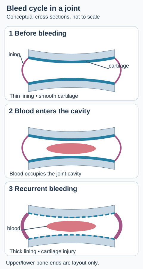
Case sources: [1] [6] [8]

Case 25

A 14-year-old with repeated spontaneous knee bleeds had normal platelets and prothrombin time. Initial activated partial thromboplastin time was normal; a specialist assay now reports factor IX activity 9% and VIII activity 94%. Which next preventive plan addresses the cause of recurrent joint bleeding?

Show answer and explanations for case 25
  1. A. Schedule factor VIII prophylaxis (Why this does not fit)

    VIII is preserved, so VIII prevention does not target the defect.

    Reasoning steps for option A
    1. Why would VIII prophylaxis miss the cause of these knee bleeds?

      VIII activity is 94%, whereas IX is 9%; normal prothrombin time and platelets do not override this factor-specific result.

    2. What alternate factor result would favor VIII prevention?

      A reproducibly low VIII activity with normal IX.

  2. B. Give desmopressin before each sport session (Why this does not fit)

    Desmopressin does not reliably raise IX to prevent B bleeds.

    Reasoning steps for option B
    1. Why can desmopressin not prevent this B phenotype?

      It mobilizes VIII and von Willebrand factor but does not reliably supply the IX needed for recurrent spontaneous hemarthroses.

    2. Does normal initial activated partial thromboplastin time make desmopressin sufficient?

      No; the specialist IX activity of 9% identifies the relevant defect despite the screen.

  3. C. Use emicizumab as IX replacement (Why this does not fit)

    Emicizumab mimics VIII activity in A rather than replacing IX in B.

    Reasoning steps for option C
    1. Why is the VIII mimetic not a B-specific substitute?

      Emicizumab mimics VIII cofactor function in A; it does not replace the deficient IX identified here.

    2. Which type of hemophilia is its prophylactic mechanism designed for?

      Hemophilia A, where VIII rather than IX is deficient.

  4. D. Individualized factor IX prophylaxis (Best answer)

    Selective low IX with recurrent deep bleeds warrants a B-specific preventive regimen despite normal screening activated partial thromboplastin time.

    Reasoning steps for option D
    1. Which data justify preventive IX rather than episodic reassurance?

      Repeated spontaneous knee bleeds with IX at 9% establish a B-specific risk even though the first activated partial thromboplastin time was normal.

    2. How should the preventive regimen be chosen?

      Individualize IX prophylaxis to bleeding history, joint status, pharmacokinetics, and specialist follow-up.

Takeaway: A normal screening clotting time does not erase measured IX deficiency or recurrent hemarthroses; plan individualized matched prevention.

Case sources: [1]

Search Bone Wizardry

Quick links