Classify reactive oxygen species, trace the phagocyte respiratory burst, and distinguish CGD from MPO and G6PD defects on board-style cases.
Free radicals become manageable when you separate three questions: what species is present, where it was generated, and which defense can reach it. This lesson uses that framework to connect redox chemistry with the phagocyte respiratory burst, chronic granulomatous disease, and red-cell oxidant injury.
1. Classify the reactive species first
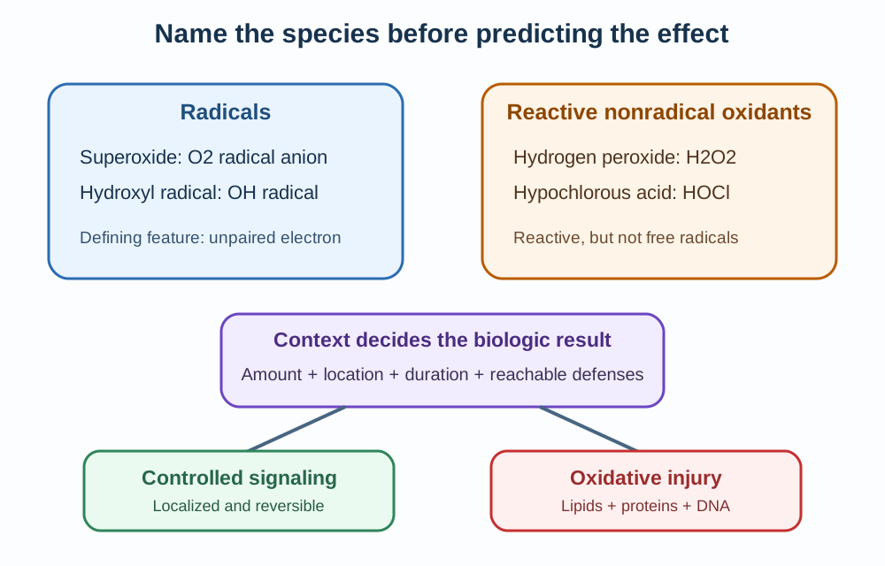
A free radical has one or more unpaired electrons. Superoxide and the hydroxyl radical meet that definition, while hydrogen peroxide and hypochlorous acid are reactive oxidants but are not radicals. The term reactive oxygen species is broader than free radical. [2]
Reactive oxygen species is broader than free radical. Name the species, then use context to decide whether it participates in controlled signaling or oxidative injury. [2]
Low, localized hydrogen peroxide can reversibly modify selected protein targets and participate in redox signaling. Injury appears when formation, location, duration, or impaired defenses allow oxidants to exceed the cell's buffering capacity. [2]
Species
Radical?
Useful first association
SpeciesSuperoxide, O2 radical anion
Radical?Yes
Useful first associationFirst product of the phagocyte NADPH oxidase
SpeciesHydrogen peroxide, H2O2
Radical?No
Useful first associationSignaling oxidant and substrate for peroxidases
SpeciesHydroxyl radical, OH radical
Radical?Yes
Useful first associationHighly reactive local injury, including metal-catalyzed formation
SpeciesHypochlorous acid, HOCl
Radical?No
Useful first associationMyeloperoxidase product inside the neutrophil phagosome
Apply the distinction now: if a stem says that H2O2 rose, do not automatically call it a free radical. First decide whether the question asks about signaling, oxidant stress, or radical chemistry. The species name controls the next inference.
2. Forecast the downstream pools
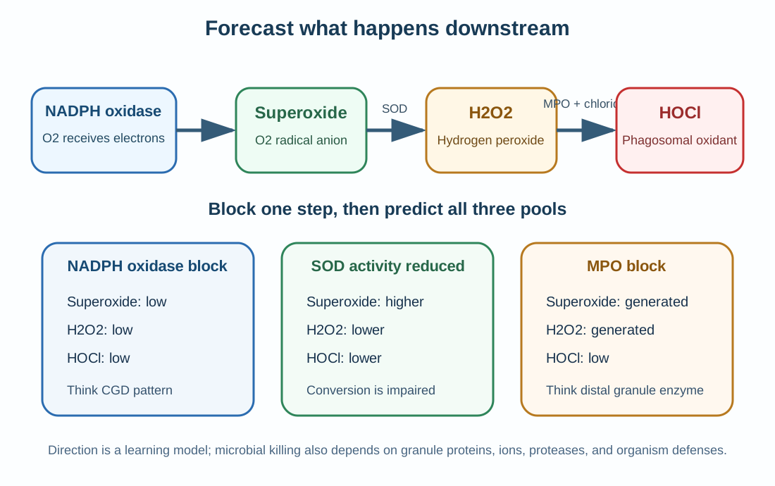
Activated phagocytes assemble NADPH oxidase at the phagosomal membrane. The complex transfers electrons to oxygen to form superoxide. Dismutation then produces hydrogen peroxide, and myeloperoxidase can use hydrogen peroxide plus chloride to form hypochlorous acid. [1][3]
Use a three-pool forecast before naming a disorder. For any blocked step, predict the direction of superoxide, hydrogen peroxide, and hypochlorous acid. A block at NADPH oxidase lowers all three downstream pools. A block at myeloperoxidase preserves upstream oxidant generation but lowers hypochlorous acid. [3]
Forecast superoxide, hydrogen peroxide, and hypochlorous acid before naming the disorder. An early oxidase block lowers every downstream pool, while an MPO block preserves upstream generation. [1][3]
Blocked function
Superoxide
H2O2
HOCl
Best first interpretation
Blocked functionPhagocyte NADPH oxidase
SuperoxideLow
H2O2Low
HOClLow
Best first interpretationCGD pattern
Blocked functionSuperoxide dismutation
SuperoxideHigher
H2O2Lower
HOClLower
Best first interpretationSuperoxide handling defect
Blocked functionMyeloperoxidase
SuperoxideGenerated
H2O2Generated
HOClLow
Best first interpretationMPO deficiency pattern
The forecast is a model, not a claim that one molecule kills every microbe by itself. Granule proteins, ion flux, proteases, oxidants, microbial defenses, and phagosomal conditions act together. [3]
Transfer the model: when a question supplies normal superoxide production but reduced hypochlorous acid, localize the defect after hydrogen peroxide, not at NADPH oxidase.
Try it here · Checkpoint 1 of 3
Make your prediction before reading the choices. A first attempt is just a starting point.
Case 5
Show answer and explanations for case 5
A. Superoxide low, hydrogen peroxide low, and HOCl low (Best answer)
The defect is at the first enzymatic step of the phagocyte burst. Reduced superoxide leaves less hydrogen peroxide and less substrate for MPO-derived hypochlorous acid.
Reasoning steps for option A
What do the genotype and functional result tell you?
The stem gives a pathogenic CYBB variant with almost no stimulated oxidase activity.
How does a block at the first step move the downstream pools?
Without superoxide there is little hydrogen peroxide, and without peroxide MPO cannot make HOCl, so all three fall.
B. Superoxide high, hydrogen peroxide high, and HOCl low (Why this does not fit)
This pattern might fit a distal MPO block with preserved upstream production. A CYBB oxidase defect prevents the initial superoxide rise.
Reasoning steps for option B
Which disorder produces preserved upstream oxidants with low HOCl?
That pattern belongs to an MPO block, where superoxide and peroxide are still generated.
Why can the upstream pools not be high in this boy?
A CYBB defect disables NADPH oxidase itself, so superoxide and peroxide cannot rise.
C. Superoxide normal, hydrogen peroxide low, and HOCl high (Why this does not fit)
Normal superoxide would require substantial oxidase activity. Hypochlorous acid cannot rise when its upstream peroxide substrate is reduced.
Reasoning steps for option C
Why might a normal superoxide value seem possible?
Some forecasts assume the first product is spared and only later steps change.
What makes this pattern internally impossible for a CYBB defect?
Superoxide cannot be normal with almost no oxidase activity, and HOCl cannot rise while its peroxide substrate falls.
D. Superoxide low, hydrogen peroxide high, and HOCl high (Why this does not fit)
Hydrogen peroxide can arise from other cellular sources, but the stimulated phagocyte pathway in the stem begins with oxidase-derived superoxide. Its downstream pools therefore fall rather than rise.
Reasoning steps for option D
Why could hydrogen peroxide still seem high?
Other cellular sources, such as mitochondria, also produce hydrogen peroxide.
Why does the phagocyte pathway still fall?
Stimulated phagosomal peroxide depends on oxidase-derived superoxide, so both peroxide and HOCl decline with the oxidase defect.
Takeaway: An early NADPH oxidase block lowers superoxide and both major downstream oxidants.
3. Recognize the CGD phenotype without overusing one mnemonic
Chronic granulomatous disease results from pathogenic variants that impair the phagocyte NADPH oxidase system. Recurrent pneumonia, lymphadenitis, skin or liver abscesses, osteomyelitis, granulomatous obstruction, and inflammatory colitis are important patterns. [1]
In North American cohorts, severe infections commonly involve Staphylococcus aureus, Burkholderia cepacia complex, Serratia marcescens, Nocardia species, and Aspergillus species. The organism list should raise suspicion, but microbiologic confirmation and the full phenotype still matter. [1][6]
Catalase positivity is a useful recall aid, not a complete causal explanation. It does not by itself predict which organism will cause severe disease, and the older story that affected neutrophils simply borrow microbial hydrogen peroxide is too narrow for modern interpretation. [1][3][6]
CGD also creates dysregulated inflammation. Granulomas and colitis can occur even when an active organism is not demonstrated, so a bowel or urinary obstruction can be part of the disease rather than proof of uncontrolled infection. [1]
Apply the pattern: recurrent deep infections with this organism spectrum plus granulomatous inflammation should prompt direct testing of the stimulated phagocyte respiratory burst.
4. Read the DHR pattern, then confirm the gene
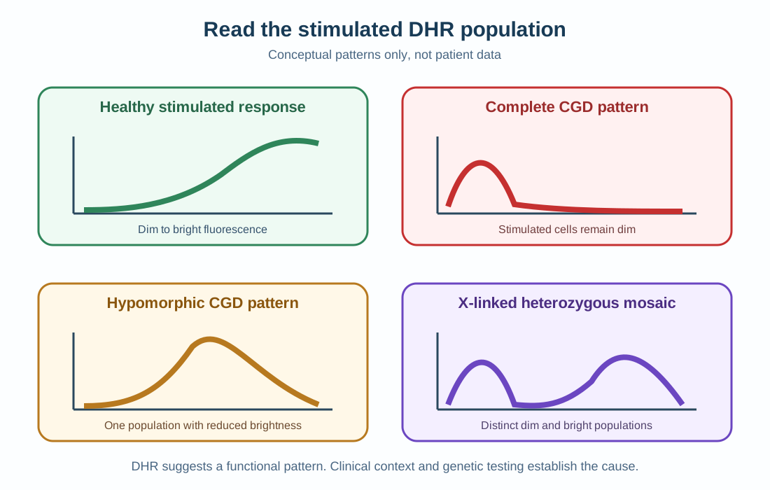
Dihydrorhodamine flow cytometry is the preferred widely available screening assay. Stimulated neutrophils oxidize DHR to a fluorescent product, allowing the laboratory to see absent, reduced, or mixed cell populations. NBT is older and mostly qualitative. [1][4]
DHR supplies a functional cellular pattern, not the causal gene by itself. Absent, reduced, and mosaic responses lead to different next questions. [1][4][5]
DHR pattern
Interpretation to consider
What it cannot prove alone
DHR patternBright stimulated population
Interpretation to considerSubstantial oxidase activity
What it cannot prove aloneEvery rare oxidase disorder is excluded
DHR patternAbsent or nearly absent fluorescence
Interpretation to considerComplete CGD phenotype
What it cannot prove aloneThe causal gene
DHR patternUniformly reduced fluorescence
Interpretation to considerHypomorphic CGD with residual activity
What it cannot prove aloneExact prognosis for one person
DHR patternTwo distinct populations
Interpretation to considerX-linked heterozygous mosaic pattern
What it cannot prove aloneClinical severity without history and genetics
The most common form is linked to CYBB on the X chromosome, but biallelic variants in several other genes cause autosomal recessive CGD. A girl or woman can therefore have clinically important disease through recessive inheritance, markedly skewed X inactivation, or other unusual genetic circumstances. [1]
Residual oxidant production is not merely a laboratory curiosity. Across CGD, greater residual production was associated with better survival, so the disorder should not be taught as universally binary. [5]
Transfer the pattern: two separated DHR populations in the mother of an affected boy support mosaic expression from X inactivation. Genetic testing establishes the familial variant and inheritance.
Try it here · Checkpoint 2 of 3
Make your prediction before reading the choices. A first attempt is just a starting point.
Case 10
Show answer and explanations for case 10
A. The assay is normal because some fluorescence is present. (Why this does not fit)
Any fluorescence does not automatically make the assay normal. The entire stimulated population is reduced compared with the normal control.
Reasoning steps for option A
Why might any fluorescence seem to mean a normal assay?
A complete CGD sample shows almost no fluorescence, so some signal can look reassuring.
What does a uniformly reduced population show?
Every stimulated neutrophil is dimmer than the control, which is abnormal even though the signal is not absent.
B. A hypomorphic CGD form with residual oxidase activity is likely. (Best answer)
Uniformly reduced DHR fluorescence indicates partial oxidase function across the neutrophil population. Residual activity can produce a milder or later-presenting CGD phenotype.
Reasoning steps for option B
Which details point to partial rather than absent oxidase activity?
He has milder, later disease than his affected brother, and DHR shows one uniformly reduced population.
How does residual activity explain the phenotype?
Hypomorphic CGD leaves some oxidase function in every cell, producing fewer severe infections than complete disease.
C. The patient is an X-linked heterozygous female with mosaic expression. (Why this does not fit)
Mosaic X inactivation usually creates two separated neutrophil populations. This male has one reduced population rather than a bright and dim mixture.
Reasoning steps for option C
Why could mosaicism be suggested by an intermediate signal?
X-linked carriers have reduced overall oxidase function, which can resemble a partial defect.
What pattern and sex argue against carrier mosaicism?
Mosaicism gives two separate bright and dim populations, and this patient is a man with one uniformly reduced population.
D. The result proves isolated myeloperoxidase deficiency. (Why this does not fit)
MPO deficiency can alter DHR interpretation, but the infection history and uniformly reduced oxidase pattern require correlation rather than a direct proof. CGD with residual activity is the stronger integration.
Reasoning steps for option D
Why might MPO deficiency be linked to an abnormal DHR?
DHR oxidation depends partly on MPO, so MPO deficiency can lower the signal.
Why does the whole picture favor CGD instead?
Recurrent bacterial lymphadenitis and an affected brother fit a hypomorphic oxidase defect, and a DHR result alone cannot prove MPO deficiency.
Takeaway: A uniformly reduced DHR population can indicate hypomorphic CGD with residual oxidase activity.
5. Separate phagocyte, granule, and red-cell defects
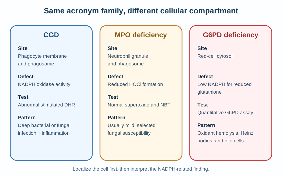
Myeloperoxidase deficiency occurs after hydrogen peroxide generation. Superoxide production and NBT reduction can remain intact, while DHR may be altered because dye oxidation depends partly on myeloperoxidase. The clinical picture is usually much milder than CGD. [1][3]
The shared NADPH and oxidant vocabulary can mislead. Localize the affected cell and intracellular site before interpreting the test. [1][3][7]
G6PD deficiency is a different NADPH problem in red blood cells. Reduced pentose phosphate pathway output limits NADPH, glutathione cannot remain adequately reduced, and an oxidant exposure can produce Heinz bodies, bite cells, jaundice, and acute hemolysis. [7]
Disorder
Primary cell or site
Main biochemical problem
Typical exam signal
DisorderCGD
Primary cell or sitePhagocyte membrane and phagosome
Main biochemical problemImpaired NADPH oxidase activity
Typical exam signalDeep bacterial or fungal infection with abnormal DHR
DisorderMPO deficiency
Primary cell or siteNeutrophil granule and phagosome
Main biochemical problemReduced HOCl formation
Typical exam signalMild phenotype with preserved superoxide generation
DisorderG6PD deficiency
Primary cell or siteRed-cell cytosol
Main biochemical problemInsufficient NADPH for reduced glutathione
Typical exam signalOxidant-triggered hemolysis with bite cells
During an acute G6PD hemolytic episode, the oldest and most deficient red cells may already be gone, while reticulocytes have more enzyme activity. A normal result at that moment can require repeat testing after recovery. [7]
Transfer the comparison: infection plus absent DHR belongs to the phagocyte branch. Dark urine, indirect hyperbilirubinemia, and bite cells after an oxidant exposure belong to the red-cell branch.
Try it here · Checkpoint 3 of 3
Make your prediction before reading the choices. A first attempt is just a starting point.
Case 21
Show answer and explanations for case 21
A. Insufficient NADPH to maintain reduced glutathione in red cells (Best answer)
G6PD activity supplies NADPH through the pentose phosphate pathway. Without adequate NADPH, reduced glutathione cannot buffer the oxidant burden and hemoglobin and membrane injury cause hemolysis.
Reasoning steps for option A
What trigger and blood findings does the stem give?
Hemolysis follows dapsone, an oxidant drug, with Heinz bodies and bite cells.
Which biochemical failure links the trigger to the smear?
G6PD deficiency limits NADPH, so glutathione cannot stay reduced and oxidized hemoglobin forms Heinz bodies that lead to hemolysis.
B. Failure of neutrophil MPO to generate HOCl (Why this does not fit)
MPO deficiency affects phagosome chemistry and usually has a much milder infection phenotype. It does not explain oxidant-triggered red-cell hemolysis with Heinz bodies.
Reasoning steps for option B
Why might an MPO defect be tempting in an oxidant question?
MPO deficiency is another disorder of oxidant chemistry and shares the same vocabulary.
Why does it not explain this episode?
MPO works in neutrophil phagosomes and causes a mild infection phenotype, not oxidant hemolysis with Heinz bodies.
C. Failure of factor VIII to support intrinsic tenase (Why this does not fit)
Factor VIII deficiency causes a bleeding disorder rather than intravascular or extravascular hemolysis. The smear and oxidant exposure point to red-cell oxidative injury.
Reasoning steps for option C
Why could a clotting factor defect seem to fit?
Both disorders present with abnormal blood findings after a trigger in a man.
What separates factor VIII deficiency from this picture?
Factor VIII deficiency causes bleeding, not hemolysis with falling haptoglobin, Heinz bodies, and bite cells.
D. Excess terminal complement assembly from a C1 inhibitor defect (Why this does not fit)
Complement dysregulation can cause other clinical syndromes. The classic bite cells and Heinz bodies after dapsone identify impaired red-cell antioxidant capacity.
Reasoning steps for option D
Why might complement dysregulation seem plausible for hemolysis?
Excess complement activity can destroy red cells.
Which findings point away from complement?
A C1 inhibitor defect causes angioedema, and Heinz bodies with bite cells after dapsone point to oxidant injury.
6. Apply redox chemistry to tissue injury and treatment
Oxidants can initiate lipid peroxidation, protein modification, and DNA injury. The result depends on the species, its location, and whether defenses such as superoxide dismutase, catalase, glutathione peroxidase, and lipid-phase antioxidants intercept it. [2]
Transition metals can convert hydrogen peroxide into highly reactive hydroxyl radicals near membranes or proteins. Reperfusion can also generate a sudden oxidant load through injured mitochondria and activated inflammatory enzymes when oxygen returns to ischemic tissue. [2]
Several classic injuries follow the same logic. Ionizing radiation splits water into hydroxyl radicals that attack nearby DNA, lipids, and proteins. [9] In acetaminophen overdose, CYP2E1 converts the drug to NAPQI, which depletes hepatic glutathione; N-acetylcysteine is most effective within 8 hours, and the Rumack-Matthew nomogram applies from 4 hours after a single acute ingestion. [8] Cytochrome P450 converts carbon tetrachloride (CCl4) to the trichloromethyl (CCl3) radical, which drives lipid peroxidation and fatty liver. [10] Prolonged high supplemental oxygen in preterm infants contributes to retinopathy of prematurity and bronchopulmonary dysplasia, [11] and glutathione peroxidase requires selenium. [12]
For CGD, prevention is practical rather than abstract. Lifelong antibacterial and antifungal prophylaxis is recommended, interferon-γ (IFN-γ) is included by many centers, and allogeneic hematopoietic stem cell transplantation is the only established cure described in the current GeneReviews chapter. [1]
No single exposure is the universal main source of free radicals. Name the patient context, the cellular source, and the measured consequence.
Final transfer: identify the species, localize the source, forecast downstream oxidants, and then match the clinical compartment. That sequence separates chemistry facts from the diagnosis.
Apply the lesson
Case 1
Show answer and explanations for case 1
A. Superoxide has an unpaired electron; hydrogen peroxide is a reactive nonradical oxidant. (Best answer)
Superoxide is a radical because it contains an unpaired electron. Hydrogen peroxide is reactive and biologically important, but it is not itself a free radical.
Reasoning steps for option A
What does the stem ask you to do before any biologic effect is considered?
Classify each molecule by its electron structure, which is the only property that defines a free radical.
How do superoxide and hydrogen peroxide differ on that criterion?
Superoxide carries an unpaired electron and is a radical; hydrogen peroxide has all electrons paired, so it is a reactive nonradical oxidant.
B. Hydrogen peroxide is a radical, while superoxide is a stable nonradical molecule. (Why this does not fit)
Hydrogen peroxide can participate in oxidant chemistry, which makes the statement sound plausible. The classifications are reversed because superoxide is the radical and hydrogen peroxide is nonradical.
Reasoning steps for option B
Why might a learner call hydrogen peroxide the radical?
Hydrogen peroxide is a well-known damaging oxidant, and damage is easy to equate with radical status.
Which electron fact reverses this pairing?
Superoxide is the species with the unpaired electron; hydrogen peroxide has none, so the labels in this choice are swapped.
C. Hypochlorous acid and hydrogen peroxide are both free radicals because they contain oxygen. (Why this does not fit)
Both molecules are reactive oxidants and can damage biologic targets. Oxygen content alone does not define a radical, and neither molecule has the radical classification used here.
Reasoning steps for option C
Why does grouping HOCl with hydrogen peroxide feel reasonable?
Both are strong oxidants made in the neutrophil phagosome and both injure microbial targets.
Is oxygen content enough to make a molecule a radical?
No. Water also contains oxygen; only an unpaired electron defines a radical, and neither HOCl nor hydrogen peroxide has one.
D. Every reactive oxygen species is a free radical by definition. (Why this does not fit)
Reactive oxygen species is a broad umbrella term that includes radicals and nonradical oxidants. Treating the two terms as synonyms erases an important chemical distinction.
Reasoning steps for option D
What makes the umbrella statement about reactive oxygen species attractive?
Textbooks often list superoxide, hydroxyl radical, and hydrogen peroxide together, so the two terms blur.
Why is the umbrella term broader than free radical?
Reactive oxygen species also include nonradical oxidants such as hydrogen peroxide and HOCl, so not every member is a radical.
Takeaway: Reactive oxygen species include both radicals and nonradical oxidants.
A. The cell has undergone irreversible oxidative destruction. (Why this does not fit)
Hydrogen peroxide can cause injury when excessive or poorly contained. The brief localized and reversible response instead supports regulated signaling.
Reasoning steps for option A
Why could a rise in hydrogen peroxide suggest destruction of the cell?
Hydrogen peroxide is an oxidant that can injure lipids, proteins, and DNA when it accumulates.
Which details of the time course argue against irreversible injury?
The rise is brief and localized, the cysteine changes are reversible, and baseline returns within seconds.
B. This is physiologic redox signaling rather than proof of cell injury. (Best answer)
Controlled hydrogen peroxide can modify selected protein targets and transmit a signal. Reversal by cellular defenses distinguishes the response from sustained oxidative distress.
Reasoning steps for option B
What features of the stem point to a regulated process?
A growth factor triggers the signal, the oxidation targets selected cysteines near a receptor, and the change is reversed.
How does this differ from oxidative distress?
Oxidative distress is sustained or widespread and overwhelms defenses; here buffering systems restore the cell promptly, which fits redox signaling.
C. The receptor has generated a hydroxyl radical because all peroxide is radical. (Why this does not fit)
Hydrogen peroxide can lead to other reactive products under specific conditions. The stem directly describes hydrogen peroxide and reversible signaling, not proof of hydroxyl radical formation.
Reasoning steps for option C
Why might hydroxyl radical formation come to mind here?
Hydrogen peroxide can give rise to hydroxyl radicals, especially in the presence of transition metals.
What does the stem actually describe?
It names hydrogen peroxide and reversible cysteine oxidation, with no metal or radical evidence; peroxide itself is not a radical.
D. The response proves that antioxidant enzymes are absent. (Why this does not fit)
Loss of antioxidant defenses would favor persistence and widespread injury. Rapid restoration of baseline shows that buffering systems are functioning.
Reasoning steps for option D
Why could the oxidant rise be blamed on missing antioxidant enzymes?
Weak defenses would allow peroxide to accumulate, so any peroxide rise can seem to imply a defense failure.
Which finding shows the defenses are working?
Cellular defenses return the signal to baseline within seconds, which could not happen if antioxidant enzymes were absent.
Takeaway: Low, localized hydrogen peroxide can serve as a reversible signaling oxidant.
A. Protein glycosylation within the Golgi apparatus (Why this does not fit)
Protein glycosylation can alter trafficking and receptor function. It does not explain propagating oxidation of polyunsaturated membrane lipids.
Reasoning steps for option A
Why might glycosylation be considered in a membrane problem?
Glycosylation modifies membrane proteins and lipids, so it is linked to membrane function.
What does glycosylation fail to explain here?
It is an enzymatic Golgi process; it does not produce lipid radicals that spread between phospholipids or generate aldehydes.
B. Direct hydrolysis of phospholipids by pancreatic lipase (Why this does not fit)
Lipase activity can digest lipid substrates in the proper compartment. The stem instead describes radical propagation and aldehyde formation in a cell membrane.
Reasoning steps for option B
Why does lipase seem to fit a lipid injury?
Lipases break down phospholipids and triglycerides, so they are a familiar cause of lipid destruction.
What separates enzymatic hydrolysis from the process in the stem?
Pancreatic lipase acts in the intestinal lumen and hydrolyzes ester bonds; it does not start a radical chain reaction or form reactive aldehydes.
C. Lipid peroxidation (Best answer)
Oxidant attack on polyunsaturated fatty acids can propagate across nearby lipids. Loss of membrane integrity and reactive aldehyde products fit lipid peroxidation.
Reasoning steps for option C
Which substrate and trigger does the stem supply?
Polyunsaturated fatty acids in a membrane are exposed to excess oxidants.
What pattern confirms lipid peroxidation?
Lipid radicals propagate to neighboring lipids, permeability rises, and aldehyde products accumulate, which is the chain reaction of lipid peroxidation.
D. ATP depletion from isolated ribosomal failure (Why this does not fit)
Severe cellular injury can lower ATP. Ribosomal failure does not directly account for lipid radicals spreading through a membrane.
Reasoning steps for option D
Why is ATP depletion a tempting explanation for membrane failure?
Loss of ATP disables ion pumps and is a central mechanism in many forms of cell injury.
Why does ribosomal failure not fit these findings?
Ribosomes make protein; their failure would not create lipid radicals propagating through phospholipids or aldehyde end products.
Takeaway: Propagating oxidation of polyunsaturated membrane lipids is lipid peroxidation.
A. MPO forms superoxide, then catalase forms hydrogen peroxide, then SOD forms HOCl. (Why this does not fit)
MPO uses hydrogen peroxide rather than making superoxide. Catalase removes hydrogen peroxide and SOD does not form hypochlorous acid.
Reasoning steps for option A
Why does a sequence starting with MPO look reasonable?
MPO is the best-known neutrophil oxidant enzyme, so it is easy to place it first.
What are the true roles of the enzymes named here?
MPO consumes hydrogen peroxide, catalase removes hydrogen peroxide, and SOD makes hydrogen peroxide from superoxide, not HOCl.
B. SOD forms superoxide, then NADPH oxidase forms hydrogen peroxide, then MPO forms chloride. (Why this does not fit)
SOD consumes superoxide rather than generating it. NADPH oxidase is the upstream source and chloride is a substrate, not the final oxidant.
Reasoning steps for option B
Why might SOD be placed at the start of the burst?
SOD is named after superoxide, so it can be mistaken for the enzyme that makes it.
Which facts break this sequence?
SOD consumes superoxide, NADPH oxidase makes superoxide rather than peroxide, and chloride is a substrate for MPO rather than its product.
C. Catalase forms superoxide, then glutathione peroxidase forms hydrogen peroxide, then MPO forms oxygen. (Why this does not fit)
Catalase and glutathione peroxidase are peroxide-removing defenses. They do not initiate the phagocyte respiratory burst.
Reasoning steps for option C
Why could catalase and glutathione peroxidase appear in a burst pathway?
Both handle oxygen-derived species, so they sound like part of the same chemistry.
What do these enzymes actually do?
They remove hydrogen peroxide as defenses; they do not generate superoxide or feed a chain ending in oxygen.
D. NADPH oxidase forms superoxide, dismutation forms hydrogen peroxide, and MPO plus chloride forms HOCl. (Best answer)
NADPH oxidase first reduces oxygen to superoxide. Hydrogen peroxide then becomes a substrate for MPO, which can generate hypochlorous acid in the phagosome.
Reasoning steps for option D
Which enzyme starts the phagocyte respiratory burst?
NADPH oxidase assembled on the phagosomal membrane transfers electrons to oxygen and forms superoxide.
How does the pathway reach the MPO product?
Superoxide dismutates to hydrogen peroxide, and MPO combines hydrogen peroxide with chloride to form HOCl.
Takeaway: The phagocyte sequence is NADPH oxidase to superoxide to hydrogen peroxide to MPO-derived HOCl.
A. Superoxide falls while hydrogen peroxide and HOCl rise. (Why this does not fit)
Superoxide dismutase normally consumes superoxide. Reducing the enzyme would not make the initial superoxide pool fall.
Reasoning steps for option A
Why might a learner expect superoxide to fall?
Many enzyme defects lower a product, so the named substrate can be mistaken for the product.
What does SOD normally do to superoxide?
SOD consumes superoxide; with less SOD, the superoxide made by an intact oxidase accumulates rather than falls.
B. Superoxide rises while hydrogen peroxide and HOCl fall. (Best answer)
Less dismutation allows superoxide to accumulate and reduces enzymatic hydrogen peroxide production. Less peroxide also limits the substrate available to myeloperoxidase.
Reasoning steps for option B
Which enzyme activities remain intact in this neutrophil?
NADPH oxidase and MPO are both intact, so superoxide is still made and HOCl can form if peroxide is available.
How does reduced dismutation shift the pools?
Superoxide accumulates, less hydrogen peroxide forms, and MPO has less substrate, so HOCl falls.
C. All three oxidants become completely absent. (Why this does not fit)
NADPH oxidase remains intact and continues to generate superoxide. The defect changes downstream conversion rather than eliminating every oxidant.
Reasoning steps for option C
Why could the stem suggest a total loss of oxidants?
A severe enzyme defect in the burst pathway can resemble complete CGD.
Which intact enzyme prevents complete loss?
NADPH oxidase still generates superoxide, and some spontaneous dismutation still occurs, so oxidants do not vanish.
D. Only chloride falls while all oxidants remain unchanged. (Why this does not fit)
Chloride availability is not the altered variable in this experiment. The direct defect is conversion of superoxide toward hydrogen peroxide.
Reasoning steps for option D
Why might chloride be singled out?
Chloride is a substrate in the final MPO step, so it looks like a variable in the pathway.
What variable was actually changed?
The experiment reduces SOD activity, which alters superoxide conversion rather than chloride availability.
Takeaway: Reduced superoxide dismutation raises superoxide and lowers the peroxide available for downstream MPO chemistry.
A. Glucose-6-phosphate dehydrogenase (Why this does not fit)
Severe G6PD deficiency can impair cellular NADPH and causes oxidant-sensitive hemolysis. The supplied neutrophil data localize the defect after hydrogen peroxide production rather than in the red-cell pentose phosphate pathway.
Reasoning steps for option A
Why could G6PD deficiency seem to fit an oxidant defect?
Severe G6PD deficiency can reduce cellular NADPH, and NADPH feeds the phagocyte oxidase.
Where do the supplied neutrophil data place the defect?
Superoxide and hydrogen peroxide are preserved, so the lesion lies after peroxide formation, not in NADPH supply.
B. Phagocyte NADPH oxidase (Why this does not fit)
NADPH oxidase deficiency would reduce superoxide at the first step. The stem explicitly states that superoxide generation is preserved.
Reasoning steps for option B
Why is NADPH oxidase the reflex answer for a burst defect?
It is the classic defect in CGD, the best-known disorder of phagocyte oxidants.
Which finding excludes an oxidase block?
Superoxide generation is preserved, which could not happen with an NADPH oxidase defect.
C. Myeloperoxidase (Best answer)
MPO uses hydrogen peroxide and chloride to form hypochlorous acid. Preserved upstream oxidants with low HOCl therefore localize the defect to MPO.
Reasoning steps for option C
Which steps are intact in this patient?
Superoxide generation and hydrogen peroxide production are both preserved.
Which enzyme converts that preserved peroxide into HOCl?
MPO combines hydrogen peroxide with chloride, so low HOCl despite normal peroxide localizes the defect to MPO.
D. Superoxide dismutase (Why this does not fit)
Loss of dismutation would reduce hydrogen peroxide production and allow superoxide accumulation. The stem instead gives preserved hydrogen peroxide with a distal product deficit.
Reasoning steps for option D
Why could SOD be suspected in a burst abnormality?
SOD sits between superoxide and hydrogen peroxide, so a defect there alters downstream oxidants.
What pattern would an SOD defect produce instead?
Superoxide would accumulate and hydrogen peroxide would fall, but here peroxide production is preserved.
Takeaway: Preserved upstream oxidants with low HOCl localize the defect to myeloperoxidase.
A. A single dim population that cannot oxidize the dye (Why this does not fit)
A dim stimulated population suggests absent or markedly reduced oxidase-dependent activity. That is not the expected healthy response.
Reasoning steps for option A
When is a single dim population seen after stimulation?
It is the pattern of complete CGD, where cells cannot oxidize DHR.
Why does this not fit the sample?
The neutrophils come from a healthy adult, whose intact oxidase should brighten the whole population.
B. Two separated populations with one bright and one dim peak (Why this does not fit)
A bimodal result can occur with mosaic X-linked heterozygosity. A healthy adult should show a largely uniform stimulated response.
Reasoning steps for option B
Where does a bright and dim bimodal pattern come from?
It is the classic result in a female carrier of X-linked CGD with mosaic X inactivation.
Why is it not expected here?
A healthy adult has oxidase activity in essentially every neutrophil, giving one uniformly bright population.
C. No fluorescence change because DHR measures antibodies rather than oxidants (Why this does not fit)
DHR is an intracellular oxidation assay, not an antibody measurement. Stimulated normal neutrophils generate a marked fluorescence increase.
Reasoning steps for option C
Why might DHR be confused with an antibody test?
Flow cytometry is widely used with fluorescent antibodies to identify cell markers.
What does DHR actually measure?
DHR is a dye oxidized inside stimulated cells by the respiratory burst, so normal neutrophils show a marked fluorescence increase.
D. A strong rightward fluorescence shift in the stimulated neutrophil population (Best answer)
Stimulated normal neutrophils oxidize DHR to a fluorescent product. The population therefore becomes substantially brighter than the unstimulated control.
Reasoning steps for option D
What happens to DHR inside normal stimulated neutrophils?
Oxidants from the respiratory burst convert DHR to fluorescent rhodamine.
How does that appear on the histogram?
The stimulated population shifts strongly to the right compared with the unstimulated control.
Takeaway: Normal stimulated neutrophils produce a strong DHR fluorescence increase.
The deep bacterial and fungal infections fit the characteristic CGD spectrum. Nearly absent stimulated DHR fluorescence directly supports severe phagocyte NADPH oxidase dysfunction.
Reasoning steps for option A
Which infections in the stem define the clinical pattern?
Recurrent Staphylococcus aureus liver abscesses and Aspergillus pneumonia are deep bacterial and fungal infections typical of CGD.
Which test result confirms the phagocyte defect?
Almost no stimulated DHR fluorescence shows a failed respiratory burst, while normal immunoglobulins and lymphocytes argue against other immunodeficiencies.
B. X-linked agammaglobulinemia (Why this does not fit)
X-linked agammaglobulinemia causes low immunoglobulins and absent or markedly reduced B cells. The immunoglobulins are normal and the abnormal test is the neutrophil burst.
Reasoning steps for option B
Why could X-linked agammaglobulinemia be considered in a boy?
It is an X-linked immunodeficiency that causes recurrent bacterial infection in young boys.
Which data rule it out?
Immunoglobulins are normal, and the abnormal test is the neutrophil DHR response rather than antibody production.
C. Leukocyte adhesion deficiency (Why this does not fit)
Leukocyte adhesion deficiency causes impaired migration, high circulating neutrophils, delayed cord separation, and little pus. It does not cause an absent intracellular DHR response.
Reasoning steps for option C
Why does leukocyte adhesion deficiency come up with deep infections?
It is a neutrophil disorder that causes recurrent bacterial infections.
What separates it from this case?
Adhesion deficiency impairs migration with neutrophilia and little pus, but stimulated cells still oxidize DHR normally.
D. Terminal complement deficiency (Why this does not fit)
Terminal complement deficiency strongly predisposes to Neisseria infection. It does not explain this organism pattern or the absent stimulated phagocyte oxidant response.
Reasoning steps for option D
Why might a complement defect be raised in recurrent infection?
Complement deficiencies are a standard part of the workup for repeated serious infections.
Which organism and test mismatch excludes it?
Terminal complement deficiency predisposes to Neisseria, not Staphylococcus and Aspergillus, and does not abolish DHR fluorescence.
Takeaway: Deep Staphylococcus and Aspergillus infections plus absent DHR support CGD.
A. Autosomal recessive inheritance with every neutrophil equally affected (Why this does not fit)
Autosomal recessive disease would not be expected to create two populations from X inactivation. The discrete bright and dim groups point to cellular mosaicism.
Reasoning steps for option A
Why might autosomal recessive CGD be considered in a family with CGD?
Autosomal recessive forms are common enough to appear on any CGD differential.
Why does it not explain two populations?
Recessive disease affects every neutrophil equally, whereas the mother shows one bright and one dim group.
B. A technical artifact that always occurs in healthy women (Why this does not fit)
Healthy controls generally show a strong, largely uniform stimulated response. A reproducible bimodal pattern in this family has biologic significance.
Reasoning steps for option B
Why might a bimodal pattern be dismissed as artifact?
Flow cytometry can show technical double peaks from preparation problems.
What makes this result biologically meaningful?
Healthy women show one uniform bright population, and this mother has a son with a CYBB variant.
C. Mosaic oxidase expression from X-chromosome inactivation (Best answer)
Random or skewed X inactivation can yield oxidase-positive and oxidase-deficient neutrophil populations in a CYBB heterozygote. DHR displays those populations separately.
Reasoning steps for option C
What family and test data does the stem give?
The mother of a boy with a CYBB variant has one bright and one dim neutrophil population.
How does X inactivation create that pattern?
In a heterozygote, some neutrophils express the normal X and some the mutant X, so DHR shows two distinct populations.
D. Complete CGD with no residual activity (Why this does not fit)
Complete CGD would produce a predominantly dim stimulated population rather than a second bright population. The bright cells demonstrate preserved activity in part of the sample.
Reasoning steps for option D
Why might a large dim population suggest complete CGD?
The dim cells look like the absent response seen in affected patients.
Which part of the result excludes complete disease?
A clearly bright population shows that many of her neutrophils have normal oxidase activity.
Takeaway: A bimodal DHR pattern in a CYBB heterozygote reflects X-inactivation mosaicism.
A. Every son will inherit the paternal CYBB variant. (Why this does not fit)
Sons receive their father's Y chromosome, not his X chromosome. Paternal transmission therefore does not send the CYBB variant to sons.
Reasoning steps for option A
Why might a learner expect every son to inherit the variant?
Sons are the usual affected children in X-linked disease, so father-to-son spread feels natural.
What does the father actually give his sons?
Sons receive the father's Y chromosome, so they cannot inherit his X-linked CYBB variant.
B. Half of the sons and half of the daughters will inherit the variant. (Why this does not fit)
A 50 percent pattern applies to a heterozygous mother, not an affected father with one X chromosome. His daughters and sons receive different paternal sex chromosomes.
Reasoning steps for option B
Why does a 50 percent pattern sound familiar?
It is the correct risk for children of a heterozygous carrier mother.
Why does the rule change when the father is affected?
He has one X chromosome, which goes to every daughter, and his Y goes to every son, so there is no 50 percent split.
C. No daughter can inherit the variant because CGD usually affects boys. (Why this does not fit)
Disease frequency in boys does not change chromosome transmission. Every daughter receives her father's X chromosome.
Reasoning steps for option C
Why might daughters seem protected?
X-linked CGD mostly produces clinical disease in boys.
Which chromosome fact defeats this?
Every daughter receives her father's only X chromosome, so every daughter carries the variant.
D. Every daughter inherits the paternal variant, while no son inherits it from him. (Best answer)
An affected father gives his only X chromosome to every daughter and his Y chromosome to every son. Clinical expression in daughters depends on X inactivation and other factors.
Reasoning steps for option D
Which chromosomes does an affected father transmit?
He passes his single X chromosome to every daughter and his Y chromosome to every son.
What does that predict for this family?
Every daughter carries the CYBB variant, no son inherits it from him, and daughters' symptoms depend on X inactivation.
Takeaway: An affected father transmits an X-linked CYBB variant to every daughter and no son.
Biallelic NCF1 pathogenic variants cause an autosomal recessive form of CGD. The patient's sex does not protect her from a recessive oxidase defect.
Reasoning steps for option A
Which genetic finding decides the inheritance pattern?
Pathogenic variants are present in both copies of NCF1, an autosomal gene.
How do her sex and family history fit?
Biallelic autosomal variants cause recessive CGD in any sex, and consanguinity raises the chance of inheriting two variants.
B. X-linked recessive CGD from paternal CYBB transmission (Why this does not fit)
CYBB causes the X-linked form, but the detected gene is NCF1.
Reasoning steps for option B
Why is X-linked CGD the first reflex for any CGD case?
CYBB variants cause the most common form of CGD.
Which finding excludes the X-linked form here?
Sequencing found the variants in NCF1, not CYBB, and both copies are affected.
C. Mitochondrial inheritance (Why this does not fit)
Mitochondrial inheritance follows maternal transmission and does not fit biallelic NCF1 variants. The oxidase defect described is nuclear and autosomal.
Reasoning steps for option C
Why might mitochondrial inheritance be considered for an oxidant disorder?
Mitochondria are a major cellular source of oxidants.
Why does it not fit the genetics?
The variants are in both copies of a nuclear gene, NCF1, which is inherited autosomally rather than maternally through mitochondria.
D. Autosomal dominant CGD from one NCF1 copy (Why this does not fit)
The stem identifies pathogenic variants in both NCF1 copies. The classic NCF1 CGD mechanism is autosomal recessive rather than a single-copy dominant pattern.
Reasoning steps for option D
Why could a single dominant NCF1 variant be suggested?
A few rare oxidase disorders have dominant mechanisms, so dominance is not unthinkable.
What does the sequencing result show instead?
Both NCF1 copies carry pathogenic variants, which is the autosomal recessive pattern.
Takeaway: Biallelic NCF1 variants cause autosomal recessive CGD in any sex.
A. Whether the patient is currently febrile during the assay (Why this does not fit)
Acute illness can affect laboratory context and needs clinical attention. It does not replace the measured long-term relationship between residual oxidant production and survival.
Reasoning steps for option A
Why might fever during testing seem to matter for prognosis?
Acute illness can change neutrophil activation and laboratory values.
Why is it not the strongest long-term predictor?
Cohort data link measured residual oxidant production, not transient fever, to long-term survival.
B. The amount of residual phagocyte oxidant production (Best answer)
Residual oxidant production behaves as a continuous functional variable. Greater residual production was associated with less severe illness and better long-term survival in the cited cohort.
Reasoning steps for option B
How do the two patients differ in the stem?
Patient 1 has almost no residual oxidant production; patient 2 has a reproducible low but greater signal.
What does cohort evidence show about that difference?
Greater residual oxidant production was associated with less severe illness and better survival, so it is a continuous prognostic variable.
C. The color of the NBT slide without quantitative follow-up (Why this does not fit)
NBT is mostly qualitative and can miss partial or mosaic patterns. Prognostic interpretation requires more direct functional measurement and clinical context.
Reasoning steps for option C
Why might the NBT slide appeal as a simple test?
NBT was the classic CGD screen and gives a quick visual result.
Why can it not rank prognosis?
NBT is mostly qualitative and can miss partial or mosaic patterns, so it does not quantify residual production.
D. The total serum immunoglobulin concentration (Why this does not fit)
Immunoglobulins may be normal in CGD because the core defect is phagocyte oxidase activity. They do not quantify residual respiratory burst function.
Reasoning steps for option D
Why might immunoglobulins be checked in recurrent infection?
Antibody levels are a standard part of any immunodeficiency workup.
Why do they not measure CGD severity?
Immunoglobulins are often normal in CGD because the defect lies in phagocyte oxidase activity.
Takeaway: Residual phagocyte oxidant production is a continuous prognostic variable in CGD.
A. The list proves that any catalase-positive organism causes equal risk in CGD. (Why this does not fit)
Many listed organisms are catalase positive, which supports the familiar association. Catalase status alone does not explain equal virulence or predict the full CGD infection spectrum.
Reasoning steps for option A
Why does a catalase-based explanation feel complete?
Most organisms in this list are catalase positive, matching the classic CGD teaching.
Why does catalase status fail to predict equal risk?
Many catalase-positive organisms rarely cause CGD infection, so species traits and host factors matter beyond catalase.
B. The pattern is specific for terminal complement deficiency. (Why this does not fit)
Terminal complement deficiency is especially associated with Neisseria. It does not fit this narrow collection of deep bacterial and fungal infections.
Reasoning steps for option B
Why might complement deficiency be considered for recurrent infection?
Complement defects are a standard cause of repeated bacterial infection.
Why does this organism list not fit?
Terminal complement deficiency predisposes mainly to Neisseria, not Staphylococcus, Burkholderia, Serratia, Nocardia, and Aspergillus.
C. The pattern supports CGD, but catalase positivity is not the whole explanation. (Best answer)
The observed CGD spectrum includes these organisms and should raise suspicion for a phagocyte oxidase defect. Species-specific traits, host factors, and residual oxidase activity matter beyond a catalase label.
Reasoning steps for option C
What does this organism list suggest?
Staphylococcus aureus, Burkholderia cepacia complex, Serratia, Nocardia, and Aspergillus are the classic severe CGD pathogens.
Why is catalase positivity only part of the story?
It is a useful recall aid, but species virulence, host inflammation, and residual oxidase activity also shape which infections occur.
D. The pattern shows isolated antibody deficiency because all listed organisms are encapsulated. (Why this does not fit)
The organisms are not simply one encapsulated-bacteria group, and Aspergillus is a fungus. CGD can occur with normal immunoglobulin concentrations.
Reasoning steps for option D
Why might antibody deficiency be raised by bacterial infections?
Recurrent bacterial infections are a hallmark of antibody deficiency.
Which organisms break the encapsulated-bacteria idea?
Aspergillus is a fungus and Nocardia and Serratia are not the encapsulated bacteria typical of antibody defects.
Takeaway: The characteristic organism pattern supports CGD, while catalase status alone is an incomplete explanation.
A. Reassure because intact antibodies prevent invasive mold disease. (Why this does not fit)
CGD is a phagocyte defect and may occur with normal antibodies. Mold exposure can cause severe pulmonary disease despite preserved immunoglobulins.
Reasoning steps for option A
Why might normal antibodies seem protective?
Antibody deficiency is a common reason for serious infections, so intact antibodies can feel reassuring.
Why do antibodies not prevent this illness?
CGD is a phagocyte defect, and invasive mold disease occurs despite normal immunoglobulins.
B. Treat only for an allergic reaction because fungal infection cannot progress this quickly. (Why this does not fit)
Mulch exposure can trigger a fulminant pneumonitis syndrome in CGD. The severity and hypoxemia require urgent infectious and inflammatory evaluation.
Reasoning steps for option B
Why could an allergic reaction explain rapid symptoms after mulch?
Inhaled organic dust can provoke a quick hypersensitivity response.
What makes allergy alone an unsafe explanation?
In CGD, mulch exposure can cause fulminant mold pneumonitis within hours, and hypoxemia with diffuse nodules demands urgent infectious evaluation.
C. Delay evaluation until a routine outpatient DHR test is repeated. (Why this does not fit)
The diagnosis is already known and the patient is acutely ill. Waiting for repeat screening would postpone necessary assessment and treatment.
Reasoning steps for option C
Why might repeating the DHR test seem methodical?
Confirming the laboratory diagnosis is a standard step in CGD care.
Why would waiting cause harm?
The CGD diagnosis is already known and the patient is hypoxemic, so delay postpones urgent treatment.
D. Urgently evaluate and treat for severe mold-associated pneumonitis in CGD. (Best answer)
Decayed organic matter can expose a person with CGD to a dense fungal inoculum. Acute respiratory compromise after mulch exposure is a medical emergency.
Reasoning steps for option D
What exposure and findings does the stem combine?
A teenager with CGD has fever, hypoxemia, and diffuse pulmonary nodules hours after spreading moldy mulch.
Why is urgent treatment the right response?
Decaying organic matter delivers a dense fungal inoculum, and acute mulch pneumonitis in CGD is a medical emergency.
Takeaway: Acute respiratory illness after moldy organic exposure is an emergency in CGD.
A. Dysregulated inflammation with granuloma formation (Best answer)
CGD causes inflammatory as well as infectious disease. Granulomatous tissue can obstruct the gastrointestinal or genitourinary tract even when an active organism is not demonstrated.
Reasoning steps for option A
What does the imaging show despite negative cultures?
The pylorus is narrowed by granulomatous tissue without a demonstrated organism.
How does CGD explain a sterile obstruction?
CGD causes dysregulated inflammation, so granulomas can obstruct the gut or urinary tract without active infection.
B. Loss of all T-cell function (Why this does not fit)
Profound T-cell defects cause a different infection pattern and do not explain the established CGD diagnosis. The obstruction is a recognized inflammatory complication of CGD.
Reasoning steps for option B
Why might a T-cell defect be linked to granulomas?
T cells organize granulomas, so their loss seems connected to granulomatous disease.
Why does it not fit this patient?
The child already has CGD, and severe T-cell loss produces a different opportunistic infection pattern rather than pyloric granulomas.
C. An antibody against gastric parietal cells (Why this does not fit)
Autoimmune gastritis affects acid production and vitamin B12 absorption. It does not typically create focal pyloric granulomatous narrowing.
Reasoning steps for option C
Why could autoimmune gastritis come to mind for gastric symptoms?
It is an autoimmune disorder of the stomach that can cause early satiety.
What does it fail to produce?
It reduces acid and vitamin B12 absorption but does not create focal granulomatous pyloric narrowing.
D. Complement-mediated intravascular hemolysis (Why this does not fit)
Complement-mediated hemolysis affects red cells and can cause anemia and dark urine. It does not produce a pyloric granuloma.
Reasoning steps for option D
Why might complement activation seem related to inflammation?
Complement is a major driver of inflammatory tissue injury.
Why does hemolysis not fit the finding?
Complement-mediated hemolysis damages red cells and causes anemia, not a granulomatous pyloric mass.
Takeaway: CGD can cause obstructive granulomatous inflammation without a cultured organism.
Antibody deficiency is a common cause of repeated bacterial infection.
Which result excludes it?
Immunoglobulins are normal, which would not be expected if B cells failed to mature into plasma cells.
B. Phagocyte NADPH oxidase activity (Best answer)
Absent stimulated DHR directly reports severe impairment of the phagocyte respiratory burst. Deep Staphylococcus abscesses fit the CGD phenotype.
Reasoning steps for option B
Which normal results narrow the defect?
Normal immunoglobulins and complement testing make antibody and complement defects unlikely.
Which result names the impaired function?
An absent stimulated DHR response shows failure of the phagocyte NADPH oxidase, fitting the liver abscesses of CGD.
C. C5 through C9 membrane attack complex assembly (Why this does not fit)
Terminal complement defects predispose strongly to Neisseria infection. They do not cause an absent neutrophil DHR response.
Reasoning steps for option C
Why might membrane attack complex defects be considered?
Terminal complement defects cause recurrent serious bacterial infections.
Which findings exclude them?
Complement testing is normal, these defects favor Neisseria, and they do not abolish neutrophil DHR fluorescence.
D. CD18-dependent leukocyte adhesion (Why this does not fit)
CD18 deficiency impairs neutrophil migration and produces high blood neutrophil counts with little pus. Intracellular oxidase activity remains measurable when those cells are tested.
Reasoning steps for option D
Why might CD18 deficiency fit neutrophil-related abscesses?
Leukocyte adhesion deficiency is another neutrophil disorder with recurrent bacterial infection.
What distinguishes it on testing?
CD18 defects block migration with neutrophilia and little pus, but the cells still produce a normal DHR response.
Takeaway: Absent stimulated DHR with deep abscesses localizes the defect to phagocyte NADPH oxidase.
A. Serum protein electrophoresis (Why this does not fit)
Serum protein electrophoresis can assess broad immunoglobulin patterns. It does not directly measure the stimulated phagocyte respiratory burst.
Reasoning steps for option A
Why might serum protein electrophoresis be ordered?
Recurrent infections prompt immune screening, and electrophoresis gives a quick view of immunoglobulin patterns.
Why does it not test for CGD?
It measures serum proteins, not the stimulated neutrophil oxidant burst that fails in CGD.
B. Bleeding time (Why this does not fit)
Bleeding time concerns primary hemostasis and is not a test of neutrophil oxidant production. It cannot diagnose CGD.
Reasoning steps for option B
Why might bleeding time be mistaken for a neutrophil test?
It is a classic bedside cell-function test, and platelets and neutrophils both circulate in blood.
What does bleeding time actually assess?
It reflects platelet and vessel function in primary hemostasis, not neutrophil oxidant production.
C. Stimulated DHR flow cytometry (Best answer)
DHR flow cytometry displays fluorescence at the single-cell level after stimulation. It can show absent, reduced, or mosaic oxidase-dependent activity and is preferred over qualitative NBT screening.
Reasoning steps for option C
What does the clinical picture ask you to test?
Recurrent Serratia and Aspergillus infections suggest a failed phagocyte respiratory burst.
Why is DHR the best first functional study?
Stimulated DHR flow cytometry shows single-cell oxidase activity and separates absent, reduced, and mosaic patterns, unlike qualitative NBT.
D. Direct antiglobulin testing (Why this does not fit)
Direct antiglobulin testing detects antibody or complement coating red cells. It does not assess phagocyte NADPH oxidase function.
Reasoning steps for option D
Why could direct antiglobulin testing seem relevant?
It is a common immune test that detects antibody or complement on cells.
Why does it not address this question?
It detects coating of red cells, not the function of phagocyte NADPH oxidase.
Takeaway: Stimulated DHR flow cytometry is the preferred widely available functional screen for CGD.
Complete CGD would markedly reduce superoxide generation and NBT reduction. Both are preserved in this patient.
Reasoning steps for option A
Why might CGD be considered in recurrent infection with an oxidant defect?
CGD is the classic disorder of neutrophil oxidant production.
Which results exclude complete CGD?
Superoxide generation and NBT reduction are normal, which complete CGD would abolish.
B. G6PD deficiency (Why this does not fit)
G6PD deficiency classically produces oxidant-sensitive red-cell hemolysis. The supplied defect is the neutrophil enzyme that uses hydrogen peroxide and chloride.
Reasoning steps for option B
Why might G6PD deficiency be linked to oxidant handling?
G6PD supplies NADPH, and severe deficiency can affect oxidant chemistry.
What does the enzyme test show instead?
The failed step is neutrophil conversion of hydrogen peroxide and chloride to HOCl, not red-cell NADPH supply.
C. Leukocyte adhesion deficiency (Why this does not fit)
Adhesion deficiency prevents normal migration into tissue and often produces little pus. It does not selectively abolish HOCl formation with preserved superoxide.
Reasoning steps for option C
Why could adhesion deficiency explain recurrent infection?
It is a neutrophil disorder that causes repeated bacterial and fungal infection.
What does adhesion deficiency not do?
It impairs migration but does not selectively abolish HOCl formation while superoxide stays normal.
D. Myeloperoxidase deficiency (Best answer)
MPO catalyzes formation of HOCl from hydrogen peroxide and chloride. Preserved superoxide and NBT with loss of that conversion fit MPO deficiency.
Reasoning steps for option D
Which steps of the burst are preserved?
Superoxide generation and NBT reduction are normal, so NADPH oxidase works.
Which enzyme failure fits the remaining defect?
MPO converts hydrogen peroxide and chloride to HOCl, and the mild Candida picture with diabetes fits MPO deficiency.
Takeaway: Normal superoxide with impaired HOCl formation supports myeloperoxidase deficiency.
A. G6PD deficiency is permanently excluded by the first value. (Why this does not fit)
A reference-range value during active hemolysis can be misleading. The most deficient older red cells may already have been destroyed.
Reasoning steps for option A
Why might a normal G6PD value seem final?
A quantitative enzyme assay within the reference range usually excludes a deficiency.
Why is this value unreliable now?
During active hemolysis the most deficient old red cells are already destroyed, leaving younger cells with more enzyme.
B. Repeat quantitative testing after recovery because reticulocytosis can mask deficiency. (Best answer)
Reticulocytes and younger red cells generally have more G6PD activity than older cells. Repeating the assay after the red-cell population stabilizes can reveal the underlying deficiency.
Reasoning steps for option B
What clues suggest the result may be falsely normal?
The test was taken during brisk hemolysis with bite cells and a high reticulocyte count.
Why does repeating the assay help?
Reticulocytes have more G6PD activity; once the red-cell population stabilizes, a repeat test can reveal the deficiency.
C. The normal value proves the episode was immune hemolysis. (Why this does not fit)
Immune hemolysis requires supporting evidence such as a positive direct antiglobulin test and does not produce a G6PD-specific inference. The timing and smear remain compatible with oxidant hemolysis.
Reasoning steps for option C
Why might immune hemolysis be considered when G6PD looks normal?
Immune hemolysis is a common alternative cause of acute hemolysis.
What evidence is missing for that diagnosis?
No positive direct antiglobulin test is given, and bite cells after an oxidant drug still point to oxidant hemolysis.
D. Order DHR flow cytometry as the definitive red-cell enzyme test. (Why this does not fit)
DHR primarily evaluates stimulated phagocyte oxidant activity. It does not replace a quantitative G6PD assay for red cells.
Reasoning steps for option D
Why might DHR flow cytometry be linked to an oxidant problem?
DHR is a well-known oxidant assay, so it seems relevant to oxidant hemolysis.
What does DHR actually test?
DHR measures the phagocyte respiratory burst in neutrophils, not red-cell G6PD activity.
Takeaway: A G6PD assay can be falsely reassuring during acute hemolysis and should be repeated after recovery when suspicion remains.
A. The urea cycle supplies carbamoyl phosphate to catalase. (Why this does not fit)
The urea cycle disposes of nitrogen and does not supply the main red-cell reducing equivalent. Catalase also does not use carbamoyl phosphate.
Reasoning steps for option A
Why might the urea cycle come up in a red-cell question?
It is a major metabolic pathway that learners often link to detoxification.
Why does it not support peroxide removal?
The urea cycle disposes of nitrogen in the liver and supplies no reducing power; catalase does not use carbamoyl phosphate.
B. β-Oxidation supplies FADH2 directly to myeloperoxidase. (Why this does not fit)
Mature red cells lack mitochondria and do not perform β-oxidation. Myeloperoxidase is a neutrophil granule enzyme rather than the red-cell peroxide defense described.
Reasoning steps for option B
Why might fatty acid oxidation seem to supply reducing power?
β-oxidation produces reduced cofactors such as FADH2.
Which red-cell facts rule it out?
Mature red cells lack mitochondria and cannot perform β-oxidation, and MPO is a neutrophil enzyme, not a red-cell defense.
C. NADPH from the pentose pathway regenerates reduced glutathione. (Best answer)
G6PD initiates the oxidative pentose phosphate pathway and generates NADPH. Glutathione reductase uses that NADPH to restore reduced glutathione for glutathione peroxidase.
Reasoning steps for option C
What does glutathione peroxidase need to keep working?
It needs reduced glutathione, which glutathione reductase regenerates using NADPH.
Which pathway supplies that NADPH in red cells?
G6PD begins the oxidative pentose phosphate pathway, the red cell's main NADPH source.
D. Glycogenolysis supplies chloride for hypochlorous acid formation. (Why this does not fit)
Glycogenolysis can supply glucose substrates but chloride and HOCl are not the relevant red-cell defense. The key reducing system is NADPH and glutathione.
Reasoning steps for option D
Why might glycogen breakdown seem to support the red cell?
Glycogenolysis supplies glucose, the red cell's fuel.
Why does it not match the question?
Glycogenolysis does not provide chloride, and HOCl formation is a neutrophil mechanism rather than a red-cell peroxide defense.
Takeaway: Red-cell peroxide defense depends on pentose phosphate pathway NADPH and reduced glutathione.
A. Oxidative phosphorylation coupling (Why this does not fit)
Oxidative phosphorylation links electron transfer to ATP synthesis. It does not specifically describe ferrous iron converting hydrogen peroxide into hydroxyl radicals.
Reasoning steps for option A
Why could oxidative phosphorylation seem linked to hepatocyte oxidants?
The mitochondrial electron transport chain is a major source of cellular reactive oxygen species.
Why does it not explain the reaction described?
Oxidative phosphorylation is an enzyme-driven energy process of the inner mitochondrial membrane; the stem instead describes a nonenzymatic, metal-catalyzed reaction between labile ferrous iron and hydrogen peroxide.
B. Superoxide dismutation (Why this does not fit)
Dismutation converts superoxide toward hydrogen peroxide. The stem already supplies hydrogen peroxide and emphasizes ferrous iron.
Reasoning steps for option B
Why might superoxide dismutation seem relevant?
It is a key reaction that produces hydrogen peroxide in cells.
Which details point past dismutation?
The stem already supplies hydrogen peroxide and emphasizes labile ferrous iron as the driver of hydroxyl radical formation.
C. Myeloperoxidase halogenation (Why this does not fit)
MPO uses hydrogen peroxide and halides mainly in phagocytes. The iron-dependent hydroxyl radical chemistry in hepatocytes is different.
Reasoning steps for option C
Why might MPO halogenation seem to fit oxidant injury?
MPO turns hydrogen peroxide into a highly damaging oxidant.
Why does it not fit hepatocytes with iron overload?
MPO works mainly in neutrophil phagosomes and makes HOCl, not hydroxyl radicals from ferrous iron.
D. Fenton chemistry (Best answer)
Ferrous iron can react with hydrogen peroxide to generate highly reactive hydroxyl radicals. The local radicals can initiate lipid and protein injury in iron-loaded tissue.
Reasoning steps for option D
Which two reactants does the stem emphasize?
Iron overload supplies labile ferrous iron alongside hydrogen peroxide in hepatocytes.
Which reaction joins them?
In Fenton chemistry, ferrous iron converts hydrogen peroxide into hydroxyl radicals, which start lipid and protein injury.
Takeaway: Ferrous iron plus hydrogen peroxide can generate hydroxyl radicals through Fenton chemistry.
A. Oxygen restoration prevents all further oxidant generation. (Why this does not fit)
Restored oxygen is necessary for tissue survival but can also become substrate for reactive species. The measured oxidant surge contradicts complete prevention.
Reasoning steps for option A
Why might restored oxygen seem purely protective?
Oxygen delivery is exactly what ischemic tissue lacks.
Which finding contradicts full protection?
The tissue shows a surge in oxidant products after flow returns, so oxidant generation increased.
B. Reoxygenation supplies substrate to injured mitochondria and inflammatory oxidant systems. (Best answer)
Ischemic injury disrupts cellular metabolism, and reoxygenation can produce a sudden oxidant load from mitochondria and activated inflammatory enzymes. This is a central feature of reperfusion injury.
Reasoning steps for option B
What happens right after flow is restored?
Oxidant products surge, mitochondria malfunction, and neutrophils activate.
How does reoxygenation cause this?
Returning oxygen fuels injured mitochondria and inflammatory oxidant enzymes, producing the sudden oxidant load of reperfusion injury.
C. The injury is caused only by absence of oxygen after blood flow returns. (Why this does not fit)
Blood flow restoration increases oxygen delivery rather than maintaining pure anoxia. The paradox is that reoxygenation itself can contribute to injury.
Reasoning steps for option C
Why might continued oxygen lack be blamed?
Ischemic injury is caused by oxygen deprivation, so ongoing damage seems to reflect the same cause.
Why does that not fit reperfusion?
Restored flow increases oxygen delivery, and the injury comes from reoxygenation itself.
D. Hydrogen peroxide becomes chemically inert during reperfusion. (Why this does not fit)
Hydrogen peroxide remains an important oxidant and signaling molecule. In the presence of damaged systems and transition metals, it can contribute to further injury.
Reasoning steps for option D
Why might hydrogen peroxide seem irrelevant after reperfusion?
It is a nonradical and is often treated as a mild signaling oxidant.
Why does it still matter here?
Hydrogen peroxide stays reactive, and with damaged systems and transition metals it can form hydroxyl radicals that add injury.
Takeaway: Reperfusion can generate a sudden oxidant load from injured mitochondria and inflammatory systems.
A. No prophylaxis is needed once the first infection resolves. (Why this does not fit)
CGD creates lifelong risk for severe bacterial and fungal infection. Recovery from one episode does not restore oxidase function.
Reasoning steps for option A
Why might recovery suggest that prophylaxis can stop?
The child has recovered, and most infections do not need ongoing treatment after they resolve.
Why does the risk persist?
Recovery does not restore oxidase function, so the risk of severe bacterial and fungal infection is lifelong.
B. Use only routine childhood vaccines and no antimicrobial prevention. (Why this does not fit)
Routine preventive care remains important, but CGD requires additional disease-specific infection prevention. Some live bacterial vaccines also warrant avoidance.
Reasoning steps for option B
Why might routine vaccines seem sufficient?
Vaccination is the standard preventive tool in childhood.
Why is it not enough in CGD?
CGD needs disease-specific antibacterial and antifungal prophylaxis, and some live bacterial vaccines should be avoided.
C. Use ongoing antibacterial and antifungal prophylaxis, with interferon-γ (IFN-γ) used by many centers. (Best answer)
Current disease-specific management includes lifelong antibacterial and antifungal prophylaxis. Interferon-γ (IFN-γ) is part of prevention in many centers, tailored by the treating team.
Reasoning steps for option C
What does the stem ask you to prevent?
A child with confirmed CGD has lifelong risk of invasive bacterial and fungal infection after recovery.
Which plan matches current recommendations?
Lifelong antibacterial and antifungal prophylaxis, with interferon-γ (IFN-γ) added by many centers, addresses both infection types.
D. Give chronic high-dose corticosteroids as the sole infection prevention. (Why this does not fit)
Corticosteroids can be used selectively for inflammatory complications with antimicrobial coverage. Used alone, they can worsen infection risk and do not replace prophylaxis.
Reasoning steps for option D
Why might corticosteroids seem useful in CGD?
They are used for inflammatory complications such as granulomatous obstruction and colitis.
Why can they not serve as prevention alone?
Used alone, corticosteroids raise infection risk and do not replace antimicrobial prophylaxis.
Takeaway: CGD prevention includes lifelong antibacterial and antifungal prophylaxis, with interferon-γ (IFN-γ) used by many centers.
A. Monthly intravenous immunoglobulin alone (Why this does not fit)
Immunoglobulin can help antibody deficiencies, but CGD usually has preserved antibody production. It does not replace the defective phagocyte oxidase system.
Reasoning steps for option A
Why might immunoglobulin seem helpful for recurrent infections?
Monthly immunoglobulin is a standard treatment for antibody deficiencies.
Why does it not treat CGD?
Antibody production is usually normal in CGD, and immunoglobulin cannot restore phagocyte oxidase activity.
B. Splenectomy (Why this does not fit)
Splenectomy does not correct the inherited oxidase defect and can add infection risk. It is not a curative CGD strategy.
Reasoning steps for option B
Why might splenectomy be considered in a severe immune disorder?
Splenectomy treats some hematologic conditions with immune destruction.
Why is it harmful here?
It does not correct the oxidase defect and further increases infection risk.
C. Long-term granulocyte transfusions as permanent monotherapy (Why this does not fit)
Granulocyte transfusions can provide temporary support in selected severe infections. They do not establish a durable donor hematopoietic system.
Reasoning steps for option C
Why might granulocyte transfusions seem to replace faulty cells?
Donor neutrophils carry functional NADPH oxidase.
Why are they not curative?
Transfused granulocytes are short-lived support and do not establish a lasting donor hematopoietic system.
D. Allogeneic hematopoietic stem cell transplantation (Best answer)
Donor hematopoietic stem cells can generate phagocytes with functional NADPH oxidase. Current GeneReviews identifies allogeneic transplantation as the established curative therapy.
Reasoning steps for option D
What does the family want the therapy to replace?
They want the defective hematopoietic phagocyte system replaced to cure the disease.
Which established therapy does that?
Allogeneic hematopoietic stem cell transplantation produces donor phagocytes with functional NADPH oxidase and is the established cure.
Takeaway: Allogeneic hematopoietic stem cell transplantation is the established curative therapy for CGD.