Trace blood cells from hematopoietic stem cells, distinguish myeloid from lymphoid patterns, read key markers and smears, and avoid lineage traps.
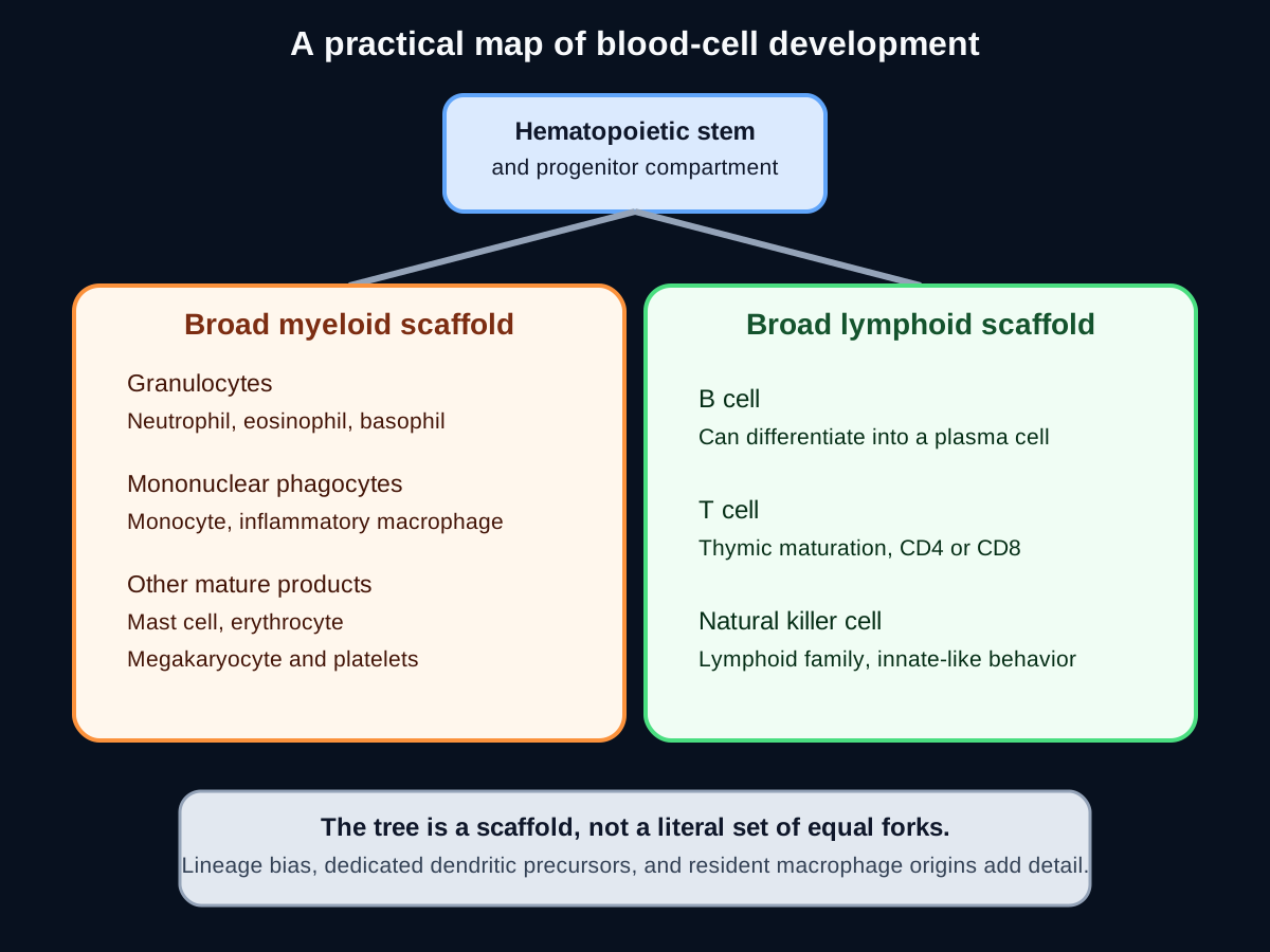
A marrow report can list CD34, CD19, CD3, CD56, myeloperoxidase, or CD138 and still feel like a pile of unrelated facts. The useful question is always the same: where did this cell come from, what did it become, and what job does it perform now? A simple family tree is a strong first scaffold, but real human hematopoiesis is a branching landscape with lineage bias and several important exceptions. [1][2]
One stem-cell compartment supplies every blood lineage
How can one graft restore neutrophils, erythrocytes, platelets, B cells, T cells, and natural killer cells? Hematopoietic stem cells can both self-renew and generate progenitors that progressively restrict their possible fates. CD34-positive cell counting is useful for collecting a transplant graft, but CD34 is an operational enrichment marker for a broad stem and progenitor population, not a unique label carried only by the rare long-term stem cell. [1][2][13]
Use the two broad groups as a first scaffold for basic lineage questions. The lower note keeps the practical map separate from the full differentiation landscape, so do not read it as a sequence of equal, irreversible forks. [1][2]
Apply the scaffold now. Sort erythrocytes, platelets, neutrophils, eosinophils, basophils, monocytes, and mast cells under myeloid. Sort B cells, T cells, and natural killer cells under lymphoid. The visible consequence is that a platelet or erythrocyte question can still test myeloid lineage even though neither mature product is an immune cell.
A new situation makes the relationship clearer. If one clonal marrow disorder raises both the platelet count and the erythrocyte mass, the shared abnormality must sit upstream of both mature products. That pattern points toward a common hematopoietic stem or myeloid progenitor compartment rather than two unrelated mature-cell diseases. [1][6]
Check the scaffold with one cell
A megakaryocyte is myeloid. Its platelets inherit that lineage even though platelets are cell fragments rather than complete nucleated cells.
Try it here · Checkpoint 1 of 3
Make your prediction before reading the choices. A first attempt is just a starting point.
Case 3
Show answer and explanations for case 3
A. A mature circulating erythrocyte (Why this does not fit)
A mature erythrocyte lacks a nucleus and cannot found a platelet-producing clone. The shared variant must be present before the two products separate.
Reasoning steps for option A
Why might the high hemoglobin draw attention to mature red cells?
Erythrocytosis of 19.2 g/dL with low erythropoietin is the most striking laboratory abnormality.
Why can a mature erythrocyte not start this clone?
It has no nucleus and cannot divide or produce megakaryocytes, yet the same JAK2 variant is in both lineages.
B. A common lymphoid progenitor restricted to B, T, and natural killer cells (Why this does not fit)
A lymphoid progenitor supports B, T, and natural killer development. It does not normally generate erythrocytes and megakaryocytes.
Reasoning steps for option B
Could a progenitor explain a variant shared by two lineages?
Yes, a shared variant implies a progenitor, which is why an early progenitor is worth considering.
Why does a lymphoid progenitor fail here?
The variant is in erythroid and megakaryocytic cells, which a B, T, and natural killer progenitor does not produce.
C. A terminal megakaryocyte only (Why this does not fit)
A megakaryocyte can generate platelets but not erythrocytes. The identical variant in both compartments requires an earlier abnormal cell.
Reasoning steps for option C
Why could the megakaryocyte seem the obvious culprit?
The platelet count of 820,000/mm3 is markedly raised and megakaryocytes carry the variant.
Which finding rules out a megakaryocyte-only origin?
The erythroid cells carry the identical variant, and a megakaryocyte cannot make erythrocytes.
D. An upstream hematopoietic stem or myeloid progenitor compartment (Best answer)
One upstream clone can transmit the variant into erythroid and megakaryocytic descendants. The combined erythrocytosis and thrombocytosis therefore localize the origin before their final separation.
Reasoning steps for option D
Which two lineages carry the same JAK2 V617F variant?
The erythroid and megakaryocytic lineages, explaining the high hemoglobin and the thrombocytosis.
Where must the variant arise to reach both lineages?
In a cell upstream of the split between erythroid and megakaryocytic development.
Which compartment sits at that point?
A hematopoietic stem or common myeloid progenitor compartment, whose descendants include both lineages.
E. A thymic T-cell precursor (Why this does not fit)
A thymic precursor belongs to T-cell development. It cannot explain a shared erythroid and megakaryocytic clone.
Reasoning steps for option E
Why might an early precursor of any kind seem reasonable?
The question asks for an initiating clone, and early precursors are the usual origin of clonal disease.
Why is a thymic precursor the wrong early cell?
It is committed to T-cell development and cannot make erythrocytes or megakaryocytes.
Takeaway: A mutation shared by erythrocytes and megakaryocytic cells arose in an upstream hematopoietic stem or myeloid progenitor.
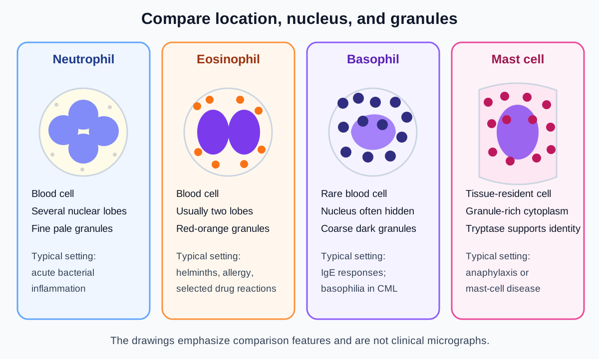
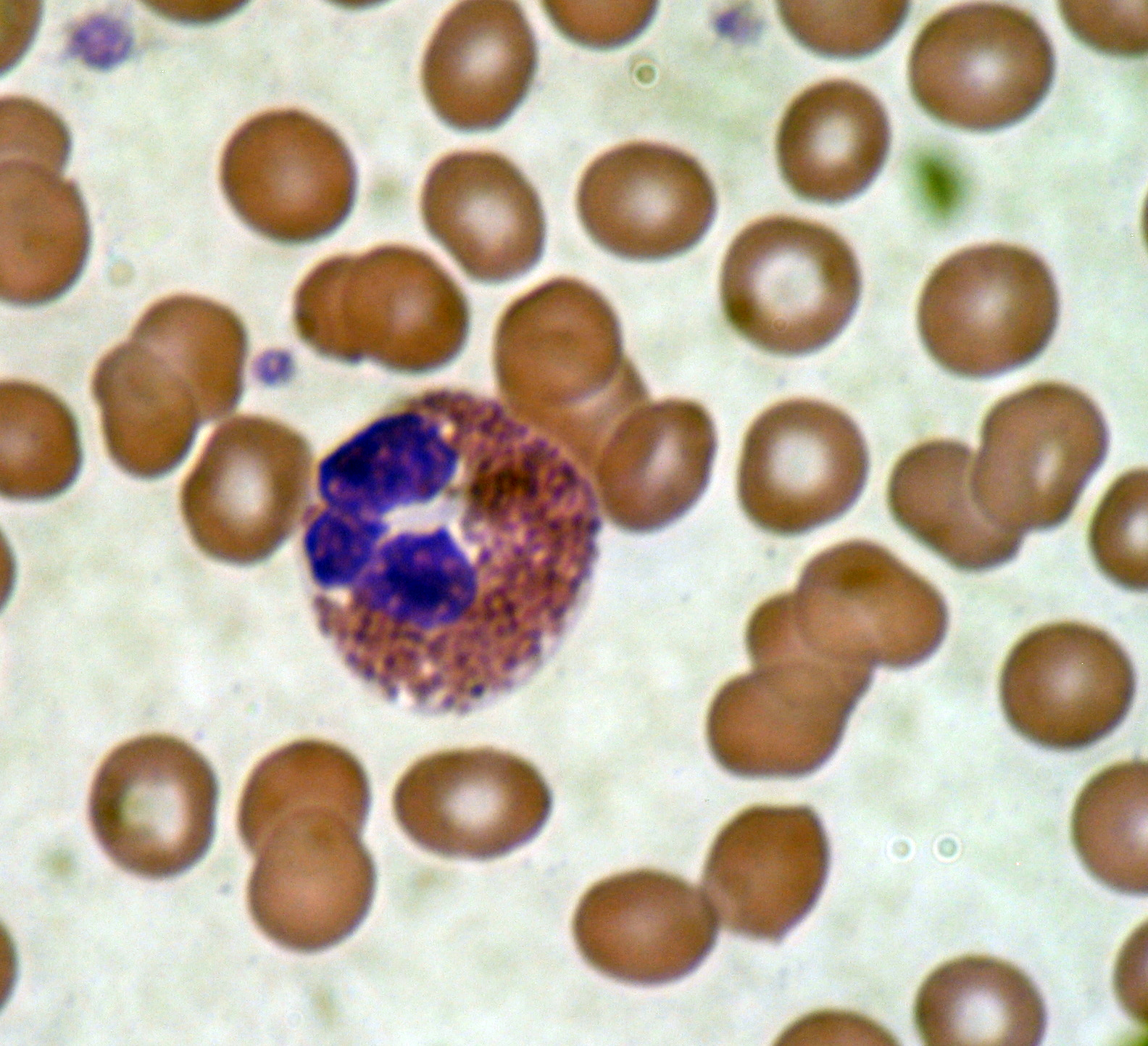
Three granulocytes can all contain cytoplasmic granules, so one feature is rarely enough. Ask three questions in order: is the cell in blood or tissue, what does the nucleus look like, and what physiologic setting is present? The drawings below compare nuclear shape and granule color using standardized morphology terms; the smear photograph after the table shows a real eosinophil. [12][17]
Read each panel from location to nucleus to granules. The drawings emphasize comparison features and do not replace a clinical smear. [7][12][17]
High-yield myeloid pattern comparison
Cell
Visual pattern
Typical setting
Lineage point
CellNeutrophil
Visual patternSegmented nucleus with several connected lobes and fine pale granules
Typical settingAcute bacterial inflammation and tissue injury
Lineage pointMyeloid granulocyte
CellEosinophil
Visual patternUsually bilobed nucleus with prominent red-orange granules
Typical settingHelminth responses, allergy, and selected drug reactions
Lineage pointMyeloid granulocyte
CellBasophil
Visual patternCoarse dark granules that can obscure the nucleus
Typical settingCirculating IgE-associated effector; basophilia can accompany CML
Lineage pointMyeloid granulocyte
CellMast cell
Visual patternGranule-rich tissue cell, identified with tissue context and mast-cell markers
Typical settingAnaphylaxis, urticaria, and mast-cell disease
Lineage pointDistinct myeloid cell, not a tissue basophil
CellMonocyte
Visual patternLarge blood cell with folded or kidney-shaped nucleus
Typical settingCirculating precursor for many inflammatory macrophages and some dendritic cells
Lineage pointMyeloid mononuclear phagocyte
Use the eosinophil smear below as a recognition check: first find the two nuclear lobes, then confirm the red-orange granules, then connect that morphology to eosinophilia in a compatible clinical setting. [12][17]
Basophils and mast cells share IgE-associated biology and mediator release, but they are distinct cells. A dark-granule cell counted in circulating blood is a basophil. A tryptase-rich cell expanding in skin or marrow with KIT D816V is a mast cell. Location and disease markers resolve the pair more reliably than the shared word histamine. [7][12]
Monocytes supply many macrophages during inflammation, but the phrase “a macrophage is simply a monocyte in tissue” is too broad. Many resident populations, including microglia, arise during development and maintain themselves locally in steady state. The correct answer depends on whether the question describes an inflammatory recruit or a long-lived resident population. [3][4]
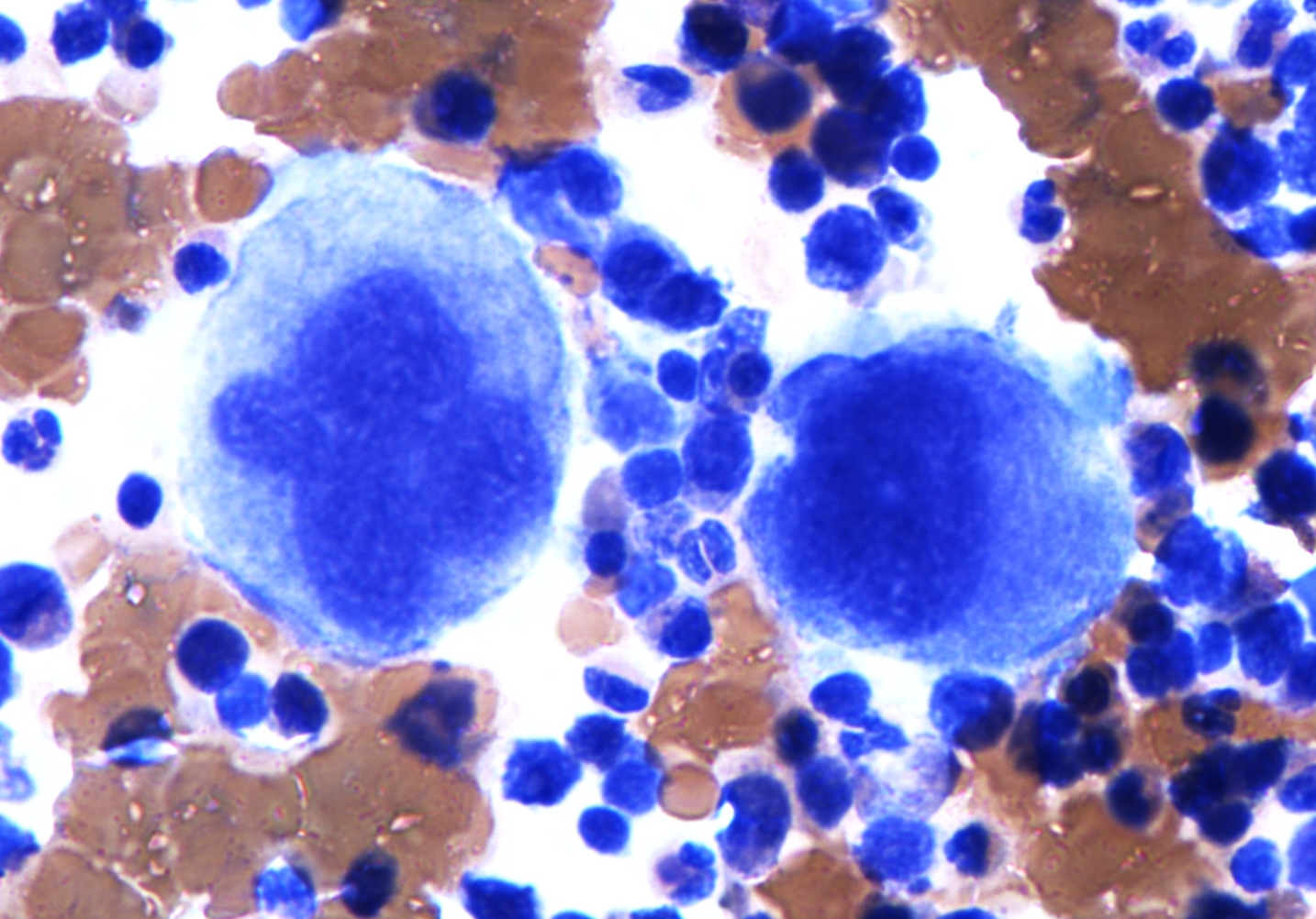
Now transfer the same method to marrow. A huge polyploid cell that extends cytoplasm toward a sinusoid and releases tiny fragments is a megakaryocyte. Its fragments are platelets, so both the giant parent and the circulating fragments belong to the myeloid side of the scaffold. [1] The marrow image below shows the scale difference directly. [14]
Use the scale difference first: megakaryocytes are much larger than surrounding marrow cells. Their cytoplasm supplies platelets. Image: Simon Caulton; original source; CC BY-SA 3.0.
Try it here · Checkpoint 2 of 3
Make your prediction before reading the choices. A first attempt is just a starting point.
Case 8
Show answer and explanations for case 8
A. Eosinophil, myeloid (Best answer)
A bilobed nucleus with red-orange granules identifies an eosinophil. Eosinophils are myeloid granulocytes and commonly rise in helminth-associated responses.
Reasoning steps for option A
What do the helminth and eosinophil count suggest?
Strongyloides infection drives eosinophilia, so an eosinophil response is expected.
Which morphology confirms the cell?
A bilobed nucleus with dense red-orange granules identifies an eosinophil.
Which lineage do eosinophils belong to?
They are granulocytes of the myeloid program.
B. Basophil, lymphoid (Why this does not fit)
Basophils are myeloid rather than lymphoid and have dark granules that may hide the nucleus. The red-orange bilobed cell is an eosinophil.
Reasoning steps for option B
Why might a basophil be considered in a pruritic parasitic infection?
Basophils also take part in IgE-associated and antiparasitic responses.
Which two errors are in this pairing?
Basophils are myeloid, not lymphoid, and their granules are dark rather than red-orange.
C. Neutrophil, myeloid (Why this does not fit)
Neutrophils are myeloid, but they usually have three to five nuclear lobes and fine pale granules. The morphology and helminth setting favor an eosinophil.
Reasoning steps for option C
Why is the myeloid half of this pairing attractive?
Neutrophils are myeloid granulocytes, which is correct as far as it goes.
Which features separate this cell from a neutrophil?
Neutrophils have three to five lobes and pale granules, and helminth infection favors eosinophils.
D. Natural killer cell, lymphoid (Why this does not fit)
Natural killer cells are lymphoid and can kill stressed targets. They do not have eosinophil granules or a bilobed granulocyte nucleus.
Reasoning steps for option D
Why might a cytotoxic lymphoid cell come to mind with a parasite?
Natural killer cells take part in immune defense and are lymphoid.
Why does the smear exclude a natural killer cell?
Natural killer cells lack a bilobed nucleus and red-orange eosinophil granules.
E. Mast cell, myeloid (Why this does not fit)
Mast cells are myeloid tissue residents and can participate in allergic responses. A counted bilobed red-granule cell in peripheral blood is an eosinophil.
Reasoning steps for option E
Why could a mast cell fit the itchy allergic-type rash?
Mast cells are myeloid and drive pruritus and allergic responses.
Which detail separates the smear cell from a mast cell?
This cell is counted in peripheral blood and is bilobed with red-orange granules, while mast cells are tissue residents.
Takeaway: Helminth exposure plus a bilobed red-orange granulocyte identifies a myeloid eosinophil.
Separate lymphoid family from mature-cell behavior
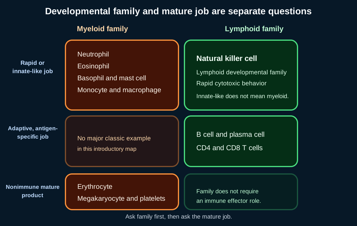
B cells, T cells, and natural killer cells share a lymphoid developmental neighborhood, but their mature behavior differs. Read each cell through four fields: origin, maturation site, surface pattern, and effector function. This prevents a common error in which an innate-like job is mistaken for myeloid ancestry. [1][5]
Lymphoid identity fields
Cell
Maturation and markers
Main mature function
CellB cell
Maturation and markersBone-marrow development; commonly CD19 and CD20
Main mature functionAntigen presentation and differentiation into antibody-secreting plasma cells
CellPlasma cell
Maturation and markersTerminal B-cell state; commonly CD138 with eccentric clock-face chromatin
Main mature functionHigh-rate antibody secretion
CellT cell
Maturation and markersThymic maturation; CD3 with CD4 or CD8 subsets
Main mature functionCoordination or antigen-specific cytotoxicity
CellNatural killer cell
Maturation and markersLymphoid lineage; commonly CD16 and CD56 without surface CD3
Main mature functionRapid cytotoxicity against stressed or MHC-I-low targets
Apply the four fields to a CD56-positive, surface-CD3-negative cell that kills an MHC-I-low target without prior sensitization. Its family is lymphoid, while its mature behavior is innate-like. Natural killer biology also includes adaptive-like and memory-associated features, so “has no memory” is not a safe universal definition. The durable distinction is lymphoid lineage with rapid cytotoxic function. [5]
Natural killer cells sit in the lymphoid column and the rapid cytotoxic row. That placement shows why mature behavior and developmental family must be answered separately. [1][5]
For T cells, connect the mature receptor context to the target: CD8 T cells recognize peptide presented by MHC class I on nucleated cells, while CD4 T cells recognize peptide presented by MHC class II on professional antigen-presenting cells. Both require antigen-specific T-cell receptor signaling; that separates them from natural killer recognition of stressed targets.
For B cells, follow differentiation forward. An activated B cell can become a plasma cell that often no longer carries CD20. This predicts why anti-CD20 therapy can clear circulating B cells while established plasma cells continue secreting antibody. A monoclonal protein plus sheets of CD138-positive clock-face cells therefore indicates a plasma-cell neoplasm, the terminal B-cell endpoint rather than a myeloid tumor. [11][16]
Make your prediction before reading the choices. A first attempt is just a starting point.
Case 19
Show answer and explanations for case 19
A. Myeloid lineage with antigen-specific adaptive function (Why this does not fit)
The marker profile does not fit a myeloid cell, and the killing occurs without antigen-specific sensitization. Both parts of this pairing are incorrect.
Reasoning steps for option A
Why might the adaptive half of this pairing seem relevant to tumor killing?
Tumor killing is often attributed to antigen-specific T cells.
Why are both halves wrong?
The CD16 and CD56 lymphocyte is not myeloid, and it killed without prior sensitization, so the function is not antigen-specific.
B. Lymphoid lineage with innate-like cytotoxic function (Best answer)
CD3 negativity with CD16 and CD56 identifies a natural killer cell. It is lymphoid by developmental family and performs rapid innate-like cytotoxicity.
Reasoning steps for option B
What does the CD3-negative, CD16-positive, CD56-positive profile identify?
A natural killer cell.
Why does the loss of MHC class I matter?
Missing self removes an inhibitory signal and triggers natural killer cytotoxicity without prior sensitization.
How should lineage and function be paired?
Lymphoid lineage with rapid innate-like cytotoxic function.
C. Lymphoid lineage with antibody-secreting function (Why this does not fit)
Antibody secretion belongs to plasma cells. The provided markers and missing-self response identify a natural killer cell.
Reasoning steps for option C
Why is the lymphoid half of this pairing attractive?
The cell is a lymphocyte, so lymphoid lineage is correct.
Which part of the pairing fails?
Antibody secretion belongs to plasma cells; this cell kills directly through a natural killer profile.
D. Myeloid lineage with phagocytic function (Why this does not fit)
Myeloid phagocytes ingest targets rather than using this CD16/CD56 natural killer profile. The cell is a cytotoxic lymphocyte.
Reasoning steps for option D
Why might a myeloid phagocyte be proposed for tumor clearance?
Macrophages can ingest tumor cells.
Why does this cell not fit a myeloid phagocyte?
It is a CD16 and CD56 lymphocyte that kills by cytotoxicity, not by phagocytosis.
E. Nonhematopoietic lineage with stromal support function (Why this does not fit)
The cell is a circulating cytotoxic lymphocyte with recognized hematopoietic markers. It is not a stromal support cell.
Reasoning steps for option E
Why might a supporting cell be considered in a tumor environment?
Stromal cells are abundant around tumors.
Why is a nonhematopoietic stromal lineage wrong?
The cell is a circulating lymphocyte with hematopoietic markers that directly kills targets.
Takeaway: Natural killer cells are lymphoid by developmental family and innate-like in rapid cytotoxic behavior.
Use the tree as a scaffold, then check the exception
Dendritic-cell development is the clearest reason not to memorize one rigid family tree. Conventional dendritic cells, plasmacytoid dendritic cells, and monocyte-derived dendritic cells are distinguished by ontogeny and function; they should not all be forced into a single “monocyte branch” or a single “lymphoid branch.” Modern nomenclature favors the developmental program and the mature subset rather than a simplistic side of the diagram. [2][3]
Question asks lineage
Use progenitor relationships and lineage-defining markers.
Question asks function
Use phagocytosis, antibody secretion, antigen presentation, or cytotoxicity.
Question asks location
Use blood, marrow, thymus, tissue, or a resident organ compartment.
Question asks neoplasm
Use the full immunophenotype and defining genetic alteration.
The same mature cell can be classified differently depending on the question being asked.
Try this classification sequence with a plasmacytoid dendritic cell producing type I interferon during viral infection. Its job identifies a specialized dendritic subset, but that fact alone does not justify placing every plasmacytoid dendritic cell on a permanently lymphoid-only route. The consequence is practical: answer the requested dimension and avoid converting a useful teaching scaffold into an absolute developmental law. [3]
The same reasoning protects against another error. Donor-derived monocytes can enter inflamed tissue and generate macrophages, yet stable brain microglia can remain recipient-derived after donor blood chimerism is established. Blood origin and resident-tissue maintenance are separate questions. [4]
Disease findings reveal the affected developmental program
Clinical questions often provide a mature-cell pattern, a surface profile, and a genetic or anatomic finding. Combine all three before naming the lineage. A very high white-cell count with a left-shifted granulocyte series, basophilia, splenomegaly, and BCR::ABL1 indicates chronic myeloid leukemia. The granulocyte pattern establishes the myeloid program; the fusion establishes the disease. [6][10]
Acute leukemia uses stricter lineage-defining evidence. Myeloperoxidase supports myeloid differentiation, while cytoplasmic or surface CD3 supports T lineage. CD19 can support B lineage only within an appropriate broader marker pattern. A blast population with convincing myeloperoxidase and cytoplasmic CD3 requires consideration of mixed-phenotype acute leukemia rather than automatic assignment from a nonspecific marker. [6][16]
Four other patterns become straightforward once the normal cell is identified. KIT D816V with mast-cell aggregates and high tryptase indicates mastocytosis. A monoclonal protein with CD138-positive plasma cells indicates a plasma-cell neoplasm. An absent or hypoplastic thymus with hypocalcemia and a conotruncal defect predicts impaired T-cell maturation in 22q11.2 deletion syndrome. Severe herpesvirus infection with absent CD16-positive and CD56-positive cells despite preserved B- and T-cell counts suggests natural killer cell deficiency. [7][8][9][11]
Use a final four-part read for every case: identify the dominant mature-cell pattern, identify the lineage-defining marker, locate the developmental or anatomic defect, then predict which related products should be preserved or lost. That approach transfers from a smear to flow cytometry, marrow pathology, immunodeficiency, and hematologic neoplasia.
Apply the lesson
Case 1
Show answer and explanations for case 1
A. Self-renewal with multilineage hematopoietic differentiation (Best answer)
This property explains durable donor output across both broad developmental programs. A mature or single-lineage progenitor could not account for all listed products.
Reasoning steps for option A
Which products carry donor genotype six months after the graft?
Neutrophils, erythrocytes, platelets, B cells, T cells, and natural killer cells, so donor output spans both the myeloid and lymphoid programs.
Why must the infused cells both self-renew and differentiate?
Output still present at six months needs a compartment that maintains itself, and output in every lineage needs cells able to generate all of them.
Which infused population has both properties?
Hematopoietic stem cells within the CD34-selected graft self-renew and give rise to every blood lineage.
B. Expansion of a mature neutrophil population (Why this does not fit)
Mature neutrophils can transiently circulate after transfusion. They cannot generate erythrocytes, platelets, or lymphocytes months later.
Reasoning steps for option B
Why could donor neutrophils alone make a graft look successful early?
Neutrophil recovery is the first visible sign of engraftment and is what clinicians track in the first weeks.
What prevents mature neutrophils from explaining the six-month result?
Mature neutrophils live only days and are terminal cells, so they cannot produce erythrocytes, platelets, or lymphocytes months later.
C. Selective proliferation of a common lymphoid progenitor (Why this does not fit)
A lymphoid progenitor can support B, T, and natural killer development. It cannot explain donor erythrocytes, platelets, and neutrophils.
Reasoning steps for option C
Which donor cells in this recipient could a common lymphoid progenitor explain?
It could account for donor B cells, T cells, and natural killer cells, half of the listed pattern.
Which donor products fall outside a lymphoid progenitor's reach?
Donor erythrocytes, platelets, and neutrophils are myeloid products that a lymphoid-restricted progenitor does not make.
D. Engraftment of thymic epithelial cells (Why this does not fit)
Thymic epithelium supports T-cell education. It does not produce the full set of blood cells listed.
Reasoning steps for option D
Why is thymic tissue relevant to donor T-cell recovery after transplant?
Thymic epithelium educates developing T cells, so it shapes how donor T cells mature.
Why can thymic epithelium not be the source of these donor cells?
It is a supporting stromal tissue that produces no blood cells, and the graft did not contain it.
E. Differentiation of marrow mesenchymal stromal cells into all blood cells (Why this does not fit)
Marrow stromal cells support the hematopoietic niche. The durable donor blood output comes from hematopoietic stem and progenitor cells.
Reasoning steps for option E
Why might marrow stroma seem important for a durable graft?
Mesenchymal stromal cells build the niche that supports hematopoietic stem cells in the recipient marrow.
What do stromal cells actually contribute to donor blood counts?
They support hematopoiesis but do not differentiate into blood cells; the multilineage output comes from hematopoietic stem and progenitor cells.
Takeaway: Multilineage donor chimerism requires a self-renewing hematopoietic stem and progenitor compartment.
A. It proves every counted CD34-positive cell is a long-term self-renewing stem cell with identical potency (Why this does not fit)
CD34 enrichment captures a broader hematopoietic stem and progenitor population. The count does not establish identical long-term potential for every counted cell.
Reasoning steps for option A
Why is it tempting to equate a high CD34 count with stem-cell dose?
CD34 is the standard graft marker, and the true long-term stem cells are among the CD34-positive cells.
What does the CD34-positive fraction actually contain?
A heterogeneous mix of stem and progenitor cells, most of which lack long-term self-renewal, so identical potency is not established.
B. It measures mature neutrophils that will supply immediate antibacterial defense (Why this does not fit)
Mature neutrophils are not defined by CD34 enumeration. The accepted value estimates the collected stem and progenitor dose.
Reasoning steps for option B
Why might a count taken after G-CSF suggest a neutrophil measurement?
G-CSF is a neutrophil growth factor and raises circulating neutrophils during mobilization.
Why does the CD34 count not measure mature neutrophils?
Mature neutrophils do not express CD34; the count estimates the early stem and progenitor graft dose.
C. It is an operational measure that enriches and quantifies hematopoietic stem and progenitor cells (Best answer)
CD34 enumeration is used to assess the collected graft dose. It is useful without being a cell-exclusive definition of the rare long-term stem cell.
Reasoning steps for option C
What does the acceptance of 6.0 million CD34-positive cells/kg tell the team?
The graft contains an adequate dose of early hematopoietic cells to restore marrow function.
Why is CD34 called an operational marker rather than a stem-cell definition?
It enriches and quantifies a broad stem and progenitor population, which is useful for dosing even though it is not exclusive to long-term stem cells.
Which interpretation matches that operational role?
The count is an enrichment and quantification measure for hematopoietic stem and progenitor cells.
D. It selectively counts common lymphoid progenitors (Why this does not fit)
The collection supports recovery of more than lymphoid products. CD34-positive collections contain heterogeneous early hematopoietic cells.
Reasoning steps for option D
Why might CD34 seem linked to lymphoid progenitors?
Early lymphoid progenitors do express CD34, so they are part of the counted population.
Why is a lymphoid-only reading too narrow?
The same CD34-positive product restores myeloid and erythroid output, so it holds heterogeneous early cells, not lymphoid progenitors alone.
E. It directly measures megakaryocytes in the apheresis bag (Why this does not fit)
Megakaryocytes are large marrow cells and are not the intended circulating apheresis target. CD34 counting estimates an early hematopoietic graft compartment.
Reasoning steps for option E
Why might a platelet-related marrow cell come to mind?
Graft CD34 dose predicts platelet recovery, and megakaryocytes are the platelet source.
Why are megakaryocytes not what the apheresis count measures?
Megakaryocytes are large marrow-resident cells, not circulating cells collected by apheresis, and CD34 counting targets early progenitors.
Takeaway: CD34 is a practical graft-enrichment and enumeration marker, not a unique identity tag for every long-term stem cell.
A. The mature B-cell program (Why this does not fit)
B-cell neoplasms can cause leukocytosis and splenomegaly. The listed cells are a left-shifted granulocytic series with basophilia, not a monoclonal B-cell population.
Reasoning steps for option A
Why could a B-cell neoplasm be considered with this presentation?
Chronic lymphocytic leukemia and B-cell lymphomas can cause very high white counts and splenomegaly.
What does the differential count show instead of B cells?
A left-shifted granulocytic series with basophilia, not a monoclonal lymphocyte population.
B. The thymic T-cell program (Why this does not fit)
T-cell disease can involve blood and lymphoid organs. It does not match a granulocyte-rich differential with BCR::ABL1-defined CML.
Reasoning steps for option B
Why might T-cell disease enter the differential?
T-cell leukemias can also produce leukocytosis, night sweats, and organ enlargement.
Which finding separates this case from T-cell disease?
The cells are granulocytes at all stages, and BCR::ABL1 defines chronic myeloid leukemia rather than a T-cell program.
C. The plasma-cell program (Why this does not fit)
Plasma-cell neoplasms produce monoclonal immunoglobulin and marrow plasma cells. They do not produce this granulocytic expansion.
Reasoning steps for option C
Why might a plasma-cell disorder seem possible in an older man with systemic symptoms?
Plasma-cell neoplasms are common at this age and cause fatigue and constitutional symptoms.
Which laboratory pattern argues against a plasma-cell disorder?
Plasma-cell disease presents with monoclonal protein and marrow plasma cells, not a WBC of 186,000/mm3 with granulocytes and basophils.
D. The natural killer cell program (Why this does not fit)
Natural killer cells are lymphoid cytotoxic cells. CD16 or CD56 expansion is not the pattern provided.
Reasoning steps for option D
Why could a lymphocyte-driven process be proposed for massive leukocytosis?
Natural killer cell leukemias exist and can involve blood and spleen.
What marker pattern would a natural killer expansion show, and is it present?
It would show CD16 and CD56 lymphocytes; this blood instead shows granulocytes and basophils.
E. The myeloid granulocytic program (Best answer)
Granulocytes and basophils belong to the myeloid program. BCR::ABL1 with this blood pattern establishes chronic myeloid leukemia.
Reasoning steps for option E
Which cells dominate the blood film?
Granulocytes at every maturation stage plus basophils, which are all myeloid cells.
What does BCR::ABL1 add?
It establishes chronic myeloid leukemia, a stem-cell disease that drives the myeloid program.
Which program is therefore expanded?
The myeloid granulocytic program, explaining the leukocytosis, basophilia, and splenomegaly.
Takeaway: Granulocytic left shift plus basophilia and BCR::ABL1 identifies expansion of the myeloid program.
B-lineage blasts may express CD19, CD79a, or PAX5 in a qualifying pattern. Auer rods and myeloperoxidase directly support myeloid differentiation.
Reasoning steps for option A
Why is B lineage a real consideration in adult acute leukemia?
Precursor B lymphoblastic leukemia also presents with cytopenias and a marrow full of blasts.
Which findings point away from B lineage?
Auer rods and myeloperoxidase are direct myeloid evidence, and no CD19-based B-cell pattern is described.
B. Natural killer (Why this does not fit)
Natural killer cells commonly express CD16 and CD56 without surface CD3. They do not form myeloperoxidase-positive Auer rods.
Reasoning steps for option B
Why might the absence of CD3 suggest natural killer cells?
Natural killer cells lack surface CD3, so a CD3-negative population can prompt that thought.
Why do these blasts not fit natural killer lineage?
Natural killer cells do not contain myeloperoxidase or form Auer rods, which are granule structures of myeloid blasts.
C. Plasma cell (Why this does not fit)
Plasma cells commonly express CD138 and produce immunoglobulin. They do not match an acute myeloperoxidase-positive blast population.
Reasoning steps for option C
Why could a plasma-cell neoplasm be considered in an older woman with anemia?
Plasma-cell disease is common at this age and can cause anemia and marrow infiltration.
Why do these marrow cells not fit plasma cells?
They are acute blasts with Auer rods and myeloperoxidase, not mature CD138-positive antibody-secreting cells.
D. Megakaryocytic (Why this does not fit)
Megakaryocytic blasts require platelet-lineage evidence such as cytoplasmic CD41 or CD61 in context. The provided lineage-defining findings instead support ordinary myeloid differentiation.
Reasoning steps for option D
Why could megakaryocytic leukemia be raised with a platelet count of 28,000/mm3?
Acute megakaryoblastic leukemia is a myeloid subtype and also causes marrow failure.
What evidence would megakaryocytic assignment need?
Platelet-lineage markers such as CD41 or CD61; the findings given support ordinary myeloid differentiation instead.
E. Myeloid (Best answer)
Auer rods and myeloperoxidase positivity are strong evidence of myeloid differentiation. The presentation therefore fits acute myeloid leukemia rather than a lymphoid blast disorder.
Reasoning steps for option E
What do Auer rods indicate about these blasts?
They are abnormal fused azurophilic granules found in myeloid blasts.
How does myeloperoxidase positivity settle the lineage?
Myeloperoxidase is the lineage-defining marker of myeloid differentiation.
Why is T lineage excluded as a second lineage here?
Cytoplasmic CD3 is absent and the myeloid evidence is direct, so this is acute myeloid leukemia rather than a mixed phenotype.
Takeaway: Myeloperoxidase-positive blasts with Auer rods are assigned to myeloid lineage.
A. Precursor B-cell development from the lymphoid program (Best answer)
TdT establishes immaturity, while CD19 and CD10 support precursor B-cell differentiation in this marker context. Absence of myeloperoxidase and cytoplasmic CD3 argues against myeloid and T lineage.
Reasoning steps for option A
What does TdT positivity establish?
The blasts are immature lymphoid precursors, typical of acute lymphoblastic leukemia in a child.
Which markers identify the lineage?
CD19 with CD10 supports precursor B differentiation, and absent myeloperoxidase and cytoplasmic CD3 exclude myeloid and T lineage.
Which normal route do the blasts mirror?
Precursor B-cell development within the lymphoid program in the marrow.
B. Granulocytic development from an early myeloid progenitor in marrow (Why this does not fit)
Granulocytic blasts require myeloid differentiation evidence, especially myeloperoxidase. The blasts are myeloperoxidase negative and carry a B-cell marker pattern.
Reasoning steps for option B
Why might granulocytic development be suggested by fever and marrow replacement?
Acute myeloid leukemia also causes fever, pallor, and petechiae from marrow failure.
Which result argues against a myeloid route?
The blasts lack myeloperoxidase and carry B-cell markers instead.
C. Thymic T-cell development (Why this does not fit)
T-lineage assignment requires cytoplasmic or surface CD3 in the proper setting. Cytoplasmic CD3 is absent while B-lineage markers are present.
Reasoning steps for option C
Why could a T-cell route be considered in a child with acute leukemia?
T-lymphoblastic leukemia is also TdT positive and occurs in childhood.
Which marker would T lineage need, and is it present?
It would need cytoplasmic CD3, which is absent while CD19 and CD10 are present.
D. Megakaryocytic development (Why this does not fit)
Megakaryocytic blasts show platelet-lineage markers in context. CD19 and CD10 do not establish megakaryocytic differentiation.
Reasoning steps for option D
Why might megakaryocytic development seem relevant to petechiae?
Petechiae reflect low platelets, and platelets come from megakaryocytes.
Why do the petechiae not identify a megakaryocytic clone?
They reflect marrow crowding by blasts, and CD19 with CD10 is a B-precursor pattern, not a platelet-lineage one.
E. Terminal plasma-cell development (Why this does not fit)
Plasma cells are terminally differentiated antibody-secreting B-lineage cells, usually CD138 positive rather than TdT positive. This is a precursor B-cell blast population.
Reasoning steps for option E
Why might plasma-cell development seem close to a B-lineage answer?
Plasma cells are the terminal stage of B-cell development, so both belong to the B program.
Why is a terminal plasma-cell route wrong for these blasts?
TdT marks immature precursors, whereas plasma cells are mature CD138-positive cells that do not express TdT.
Eosinophils usually have a bilobed nucleus and conspicuous red-orange granules. The segmented pale-granule cell in acute bacterial inflammation is different.
Reasoning steps for option A
Why might a granulocyte with a lobed nucleus be mistaken for an eosinophil?
Eosinophils are also granulocytes with a lobed nucleus.
Which features separate this cell from an eosinophil?
It has four lobes and fine pale granules, while an eosinophil is usually bilobed with red-orange granules.
B. Neutrophil (Best answer)
A segmented nucleus with several connected lobes and fine pale granules identifies a neutrophil. Acute bacterial inflammation also fits its rapid myeloid response.
Reasoning steps for option B
What does a four-lobed nucleus with fine pale granules identify?
A segmented neutrophil.
How does the clinical setting fit?
Aspiration pneumonia is an acute bacterial infection, and neutrophilia is the expected rapid myeloid response.
C. Basophil (Why this does not fit)
Basophils contain coarse dark granules that can obscure the nucleus. The nucleus here remains clearly segmented and the granules are pale.
Reasoning steps for option C
Why might a basophil be considered for any circulating granulocyte?
Basophils are granulocytes and appear in the same leukocyte differential.
Which features exclude a basophil?
Basophil granules are coarse and dark and obscure the nucleus, whereas these granules are pale and the lobes are clearly visible.
D. Monocyte (Why this does not fit)
Monocytes are large cells with folded or kidney-shaped nuclei and abundant gray-blue cytoplasm. They are not the multilobed granulocyte described.
Reasoning steps for option D
Why could a monocyte be considered in pneumonia?
Monocytes also rise in infection and migrate to inflamed lungs.
Which nuclear shape rules out a monocyte?
Monocytes have a folded or kidney-shaped nucleus, not four connected lobes.
E. Natural killer cell (Why this does not fit)
Natural killer cells are lymphoid cytotoxic cells and resemble lymphocytes morphologically. They do not have a four-lobed granulocyte nucleus.
Reasoning steps for option E
Why might a cytotoxic lymphocyte seem relevant in acute infection?
Natural killer cells are part of early innate defense.
Which morphology excludes a natural killer cell?
Natural killer cells look like lymphocytes with a round nucleus, not a four-lobed granulocyte nucleus.
Takeaway: A segmented multilobed nucleus with fine pale granules in acute bacterial inflammation identifies a neutrophil.
Mast cells contain histamine and can be granule rich. They are primarily tissue residents, whereas the described cells are counted in circulating blood.
Reasoning steps for option A
Why could histamine-containing granules suggest a mast cell?
Mast cells are the best-known histamine-rich cells and are also granule rich.
Which detail in the stem favors a different cell?
These cells are counted in circulating blood, whereas mast cells are tissue residents.
B. Eosinophil (Why this does not fit)
Eosinophils usually show red-orange granules and a visible bilobed nucleus. Dark granules that obscure the nucleus favor a basophil.
Reasoning steps for option B
Why might an eosinophil be considered in a granulocyte-rich CML film?
Eosinophils are granulocytes and can also increase in CML.
Which granule color excludes an eosinophil?
Eosinophil granules are red-orange with a visible bilobed nucleus, not blue-black and obscuring.
C. Basophil (Best answer)
A rare circulating granulocyte with coarse blue-black granules and histamine is a basophil. Basophilia is also a characteristic associated finding in CML.
Reasoning steps for option C
What does a circulating granulocyte with coarse blue-black granules represent?
A basophil, whose dark granules often hide the nucleus.
Which additional findings fit?
Basophils store histamine, and basophilia is a characteristic finding in chronic myeloid leukemia.
D. Monocyte (Why this does not fit)
Monocytes are large agranular-appearing cells with folded nuclei and phagocytic potential. They do not have dense histamine-containing granules that obscure the nucleus.
Reasoning steps for option D
Why might a monocyte be considered among rare circulating cells?
Monocytes are also a minority population in the blood differential.
Why does the morphology exclude a monocyte?
Monocytes have a folded nucleus and gray-blue cytoplasm without dense dark histamine granules.
E. Plasma cell (Why this does not fit)
Plasma cells are antibody-secreting lymphoid descendants with eccentric nuclei. They are not circulating dark-granule granulocytes.
Reasoning steps for option E
Why might a plasma cell seem possible in a hematologic malignancy?
Plasma cells can occasionally circulate in marrow disorders.
Why is a plasma cell wrong here?
Plasma cells are B-lineage antibody secretors with eccentric nuclei, not granulocytes with dark histamine granules.
Takeaway: A rare dark-granule cell in blood is a basophil; tissue residency would instead favor a mast cell.
A. It is a tissue basophil created when a circulating basophil enters skin (Why this does not fit)
Basophils and mast cells share IgE-associated mediator biology. They remain distinct cell types rather than sequential blood and tissue forms of one cell.
Reasoning steps for option A
Why does a tissue basophil sound reasonable for these symptoms?
Basophils and mast cells both release histamine through IgE-associated pathways, and the shared biology invites that link.
Why is the migration explanation wrong?
Mast cells are a distinct lineage with their own precursors; circulating basophils do not become tissue mast cells.
B. It is a lymphoid natural killer cell with granules (Why this does not fit)
Natural killer cells contain cytotoxic granules, but they do not produce this tryptase-rich KIT D816V mast-cell aggregate pattern. The skin lesions and mediator symptoms identify mast-cell disease.
Reasoning steps for option B
Why could granule-rich cytotoxic lymphocytes be considered?
Natural killer cells also contain cytoplasmic granules.
C. It is an eosinophil undergoing tissue maturation (Why this does not fit)
Eosinophils can enter tissues during allergy and parasitic disease. KIT D816V-positive tryptase-rich aggregates are mast cells, not maturing eosinophils.
Reasoning steps for option C
Why might eosinophils be considered in an allergic-type skin disorder?
Eosinophils also infiltrate skin in allergic and parasitic disease.
Which markers identify the lesional cells instead?
Tryptase-rich KIT D816V-positive aggregates are mast cells, not maturing eosinophils.
D. It is a distinct myeloid mast cell (Best answer)
Tissue aggregates, persistent tryptase elevation, and KIT D816V identify a mast-cell proliferation. Mast cells are distinct myeloid cells and are not displaced basophils.
Reasoning steps for option D
Which clinical features point to mast-cell mediator release?
Flushing, presyncope, and macules that urticate when rubbed.
What do the laboratory and biopsy findings add?
Persistently high tryptase and KIT D816V-positive tissue aggregates establish a mast-cell proliferation.
How should that cell be classified?
As a distinct myeloid mast cell, not as a displaced basophil.
E. It is a terminal plasma cell (Why this does not fit)
Plasma cells produce immunoglobulin and commonly express CD138. They do not produce the tryptase and KIT pattern described.
Reasoning steps for option E
Why might a plasma cell be considered for a clonal tissue proliferation?
Plasma-cell neoplasms are also clonal and can infiltrate tissues.
Which markers separate the lesion from plasma cells?
Plasma cells express CD138 and secrete immunoglobulin, not tryptase with KIT D816V.
Neutrophils can enter infected tissue and phagocytose microbes. They have segmented nuclei rather than a CD14-positive kidney-shaped monocyte pattern and do not become macrophages.
Reasoning steps for option A
Why could neutrophils be expected in infected valve tissue?
Neutrophils are the first phagocytes recruited to bacterial infection.
Which features of the blood population exclude neutrophils?
The cells are CD14-positive with kidney-shaped nuclei, and neutrophils do not become antigen-presenting macrophages.
B. B lymphocyte (Why this does not fit)
B cells can present antigen and produce antibody after differentiation. They do not match CD14-positive kidney-shaped blood cells or become tissue macrophages.
Reasoning steps for option B
Why might B lymphocytes be considered for antigen-presenting cells?
B cells can present antigen to T cells.
Why do B cells not fit the blood or tissue findings?
They lack CD14 and kidney-shaped nuclei and do not become large debris-ingesting macrophages.
C. Monocyte (Best answer)
CD14 positivity and a folded kidney-shaped nucleus identify a monocyte. During inflammation, monocytes can enter tissue and differentiate into phagocytic macrophages.
Reasoning steps for option C
What do CD14 and a kidney-shaped nucleus identify in blood?
A monocyte.
What do monocytes become in inflamed tissue?
They leave the blood and differentiate into macrophages that phagocytose debris and present antigen.
Which blood cell therefore supplied the valve macrophages?
The circulating monocyte.
D. Megakaryocyte (Why this does not fit)
Megakaryocytes are giant marrow cells that release platelets. They are not circulating CD14-positive precursors for inflammatory macrophages.
Reasoning steps for option D
Why might a marrow giant cell be linked to endocarditis?
Platelets from megakaryocytes build the vegetations on infected valves.
Why is a megakaryocyte not the precursor here?
It stays in marrow and releases platelets; it is not a circulating CD14-positive cell that becomes a macrophage.
E. Mast cell (Why this does not fit)
Mast cells are granule-rich tissue residents involved in mediator release. They are not the kidney-shaped circulating precursor described.
Reasoning steps for option E
Why might mast cells be considered as tissue immune cells?
Mast cells reside in tissues and help recruit other cells during infection.
Why do mast cells not match the circulating precursor?
They are tissue-resident granule cells, not CD14-positive kidney-shaped blood cells that become phagocytes.
Takeaway: CD14-positive kidney-shaped blood monocytes can supply macrophages during tissue inflammation.
C. The transplant failed to establish donor hematopoiesis (Why this does not fit)
Circulating monocytes show complete donor chimerism, so donor hematopoiesis is established. The discordance is specific to the resident brain population.
Persisting recipient cells can signal loss of donor engraftment.
Which result shows donor hematopoiesis is established?
Every circulating monocyte now carries the donor genotype, so the graft is working; only the long-lived brain residents kept the old genotype.
D. Stable microglia can be long-lived developmentally seeded residents that self-maintain (Best answer)
Many microglia arise during development and can maintain themselves locally in steady state. Donor blood chimerism therefore need not replace stable recipient microglia.
Reasoning steps for option D
What does complete donor monocyte chimerism tell you?
Donor hematopoiesis now supplies circulating monocytes.
Why do microglia still carry the recipient genotype?
Many microglia are seeded during embryonic development and renew locally, so blood-derived replacement is not needed in the absence of inflammation.
What general principle does this illustrate?
Blood origin and resident-tissue maintenance are separate questions for macrophage populations.
E. Microglia are generated continuously by thymic epithelium (Why this does not fit)
Thymic epithelium supports T-cell maturation. It does not generate resident brain macrophages.
Reasoning steps for option E
Why might an organ that shapes immune cells be proposed?
The thymus is a site where immune cells develop.
Why does thymic epithelium not produce microglia?
The thymus is an epithelial scaffold for developing T cells, whereas microglia descend from early yolk-sac macrophage precursors that colonize the brain before birth.
Takeaway: Stable microglia can remain recipient-derived because many are developmentally seeded and locally self-maintaining.
A. Every dendritic cell is a circulating monocyte that entered inflamed tissue and changed its identity (Why this does not fit)
The persistence of conventional dendritic cells after monocyte depletion contradicts a universal monocyte intermediate. Monocyte-derived cells represent one inflammatory route, not all dendritic development.
Reasoning steps for option A
Why is a monocyte origin for all dendritic cells tempting?
The second observation shows monocytes can become dendritic-like cells during inflammation.
Which observation contradicts a universal monocyte route?
Conventional dendritic cells keep developing when circulating monocytes are depleted.
B. Every dendritic cell belongs to a lymphoid-only route (Why this does not fit)
The observations include monocyte-derived inflammatory cells and dedicated dendritic precursors. A lymphoid-only rule cannot account for both.
Reasoning steps for option B
Why might a lymphoid-only route be proposed for dendritic cells?
Older teaching placed some dendritic subsets on the lymphoid side of the tree.
Why does a lymphoid-only rule fail these data?
It cannot explain monocyte-derived inflammatory dendritic-like cells or dedicated Flt3-dependent precursors.
C. Dendritic populations should be classified by subset and ontogeny rather than one universal parent (Best answer)
Conventional dendritic cells can arise through dedicated precursor programs, while inflammation can generate monocyte-derived dendritic-like cells. The combined data require an ontogeny-based classification.
Reasoning steps for option C
What does monocyte depletion show about conventional dendritic cells?
They arise from dedicated Flt3-dependent precursors, not through monocytes.
What does the inflammatory observation show?
Monocytes can generate dendritic-like cells under inflammation.
Which classification integrates both observations?
Dendritic populations are classified by subset and ontogeny rather than by one universal parent.
D. Conventional dendritic cells are terminal plasma cells (Why this does not fit)
Plasma cells are antibody-secreting B-lineage descendants. They do not explain Flt3-dependent dendritic development.
Reasoning steps for option D
Why could the word conventional suggest a mature terminal cell?
Plasma cells are the classic terminal immune cell, and both are antigen-related.
Why are conventional dendritic cells not plasma cells?
Conventional dendritic cells present antigen to T cells and need Flt3 signaling to develop, whereas plasma cells are end-stage B cells that make immunoglobulin.
E. Monocyte depletion proves dendritic cells are nonhematopoietic (Why this does not fit)
A cell can remain hematopoietic without passing through a circulating monocyte. Dedicated hematopoietic dendritic precursors account for the persistence.
Reasoning steps for option E
Why might persistence without monocytes suggest a separate origin?
It shows dendritic cells do not depend on circulating monocytes.
Why does that not make them nonhematopoietic?
Dedicated Flt3-dependent dendritic precursors are themselves hematopoietic cells.
Takeaway: Dendritic cells are not assigned to one universal parent; subset and ontogeny determine the developmental route.
CD19, CD20, CD79a, and surface immunoglobulin form a coherent mature B-cell profile. The negative T- and myeloid-lineage findings support that assignment.
Reasoning steps for option A
Which positive markers are present on the clone?
CD19, CD20, CD79a, and surface immunoglobulin.
What do those markers form together?
A coherent mature B-cell profile, with surface immunoglobulin confirming a mature stage.
How do the negative markers help?
Absent surface CD3 and myeloperoxidase exclude T and myeloid assignment.
B. Mature T cell (Why this does not fit)
A mature T-cell population should show surface CD3. The provided markers instead form a B-cell pattern.
Reasoning steps for option B
Why might a lymph-node lymphoma be a T-cell process?
T-cell lymphomas also involve lymph nodes in older adults.
Which finding rules out mature T lineage?
A mature T-cell population expresses surface CD3, which is absent here.
C. Myeloid blast (Why this does not fit)
Myeloid blasts require appropriate myeloid differentiation evidence, often including myeloperoxidase. The cells are myeloperoxidase negative and express multiple B-cell markers.
Reasoning steps for option C
Why might a myeloid blast be considered in an older adult?
Myeloid sarcoma can occasionally present as a nodal mass.
Which findings exclude myeloid blasts?
The cells lack myeloperoxidase and express several B-cell markers with surface immunoglobulin.
D. Natural killer cell (Why this does not fit)
Natural killer cells are commonly CD56 positive and lack surface CD3, but they do not express this surface-immunoglobulin B-cell profile. The positive markers identify B lineage.
Reasoning steps for option D
Why could a CD3-negative lymphocyte suggest a natural killer cell?
Natural killer cells are lymphoid and lack surface CD3.
Which markers separate this clone from natural killer cells?
Natural killer cells do not express CD19, CD20, CD79a, or surface immunoglobulin.
E. Megakaryocyte (Why this does not fit)
Megakaryocytes express platelet-associated markers and have distinctive marrow morphology. They do not express surface immunoglobulin as a monoclonal lymph-node population.
Reasoning steps for option E
Why might a large marrow-derived cell be considered?
Megakaryocytes can appear in extramedullary tissues.
Why does the immunophenotype exclude megakaryocytes?
Megakaryocytes express platelet markers, not surface immunoglobulin and B-cell antigens.
Takeaway: CD19, CD20, CD79a, and surface immunoglobulin identify a mature B-cell population.
Neutrophils are myeloid granulocytes and do not secrete monoclonal immunoglobulin. Their segmented nuclei do not match clock-face plasma cells.
Reasoning steps for option A
Why might a marrow-derived white cell be considered?
Neutrophils are the most common marrow white-cell product.
Why can neutrophils not explain this disease?
They do not secrete immunoglobulin and have segmented nuclei, not clock-face chromatin.
B. Mature B cell (Best answer)
A plasma cell is the terminal antibody-secreting state of an activated mature B cell. The monoclonal IgG and CD138-positive clock-face morphology identify that endpoint.
Reasoning steps for option B
What do the monoclonal IgG and CD138-positive clock-face cells identify?
A plasma-cell neoplasm such as multiple myeloma.
From which mature cell does a plasma cell arise?
Antigen-activated mature B cells differentiate into plasma cells, so the malignant clone traces back to the B lineage.
How do the clinical features fit?
Bone pain, anemia, kidney injury, and constipation that can reflect hypercalcemia are classic myeloma effects.
C. Natural killer cell (Why this does not fit)
Natural killer cells are cytotoxic lymphoid cells and commonly express CD16 and CD56. They do not become antibody-secreting plasma cells.
Reasoning steps for option C
Why might a lymphoid cytotoxic cell be considered?
Natural killer cells are lymphoid like B cells.
Why do natural killer cells not give rise to plasma cells?
They are CD16 and CD56 cytotoxic cells that never secrete antibody.
D. Megakaryocyte (Why this does not fit)
Megakaryocytes release platelets and are giant polyploid marrow cells. They do not produce monoclonal immunoglobulin.
Reasoning steps for option D
Why might a giant marrow cell be linked to marrow sheets of abnormal cells?
Megakaryocytes are prominent marrow cells visible on the same aspirate.
Why is the megakaryocyte excluded?
It releases platelets and does not produce monoclonal immunoglobulin.
E. Monocyte (Why this does not fit)
Monocytes can become inflammatory macrophages or related phagocytes. They do not differentiate into CD138-positive antibody-secreting plasma cells.
Reasoning steps for option E
Why could monocytes be considered for a tissue-infiltrating cell?
Monocytes differentiate into several tissue cell types.
Why do monocytes not become plasma cells?
They become macrophages or related phagocytes, not CD138-positive antibody-secreting cells.
Takeaway: A plasma-cell neoplasm arises from the terminal antibody-secreting state of a mature B cell.
A. CD20-positive memory B cells that escaped complete depletion (Why this does not fit)
Memory B cells can contribute to later antibody responses, but CD20-directed therapy targets them. They do not best explain persistent secretion during profound circulating B-cell depletion.
Reasoning steps for option A
Why might memory B cells seem to explain later antibody?
Memory B cells can generate new antibody-secreting cells after re-exposure.
Why are memory B cells not the best explanation?
They carry CD20 and are depleted by the drug, yet IgG persists during profound B-cell depletion.
B. Thymic epithelial cells (Why this does not fit)
Thymic epithelium supports T-cell maturation. It does not secrete immunoglobulin.
Reasoning steps for option B
Why could a lymphoid-organ tissue seem relevant to immunity after therapy?
The thymus shapes adaptive immune development.
Why does thymic epithelium not explain persistent IgG?
It supports T-cell maturation and secretes no immunoglobulin.
C. Long-lived plasma cells that lack CD20 (Best answer)
Long-lived plasma cells can continue producing preexisting antibody and often lack CD20. They can therefore persist when CD20-positive B cells are depleted.
Reasoning steps for option C
What does loss of circulating CD19 and CD20 cells show?
The anti-CD20 drug has depleted circulating B cells.
Which cells can keep making the preexisting IgG?
Long-lived plasma cells in marrow continue antibody secretion.
Why do they survive anti-CD20 therapy?
Long-lived plasma cells often lack CD20, so the drug does not target them.
D. Natural killer cells (Why this does not fit)
Natural killer cells can mediate antibody-dependent cellular cytotoxicity through CD16. They do not synthesize the persistent serum IgG.
Reasoning steps for option D
Why could natural killer cells be linked to IgG?
Natural killer cells bind IgG through CD16 for antibody-dependent killing.
Why do natural killer cells not maintain serum IgG?
Binding antibody is not making it; they do not synthesize immunoglobulin.
E. Megakaryocytes (Why this does not fit)
Megakaryocytes generate platelets. They have no role in maintaining serum immunoglobulin.
Reasoning steps for option E
Why might a long-lived marrow cell be proposed?
Megakaryocytes persist in marrow during B-cell depletion.
Why are megakaryocytes excluded?
They produce platelets and have no role in immunoglobulin production.
Takeaway: Persistent antibody after anti-CD20 therapy can come from long-lived CD20-negative plasma cells.
A. B-cell maturation in bone marrow (Why this does not fit)
B-cell numbers are relatively preserved and B cells mature in marrow. The absent organ in the stem is the thymus.
Reasoning steps for option A
Why might a lymphocyte maturation defect suggest B cells?
The infant has an immune deficiency, and B cells are a major lymphocyte group.
Which finding argues against B-cell maturation failure?
Only the CD3-positive cells are markedly reduced; B cells, which finish developing in marrow without thymic help, are relatively spared.
B. T-cell maturation in the thymus (Best answer)
An absent thymic shadow with the classic associated findings indicates thymic hypoplasia. The reduced CD3-positive population follows impaired T-cell maturation.
Reasoning steps for option B
What do the conotruncal defect, cleft palate, low calcium, and absent thymic shadow suggest?
22q11.2 deletion syndrome with thymic and parathyroid hypoplasia.
What does the low CD3-positive count reflect?
Too few T cells, because the hypoplastic thymus cannot support their maturation.
Which developmental process is impaired?
T-cell maturation in the thymus.
C. Neutrophil segmentation in blood (Why this does not fit)
Neutrophils develop through the myeloid marrow program. Their nuclear segmentation does not depend on the thymus.
Reasoning steps for option C
Why might neutrophils be considered in an infant at risk of infection?
Neutrophil defects also cause serious infections in infancy.
Why is neutrophil segmentation not the affected process?
Neutrophils mature in marrow and do not depend on the thymus.
D. Megakaryocyte production of platelets (Why this does not fit)
Megakaryocytes mature in marrow and release platelets. Thymic hypoplasia does not directly block this process.
Reasoning steps for option D
Why might a marrow product be considered in a newborn with a syndrome?
Some congenital syndromes affect platelet production.
Why is platelet production not the primary defect here?
Megakaryocytes mature in marrow, and thymic hypoplasia does not block platelet release.
E. Mast-cell residence in skin (Why this does not fit)
Mast cells are tissue-resident myeloid cells. Their development is not the primary consequence of an absent thymus.
Reasoning steps for option E
Why might mast cells be considered in a cleft and cardiac syndrome?
Mast cells are myeloid residents present in developing tissues.
Why is mast-cell residence not the primary consequence?
Mast-cell development does not depend on the thymus, while CD3-positive cells are markedly reduced.
Takeaway: Thymic hypoplasia in 22q11.2 deletion syndrome impairs T-cell maturation.
CD4 T cells primarily recognize peptide on MHC class II. The target and marker pattern here specify CD8-mediated killing.
Reasoning steps for option A
Why might a CD4 T cell be considered for a viral antigen response?
CD4 T cells coordinate antiviral immunity and also use T-cell receptors.
Which details exclude a CD4 helper cell?
The peptide is on MHC class I and the killer expresses CD8; CD4 cells recognize MHC class II.
B. Natural killer cell (Why this does not fit)
Natural killer cells can kill stressed or MHC-I-low targets without a conventional antigen-specific T-cell receptor. Surface CD3 and CD8 with peptide-MHC-I recognition identify a T cell.
Reasoning steps for option B
Why could a natural killer cell kill an infected hepatocyte?
Natural killer cells kill virus-infected and stressed cells.
Which receptor evidence points to a T cell instead?
The cell binds peptide-MHC class I through a T-cell receptor and expresses surface CD3, which natural killer cells lack.
C. B cell (Why this does not fit)
B cells can recognize antigen and present peptide but do not perform this CD8 T-cell receptor-mediated cytotoxic response. The surface profile is not a B-cell pattern.
Reasoning steps for option C
Why might B cells be considered in a viral infection?
B cells recognize viral antigen and make antibody.
Why is this cytotoxic response not a B-cell action?
B cells do not use CD3 and CD8 or kill infected cells through a T-cell receptor.
D. CD8 cytotoxic T cell (Best answer)
CD8 T cells recognize peptide on MHC class I through an antigen-specific T-cell receptor and express surface CD3. They can induce apoptosis of infected nucleated cells.
Reasoning steps for option D
What does the hepatocyte display, and on which molecule?
A viral peptide on MHC class I, which all nucleated cells express.
Which lymphocyte recognizes peptide on MHC class I?
A CD8 T cell using its antigen-specific T-cell receptor with surface CD3.
How does the outcome confirm the answer?
CD8 cytotoxic T cells induce apoptosis of infected nucleated cells.
E. Neutrophil (Why this does not fit)
Neutrophils phagocytose and release antimicrobial products but do not use CD3-positive antigen-specific receptors. The described cell is lymphoid.
Reasoning steps for option E
Why might neutrophils be linked to hepatocyte injury?
Neutrophils damage tissue during inflammation.
Which features rule out a neutrophil?
Neutrophils are myeloid phagocytes without CD3 or antigen-specific receptors.
Takeaway: Surface CD3 and CD8 with peptide-MHC-I recognition identifies an antigen-specific cytotoxic T cell.
CD16 and CD56 with cytotoxic target-cell activity identify natural killer cells. Their absence fits severe recurrent herpesvirus infection despite preserved B- and T-cell counts.
Reasoning steps for option A
Which infections dominate this child's history?
Severe recurrent herpesvirus infections: herpes simplex, varicella-zoster, and cytomegalovirus.
Which population is missing on flow cytometry?
CD16-positive, CD56-positive lymphocytes, with severely reduced target-cell killing.
What diagnosis links these findings?
Natural killer cell deficiency, with B and T cells and immunoglobulins preserved.
B. Plasma cells (Why this does not fit)
Plasma-cell failure would reduce immunoglobulin levels and predispose especially to extracellular bacterial infection. Immunoglobulins are normal and the missing markers are natural killer markers.
Reasoning steps for option B
Why might plasma-cell failure be considered with recurrent infections?
Antibody deficiency is a common cause of repeated infection in infancy.
Which results exclude a plasma-cell problem?
Immunoglobulin levels are normal, and the missing markers belong to natural killer cells.
C. CD4 T cells (Why this does not fit)
CD4 T-cell deficiency would reduce the measured T-cell compartment and impair broader cellular coordination. T-cell counts are normal and the absent population is CD16/CD56 positive.
Reasoning steps for option C
Why might CD4 T-cell deficiency explain severe viral infection?
Low CD4 T cells predispose to severe herpesvirus and cytomegalovirus infection.
Which finding excludes CD4 deficiency?
T-cell counts are normal, while the absent population is CD16 and CD56 positive.
D. Neutrophils (Why this does not fit)
Neutrophil deficiency commonly causes bacterial and fungal infection and is assessed through myeloid counts. It does not explain absent CD16/CD56 lymphocytes.
Reasoning steps for option D
Why could neutropenia explain recurrent severe infections?
Neutrophil deficiency is a major cause of serious infection in infants.
Why does the pattern not fit neutropenia?
Neutropenia causes bacterial and fungal infections and does not remove CD16 and CD56 lymphocytes.
E. Mast cells (Why this does not fit)
Mast cells are tissue-resident mediator cells and are not measured as a circulating CD16/CD56 lymphocyte population. Their deficiency does not fit this virologic pattern.
Reasoning steps for option E
Why might a mediator cell be considered in immune deficiency?
Mast cells participate in early antiviral responses in tissue.
Why are mast cells not the deficient population here?
They are tissue residents not measured as circulating CD16 and CD56 lymphocytes.
Takeaway: Severe herpesvirus disease with absent CD16/CD56 cytotoxic lymphocytes indicates natural killer cell deficiency.
A. It is a plasmacytoid dendritic cell, and every such cell follows an obligatory lymphoid-only developmental route (Why this does not fit)
The mature subset identification is reasonable, but the universal developmental claim is too rigid. Dendritic ontogeny is better described by subset and precursor program.
Reasoning steps for option A
Why is the first half of this statement appealing?
Plasmacytoid dendritic cell is the right name for an interferon-producing cell with plasmacytoid morphology.
Which part of the statement is outdated?
Claiming an obligatory lymphoid-only route; dendritic ontogeny is described by subset and precursor program.
B. It is a conventional neutrophil because type I interferon is a granulocyte product (Why this does not fit)
Neutrophils do not fit plasmacytoid morphology or the characteristic high type I interferon output. The mature subset is dendritic.
Reasoning steps for option B
Why might neutrophils be considered in acute viral infection?
Neutrophils respond early to infection and release inflammatory mediators.
Which findings exclude a neutrophil?
Neutrophils lack plasmacytoid morphology and are not the major source of type I interferon.
C. It is a plasmacytoid dendritic cell whose ontogeny should be described by subset and precursor program (Best answer)
The functional and morphologic findings identify a plasmacytoid dendritic cell. Its development should not be reduced to an absolute lymphoid-only side of a simple tree.
Reasoning steps for option C
What does high type I interferon production with plasmacytoid morphology identify?
A plasmacytoid dendritic cell.
Why should its development not be fixed to one side of the tree?
Dendritic ontogeny varies by subset and precursor program rather than following an absolute lymphoid-only route.
Which statement keeps both points?
The one that names the subset and describes its ontogeny by precursor program.
D. It is a plasma cell because both names contain plasmacytoid (Why this does not fit)
Plasma cells secrete immunoglobulin and commonly express CD138. Shared wording does not make the interferon-producing dendritic subset a plasma cell.
Reasoning steps for option D
Why might the name suggest a plasma cell?
Both terms contain the root plasma.
Why is the cell not a plasma cell?
Plasma cells secrete immunoglobulin and express CD138, while this cell produces type I interferon.
E. It is a monocyte, and every dendritic cell must pass through circulating monocytes (Why this does not fit)
Some inflammatory dendritic-like cells can arise from monocytes, but dedicated dendritic precursors also exist. A universal monocyte intermediate is incorrect.
Reasoning steps for option E
Why is a monocyte origin partly true for some dendritic-like cells?
Monocytes can generate dendritic-like cells during inflammation.
Which part of this statement is wrong?
The claim that every dendritic cell passes through monocytes; dedicated dendritic precursors exist.
Takeaway: Identify plasmacytoid dendritic cells by subset and function without assigning every cell to a rigid lymphoid-only route.
A. Every stem cell follows identical equal binary forks (Why this does not fit)
Identical equal forks predict similar proportional output from comparable clones. The observed platelet and lymphoid biases contradict that uniform model.
Reasoning steps for option A
Why does an equal-fork tree seem like the standard model?
The classic diagram shows each stem cell splitting symmetrically into myeloid and lymphoid branches.
Which observation contradicts identical equal forks?
Comparable clones show strong platelet or lymphoid bias rather than similar proportional output.
B. Each mature blood cell has an unrelated nonhematopoietic founder (Why this does not fit)
All tracked outputs arise from hematopoietic stem-cell clones. The variation reflects biased hematopoiesis, not unrelated tissue origins.
Reasoning steps for option B
Why might different outputs suggest different founders?
Distinct output patterns could seem to imply unrelated origins.
Why are unrelated founders excluded?
All outputs are traced to hematopoietic stem-cell clones, so the variation is lineage bias.
C. Only lymphoid cells arise from hematopoietic stem cells (Why this does not fit)
The traced clones also supply platelets and multiple other blood products. Hematopoietic stem cells support both broad developmental programs.
Reasoning steps for option C
Why might lymphoid output be singled out?
One clone supplies mostly lymphoid cells.
Why is a lymphoid-only model wrong?
Other clones supply platelets and several lineages, so stem cells feed both programs.
D. A branching differentiation landscape with heterogeneous lineage bias (Best answer)
Clonal output can be multilineage yet quantitatively biased toward selected fates. A branching landscape fits this heterogeneity better than identical equal forks.
Reasoning steps for option D
What do the three clone patterns show?
Platelet-biased, lymphoid-biased, and multilineage clones, all retaining some stem-cell features.
Which model fits multilineage but biased output?
A branching differentiation landscape with heterogeneous lineage bias, rather than identical equal forks.
E. Platelets must be lymphoid because one clone makes mostly platelets (Why this does not fit)
Output bias does not reassign the lineage identity of the product. Platelets remain myeloid megakaryocyte fragments.
Reasoning steps for option E
Why might a platelet-biased clone seem to change platelet lineage?
A clone with mostly platelet output could seem to redefine that product.
Why does output bias not reassign lineage?
Platelets remain fragments of myeloid megakaryocytes whichever clone produces them.
Takeaway: Hematopoiesis is a branching landscape with heterogeneous lineage bias, not a set of identical equal forks.
A. Ordinary B-lymphoblastic leukemia (Why this does not fit)
CD19 is absent and no coherent B-lineage marker pattern is supplied. The defining findings instead span T and myeloid programs.
Reasoning steps for option A
Why might B-lymphoblastic leukemia be considered with CD34 and TdT?
Both are immature blast markers typical of lymphoblastic leukemia.
Which findings exclude ordinary B-lymphoblastic leukemia?
CD19 is absent, and the defining findings are cytoplasmic CD3 and myeloperoxidase.
B. Ordinary acute myeloid leukemia with an irrelevant T marker (Why this does not fit)
Cytoplasmic CD3 is a lineage-defining T-cell finding rather than a nonspecific accessory marker. Its coexistence with myeloperoxidase requires evaluation for mixed phenotype.
Reasoning steps for option B
Why is myeloid leukemia attractive with myeloperoxidase activity?
Myeloperoxidase is the defining myeloid marker.
Why can cytoplasmic CD3 not be dismissed here?
Cytoplasmic CD3 is lineage-defining T-cell evidence, not an incidental aberrant marker.
C. Ordinary T-lymphoblastic leukemia with an irrelevant myeloid marker (Why this does not fit)
Myeloperoxidase is lineage-defining myeloid evidence rather than a minor aberrancy. Its coexistence with cytoplasmic CD3 cannot be ignored.
Reasoning steps for option C
Why is T-lymphoblastic leukemia attractive with cytoplasmic CD3?
Cytoplasmic CD3 is the defining T-lineage marker.
Why can myeloperoxidase not be dismissed here?
Convincing myeloperoxidase is lineage-defining myeloid evidence.
D. Chronic myeloid leukemia in blast phase (Why this does not fit)
The stem states that testing does not support CML blast phase. The current issue is simultaneous acute-leukemia evidence for T and myeloid differentiation.
Reasoning steps for option D
Why might CML in blast phase be considered for a mixed blast phenotype?
CML blast phase can produce blasts expressing more than one lineage.
Which stem detail excludes it?
Testing does not support CML blast phase.
E. Mixed-phenotype acute leukemia with T and myeloid differentiation (Best answer)
Cytoplasmic CD3 supports T lineage and myeloperoxidase supports myeloid lineage. An immature blast population with both defining findings raises mixed-phenotype acute leukemia.
Reasoning steps for option E
Which lineage-defining markers are present on one blast population?
Cytoplasmic CD3 for T lineage and myeloperoxidase for myeloid lineage.
What classification do two defining lineages raise?
Mixed-phenotype acute leukemia, T and myeloid, once defined genetic entities are excluded.
Takeaway: Lineage-defining cytoplasmic CD3 plus myeloperoxidase in one acute blast population raises T/myeloid mixed-phenotype acute leukemia.