
Choose an answer, then open any option to work its reasoning.
MSK · Hand and Forearm
Forearm Flexors & FDS vs FDP Injury
Two flexor tendons share every finger. FDS stops at the middle joint; FDP runs all the way to the tip. One isolation maneuver tells them apart, and one nerve split is the classic board trap.
What this page makes you able to do
- Apply the two-tendon rule: FDS flexes the PIP, FDP flexes the DIP
- Run the FDS and FDP isolation maneuvers and read the tenodesis effect correctly
- Localize a nerve lesion by FDP's split supply: median (AIN) to index and middle, ulnar to ring and little
- Name the Verdan zone of a laceration and what it means for repair
- Recognize jersey finger and choose repair timing for acute and delayed flexor injuries
- Dr. Fatima Ali, DOPsychiatry residentPrimary reviewer
Last reviewed
From the Attending:
The whole topic is one rule: FDS flexes the PIP, FDP flexes the DIP. The tests, the nerve supply, and every injury pattern fall out of that single sentence.
Prove it
Opening question
A 28-year-old man is doing pull-ups when he feels a sudden pop and sharp pain in his forearm. He cannot actively flex the middle joint (PIP) of his ring finger, but the tip joint (DIP) flexes normally and MCP flexion is full.Which structure is most likely injured?
- Why this failsFDP controls the DIP: the tip joint. His tip joint works perfectly, so FDP is intact. The failing joint is the PIP, which is FDS territory. The joint that fails names the tendon.
- Why this is rightFDS inserts on the middle phalanx and flexes the PIP (middle) joint. Isolated loss of PIP flexion with intact DIP flexion means FDS is torn while FDP keeps working: two tendons, two joints, and only one is down.
- Why this failsThe palmar interossei adduct the fingers at the MCP joint; they do not run through the finger to flex the PIP. His MCP flexion is full, and the deficit is isolated to the middle joint.
- Why this failsThe ulnar nerve supplies FDP to the ring and little fingers (DIP flexion), not FDS. His DIP works, so the ulnar-supplied FDP is intact; an ulnar lesion would also bring intrinsic muscle weakness.
Work the reasoning
The PIP (middle joint). FDS inserts on the middle phalanx after splitting at Camper's chiasma to let FDP pass through.
FDP is intact. FDS and FDP serve different joints, so a working DIP tells you nothing about FDS.
Hold the other three fingers in full extension to block FDP mass action, then ask for isolated ring-finger PIP flexion. No PIP movement = FDS rupture.
The answer is the flexor digitorum superficialis (FDS) tendon of the ring finger: PIP fails, DIP works, and the rule is two joints, two tendons, one test.
THE SETUP
Two Tendons, Two Joints, One Rule
How FDS and FDP share one tight tunnel, and where they part ways.
Every finger except the thumb carries two flexor tendons through the same fibrous sheath. FDS takes the common flexor origin at the medial epicondyle and inserts on the middle phalanx, so it owns the PIP joint; each finger has its own FDS belly, which is why isolated PIP flexion is testable. FDP runs deeper and inserts on the distal phalanx, so it owns the DIP joint; its bellies are shared (index plus middle, ring plus little), which is why its action is a mass action. At the level of the PIP, FDS splits at Camper's chiasma and lets FDP pass through to continue distally.
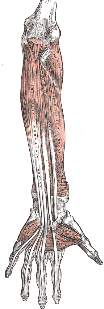
Match each tendon to its insertion and joint.
Match each tendon to its insertion.
Four cards to lock the anatomy.
The two tendons, side by side.
THE TEST
Isolating FDS: The One Maneuver
Block FDP's mass action, then ask the middle joint to move.
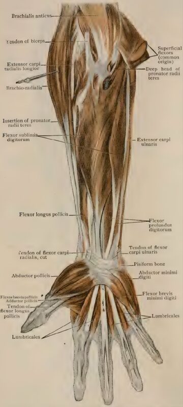
The patient rests the hand flat on the table, palm up. Identify the target finger, for example the ring finger.
Hold the other three fingers in full extension. FDP acts as a mass-action unit, so with the neighbors extended it cannot contribute to the target finger's PIP flexion.
Ask the patient to flex only the target finger at the middle joint. FDS has more independent digital control and can still attempt the move.
No PIP flexion with the neighbors held out = FDS rupture. DIP flexion should still work, because FDP is a separate tendon for a separate joint. Hold the others out, ask for PIP: that is the FDS test.
To test FDP, stabilize the PIP in extension and ask for DIP flexion: the deep tendon is the only one that can bend the tip. One more trap: the tenodesis effect. Wrist extension tenses the long flexors and passively flexes the fingers, and wrist flexion extends them. A patient can fool both tests by moving the wrist, so always stabilize the wrist before reading the result.
Run the exam in the order that prevents a missed tendon.
Tap the exam steps in order.
Which tendon is tested by holding the other fingers extended?
The examiner holds the adjacent fingers in full extension, then asks the patient to flex the injured finger at the PIP. Which tendon is being isolated?
FDS. Holding the neighbors extended blocks FDP's shared-belly mass action, so any PIP flexion that remains belongs to FDS alone. FDP. FDP is the tendon the maneuver is designed to exclude. FPL. FPL flexes the thumb, not the finger PIP. Lumbrical. The lumbrical extends the PIP through the extensor apparatus; it is not a finger flexor.
THE FLEXOR ZONES
Verdan's Map: Where the Cut Lives
The zone of a laceration predicts the repair and the prognosis.
Verdan divided the flexor system into five zones, and two of them dominate board questions. Zone I lies distal to the FDS insertion: only FDP runs there, so a Zone I injury is a pure FDP problem: the jersey finger territory. Zone II, between the distal palmar crease and the FDS insertion, is the old "no man's land": both tendons share one tight sheath, and repairs here were historically disastrous. Modern practice is primary repair with early controlled motion, and the zone is no longer a no-repair zone. Zone III is the palm, Zone IV the carpal tunnel (nine tendons plus the median nerve), and Zone V the forearm, where tendons and nerves can be cut together.
Tap each zone to see what lives there.
Tap a zone.
THE NERVE SPLIT
FDP's Split Supply
The most-tested trap on the page: FDS is all median, FDP is divided.
FDS is 100% median nerve. FDP is the trap: a split supply. The anterior interosseous nerve (AIN), a pure motor branch of the median nerve that leaves it below the elbow, drives FDP to the index and middle fingers plus flexor pollicis longus and pronator quadratus. The ulnar nerve drives FDP to the ring and little fingers. So a failed pinch (the "OK sign", thumb IP plus index DIP) is an AIN story with sensation intact, while ring and little DIP loss is an ulnar story. FDP is the litmus test that tells you which nerve is down.
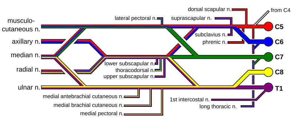
Route the finger deficit to the nerve.
Choose a deficit pattern to route it.
Lock the pure-motor branch.
A patient cannot make the pinch (OK sign): the index DIP and thumb IP do not flex, and pronation is weak, but sensation is intact. Which nerve is injured?
Anterior interosseous nerve (AIN). AIN supplies index and middle FDP, FPL, and pronator quadratus, and has no cutaneous fibers, so the failed OK sign comes with intact sensation. Ulnar nerve. Ulnar drives ring and little FDP, not the index and thumb. Radial nerve. Radial never crosses to the flexor side. Recurrent thenar branch. That branch moves the thumb muscles, not FPL or index FDP.
THE RECAP
Cloze the Loop
Fill the blanks and the whole page locks in.
FDS controls the PIP (middle) joint and is innervated 100% by the median nerve. FDP controls the DIP (tip) joint; its radial half (index + middle) is supplied by the anterior interosseous (AIN) branch of the median nerve, and its ulnar half (ring + little) by the ulnar nerve. At the PIP level, FDS splits at Camper's chiasma to let FDP pass through. Test FDS by holding the other fingers in extension and asking for PIP flexion; test FDP by stabilizing the PIP and asking for DIP flexion. Two joints, two tendons, one split nerve: the rest is detail.
Tap the letters to expand the hook.
SSMDDD
Superficialis: the shallow tendon in each finger's sheath
Stops at the middle phalanx
Middle joint (PIP) is its job
Deep tendon: the profundus
Delivers all the way to the fingertip
Distal phalanx (DIP) is its job
One question to close the loop.
A 24-year-old has a volar wrist laceration. The ring finger DIP cannot flex, but its PIP flexes fully and there is no bony tenderness. Which structure is injured, and in which zone?
FDP, Zone I. Only FDP runs distal to the FDS insertion, so isolated DIP flexion loss is an FDP injury in Zone I, the jersey finger territory. FDS, Zone II. FDS loss takes the PIP, which works here. FDP, Zone II. Zone II holds both tendons, but the isolated DIP loss localizes to Zone I. Lumbrical, Zone III. Lumbricals extend the PIP; they never flex the DIP.
JERSEY FINGER
The FDP Avulsion
Grab the jersey, hear the pop, lose the tip.
Jersey finger is an avulsion of FDP from the distal phalanx, and the ring finger is the classic victim. The mechanism is a forced DIP extension against a contracting FDP: grabbing an opponent's jersey while they pull away. The patient cannot flex the DIP, and a tender lump in the palm may mark the retracted tendon end. Radiograph the finger, because the avulsion often carries a small bony fragment. Treatment is surgical reattachment with a suture anchor plus protected motion, and it is urgent: every day of delay lets the muscle shorten and the sheath collapse, and a retracted tendon is much harder to bring back.
Two tip injuries, opposite directions.
Lock the classic presentation.
Which digit is most commonly injured in jersey finger, and what is the classic mechanism?
Ring finger; forced DIP extension against a contracting FDP. Grabbing a jersey while the opponent pulls away tears FDP off the distal phalanx, most often in the ring finger. Index finger. Index FDP avulsions happen but are not the classic story. Thumb. Thumb IP hyperextension injures the volar plate or FPL, not the classic jersey finger. Little finger. A sheath laceration is a Zone II injury, not an avulsion.
ACUTE LACERATION
Repair Timing and Rehab
Primary repair early, protected motion always, and a plan for the delayed patient.
A clean flexor laceration is repaired primarily within days. Zone II is no longer a no-repair zone: the standard is primary repair with early controlled motion (Kleinert or Duran protocols) under a dorsal blocking splint, because motion keeps the repair from sticking. The most common complication is adhesions, followed by rerupture and bowstringing. The patient who presents weeks late is a different problem: the muscle has shortened and the sheath has collapsed, so staged reconstruction with a silicone (Hunter) rod, then a tendon graft, replaces one-stage repair.
Route the presentation to the repair plan.
Choose a presentation to route it.
Now the delayed patient, step by step.
Prove it
Walkthrough: name the mechanism
Original practice scenarios, one at a time. Work each option to see why it wins or fails.
Clinical walkthrough

