
Choose an answer, then open any option to work its reasoning.
MUSCULOSKELETAL / UPPER EXTREMITY
The Shoulder Beyond the Cuff: Frozen, Separated, and the Axillary Trap
Not every painful shoulder is the rotator cuff. The frozen shoulder that locks external rotation, the AC joint that steps off, and the proximal humerus fracture that can take the axillary nerve with it.
What this page makes you able to do
- Separate frozen shoulder from rotator cuff disease by the pattern of passive motion loss
- Grade AC separation with the Rockwood ladder and pick conservative versus surgical care
- Catch the axillary nerve injury hiding behind a proximal humerus fracture
- Read the shoulder exam for the red flags that cannot wait
- Dr. Fatima Ali, DOPsychiatry residentPrimary reviewer
Last reviewed
From the Attending:
The shoulder exam is a motion story. Passive external rotation loss is the frozen-shoulder signature, a painful arc says pinch rather than tear, and a step-off says the AC joint, not the cuff.
Prove it
Opening question
A 54-year-old woman with type 2 diabetes mellitus is brought to the clinic with four months of progressive right shoulder pain and stiffness. She reports the pain was worst at night during the first six weeks, and now she cannot reach behind her back to tuck in her shirt. Examination reveals marked restriction of passive external rotation to 15 degrees compared with 70 degrees on the left, with flexion and abduction restricted to a lesser degree. Radiographs show no fracture, no joint-space narrowing, and no calcific deposit.Which of the following is the most likely diagnosis?
- Why this is rightGlobal passive restriction with external rotation lost first and worst is the capsular signature, and diabetes raises the risk of adhesive capsulitis about fivefold. The clean radiograph rules out arthritis and fracture.
- Why this failsFull-thickness tears hurt at night and weaken active elevation, but the examiner can still move the arm passively. A tear does not lock passive external rotation.
- Why this failsBursitis reproduces pain on the 60 to 120 degree arc with positive Neer and Hawkins tests, but passive range stays near full. It does not produce a global capsular restriction.
- Why this failsOsteoarthritis shows joint-space narrowing and osteophytes on radiographs and grinds with crepitus. Here the radiograph is clean and the history is a capsular timeline, not wear.
Work the reasoning
Passive external rotation. The anteroinferior capsule contracts first, so the arm will not rotate outward before any other direction is limited.
Diabetes raises the risk of frozen shoulder roughly fivefold, and the stiffness tends to be more severe and more often bilateral.
DIABETES LOCKS THE SHOULDER
Adhesive capsulitis: the capsule that will not give
A global motion loss with external rotation first, a three-phase timeline, and a treatment plan that starts with therapy, not surgery.
The frozen shoulder
The capsule scars down, and the arm stops moving in every direction.
Adhesive capsulitis is a contracture of the glenohumeral joint capsule, not a tendon problem. It strikes between ages 40 and 60, and diabetes is the strongest risk factor, raising the odds about fivefold.
Thyroid disease and prior shoulder immobilization also load the dice. The signature is a global restriction of both active and passive motion, and external rotation goes first and stays worst, because the anteroinferior capsule is the part that scars down earliest.
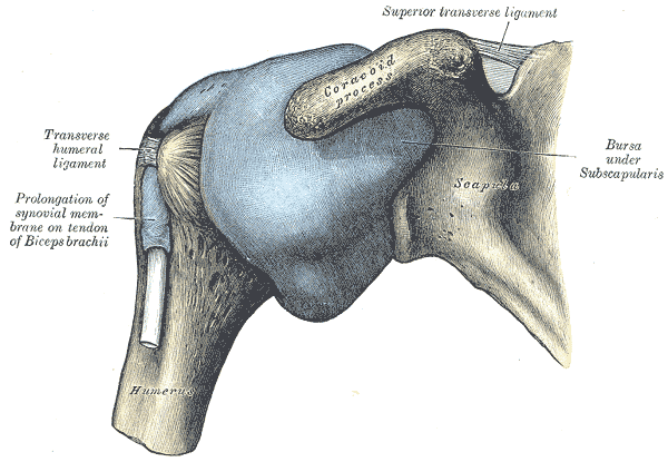
The disease runs a three-phase course: freezing hurts, frozen locks, and thawing slowly gives range back. Most patients recover with time, and the plan starts with physical therapy and a corticosteroid injection for pain.
Hydrodilatation or manipulation under anesthesia is reserved for refractory cases. Warn every patient that the other shoulder becomes involved in roughly 20 to 30 percent of cases, usually within five years.
Put the three phases of adhesive capsulitis in order.
Tap the phases in the order they happen.
From the Attending
Frozen shoulder is a diagnosis of passive motion. If the examiner can rotate the arm outward to full range, it is not adhesive capsulitis. Start hunting the cuff, the bursa, or the joint instead.
The frozen timeline: Diane, 52
Three stages, one arc: watch external rotation disappear, then crawl back.
The frozen shoulder timeline
Diane, 52
Type 2 diabetes, right shoulder, four months in. The external rotation arc is the clock.
Stage 1
Freezing
Pain dominates, night waking is the rule, and external rotation is already slipping. The capsule is inflamed, not yet rigid.
Stage 2
Frozen
The pain quiets, but the capsule is a cast. External rotation is nearly gone and the arm will not move passively in any plane.
Stage 3
Thawing
Range creeps back over months. The arc returns last to external rotation, the direction that left first.
RoutePainful phase, stiff phase, recovery phase; therapy plus injection first.
PatternGlobal passive restriction with external rotation first and worst.
PearlDiabetes multiplies the risk fivefold, and the other shoulder follows in 20 to 30 percent.
The AC joint: the step-off speaks
Fall on the point of the shoulder, a piano-key clavicle, and a ligament ladder that decides who operates.
The acromioclavicular joint is the most common shoulder injury in contact and collision athletes. The mechanism is a direct blow to the point of the shoulder, and the pain is worst with cross-body adduction, which loads the joint.
A visible step-off means the clavicle has moved relative to the acromion. Press the clavicle and it springs back like a piano key, and the Rockwood grade follows the ligament ladder: the AC ligaments go first, then the coracoclavicular ligaments, then the deltoid and trapezius fascia.
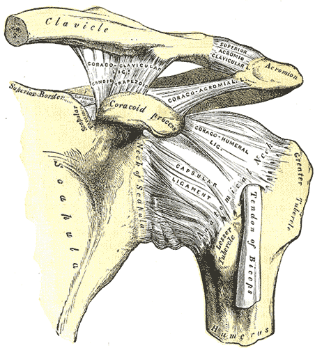
Types I and II are sprains with intact CC ligaments and are treated conservatively with a sling and early motion. Type III, a complete AC plus CC tear with less than 100 percent superior displacement, is the controversy, and most are still managed nonoperatively.
Types IV through VI carry the clavicle posteriorly, more than 100 percent superiorly, or inferiorly, and they usually go to surgery. Clavicle shaft fractures live next door; the middle-third break belongs to fractures-trauma.
Which injury fits which picture?
The proximal humerus: the axillary nerve rides along
Surgical neck, valgus impaction, Neer parts, and the deltoid patch that tells you the nerve survived.
The proximal humerus is one of the most common fracture sites in older adults, and the surgical neck is the usual break: a low-energy fall in an osteoporotic bone does it. The axillary nerve wraps that neck, and it is the nerve at risk.
Deltoid weakness plus a numb patch over the outer shoulder, the regimental badge area, means axillary nerve injury until proven otherwise. The Neer classification then counts displaced parts: the articular segment, the greater tuberosity, the lesser tuberosity, and the shaft, and a part counts only if it is displaced more than 1 cm or angulated more than 45 degrees.
From the Attending
After any proximal humerus fracture, document deltoid sensation and contraction before and after every reduction. A numb deltoid patch is the axillary nerve calling out, and it is the one finding the surgeon needs most.
Most proximal humerus fractures are minimally displaced and heal in a sling with early pendulum exercises. Valgus-impacted two-part fractures are the favorable pattern, because the head stays seated and its blood supply usually survives.
Displaced three- and four-part fractures in older adults often go to arthroplasty, because the blood supply to the humeral head is gone. For a young, active patient with a displaced two-part fracture, open reduction and internal fixation is the usual answer.
Follow the displacement to the treatment.
Tap a fracture pattern.
The biceps and the Popeye
Groove tenderness, Speed's and Yergason, and a rupture that most people can live with.
The long head of the biceps runs through the bicipital groove and is a frequent co-conspirator in anterior shoulder pain, so groove tenderness is the bedside clue. Speed's test loads the tendon with resisted forward flexion of the straight arm, and Yergason's test loads it with resisted supination of the flexed elbow.
The proximal tendon can rupture as it wears against the acromion, and the muscle belly slides down the arm and bunches into the Popeye bulge. A proximal rupture is usually managed conservatively, because the short head and the brachialis keep elbow flexion and supination strong; the main complaint is the cosmetic deformity.
Distal biceps ruptures are the surgical ones, tearing off the radial tuberosity with a loss of supination power. SLAP tears live in the labrum, belong to young overhead athletes, and come with a painful click; both belong to the soft-tissue-trauma page.
A 40-year-old laborer felt a pop while lifting and now has a bulge in his upper arm. Work the case.
The subacromial space: bursitis under the arch
The painful arc from 60 to 120 degrees, the Neer and Hawkins provocation, and the injection that both treats and tells.
The subacromial bursa sits between the humeral head and the coracoacromial arch. When the cuff or bursa swells, abduction squeezes the bursa between 60 and 120 degrees: that is the painful arc, pain that appears mid-range and clears as the arm reaches overhead.
Neer's test forces the arm forward under the arch, and Hawkins' test internally rotates the flexed arm. Both reproduce the pinch, and a subacromial injection that relieves the pain confirms the space as the source.
Calcific tendinitis is the same space with hydroxyapatite deposits, visible on plain radiographs, and the Milwaukee shoulder is its destructive, elderly, cuff-tear-associated cousin. The impingement continuum runs from bursitis to partial tears to full-thickness tears, so the cuff half of the story lives on the rotator-cuff-impingement page.
On clinic afternoon, your attending points at the last patient of the day.
Attending
Raising her arm hurts between 60 and 120 degrees, then it smooths out overhead. No night pain. What does that arc tell you?
You
The bursa and the cuff are being pinched under the coracoacromial arch. Classic subacromial impingement, not a full-thickness tear.
Attending
And if the same patient woke at 2 AM with the arm dead?
You
Then I stop trusting the arc and image the cuff.
Attending
Exactly. The arc localizes the pinch. The night pain indicts the tendon.
Painful arc picks the space. Night pain picks the tendon.
Where is the pinch?
A 48-year-old swimmer has pain only when raising her arm between 60 and 120 degrees of abduction. Which structure is being pinched?
The subacromial bursa beneath the coracoacromial arch. The swollen bursa is squeezed between the humeral head and the arch only in mid-abduction, which is exactly the 60 to 120 degree window. The glenohumeral joint capsule. Capsular disease restricts passive motion globally, especially external rotation, and does not produce a clean arc. The acromioclavicular joint. AC pain sits at the top of the shoulder and is worst with cross-body adduction, not mid-range abduction. The long head of the biceps in the groove. Biceps pain is anterior and reproduces with resisted supination or forward flexion, not with a mid-arc.
The winging scapula: which nerve, which border
Wall push separates the serratus story from the trapezius story, and the border that lifts names the nerve.
Scapular winging is the scapula losing its anchor to the chest wall, so have the patient push against a wall and watch which border lifts. Medial border winging with the arm pushing forward means serratus anterior weakness, which means the long thoracic nerve, and it classically follows axillary surgery, thoracotomy, or heavy backpack use.
Lateral border winging with a weak shrug means trapezius weakness, which means the spinal accessory nerve, and it follows neck surgery or lymph node biopsy. Rhomboid weakness from the dorsal scapular nerve is a rarer medial pattern that shows up with arm elevation more than wall push.
Treatment is usually observation and physical therapy. Long thoracic palsy recovers over months to a couple of years in most patients, and nerve transfer is the late resort for the ones who do not.
Match each wing pattern to its nerve.
Tap a pattern, then its nerve.
The red-flag split: hot, traumatic, or weak
Three presentations that change the plan the moment you see them.
A hot, swollen shoulder with fever and pain on any motion is septic arthritis until it is tapped. It can be the glenohumeral joint or, in the older or immunosuppressed patient, the AC joint, and the tap and cultures live on the msk-infections page.
An acutely dislocated shoulder is anterior in about 95 percent of cases. Confirm no fracture, reduce with traction and countertraction, and make the axillary nerve check part of the aftercare; the full dislocation story is on the soft-tissue-trauma page.
Night pain that wakes the patient plus weakness of elevation means a full-thickness cuff tear until the MRI says otherwise, because full-thickness tears do not heal on their own timeline.
Rest pain in an older adult with a mass and a lytic lesion is a tumor until proven otherwise, especially with a history of malignancy; that workup lives on the bone-tumors page.
Run the can't-miss shoulder checklist.
The one-screen discriminator
Four exam findings, four diagnoses: external rotation loss, step-off, deltoid patch, Popeye bulge.
External rotation loss with globally restricted passive motion is frozen shoulder until proven otherwise. A step-off with cross-body pain is the AC joint: grade it with Rockwood and act on the ligament ladder.
A deltoid patch of numbness with a proximal humerus fracture is the axillary nerve: document it, protect it, and follow it. A Popeye bulge is the long head of the biceps: reassure, because flexion strength survives.
Score the AC injury: check every finding that applies.
Rockwood score 0
Decode the shoulder in four letters.
STEP: STEP
SStep-off at the AC joint with cross-body pain means acromioclavicular separation.
TTight external rotation, first and worst, means adhesive capsulitis.
EEmpty deltoid patch of numbness after a proximal humerus fracture means axillary nerve injury.
PPopeye bulge in the upper arm means long head biceps rupture.
Clinical walkthrough

