⌘ KStart free
0%
Skip to lesson

Neurology

Basal ganglia anatomy, circuits, and clinical localization

A useful first distinction: is the problem loss of strength, or difficulty regulating an intended action? Build the nuclear map or try the signed circuit.

Locate deep nuclei and capsular neighbors, trace basal ganglia circuits, and distinguish vascular, degenerative, toxic, genetic, and treatable motor syndromes.

A putaminal hemorrhage can cause dense weakness because of the internal capsule beside it. A basal ganglia circuit disorder can instead produce abnormal initiation, rigidity, chorea, or dystonia with relatively preserved strength. First locate the gray nucleus and its white-matter neighbors. Then distinguish destruction of a descending motor tract from dysfunction of the loops that regulate cortical motor activity.

Build the map from lateral surface to ventricular neighbors

The putamen and globus pallidus form the lentiform nucleus. The putamen lies lateral to the pallidum. The dorsal striatum is a different grouping, comprising caudate and putamen, which share major cortical input and related cellular organization. Ventral striatum includes nucleus accumbens. The historical term corpus striatum has been used for caudate, putamen, and pallidum; it should not be substituted indiscriminately for every modern basal ganglia structure. [1]

Published schematic lateral projection of caudate, putamen, globus pallidus, nucleus accumbens and thalamus; the caudate curves around the deeper nuclei.Enlarge the whole image
Use this schematic lateral view to orient the curved caudate. It is not an axial capsule map. Putamen and pallidum form lentiform nucleus; caudate and putamen form dorsal striatum. Lim S-J, Fiez JA and Holt LL (2014). CC BY 3.0.
Image: Source and provenance. None; original image bytes.

Two groupings share one nucleus

  1. Caudate and putamen form which grouping?

    Dorsal striatum combines caudate with putamen. Pallidum is not the other half of that pair.

  2. Replace caudate with globus pallidus. What is the pair now?

    Putamen plus globus pallidus forms lentiform nucleus. Thalamus is a separate relay complex.

  3. At the posterior capsule, which structure is medial?

    The posterior limb separates medial thalamus from lateral lentiform nucleus.

Name the pair, then check its white-matter neighbors. [1] [21]

Read the complete worked explanation
  1. Caudate and putamen form which grouping?

    Dorsal striatum combines caudate with putamen. Pallidum is not the other half of that pair.

  2. Replace caudate with globus pallidus. What is the pair now?

    Putamen plus globus pallidus forms lentiform nucleus. Thalamus is a separate relay complex.

  3. At the posterior capsule, which structure is medial?

    The posterior limb separates medial thalamus from lateral lentiform nucleus.

Name the pair, then check its white-matter neighbors.

A lateral-to-medial sequence near the lentiform nucleus
  1. Insular cortex
  2. Extreme capsule, a white-matter layer
  3. Claustrum, a thin gray sheet
  4. External capsule, another white-matter layer
  5. Putamen
  6. Globus pallidus
  7. Internal capsule
  8. Caudate at the anterior-limb level, or thalamus at the posterior-limb level

This sequence is an orientation aid, not one straight line valid in every slice. The internal capsule bends, and its medial neighbor changes with the level. [21]

The putamen is lateral within the principal motor nuclear grouping, but the claustrum is gray matter farther lateral. The external capsule does not separate putamen from pallidum; a thin medullary lamina separates those components. Within the pallidum, GPe names the external segment and GPi the internal segment. GPi is a major inhibitory output nucleus. Recognizing these boundaries prevents confusing a nuclear lesion with damage to an adjacent fiber tract.

The caudate head borders the frontal horn of the lateral ventricle, while its body and tail follow a curved ventricular relationship. The anterior limb of the internal capsule separates caudate from lentiform nucleus. The posterior limb separates thalamus from lentiform nucleus and contains closely packed motor and sensory pathways. Corticobulbar fibers are associated with the genu, while corticospinal fibers course through the posterior limb. A small lesion can therefore cause broad contralateral deficits, but its exact extent determines whether the syndrome is purely motor.

The thalamus is a diencephalic relay complex beside the third ventricle, not a basal ganglia nucleus simply because it is deep gray matter. The subthalamic nucleus lies below it. The substantia nigra lies in the midbrain; its pars compacta supplies dopamine to striatum, while pars reticulata provides inhibitory output. Functionally related nuclei can be far apart anatomically. [1]

Trace the sign of each synapse

GPi and SNr provide tonic GABAergic inhibition to downstream targets, including motor thalamus. Motor thalamus excites cortex through glutamatergic projections. A pathway that inhibits an already inhibitory output can disinhibit thalamus. This double-negative logic is more reliable than assuming that every basal ganglia structure simply activates or suppresses all motor activity.

Connection schematic: active GPi inhibits thalamus; direct-pathway inhibition of GPi weakens that inhibition and permits thalamic activity.Enlarge the whole image

The bar ends mean inhibition. Weakening an inhibitory output releases its target from some restraint. Line widths show qualitative states only.

Bone Wizardry, original construction (2026). Source and provenance. Follow the labeled connections to localize the pathway.

An inhibition can release activity

  1. GPi normally inhibits thalamus. If GPi output falls, what happens to thalamic inhibition?

    Less GPi output means less inhibition reaching the thalamus.

  2. Less inhibition permits what simplified thalamic response?

    Removing some tonic inhibition permits greater thalamic activity. This is disinhibition.

  3. Do the direct and indirect routes work as mutually exclusive switches?

    Both populations can participate in real actions. The signed model isolates a connection, not a complete behavior.

Track the sign of each connection before judging the net effect. [1] [2] [18]

Read the complete worked explanation
  1. GPi normally inhibits thalamus. If GPi output falls, what happens to thalamic inhibition?

    Less GPi output means less inhibition reaching the thalamus.

  2. Less inhibition permits what simplified thalamic response?

    Removing some tonic inhibition permits greater thalamic activity. This is disinhibition.

  3. Do the direct and indirect routes work as mutually exclusive switches?

    Both populations can participate in real actions. The signed model isolates a connection, not a complete behavior.

Track the sign of each connection before judging the net effect.

Direct pathway

  1. Cortex excites striatal neurons with glutamate.
  2. Direct-pathway striatal neurons inhibit GPi/SNr with GABA.
  3. Reduced GPi/SNr output releases thalamus from some tonic inhibition.
  4. Thalamocortical facilitation increases in the simplified model.

Indirect pathway

  1. Cortex excites indirect-pathway striatal neurons.
  2. Striatum inhibits GPe with GABA.
  3. Reduced GPe inhibition permits greater STN activity.
  4. STN excites GPi/SNr with glutamate.
  5. Greater inhibitory output restrains thalamocortical facilitation.

SNc dopamine favors direct-pathway activity through D1 signaling and suppresses indirect-pathway striatal activity through D2 signaling. D1 receptors principally engage Gs/olf-related signaling; D2 receptors engage Gi/o-related signaling. Although their local cellular effects differ, both can facilitate selected motor activity in the simplified net model. Loss of dopamine reduces direct-pathway facilitation and increases indirect-pathway influence, helping explain hypokinetic features of Parkinson disease. Striatal acetylcholine arises importantly from local interneurons, not from SNc dopamine neurons. [1] [2]

The hyperdirect route connects cortex to STN without the first striatal relay. Real behavior also depends on timing, oscillation, action-specific populations, and interconnected motor, associative, and limbic loops. Direct and indirect pathways are not mutually exclusive switches, and every hyperkinetic disorder cannot be reduced to “too much dopamine.” DBS acts on this network; treating stimulation as identical to destroying one nucleus is an oversimplification. [18]

Try a transfer question

Case 6

A patient suddenly develops large-amplitude flinging of the left arm and leg. Imaging shows a small right subthalamic lesion. Which circuit consequence best explains the classic association?

Explore every option and its reasoning
  1. A. Increased inhibitory GPi output that suppresses thalamocortical facilitation (Why this does not fit)

    Reason through this option

    Make one small inference

    1. What is the normal sign of the connection being invoked?

      GPi inhibits its thalamic targets.

    2. What happens to that drive when the right STN is injured?

      That net change would favor reduced motor facilitation in this simplified model, not the hyperkinetic consequence associated with loss of STN excitation.

    Contralateral STN injury is a classic cause of hemiballism, but not its only cause. [2] [14]

    Read the complete worked explanation
    1. What is the normal sign of the connection being invoked?

      GPi inhibits its thalamic targets.

    2. What happens to that drive when the right STN is injured?

      That net change would favor reduced motor facilitation in this simplified model, not the hyperkinetic consequence associated with loss of STN excitation.

    Contralateral STN injury is a classic cause of hemiballism, but not its only cause.

    Original source explanation

    That net change would favor reduced motor facilitation in this simplified model, not the hyperkinetic consequence associated with loss of STN excitation.

  2. B. Increased GPe inhibition of STN as the direct output of the damaged STN neurons (Why this does not fit)

    Reason through this option

    Make one small inference

    1. What is the normal sign of the connection being invoked?

      GPe inhibits STN; STN has excitatory projections to pallidal output nuclei.

    2. What happens to that drive when the right STN is injured?

      The STN does not provide GPe’s inhibitory output; its projection to pallidal output nuclei is excitatory, and its loss reduces that drive.

    Contralateral STN injury is a classic cause of hemiballism, but not its only cause. [2] [14]

    Read the complete worked explanation
    1. What is the normal sign of the connection being invoked?

      GPe inhibits STN; STN has excitatory projections to pallidal output nuclei.

    2. What happens to that drive when the right STN is injured?

      The STN does not provide GPe’s inhibitory output; its projection to pallidal output nuclei is excitatory, and its loss reduces that drive.

    Contralateral STN injury is a classic cause of hemiballism, but not its only cause.

    Original source explanation

    The STN does not provide GPe’s inhibitory output; its projection to pallidal output nuclei is excitatory, and its loss reduces that drive.

  3. C. Reduced excitatory input from STN to inhibitory basal ganglia output nuclei (Best answer)

    Reason through this option

    Make one small inference

    1. What is the normal sign of the connection being invoked?

      STN excites GPi/SNr output nuclei.

    2. What happens to that drive when the right STN is injured?

      Less STN drive can reduce GPi/SNr inhibition of the thalamus and favor excessive contralateral motor activity in the simplified model.

    3. What useful rule carries to the next patient?

      Contralateral STN injury is a classic cause of hemiballism, but not its only cause.

    Contralateral STN injury is a classic cause of hemiballism, but not its only cause. [2] [14]

    Read the complete worked explanation
    1. What is the normal sign of the connection being invoked?

      STN excites GPi/SNr output nuclei.

    2. What happens to that drive when the right STN is injured?

      Less STN drive can reduce GPi/SNr inhibition of the thalamus and favor excessive contralateral motor activity in the simplified model.

    3. What useful rule carries to the next patient?

      Contralateral STN injury is a classic cause of hemiballism, but not its only cause.

    Contralateral STN injury is a classic cause of hemiballism, but not its only cause.

    Original source explanation

    Less STN drive can reduce GPi/SNr inhibition of the thalamus and favor excessive contralateral motor activity in the simplified model.

  4. D. Increased STN glutamatergic excitation as the direct result of destroying STN neurons (Why this does not fit)

    Reason through this option

    Make one small inference

    1. What is the normal sign of the connection being invoked?

      STN output normally supplies excitatory drive.

    2. What happens to that drive when the right STN is injured?

      Loss of the nucleus does not directly increase its output.

    Contralateral STN injury is a classic cause of hemiballism, but not its only cause. [2] [14]

    Read the complete worked explanation
    1. What is the normal sign of the connection being invoked?

      STN output normally supplies excitatory drive.

    2. What happens to that drive when the right STN is injured?

      Loss of the nucleus does not directly increase its output.

    Contralateral STN injury is a classic cause of hemiballism, but not its only cause.

    Original source explanation

    Loss of the nucleus does not directly increase its output.

Takeaway: Contralateral STN injury is a classic cause of hemiballism, but not its only cause.

Case sources: [2] [14]

Try a transfer question

Case 25

In Parkinson disease, loss of SNc dopamine reduces D2-mediated suppression of indirect-pathway striatal neurons. In the simplified rate model, what follows?

Explore every option and its reasoning
  1. A. Complete loss of every inhibitory synapse in the basal ganglia (Why this does not fit)

    Reason through this option

    Make one small inference

    1. Which signal is normally present in this part of the circuit?

      GABAergic connections remain even when dopamine falls.

    2. What follows when D2-mediated suppression is reduced?

      Dopamine loss changes modulation; it does not remove all GABAergic signaling.

    Trace each inhibitory and excitatory step before predicting the net effect. [1] [2]

    Read the complete worked explanation
    1. Which signal is normally present in this part of the circuit?

      GABAergic connections remain even when dopamine falls.

    2. What follows when D2-mediated suppression is reduced?

      Dopamine loss changes modulation; it does not remove all GABAergic signaling.

    Trace each inhibitory and excitatory step before predicting the net effect.

    Original source explanation

    Dopamine loss changes modulation; it does not remove all GABAergic signaling.

  2. B. Direct excitation of the thalamus by GPe as the main consequence (Why this does not fit)

    Reason through this option

    Make one small inference

    1. Which signal is normally present in this part of the circuit?

      GPe supplies inhibitory output.

    2. What follows when D2-mediated suppression is reduced?

      GPe is inhibitory and is not the excitatory thalamic relay proposed here.

    Trace each inhibitory and excitatory step before predicting the net effect. [1] [2]

    Read the complete worked explanation
    1. Which signal is normally present in this part of the circuit?

      GPe supplies inhibitory output.

    2. What follows when D2-mediated suppression is reduced?

      GPe is inhibitory and is not the excitatory thalamic relay proposed here.

    Trace each inhibitory and excitatory step before predicting the net effect.

    Original source explanation

    GPe is inhibitory and is not the excitatory thalamic relay proposed here.

  3. C. The STN begins releasing acetylcholine instead of glutamate (Why this does not fit)

    Reason through this option

    Make one small inference

    1. Which signal is normally present in this part of the circuit?

      STN supplies excitatory glutamatergic drive.

    2. What follows when D2-mediated suppression is reduced?

      A disease-related change in modulation does not imply that transmitter switch.

    Trace each inhibitory and excitatory step before predicting the net effect. [1] [2]

    Read the complete worked explanation
    1. Which signal is normally present in this part of the circuit?

      STN supplies excitatory glutamatergic drive.

    2. What follows when D2-mediated suppression is reduced?

      A disease-related change in modulation does not imply that transmitter switch.

    Trace each inhibitory and excitatory step before predicting the net effect.

    Original source explanation

    A disease-related change in modulation does not imply that transmitter switch.

  4. D. Greater indirect-pathway influence and increased inhibitory output toward the thalamus (Best answer)

    Reason through this option

    Make one small inference

    1. Which signal is normally present in this part of the circuit?

      D2 normally suppresses indirect-pathway striatal activity.

    2. What follows when D2-mediated suppression is reduced?

      More striatal inhibition of GPe can release STN activity, increasing GPi/SNr output and reducing thalamocortical facilitation.

    3. What useful rule carries to the next patient?

      Trace each inhibitory and excitatory step before predicting the net effect.

    Trace each inhibitory and excitatory step before predicting the net effect. [1] [2]

    Read the complete worked explanation
    1. Which signal is normally present in this part of the circuit?

      D2 normally suppresses indirect-pathway striatal activity.

    2. What follows when D2-mediated suppression is reduced?

      More striatal inhibition of GPe can release STN activity, increasing GPi/SNr output and reducing thalamocortical facilitation.

    3. What useful rule carries to the next patient?

      Trace each inhibitory and excitatory step before predicting the net effect.

    Trace each inhibitory and excitatory step before predicting the net effect.

    Original source explanation

    More striatal inhibition of GPe can release STN activity, increasing GPi/SNr output and reducing thalamocortical facilitation.

Takeaway: Trace each inhibitory and excitatory step before predicting the net effect.

Case sources: [1] [2]

Distinguish deep hemorrhage, capsule injury, and thalamic symptoms

Chronic hypertension can damage deep perforating arterioles through arteriolosclerotic changes. Lenticulostriate branches from the MCA supply much of the putaminal region and portions of nearby nuclei and capsule. Contributions from other vessels, including the recurrent artery of Heubner and anterior choroidal artery in relevant territories, mean that no single artery supplies every deep structure. Avoid explanations claiming that the putamen bleeds because it is necessarily the first tissue struck by blood.

Noncontrast head CT with a large bright deep hemorrhage on the right of the displayed image, in the patient’s left basal ganglia region.Enlarge the whole image
The axial CT shows a left basal ganglia hemorrhage with surrounding edema. In standard radiologic display, the patient’s left is on your right. The large deep lesion can affect neighboring motor pathways; the image alone does not prove its cause.
Image: Dahlqvist MB, Andres RH, Raabe A, Jakob SM, Takala J and Dünser MW; original source; CC BY 2.0.

A nucleus is beside a motor highway

  1. The CT shows deep blood beside the capsule. Does weakness prove that putamen directly supplies muscle?

    Dense weakness can reflect involvement or compression of adjacent capsular motor fibers.

  2. Does this CT location prove one unique hemorrhage cause?

    Deep location suggests a mechanism; clinical context and etiologic evaluation are still required.

Localize both the damaged nucleus and its affected neighbors. [3] [17]

Read the complete worked explanation
  1. The CT shows deep blood beside the capsule. Does weakness prove that putamen directly supplies muscle?

    Dense weakness can reflect involvement or compression of adjacent capsular motor fibers.

  2. Does this CT location prove one unique hemorrhage cause?

    Deep location suggests a mechanism; clinical context and etiologic evaluation are still required.

Localize both the damaged nucleus and its affected neighbors.

A putaminal hemorrhage is a classic deep hypertensive pattern. Historic Charcot-Bouchard microaneurysms help explain one mechanism, but they should not be asserted as a demonstrated requirement in every patient. Older adults with lobar hemorrhage may instead have cerebral amyloid angiopathy, although location and age do not settle the diagnosis alone. A ruptured saccular aneurysm classically causes subarachnoid hemorrhage, a different bleeding compartment. Acute deficits require urgent imaging and hemorrhage-specific care. [3]

Dense contralateral weakness from a putaminal hemorrhage often reflects capsular involvement or compression rather than proof that putamen directly supplies muscle. Small capsular infarcts can cause pure motor syndromes, while larger or differently situated lesions can also involve sensation. A thalamic lesion may cause contralateral sensory loss and later central poststroke pain with burning dysesthesia or allodynia. Weakness can occur with nearby involvement; a rigid “thalamus means sensory only” rule is unsafe. Intraventricular hemorrhage extension can obstruct CSF circulation and cause hydrocephalus. [17] [3]

Try a transfer question

Case 1

A patient with longstanding hypertension develops sudden right-sided weakness. CT shows an acute hemorrhage centered in the left putamen. Which vascular process is a classic explanation?

Explore every option and its reasoning
  1. A. Rupture of a saccular aneurysm necessarily confined to the subarachnoid space (Why this does not fit)

    Reason through this option

    Make one small inference

    1. Which compartment or vessel does this option point toward?

      A ruptured saccular aneurysm classically produces blood in the subarachnoid space.

    2. Does the deep putaminal location support this explanation?

      The described primary deep intraparenchymal location fits perforator disease better than the classic aneurysmal subarachnoid pattern.

    Location suggests a small-vessel mechanism, while the clinical setting determines further etiologic evaluation. [3]

    Read the complete worked explanation
    1. Which compartment or vessel does this option point toward?

      A ruptured saccular aneurysm classically produces blood in the subarachnoid space.

    2. Does the deep putaminal location support this explanation?

      The described primary deep intraparenchymal location fits perforator disease better than the classic aneurysmal subarachnoid pattern.

    Location suggests a small-vessel mechanism, while the clinical setting determines further etiologic evaluation.

    Original source explanation

    The described primary deep intraparenchymal location fits perforator disease better than the classic aneurysmal subarachnoid pattern.

  2. B. Isolated occlusion of the superior sagittal sinus proven by location alone (Why this does not fit)

    Reason through this option

    Make one small inference

    1. Which compartment or vessel does this option point toward?

      Venous thrombosis concerns venous drainage, not a demonstrated deep perforator lesion.

    2. Does the deep putaminal location support this explanation?

      A putaminal hemorrhage does not by itself establish cerebral venous thrombosis.

    Location suggests a small-vessel mechanism, while the clinical setting determines further etiologic evaluation. [3]

    Read the complete worked explanation
    1. Which compartment or vessel does this option point toward?

      Venous thrombosis concerns venous drainage, not a demonstrated deep perforator lesion.

    2. Does the deep putaminal location support this explanation?

      A putaminal hemorrhage does not by itself establish cerebral venous thrombosis.

    Location suggests a small-vessel mechanism, while the clinical setting determines further etiologic evaluation.

    Original source explanation

    A putaminal hemorrhage does not by itself establish cerebral venous thrombosis.

  3. C. Cerebral amyloid angiopathy proven by every hemorrhage in an older adult (Why this does not fit)

    Reason through this option

    Make one small inference

    1. Which compartment or vessel does this option point toward?

      CAA is a small-vessel disorder associated especially with lobar hemorrhage.

    2. Does the deep putaminal location support this explanation?

      CAA more characteristically produces lobar hemorrhage; age alone cannot establish it here.

    Location suggests a small-vessel mechanism, while the clinical setting determines further etiologic evaluation. [3]

    Read the complete worked explanation
    1. Which compartment or vessel does this option point toward?

      CAA is a small-vessel disorder associated especially with lobar hemorrhage.

    2. Does the deep putaminal location support this explanation?

      CAA more characteristically produces lobar hemorrhage; age alone cannot establish it here.

    Location suggests a small-vessel mechanism, while the clinical setting determines further etiologic evaluation.

    Original source explanation

    CAA more characteristically produces lobar hemorrhage; age alone cannot establish it here.

  4. D. Deep perforating arteriopathy affecting lenticulostriate vessels (Best answer)

    Reason through this option

    Make one small inference

    1. Which compartment or vessel does this option point toward?

      Lenticulostriate arteries are small penetrating branches supplying the putaminal region.

    2. Does the deep putaminal location support this explanation?

      Chronic hypertension can injure small deep perforators supplying the putaminal region.

    3. What useful rule carries to the next patient?

      Location suggests a small-vessel mechanism, while the clinical setting determines further etiologic evaluation.

    Location suggests a small-vessel mechanism, while the clinical setting determines further etiologic evaluation. [3]

    Read the complete worked explanation
    1. Which compartment or vessel does this option point toward?

      Lenticulostriate arteries are small penetrating branches supplying the putaminal region.

    2. Does the deep putaminal location support this explanation?

      Chronic hypertension can injure small deep perforators supplying the putaminal region.

    3. What useful rule carries to the next patient?

      Location suggests a small-vessel mechanism, while the clinical setting determines further etiologic evaluation.

    Location suggests a small-vessel mechanism, while the clinical setting determines further etiologic evaluation.

    Original source explanation

    Chronic hypertension can injure small deep perforators supplying the putaminal region.

Takeaway: Location suggests a small-vessel mechanism, while the clinical setting determines further etiologic evaluation.

Case sources: [3]

Use accompanying signs and treatment timing

Parkinsonism requires bradykinesia with rigidity or rest tremor in the usual clinical framework. In Parkinson disease, degeneration of SNc dopaminergic neurons alters striatal modulation. Postural instability is often a later feature; early recurrent falls or early severe autonomic dysfunction should broaden the differential. Tremor alone does not establish PD, and an absence of tremor does not exclude a parkinsonian syndrome.

Schematic medication cycle: peak benefit can coincide with dyskinesia, while slowness may return as benefit wears off.Enlarge the whole image

Match symptom timing to dose timing. This curve is a qualitative teaching sketch, not measured concentration, severity, or a dosing recommendation.

Bone Wizardry, original construction (2026). Source and provenance. Original timeline.

Place the symptom beside the dose

  1. Flowing involuntary activity peaks with levodopa benefit. Which timing fits?

    Peak benefit with involuntary activity supports a medication-related dyskinesia pattern.

  2. Does that timing justify abrupt withdrawal?

    Specialist adjustment must balance benefit and adverse effects. Abrupt withdrawal can be harmful.

The dose timeline helps distinguish benefit, wearing off, and dyskinesia. [7] [9]

Read the complete worked explanation
  1. Flowing involuntary activity peaks with levodopa benefit. Which timing fits?

    Peak benefit with involuntary activity supports a medication-related dyskinesia pattern.

  2. Does that timing justify abrupt withdrawal?

    Specialist adjustment must balance benefit and adverse effects. Abrupt withdrawal can be harmful.

The dose timeline helps distinguish benefit, wearing off, and dyskinesia.

  • PSP is suggested by early postural instability, axial features, and vertical supranuclear gaze dysfunction. A supranuclear deficit may improve with vestibulo-ocular stimulation because the final ocular motor apparatus remains capable of responding.
  • MSA combines prominent autonomic failure with parkinsonism or a cerebellar syndrome under formal diagnostic criteria.
  • Corticobasal syndrome may include marked asymmetry, rigidity, apraxia, cortical sensory findings, and alien-limb phenomena. The syndrome does not by itself prove corticobasal degeneration pathology.

[8] [15] [19]

Levodopa can improve PD motor symptoms but later produce fluctuations and dyskinesia. A flowing involuntary pattern near peak dose benefit differs from worsening bradykinesia when a dose wears off. Specialist medication adjustment should balance both states. Selected patients with disabling complications despite optimized therapy may benefit from STN or GPi DBS; it is not a cure for the underlying degeneration and does not reliably correct every cognitive or autonomic problem. [7]

Dopamine-receptor-blocking drugs can produce parkinsonism, acute dystonia, akathisia, or tardive syndromes depending on the clinical pattern and timing. Persistent orofacial stereotypies after prolonged exposure suggest tardive dyskinesia. Postsynaptic adaptations are part of proposed mechanisms, but a one-sentence dopamine-excess explanation is incomplete. Review the medication and functional burden. VMAT2 inhibitors have approved uses for adult tardive dyskinesia and selected chorea indications; treatment requires attention to the specific agent’s precautions. Anticholinergics are not a generic treatment for tardive dyskinesia. [9]

Try a transfer question

Case 16

A person with Parkinson disease develops flowing involuntary limb activity most prominently near peak benefit from each levodopa dose. What is the best interpretation?

Explore every option and its reasoning
  1. A. Levodopa-associated dyskinesia requiring specialist regimen review (Best answer)

    Reason through this option

    Make one small inference

    1. What relation to medication does this option imply?

      Levodopa can produce motor benefit and treatment-related dyskinesia.

    2. What does the peak-dose timing tell you?

      The timing with peak medication effect supports a dopaminergic treatment complication.

    3. What useful rule carries to the next patient?

      Use timing relative to treatment to distinguish dyskinesia from untreated parkinsonism.

    Use timing relative to treatment to distinguish dyskinesia from untreated parkinsonism. [7]

    Read the complete worked explanation
    1. What relation to medication does this option imply?

      Levodopa can produce motor benefit and treatment-related dyskinesia.

    2. What does the peak-dose timing tell you?

      The timing with peak medication effect supports a dopaminergic treatment complication.

    3. What useful rule carries to the next patient?

      Use timing relative to treatment to distinguish dyskinesia from untreated parkinsonism.

    Use timing relative to treatment to distinguish dyskinesia from untreated parkinsonism.

    Original source explanation

    The timing with peak medication effect supports a dopaminergic treatment complication.

  2. B. Proof that the original Parkinson diagnosis was impossible (Why this does not fit)

    Reason through this option

    Make one small inference

    1. What relation to medication does this option imply?

      A treatment complication does not erase the preceding diagnosis.

    2. What does the peak-dose timing tell you?

      Dyskinesia can occur during treatment of established PD.

    Use timing relative to treatment to distinguish dyskinesia from untreated parkinsonism. [7]

    Read the complete worked explanation
    1. What relation to medication does this option imply?

      A treatment complication does not erase the preceding diagnosis.

    2. What does the peak-dose timing tell you?

      Dyskinesia can occur during treatment of established PD.

    Use timing relative to treatment to distinguish dyskinesia from untreated parkinsonism.

    Original source explanation

    Dyskinesia can occur during treatment of established PD.

  3. C. Tardive dyskinesia necessarily caused by levodopa blocking D2 receptors (Why this does not fit)

    Reason through this option

    Make one small inference

    1. What relation to medication does this option imply?

      D2-blocking drugs differ from a dopamine precursor.

    2. What does the peak-dose timing tell you?

      Levodopa is a dopamine precursor, not a D2-blocking antipsychotic.

    Use timing relative to treatment to distinguish dyskinesia from untreated parkinsonism. [7]

    Read the complete worked explanation
    1. What relation to medication does this option imply?

      D2-blocking drugs differ from a dopamine precursor.

    2. What does the peak-dose timing tell you?

      Levodopa is a dopamine precursor, not a D2-blocking antipsychotic.

    Use timing relative to treatment to distinguish dyskinesia from untreated parkinsonism.

    Original source explanation

    Levodopa is a dopamine precursor, not a D2-blocking antipsychotic.

  4. D. A reason to stop all dopaminergic treatment abruptly without review (Why this does not fit)

    Reason through this option

    Make one small inference

    1. What relation to medication does this option imply?

      Abrupt withdrawal is different from a planned regimen adjustment.

    2. What does the peak-dose timing tell you?

      Abrupt withdrawal can be harmful; adjustments should account for both motor benefit and adverse effects.

    Use timing relative to treatment to distinguish dyskinesia from untreated parkinsonism. [7]

    Read the complete worked explanation
    1. What relation to medication does this option imply?

      Abrupt withdrawal is different from a planned regimen adjustment.

    2. What does the peak-dose timing tell you?

      Abrupt withdrawal can be harmful; adjustments should account for both motor benefit and adverse effects.

    Use timing relative to treatment to distinguish dyskinesia from untreated parkinsonism.

    Original source explanation

    Abrupt withdrawal can be harmful; adjustments should account for both motor benefit and adverse effects.

Takeaway: Use timing relative to treatment to distinguish dyskinesia from untreated parkinsonism.

Case sources: [7]

Separate progressive, focal, and postinfectious hyperkinesia

Huntington disease combines progressive motor, cognitive, and psychiatric manifestations with an HTT CAG expansion on chromosome 4. Early loss of indirect-pathway striatal projection neurons helps explain chorea in the simplified model. Caudate atrophy can enlarge the frontal horns by tissue loss, termed ex vacuo enlargement; that imaging finding does not by itself call for a CSF shunt.

HTT categories: up to 26 normal, 27 to 35 intermediate, 36 to 39 pathogenic with reduced penetrance, 40 or more fully penetrant with age-dependent expression.Enlarge the whole image

Find 38 within its interval before predicting anything else. Category, chance of inheritance, and individual onset are different questions. Equal band widths do not encode probability.

Bone Wizardry, original construction (2026). Source and provenance. Original interval diagram.

Separate the allele from the prediction

  1. An HTT allele has 38 repeats. Which category contains it?

    The 36 through 39 range is pathogenic with reduced penetrance. The fully penetrant category starts at 40.

  2. Does that repeat count give an exact onset date?

    Repeat category is not an individual disease calendar. Genetic counseling must preserve that uncertainty.

  3. One heterozygous parent carries the allele. What is each child’s inheritance probability?

    Each child has a 50% chance of receiving that allele. This probability differs from age of onset or penetrance.

Keep inheritance probability, penetrance, and onset separate. [5]

Read the complete worked explanation
  1. An HTT allele has 38 repeats. Which category contains it?

    The 36 through 39 range is pathogenic with reduced penetrance. The fully penetrant category starts at 40.

  2. Does that repeat count give an exact onset date?

    Repeat category is not an individual disease calendar. Genetic counseling must preserve that uncertainty.

  3. One heterozygous parent carries the allele. What is each child’s inheritance probability?

    Each child has a 50% chance of receiving that allele. This probability differs from age of onset or penetrance.

Keep inheritance probability, penetrance, and onset separate.

Repeat categories matter. Up to 26 CAG repeats is the normal range, 27 through 35 is intermediate, 36 through 39 is a reduced-penetrance pathogenic range, and 40 or more is the fully penetrant category, with age-dependent expression. A heterozygous parent transmits the allele with a 50% probability to each child.

Expansion and anticipation are often more pronounced with paternal transmission, but repeat size does not supply an exact individual onset date. Genetic counseling supports predictive testing and family decisions. Symptomatic therapies exist even though no cure is established. [5] [9]

Hemiballism describes large-amplitude flinging, usually on one side. A contralateral STN lesion is the classic association because loss of its excitatory drive can reduce inhibitory output toward thalamus. Clinical series also show lesions elsewhere in the network. Severe hyperglycemia can produce hemichorea or hemiballism with striatal imaging abnormalities. Acute onset requires assessment for vascular and metabolic causes, not a compulsory search for one visibly injured nucleus. [13] [14]

Sydenham chorea is a poststreptococcal rheumatic manifestation that can appear after a delay, with irregular purposeless activity, emotional changes, and motor impersistence such as an intermittently relaxing grip. Evaluate for rheumatic fever and carditis, including potentially subclinical cardiac disease. Chorea differs from a stereotyped tic that may be preceded by an urge and briefly suppressed. Tourette syndrome requires an appropriate chronic motor and vocal tic history; PANDAS is not another name for Sydenham chorea. [10]

Try a transfer question

Case 20

An adult undergoing predictive testing has one HTT allele with 38 CAG repeats. Which counseling category applies?

Explore every option and its reasoning
  1. A. Reduced-penetrance pathogenic range (Best answer)

    Reason through this option

    Make one small inference

    1. Which CAG interval belongs to this category?

      Reduced penetrance: 36 through 39 repeats.

    2. Where does 38 fall, and what does that permit you to predict?

      Alleles with 36 through 39 repeats have reduced penetrance; development and timing of disease are not certain for every carrier.

    3. What useful rule carries to the next patient?

      Separate repeat-size category, inheritance probability, and individual onset uncertainty.

    Separate repeat-size category, inheritance probability, and individual onset uncertainty. [5]

    Read the complete worked explanation
    1. Which CAG interval belongs to this category?

      Reduced penetrance: 36 through 39 repeats.

    2. Where does 38 fall, and what does that permit you to predict?

      Alleles with 36 through 39 repeats have reduced penetrance; development and timing of disease are not certain for every carrier.

    3. What useful rule carries to the next patient?

      Separate repeat-size category, inheritance probability, and individual onset uncertainty.

    Separate repeat-size category, inheritance probability, and individual onset uncertainty.

    Original source explanation

    Alleles with 36 through 39 repeats have reduced penetrance; development and timing of disease are not certain for every carrier.

  2. B. Normal range with no expansion-related counseling needed (Why this does not fit)

    Reason through this option

    Make one small inference

    1. Which CAG interval belongs to this category?

      Normal range: up to 26 repeats.

    2. Where does 38 fall, and what does that permit you to predict?

      38 repeats is above the normal and intermediate ranges.

    Separate repeat-size category, inheritance probability, and individual onset uncertainty. [5]

    Read the complete worked explanation
    1. Which CAG interval belongs to this category?

      Normal range: up to 26 repeats.

    2. Where does 38 fall, and what does that permit you to predict?

      38 repeats is above the normal and intermediate ranges.

    Separate repeat-size category, inheritance probability, and individual onset uncertainty.

    Original source explanation

    38 repeats is above the normal and intermediate ranges.

  3. C. Fully penetrant range beginning at exactly 36 repeats (Why this does not fit)

    Reason through this option

    Make one small inference

    1. Which CAG interval belongs to this category?

      Fully penetrant category: 40 or more repeats, with age-dependent expression.

    2. Where does 38 fall, and what does that permit you to predict?

      The fully penetrant category begins at 40 repeats in standard GeneReviews classification.

    Separate repeat-size category, inheritance probability, and individual onset uncertainty. [5]

    Read the complete worked explanation
    1. Which CAG interval belongs to this category?

      Fully penetrant category: 40 or more repeats, with age-dependent expression.

    2. Where does 38 fall, and what does that permit you to predict?

      The fully penetrant category begins at 40 repeats in standard GeneReviews classification.

    Separate repeat-size category, inheritance probability, and individual onset uncertainty.

    Original source explanation

    The fully penetrant category begins at 40 repeats in standard GeneReviews classification.

  4. D. Intermediate repeat range with possible expansion in offspring but no usual disease expression in the carrier (Why this does not fit)

    Reason through this option

    Make one small inference

    1. Which CAG interval belongs to this category?

      Intermediate range: 27 through 35 repeats.

    2. Where does 38 fall, and what does that permit you to predict?

      The intermediate range is 27 through 35 repeats; 38 belongs to the reduced-penetrance pathogenic category.

    Separate repeat-size category, inheritance probability, and individual onset uncertainty. [5]

    Read the complete worked explanation
    1. Which CAG interval belongs to this category?

      Intermediate range: 27 through 35 repeats.

    2. Where does 38 fall, and what does that permit you to predict?

      The intermediate range is 27 through 35 repeats; 38 belongs to the reduced-penetrance pathogenic category.

    Separate repeat-size category, inheritance probability, and individual onset uncertainty.

    Original source explanation

    The intermediate range is 27 through 35 repeats; 38 belongs to the reduced-penetrance pathogenic category.

Takeaway: Separate repeat-size category, inheritance probability, and individual onset uncertainty.

Case sources: [5]

Use symmetric imaging with the clinical context

Carbon monoxide poisoning can injure the globus pallidus bilaterally and can also affect white matter and other regions. The pattern is characteristic, not specific. Exposure history and clinical assessment remain essential. Conventional pulse oximetry may look reassuring despite carboxyhemoglobin; use appropriate CO-oximetry and interpret the level with elapsed time and prior oxygen treatment. Delayed neurologic deterioration can occur. The pallidal predilection should not be justified by an unsupported claim that this structure universally has the highest metabolic rate. [4] [16]

Wilson disease results from ATP7B-related autosomal recessive copper-handling dysfunction and can combine liver disease, psychiatric symptoms, tremor, dystonia, or other neurologic signs. Putaminal and other deep nuclear abnormalities may occur. Kayser-Fleischer rings and ceruloplasmin contribute to assessment but are not solitary universal diagnostic tests. The classic giant-panda MRI sign refers to a midbrain appearance, not a putaminal shape. Specialist-directed copper reduction may use chelation or zinc according to the disease state; routine simultaneous treatment with both should not be invented. [6] [20]

Whole clinical photograph of an eye with a brown peripheral corneal Kayser-Fleischer ring surrounding the iris region.Enlarge the whole image
The peripheral corneal ring is copper deposition in Descemet’s membrane. The corneal finding can support Wilson assessment; neither its presence nor a single laboratory result completes every diagnosis.
Image: Herbert L. Fred and Hendrik A. van Dijk; original source; CC BY 3.0.

A finding contributes evidence

  1. Does a peripheral corneal copper ring alone complete a Wilson diagnosis?

    The ocular finding contributes to a combined clinical, copper, hepatic, and genetic assessment as indicated.

  2. Can one normal ceruloplasmin result universally exclude Wilson disease?

    No single normal ceruloplasmin result excludes every case. Interpret it with the rest of the evidence.

Use a recognizable finding to guide an integrated assessment. [6] [20]

Read the complete worked explanation
  1. Does a peripheral corneal copper ring alone complete a Wilson diagnosis?

    The ocular finding contributes to a combined clinical, copper, hepatic, and genetic assessment as indicated.

  2. Can one normal ceruloplasmin result universally exclude Wilson disease?

    No single normal ceruloplasmin result excludes every case. Interpret it with the rest of the evidence.

Use a recognizable finding to guide an integrated assessment.

In chronic liver disease with portosystemic shunting, manganese accumulation is associated with symmetric pallidal T1 hyperintensity and can accompany persistent parkinsonian features. This acquired hepatocerebral pattern differs from simply equating every symptom with fluctuating hepatic encephalopathy. Imaging does not replace a clinical evaluation, and age alone is not a valid exclusion of Wilson disease. [11]

GCH1-related dopa-responsive dystonia offers another treatable contrast. Childhood foot dystonia, diurnal worsening, improvement after sleep, and a striking response to low-dose levodopa suggest impaired dopamine synthesis. Reduced tetrahydrobiopterin availability affects tyrosine hydroxylase function. The common dominant GCH1 form is not the only cause of dopa-responsive dystonia, and atypical presentations occur. A strong sustained response is useful without promising that treatment complications are impossible. [12]

Try a transfer question

Case 26

A child develops foot dystonia that worsens later in the day and improves after sleep. A supervised low-dose levodopa trial produces a striking sustained response. A pathogenic GCH1 variant is found. Which biochemical explanation fits?

Explore every option and its reasoning
  1. A. Copper accumulation caused by ATP7B loss (Why this does not fit)

    Reason through this option

    Make one small inference

    1. What biological mechanism does this alternative name?

      ATP7B concerns copper handling.

    2. Which mechanism matches the GCH1 result and sustained levodopa response?

      That is Wilson disease, not the specified genotype.

    Recognize dopa-responsive dystonia as a treatable cause of childhood dystonia. [12]

    Read the complete worked explanation
    1. What biological mechanism does this alternative name?

      ATP7B concerns copper handling.

    2. Which mechanism matches the GCH1 result and sustained levodopa response?

      That is Wilson disease, not the specified genotype.

    Recognize dopa-responsive dystonia as a treatable cause of childhood dystonia.

    Original source explanation

    That is Wilson disease, not the specified genotype.

  2. B. An HTT CAG expansion with obligatory early chorea (Why this does not fit)

    Reason through this option

    Make one small inference

    1. What biological mechanism does this alternative name?

      HTT expansion concerns Huntington disease.

    2. Which mechanism matches the GCH1 result and sustained levodopa response?

      The genetic finding and levodopa-responsive dystonia support a different mechanism.

    Recognize dopa-responsive dystonia as a treatable cause of childhood dystonia. [12]

    Read the complete worked explanation
    1. What biological mechanism does this alternative name?

      HTT expansion concerns Huntington disease.

    2. Which mechanism matches the GCH1 result and sustained levodopa response?

      The genetic finding and levodopa-responsive dystonia support a different mechanism.

    Recognize dopa-responsive dystonia as a treatable cause of childhood dystonia.

    Original source explanation

    The genetic finding and levodopa-responsive dystonia support a different mechanism.

  3. C. Reduced tetrahydrobiopterin availability impairs dopamine synthesis (Best answer)

    Reason through this option

    Make one small inference

    1. What biological mechanism does this alternative name?

      Tetrahydrobiopterin is a cofactor needed for dopamine synthesis.

    2. Which mechanism matches the GCH1 result and sustained levodopa response?

      GCH1 deficiency can reduce a cofactor needed by tyrosine hydroxylase, producing a treatable dopamine synthesis disorder.

    3. What useful rule carries to the next patient?

      Recognize dopa-responsive dystonia as a treatable cause of childhood dystonia.

    Recognize dopa-responsive dystonia as a treatable cause of childhood dystonia. [12]

    Read the complete worked explanation
    1. What biological mechanism does this alternative name?

      Tetrahydrobiopterin is a cofactor needed for dopamine synthesis.

    2. Which mechanism matches the GCH1 result and sustained levodopa response?

      GCH1 deficiency can reduce a cofactor needed by tyrosine hydroxylase, producing a treatable dopamine synthesis disorder.

    3. What useful rule carries to the next patient?

      Recognize dopa-responsive dystonia as a treatable cause of childhood dystonia.

    Recognize dopa-responsive dystonia as a treatable cause of childhood dystonia.

    Original source explanation

    GCH1 deficiency can reduce a cofactor needed by tyrosine hydroxylase, producing a treatable dopamine synthesis disorder.

  4. D. Accelerated destruction of all nigral neurons identical to adult PD (Why this does not fit)

    Reason through this option

    Make one small inference

    1. What biological mechanism does this alternative name?

      Dopamine synthesis failure differs from loss of nigral neurons.

    2. Which mechanism matches the GCH1 result and sustained levodopa response?

      The classic GCH1 syndrome is a synthesis disorder and need not represent the same neurodegenerative process.

    Recognize dopa-responsive dystonia as a treatable cause of childhood dystonia. [12]

    Read the complete worked explanation
    1. What biological mechanism does this alternative name?

      Dopamine synthesis failure differs from loss of nigral neurons.

    2. Which mechanism matches the GCH1 result and sustained levodopa response?

      The classic GCH1 syndrome is a synthesis disorder and need not represent the same neurodegenerative process.

    Recognize dopa-responsive dystonia as a treatable cause of childhood dystonia.

    Original source explanation

    The classic GCH1 syndrome is a synthesis disorder and need not represent the same neurodegenerative process.

Takeaway: Recognize dopa-responsive dystonia as a treatable cause of childhood dystonia.

Case sources: [12]

Localize the anatomy and explain the phenotype

Case 2

Two people are found confused near a malfunctioning fuel-burning heater. One later has bilateral pallidal abnormalities on MRI despite an initially normal conventional pulse-oximeter reading. Which exposure is most concerning?

Explore every option and its reasoning
  1. A. A unilateral MCA embolus in both people (Why this does not fit)

    Reason through this option

    Make one small inference

    1. What time course would this explanation suggest?

      An embolus produces a focal vascular event.

    2. Which explanation accounts for two people beside the heater?

      The bilateral symmetric imaging and shared environmental context argue against that explanation.

    Use exposure history and appropriate CO-oximetry; a characteristic MRI pattern is not a stand-alone diagnosis. [4] [16]

    Read the complete worked explanation
    1. What time course would this explanation suggest?

      An embolus produces a focal vascular event.

    2. Which explanation accounts for two people beside the heater?

      The bilateral symmetric imaging and shared environmental context argue against that explanation.

    Use exposure history and appropriate CO-oximetry; a characteristic MRI pattern is not a stand-alone diagnosis.

    Original source explanation

    The bilateral symmetric imaging and shared environmental context argue against that explanation.

  2. B. Wilson disease established by any pallidal signal change (Why this does not fit)

    Reason through this option

    Make one small inference

    1. What time course would this explanation suggest?

      Wilson disease is an inherited copper-handling disorder with a systemic clinical context.

    2. Which explanation accounts for two people beside the heater?

      The acute shared exposure is much more compelling, and pallidal abnormalities are not specific for Wilson disease.

    Use exposure history and appropriate CO-oximetry; a characteristic MRI pattern is not a stand-alone diagnosis. [4] [16]

    Read the complete worked explanation
    1. What time course would this explanation suggest?

      Wilson disease is an inherited copper-handling disorder with a systemic clinical context.

    2. Which explanation accounts for two people beside the heater?

      The acute shared exposure is much more compelling, and pallidal abnormalities are not specific for Wilson disease.

    Use exposure history and appropriate CO-oximetry; a characteristic MRI pattern is not a stand-alone diagnosis.

    Original source explanation

    The acute shared exposure is much more compelling, and pallidal abnormalities are not specific for Wilson disease.

  3. C. Carbon monoxide (Best answer)

    Reason through this option

    Make one small inference

    1. What time course would this explanation suggest?

      CO can expose several people at the same time in one enclosed environment.

    2. Which explanation accounts for two people beside the heater?

      The shared exposure and bilateral pallidal injury are compatible with CO poisoning, and ordinary pulse oximetry cannot reliably exclude carboxyhemoglobinemia.

    3. What useful rule carries to the next patient?

      Use exposure history and appropriate CO-oximetry; a characteristic MRI pattern is not a stand-alone diagnosis.

    Use exposure history and appropriate CO-oximetry; a characteristic MRI pattern is not a stand-alone diagnosis. [4] [16]

    Read the complete worked explanation
    1. What time course would this explanation suggest?

      CO can expose several people at the same time in one enclosed environment.

    2. Which explanation accounts for two people beside the heater?

      The shared exposure and bilateral pallidal injury are compatible with CO poisoning, and ordinary pulse oximetry cannot reliably exclude carboxyhemoglobinemia.

    3. What useful rule carries to the next patient?

      Use exposure history and appropriate CO-oximetry; a characteristic MRI pattern is not a stand-alone diagnosis.

    Use exposure history and appropriate CO-oximetry; a characteristic MRI pattern is not a stand-alone diagnosis.

    Original source explanation

    The shared exposure and bilateral pallidal injury are compatible with CO poisoning, and ordinary pulse oximetry cannot reliably exclude carboxyhemoglobinemia.

  4. D. Isolated hereditary Huntington disease (Why this does not fit)

    Reason through this option

    Make one small inference

    1. What time course would this explanation suggest?

      Huntington disease produces an inherited, progressive syndrome.

    2. Which explanation accounts for two people beside the heater?

      A shared acute exposure event is not explained by inherited striatal degeneration.

    Use exposure history and appropriate CO-oximetry; a characteristic MRI pattern is not a stand-alone diagnosis. [4] [16]

    Read the complete worked explanation
    1. What time course would this explanation suggest?

      Huntington disease produces an inherited, progressive syndrome.

    2. Which explanation accounts for two people beside the heater?

      A shared acute exposure event is not explained by inherited striatal degeneration.

    Use exposure history and appropriate CO-oximetry; a characteristic MRI pattern is not a stand-alone diagnosis.

    Original source explanation

    A shared acute exposure event is not explained by inherited striatal degeneration.

Takeaway: Use exposure history and appropriate CO-oximetry; a characteristic MRI pattern is not a stand-alone diagnosis.

Case sources: [4] [16]

Case 3

A patient with progressive chorea, behavioral change, and a pathogenic HTT expansion has enlarged frontal horns on MRI with marked caudate-head atrophy. What explains the ventricular appearance?

Explore every option and its reasoning
  1. A. Expansion of the caudate compressing the ventricle (Why this does not fit)

    Reason through this option

    Make one small inference

    1. What physical change does this option propose?

      An expanding caudate would occupy more space.

    2. Does the scan describe tissue gain, tissue loss, or obstruction?

      The caudate is described as atrophic, not enlarged.

    Distinguish ventricular enlargement caused by tissue loss from pressure-driven obstruction. [5]

    Read the complete worked explanation
    1. What physical change does this option propose?

      An expanding caudate would occupy more space.

    2. Does the scan describe tissue gain, tissue loss, or obstruction?

      The caudate is described as atrophic, not enlarged.

    Distinguish ventricular enlargement caused by tissue loss from pressure-driven obstruction.

    Original source explanation

    The caudate is described as atrophic, not enlarged.

  2. B. Ex vacuo enlargement from loss of adjacent tissue (Best answer)

    Reason through this option

    Make one small inference

    1. What physical change does this option propose?

      An atrophic caudate occupies less space next to the frontal horn.

    2. Does the scan describe tissue gain, tissue loss, or obstruction?

      Caudate loss creates additional space near the frontal horns without requiring an obstructed CSF pathway.

    3. What useful rule carries to the next patient?

      Distinguish ventricular enlargement caused by tissue loss from pressure-driven obstruction.

    Distinguish ventricular enlargement caused by tissue loss from pressure-driven obstruction. [5]

    Read the complete worked explanation
    1. What physical change does this option propose?

      An atrophic caudate occupies less space next to the frontal horn.

    2. Does the scan describe tissue gain, tissue loss, or obstruction?

      Caudate loss creates additional space near the frontal horns without requiring an obstructed CSF pathway.

    3. What useful rule carries to the next patient?

      Distinguish ventricular enlargement caused by tissue loss from pressure-driven obstruction.

    Distinguish ventricular enlargement caused by tissue loss from pressure-driven obstruction.

    Original source explanation

    Caudate loss creates additional space near the frontal horns without requiring an obstructed CSF pathway.

  3. C. Obstructive hydrocephalus requiring a shunt solely from this image description (Why this does not fit)

    Reason through this option

    Make one small inference

    1. What physical change does this option propose?

      Obstruction impedes CSF circulation.

    2. Does the scan describe tissue gain, tissue loss, or obstruction?

      The stated tissue atrophy supports ex vacuo enlargement rather than proving treatable obstruction.

    Distinguish ventricular enlargement caused by tissue loss from pressure-driven obstruction. [5]

    Read the complete worked explanation
    1. What physical change does this option propose?

      Obstruction impedes CSF circulation.

    2. Does the scan describe tissue gain, tissue loss, or obstruction?

      The stated tissue atrophy supports ex vacuo enlargement rather than proving treatable obstruction.

    Distinguish ventricular enlargement caused by tissue loss from pressure-driven obstruction.

    Original source explanation

    The stated tissue atrophy supports ex vacuo enlargement rather than proving treatable obstruction.

  4. D. Normal aging as the complete explanation despite marked caudate loss (Why this does not fit)

    Reason through this option

    Make one small inference

    1. What physical change does this option propose?

      Normal aging does not explain every focal pattern of tissue loss.

    2. Does the scan describe tissue gain, tissue loss, or obstruction?

      The confirmed disorder and regional atrophy provide a more specific explanation.

    Distinguish ventricular enlargement caused by tissue loss from pressure-driven obstruction. [5]

    Read the complete worked explanation
    1. What physical change does this option propose?

      Normal aging does not explain every focal pattern of tissue loss.

    2. Does the scan describe tissue gain, tissue loss, or obstruction?

      The confirmed disorder and regional atrophy provide a more specific explanation.

    Distinguish ventricular enlargement caused by tissue loss from pressure-driven obstruction.

    Original source explanation

    The confirmed disorder and regional atrophy provide a more specific explanation.

Takeaway: Distinguish ventricular enlargement caused by tissue loss from pressure-driven obstruction.

Case sources: [5]

Case 4

A small left deep infarct causes right face, arm, and leg weakness without aphasia, neglect, or a clear sensory deficit. Which structure can account for the concentrated motor findings?

Explore every option and its reasoning
  1. A. Internal capsule motor fibers (Best answer)

    Reason through this option

    Make one small inference

    1. What function is associated with this structure?

      The capsule carries descending motor fibers close together.

    2. Which structure explains broad right-sided weakness from one small left lesion?

      Closely packed descending fibers allow a small lesion to produce broad contralateral weakness without cortical signs.

    3. What useful rule carries to the next patient?

      Pure motor lacunar syndromes can localize to the capsule, but not every capsular lesion is purely motor.

    Pure motor lacunar syndromes can localize to the capsule, but not every capsular lesion is purely motor. [1] [2]

    Read the complete worked explanation
    1. What function is associated with this structure?

      The capsule carries descending motor fibers close together.

    2. Which structure explains broad right-sided weakness from one small left lesion?

      Closely packed descending fibers allow a small lesion to produce broad contralateral weakness without cortical signs.

    3. What useful rule carries to the next patient?

      Pure motor lacunar syndromes can localize to the capsule, but not every capsular lesion is purely motor.

    Pure motor lacunar syndromes can localize to the capsule, but not every capsular lesion is purely motor.

    Original source explanation

    Closely packed descending fibers allow a small lesion to produce broad contralateral weakness without cortical signs.

  2. B. Left primary visual cortex alone (Why this does not fit)

    Reason through this option

    Make one small inference

    1. What function is associated with this structure?

      Primary visual cortex processes vision.

    2. Which structure explains broad right-sided weakness from one small left lesion?

      An isolated visual lesion would more likely cause a field defect than this motor syndrome.

    Pure motor lacunar syndromes can localize to the capsule, but not every capsular lesion is purely motor. [1] [2]

    Read the complete worked explanation
    1. What function is associated with this structure?

      Primary visual cortex processes vision.

    2. Which structure explains broad right-sided weakness from one small left lesion?

      An isolated visual lesion would more likely cause a field defect than this motor syndrome.

    Pure motor lacunar syndromes can localize to the capsule, but not every capsular lesion is purely motor.

    Original source explanation

    An isolated visual lesion would more likely cause a field defect than this motor syndrome.

  3. C. Right caudate head alone (Why this does not fit)

    Reason through this option

    Make one small inference

    1. What function is associated with this structure?

      Caudate participates in basal ganglia loops.

    2. Which structure explains broad right-sided weakness from one small left lesion?

      This is the wrong side for the described descending motor deficit and is not the compact corticospinal route.

    Pure motor lacunar syndromes can localize to the capsule, but not every capsular lesion is purely motor. [1] [2]

    Read the complete worked explanation
    1. What function is associated with this structure?

      Caudate participates in basal ganglia loops.

    2. Which structure explains broad right-sided weakness from one small left lesion?

      This is the wrong side for the described descending motor deficit and is not the compact corticospinal route.

    Pure motor lacunar syndromes can localize to the capsule, but not every capsular lesion is purely motor.

    Original source explanation

    This is the wrong side for the described descending motor deficit and is not the compact corticospinal route.

  4. D. Cerebellar vermis alone (Why this does not fit)

    Reason through this option

    Make one small inference

    1. What function is associated with this structure?

      The vermis contributes to balance and axial coordination.

    2. Which structure explains broad right-sided weakness from one small left lesion?

      Vermian injury principally impairs balance and does not explain dense unilateral pyramidal weakness.

    Pure motor lacunar syndromes can localize to the capsule, but not every capsular lesion is purely motor. [1] [2]

    Read the complete worked explanation
    1. What function is associated with this structure?

      The vermis contributes to balance and axial coordination.

    2. Which structure explains broad right-sided weakness from one small left lesion?

      Vermian injury principally impairs balance and does not explain dense unilateral pyramidal weakness.

    Pure motor lacunar syndromes can localize to the capsule, but not every capsular lesion is purely motor.

    Original source explanation

    Vermian injury principally impairs balance and does not explain dense unilateral pyramidal weakness.

Takeaway: Pure motor lacunar syndromes can localize to the capsule, but not every capsular lesion is purely motor.

Case sources: [1] [2]

Case 5

While reviewing an axial scan through the lentiform nucleus, a trainee traces inward from the putamen toward the thalamus at the posterior-limb level. Which sequence is anatomically appropriate?

Explore every option and its reasoning
  1. A. Putamen, thalamus, globus pallidus, internal capsule (Why this does not fit)

    Reason through this option

    Make one small inference

    1. Where does this proposed sequence place the next structure?

      This sequence places thalamus immediately medial to putamen.

    2. Does the sequence follow the stated lateral-to-medial direction?

      The thalamus is not interposed between putamen and pallidum.

    Specify the slice level before assigning a lateral-to-medial sequence. [1]

    Read the complete worked explanation
    1. Where does this proposed sequence place the next structure?

      This sequence places thalamus immediately medial to putamen.

    2. Does the sequence follow the stated lateral-to-medial direction?

      The thalamus is not interposed between putamen and pallidum.

    Specify the slice level before assigning a lateral-to-medial sequence.

    Original source explanation

    The thalamus is not interposed between putamen and pallidum.

  2. B. Internal capsule, putamen, globus pallidus, thalamus (Why this does not fit)

    Reason through this option

    Make one small inference

    1. Where does this proposed sequence place the next structure?

      This sequence starts at the capsule before the stated starting point.

    2. Does the sequence follow the stated lateral-to-medial direction?

      This begins with a medial structure before reaching the putamen and does not match the stated inward path.

    Specify the slice level before assigning a lateral-to-medial sequence. [1]

    Read the complete worked explanation
    1. Where does this proposed sequence place the next structure?

      This sequence starts at the capsule before the stated starting point.

    2. Does the sequence follow the stated lateral-to-medial direction?

      This begins with a medial structure before reaching the putamen and does not match the stated inward path.

    Specify the slice level before assigning a lateral-to-medial sequence.

    Original source explanation

    This begins with a medial structure before reaching the putamen and does not match the stated inward path.

  3. C. Putamen, external capsule, thalamus, globus pallidus (Why this does not fit)

    Reason through this option

    Make one small inference

    1. Where does this proposed sequence place the next structure?

      This sequence puts external capsule medial to putamen.

    2. Does the sequence follow the stated lateral-to-medial direction?

      The external capsule is lateral to putamen, not between putamen and thalamus.

    Specify the slice level before assigning a lateral-to-medial sequence. [1]

    Read the complete worked explanation
    1. Where does this proposed sequence place the next structure?

      This sequence puts external capsule medial to putamen.

    2. Does the sequence follow the stated lateral-to-medial direction?

      The external capsule is lateral to putamen, not between putamen and thalamus.

    Specify the slice level before assigning a lateral-to-medial sequence.

    Original source explanation

    The external capsule is lateral to putamen, not between putamen and thalamus.

  4. D. Putamen, globus pallidus, posterior limb of internal capsule, thalamus (Best answer)

    Reason through this option

    Make one small inference

    1. Where does this proposed sequence place the next structure?

      This sequence places pallidum and posterior capsule between putamen and thalamus.

    2. Does the sequence follow the stated lateral-to-medial direction?

      The pallidum is medial to putamen, and the posterior capsule separates the lentiform nucleus from the thalamus.

    3. What useful rule carries to the next patient?

      Specify the slice level before assigning a lateral-to-medial sequence.

    Specify the slice level before assigning a lateral-to-medial sequence. [1]

    Read the complete worked explanation
    1. Where does this proposed sequence place the next structure?

      This sequence places pallidum and posterior capsule between putamen and thalamus.

    2. Does the sequence follow the stated lateral-to-medial direction?

      The pallidum is medial to putamen, and the posterior capsule separates the lentiform nucleus from the thalamus.

    3. What useful rule carries to the next patient?

      Specify the slice level before assigning a lateral-to-medial sequence.

    Specify the slice level before assigning a lateral-to-medial sequence.

    Original source explanation

    The pallidum is medial to putamen, and the posterior capsule separates the lentiform nucleus from the thalamus.

Takeaway: Specify the slice level before assigning a lateral-to-medial sequence.

Case sources: [1]

Case 7

A trainee sees gray matter bridges between the caudate and putamen interrupted by internal-capsule fibers. Which term best identifies these two major input nuclei together?

Explore every option and its reasoning
  1. A. Substantia nigra (Why this does not fit)

    Reason through this option

    Make one small inference

    1. Which structures belong to this name?

      Substantia nigra is a midbrain structure.

    2. Does this grouping include both caudate and putamen?

      The substantia nigra is in the midbrain and is not the pair described.

    The visual striations reflect interdigitating gray and white matter, not a separate extra nucleus. [1]

    Read the complete worked explanation
    1. Which structures belong to this name?

      Substantia nigra is a midbrain structure.

    2. Does this grouping include both caudate and putamen?

      The substantia nigra is in the midbrain and is not the pair described.

    The visual striations reflect interdigitating gray and white matter, not a separate extra nucleus.

    Original source explanation

    The substantia nigra is in the midbrain and is not the pair described.

  2. B. Dorsal striatum (Best answer)

    Reason through this option

    Make one small inference

    1. Which structures belong to this name?

      Dorsal striatum contains caudate and putamen.

    2. Does this grouping include both caudate and putamen?

      Caudate and putamen form the dorsal striatum and share major cortical input and intrinsic neuronal organization.

    3. What useful rule carries to the next patient?

      The visual striations reflect interdigitating gray and white matter, not a separate extra nucleus.

    The visual striations reflect interdigitating gray and white matter, not a separate extra nucleus. [1]

    Read the complete worked explanation
    1. Which structures belong to this name?

      Dorsal striatum contains caudate and putamen.

    2. Does this grouping include both caudate and putamen?

      Caudate and putamen form the dorsal striatum and share major cortical input and intrinsic neuronal organization.

    3. What useful rule carries to the next patient?

      The visual striations reflect interdigitating gray and white matter, not a separate extra nucleus.

    The visual striations reflect interdigitating gray and white matter, not a separate extra nucleus.

    Original source explanation

    Caudate and putamen form the dorsal striatum and share major cortical input and intrinsic neuronal organization.

  3. C. Lentiform nucleus (Why this does not fit)

    Reason through this option

    Make one small inference

    1. Which structures belong to this name?

      Lentiform nucleus contains putamen and globus pallidus.

    2. Does this grouping include both caudate and putamen?

      Lentiform refers to putamen plus globus pallidus, not caudate plus putamen.

    The visual striations reflect interdigitating gray and white matter, not a separate extra nucleus. [1]

    Read the complete worked explanation
    1. Which structures belong to this name?

      Lentiform nucleus contains putamen and globus pallidus.

    2. Does this grouping include both caudate and putamen?

      Lentiform refers to putamen plus globus pallidus, not caudate plus putamen.

    The visual striations reflect interdigitating gray and white matter, not a separate extra nucleus.

    Original source explanation

    Lentiform refers to putamen plus globus pallidus, not caudate plus putamen.

  4. D. Thalamus (Why this does not fit)

    Reason through this option

    Make one small inference

    1. Which structures belong to this name?

      Thalamus is a distinct relay complex.

    2. Does this grouping include both caudate and putamen?

      The thalamus is a distinct diencephalic relay complex.

    The visual striations reflect interdigitating gray and white matter, not a separate extra nucleus. [1]

    Read the complete worked explanation
    1. Which structures belong to this name?

      Thalamus is a distinct relay complex.

    2. Does this grouping include both caudate and putamen?

      The thalamus is a distinct diencephalic relay complex.

    The visual striations reflect interdigitating gray and white matter, not a separate extra nucleus.

    Original source explanation

    The thalamus is a distinct diencephalic relay complex.

Takeaway: The visual striations reflect interdigitating gray and white matter, not a separate extra nucleus.

Case sources: [1]

Case 8

Months after a right thalamic stroke that initially caused left-sided sensory loss, a patient develops burning pain and pain from light touch on the left. Which mechanism best fits?

Explore every option and its reasoning
  1. A. Central poststroke pain associated with a thalamic sensory pathway lesion (Best answer)

    Reason through this option

    Make one small inference

    1. What part of the nervous system does this explanation involve?

      A thalamic sensory lesion is a central nervous system injury.

    2. What does delayed allodynia on the previously numb side suggest?

      Delayed painful dysesthesia and allodynia can follow central sensory injury.

    3. What useful rule carries to the next patient?

      Thalamic injury can produce delayed positive sensory symptoms as well as initial sensory loss.

    Thalamic injury can produce delayed positive sensory symptoms as well as initial sensory loss. [1] [17]

    Read the complete worked explanation
    1. What part of the nervous system does this explanation involve?

      A thalamic sensory lesion is a central nervous system injury.

    2. What does delayed allodynia on the previously numb side suggest?

      Delayed painful dysesthesia and allodynia can follow central sensory injury.

    3. What useful rule carries to the next patient?

      Thalamic injury can produce delayed positive sensory symptoms as well as initial sensory loss.

    Thalamic injury can produce delayed positive sensory symptoms as well as initial sensory loss.

    Original source explanation

    Delayed painful dysesthesia and allodynia can follow central sensory injury.

  2. B. A new peripheral nerve lesion affecting every left-sided dermatome proved by touch pain (Why this does not fit)

    Reason through this option

    Make one small inference

    1. What part of the nervous system does this explanation involve?

      Peripheral nerves serve more limited territories than an entire side of the body.

    2. What does delayed allodynia on the previously numb side suggest?

      The broad contralateral distribution and prior thalamic injury support a central mechanism.

    Thalamic injury can produce delayed positive sensory symptoms as well as initial sensory loss. [1] [17]

    Read the complete worked explanation
    1. What part of the nervous system does this explanation involve?

      Peripheral nerves serve more limited territories than an entire side of the body.

    2. What does delayed allodynia on the previously numb side suggest?

      The broad contralateral distribution and prior thalamic injury support a central mechanism.

    Thalamic injury can produce delayed positive sensory symptoms as well as initial sensory loss.

    Original source explanation

    The broad contralateral distribution and prior thalamic injury support a central mechanism.

  3. C. Pure injury of the left anterior horn cells (Why this does not fit)

    Reason through this option

    Make one small inference

    1. What part of the nervous system does this explanation involve?

      Anterior horn cells are lower motor neurons.

    2. What does delayed allodynia on the previously numb side suggest?

      Motor neuron loss does not account for this central sensory pain pattern.

    Thalamic injury can produce delayed positive sensory symptoms as well as initial sensory loss. [1] [17]

    Read the complete worked explanation
    1. What part of the nervous system does this explanation involve?

      Anterior horn cells are lower motor neurons.

    2. What does delayed allodynia on the previously numb side suggest?

      Motor neuron loss does not account for this central sensory pain pattern.

    Thalamic injury can produce delayed positive sensory symptoms as well as initial sensory loss.

    Original source explanation

    Motor neuron loss does not account for this central sensory pain pattern.

  4. D. Normal recovery that never warrants treatment assessment (Why this does not fit)

    Reason through this option

    Make one small inference

    1. What part of the nervous system does this explanation involve?

      Recovery does not make persistent pain unimportant.

    2. What does delayed allodynia on the previously numb side suggest?

      Persistent central pain can be disabling and requires evaluation.

    Thalamic injury can produce delayed positive sensory symptoms as well as initial sensory loss. [1] [17]

    Read the complete worked explanation
    1. What part of the nervous system does this explanation involve?

      Recovery does not make persistent pain unimportant.

    2. What does delayed allodynia on the previously numb side suggest?

      Persistent central pain can be disabling and requires evaluation.

    Thalamic injury can produce delayed positive sensory symptoms as well as initial sensory loss.

    Original source explanation

    Persistent central pain can be disabling and requires evaluation.

Takeaway: Thalamic injury can produce delayed positive sensory symptoms as well as initial sensory loss.

Case sources: [1] [17]

Case 9

A scan shows a lesion in the lateral component of the lentiform nucleus, lateral to the globus pallidus and medial to the external capsule. Which nucleus is involved?

Explore every option and its reasoning
  1. A. Caudate head (Why this does not fit)

    Reason through this option

    Make one small inference

    1. Where is this structure relative to the capsule?

      Caudate head is medial to the anterior limb.

    2. Which candidate lies inside the stated pair of boundaries?

      The caudate head lies near the lateral ventricle and medial to the anterior capsule.

    Use neighboring white-matter boundaries to identify deep gray nuclei. [1]

    Read the complete worked explanation
    1. Where is this structure relative to the capsule?

      Caudate head is medial to the anterior limb.

    2. Which candidate lies inside the stated pair of boundaries?

      The caudate head lies near the lateral ventricle and medial to the anterior capsule.

    Use neighboring white-matter boundaries to identify deep gray nuclei.

    Original source explanation

    The caudate head lies near the lateral ventricle and medial to the anterior capsule.

  2. B. Thalamus (Why this does not fit)

    Reason through this option

    Make one small inference

    1. Where is this structure relative to the capsule?

      Thalamus is medial to the posterior limb.

    2. Which candidate lies inside the stated pair of boundaries?

      The thalamus lies medial to the posterior capsule, not lateral to the pallidum.

    Use neighboring white-matter boundaries to identify deep gray nuclei. [1]

    Read the complete worked explanation
    1. Where is this structure relative to the capsule?

      Thalamus is medial to the posterior limb.

    2. Which candidate lies inside the stated pair of boundaries?

      The thalamus lies medial to the posterior capsule, not lateral to the pallidum.

    Use neighboring white-matter boundaries to identify deep gray nuclei.

    Original source explanation

    The thalamus lies medial to the posterior capsule, not lateral to the pallidum.

  3. C. Claustrum (Why this does not fit)

    Reason through this option

    Make one small inference

    1. Where is this structure relative to the capsule?

      Claustrum is lateral to the external capsule.

    2. Which candidate lies inside the stated pair of boundaries?

      The claustrum is lateral to the external capsule, outside the lentiform nucleus.

    Use neighboring white-matter boundaries to identify deep gray nuclei. [1]

    Read the complete worked explanation
    1. Where is this structure relative to the capsule?

      Claustrum is lateral to the external capsule.

    2. Which candidate lies inside the stated pair of boundaries?

      The claustrum is lateral to the external capsule, outside the lentiform nucleus.

    Use neighboring white-matter boundaries to identify deep gray nuclei.

    Original source explanation

    The claustrum is lateral to the external capsule, outside the lentiform nucleus.

  4. D. Putamen (Best answer)

    Reason through this option

    Make one small inference

    1. Where is this structure relative to the capsule?

      Putamen is lateral to pallidum and medial to external capsule.

    2. Which candidate lies inside the stated pair of boundaries?

      These borders define the putamen within the lentiform complex.

    3. What useful rule carries to the next patient?

      Use neighboring white-matter boundaries to identify deep gray nuclei.

    Use neighboring white-matter boundaries to identify deep gray nuclei. [1]

    Read the complete worked explanation
    1. Where is this structure relative to the capsule?

      Putamen is lateral to pallidum and medial to external capsule.

    2. Which candidate lies inside the stated pair of boundaries?

      These borders define the putamen within the lentiform complex.

    3. What useful rule carries to the next patient?

      Use neighboring white-matter boundaries to identify deep gray nuclei.

    Use neighboring white-matter boundaries to identify deep gray nuclei.

    Original source explanation

    These borders define the putamen within the lentiform complex.

Takeaway: Use neighboring white-matter boundaries to identify deep gray nuclei.

Case sources: [1]

Case 10

An adult with uncontrolled hypertension has an acute hemorrhage in a deep gray structure lateral to the internal capsule. Which site best fits this location and clinical setting?

Explore every option and its reasoning
  1. A. Cerebellar vermis (Why this does not fit)

    Reason through this option

    Make one small inference

    1. Is this structure cerebral deep gray matter beside the internal capsule?

      The vermis is in the cerebellum.

    2. Which location fits a deep hypertensive hemorrhage beside the capsule?

      The cerebellum is not lateral to the cerebral internal capsule.

    Use the anatomic description instead of an unsupported universal ranking of hemorrhage sites. [1] [3]

    Read the complete worked explanation
    1. Is this structure cerebral deep gray matter beside the internal capsule?

      The vermis is in the cerebellum.

    2. Which location fits a deep hypertensive hemorrhage beside the capsule?

      The cerebellum is not lateral to the cerebral internal capsule.

    Use the anatomic description instead of an unsupported universal ranking of hemorrhage sites.

    Original source explanation

    The cerebellum is not lateral to the cerebral internal capsule.

  2. B. Corpus callosum splenium (Why this does not fit)

    Reason through this option

    Make one small inference

    1. Is this structure cerebral deep gray matter beside the internal capsule?

      The splenium belongs to the white matter of the corpus callosum.

    2. Which location fits a deep hypertensive hemorrhage beside the capsule?

      The splenium is commissural white matter, not the described deep gray location.

    Use the anatomic description instead of an unsupported universal ranking of hemorrhage sites. [1] [3]

    Read the complete worked explanation
    1. Is this structure cerebral deep gray matter beside the internal capsule?

      The splenium belongs to the white matter of the corpus callosum.

    2. Which location fits a deep hypertensive hemorrhage beside the capsule?

      The splenium is commissural white matter, not the described deep gray location.

    Use the anatomic description instead of an unsupported universal ranking of hemorrhage sites.

    Original source explanation

    The splenium is commissural white matter, not the described deep gray location.

  3. C. Putamen (Best answer)

    Reason through this option

    Make one small inference

    1. Is this structure cerebral deep gray matter beside the internal capsule?

      Putamen is deep cerebral gray matter lateral to the capsule.

    2. Which location fits a deep hypertensive hemorrhage beside the capsule?

      A putaminal hemorrhage is a classic deep hypertensive hemorrhage location adjacent to the capsule.

    3. What useful rule carries to the next patient?

      Use the anatomic description instead of an unsupported universal ranking of hemorrhage sites.

    Use the anatomic description instead of an unsupported universal ranking of hemorrhage sites. [1] [3]

    Read the complete worked explanation
    1. Is this structure cerebral deep gray matter beside the internal capsule?

      Putamen is deep cerebral gray matter lateral to the capsule.

    2. Which location fits a deep hypertensive hemorrhage beside the capsule?

      A putaminal hemorrhage is a classic deep hypertensive hemorrhage location adjacent to the capsule.

    3. What useful rule carries to the next patient?

      Use the anatomic description instead of an unsupported universal ranking of hemorrhage sites.

    Use the anatomic description instead of an unsupported universal ranking of hemorrhage sites.

    Original source explanation

    A putaminal hemorrhage is a classic deep hypertensive hemorrhage location adjacent to the capsule.

  4. D. Occipital cortical ribbon (Why this does not fit)

    Reason through this option

    Make one small inference

    1. Is this structure cerebral deep gray matter beside the internal capsule?

      The occipital ribbon is cerebral cortex.

    2. Which location fits a deep hypertensive hemorrhage beside the capsule?

      This is a lobar location and does not match the described deep relation to the capsule.

    Use the anatomic description instead of an unsupported universal ranking of hemorrhage sites. [1] [3]

    Read the complete worked explanation
    1. Is this structure cerebral deep gray matter beside the internal capsule?

      The occipital ribbon is cerebral cortex.

    2. Which location fits a deep hypertensive hemorrhage beside the capsule?

      This is a lobar location and does not match the described deep relation to the capsule.

    Use the anatomic description instead of an unsupported universal ranking of hemorrhage sites.

    Original source explanation

    This is a lobar location and does not match the described deep relation to the capsule.

Takeaway: Use the anatomic description instead of an unsupported universal ranking of hemorrhage sites.

Case sources: [1] [3]

Case 11

A radiology report describes a lesion involving both putamen and globus pallidus. Which collective anatomic name correctly describes those structures?

Explore every option and its reasoning
  1. A. Internal capsule (Why this does not fit)

    Reason through this option

    Make one small inference

    1. What structures does this term collect together?

      Internal capsule names a white-matter tract.

    2. Does that definition contain the two nuclei in the report?

      The capsule is white matter adjacent to, rather than identical with, these gray nuclei.

    Keep the lentiform and striatal groupings distinct. [1]

    Read the complete worked explanation
    1. What structures does this term collect together?

      Internal capsule names a white-matter tract.

    2. Does that definition contain the two nuclei in the report?

      The capsule is white matter adjacent to, rather than identical with, these gray nuclei.

    Keep the lentiform and striatal groupings distinct.

    Original source explanation

    The capsule is white matter adjacent to, rather than identical with, these gray nuclei.

  2. B. Lentiform nucleus (Best answer)

    Reason through this option

    Make one small inference

    1. What structures does this term collect together?

      Lentiform nucleus groups putamen with globus pallidus.

    2. Does that definition contain the two nuclei in the report?

      The lentiform complex consists of putamen and globus pallidus.

    3. What useful rule carries to the next patient?

      Keep the lentiform and striatal groupings distinct.

    Keep the lentiform and striatal groupings distinct. [1]

    Read the complete worked explanation
    1. What structures does this term collect together?

      Lentiform nucleus groups putamen with globus pallidus.

    2. Does that definition contain the two nuclei in the report?

      The lentiform complex consists of putamen and globus pallidus.

    3. What useful rule carries to the next patient?

      Keep the lentiform and striatal groupings distinct.

    Keep the lentiform and striatal groupings distinct.

    Original source explanation

    The lentiform complex consists of putamen and globus pallidus.

  3. C. Dorsal striatum (Why this does not fit)

    Reason through this option

    Make one small inference

    1. What structures does this term collect together?

      Dorsal striatum groups caudate with putamen.

    2. Does that definition contain the two nuclei in the report?

      That term includes caudate and putamen, not pallidum as the defining pair.

    Keep the lentiform and striatal groupings distinct. [1]

    Read the complete worked explanation
    1. What structures does this term collect together?

      Dorsal striatum groups caudate with putamen.

    2. Does that definition contain the two nuclei in the report?

      That term includes caudate and putamen, not pallidum as the defining pair.

    Keep the lentiform and striatal groupings distinct.

    Original source explanation

    That term includes caudate and putamen, not pallidum as the defining pair.

  4. D. Subthalamic nucleus (Why this does not fit)

    Reason through this option

    Make one small inference

    1. What structures does this term collect together?

      STN names a separate nucleus below the thalamus.

    2. Does that definition contain the two nuclei in the report?

      This is a separate nucleus below the thalamus.

    Keep the lentiform and striatal groupings distinct. [1]

    Read the complete worked explanation
    1. What structures does this term collect together?

      STN names a separate nucleus below the thalamus.

    2. Does that definition contain the two nuclei in the report?

      This is a separate nucleus below the thalamus.

    Keep the lentiform and striatal groupings distinct.

    Original source explanation

    This is a separate nucleus below the thalamus.

Takeaway: Keep the lentiform and striatal groupings distinct.

Case sources: [1]

Case 12

During a discussion of reward and motor circuits, a trainee says that every use of the word striatum means only caudate and putamen. Which refinement is correct?

Explore every option and its reasoning
  1. A. Caudate and putamen are dorsal striatum; ventral striatum includes nucleus accumbens (Best answer)

    Reason through this option

    Make one small inference

    1. What is the defining distinction in this option?

      Dorsal and ventral are different striatal subdivisions.

    2. Does the definition leave room for nucleus accumbens?

      This preserves the useful motor grouping without excluding ventral striatal structures.

    3. What useful rule carries to the next patient?

      A regional qualifier prevents an otherwise useful shorthand from becoming a false definition.

    A regional qualifier prevents an otherwise useful shorthand from becoming a false definition. [1] [2]

    Read the complete worked explanation
    1. What is the defining distinction in this option?

      Dorsal and ventral are different striatal subdivisions.

    2. Does the definition leave room for nucleus accumbens?

      This preserves the useful motor grouping without excluding ventral striatal structures.

    3. What useful rule carries to the next patient?

      A regional qualifier prevents an otherwise useful shorthand from becoming a false definition.

    A regional qualifier prevents an otherwise useful shorthand from becoming a false definition.

    Original source explanation

    This preserves the useful motor grouping without excluding ventral striatal structures.

  2. B. The thalamus is the entire ventral striatum (Why this does not fit)

    Reason through this option

    Make one small inference

    1. What is the defining distinction in this option?

      Thalamus is a relay complex, not a striatal subdivision.

    2. Does the definition leave room for nucleus accumbens?

      Thalamic nuclei are not the ventral striatum.

    A regional qualifier prevents an otherwise useful shorthand from becoming a false definition. [1] [2]

    Read the complete worked explanation
    1. What is the defining distinction in this option?

      Thalamus is a relay complex, not a striatal subdivision.

    2. Does the definition leave room for nucleus accumbens?

      Thalamic nuclei are not the ventral striatum.

    A regional qualifier prevents an otherwise useful shorthand from becoming a false definition.

    Original source explanation

    Thalamic nuclei are not the ventral striatum.

  3. C. Globus pallidus and substantia nigra alone form the dorsal striatum (Why this does not fit)

    Reason through this option

    Make one small inference

    1. What is the defining distinction in this option?

      Pallidum and substantia nigra are not the caudate-putamen input pair.

    2. Does the definition leave room for nucleus accumbens?

      These are not the two major dorsal striatal input nuclei.

    A regional qualifier prevents an otherwise useful shorthand from becoming a false definition. [1] [2]

    Read the complete worked explanation
    1. What is the defining distinction in this option?

      Pallidum and substantia nigra are not the caudate-putamen input pair.

    2. Does the definition leave room for nucleus accumbens?

      These are not the two major dorsal striatal input nuclei.

    A regional qualifier prevents an otherwise useful shorthand from becoming a false definition.

    Original source explanation

    These are not the two major dorsal striatal input nuclei.

  4. D. Striatum is another name for all cerebral white matter (Why this does not fit)

    Reason through this option

    Make one small inference

    1. What is the defining distinction in this option?

      Gray nuclear tissue differs from white-matter fiber tracts.

    2. Does the definition leave room for nucleus accumbens?

      It refers to gray nuclear structures rather than all white matter.

    A regional qualifier prevents an otherwise useful shorthand from becoming a false definition. [1] [2]

    Read the complete worked explanation
    1. What is the defining distinction in this option?

      Gray nuclear tissue differs from white-matter fiber tracts.

    2. Does the definition leave room for nucleus accumbens?

      It refers to gray nuclear structures rather than all white matter.

    A regional qualifier prevents an otherwise useful shorthand from becoming a false definition.

    Original source explanation

    It refers to gray nuclear structures rather than all white matter.

Takeaway: A regional qualifier prevents an otherwise useful shorthand from becoming a false definition.

Case sources: [1] [2]

Case 13

A 22-year-old has tremor, dystonia, behavioral change, and unexplained liver disease. Slit-lamp examination shows corneal copper deposition. Which investigation strategy is appropriate?

Explore every option and its reasoning
  1. A. Diagnose Wilson disease from age alone and omit copper testing (Why this does not fit)

    Reason through this option

    Make one small inference

    1. What evidence is this approach relying on?

      Age is one part of context.

    2. Can this evidence alone establish or exclude Wilson disease?

      Age contributes context but is not diagnostic.

    Wilson disease is a treatable systemic disorder requiring integrated diagnosis and specialist treatment. [6]

    Read the complete worked explanation
    1. What evidence is this approach relying on?

      Age is one part of context.

    2. Can this evidence alone establish or exclude Wilson disease?

      Age contributes context but is not diagnostic.

    Wilson disease is a treatable systemic disorder requiring integrated diagnosis and specialist treatment.

    Original source explanation

    Age contributes context but is not diagnostic.

  2. B. Exclude Wilson disease if ceruloplasmin is not low on one test (Why this does not fit)

    Reason through this option

    Make one small inference

    1. What evidence is this approach relying on?

      Ceruloplasmin is one test in a combined assessment.

    2. Can this evidence alone establish or exclude Wilson disease?

      A normal value does not universally exclude the disorder.

    Wilson disease is a treatable systemic disorder requiring integrated diagnosis and specialist treatment. [6]

    Read the complete worked explanation
    1. What evidence is this approach relying on?

      Ceruloplasmin is one test in a combined assessment.

    2. Can this evidence alone establish or exclude Wilson disease?

      A normal value does not universally exclude the disorder.

    Wilson disease is a treatable systemic disorder requiring integrated diagnosis and specialist treatment.

    Original source explanation

    A normal value does not universally exclude the disorder.

  3. C. Treat as Huntington disease solely because abnormal activity is present (Why this does not fit)

    Reason through this option

    Make one small inference

    1. What evidence is this approach relying on?

      Involuntary activity occurs in more than one disease.

    2. Can this evidence alone establish or exclude Wilson disease?

      The hepatic and corneal findings strongly favor a copper-handling disorder over that alternative.

    Wilson disease is a treatable systemic disorder requiring integrated diagnosis and specialist treatment. [6]

    Read the complete worked explanation
    1. What evidence is this approach relying on?

      Involuntary activity occurs in more than one disease.

    2. Can this evidence alone establish or exclude Wilson disease?

      The hepatic and corneal findings strongly favor a copper-handling disorder over that alternative.

    Wilson disease is a treatable systemic disorder requiring integrated diagnosis and specialist treatment.

    Original source explanation

    The hepatic and corneal findings strongly favor a copper-handling disorder over that alternative.

  4. D. Evaluate for Wilson disease using a combined copper, hepatic, ophthalmic, and genetic assessment as indicated (Best answer)

    Reason through this option

    Make one small inference

    1. What evidence is this approach relying on?

      Hepatic, ocular, neurologic, copper, and genetic evidence can be combined.

    2. Can this evidence alone establish or exclude Wilson disease?

      The clinical combination is compelling, but no single screening result establishes or excludes every case.

    3. What useful rule carries to the next patient?

      Wilson disease is a treatable systemic disorder requiring integrated diagnosis and specialist treatment.

    Wilson disease is a treatable systemic disorder requiring integrated diagnosis and specialist treatment. [6]

    Read the complete worked explanation
    1. What evidence is this approach relying on?

      Hepatic, ocular, neurologic, copper, and genetic evidence can be combined.

    2. Can this evidence alone establish or exclude Wilson disease?

      The clinical combination is compelling, but no single screening result establishes or excludes every case.

    3. What useful rule carries to the next patient?

      Wilson disease is a treatable systemic disorder requiring integrated diagnosis and specialist treatment.

    Wilson disease is a treatable systemic disorder requiring integrated diagnosis and specialist treatment.

    Original source explanation

    The clinical combination is compelling, but no single screening result establishes or excludes every case.

Takeaway: Wilson disease is a treatable systemic disorder requiring integrated diagnosis and specialist treatment.

Case sources: [6]

Case 14

A patient with a basal ganglia disorder has difficulty scaling and initiating intended actions but retains full strength on brief testing. Which explanation best fits the motor system’s organization?

Explore every option and its reasoning
  1. A. The finding is best explained by interruption of compact corticospinal fibers (Why this does not fit)

    Reason through this option

    Make one small inference

    1. Does the proposed pathway regulate actions or directly carry the motor command?

      Corticospinal fibers carry descending commands.

    2. Which account fits abnormal initiation with preserved tested strength?

      Capsular corticospinal interruption would be expected to produce a weakness syndrome rather than the preserved strength emphasized here.

    A movement disorder and a pyramidal weakness syndrome are different examination problems. [1] [2]

    Read the complete worked explanation
    1. Does the proposed pathway regulate actions or directly carry the motor command?

      Corticospinal fibers carry descending commands.

    2. Which account fits abnormal initiation with preserved tested strength?

      Capsular corticospinal interruption would be expected to produce a weakness syndrome rather than the preserved strength emphasized here.

    A movement disorder and a pyramidal weakness syndrome are different examination problems.

    Original source explanation

    Capsular corticospinal interruption would be expected to produce a weakness syndrome rather than the preserved strength emphasized here.

  2. B. Any basal ganglia lesion must abolish all voluntary strength (Why this does not fit)

    Reason through this option

    Make one small inference

    1. Does the proposed pathway regulate actions or directly carry the motor command?

      Basal ganglia effects depend on the circuit and neighboring tissue affected.

    2. Which account fits abnormal initiation with preserved tested strength?

      Clinical effects depend on the affected circuits and neighboring structures.

    A movement disorder and a pyramidal weakness syndrome are different examination problems. [1] [2]

    Read the complete worked explanation
    1. Does the proposed pathway regulate actions or directly carry the motor command?

      Basal ganglia effects depend on the circuit and neighboring tissue affected.

    2. Which account fits abnormal initiation with preserved tested strength?

      Clinical effects depend on the affected circuits and neighboring structures.

    A movement disorder and a pyramidal weakness syndrome are different examination problems.

    Original source explanation

    Clinical effects depend on the affected circuits and neighboring structures.

  3. C. Basal ganglia loops modulate cortical motor activity rather than directly innervating skeletal muscle (Best answer)

    Reason through this option

    Make one small inference

    1. Does the proposed pathway regulate actions or directly carry the motor command?

      Basal ganglia participate in loops that regulate cortical activity.

    2. Which account fits abnormal initiation with preserved tested strength?

      They help select and regulate actions through distributed loops, so dysfunction need not produce primary paralysis.

    3. What useful rule carries to the next patient?

      A movement disorder and a pyramidal weakness syndrome are different examination problems.

    A movement disorder and a pyramidal weakness syndrome are different examination problems. [1] [2]

    Read the complete worked explanation
    1. Does the proposed pathway regulate actions or directly carry the motor command?

      Basal ganglia participate in loops that regulate cortical activity.

    2. Which account fits abnormal initiation with preserved tested strength?

      They help select and regulate actions through distributed loops, so dysfunction need not produce primary paralysis.

    3. What useful rule carries to the next patient?

      A movement disorder and a pyramidal weakness syndrome are different examination problems.

    A movement disorder and a pyramidal weakness syndrome are different examination problems.

    Original source explanation

    They help select and regulate actions through distributed loops, so dysfunction need not produce primary paralysis.

  4. D. The putamen contains lower motor neurons that directly supply all limb muscles (Why this does not fit)

    Reason through this option

    Make one small inference

    1. Does the proposed pathway regulate actions or directly carry the motor command?

      Lower motor neurons connect the brainstem or spinal cord to skeletal muscle.

    2. Which account fits abnormal initiation with preserved tested strength?

      Lower motor neurons are in brainstem motor nuclei and spinal anterior horns.

    A movement disorder and a pyramidal weakness syndrome are different examination problems. [1] [2]

    Read the complete worked explanation
    1. Does the proposed pathway regulate actions or directly carry the motor command?

      Lower motor neurons connect the brainstem or spinal cord to skeletal muscle.

    2. Which account fits abnormal initiation with preserved tested strength?

      Lower motor neurons are in brainstem motor nuclei and spinal anterior horns.

    A movement disorder and a pyramidal weakness syndrome are different examination problems.

    Original source explanation

    Lower motor neurons are in brainstem motor nuclei and spinal anterior horns.

Takeaway: A movement disorder and a pyramidal weakness syndrome are different examination problems.

Case sources: [1] [2]

Case 15

In a surgical planning discussion, GPi is identified as a major inhibitory output nucleus. What does the abbreviation specify?

Explore every option and its reasoning
  1. A. Posterior limb of the internal capsule (Why this does not fit)

    Reason through this option

    Make one small inference

    1. What does each abbreviation or name identify?

      Posterior internal capsule is a fiber tract.

    2. Which one provides major tonic inhibitory output?

      The capsule is a neighboring projection-fiber tract, whereas GPi names a gray pallidal output nucleus.

    Distinguish the internal output segment from the external pallidal relay. [2]

    Read the complete worked explanation
    1. What does each abbreviation or name identify?

      Posterior internal capsule is a fiber tract.

    2. Which one provides major tonic inhibitory output?

      The capsule is a neighboring projection-fiber tract, whereas GPi names a gray pallidal output nucleus.

    Distinguish the internal output segment from the external pallidal relay.

    Original source explanation

    The capsule is a neighboring projection-fiber tract, whereas GPi names a gray pallidal output nucleus.

  2. B. Internal segment of the globus pallidus (Best answer)

    Reason through this option

    Make one small inference

    1. What does each abbreviation or name identify?

      GPi means globus pallidus, internal segment.

    2. Which one provides major tonic inhibitory output?

      GPi provides tonic GABAergic output to downstream targets including the motor thalamus.

    3. What useful rule carries to the next patient?

      Distinguish the internal output segment from the external pallidal relay.

    Distinguish the internal output segment from the external pallidal relay. [2]

    Read the complete worked explanation
    1. What does each abbreviation or name identify?

      GPi means globus pallidus, internal segment.

    2. Which one provides major tonic inhibitory output?

      GPi provides tonic GABAergic output to downstream targets including the motor thalamus.

    3. What useful rule carries to the next patient?

      Distinguish the internal output segment from the external pallidal relay.

    Distinguish the internal output segment from the external pallidal relay.

    Original source explanation

    GPi provides tonic GABAergic output to downstream targets including the motor thalamus.

  3. C. External segment of the globus pallidus (Why this does not fit)

    Reason through this option

    Make one small inference

    1. What does each abbreviation or name identify?

      GPe means globus pallidus, external segment.

    2. Which one provides major tonic inhibitory output?

      GPe is the external segment and is an important intrinsic relay in the indirect pathway.

    Distinguish the internal output segment from the external pallidal relay. [2]

    Read the complete worked explanation
    1. What does each abbreviation or name identify?

      GPe means globus pallidus, external segment.

    2. Which one provides major tonic inhibitory output?

      GPe is the external segment and is an important intrinsic relay in the indirect pathway.

    Distinguish the internal output segment from the external pallidal relay.

    Original source explanation

    GPe is the external segment and is an important intrinsic relay in the indirect pathway.

  4. D. Substantia nigra pars compacta (Why this does not fit)

    Reason through this option

    Make one small inference

    1. What does each abbreviation or name identify?

      SNc means substantia nigra, pars compacta.

    2. Which one provides major tonic inhibitory output?

      SNc supplies dopamine rather than being the structure named GPi.

    Distinguish the internal output segment from the external pallidal relay. [2]

    Read the complete worked explanation
    1. What does each abbreviation or name identify?

      SNc means substantia nigra, pars compacta.

    2. Which one provides major tonic inhibitory output?

      SNc supplies dopamine rather than being the structure named GPi.

    Distinguish the internal output segment from the external pallidal relay.

    Original source explanation

    SNc supplies dopamine rather than being the structure named GPi.

Takeaway: Distinguish the internal output segment from the external pallidal relay.

Case sources: [2]

Case 17

A patient has early unexplained backward falls, axial rigidity, and impaired voluntary vertical gaze that can be improved by a vestibulo-ocular maneuver. Levodopa benefit is limited. Which diagnosis is most strongly suggested?

Explore every option and its reasoning
  1. A. Typical early Parkinson disease established by any rigidity (Why this does not fit)

    Reason through this option

    Make one small inference

    1. Which part of the pattern matters for this alternative?

      Rigidity can occur in typical and atypical parkinsonism.

    2. Which explanation connects the early falls with the eye findings?

      The early falls and ocular findings are reasons to assess an atypical parkinsonian syndrome.

    Atypical timing and eye findings are more discriminating than bradykinesia alone. [8]

    Read the complete worked explanation
    1. Which part of the pattern matters for this alternative?

      Rigidity can occur in typical and atypical parkinsonism.

    2. Which explanation connects the early falls with the eye findings?

      The early falls and ocular findings are reasons to assess an atypical parkinsonian syndrome.

    Atypical timing and eye findings are more discriminating than bradykinesia alone.

    Original source explanation

    The early falls and ocular findings are reasons to assess an atypical parkinsonian syndrome.

  2. B. Pure autonomic failure (Why this does not fit)

    Reason through this option

    Make one small inference

    1. Which part of the pattern matters for this alternative?

      Pure autonomic failure centers on autonomic dysfunction.

    2. Which explanation connects the early falls with the eye findings?

      The principal abnormalities here are central motor and ocular features.

    Atypical timing and eye findings are more discriminating than bradykinesia alone. [8]

    Read the complete worked explanation
    1. Which part of the pattern matters for this alternative?

      Pure autonomic failure centers on autonomic dysfunction.

    2. Which explanation connects the early falls with the eye findings?

      The principal abnormalities here are central motor and ocular features.

    Atypical timing and eye findings are more discriminating than bradykinesia alone.

    Original source explanation

    The principal abnormalities here are central motor and ocular features.

  3. C. Isolated lower motor neuron disease (Why this does not fit)

    Reason through this option

    Make one small inference

    1. Which part of the pattern matters for this alternative?

      Lower motor neuron disease produces a denervation pattern.

    2. Which explanation connects the early falls with the eye findings?

      Rigidity, early falls, and supranuclear gaze dysfunction do not fit a pure denervation syndrome.

    Atypical timing and eye findings are more discriminating than bradykinesia alone. [8]

    Read the complete worked explanation
    1. Which part of the pattern matters for this alternative?

      Lower motor neuron disease produces a denervation pattern.

    2. Which explanation connects the early falls with the eye findings?

      Rigidity, early falls, and supranuclear gaze dysfunction do not fit a pure denervation syndrome.

    Atypical timing and eye findings are more discriminating than bradykinesia alone.

    Original source explanation

    Rigidity, early falls, and supranuclear gaze dysfunction do not fit a pure denervation syndrome.

  4. D. Progressive supranuclear palsy (Best answer)

    Reason through this option

    Make one small inference

    1. Which part of the pattern matters for this alternative?

      PSP can affect postural stability and supranuclear vertical gaze control.

    2. Which explanation connects the early falls with the eye findings?

      Early postural instability and vertical supranuclear ocular dysfunction are characteristic diagnostic features.

    3. What useful rule carries to the next patient?

      Atypical timing and eye findings are more discriminating than bradykinesia alone.

    Atypical timing and eye findings are more discriminating than bradykinesia alone. [8]

    Read the complete worked explanation
    1. Which part of the pattern matters for this alternative?

      PSP can affect postural stability and supranuclear vertical gaze control.

    2. Which explanation connects the early falls with the eye findings?

      Early postural instability and vertical supranuclear ocular dysfunction are characteristic diagnostic features.

    3. What useful rule carries to the next patient?

      Atypical timing and eye findings are more discriminating than bradykinesia alone.

    Atypical timing and eye findings are more discriminating than bradykinesia alone.

    Original source explanation

    Early postural instability and vertical supranuclear ocular dysfunction are characteristic diagnostic features.

Takeaway: Atypical timing and eye findings are more discriminating than bradykinesia alone.

Case sources: [8]

Case 18

In the simplified basal ganglia model, nigrostriatal dopamine facilitates selected motor activity through two striatal receptor populations. Which pairing is correct?

Explore every option and its reasoning
  1. A. Both D1 and D2 directly excite skeletal muscle end plates (Why this does not fit)

    Reason through this option

    Make one small inference

    1. Where and how does this option place the signal?

      Neuromuscular junctions are outside these central striatal circuits.

    2. Can opposite local receptor effects both favor motor facilitation?

      These are central modulatory receptors, not skeletal neuromuscular junction receptors.

    Opposite receptor effects can have a similar net effect on thalamocortical facilitation. [1] [2]

    Read the complete worked explanation
    1. Where and how does this option place the signal?

      Neuromuscular junctions are outside these central striatal circuits.

    2. Can opposite local receptor effects both favor motor facilitation?

      These are central modulatory receptors, not skeletal neuromuscular junction receptors.

    Opposite receptor effects can have a similar net effect on thalamocortical facilitation.

    Original source explanation

    These are central modulatory receptors, not skeletal neuromuscular junction receptors.

  2. B. Dopamine is the main excitatory transmitter released by the STN (Why this does not fit)

    Reason through this option

    Make one small inference

    1. Where and how does this option place the signal?

      STN excitation is principally glutamatergic in this model.

    2. Can opposite local receptor effects both favor motor facilitation?

      The STN’s principal excitatory transmitter in these circuits is glutamate.

    Opposite receptor effects can have a similar net effect on thalamocortical facilitation. [1] [2]

    Read the complete worked explanation
    1. Where and how does this option place the signal?

      STN excitation is principally glutamatergic in this model.

    2. Can opposite local receptor effects both favor motor facilitation?

      The STN’s principal excitatory transmitter in these circuits is glutamate.

    Opposite receptor effects can have a similar net effect on thalamocortical facilitation.

    Original source explanation

    The STN’s principal excitatory transmitter in these circuits is glutamate.

  3. C. D1 promotes the direct pathway and D2 suppresses the indirect pathway (Best answer)

    Reason through this option

    Make one small inference

    1. Where and how does this option place the signal?

      D1 promotes direct-pathway activity; D2 suppresses indirect-pathway striatal activity.

    2. Can opposite local receptor effects both favor motor facilitation?

      The receptor effects differ, but both reduce excessive inhibitory output in the simplified net model.

    3. What useful rule carries to the next patient?

      Opposite receptor effects can have a similar net effect on thalamocortical facilitation.

    Opposite receptor effects can have a similar net effect on thalamocortical facilitation. [1] [2]

    Read the complete worked explanation
    1. Where and how does this option place the signal?

      D1 promotes direct-pathway activity; D2 suppresses indirect-pathway striatal activity.

    2. Can opposite local receptor effects both favor motor facilitation?

      The receptor effects differ, but both reduce excessive inhibitory output in the simplified net model.

    3. What useful rule carries to the next patient?

      Opposite receptor effects can have a similar net effect on thalamocortical facilitation.

    Opposite receptor effects can have a similar net effect on thalamocortical facilitation.

    Original source explanation

    The receptor effects differ, but both reduce excessive inhibitory output in the simplified net model.

  4. D. D1 suppresses the direct pathway and D2 stimulates the indirect pathway (Why this does not fit)

    Reason through this option

    Make one small inference

    1. Where and how does this option place the signal?

      Reversing both dopamine effects reverses the conventional model.

    2. Can opposite local receptor effects both favor motor facilitation?

      This reverses the conventional dopamine effects used in the model.

    Opposite receptor effects can have a similar net effect on thalamocortical facilitation. [1] [2]

    Read the complete worked explanation
    1. Where and how does this option place the signal?

      Reversing both dopamine effects reverses the conventional model.

    2. Can opposite local receptor effects both favor motor facilitation?

      This reverses the conventional dopamine effects used in the model.

    Opposite receptor effects can have a similar net effect on thalamocortical facilitation.

    Original source explanation

    This reverses the conventional dopamine effects used in the model.

Takeaway: Opposite receptor effects can have a similar net effect on thalamocortical facilitation.

Case sources: [1] [2]

Case 19

After years of dopamine-receptor-blocking treatment, a patient develops persistent lip smacking, chewing motions, and tongue protrusion. Which syndrome is most likely?

Explore every option and its reasoning
  1. A. A motor tic disorder proven by the mouth location alone (Why this does not fit)

    Reason through this option

    Make one small inference

    1. What feature defines this proposed syndrome?

      Tics require a compatible history; mouth location alone is insufficient.

    2. How does prolonged drug exposure change the interpretation of lip smacking?

      Location alone does not establish tics, and the drug exposure supports a tardive cause.

    Review the causative medication and consider evidence-based symptomatic treatment without equating tardive disease with simple dopamine excess. [9]

    Read the complete worked explanation
    1. What feature defines this proposed syndrome?

      Tics require a compatible history; mouth location alone is insufficient.

    2. How does prolonged drug exposure change the interpretation of lip smacking?

      Location alone does not establish tics, and the drug exposure supports a tardive cause.

    Review the causative medication and consider evidence-based symptomatic treatment without equating tardive disease with simple dopamine excess.

    Original source explanation

    Location alone does not establish tics, and the drug exposure supports a tardive cause.

  2. B. Tardive dyskinesia (Best answer)

    Reason through this option

    Make one small inference

    1. What feature defines this proposed syndrome?

      Tardive syndromes follow dopamine-receptor-blocking exposure and may persist.

    2. How does prolonged drug exposure change the interpretation of lip smacking?

      The delayed repetitive orofacial pattern after chronic dopamine receptor blockade is characteristic.

    3. What useful rule carries to the next patient?

      Review the causative medication and consider evidence-based symptomatic treatment without equating tardive disease with simple dopamine excess.

    Review the causative medication and consider evidence-based symptomatic treatment without equating tardive disease with simple dopamine excess. [9]

    Read the complete worked explanation
    1. What feature defines this proposed syndrome?

      Tardive syndromes follow dopamine-receptor-blocking exposure and may persist.

    2. How does prolonged drug exposure change the interpretation of lip smacking?

      The delayed repetitive orofacial pattern after chronic dopamine receptor blockade is characteristic.

    3. What useful rule carries to the next patient?

      Review the causative medication and consider evidence-based symptomatic treatment without equating tardive disease with simple dopamine excess.

    Review the causative medication and consider evidence-based symptomatic treatment without equating tardive disease with simple dopamine excess.

    Original source explanation

    The delayed repetitive orofacial pattern after chronic dopamine receptor blockade is characteristic.

  3. C. An acute dystonic reaction necessarily occurring only within minutes of the first dose (Why this does not fit)

    Reason through this option

    Make one small inference

    1. What feature defines this proposed syndrome?

      Acute dystonia differs in timing and pattern from delayed stereotypies.

    2. How does prolonged drug exposure change the interpretation of lip smacking?

      The long exposure and persistent stereotyped pattern better fit a tardive syndrome.

    Review the causative medication and consider evidence-based symptomatic treatment without equating tardive disease with simple dopamine excess. [9]

    Read the complete worked explanation
    1. What feature defines this proposed syndrome?

      Acute dystonia differs in timing and pattern from delayed stereotypies.

    2. How does prolonged drug exposure change the interpretation of lip smacking?

      The long exposure and persistent stereotyped pattern better fit a tardive syndrome.

    Review the causative medication and consider evidence-based symptomatic treatment without equating tardive disease with simple dopamine excess.

    Original source explanation

    The long exposure and persistent stereotyped pattern better fit a tardive syndrome.

  4. D. Huntington disease proven by any involuntary activity (Why this does not fit)

    Reason through this option

    Make one small inference

    1. What feature defines this proposed syndrome?

      Huntington disease is an inherited progressive syndrome.

    2. How does prolonged drug exposure change the interpretation of lip smacking?

      The medication history is highly relevant and no hereditary syndrome is established.

    Review the causative medication and consider evidence-based symptomatic treatment without equating tardive disease with simple dopamine excess. [9]

    Read the complete worked explanation
    1. What feature defines this proposed syndrome?

      Huntington disease is an inherited progressive syndrome.

    2. How does prolonged drug exposure change the interpretation of lip smacking?

      The medication history is highly relevant and no hereditary syndrome is established.

    Review the causative medication and consider evidence-based symptomatic treatment without equating tardive disease with simple dopamine excess.

    Original source explanation

    The medication history is highly relevant and no hereditary syndrome is established.

Takeaway: Review the causative medication and consider evidence-based symptomatic treatment without equating tardive disease with simple dopamine excess.

Case sources: [9]

Case 21

A patient with levodopa-responsive Parkinson disease has disabling motor fluctuations despite optimized medication and is evaluated for STN deep brain stimulation. Which explanation is most accurate?

Explore every option and its reasoning
  1. A. DBS is exactly equivalent to permanently destroying every STN neuron (Why this does not fit)

    Reason through this option

    Make one small inference

    1. What kind of intervention is being proposed?

      Ablation destroys tissue; stimulation is not identical to ablation.

    2. What benefit can a selected patient reasonably expect?

      Stimulation has complex effects on local and connected circuits and is not simply an anatomic ablation.

    The circuit model can motivate a target without fully explaining the effects of stimulation. [7] [18]

    Read the complete worked explanation
    1. What kind of intervention is being proposed?

      Ablation destroys tissue; stimulation is not identical to ablation.

    2. What benefit can a selected patient reasonably expect?

      Stimulation has complex effects on local and connected circuits and is not simply an anatomic ablation.

    The circuit model can motivate a target without fully explaining the effects of stimulation.

    Original source explanation

    Stimulation has complex effects on local and connected circuits and is not simply an anatomic ablation.

  2. B. DBS reverses all cognitive decline and autonomic failure (Why this does not fit)

    Reason through this option

    Make one small inference

    1. What kind of intervention is being proposed?

      Motor treatment does not guarantee reversal of cognitive or autonomic impairment.

    2. What benefit can a selected patient reasonably expect?

      These are not guaranteed targets of motor DBS and can influence suitability.

    The circuit model can motivate a target without fully explaining the effects of stimulation. [7] [18]

    Read the complete worked explanation
    1. What kind of intervention is being proposed?

      Motor treatment does not guarantee reversal of cognitive or autonomic impairment.

    2. What benefit can a selected patient reasonably expect?

      These are not guaranteed targets of motor DBS and can influence suitability.

    The circuit model can motivate a target without fully explaining the effects of stimulation.

    Original source explanation

    These are not guaranteed targets of motor DBS and can influence suitability.

  3. C. The operation is indicated for every new PD diagnosis before medication is tried (Why this does not fit)

    Reason through this option

    Make one small inference

    1. What kind of intervention is being proposed?

      DBS requires individualized selection, rather than automatic use at diagnosis.

    2. What benefit can a selected patient reasonably expect?

      Selection typically follows assessment of symptoms, medication response, risks, and goals.

    The circuit model can motivate a target without fully explaining the effects of stimulation. [7] [18]

    Read the complete worked explanation
    1. What kind of intervention is being proposed?

      DBS requires individualized selection, rather than automatic use at diagnosis.

    2. What benefit can a selected patient reasonably expect?

      Selection typically follows assessment of symptoms, medication response, risks, and goals.

    The circuit model can motivate a target without fully explaining the effects of stimulation.

    Original source explanation

    Selection typically follows assessment of symptoms, medication response, risks, and goals.

  4. D. DBS modulates abnormal network activity and can improve selected motor complications in appropriately selected patients (Best answer)

    Reason through this option

    Make one small inference

    1. What kind of intervention is being proposed?

      DBS alters activity in a connected network.

    2. What benefit can a selected patient reasonably expect?

      Benefit depends on selection and programming; the procedure does not regenerate nigral neurons or cure the progressive disorder.

    3. What useful rule carries to the next patient?

      The circuit model can motivate a target without fully explaining the effects of stimulation.

    The circuit model can motivate a target without fully explaining the effects of stimulation. [7] [18]

    Read the complete worked explanation
    1. What kind of intervention is being proposed?

      DBS alters activity in a connected network.

    2. What benefit can a selected patient reasonably expect?

      Benefit depends on selection and programming; the procedure does not regenerate nigral neurons or cure the progressive disorder.

    3. What useful rule carries to the next patient?

      The circuit model can motivate a target without fully explaining the effects of stimulation.

    The circuit model can motivate a target without fully explaining the effects of stimulation.

    Original source explanation

    Benefit depends on selection and programming; the procedure does not regenerate nigral neurons or cure the progressive disorder.

Takeaway: The circuit model can motivate a target without fully explaining the effects of stimulation.

Case sources: [7] [18]

Case 22

A school-aged child develops irregular purposeless limb activity, emotional lability, and difficulty sustaining a hand grip several weeks after a streptococcal illness. Which next diagnostic association is most appropriate?

Explore every option and its reasoning
  1. A. Huntington disease established solely because the child has involuntary activity (Why this does not fit)

    Reason through this option

    Make one small inference

    1. Which history distinguishes this alternative?

      An HTT disorder needs its own evidence beyond involuntary activity.

    2. What does poststreptococcal chorea require you to assess outside the brain?

      The postinfectious pattern needs its own evaluation and does not prove an HTT disorder.

    Examine the type of involuntary activity and look beyond the nervous system. [10]

    Read the complete worked explanation
    1. Which history distinguishes this alternative?

      An HTT disorder needs its own evidence beyond involuntary activity.

    2. What does poststreptococcal chorea require you to assess outside the brain?

      The postinfectious pattern needs its own evaluation and does not prove an HTT disorder.

    Examine the type of involuntary activity and look beyond the nervous system.

    Original source explanation

    The postinfectious pattern needs its own evaluation and does not prove an HTT disorder.

  2. B. PANDAS and Sydenham chorea are interchangeable names for any poststreptococcal symptom (Why this does not fit)

    Reason through this option

    Make one small inference

    1. Which history distinguishes this alternative?

      PANDAS and Sydenham chorea are distinct clinical concepts.

    2. What does poststreptococcal chorea require you to assess outside the brain?

      The syndromes are not synonymous, and chorea has a specific rheumatic association.

    Examine the type of involuntary activity and look beyond the nervous system. [10]

    Read the complete worked explanation
    1. Which history distinguishes this alternative?

      PANDAS and Sydenham chorea are distinct clinical concepts.

    2. What does poststreptococcal chorea require you to assess outside the brain?

      The syndromes are not synonymous, and chorea has a specific rheumatic association.

    Examine the type of involuntary activity and look beyond the nervous system.

    Original source explanation

    The syndromes are not synonymous, and chorea has a specific rheumatic association.

  3. C. Sydenham chorea with evaluation for acute rheumatic fever and carditis (Best answer)

    Reason through this option

    Make one small inference

    1. Which history distinguishes this alternative?

      Delayed chorea can be a manifestation of rheumatic fever.

    2. What does poststreptococcal chorea require you to assess outside the brain?

      The delayed poststreptococcal chorea pattern is a recognized rheumatic manifestation, and cardiac involvement may be subclinical.

    3. What useful rule carries to the next patient?

      Examine the type of involuntary activity and look beyond the nervous system.

    Examine the type of involuntary activity and look beyond the nervous system. [10]

    Read the complete worked explanation
    1. Which history distinguishes this alternative?

      Delayed chorea can be a manifestation of rheumatic fever.

    2. What does poststreptococcal chorea require you to assess outside the brain?

      The delayed poststreptococcal chorea pattern is a recognized rheumatic manifestation, and cardiac involvement may be subclinical.

    3. What useful rule carries to the next patient?

      Examine the type of involuntary activity and look beyond the nervous system.

    Examine the type of involuntary activity and look beyond the nervous system.

    Original source explanation

    The delayed poststreptococcal chorea pattern is a recognized rheumatic manifestation, and cardiac involvement may be subclinical.

  4. D. Tourette syndrome established without a chronic motor and vocal tic history (Why this does not fit)

    Reason through this option

    Make one small inference

    1. Which history distinguishes this alternative?

      Tourette syndrome needs the appropriate chronic motor and vocal tic history.

    2. What does poststreptococcal chorea require you to assess outside the brain?

      The described chorea and infectious timing differ from the required tic history.

    Examine the type of involuntary activity and look beyond the nervous system. [10]

    Read the complete worked explanation
    1. Which history distinguishes this alternative?

      Tourette syndrome needs the appropriate chronic motor and vocal tic history.

    2. What does poststreptococcal chorea require you to assess outside the brain?

      The described chorea and infectious timing differ from the required tic history.

    Examine the type of involuntary activity and look beyond the nervous system.

    Original source explanation

    The described chorea and infectious timing differ from the required tic history.

Takeaway: Examine the type of involuntary activity and look beyond the nervous system.

Case sources: [10]

Case 23

A trainee is orienting to the posterior limb of the internal capsule on an axial scan. Which structures lie immediately on its medial and lateral sides in the classic deep nuclear relationship?

Explore every option and its reasoning
  1. A. Caudate head medially at every posterior-limb level (Why this does not fit)

    Reason through this option

    Make one small inference

    1. Which side and capsular limb does this pair describe?

      Caudate head is the medial neighbor of the anterior limb.

    2. Does that relationship match the posterior limb named in the question?

      Caudate is the key medial neighbor for the anterior limb; slice level matters.

    Capsular anatomy is easier to retain when each limb has named neighbors. [1]

    Read the complete worked explanation
    1. Which side and capsular limb does this pair describe?

      Caudate head is the medial neighbor of the anterior limb.

    2. Does that relationship match the posterior limb named in the question?

      Caudate is the key medial neighbor for the anterior limb; slice level matters.

    Capsular anatomy is easier to retain when each limb has named neighbors.

    Original source explanation

    Caudate is the key medial neighbor for the anterior limb; slice level matters.

  2. B. Thalamus medially and lentiform nucleus laterally (Best answer)

    Reason through this option

    Make one small inference

    1. Which side and capsular limb does this pair describe?

      Thalamus is medial and lentiform nucleus lateral to the posterior limb.

    2. Does that relationship match the posterior limb named in the question?

      This relation distinguishes the posterior limb from the anterior limb near the caudate head.

    3. What useful rule carries to the next patient?

      Capsular anatomy is easier to retain when each limb has named neighbors.

    Capsular anatomy is easier to retain when each limb has named neighbors. [1]

    Read the complete worked explanation
    1. Which side and capsular limb does this pair describe?

      Thalamus is medial and lentiform nucleus lateral to the posterior limb.

    2. Does that relationship match the posterior limb named in the question?

      This relation distinguishes the posterior limb from the anterior limb near the caudate head.

    3. What useful rule carries to the next patient?

      Capsular anatomy is easier to retain when each limb has named neighbors.

    Capsular anatomy is easier to retain when each limb has named neighbors.

    Original source explanation

    This relation distinguishes the posterior limb from the anterior limb near the caudate head.

  3. C. Putamen medially and thalamus laterally (Why this does not fit)

    Reason through this option

    Make one small inference

    1. Which side and capsular limb does this pair describe?

      This option reverses the medial and lateral sides.

    2. Does that relationship match the posterior limb named in the question?

      This reverses the principal relationship.

    Capsular anatomy is easier to retain when each limb has named neighbors. [1]

    Read the complete worked explanation
    1. Which side and capsular limb does this pair describe?

      This option reverses the medial and lateral sides.

    2. Does that relationship match the posterior limb named in the question?

      This reverses the principal relationship.

    Capsular anatomy is easier to retain when each limb has named neighbors.

    Original source explanation

    This reverses the principal relationship.

  4. D. Claustrum medially and insula laterally (Why this does not fit)

    Reason through this option

    Make one small inference

    1. Which side and capsular limb does this pair describe?

      Claustrum and insula lie farther lateral.

    2. Does that relationship match the posterior limb named in the question?

      Those structures are farther lateral and define a different set of boundaries.

    Capsular anatomy is easier to retain when each limb has named neighbors. [1]

    Read the complete worked explanation
    1. Which side and capsular limb does this pair describe?

      Claustrum and insula lie farther lateral.

    2. Does that relationship match the posterior limb named in the question?

      Those structures are farther lateral and define a different set of boundaries.

    Capsular anatomy is easier to retain when each limb has named neighbors.

    Original source explanation

    Those structures are farther lateral and define a different set of boundaries.

Takeaway: Capsular anatomy is easier to retain when each limb has named neighbors.

Case sources: [1]

Case 24

A patient with advanced cirrhosis and portosystemic shunting develops a persistent parkinsonian syndrome. MRI shows symmetric high T1 signal in the globus pallidus. Which deposited metal is a recognized association?

Explore every option and its reasoning
  1. A. Manganese (Best answer)

    Reason through this option

    Make one small inference

    1. What would the proposed material need to explain?

      Manganese accumulation can accompany pallidal T1 hyperintensity in liver disease.

    2. Which association fits cirrhosis, shunting, and this T1 pattern?

      Impaired hepatic handling and shunting can promote manganese accumulation associated with pallidal T1 hyperintensity.

    3. What useful rule carries to the next patient?

      Distinguish a persistent hepatocerebral syndrome from simply assigning every symptom to fluctuating encephalopathy.

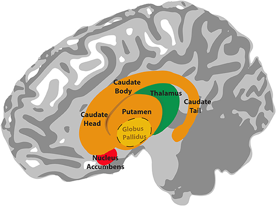
    Distinguish a persistent hepatocerebral syndrome from simply assigning every symptom to fluctuating encephalopathy. [11]
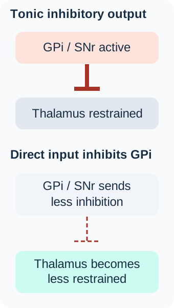
    Read the complete worked explanation
    1. What would the proposed material need to explain?

      Manganese accumulation can accompany pallidal T1 hyperintensity in liver disease.

    2. Which association fits cirrhosis, shunting, and this T1 pattern?

      Impaired hepatic handling and shunting can promote manganese accumulation associated with pallidal T1 hyperintensity.

    3. What useful rule carries to the next patient?

      Distinguish a persistent hepatocerebral syndrome from simply assigning every symptom to fluctuating encephalopathy.
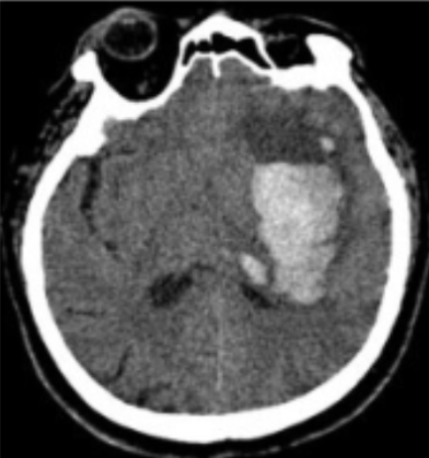
    Distinguish a persistent hepatocerebral syndrome from simply assigning every symptom to fluctuating encephalopathy.
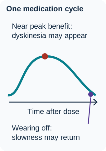
    Original source explanation

    Impaired hepatic handling and shunting can promote manganese accumulation associated with pallidal T1 hyperintensity.

  2. B. Copper proven by the MRI signal alone (Why this does not fit)

    Reason through this option

    Make one small inference

    1. What would the proposed material need to explain?

      Copper disorders require a combined clinical and biochemical assessment.

    2. Which association fits cirrhosis, shunting, and this T1 pattern?

      Wilson disease requires an integrated assessment; this signal and context are classically associated with manganese and are not a stand-alone copper assay.

    Distinguish a persistent hepatocerebral syndrome from simply assigning every symptom to fluctuating encephalopathy. [11]
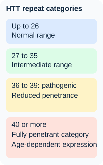
    Read the complete worked explanation
    1. What would the proposed material need to explain?

      Copper disorders require a combined clinical and biochemical assessment.

    2. Which association fits cirrhosis, shunting, and this T1 pattern?

      Wilson disease requires an integrated assessment; this signal and context are classically associated with manganese and are not a stand-alone copper assay.

    Distinguish a persistent hepatocerebral syndrome from simply assigning every symptom to fluctuating encephalopathy.
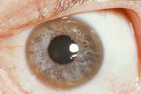
    Original source explanation

    Wilson disease requires an integrated assessment; this signal and context are classically associated with manganese and are not a stand-alone copper assay.

  3. C. Calcium established without CT or other evidence (Why this does not fit)

    Reason through this option

    Make one small inference

    1. What would the proposed material need to explain?

      Calcium deposition cannot be inferred from every bright MRI signal.

    2. Which association fits cirrhosis, shunting, and this T1 pattern?

      T1 pallidal signal in this context does not by itself establish calcium deposition.

    Distinguish a persistent hepatocerebral syndrome from simply assigning every symptom to fluctuating encephalopathy. [11]

    Read the complete worked explanation
    1. What would the proposed material need to explain?

      Calcium deposition cannot be inferred from every bright MRI signal.

    2. Which association fits cirrhosis, shunting, and this T1 pattern?

      T1 pallidal signal in this context does not by itself establish calcium deposition.

    Distinguish a persistent hepatocerebral syndrome from simply assigning every symptom to fluctuating encephalopathy.

    Original source explanation

    T1 pallidal signal in this context does not by itself establish calcium deposition.

  4. D. Iron proven by any bright T1 basal ganglia signal (Why this does not fit)

    Reason through this option

    Make one small inference

    1. What would the proposed material need to explain?

      MRI signal depends on the sequence and clinical context.

    2. Which association fits cirrhosis, shunting, and this T1 pattern?

      Signal interpretation depends on sequence and context; the given association favors manganese.

    Distinguish a persistent hepatocerebral syndrome from simply assigning every symptom to fluctuating encephalopathy. [11]

    Read the complete worked explanation
    1. What would the proposed material need to explain?

      MRI signal depends on the sequence and clinical context.

    2. Which association fits cirrhosis, shunting, and this T1 pattern?

      Signal interpretation depends on sequence and context; the given association favors manganese.

    Distinguish a persistent hepatocerebral syndrome from simply assigning every symptom to fluctuating encephalopathy.

    Original source explanation

    Signal interpretation depends on sequence and context; the given association favors manganese.

Takeaway: Distinguish a persistent hepatocerebral syndrome from simply assigning every symptom to fluctuating encephalopathy.

Case sources: [11]

Case 27

A patient with no previous movement disorder develops right-sided chorea and flinging. Glucose is markedly high, and imaging shows a left striatal abnormality without an STN lesion. Which conclusion is best?

Explore every option and its reasoning
  1. A. The findings prove hereditary Huntington disease (Why this does not fit)

    Reason through this option

    Make one small inference

    1. What does this alternative assume about the cause?

      Inherited disease requires evidence beyond acute hyperkinesia.

    2. Can the syndrome be explained without a visible STN lesion?

      The acute course and metabolic context do not establish an inherited progressive disorder.

    The classic lesion is a starting association, not a compulsory imaging finding. [13] [14]

    Read the complete worked explanation
    1. What does this alternative assume about the cause?

      Inherited disease requires evidence beyond acute hyperkinesia.

    2. Can the syndrome be explained without a visible STN lesion?

      The acute course and metabolic context do not establish an inherited progressive disorder.

    The classic lesion is a starting association, not a compulsory imaging finding.

    Original source explanation

    The acute course and metabolic context do not establish an inherited progressive disorder.

  2. B. Hyperglycemia-associated striatal dysfunction can cause hemichorea and hemiballism (Best answer)

    Reason through this option

    Make one small inference

    1. What does this alternative assume about the cause?

      Striatal dysfunction can accompany severe hyperglycemia.

    2. Can the syndrome be explained without a visible STN lesion?

      The syndrome can arise outside the STN, and acute metabolic and vascular causes require assessment.

    3. What useful rule carries to the next patient?

      The classic lesion is a starting association, not a compulsory imaging finding.

    The classic lesion is a starting association, not a compulsory imaging finding. [13] [14]

    Read the complete worked explanation
    1. What does this alternative assume about the cause?

      Striatal dysfunction can accompany severe hyperglycemia.

    2. Can the syndrome be explained without a visible STN lesion?

      The syndrome can arise outside the STN, and acute metabolic and vascular causes require assessment.

    3. What useful rule carries to the next patient?

      The classic lesion is a starting association, not a compulsory imaging finding.

    The classic lesion is a starting association, not a compulsory imaging finding.

    Original source explanation

    The syndrome can arise outside the STN, and acute metabolic and vascular causes require assessment.

  3. C. The activity cannot be hemiballism unless an STN lesion is visible (Why this does not fit)

    Reason through this option

    Make one small inference

    1. What does this alternative assume about the cause?

      The classic STN association is not the only network location.

    2. Can the syndrome be explained without a visible STN lesion?

      Clinical series demonstrate lesions outside the classic nucleus.

    The classic lesion is a starting association, not a compulsory imaging finding. [13] [14]

    Read the complete worked explanation
    1. What does this alternative assume about the cause?

      The classic STN association is not the only network location.

    2. Can the syndrome be explained without a visible STN lesion?

      Clinical series demonstrate lesions outside the classic nucleus.

    The classic lesion is a starting association, not a compulsory imaging finding.

    Original source explanation

    Clinical series demonstrate lesions outside the classic nucleus.

  4. D. The glucose result is irrelevant to the neurologic presentation (Why this does not fit)

    Reason through this option

    Make one small inference

    1. What does this alternative assume about the cause?

      Metabolic abnormalities can contribute to acute neurologic symptoms.

    2. Can the syndrome be explained without a visible STN lesion?

      Severe hyperglycemia is a recognized associated cause and requires treatment.

    The classic lesion is a starting association, not a compulsory imaging finding. [13] [14]

    Read the complete worked explanation
    1. What does this alternative assume about the cause?

      Metabolic abnormalities can contribute to acute neurologic symptoms.

    2. Can the syndrome be explained without a visible STN lesion?

      Severe hyperglycemia is a recognized associated cause and requires treatment.

    The classic lesion is a starting association, not a compulsory imaging finding.

    Original source explanation

    Severe hyperglycemia is a recognized associated cause and requires treatment.

Takeaway: The classic lesion is a starting association, not a compulsory imaging finding.

Case sources: [13] [14]

Case 28

During a circuit discussion, a resident asks which route lets cortex influence STN without first passing through striatum. Which answer is correct?

Explore every option and its reasoning
  1. A. The hyperdirect pathway (Best answer)

    Reason through this option

    Make one small inference

    1. What are the beginning and next relay of this route?

      Hyperdirect: cortex to STN.

    2. Which route bypasses the first striatal relay?

      Cortical excitation of STN provides a route that bypasses the first striatal relay.

    3. What useful rule carries to the next patient?

      Direct and indirect pathways are useful models but do not exhaust basal ganglia connections.

    Direct and indirect pathways are useful models but do not exhaust basal ganglia connections. [18]

    Read the complete worked explanation
    1. What are the beginning and next relay of this route?

      Hyperdirect: cortex to STN.

    2. Which route bypasses the first striatal relay?

      Cortical excitation of STN provides a route that bypasses the first striatal relay.

    3. What useful rule carries to the next patient?

      Direct and indirect pathways are useful models but do not exhaust basal ganglia connections.

    Direct and indirect pathways are useful models but do not exhaust basal ganglia connections.

    Original source explanation

    Cortical excitation of STN provides a route that bypasses the first striatal relay.

  2. B. The direct striatopallidal pathway (Why this does not fit)

    Reason through this option

    Make one small inference

    1. What are the beginning and next relay of this route?

      Direct: cortex to striatum, then GPi/SNr.

    2. Which route bypasses the first striatal relay?

      The direct pathway in the classic model passes through striatum before GPi/SNr.

    Direct and indirect pathways are useful models but do not exhaust basal ganglia connections. [18]

    Read the complete worked explanation
    1. What are the beginning and next relay of this route?

      Direct: cortex to striatum, then GPi/SNr.

    2. Which route bypasses the first striatal relay?

      The direct pathway in the classic model passes through striatum before GPi/SNr.

    Direct and indirect pathways are useful models but do not exhaust basal ganglia connections.

    Original source explanation

    The direct pathway in the classic model passes through striatum before GPi/SNr.

  3. C. The nigrostriatal dopamine pathway (Why this does not fit)

    Reason through this option

    Make one small inference

    1. What are the beginning and next relay of this route?

      Nigrostriatal: SNc to striatum.

    2. Which route bypasses the first striatal relay?

      This runs from SNc toward striatum, not from cortex directly to STN.

    Direct and indirect pathways are useful models but do not exhaust basal ganglia connections. [18]

    Read the complete worked explanation
    1. What are the beginning and next relay of this route?

      Nigrostriatal: SNc to striatum.

    2. Which route bypasses the first striatal relay?

      This runs from SNc toward striatum, not from cortex directly to STN.

    Direct and indirect pathways are useful models but do not exhaust basal ganglia connections.

    Original source explanation

    This runs from SNc toward striatum, not from cortex directly to STN.

  4. D. The excitatory thalamocortical return pathway (Why this does not fit)

    Reason through this option

    Make one small inference

    1. What are the beginning and next relay of this route?

      Thalamocortical: thalamus to cortex.

    2. Which route bypasses the first striatal relay?

      The thalamocortical route returns signals to cortex and is not the cortical projection to STN described in the question.

    Direct and indirect pathways are useful models but do not exhaust basal ganglia connections. [18]

    Read the complete worked explanation
    1. What are the beginning and next relay of this route?

      Thalamocortical: thalamus to cortex.

    2. Which route bypasses the first striatal relay?

      The thalamocortical route returns signals to cortex and is not the cortical projection to STN described in the question.

    Direct and indirect pathways are useful models but do not exhaust basal ganglia connections.

    Original source explanation

    The thalamocortical route returns signals to cortex and is not the cortical projection to STN described in the question.

Takeaway: Direct and indirect pathways are useful models but do not exhaust basal ganglia connections.

Case sources: [18]

Case 29

A patient with atypical parkinsonism has markedly asymmetric limb rigidity, apraxia, and an alien-limb phenomenon. What can the clinical team reasonably name before pathology is known?

Explore every option and its reasoning
  1. A. Pathologically proven corticobasal degeneration from the examination alone (Why this does not fit)

    Reason through this option

    Make one small inference

    1. Does this label name a clinical syndrome or prove a tissue diagnosis?

      A pathology label requires more than the examination alone.

    2. What can apraxia and alien-limb findings establish before pathology is known?

      A clinical syndrome does not establish one histopathologic diagnosis.

    Keep a clinical syndrome separate from its possible pathology. [15]

    Read the complete worked explanation
    1. Does this label name a clinical syndrome or prove a tissue diagnosis?

      A pathology label requires more than the examination alone.

    2. What can apraxia and alien-limb findings establish before pathology is known?

      A clinical syndrome does not establish one histopathologic diagnosis.

    Keep a clinical syndrome separate from its possible pathology.

    Original source explanation

    A clinical syndrome does not establish one histopathologic diagnosis.

  2. B. Pure autonomic failure (Why this does not fit)

    Reason through this option

    Make one small inference

    1. Does this label name a clinical syndrome or prove a tissue diagnosis?

      Pure autonomic failure is not a cortical-motor syndrome.

    2. What can apraxia and alien-limb findings establish before pathology is known?

      The defining findings are cortical and motor, not a purely autonomic presentation.

    Keep a clinical syndrome separate from its possible pathology. [15]

    Read the complete worked explanation
    1. Does this label name a clinical syndrome or prove a tissue diagnosis?

      Pure autonomic failure is not a cortical-motor syndrome.

    2. What can apraxia and alien-limb findings establish before pathology is known?

      The defining findings are cortical and motor, not a purely autonomic presentation.

    Keep a clinical syndrome separate from its possible pathology.

    Original source explanation

    The defining findings are cortical and motor, not a purely autonomic presentation.

  3. C. Typical uncomplicated PD established by rigidity alone (Why this does not fit)

    Reason through this option

    Make one small inference

    1. Does this label name a clinical syndrome or prove a tissue diagnosis?

      Rigidity alone is not specific for typical PD.

    2. What can apraxia and alien-limb findings establish before pathology is known?

      Apraxia and alien-limb features warrant evaluation of an atypical syndrome.

    Keep a clinical syndrome separate from its possible pathology. [15]

    Read the complete worked explanation
    1. Does this label name a clinical syndrome or prove a tissue diagnosis?

      Rigidity alone is not specific for typical PD.

    2. What can apraxia and alien-limb findings establish before pathology is known?

      Apraxia and alien-limb features warrant evaluation of an atypical syndrome.

    Keep a clinical syndrome separate from its possible pathology.

    Original source explanation

    Apraxia and alien-limb features warrant evaluation of an atypical syndrome.

  4. D. Corticobasal syndrome (Best answer)

    Reason through this option

    Make one small inference

    1. Does this label name a clinical syndrome or prove a tissue diagnosis?

      Corticobasal syndrome names a recognizable clinical pattern.

    2. What can apraxia and alien-limb findings establish before pathology is known?

      The clinical pattern can be described as CBS, while several underlying pathologies can produce it.

    3. What useful rule carries to the next patient?

      Keep a clinical syndrome separate from its possible pathology.

    Keep a clinical syndrome separate from its possible pathology. [15]

    Read the complete worked explanation
    1. Does this label name a clinical syndrome or prove a tissue diagnosis?

      Corticobasal syndrome names a recognizable clinical pattern.

    2. What can apraxia and alien-limb findings establish before pathology is known?

      The clinical pattern can be described as CBS, while several underlying pathologies can produce it.

    3. What useful rule carries to the next patient?

      Keep a clinical syndrome separate from its possible pathology.

    Keep a clinical syndrome separate from its possible pathology.

    Original source explanation

    The clinical pattern can be described as CBS, while several underlying pathologies can produce it.

Takeaway: Keep a clinical syndrome separate from its possible pathology.

Case sources: [15]

Case 30

A trainee identifies a thin gray sheet between the extreme capsule and external capsule, lateral to the putamen. Which structure has been found?

Explore every option and its reasoning
  1. A. Posterior limb of the internal capsule (Why this does not fit)

    Reason through this option

    Make one small inference

    1. Where is this structure relative to putamen?

      Posterior internal capsule is medial to lentiform nucleus.

    2. Which structure is a gray sheet between the two named capsules?

      That is white matter medial to the lentiform nucleus rather than the thin gray sheet described.

    Putamen is lateral within the main motor nuclear grouping, but it is not the most lateral deep gray structure in every definition. [1] [21]

    Read the complete worked explanation
    1. Where is this structure relative to putamen?

      Posterior internal capsule is medial to lentiform nucleus.

    2. Which structure is a gray sheet between the two named capsules?

      That is white matter medial to the lentiform nucleus rather than the thin gray sheet described.

    Putamen is lateral within the main motor nuclear grouping, but it is not the most lateral deep gray structure in every definition.

    Original source explanation

    That is white matter medial to the lentiform nucleus rather than the thin gray sheet described.

  2. B. Subthalamic nucleus (Why this does not fit)

    Reason through this option

    Make one small inference

    1. Where is this structure relative to putamen?

      STN lies below thalamus.

    2. Which structure is a gray sheet between the two named capsules?

      STN lies below the thalamus and is not this lateral sheet.

    Putamen is lateral within the main motor nuclear grouping, but it is not the most lateral deep gray structure in every definition. [1] [21]

    Read the complete worked explanation
    1. Where is this structure relative to putamen?

      STN lies below thalamus.

    2. Which structure is a gray sheet between the two named capsules?

      STN lies below the thalamus and is not this lateral sheet.

    Putamen is lateral within the main motor nuclear grouping, but it is not the most lateral deep gray structure in every definition.

    Original source explanation

    STN lies below the thalamus and is not this lateral sheet.

  3. C. Claustrum (Best answer)

    Reason through this option

    Make one small inference

    1. Where is this structure relative to putamen?

      Claustrum is lateral to putamen, between external and extreme capsules.

    2. Which structure is a gray sheet between the two named capsules?

      The claustrum lies between those two white-matter layers and lateral to the main lentiform motor nuclei.

    3. What useful rule carries to the next patient?

      Putamen is lateral within the main motor nuclear grouping, but it is not the most lateral deep gray structure in every definition.

    Putamen is lateral within the main motor nuclear grouping, but it is not the most lateral deep gray structure in every definition. [1] [21]

    Read the complete worked explanation
    1. Where is this structure relative to putamen?

      Claustrum is lateral to putamen, between external and extreme capsules.

    2. Which structure is a gray sheet between the two named capsules?

      The claustrum lies between those two white-matter layers and lateral to the main lentiform motor nuclei.

    3. What useful rule carries to the next patient?

      Putamen is lateral within the main motor nuclear grouping, but it is not the most lateral deep gray structure in every definition.

    Putamen is lateral within the main motor nuclear grouping, but it is not the most lateral deep gray structure in every definition.

    Original source explanation

    The claustrum lies between those two white-matter layers and lateral to the main lentiform motor nuclei.

  4. D. Globus pallidus (Why this does not fit)

    Reason through this option

    Make one small inference

    1. Where is this structure relative to putamen?

      Pallidum lies medial to putamen.

    2. Which structure is a gray sheet between the two named capsules?

      The pallidum lies medial to putamen, not outside the external capsule.

    Putamen is lateral within the main motor nuclear grouping, but it is not the most lateral deep gray structure in every definition. [1] [21]

    Read the complete worked explanation
    1. Where is this structure relative to putamen?

      Pallidum lies medial to putamen.

    2. Which structure is a gray sheet between the two named capsules?

      The pallidum lies medial to putamen, not outside the external capsule.

    Putamen is lateral within the main motor nuclear grouping, but it is not the most lateral deep gray structure in every definition.

    Original source explanation

    The pallidum lies medial to putamen, not outside the external capsule.

Takeaway: Putamen is lateral within the main motor nuclear grouping, but it is not the most lateral deep gray structure in every definition.

Case sources: [1] [21]

Whole teaching image

Search Bone Wizardry

Quick links