Histology is the board question. Every tumor has ONE finding that gives it away. Learn those, and you'll never confuse GBM with medulloblastoma again.
Before you learn anything · take the shot:
A 49-year-old man presents with progressive headaches and blurry vision over 6 weeks. Exam shows papilledema. MRI reveals a large temporal lobe mass crossing the midline with rim enhancement, central hemorrhage, and necrosis. Biopsy is performed. Which histological finding is most likely?
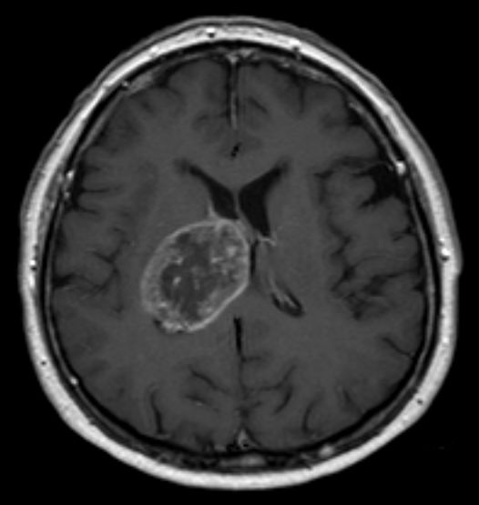
That's it. The trifecta of pseudopalisading necrosis + microvascular proliferation + crossing the midline = GBM (glioblastoma), every single time. In a middle-aged adult with a temporal/frontal mass that hemorrhages and necrotizes, this is the only answer. The rim enhancement on MRI is the viable tumor surrounding the dead center.
Good instinct · whorls and psammoma bodies are real, but that's meningioma. Think of it like a really organized knitting circle · cells wrapping neatly around each other. But meningiomas are dural-based (attached to the covering of the brain, not inside it), and they don't cross the midline or necrose. If the mass is INSIDE the brain parenchyma with hemorrhage and necrosis, it's not a meningioma.
That was the trap answer · and it's a really common one to pick. Small blue cells in rosettes = medulloblastoma. But think about it: medulloblastoma is a kid's tumor (usually under 20), and it lives in the cerebellum (back of the brain), not the temporal lobe. A 49-year-old with a temporal mass crossing midline? That's GBM territory. Rosettes in the cerebellum = kid. Pseudopalisades in the temporal lobe = adult. Age + location kills this answer.
Nice thought · fried egg cells scream oligodendroglioma. Imagine cracking eggs on a microscope slide · clear halos around round nuclei. But oligos are slow-growing, live in the frontal lobe, and love to calcify. They don't hemorrhage, necrose, or cross the midline aggressively like this. Fried eggs + calcification + frontal = oligo. Necrosis + midline crossing = GBM.
Makes sense that you'd consider this, but Rosenthal fibers are the calling card of pilocytic astrocytoma · the most common brain tumor in kids. It's a friendly tumor: cystic, well-circumscribed, sits in the cerebellum, and basically never kills anyone. A 49-year-old with hemorrhagic necrosis crossing the midline? That's the opposite of friendly. Rosenthal fibers + cystic + cerebellum + kid = pilocytic. NOT adult temporal lobe necrosis.
Clinical Images
📷 GBM: ring-enhancing mass with central necrosis · tap to expand
📷 Meningioma: extra-axial, dural-based mass · tap to expand
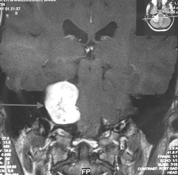
📷 Acoustic neuroma: cerebellopontine angle mass · tap to expand
THE PATTERN
Adult vs Pediatric. Supra vs Infra.
Two rules that eliminate half the wrong answers before you even look at histology.
The Two-Question Filter
Before you even think about what's under the microscope, answer two questions:
How old? Adults get supratentorial tumors (above the tentorium · cerebral hemispheres). Kids get infratentorial tumors (below · cerebellum, 4th ventricle, brainstem).
Where is it? Supratentorial = think GBM, meningioma, oligodendroglioma. Infratentorial = think medulloblastoma, pilocytic astrocytoma, ependymoma.
The exception is craniopharyngioma · a kid's tumor that's suprasellar (above the pituitary). But it's the ONLY common pediatric supratentorial tumor clinical medicine test.
Adults = supratentorial. Kids = infratentorial. If a board question gives you a 7-year-old with a cerebellar mass, you're choosing between pilocytic astrocytoma, medulloblastoma, and ependymoma · NOT GBM or meningioma. Age + location narrows to 2-3 choices instantly.
Board Trap: "Most common" depends on what they're asking. Most common primary brain tumor overall = meningioma (benign). Most common primary MALIGNANT brain tumor in adults = GBM. Most common brain tumor in kids overall = pilocytic astrocytoma. Most common MALIGNANT in kids = medulloblastoma. They ask "most common" differently every time.
🔑Adults are Above (supratentorial). Kids are below (Kicking in the Cerebellar Kitchen = infratentorial).
LOCATION MAP
Where Each Tumor Lives
Tap a pin. The region lights up, and the panel below tells you who gets it, how it shows, and the trap.
AdultsChildrenBoth
Tap any pin
Glioblastoma (GBM)
Temporal/Parietal · crosses corpus callosum
Who Gets It
Adults 50-70. Most common primary malignant brain tumor in adults.
Classic Presentation
Progressive headache, papilledema, focal deficit. Ring-enhancing mass with central necrosis on MRI. "Butterfly glioma" when it crosses the midline.
clinical medicine trap: Multiple ring-enhancing lesions in someone with known cancer is METASTASIS, not GBM. GBM is usually a single mass even when it crosses midline.
HISTOLOGY ID
Tumor Battle Cards
Tap any card to flip it. Front = the stats you need for clinical practice. Back = the deep dive.
Ring-enhancing mass + necrosis + hemorrhage in adult
FLIP CARD
GBM · Deep Dive
THE CLUE: 49-year-old, headache, papilledema, temporal mass crossing midline with rim enhancement. That's GBM until proven otherwise.
THE CHAIN: Astrocyte goes rogue → grows insanely fast (WHO grade IV) → outgrows its blood supply → center dies (necrosis) → surviving cells line up around the dead zone like a fence (pseudopalisades) → tumor screams for new blood vessels (VEGF) → microvascular proliferation.
THE HIGH-YIELD DETAIL: GBM crosses the midline through the corpus callosum, making it look like a butterfly on MRI. No other brain tumor does this. When you see "butterfly glioma" · it's GBM.
THE MOVE: Pseudopalisades = GBM. Rim enhancement + necrosis in an adult = GBM. Crosses midline = GBM. Pick it and move on.
FLIP CARD BACK
Adult · Benign (usually)
Meningioma
Histology
Whorled spindle cells + psammoma bodies
Location
Dural-based (attached to brain's covering), well-defined
Extra-axial
OUTSIDE the brain parenchyma. Compresses, doesn't invade.
Board Clue
Middle-aged woman + well-defined dural mass + "dural tail" sign
FLIP CARD
Meningioma · Deep Dive
THE CLUE: Well-circumscribed mass attached to the dura, often in a middle-aged woman. Homogeneously enhancing. No necrosis, no hemorrhage.
THE CHAIN: Arises from arachnoid cap cells (the meninges, not the brain itself) → grows slowly on the OUTSIDE → pushes on brain tissue → causes symptoms by compression, not invasion → usually completely resectable.
THE HIGH-YIELD DETAIL: Most common PRIMARY brain tumor overall. But it's benign. When clinical medicine say "most common malignant" · that's GBM. The word "malignant" changes the answer completely. Also: meningiomas have estrogen/progesterone receptors · that's why they're more common in women and can grow during pregnancy.
Antoni A (hypercellular, organized) + Antoni B (hypocellular, loose)
Location
CP angle (cerebellopontine angle) · CN VIII most common
Stain
S-100+ (neural crest origin)
Board Clue
Unilateral hearing loss + tinnitus + CP angle mass
FLIP CARD
Schwannoma · Deep Dive
THE CLUE: Adult with gradual unilateral hearing loss and a mass at the CP angle. Classic acoustic neuroma (vestibular schwannoma).
THE CHAIN: Schwann cells wrap peripheral nerves in myelin → one goes rogue → grows on CN VIII at the CP angle → compresses CN VIII (hearing loss, tinnitus) → if big enough, compresses CN VII (facial droop) and CN V (facial numbness).
THE HIGH-YIELD DETAIL: BILATERAL schwannomas = NF2 (neurofibromatosis type 2). The mnemonic: NF2 = 2 schwannomas (bilateral acoustic neuromas). If they say bilateral CP angle masses, stop thinking · it's NF2.
THE MOVE: Antoni A + Antoni B = schwannoma. S-100+ = schwannoma. CP angle + hearing loss = schwannoma. Bilateral = NF2.
Frontal mass + calcification + seizures + fried egg cells
FLIP CARD
Oligodendroglioma · Deep Dive
THE CLUE: Young-to-middle-aged adult with new-onset seizures and a calcified frontal lobe mass. Biopsy shows round cells with clear halos.
THE CHAIN: Oligodendrocytes normally make myelin in the CNS → one transforms → grows slowly in the frontal lobe → calcifies (almost all oligos do) → irritates surrounding cortex → seizures as the first symptom.
THE HIGH-YIELD DETAIL: The "fried egg" appearance is actually a fixation artifact · the cytoplasm swells and clears during processing. But clinical medicine don't care. See "fried egg" = oligo. Also: 1p/19q codeletion is diagnostic AND prognostic. These tumors respond way better to chemo than astrocytomas.
THE CLUE: Adult with a cerebellar mass AND unexplained polycythemia (high RBC count). That's the combo that points to it.
THE CHAIN: Tumor makes EPO (erythropoietin) → bone marrow cranks out red blood cells → polycythemia. It's the only brain tumor that does this. The tumor itself is a tangle of blood vessels with foamy stromal cells stuffed between them.
THE HIGH-YIELD DETAIL: If the question mentions VHL syndrome (retinal angiomas + renal cell carcinoma + pheochromocytoma), the cerebellar mass is hemangioblastoma. VHL = chromosome 3p deletion = loss of tumor suppressor. Most common CNS tumor in VHL.
Cerebellum (posterior fossa), can fill 4th ventricle
Spread
Drop metastases via CSF to spinal cord
Board Clue
Child + cerebellar mass + hydrocephalus + small blue cells
FLIP CARD
Medulloblastoma · Deep Dive
THE CLUE: Child with morning headaches, vomiting, and ataxia. MRI shows cerebellar mass compressing the 4th ventricle. Biopsy: small blue cells.
THE CHAIN: Embryonal tumor (primitive, undifferentiated) → grows fast in the cerebellum → compresses 4th ventricle → blocks CSF flow → hydrocephalus (headache, vomiting, papilledema) → cells shed into CSF → drop mets rain down the spinal cord.
THE HIGH-YIELD DETAIL: "Small round blue cells" is a pattern that shows up in MULTIPLE pediatric tumors · neuroblastoma, Ewing sarcoma, medulloblastoma, Wilms (blastemal). The LOCATION tells you which one: small blue cells in the CEREBELLUM = medulloblastoma. In the adrenal = neuroblastoma. In bone = Ewing.
THE MOVE: Kid + cerebellum + small blue cells + rosettes = medulloblastoma. Drop mets = medulloblastoma. Most common malignant brain tumor in kids = medulloblastoma.
FLIP CARD BACK
Pediatric · Benign
Pilocytic Astrocytoma
Histology
Rosenthal fibers + eosinophilic granular bodies
Location
Cerebellum. Cystic with enhancing mural nodule.
Behavior
WHO grade I. Excellent prognosis. Often curable with surgery.
Board Clue
Child + cystic cerebellar mass + Rosenthal fibers
FLIP CARD
Pilocytic Astrocytoma · Deep Dive
THE CLUE: Child with cerebellar symptoms (ataxia, wide-based gait). MRI shows a cystic mass with a bright enhancing nodule on the wall. Looks scary, but it's the good one.
THE CHAIN: Well-differentiated astrocyte tumor → grows slowly as a cyst with a solid bump (mural nodule) → the cyst is just fluid, the nodule is the tumor → scoop it out → cured. No chemo needed in most cases.
THE HIGH-YIELD DETAIL: Most common brain tumor in kids OVERALL (not malignant · just overall). When they ask "most common brain tumor in children" without the word "malignant" · pilocytic. When they add "malignant" · medulloblastoma. One word changes the answer. Also: associated with NF1 (optic pathway gliomas in NF1 are often pilocytic astrocytomas).
Child + 4th ventricle mass + perivascular pseudorosettes
FLIP CARD
Ependymoma · Deep Dive
THE CLUE: Child with hydrocephalus symptoms. MRI shows a mass ARISING FROM the floor of the 4th ventricle · not just compressing it (that's medulloblastoma), but growing OUT of the ventricle wall.
THE CHAIN: Ependymal cells line the ventricles → one transforms → grows into the 4th ventricle → blocks CSF outflow → hydrocephalus. Can extend through the foramina of Luschka/Magendie like fingers reaching out.
THE HIGH-YIELD DETAIL: Both ependymoma and medulloblastoma are in the posterior fossa in kids. The difference: ependymoma has perivascular pseudorosettes (cells radiating around blood vessels like a daisy). Medulloblastoma has Homer-Wright rosettes (cells circling an empty center). The rosette type tells you which one.
THE MOVE: Perivascular pseudorosettes = ependymoma. 4th ventricle + hydrocephalus = ependymoma. In adults, ependymomas favor the spinal cord instead.
FLIP CARD BACK
Pediatric · Benign (but tricky)
Craniopharyngioma
Histology
Cholesterol crystals in "motor oil" fluid + calcification
Location
Suprasellar (above pituitary). From Rathke's pouch remnant.
Child + calcified suprasellar mass + visual field defect
FLIP CARD
Craniopharyngioma · Deep Dive
THE CLUE: Child or teenager who stopped growing AND has visual field problems. CT shows a calcified mass sitting right above the sella (where the pituitary lives).
THE CHAIN: Rathke's pouch is an embryonic structure that becomes the anterior pituitary → remnant cells left behind → grow into a cystic, calcified mass → sits right above the pituitary → compresses the optic chiasm (bitemporal hemianopia) → compresses the pituitary (growth hormone deficiency, diabetes insipidus).
THE HIGH-YIELD DETAIL: This is the ONE common pediatric supratentorial tumor. It breaks the "kids = infratentorial" rule. But the location is specific: suprasellar, not just anywhere supratentorial. The cyst fluid looks like motor oil and is loaded with cholesterol crystals. Benign histologically but a surgical nightmare · stuck to everything important.
Pediatric posterior fossaMedulloblastoma has Homer-Wright rosettes; pilocytic astrocytoma has Rosenthal fibers; ependymoma has perivascular pseudorosettes.
Suprasellar childCraniopharyngioma has cholesterol crystals and machinery-oil fluid.
Clinical Imaging
GBM: ring-enhancing mass, central necrosis
Meningioma: extra-axial, dural-based
Vestibular schwannoma: CP angle
DANGER ZONES
Board Traps
The wrong answers that feel right · and how to kill them.
Trap #1: Medulloblastoma in an adult. They give you a 45-year-old with a cerebellar mass and small blue cells. You think medulloblastoma because "small blue cells + cerebellum." But adults CAN get medulloblastoma · it's just rare. The real trap: they're testing whether you know that in ADULTS, the most common posterior fossa tumor is metastasis (from lung, breast, melanoma). Always consider mets first in adults.
Trap #2: "Most common brain tumor" without context. Overall = meningioma. Overall malignant in adults = GBM. Overall in kids = pilocytic astrocytoma. Malignant in kids = medulloblastoma. Most common brain tumor period (including secondary) = metastasis. They love switching the qualifier.
Trap #3: Two rosette types. Homer-Wright rosettes (cells around a central EMPTY space) = medulloblastoma/neuroblastoma. Perivascular pseudorosettes (cells around a BLOOD VESSEL) = ependymoma. The word "perivascular" is the giveaway. If it says "pseudo" or "perivascular" · ependymoma. If it says "Homer-Wright" or just "rosettes" · medulloblastoma.
Trap #4: Ring-enhancing lesion. Not everything that ring-enhances is GBM. Differential: GBM, brain abscess, metastasis, toxoplasmosis (in HIV). Board clue for abscess = restricted diffusion on DWI (bright on diffusion-weighted imaging). GBM = no restricted diffusion. They can both ring-enhance, but DWI separates them.
🔑Ring-enhancing differential: GBM, Abscess, Mets, Toxo. GAMT · "the GAMT enhances in a ring."
Answer each question to narrow the diagnosis. Work through the clues the way a real board stem feeds them to you.
Step 1: A patient presents with a brain mass. What is the patient's age group?
Adults lean supratentorial. In adults, most primary brain tumors live ABOVE the tentorium: GBM, meningioma, oligodendroglioma. Exception: hemangioblastoma favors the cerebellum in adults. Move to location.
Kids lean infratentorial. In children, the action is BELOW the tentorium: medulloblastoma, pilocytic astrocytoma, ependymoma all live in the posterior fossa. One big exception: craniopharyngioma is suprasellar (above the pituitary) and still a pediatric tumor. Move to location.
Step 2: Where exactly is the mass located on imaging?
Intra-axial supratentorial in an adult: the short list is GBM (aggressive, necrotic) and oligodendroglioma (slow, calcified). Look at how it behaves and what it looks like.
Extra-axial, dural-based: this is almost always meningioma. Confirm with whorled cells and psammoma bodies on biopsy.
Posterior fossa: in a child, pick between medulloblastoma (solid cerebellar vermis, small blue cells), pilocytic astrocytoma (cystic with mural nodule, Rosenthal fibers), and ependymoma (4th ventricle floor, perivascular rosettes). In an adult, posterior fossa mass = think metastasis first.
Suprasellar: craniopharyngioma (calcified, cholesterol cyst fluid, kid or young adult). CP angle: schwannoma (hearing loss, S-100+, Antoni A/B). Different locations, different tumors.
Step 3: What does the MRI show?
Ring-enhancing + necrosis: GBM until proven otherwise in an adult (especially if it crosses the midline = butterfly glioma). Differential includes brain abscess (check DWI: bright center = abscess) and metastasis (multiple lesions, known primary).
Calcified + frontal + seizures: oligodendroglioma. Fried egg cells on biopsy, 1p/19q codeletion on genetics. Slow, relatively friendly.
Cystic with mural nodule: pilocytic astrocytoma (kid, cerebellum, Rosenthal fibers). In adults with cerebellar cyst, add hemangioblastoma to the list (check for polycythemia, VHL history).
Multiple lesions: metastases. Brain mets outnumber all primary tumors combined. Gray-white junction seeding = hematogenous spread. Ask about known cancer history, lung, breast, melanoma.
Step 4: Biopsy result. Which histological finding clinches the diagnosis?
Pseudopalisades = GBM.Pseudopalisading necrosis is the grade IV astrocytoma hallmark. Tumor cells line up around zones of necrosis like spectators around a crash site. No other brain tumor does this. Done.
Whorls + psammoma bodies = meningioma. Concentric rings of spindle cells (whorls) + calcified laminated bodies (psammoma bodies). Extra-axial, GFAP-negative, EMA-positive. Most common primary brain tumor overall.
Fried egg cells = oligodendroglioma.Perinuclear halos are a processing artifact but completely diagnostic in clinical practice. Add 1p/19q codeletion = confirmed oligo. Better prognosis, responds to chemo.
Small blue cells + Homer-Wright rosettes = medulloblastoma.Cells circling an empty center = Homer-Wright. Not perivascular (that's ependymoma). Kid + cerebellum + these rosettes = medulloblastoma, most common malignant pediatric brain tumor.
Workup complete. The four-step ladder: Age → Location → Imaging character → Histology. Each step cuts the differential in half. On exam, you won't always get all four layers, but you'll get enough. Anchor the pattern: you can derive the answer from 2-3 clues without memorizing every detail.
Key finding: MRI multiple ring-enhancing lesions at gray-white junction
Treatment: Steroids (dexamethasone), whole-brain radiation, stereotactic radiosurgery, surgery
Board pearl: Multiple ring-enhancing lesions at gray-white junction = metastases first (not GBM, which is usually single)
Medically reviewed by Kaitlyn Cocuzzo, MD and Fatima Ali, DO
Bone Wizardry is an independent educational resource for visual learning in the medical sciences. It is not affiliated with, endorsed by, or sponsored by any licensing or examination board, contains no real or recalled examination questions, and does not guarantee any educational or examination outcome.
 Bone WizardryMEDICAL EDUCATION