Demyelinating diseases: localization, timing and treatment
Localize CNS or peripheral disease, assess MS and opticospinal antibody disorders, detect GBS danger, and separate chronic progression from slow recovery.
A patient cannot walk. Another cannot see clearly. A third has double vision. The useful first question is not which disease name sounds familiar, but where the nervous system is failing, how that failure evolved, and which evidence changes the next decision. Follow six connected comparisons, then apply them to original clinical cases.
1. Locate the failure before naming the disease
Does absent myelin always mean the same illness? No. Oligodendrocytes provide CNS myelin; Schwann cells provide peripheral myelin. Damage can slow or interrupt conduction, while axonal loss removes functioning fibers. Optic nerves belong to the CNS even though they extend toward the eyes. AQP4-associated disease primarily injures astrocytes, and some GBS variants primarily affect axons or nodes rather than compact myelin. [3][4][5][17]
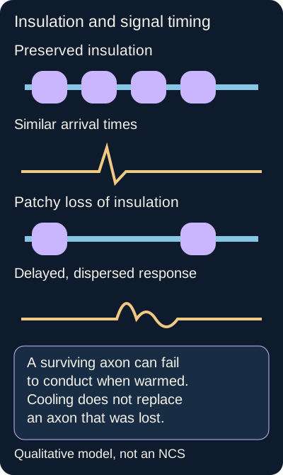
Compare the arrival time and width, not just the size, of the two response traces. Compare delayed arrival and broadened responses after demyelination. Original illustration: Bone Wizardry. [3][4][15]Open full-size diagram
Try the comparison: trace the two responses with a finger or cursor. The lower response arrives later and spreads out. Loss of insulation changes timing; a smaller response can also reflect block, dispersion or fewer working axons. A low amplitude alone cannot establish which of those occurred. [3][4]
Conduction laboratory
Predict what changes when a previously damaged pathway warms. Then compare it with actual fiber loss. This deliberately simplified model uses teaching units, not patient measurements or diagnostic thresholds.
Read the result without controls: preserved insulation supports coordinated arrival. Patchy damage delays and disperses the response. Warming a vulnerable surviving pathway can temporarily reduce conduction; returning to baseline can restore its prior function. Reducing the number of surviving fibers leaves a smaller response even after cooling. The model illustrates these directions, not their magnitude in a real person. [15]
Central pattern
Optic neuropathy, a sensory level, spasticity, brisk reflexes or an extensor plantar response suggest CNS involvement. Symptoms in different CNS regions require localization before a unifying diagnosis.
Peripheral pattern
Flaccid weakness, reduced reflexes, distal sensory loss and abnormal peripheral nerve studies suggest nerve or root disease. Areflexia reflects disruption of the reflex circuit; it does not identify one immune target.
Important exception: acute spinal cord injury can begin with areflexia and flaccidity. A sensory level, early urinary retention or rapidly developing myelopathy warrants urgent cord assessment and exclusion of compression. A normal brain MRI does not exclude spinal disease. [3][5]
Apply it elsewhere: compare an areflexic patient with distal tingling against an areflexic patient with a clear chest-level sensory boundary and retention. The second presentation changes the imaging priority even though both patients have absent leg reflexes.
2. Build an MS interpretation from anatomy and evidence
Can one symptom or one bright MRI spot establish MS? Neither can. MS involves immune-mediated CNS injury with myelin and axonal damage. T cells, B cells and innate inflammatory processes contribute; a single hypersensitivity label is not a complete explanation. HLA-DRB1*15 susceptibility, historically linked to HLA-DR2, and population associations with sex, age and geography are background probabilities, not diagnostic requirements. [1][13][17]
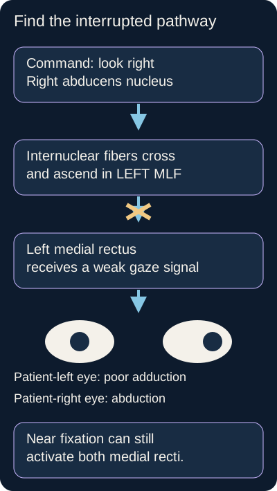
Painful visual loss with reduced color perception and a relative afferent pupillary defect supports optic neuropathy. The optic disc can be normal in retrobulbar neuritis or swollen when the visible nerve head is involved. By contrast, an eye that cannot adduct during horizontal gaze has an ocular motor localization. In internuclear ophthalmoplegia, the affected MLF is on the side of impaired adduction; the other eye may show abducting nystagmus. Convergence often remains intact, but its absence does not automatically exclude INO. [5][12]
The sides are labeled from the patient perspective; the eye symbols show the direction of movement. INO is named for the side of impaired adduction. Convergence is often, but not invariably, preserved. Original illustration: Bone Wizardry. [12]Open full-size diagram
Trace and test: follow a rightward gaze command through the diagram, then cover the labels. A left MLF interruption weakens left adduction while right abduction persists. Preserved near adduction favors a functioning medial rectus motor apparatus with a disrupted conjugate gaze connection. MS and vascular disease are important causes of INO, but age alone does not settle its cause. [12]
Electric-shock sensations down the spine during neck flexion are called Lhermitte sign and suggest a cervical cord process, not uniquely MS. Intention tremor, nystagmus and scanning speech form the historical cerebellar Charcot triad, also not a diagnostic fingerprint. Clinical findings identify affected systems before disease criteria are applied. [1][5][17]
Two kinds of dissemination, more than one way to show them
Characteristic lesions in different CNS regions support dissemination in space. A new lesion on follow-up or simultaneous enhancing and nonenhancing lesions can supply temporal evidence. Ovoid periventricular lesions oriented along veins, often called Dawson fingers, support the interpretation but are not individually pathognomonic. Nonspecific subcortical spots in migraine or vascular disease should not be counted indiscriminately. [1][2][5]
In the traditional 2017 framework, a typical first event with spatial dissemination and CSF-specific oligoclonal bands can meet diagnostic requirements without waiting for another symptomatic event. The 2024 McDonald revisions, published in 2025, add the optic nerve as a fifth anatomical location and incorporate selected newer MRI and CSF evidence. This lesson teaches the evidence logic, not every pathway in that specialist framework. Use the current criteria and exclude a better explanation rather than applying a mandatory two-attack rule. [1][2]
Compare paired serum and CSF band patterns. Bands confined to CSF support intrathecal IgG synthesis; matching bands in both samples do not provide the same evidence of restriction. The assay does not identify a uniquely MS-specific antigen. Infection and other inflammatory CNS disorders can also produce restricted bands. CSF glucose is usually normal and protein may be normal or mildly increased. Marked pleocytosis or low CSF glucose should widen the differential. [1][2][5]
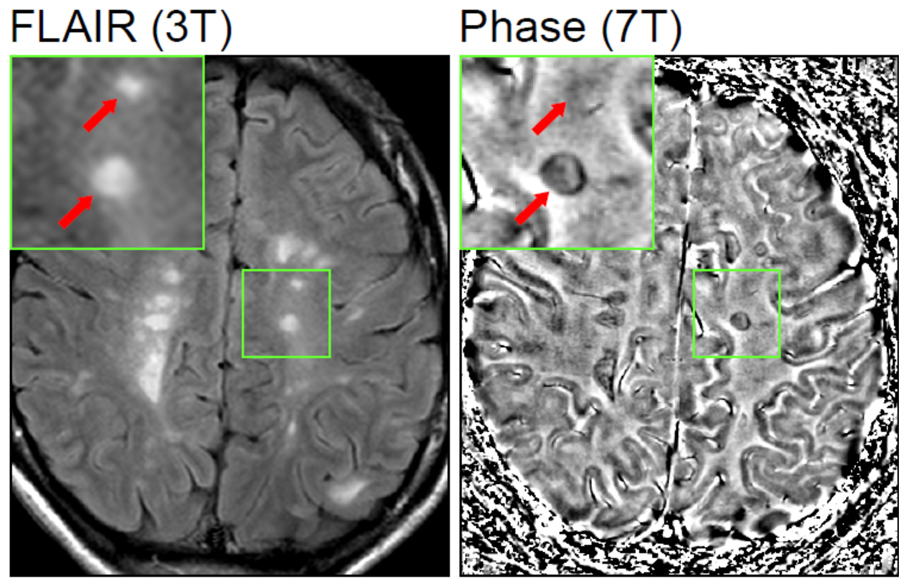
Lesions conspicuous on FLAIR do not necessarily share the same phase appearance. The source study investigated iron and inflammatory lesion features. This is not a gadolinium-enhancement comparison, a standalone MS diagnostic test, or a demonstration that every bright spot has the same age. Image: Mehta et al., 2013. Original image and attribution, CC BY 2.5. [11].Open full-size clinical image
Inspect the paired image: find one region visible on FLAIR, then locate its corresponding phase inset. The visible consequence is sequence-dependent information, not a diagnosis appearing twice. Transfer: when a new scan looks different, first ask whether the sequence changed, whether a true new lesion appeared, and whether the clinical syndrome still fits.
Try it here · Checkpoint 1 of 3
Make your prediction before reading the choices. A first attempt is just a starting point.
Case 1
Show answer and explanations for case 1
A. Left oculomotor nerve (Why this does not fit)
A left third-nerve lesion can impair left medial rectus function. Preserved near adduction demonstrates useful left medial rectus output despite impaired conjugate adduction. Separate the conjugate gaze connection from the final motor nerve, using convergence and the other third-nerve functions.
Reasoning steps for option A
How could a left third-nerve lesion affect this patient's medial rectus?
A left third-nerve lesion can impair left medial rectus function.
Why does preserved near adduction argue against a left third-nerve palsy?
Preserved near adduction demonstrates useful left medial rectus output despite impaired conjugate adduction.
Which gaze pathway, rather than the final motor nerve, is disrupted?
Preserved near adduction argues against a left third-nerve output lesion; deficient conjugate adduction instead implicates the left medial longitudinal fasciculus.
B. Right abducens nucleus (Why this does not fit)
The right abducens nucleus coordinates rightward conjugate gaze. It impairs right eye abduction as well as the linked left eye adduction; this patient still abducts the right eye. A nuclear horizontal gaze palsy affects the gaze pair, not only the adducting eye.
Reasoning steps for option B
What rightward gaze command originates in the right abducens nucleus?
The right abducens nucleus coordinates rightward conjugate gaze.
Why does preserved right eye abduction argue against nuclear damage?
It impairs right eye abduction as well as the linked left eye adduction; this patient still abducts the right eye.
Would a right abducens nuclear palsy spare one member of the gaze pair?
A nuclear horizontal gaze palsy affects the gaze pair, not only the adducting eye.
C. Left medial longitudinal fasciculus (Best answer)
The left eye has impaired adduction during rightward gaze. Internuclear fibers from the right abducens nucleus ascend in the left medial longitudinal fasciculus to the left medial rectus subnucleus. Preserved convergence supports a functioning medial rectus apparatus with a deficient internuclear command. The pattern localizes to the left medial longitudinal fasciculus; the cause still requires evaluation.
Reasoning steps for option C
Which eye adducts slowly on rightward gaze?
The left eye has impaired adduction during rightward gaze.
Which internuclear tract links the right abducens nucleus to left medial rectus activation?
Internuclear fibers from the right abducens nucleus ascend in the left medial longitudinal fasciculus to the left medial rectus subnucleus.
Why does preserved near adduction favor internuclear disconnection?
Preserved convergence supports a functioning medial rectus apparatus with a deficient internuclear command.
Which side of the MLF is implicated, and does this establish etiology?
The pattern localizes to the left medial longitudinal fasciculus; the cause still requires evaluation.
D. Right oculomotor nerve (Why this does not fit)
Right third-nerve dysfunction can impair right adduction, elevation and depression. Conjugate adduction is abnormal in the left eye, not the right eye. Identify the eye with the adduction deficit before assigning the side of an internuclear lesion.
Reasoning steps for option D
What eye movements would a right third-nerve lesion impair?
Right third-nerve dysfunction can impair right adduction, elevation and depression.
Is the impaired adduction in the right or left eye?
Conjugate adduction is abnormal in the left eye, not the right eye.
Which eye's adduction deficit determines the side of this internuclear lesion?
The left eye, not the right, has the adduction deficit, placing the internuclear lesion on the left.
Takeaway: An internuclear lesion is named for the side of the adduction deficit, not the abducting nystagmus.
3. Separate a relapse, progression and a treatment complication
Does worsening always mean a new MS attack? Compare three histories. An old visual deficit returns in a hot shower and resolves after cooling. New weakness persists for several days without fever or infection. Walking gradually deteriorates over a year between discrete attacks. These patterns raise different questions: reversible conduction failure, a possible relapse, and disability progression. [8][9][15]
Make the prediction first: which of those patterns should improve promptly with cooling? Heat can temporarily impair conduction in surviving demyelinated pathways, called Uhthoff phenomenon. This does not require new tissue injury and is not unique proof of MS. A fever or infection can similarly worsen old deficits. New or sustained symptoms need reassessment rather than automatic reassurance or automatic steroids. A clinical relapse generally lasts at least 24 hours without a better explanation; an MRI-visible new lesion is not obligatory for every clinically meaningful relapse. [1][8][15]
Read the history from onset to the present
Course
What the history establishes
CourseRelapsing-remitting
What the history establishesDiscrete attacks with partial or substantial recovery; the most common onset pattern.
CourseSecondary progressive
What the history establishesGradual worsening after an earlier relapsing course. Superimposed inflammatory activity can still occur.
CoursePrimary progressive
What the history establishesProgression from onset, without a preceding relapsing-remitting phase. Later age at onset and a more balanced sex distribution are tendencies, not criteria.
Activity, shown by relapses or relevant MRI findings, and progression are separate descriptors. There is no reliable universal countdown to secondary progression, and a course label alone cannot determine an individual prognosis. [9][13][17]
Treat the current problem
For a functionally important MS relapse, high-dose methylprednisolone can accelerate recovery. Appropriate high-dose oral treatment can be an alternative to intravenous treatment; the COPOUSEP trial did not test an ordinary low-dose prednisone prescription. Severe inadequately responsive attacks can require plasma-exchange assessment. Acute treatment is distinct from long-term disease modification and does not promise reversal of accumulated axonal loss. [8][5]
Maintenance selection is individualized. Interferon beta and glatiramer are established options in relapsing disease; natalizumab blocks alpha-4 integrin-mediated leukocyte trafficking, while ocrelizumab depletes CD20-expressing B cells and has trial evidence in primary progressive MS. Fingolimod alters S1P-dependent lymphocyte trafficking and requires attention to initiation-related bradycardia and conduction abnormalities. Efficacy, infection risk, pregnancy considerations, monitoring and the individual course matter more than memorizing one universal first-line drug. [13][14][16][17]
When the treatment changes the differential
A steadily progressive focal or cognitive syndrome over weeks in an immunocompromised person raises concern for progressive multifocal leukoencephalopathy. JC virus infects oligodendrocytes. HIV-related immune impairment and natalizumab are important contexts. MRI commonly shows asymmetric white matter and U-fiber involvement with limited mass effect or enhancement, but inflammatory PML can enhance. Fever is often absent, not prohibited. A negative first CSF JC-virus PCR does not exclude a strongly compatible presentation; repeat sensitive testing or further investigation may be needed. [10][14]
Anti-JC-virus antibody status or index helps stratify natalizumab-associated risk together with treatment duration and prior immunosuppression; seropositivity does not diagnose PML. Suspected PML requires urgent expert evaluation and withholding the implicated therapy. In HIV, effective antiretroviral treatment is central. Immune restoration can produce inflammatory worsening with edema, and stopping natalizumab also requires a plan for subsequent MS activity. Neither finding justifies unsupervised medication changes. [10][14]
Transfer: a patient whose language function steadily declines during natalizumab treatment does not belong automatically in the familiar relapse pathway. Compare the tempo, lesion pattern and immune context before adding more immunosuppression.
4. Similar locations can conceal different immune targets
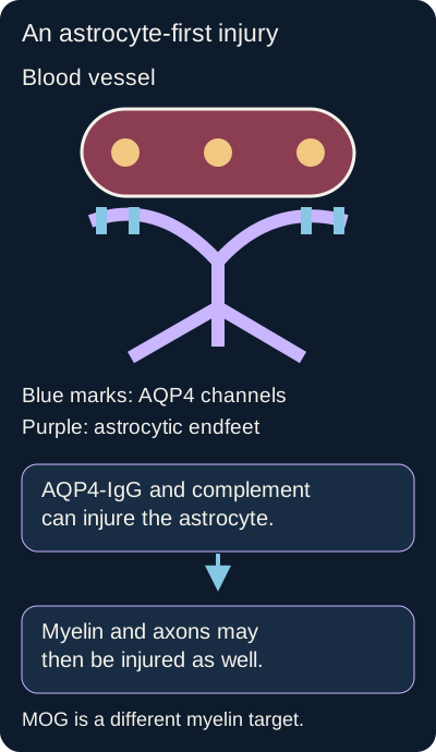
Does optic neuritis plus myelitis automatically mean MS? No. Severe optic neuropathy, substantial spinal cord disability, persistent unexplained hiccups or vomiting, and a lesion involving the area postrema should prompt consideration of neuromyelitis optica spectrum disorder. AQP4-IgG-positive NMOSD is an astrocytopathy with secondary myelin and axonal injury, not simply a more severe MS phenotype. [5]
The drawing locates the initial target rather than making every white matter disorder a primary myelin disease. MOGAD targets a distinct CNS myelin-associated antigen. Original illustration: Bone Wizardry. [5][6][7]Open full-size diagram
Point to the first injured structure: begin at the blue water-channel marks, then follow the astrocyte to the surrounding tissue. The consequence is that an antibody targeting astrocytic endfeet can still produce white matter dysfunction. Now substitute MOG, a myelin-associated protein, for AQP4. The tissue target and diagnostic interpretation change even though visual symptoms may look similar. [5][7]
MS pattern
Characteristic brain lesions and often relatively short, partial spinal lesions. Optic neuritis is commonly unilateral. These are tendencies within a complete diagnostic assessment.
AQP4-associated NMOSD
Optic attacks can be severe and bilateral or unilateral. Central cord lesions often extend over at least three vertebral segments. Area postrema and other brain lesions can occur; a normal brain MRI neither establishes nor excludes it.
MOGAD
A distinct syndrome supported by compatible clinical findings and reliable MOG-IgG testing. Optic neuritis may show prominent disc swelling or anterior and perineural involvement; myelitis and ADEM presentations also occur. It can be monophasic or relapsing and is not uniformly mild.
Longitudinally extensive transverse myelitis is a lesion pattern, not one etiology. Conversely, a short lesion does not exclude AQP4-associated NMOSD. Serum cell-based AQP4-IgG or MOG-IgG testing should be directed by the phenotype; low-positive MOG screening results in incompatible presentations need particular caution. Seronegative NMOSD requires stricter clinical and imaging assessment rather than an automatic diagnosis from lesion length. These disorders occur across demographic groups. [5][7]
Compare the consequence of treatment selection: a severe NMOSD attack calls for prompt high-dose intravenous corticosteroids and early apheresis assessment when severe or inadequately responsive. Long-term prevention may involve rituximab, C5 inhibition with eculizumab or ravulizumab, CD19 targeting with inebilizumab, or IL-6 receptor targeting with satralizumab. Azathioprine and mycophenolate remain options in some settings. Label eligibility, infection prevention and specialist selection matter. Interferon beta and natalizumab are not interchangeable NMOSD treatments and can be inappropriate or harmful. [6]
In a child, encephalopathy with multifocal deficits and large poorly demarcated lesions suggests an ADEM presentation. MOG testing and follow-up may clarify the disease; it is inaccurate to require every lesion to have precisely the same age or promise that no later attack can occur. Broader mimics include infection, vascular myelopathy, sarcoidosis and neoplasm. Absence of systemic sarcoid signs or a particular enhancement pattern does not by itself exclude those alternatives. [5][7]
Transfer: severe optic neuritis plus a two-segment cord lesion should not be labeled MS solely because the spinal lesion is short. A compatible AQP4-IgG result can change the diagnosis and preventive treatment.
Try it here · Checkpoint 2 of 3
Make your prediction before reading the choices. A first attempt is just a starting point.
Case 30
Show answer and explanations for case 30
A. A lesion shorter than three segments excludes NMOSD regardless of serology (Why this does not fit)
Longitudinally extensive cord lesions are strongly associated with NMOSD. Shorter lesions can occur, so length cannot negate a compatible core syndrome with convincing AQP4-IgG evidence. Use common imaging patterns as probability modifiers rather than absolute diagnostic vetoes.
Reasoning steps for option A
Why are long cord lesions associated with NMOSD?
Longitudinally extensive spinal lesions are strongly associated with NMOSD, but shorter lesions occur.
Can a two-segment lesion coexist with convincing AQP4-IgG?
Shorter lesions can occur, so length cannot negate a compatible core syndrome with convincing AQP4-IgG evidence.
Why is three-segment length not an absolute exclusion rule?
NMOSD can produce a short cord lesion; repeat strong AQP4-IgG and optic myelitis should not be vetoed by a three-segment cutoff.
B. Normal brain MRI proves a peripheral neuropathy (Why this does not fit)
A normal brain MRI can reduce evidence for some cerebral disorders. The optic neuropathy and intramedullary spinal lesion already establish CNS involvement. A normal study of one CNS region does not relocate disease to peripheral nerves.
Reasoning steps for option B
Could a normal brain MRI limit evidence of cerebral disease?
A normal brain MRI can limit cerebral evidence but cannot negate the optic and cord abnormalities.
What optic and cord findings still establish CNS involvement?
The optic neuropathy and intramedullary spinal lesion already establish CNS involvement.
Why does normal brain imaging not imply peripheral neuropathy?
Optic neuritis and intramedullary myelitis establish CNS disease even when brain MRI is normal.
C. The two-segment lesion establishes MS because its length is typical (Why this does not fit)
MS often causes relatively short spinal lesions. Convincing AQP4-IgG positivity and the severe optic-spinal phenotype favor a different diagnosis after exclusions. A common feature of one disease is not sufficient when stronger disease-specific evidence points elsewhere.
Reasoning steps for option C
Why might a short cord lesion suggest MS?
MS may produce short spinal cord lesions, but length is not the only diagnostic evidence.
Which repeat antibody result and severe optic-spinal features weigh against MS?
Convincing AQP4-IgG positivity and the severe optic-spinal phenotype favor a different diagnosis after exclusions.
Why does lesion length alone not establish MS here?
Repeat strong AQP4-IgG and severe optic-spinal disease favor NMOSD over diagnosing MS solely from a two-segment lesion.
D. AQP4-IgG-positive NMOSD remains supported despite the short cord lesion (Best answer)
The patient has optic neuritis and inflammatory myelitis. Strongly positive AQP4-IgG on cell-based testing provides substantial disease-specific support in this setting. No; NMOSD cord lesions are often long but can be short, depending on the attack and imaging timing. Integrate phenotype, reliable serology, imaging and exclusions rather than diagnosing from lesion length alone.
Reasoning steps for option D
Which core syndromes support an NMOSD phenotype?
The patient has optic neuritis and inflammatory myelitis.
What is the significance of repeat strong cell-based AQP4-IgG?
Strongly positive AQP4-IgG on cell-based testing provides substantial disease-specific support in this setting.
Can a two-segment cord lesion occur in NMOSD?
Yes; NMOSD cord lesions are often long but can be short, depending on the attack and imaging timing.
How should reliable serology, phenotype, imaging and exclusions be integrated?
Integrate phenotype, reliable serology, imaging and exclusions rather than diagnosing from lesion length alone.
Takeaway: AQP4-associated NMOSD is not ruled out by a short cord lesion or a normal brain MRI.
5. In acute peripheral weakness, protect breathing while defining the injury
Can oxygen saturation look reassuring while respiratory strength is failing? Yes. Guillain-Barre syndrome is an acute immune-mediated polyradiculoneuropathy that usually reaches its greatest weakness within four weeks. Symmetric ascending weakness and reduced reflexes are common, but not compulsory in every variant. Facial, bulbar, respiratory and autonomic involvement can be prominent. A gastrointestinal or respiratory illness in the preceding one to three weeks is common; Campylobacter, CMV and EBV are recognized associations. [3]
A bacterial surface structure can resemble a neural ganglioside, producing cross-reactive immunity after the infection resolves. The Campylobacter-ganglioside connection is particularly important in some axonal or nodal phenotypes; anti-GM1 and anti-GD1a antibodies are associated with AMAN. It does not mean that every GBS case has one proven antimyelin antibody or ongoing bacterial invasion. Antibiotics for an active infection do not replace treatment of the neurological immune process. [3]
Read nerve responses as physiology, not just labels
Pattern
What the evidence can show
PatternAIDP
What the evidence can showA demyelinating GBS pattern: prolonged distal or late-response latencies, slowing, conduction block and temporal dispersion across appropriate nerves.
PatternAMAN
What the evidence can showMotor axonal or nodal dysfunction with sensory responses preserved. Reduced motor amplitudes need serial interpretation because reversible nodal failure can resemble early block.
PatternAMSAN
What the evidence can showMotor and sensory axonal involvement, with reduced responses in both populations. Prognosis depends on severity and trajectory, not the name alone.
PatternMiller Fisher
What the evidence can showOphthalmoplegia, ataxia and areflexia, often with relatively preserved limb strength and anti-GQ1b antibodies. Peripheral proprioceptive dysfunction contributes; ataxia does not prove an isolated cerebellar lesion.
Reconstruct the trace: return to the conduction drawing and predict which measurement reflects arrival time, spread of arrival, and the amount of functioning output. Then add preserved sensory responses to a low-motor-amplitude pattern. That new information favors a motor-selective process rather than AMSAN. Nerve temperature, technique, entrapment sites and evolving nodal physiology can affect classification. [3][4]
CSF often shows increased protein with few cells, called albuminocytologic dissociation. Root and barrier abnormalities help explain the protein rise; a low cell count does not prove that the immune injury is noncellular. Early protein may be normal. Marked pleocytosis, low glucose or an incompatible systemic syndrome should prompt investigation for alternatives. CSF protein is neither a standalone GBS confirmation nor a respiratory safety test. [3]
Calculate, then inspect the patient
A 75-kg adult has an FVC of 1500 mL: 1500 divided by 75 equals 20 mL/kg. If the value fell rapidly and cough is weak, respiratory reserve and secretion clearance are both threatened. Serial FVC, inspiratory and expiratory strength, bulbar function, secretion handling and the clinical trajectory guide escalation. A value around or below 20 mL/kg can prompt elective ventilation assessment; it is not an isolated universal intubation switch. Do not wait for oxygen desaturation or abnormal blood gases when other findings show impending failure. [3]
For patients meeting severity and timing indications, IVIG or plasma exchange are effective options. Routine sequential combination is not established as better, and routine additional IVIG solely because a prognosis is poor is not recommended. Corticosteroids are not effective GBS treatment according to trial evidence; an invented explanation about failure to block one immune pathway is not a substitute for those results. Recovery often takes weeks to months because stopping immune injury does not instantly restore nerve function. [3]
Supportive care includes respiratory and swallowing assessment, ECG and blood-pressure surveillance for autonomic instability, pain management, prevention of immobility complications, and rehabilitation. Watch for mimics: fatigable ocular-bulbar weakness without sensory loss suggests a junction disorder; cranial-predominant descending weakness with autonomic or pupillary abnormalities can suggest botulism. Anterior horn disease may produce a motor-predominant denervation pattern, while objective sensory abnormalities argue against a purely motor-neuron explanation.
Myasthenia gravis typically preserves sensation and tendon reflexes. Lambert-Eaton syndrome can cause proximal weakness, autonomic symptoms and depressed reflexes that facilitate after activation. These comparisons require the full examination rather than a single direction of weakness. [3]
Transfer: preserved limb power in a Miller Fisher presentation does not end monitoring. Ask whether swallowing, cough or respiratory function is changing, and assess overlap with the broader GBS spectrum.
Try it here · Checkpoint 3 of 3
Make your prediction before reading the choices. A first attempt is just a starting point.
Case 16
Show answer and explanations for case 16
A. Urgent ICU airway assessment and preparation for elective ventilation (Best answer)
1500 mL divided by 75 kg is 20 mL/kg. FVC has fallen by half over 12 hours, indicating rapidly declining reserve. Nasal speech, weak cough and retained secretions suggest bulbar and airway-protection impairment. Normal oxygen saturation does not exclude impending neuromuscular ventilatory failure; the combined findings warrant urgent airway assessment before a crisis.
Reasoning steps for option A
What is the current FVC per kilogram?
1500 mL divided by 75 kg is 20 mL/kg.
What does the FVC fall from 3.0 L to 1.5 L over 12 hours add?
FVC has fallen by half over 12 hours, indicating rapidly declining reserve.
Which bulbar findings increase airway risk beyond the 20 mL/kg FVC?
Nasal speech, weak cough and retained secretions suggest bulbar and airway-protection impairment.
Why is urgent airway assessment needed despite oxygen saturation of 98%?
Normal oxygen saturation does not exclude impending neuromuscular ventilatory failure; the combined findings warrant urgent airway assessment before a crisis.
B. Continue ward observation until oxygen saturation falls below 90% (Why this does not fit)
Pulse oximetry measures an important component of respiratory status. It can remain normal while respiratory muscle reserve and airway protection deteriorate. Do not wait for hypoxemia when mechanical and bulbar findings already indicate danger.
Reasoning steps for option B
What does pulse oximetry measure in this GBS patient?
Pulse oximetry estimates arterial oxygen saturation, not respiratory muscle reserve or the ability to clear secretions.
Can normal oxygenation exclude falling ventilatory reserve and secretion retention?
It can remain normal while respiratory muscle reserve and airway protection deteriorate.
Why not await saturation below 90% before escalating care?
The rapid FVC drop to 20 mL/kg, weak cough and retained secretions warrant airway evaluation before hypoxemia develops.
C. Reassure because an FVC of 1.5 L is adequate for every adult (Why this does not fit)
An absolute lung volume is easy to compare with a familiar number. The measured volume is only 20 mL/kg in this patient and has fallen rapidly. Interpret body-size-normalized values, measurement quality and trajectory together.
Reasoning steps for option C
Why can an absolute FVC of 1.5 L seem reassuring?
An absolute FVC of 1.5 L can appear reassuring if body size and the rapid fall are ignored, but here it is only 20 mL/kg.
What is 1.5 L relative to this patient’s 75-kg body weight and 12-hour trend?
The measured volume is only 20 mL/kg in this patient and has fallen rapidly.
Which FVC characteristics should inform airway decisions?
FVC is 20 mL/kg and has halved in 12 hours; body-size adjustment, measurement reliability and trajectory all matter.
D. Wait for CSF protein to rise further before changing the treatment setting (Why this does not fit)
CSF protein may rise as a GBS episode evolves. CSF protein does not quantify airway protection or respiratory muscle function. Escalate respiratory care using clinical and physiological evidence, not the magnitude of CSF protein.
Reasoning steps for option D
Why might CSF protein change as GBS evolves?
CSF protein may rise as a GBS episode evolves.
Can CSF protein measure cough strength or ventilatory reserve?
CSF protein does not quantify airway protection or respiratory muscle function.
Which respiratory and bulbar findings, rather than CSF protein, mandate action?
The falling FVC, nasal speech, weak cough and retained secretions support urgent airway evaluation irrespective of CSF protein.
Takeaway: A rapidly falling FVC plus impaired cough can demand action before oxygen saturation changes.
6. Continued progression is different from slow recovery
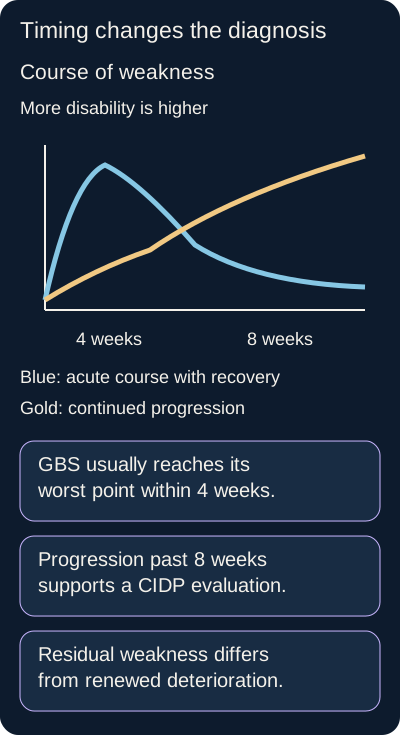
Does weakness after two months automatically become CIDP? No. Residual GBS disability can last much longer. Chronic inflammatory demyelinating polyradiculoneuropathy is supported by a compatible progressive or relapsing course, usually over at least eight weeks, and appropriate electrodiagnostic findings. Typical CIDP produces symmetric proximal and distal weakness, sensory dysfunction and reduced reflexes; variants require their own criteria. [3][4]
Trace each curve from left to right. The interval from 4 to 8 weeks requires follow-up rather than an automatic binary label. Repeated deteriorations after an acute onset can suggest acute-onset CIDP. Original illustration: Bone Wizardry. [3][4]Open full-size diagram
Trace the difference: the blue curve improves slowly after an early worst point; the gold curve continues worsening. A patient recovering at week 10 does not have the same course as a patient with new deterioration at week 10. Repeated fluctuations or renewed deterioration beyond eight weeks after an initially acute presentation should prompt reassessment for acute-onset CIDP. The four-to-eight-week interval is not solved by a rigid two-box label. [3][4]
Look for an acquired pattern across nerves: nonuniform slowing, block, dispersion and prolonged latencies interpreted against formal criteria and technical factors. Increased CSF protein is supportive, not sufficient. Longstanding familial distal weakness with high arches and uniform slowing suggests an inherited neuropathy. Diabetes and monoclonal gammopathies can complicate the differential; they do not automatically explain every progressive neuropathy, nor does their presence prove CIDP. [4]
IVIG and corticosteroids are established initial options in typical CIDP; plasma exchange is effective when appropriate. Maintenance may use immunoglobulin, including subcutaneous regimens in suitable patients, or another individualized approach. Motor CIDP deserves particular attention because IVIG is preferred initially and corticosteroids can worsen some patients. Measure objective improvement in strength and function and reassess the diagnosis when the response or course is discordant. [4]
Transfer: change only one fact in a chronic weakness case. If sensation is preserved and the specialist confirms motor CIDP, the preferred initial treatment can change. If the slowing is uniform and a parent has the same foot deformities, the priority becomes an inherited-neuropathy assessment rather than automatic immunotherapy.
7. Apply the comparisons to new patients
For each case, identify the anatomical location, the time course and the finding that most changes the decision. Commit to an answer before reading the rationale. Then explain why the strongest competing option fits less well. The questions are original educational cases and do not reproduce examination material.
Case 2
Show answer and explanations for case 2
A. It supports both dissemination in space and dissemination in time (Best answer)
Periventricular and juxtacortical lesions involve distinct characteristic CNS regions. Simultaneous enhancing and nonenhancing lesions can support dissemination in time in the appropriate diagnostic setting. A second clinical attack is not invariably required when clinical and paraclinical evidence meets the applicable criteria. The morphology, syndrome and exclusion of better explanations remain essential; the MRI pattern is not a diagnosis in isolation.
Reasoning steps for option A
Which two characteristic brain regions contain lesions in this first optic neuritis episode?
Periventricular and juxtacortical lesions involve distinct characteristic CNS regions.
How do enhancing and nonenhancing lesions on one scan inform timing?
Simultaneous enhancing and nonenhancing lesions can support dissemination in time in the appropriate diagnostic setting.
Does the first clinical attack necessarily preclude temporal dissemination?
A second clinical attack is not invariably required when clinical and paraclinical evidence meets the applicable criteria.
Why does this MRI evidence not establish MS by itself?
The morphology, syndrome and exclusion of better explanations remain essential; the MRI pattern is not a diagnosis in isolation.
B. It supports dissemination in space but cannot support dissemination in time (Why this does not fit)
The lesions occupy different characteristic regions. The coexistence of enhancing and nonenhancing lesions supplies temporal evidence rather than requiring two MRI dates. Distinguish two clinical attacks from the different accepted ways to establish temporal dissemination.
Reasoning steps for option B
Which part of the periventricular and juxtacortical distribution does this option recognize?
Periventricular and juxtacortical lesions provide the spatial evidence acknowledged by this option.
Which simultaneous contrast findings contradict its denial of temporal evidence?
The coexistence of enhancing and nonenhancing lesions supplies temporal evidence rather than requiring two MRI dates.
Why are two clinical attacks not the only route to temporal dissemination?
Enhancing and nonenhancing lesions on this same scan can support dissemination in time without two clinical attacks.
C. It supports dissemination in time but cannot support dissemination in space (Why this does not fit)
Enhancing and nonenhancing lesions can indicate different inflammatory timing. The brain MRI explicitly includes both periventricular and juxtacortical lesions. Count characteristic anatomical regions, not only the number of symptomatic locations.
Reasoning steps for option C
What does the contrast difference show about lesion timing?
Enhancing and nonenhancing lesions can indicate different inflammatory timing.
Which two brain regions demonstrate spatial dissemination?
The brain MRI explicitly includes both periventricular and juxtacortical lesions.
Why count periventricular and juxtacortical sites rather than symptomatic sites alone?
Periventricular and juxtacortical lesions occupy distinct characteristic CNS regions, supporting dissemination in space.
D. It supports neither form of dissemination until visual symptoms recur (Why this does not fit)
Longitudinal observation can clarify an uncertain first neurological event. The supplied MRI already provides characteristic spatial and temporal evidence after a typical event and exclusion of alternatives. Do not convert the teaching phrase multiple episodes into a mandatory second symptomatic attack.
Reasoning steps for option D
When could observing a first visual event over time be useful?
Longitudinal observation can clarify an uncertain first neurological event.
Why does this patient's MRI already supply both forms of dissemination evidence?
The supplied MRI already provides characteristic spatial and temporal evidence after a typical event and exclusion of alternatives.
Why need visual symptoms not recur to demonstrate time dissemination?
The enhancing periventricular and nonenhancing juxtacortical lesions can supply temporal evidence without recurrent visual symptoms.
Takeaway: One clinical event can coexist with substantial evidence of spatial and temporal dissemination.
A. Both patterns provide equivalent evidence of CSF-restricted IgG synthesis (Why this does not fit)
IgG patterns in serum and CSF must be compared to identify restriction to the CNS compartment. Patient A has matched bands, whereas patient B has bands absent from serum. Matched systemic bands are not equivalent to CSF-restricted oligoclonal bands.
Reasoning steps for option A
Why compare serum with CSF in these two patients?
IgG patterns in serum and CSF must be compared to identify restriction to the CNS compartment.
How do A's five matched bands differ from B's four unmatched CSF bands?
Patient A has matched bands, whereas patient B has bands absent from serum.
Do matched serum and CSF bands establish CNS-restricted synthesis?
Matched systemic bands are not equivalent to CSF-restricted oligoclonal bands.
B. Only patient A has evidence that the IgG was produced within the CNS (Why this does not fit)
Patient A has the same detectable IgG band pattern in blood and CSF. Only patient B has bands not detected in serum, supporting intrathecal synthesis. Determine whether the CSF contains additional restricted bands rather than simply whether bands are present.
Reasoning steps for option B
What does patient A's matched band pattern indicate?
Patient A has the same detectable IgG band pattern in blood and CSF.
Which patient's CSF has bands absent from serum?
Only patient B has bands not detected in serum, supporting intrathecal synthesis.
Why does presence of bands alone not identify intrathecal production?
Patient B has bands restricted to CSF, while patient A has only matched serum and CSF bands; the paired comparison identifies intrathecal synthesis.
C. Patient B has established that the IgG specifically targets myelin (Why this does not fit)
CSF-restricted bands support intrathecal immunoglobulin synthesis. The band pattern does not establish the antigen specificity of those antibodies. Do not equate the presence of restricted bands with proof of a particular antimyelin antibody.
Reasoning steps for option C
What biological process do B's restricted bands support?
CSF-restricted bands support intrathecal immunoglobulin synthesis.
Can the band pattern establish an antibody's myelin specificity?
The band pattern does not establish the antigen specificity of those antibodies.
Why is an antimyelin target not proven by these bands?
Although patient B has CSF-restricted bands, those bands do not prove that the antibodies target myelin.
D. Only patient B has CSF-restricted bands that support but do not prove MS (Best answer)
Patient B has four bands absent from serum. The pattern supports intrathecal immunoglobulin synthesis. In an appropriate typical presentation, restricted bands can contribute to the diagnostic framework alongside MRI and clinical evidence. Other inflammatory and infectious CNS disorders can also produce restricted bands, so the entire presentation must remain compatible.
Reasoning steps for option D
Which patient's bands are absent from serum?
Patient B has four bands absent from serum.
What kind of synthesis does B's pattern support?
Patient B's four CSF-only bands support intrathecal immunoglobulin synthesis.
How can B's bands contribute to an MS evaluation?
In an appropriate typical presentation, restricted bands can contribute to the diagnostic framework alongside MRI and clinical evidence.
Why do B's restricted bands not independently prove MS?
Other inflammatory and infectious CNS disorders can also produce restricted bands, so the entire presentation must remain compatible.
Takeaway: CSF-restricted bands identify a compartmental immune response, not a uniquely MS-specific antibody.
A. New inflammatory myelin destruction followed by rapid remyelination (Why this does not fit)
New inflammatory injury can impair conduction in an optic nerve. Return to the old baseline within 20 minutes is too rapid to be explained by destruction followed by remyelination. Rapidly reversible old symptoms favor altered conduction in existing lesions rather than rapid structural destruction and repair.
Reasoning steps for option A
Can new optic nerve inflammation impair conduction?
New inflammatory injury can impair conduction in an optic nerve.
Could remyelination explain recovery after 20 minutes in a cool room?
Return to the old baseline within 20 minutes is too rapid to be explained by destruction followed by remyelination.
What does rapid return of the old visual baseline favor over new destruction?
Rapidly reversible old symptoms favor altered conduction in existing lesions rather than rapid structural destruction and repair.
B. Reduced conduction reserve in previously injured axons during warming (Best answer)
A previously affected visual function worsened during warming rather than a new anatomical deficit appearing. Demyelinated axons can have reduced conduction reserve, making temperature-related failure more likely. Prompt recovery supports reversible conduction dysfunction without requiring new lesion formation. Persistent or new symptoms, fever or other illness would require fresh assessment rather than automatic attribution to heat.
Reasoning steps for option B
Was the blurred vision an old visual deficit triggered by warming or a new deficit?
A previously affected visual function worsened during warming rather than a new anatomical deficit appearing.
Why can heat disrupt conduction in a previously demyelinated optic nerve?
Demyelinated axons can have reduced conduction reserve, making temperature-related failure more likely.
What does rapid recovery on cooling imply about new lesion formation?
Prompt recovery supports reversible conduction dysfunction without requiring new lesion formation.
Which persistent symptoms or systemic signs would call for reassessment?
Persistent or new symptoms, fever or other illness would require fresh assessment rather than automatic attribution to heat.
C. Irreversible optic axon loss caused by the exercise session (Why this does not fit)
Loss of optic axons can leave persistent visual impairment. The visual function returns to its previous baseline shortly after cooling. Use reversibility to separate temporary failure of existing fibers from new permanent axonal loss.
Reasoning steps for option C
What kind of vision loss can optic axon destruction leave?
Loss of optic axons can leave persistent visual impairment.
Does return to baseline after cooling fit irreversible new axon loss?
The visual function returns to its previous baseline shortly after cooling.
How does reversibility distinguish conduction block from lost axons?
Return to the prior visual baseline after cooling favors transient conduction failure in surviving fibers, not irreversible new optic axon loss.
D. New cerebral infarction with myelin repair during cooling (Why this does not fit)
Cerebral ischemia can produce acute visual or other focal deficits. This reproducible warming-linked return of an old deficit does not require infarction, and cooling does not rapidly repair infarcted tissue. A first or persistent acute focal deficit still needs urgent evaluation even when a patient also has MS.
Reasoning steps for option D
Can cerebral infarction cause a focal visual deficit?
Cerebral ischemia can produce acute visual or other focal deficits.
Why do jogging-related recurrence and rapid cooling recovery disfavor infarction?
This reproducible warming-linked return of an old deficit does not require infarction, and cooling does not rapidly repair infarcted tissue.
When would a new focal deficit still need urgent evaluation?
A first or persistent acute focal deficit still needs urgent evaluation even when a patient also has MS.
Takeaway: Brief heat-related return of an old deficit suggests reduced conduction reserve, not proof of a new relapse.
A. A has primary progressive MS; B has secondary progressive MS (Why this does not fit)
The distinction depends on whether a relapsing course preceded the progressive phase. Only patient A had a relapsing-remitting phase before gradual deterioration. Assign secondary progression to a later progressive phase and primary progression to progression from onset.
Reasoning steps for option A
What prior disease phase separates primary from secondary progression?
The distinction depends on whether a relapsing course preceded the progressive phase.
Which patient had attacks before gradual walking decline?
Only patient A had a relapsing-remitting phase before gradual deterioration.
Which course begins with progression and which follows relapses?
A developed progression after separated attacks, consistent with secondary progression; B progressed from onset, consistent with primary progression.
B. Both have relapsing-remitting MS because their new lesions enhance (Why this does not fit)
A new enhancing lesion supports current inflammatory activity. Neither enhancement nor a superimposed relapse excludes a progressive course. Describe inflammatory activity and longitudinal disability progression as separate dimensions.
Reasoning steps for option B
What does the new enhancing lesion indicate in both patients?
A new enhancing lesion supports current inflammatory activity.
Can enhancing activity coexist with progressive disability?
Neither enhancement nor a superimposed relapse excludes a progressive course.
Which separate dimensions describe activity and progression?
Both patients have enhancing inflammatory activity and independently documented progressive walking disability; these are separate descriptors.
C. A has active secondary progressive MS; B has active primary progressive MS (Best answer)
Gradual deterioration followed an earlier relapsing-remitting phase. Gradual deterioration was present from symptom onset without a preceding relapsing period. A new enhancing lesion supports activity in each patient. A progressive course can be active; activity does not erase the history that distinguishes primary from secondary progression.
Reasoning steps for option C
Why is A's walking decline secondary progression?
Gradual deterioration followed an earlier relapsing-remitting phase.
Why is B's walking decline primary progression?
Gradual deterioration was present from symptom onset without a preceding relapsing period.
What does enhancement add to each progressive course?
A new enhancing lesion supports activity in each patient.
Can both primary and secondary progressive MS be active?
A progressive course can be active; activity does not erase the history that distinguishes primary from secondary progression.
D. Both have secondary progressive MS because disability has increased for 2 years (Why this does not fit)
Two years of gradual deterioration supports a sustained progressive component. Patient B has no earlier relapsing-remitting phase. The sequence of disease phases, rather than the severity or duration alone, distinguishes primary from secondary progression.
Reasoning steps for option D
What does two years of worsening walking establish?
Two years of gradual deterioration supports a sustained progressive component.
Which historical feature does B lack for secondary progression?
Patient B has no earlier relapsing-remitting phase.
Why is chronology more informative than duration for labeling B?
B progressed from onset despite two years of worsening, whereas A had prior attacks; only A has secondary progression.
Takeaway: Progression describes disability over time; activity describes recent relapses or MRI inflammation.
A. A routine low-dose oral prednisone course is equivalent to the tested intravenous regimen (Why this does not fit)
The study compared high-dose methylprednisolone by two routes. Routine low-dose oral prednisone was not the tested oral regimen. Match the drug, dose, regimen and population rather than generalizing from route alone.
Reasoning steps for option A
What dose and drug were compared by oral and intravenous routes?
The study compared methylprednisolone 1000 mg daily for 3 days by oral and intravenous routes.
Was ordinary low-dose prednisone the studied oral regimen?
Routine low-dose oral prednisone was not the tested oral regimen.
Why can the trial not justify swapping in a lower-dose drug course?
Non-inferiority of oral methylprednisolone 1000 mg daily for 3 days cannot be transferred to routine low-dose prednisone.
B. The oral regimen prevents long-term disability progression better than the intravenous regimen (Why this does not fit)
The reported outcome was improvement without retreatment at 28 days. The trial data supplied do not establish superior prevention of long-term disability progression. Keep the conclusion aligned with the measured endpoint and the non-inferiority design.
Reasoning steps for option B
What was measured at day 28?
The reported outcome was improvement without retreatment at 28 days.
Did this endpoint test long-term disability prevention?
The trial data supplied do not establish superior prevention of long-term disability progression.
Why does non-inferiority not prove superior long-term benefit?
Non-inferiority for improvement without retreatment at 28 days does not establish superior prevention of long-term disability.
C. The oral regimen has proven benefit for Guillain-Barre syndrome as well (Why this does not fit)
Both MS and GBS can involve immune-mediated neurological injury. An MS relapse trial does not establish efficacy in GBS, for which corticosteroids are not recommended. Use disease-specific outcome evidence rather than the shared word immune.
Reasoning steps for option C
Why might immune involvement in MS and GBS invite extrapolation?
Both MS and GBS can involve immune-mediated neurological injury.
Why does an MS relapse steroid trial not establish GBS efficacy?
An MS relapse trial does not establish efficacy in GBS, for which corticosteroids are not recommended.
Which condition-specific treatment evidence governs steroid use?
Corticosteroids can treat a suitable MS relapse, but they are not recommended for Guillain-Barre syndrome; this MS trial cannot change GBS practice.
D. The tested high-dose oral regimen can be a reasonable alternative for a suitable MS relapse (Best answer)
High-dose oral treatment met the trial criterion for non-inferiority at the 28-day endpoint. The patient can swallow, absorb medication and participate in outpatient follow-up. A specialist can consider the tested high-dose oral approach instead of intravenous treatment in a suitable MS relapse. Severity, monitoring needs, contraindications and practical access still influence the regimen and treatment setting.
Reasoning steps for option D
What did the 81% versus 80% comparison establish?
High-dose oral treatment met the trial criterion for non-inferiority at the 28-day endpoint.
Do normal swallowing, absorption and reliable follow-up support oral treatment here?
The patient can swallow, absorb medication and participate in outpatient follow-up.
Which specific oral regimen may a specialist consider for this disabling MS relapse?
A specialist may consider methylprednisolone 1000 mg orally daily for 3 days rather than the tested intravenous regimen in this suitable MS relapse.
What severity and monitoring considerations still affect route selection?
Severity, monitoring needs, contraindications and practical access still influence the regimen and treatment setting.
Takeaway: High-dose oral relapse treatment is not interchangeable with any oral steroid prescription.
A. Withhold natalizumab; urgently assess PML with repeat sensitive CSF PCR (Best answer)
The steadily progressive language and motor syndrome occurs during a therapy associated with PML risk. An asymmetric subcortical lesion with U-fiber involvement and little mass effect is compatible with PML. A negative initial CSF PCR can occur when viral burden is low or assay sensitivity is insufficient. Withhold natalizumab while urgently pursuing expert evaluation, repeat sensitive testing and further investigation if suspicion persists.
Reasoning steps for option A
Why is progressive aphasia and hand clumsiness on natalizumab concerning?
The steadily progressive language and motor syndrome occurs during a therapy associated with PML risk.
What does asymmetric U-fiber white matter involvement suggest?
An asymmetric subcortical lesion with U-fiber involvement and little mass effect is compatible with PML.
Can a negative routine CSF JC-virus PCR rule out PML?
A negative initial CSF PCR can occur when viral burden is low or assay sensitivity is insufficient.
What should happen to natalizumab while PML is urgently investigated?
Withhold natalizumab while urgently pursuing expert evaluation, repeat sensitive testing and further investigation if suspicion persists.
B. Continue natalizumab because the negative PCR excludes PML (Why this does not fit)
Detection of JC-virus DNA in compatible CSF strongly supports the diagnosis. The first assay can miss low viral concentrations despite a concerning clinical and MRI presentation. A negative test cannot automatically outweigh substantial pretest concern when the assay has meaningful sensitivity limitations.
Reasoning steps for option B
What would detectable JC-virus DNA in CSF support?
Detection of JC-virus DNA in compatible CSF strongly supports the diagnosis.
Why could an initial negative PCR miss PML in this patient?
The first assay can miss low viral concentrations despite a concerning clinical and MRI presentation.
Why should the concerning course and MRI outweigh a single negative assay?
A negative routine CSF JC-virus PCR may miss low-level virus; progressive symptoms and a compatible U-fiber lesion warrant continued PML investigation.
C. Increase natalizumab frequency to treat presumed breakthrough MS (Why this does not fit)
Breakthrough inflammatory MS activity is one possibility. This subacute progressive presentation and lesion pattern raise concern for an opportunistic infection during natalizumab treatment. Assess a plausible treatment-associated infection before intensifying the implicated immunotherapy.
Reasoning steps for option C
Could new deficits during natalizumab represent MS breakthrough?
Breakthrough inflammatory MS activity is one possibility.
Which treatment-associated infection must be considered with this MRI pattern?
This subacute progressive presentation and lesion pattern raise concern for an opportunistic infection during natalizumab treatment.
Why not intensify natalizumab before evaluating that possibility?
Because the natalizumab-associated lesion could be PML, investigate infection before increasing natalizumab exposure.
D. Use serum JC-virus antibodies alone to decide whether the lesion is PML (Why this does not fit)
Anti-JC-virus antibody status contributes to PML risk stratification during natalizumab treatment. Seropositivity can reflect prior exposure and does not establish active CNS infection. Combine the neurological course and MRI with virological or tissue evidence as clinically indicated.
Reasoning steps for option D
What is the role of serum JC-virus antibody status during natalizumab?
Anti-JC-virus antibody status contributes to PML risk stratification during natalizumab treatment.
Does serum exposure establish that this U-fiber lesion is active PML?
Seropositivity can reflect prior exposure and does not establish active CNS infection.
Which clinical, imaging and infection-specific evidence can clarify this lesion?
Progressive deficits and the U-fiber MRI lesion require PML evaluation with sensitive CSF JC-virus testing and further investigation if needed; serum antibody index alone is insufficient.
Takeaway: A negative first CSF PCR does not dismiss a clinically concerning PML presentation.
A. Conversion of PML into multiple sclerosis (Why this does not fit)
MS lesions can enhance during active inflammation. The patient has a compatible progressive syndrome with CSF JC-virus DNA and a temporal relationship to immune recovery. Enhancement alone does not convert a virologically supported infection into MS.
Reasoning steps for option A
Why might new enhancement superficially suggest MS?
MS lesions can enhance during active inflammation.
Which CSF and clinical evidence already supports PML instead?
The patient has a compatible progressive syndrome with CSF JC-virus DNA and a temporal relationship to immune recovery.
Does enhancement after ART convert confirmed PML into MS?
Enhancement alone does not convert a virologically supported infection into MS.
B. Immune reconstitution inflammatory syndrome associated with PML (Best answer)
Antiretroviral treatment reduced HIV replication and permitted immune recovery. They suggest an inflammatory response in the previously infected tissue. PML-associated immune reconstitution inflammatory syndrome can worsen deficits and produce enhancement or edema. Urgent specialist assessment is needed; severe inflammatory edema may require corticosteroid treatment while effective ART is generally continued.
Reasoning steps for option B
What does falling HIV RNA suggest after ART begins?
Antiretroviral treatment reduced HIV replication and permitted immune recovery.
Why might the existing PML lesions acquire edema and enhancement?
New enhancement and edema suggest an inflammatory response in the previously infected PML tissue during immune recovery.
Which immune-restoration complication explains the deterioration?
PML-associated immune reconstitution inflammatory syndrome can worsen deficits and produce enhancement or edema.
What urgent assessment and treatment considerations follow severe PML-associated inflammation?
Urgent specialist assessment is needed; severe inflammatory edema may require corticosteroid treatment while effective ART is generally continued.
C. Immediate proof that antiretroviral therapy cannot suppress HIV (Why this does not fit)
Persistent uncontrolled HIV can prevent useful immune recovery. The substantial fall in HIV RNA indicates virological response rather than immediate proof of treatment failure. Virological improvement and inflammatory neurological worsening can occur together.
Reasoning steps for option C
How could persistently uncontrolled HIV affect immunity?
Persistent uncontrolled HIV can prevent useful immune recovery.
Does substantially falling HIV RNA demonstrate immediate ART failure?
The substantial fall in HIV RNA indicates virological response rather than immediate proof of treatment failure.
Can virological response coexist with neurological worsening?
Virological improvement and inflammatory neurological worsening can occur together.
D. Exclusion of PML because enhancing lesions cannot occur in that infection (Why this does not fit)
PML often has little enhancement or mass effect before an inflammatory response develops. Immune restoration can produce enhancement and edema in PML lesions. Treat typical patterns as contextual evidence, not absolute exclusions that ignore immune status and timing.
Reasoning steps for option D
How much enhancement is typical in PML before immune restoration?
PML often has little enhancement or mass effect before an inflammatory response develops.
What changes in PML imaging after immune recovery?
Immune restoration can produce enhancement and edema in PML lesions.
Why does enhancement not exclude PML in this treated HIV patient?
PML can acquire enhancement and edema during immune recovery after ART, so new enhancement does not exclude the established infection.
Takeaway: Enhancement can emerge in PML during immune reconstitution; it does not automatically exclude the infection.
A. Peripheral Schwann-cell compact myelin (Why this does not fit)
Schwann cells myelinate peripheral axons. The optic nerves and spinal cord belong to the CNS, and the antibody is directed against aquaporin-4. A central optic-spinal syndrome cannot be explained by selecting a peripheral myelin target alone.
Reasoning steps for option A
Where is compact myelin made by Schwann cells?
Schwann cells myelinate peripheral axons.
Are optic nerves and spinal cord peripheral structures?
The optic nerves and spinal cord belong to the CNS, and the antibody is directed against aquaporin-4.
Why does peripheral myelin fail to match AQP4-positive optic-spinal disease?
A central optic-spinal syndrome cannot be explained by selecting a peripheral myelin target alone.
B. Presynaptic voltage-gated calcium channels at the neuromuscular junction (Why this does not fit)
Presynaptic calcium-channel autoimmunity can reduce acetylcholine release and cause Lambert-Eaton weakness. Optic neuropathy, a long intramedullary cord lesion and aquaporin-4 IgG indicate a CNS astrocytic process. Match the target to the involved tissue, not merely to an immune cause of weakness.
Reasoning steps for option B
What clinical syndrome follows presynaptic calcium-channel autoimmunity?
Presynaptic calcium-channel autoimmunity can reduce acetylcholine release and cause Lambert-Eaton weakness.
Would that junctional disorder explain optic neuritis, long cord lesion and AQP4 IgG?
Optic neuropathy, a long intramedullary cord lesion and aquaporin-4 IgG indicate a CNS astrocytic process.
Why must the target match the CNS rather than merely cause weakness?
The optic nerve and cord findings with aquaporin-4 IgG implicate astrocytic AQP4, not presynaptic calcium channels at the neuromuscular junction.
C. Postsynaptic acetylcholine receptors on skeletal muscle (Why this does not fit)
Acetylcholine-receptor antibodies impair neuromuscular transmission and cause fatigable weakness. They do not explain the optic-spinal inflammatory lesions or aquaporin-4-specific serology. Neuromuscular transmission failure does not produce the stated CNS MRI lesions.
Reasoning steps for option C
What impairment follows acetylcholine-receptor autoimmunity?
Acetylcholine-receptor antibodies impair neuromuscular transmission and cause fatigable weakness.
Can neuromuscular transmission failure explain this AQP4-positive optic-spinal syndrome?
They do not explain the optic-spinal inflammatory lesions or aquaporin-4-specific serology.
Would a junctional antibody cause these CNS MRI lesions?
Acetylcholine-receptor dysfunction at the neuromuscular junction cannot cause the described optic-spinal CNS lesions or account for AQP4 IgG.
D. Aquaporin-4 on astrocytic endfeet (Best answer)
The validated assay detects IgG directed against aquaporin-4. Aquaporin-4 water channels are highly expressed on astrocytic endfeet at interfaces including the blood-brain barrier. Antibody-associated complement injury can damage astrocytes with secondary injury to oligodendrocytes, myelin and neurons. AQP4-IgG-positive NMOSD is principally an astrocytopathy, not simply a severe subtype of MS.
Reasoning steps for option D
Which antigen is detected by the validated cell-based serum assay?
The validated assay detects IgG directed against aquaporin-4.
Where in the CNS is aquaporin-4 especially expressed?
Aquaporin-4 water channels are highly expressed on astrocytic endfeet at interfaces including the blood-brain barrier.
How does perivascular complement injury relate to astrocyte damage?
Antibody-associated complement injury can damage astrocytes with secondary injury to oligodendrocytes, myelin and neurons.
Why is AQP4-positive NMOSD an astrocytopathy rather than simply MS?
AQP4-IgG-positive NMOSD is principally an astrocytopathy, not simply a severe subtype of MS.
Takeaway: AQP4-associated NMOSD begins with astrocytic injury and can produce secondary demyelination.
A. Serum acetylcholine-receptor antibodies (Why this does not fit)
Acetylcholine-receptor antibodies support a diagnosis of myasthenia gravis in a compatible fatigable motor syndrome. The patient has a dorsal medullary lesion and prior inflammatory optic neuropathy rather than isolated neuromuscular transmission failure. Choose a test for the disease that explains both CNS events.
Reasoning steps for option A
What syndrome is acetylcholine-receptor testing designed to support?
Acetylcholine-receptor antibodies support a diagnosis of myasthenia gravis in a compatible fatigable motor syndrome.
Why do optic neuritis and a dorsal medullary lesion not suggest isolated myasthenia?
The patient has a dorsal medullary lesion and prior inflammatory optic neuropathy rather than isolated neuromuscular transmission failure.
Which test could link both CNS events instead?
Serum AQP4-IgG by cell-based assay addresses both area postrema symptoms from the dorsal medullary lesion and prior severe optic neuritis.
B. Serum anti-GQ1b antibodies (Why this does not fit)
Anti-GQ1b antibodies can support Miller Fisher syndrome in a patient with ophthalmoplegia, ataxia and areflexia. The patient has optic neuritis and a focal dorsal medullary lesion with persistent hiccups and vomiting, not the Miller Fisher pattern. Optic nerve dysfunction and a medullary lesion are not equivalent to peripheral ophthalmoplegia.
Reasoning steps for option B
Which triad would make anti-GQ1b testing relevant?
Anti-GQ1b antibodies can support Miller Fisher syndrome in a patient with ophthalmoplegia, ataxia and areflexia.
Does this patient have Miller Fisher features or optic neuritis with area postrema symptoms?
The patient has optic neuritis and a focal dorsal medullary lesion with persistent hiccups and vomiting, not the Miller Fisher pattern.
Why is peripheral ophthalmoplegia different from optic nerve and medullary disease?
Optic nerve dysfunction and a medullary lesion are not equivalent to peripheral ophthalmoplegia.
C. Serum aquaporin-4 IgG by cell-based assay (Best answer)
Persistent unexplained hiccups and vomiting with a dorsal medullary lesion implicate the area postrema region. Severe optic neuritis provides a second characteristic CNS syndrome associated with NMOSD. Serum AQP4-IgG testing using a cell-based assay is appropriate for this compatible phenotype. The antibody result must be integrated with the clinical and MRI presentation and exclusion of better explanations.
Reasoning steps for option C
Where does the lesion causing persistent hiccups and vomiting localize?
Persistent unexplained hiccups and vomiting with a dorsal medullary lesion implicate the area postrema region.
How does prior severe optic neuritis strengthen suspicion for NMOSD?
Severe optic neuritis provides a second characteristic CNS syndrome associated with NMOSD.
Which serum assay targets the suspected AQP4-associated disorder?
Serum AQP4-IgG testing using a cell-based assay is appropriate for this compatible phenotype.
What clinical and MRI context is needed to interpret AQP4-IgG?
The antibody result must be integrated with the clinical and MRI presentation and exclusion of better explanations.
D. Serum JC-virus antibody index as the diagnostic test (Why this does not fit)
The anti-JC-virus antibody index can contribute to PML risk assessment during natalizumab therapy. It does not diagnose a dorsal medullary inflammatory episode or establish CNS JC-virus infection. Risk-stratification assays should not replace disease-directed diagnostic testing.
Reasoning steps for option D
When is a serum JC-virus antibody index useful?
The anti-JC-virus antibody index can contribute to PML risk assessment during natalizumab therapy.
Could that index diagnose this medullary lesion and preceding optic neuritis?
It does not diagnose a dorsal medullary inflammatory episode or establish CNS JC-virus infection.
Why choose a disease-directed assay instead of a natalizumab PML risk marker?
Serum AQP4-IgG by cell-based assay evaluates the suspected NMOSD phenotype; the JC-virus antibody index assesses natalizumab-associated PML risk instead.
Takeaway: The area postrema can connect otherwise unexplained vomiting and hiccups to an optic-spinal syndrome.
A. The findings favor MS with an incidental MOG antibody result (Why this does not fit)
Optic neuritis can be an initial MS event, and some antibody results are incidental. Bilateral disc swelling, anterior and perineural involvement, and a clearly positive high-titer cell-based result form a compatible MOGAD phenotype. A compatible high-quality antibody result deserves more weight than a nonspecific assumption that every optic attack is MS.
Reasoning steps for option A
Why might optic neuritis initially suggest MS?
Optic neuritis can be an initial MS event, and some antibody results are incidental.
Which orbital and serologic features contradict incidental MOG-IgG?
Bilateral disc swelling, anterior and perineural involvement, and a clearly positive high-titer cell-based result form a compatible MOGAD phenotype.
Why should a validated high-titer MOG result outweigh the MS assumption?
The high-titer live cell-based MOG-IgG agrees with bilateral disc edema and anterior perineural optic inflammation, making incidental serology less plausible.
B. The clinical and antibody findings support MOGAD (Best answer)
The patient has bilateral inflammatory optic neuropathy with prominent disc swelling and compatible orbital MRI features. A clearly positive serum MOG-IgG result from a validated cell-based assay supports MOGAD. AQP4-IgG is negative and no better explanation was identified. MOGAD is distinct from MS and AQP4-associated NMOSD; this presentation does not guarantee a monophasic or benign future course.
Reasoning steps for option B
Which optic features make this a compatible MOGAD attack?
The patient has bilateral inflammatory optic neuropathy with prominent disc swelling and compatible orbital MRI features.
How does high-titer live cell-based MOG-IgG affect interpretation?
A clearly positive serum MOG-IgG result from a validated cell-based assay supports MOGAD.
What does negative AQP4-IgG add to this optic neuritis differential?
AQP4-IgG is negative and no better explanation was identified.
Does this first MOGAD optic attack establish a benign monophasic prognosis?
MOGAD is distinct from MS and AQP4-associated NMOSD; this presentation does not guarantee a monophasic or benign future course.
C. The findings favor AQP4-IgG-negative NMOSD over MOGAD (Why this does not fit)
Severe bilateral optic neuritis can raise concern for NMOSD. A compatible syndrome with convincing MOG-IgG identifies a distinct alternative that must be considered before assigning seronegative NMOSD. Shared optic involvement does not make MOGAD and AQP4-associated disease interchangeable.
Reasoning steps for option C
Why can bilateral severe optic neuritis evoke NMOSD?
Severe bilateral optic neuritis can raise concern for NMOSD.
What finding weighs against labeling this seronegative NMOSD?
A compatible syndrome with convincing MOG-IgG identifies a distinct alternative that must be considered before assigning seronegative NMOSD.
Why are MOGAD and AQP4-associated disease not interchangeable?
Convincing MOG-IgG in this compatible optic phenotype favors MOGAD, a diagnosis distinct from AQP4-associated NMOSD.
D. The antibody should be dismissed unless a second clinical attack occurs (Why this does not fit)
Follow-up is important to establish whether an inflammatory disease relapses. MOGAD can be monophasic and the compatible first event with reliable serology can support the diagnosis. A second attack determines a relapsing course; it is not obligatory for recognizing every antibody-associated first event.
Reasoning steps for option D
Why is longitudinal follow-up still needed after this optic attack?
Follow-up is important to establish whether an inflammatory disease relapses.
Can a first compatible optic event with reliable MOG-IgG support MOGAD?
MOGAD can be monophasic and the compatible first event with reliable serology can support the diagnosis.
What does a second attack establish that the first need not?
A second episode would document relapse, whereas this first compatible optic attack with reliable MOG-IgG already supports MOGAD.
Takeaway: MOG-IgG is meaningful when assay quality and the clinical phenotype agree; prognosis remains individualized.
A. Initiate urgent plasma-exchange assessment alongside attack care (Best answer)
The patient has a severe active spinal cord attack with worsening motor function. High-dose corticosteroids have not controlled the attack. Early apheresis, commonly plasma exchange, should be assessed urgently rather than postponed while disability accumulates. Long-term relapse prevention must also be planned, but it does not replace treatment of this attack.
Reasoning steps for option A
Which worsening motor and bladder deficits make this NMOSD attack urgent?
Severe paraparesis and urinary retention reflect an active spinal cord attack, with leg strength continuing to worsen despite corticosteroids.
What does worsening leg strength after three days of IV methylprednisolone show?
High-dose corticosteroids have not controlled the attack.
Which escalation should be assessed for this steroid-refractory NMOSD myelitis?
Early apheresis, commonly plasma exchange, should be assessed urgently rather than postponed while disability accumulates.
Why does NMOSD relapse prevention not replace rescue of this worsening myelitis?
Long-term prevention addresses future AQP4-associated relapses, but worsening motor function despite IV steroids calls for urgent apheresis assessment now.
B. Begin interferon beta as the acute rescue treatment (Why this does not fit)
Interferon beta is used in selected patients with MS. The established disorder is AQP4-associated NMOSD, where interferon beta is inappropriate and may worsen disease. Do not transfer an MS maintenance treatment into NMOSD attack care.
Reasoning steps for option B
Why might interferon beta be suggested for demyelinating disease?
Interferon beta is used in selected patients with MS.
Why is interferon beta inappropriate in established AQP4-IgG NMOSD?
The established disorder is AQP4-associated NMOSD, where interferon beta is inappropriate and may worsen disease.
Why is an MS maintenance agent not a rescue for this myelitis?
Interferon beta is an MS maintenance agent and may worsen established AQP4-IgG NMOSD; it does not rescue steroid-refractory myelitis.
C. Wait several weeks for an oral maintenance drug to become effective (Why this does not fit)
Long-term immunotherapy can reduce future NMOSD attacks. The current severe attack is progressing despite initial treatment and requires timely rescue assessment. Prevention and acute rescue operate on different time scales.
Reasoning steps for option C
Why should future NMOSD relapse prevention still be planned?
Long-term immunotherapy can reduce future NMOSD attacks.
Why does progression after 3 days of IV steroids require action now?
The current severe attack is progressing despite initial treatment and requires timely rescue assessment.
How do maintenance prevention and acute plasma exchange differ in timing?
Maintenance treatment aims to prevent later NMOSD attacks, whereas progressive current myelitis requires urgent rescue assessment.
D. Continue observation until the cord lesion becomes shorter on MRI (Why this does not fit)
Follow-up MRI can document evolution of an inflammatory lesion. Clinical deterioration during a severe attack cannot safely be managed by waiting for a radiographic improvement. Treatment urgency follows neurological function as well as imaging.
Reasoning steps for option D
What can serial spinal MRI establish in this attack?
Follow-up MRI can document evolution of an inflammatory lesion.
Why is worsening paraparesis unsafe to observe while waiting for lesion shortening?
Clinical deterioration during a severe attack cannot safely be managed by waiting for a radiographic improvement.
Which clinical finding makes imaging improvement an inadequate prerequisite?
Worsening leg power and urinary retention after IV steroids require urgent clinical escalation regardless of MRI lesion length.
Takeaway: In severe NMOSD, an inadequate steroid response should prompt early apheresis assessment.
A. Assign MOGAD and begin a maintenance immunotherapy discussion (Why this does not fit)
MOG-IgG is a central component of MOGAD diagnostic criteria. There is no compatible core inflammatory event, and a low-positive result in this setting may be incidental or false positive. Do not assign a treatment-driving disease label from serology alone.
Reasoning steps for option A
Why is MOG-IgG relevant to a MOGAD diagnosis?
MOG-IgG is a central component of MOGAD diagnostic criteria.
What clinical feature is absent despite this low-positive screening assay?
There is no compatible core inflammatory event, and a low-positive result in this setting may be incidental or false positive.
Why should low-positive serology alone not trigger immunotherapy?
The isolated low-positive screen without optic neuritis, myelitis or encephalopathy cannot justify MOGAD maintenance treatment.
B. Classify the MRI as a typical clinically isolated syndrome and select an MS therapy (Why this does not fit)
A clinically isolated syndrome can be the first inflammatory CNS event in MS. This patient has neither a typical clinical event nor a characteristic MRI pattern establishing that interpretation. Nonspecific MRI spots are not themselves a clinically isolated syndrome.
Reasoning steps for option B
When can a clinically isolated syndrome precede MS?
A clinically isolated syndrome can be the first inflammatory CNS event in MS.
Does migraine with nonspecific subcortical spots constitute a typical inflammatory event?
This patient has neither a typical clinical event nor a characteristic MRI pattern establishing that interpretation.
Why are nonspecific MRI spots insufficient to call this a CIS?
Small nonspecific migraine-associated subcortical spots without a clinical attack do not constitute a clinically isolated syndrome.
C. Reassess the indication, assay result and clinical phenotype before assigning MOGAD (Best answer)
No compatible inflammatory CNS event has been identified. Testing in a low-pretest-probability population increases the risk of an incidental or false-positive interpretation. Review the assay and titer, obtain expert clinical and MRI assessment, and consider confirmatory testing when indicated. A diagnosis of MOGAD and long-term immunotherapy should not be assigned solely from the screening result.
Reasoning steps for option C
Which core inflammatory CNS event is missing in this migraine presentation?
No compatible inflammatory CNS event has been identified.
Why is low-positive MOG-IgG less persuasive without a compatible attack?
Testing in a low-pretest-probability population increases the risk of an incidental or false-positive interpretation.
How should the unexpected low-positive MOG result and MRI be reassessed?
Review the assay and titer, obtain expert clinical and MRI assessment, and consider confirmatory testing when indicated.
Can this migraine-associated screening result alone justify MOGAD immunotherapy?
A diagnosis of MOGAD and long-term immunotherapy should not be assigned solely from the screening result.
D. Repeat the antibody assay and diagnose MOGAD if low positivity persists (Why this does not fit)
Repeat or confirmatory testing can clarify the reliability of an unexpected result. Persistent low positivity still does not supply the missing compatible core clinical event. An improved laboratory assessment cannot substitute for a required clinical phenotype.
Reasoning steps for option D
What could repeating this unexpected MOG-IgG assay clarify?
Repeat or confirmatory testing can clarify the reliability of an unexpected result.
Would persistent low positivity supply an optic, cord or encephalitic event?
Persistent low positivity still does not supply the missing compatible core clinical event.
What clinical requirement cannot be replaced by a repeated antibody test?
Even confirmed low-positive MOG-IgG cannot establish MOGAD without a compatible core inflammatory CNS event.
Takeaway: Low-positive serology in an incompatible phenotype is a reason to reassess, not a disease label.
A. A typical relapsing-remitting MS course (Why this does not fit)
MS can produce inflammatory lesions in children. There is one encephalopathic multifocal episode rather than a demonstrated relapsing-remitting course. Encephalopathy and the lesion pattern should broaden the evaluation beyond a routine first MS attack.
Reasoning steps for option A
Why might pediatric white matter lesions prompt consideration of MS?
MS can produce inflammatory lesions in children.
What about this single encephalopathic event fails to show a relapsing course?
There is one encephalopathic multifocal episode rather than a demonstrated relapsing-remitting course.
Which MRI and cognitive findings instead support broader ADEM evaluation?
Confusion, abrupt multifocal deficits and large poorly marginated deep-gray and white-matter lesions favor ADEM evaluation rather than a proven MS relapse pattern.
B. Acute disseminated encephalomyelitis (Best answer)
The child has an acute multifocal neurological episode with encephalopathy. Large poorly defined lesions involving white and deep gray matter fit an ADEM presentation. ADEM can occur in MOGAD and other settings, so clinical context and appropriate testing remain important. Subsequent events and imaging help determine the longer-term diagnosis and course; one episode does not guarantee that no future attack can occur.
Reasoning steps for option B
How do confusion, ataxia and weakness define this acute episode?
The child has an acute multifocal neurological episode with encephalopathy.
Which large MRI lesion features support an ADEM presentation?
Large poorly defined lesions involving white and deep gray matter fit an ADEM presentation.
Why does an ADEM syndrome not establish a specific MOG antibody etiology?
ADEM can occur in MOGAD and other settings, so clinical context and appropriate testing remain important.
What can follow-up determine about recurrence after this first ADEM episode?
Subsequent events and imaging help determine the longer-term diagnosis and course; one episode does not guarantee that no future attack can occur.
C. Miller Fisher syndrome (Why this does not fit)
Postinfectious ataxia can occur in Miller Fisher syndrome. Encephalopathy and multifocal CNS MRI lesions are not explained by an isolated peripheral ophthalmoplegia-ataxia-areflexia syndrome. Use cognition and imaging to distinguish central inflammatory disease from a peripheral variant.
Reasoning steps for option C
Why might postinfectious ataxia evoke Miller Fisher syndrome?
Postinfectious ataxia can occur in Miller Fisher syndrome.
Which encephalopathic and MRI findings are outside isolated Miller Fisher syndrome?
Encephalopathy and multifocal CNS MRI lesions are not explained by an isolated peripheral ophthalmoplegia-ataxia-areflexia syndrome.
How does central MRI involvement distinguish this from a peripheral variant?
Confusion with multifocal brain lesions localizes centrally, unlike the peripheral ophthalmoplegia-ataxia-areflexia syndrome.
D. Primary progressive MS (Why this does not fit)
MS can sometimes have a progressive course. This abrupt postinfectious multifocal encephalopathic episode is not sustained progression from onset. A course descriptor must match the observed time course, not merely the presence of several deficits.
Reasoning steps for option D
What feature of MS permits a primary progressive classification?
Sustained progression from symptom onset supports a primary progressive course, unlike this abrupt encephalopathic episode.
Does abrupt illness-associated encephalopathy represent progression from onset?
This abrupt postinfectious multifocal encephalopathic episode is not sustained progression from onset.
Why must an MS course label reflect sustained temporal evolution?
The abrupt postinfectious encephalopathic episode is not sustained progression from onset.
Takeaway: An ADEM presentation requires etiologic assessment and follow-up, not an automatic promise of a monophasic course.
A. Exclude GBS because CSF protein is within the reference range (Why this does not fit)
Raised protein with few cells is a supportive GBS finding. CSF protein can remain normal early in the illness. A supportive test that develops over time cannot reliably exclude an early compatible presentation.
Reasoning steps for option A
Why might albuminocytologic dissociation be sought in suspected GBS?
Raised protein with few cells is a supportive GBS finding.
Can day-three CSF protein remain normal despite evolving GBS?
CSF protein can remain normal early in the illness.
Why does normal early protein fail to rule out the areflexic syndrome?
On day three, 38 mg/dL CSF protein does not exclude clinically compatible, worsening GBS.
B. Diagnose CIDP immediately because the lumbar puncture is normal (Why this does not fit)
CIDP can produce symmetric areflexic weakness. The observed progression is only 3 days, without a documented chronic or relapsing course. Do not use an early normal CSF result to transform an acute neuropathy into a chronic diagnosis.
Reasoning steps for option B
Why can CIDP cause symmetric weakness and absent reflexes?
CIDP can produce symmetric areflexic weakness.
How does a three-day trajectory differ from chronic or relapsing CIDP?
The observed progression is only 3 days, without a documented chronic or relapsing course.
Why does normal early CSF not establish a chronic neuropathy?
Only three days of worsening weakness are documented, not the chronic or relapsing time course needed for CIDP.
C. Discharge with reassurance because normal CSF excludes respiratory deterioration (Why this does not fit)
CSF helps evaluate inflammatory and infectious neurological disease. Its protein concentration does not measure respiratory muscle reserve, and the clinical weakness is progressing. Assess respiratory and bulbar function directly rather than using CSF as a safety monitor.
Reasoning steps for option C
Why obtain CSF in a progressive areflexic neuropathy?
CSF helps evaluate inflammatory and infectious neurological disease.
Does normal CSF protein assess the respiratory muscles as weakness spreads?
Its protein concentration does not measure respiratory muscle reserve, and the clinical weakness is progressing.
Which functions require direct serial assessment before discharge?
Serial respiratory mechanics, bulbar function and autonomic status must be assessed directly during spreading weakness.
D. Continue urgent GBS evaluation with admission and serial respiratory and autonomic assessment (Best answer)
Symmetric areflexic weakness with distal sensory symptoms and no level supports a peripheral neuropathic process. Rapid progression after infection is compatible with GBS. It does not exclude GBS during the first days of weakness. Continuing progression warrants monitored evaluation, electrophysiology as appropriate, and treatment decisions based on functional severity and trajectory.
Reasoning steps for option D
How do areflexia, paresthesia and absence of a sensory level localize weakness?
Symmetric areflexic weakness with distal sensory symptoms and no level supports a peripheral neuropathic process.
Why does three-day post-diarrheal progression support GBS?
Rapid progression after infection is compatible with GBS.
Can protein 38 mg/dL on day three exclude GBS?
It does not exclude GBS during the first days of weakness.
What monitored evaluation is needed while weakness keeps spreading?
Continuing progression warrants monitored evaluation, electrophysiology as appropriate, and treatment decisions based on functional severity and trajectory.
Takeaway: Early normal CSF protein does not exclude GBS or establish that outpatient observation is safe.
A. Give corticosteroids alone because all autoimmune neuropathies respond similarly (Why this does not fit)
Immune-mediated disease often prompts consideration of corticosteroids. GBS trials do not establish benefit from corticosteroids, unlike treatment evidence for typical CIDP. Treatment response must come from disease-specific evidence, not the autoimmune label.
Reasoning steps for option A
Why might autoimmune GBS prompt consideration of steroids?
Immune-mediated disease often prompts consideration of corticosteroids.
How does GBS steroid evidence differ from CIDP treatment evidence?
GBS trials do not establish benefit from corticosteroids, unlike treatment evidence for typical CIDP.
Why should autoimmune classification not substitute for GBS-specific trials?
Trials do not support steroids alone for acute GBS even though corticosteroids can benefit typical CIDP.
B. Give plasma exchange immediately after a routine full IVIG course to guarantee greater benefit (Why this does not fit)
Both plasma exchange and IVIG are individually effective treatments for GBS. Routine sequential combination has not established additional benefit and exchange can remove administered immunoglobulin. Choose an appropriate proven treatment without assuming that stacking effective treatments improves outcomes.
Reasoning steps for option B
Why are both IVIG and plasma exchange candidates for GBS?
Both plasma exchange and IVIG are individually effective treatments for GBS.
Why is routine exchange after IVIG not proven superior and potentially counterproductive?
Routine sequential combination has not established additional benefit and exchange can remove administered immunoglobulin.
What guides selection of one established therapy instead of automatic stacking?
An appropriate single course of IVIG or exchange is supported; routine exchange after IVIG can remove the infused immunoglobulin without proven added benefit.
C. Start an appropriate course of IVIG or plasma exchange with continued supportive monitoring (Best answer)
The patient is within the acute treatment period and cannot walk independently. IVIG and plasma exchange are accepted effective options with broadly comparable efficacy. Contraindications, practical availability and the clinical situation guide the choice. Respiratory, autonomic and bulbar deterioration can still occur, so supportive care continues.
Reasoning steps for option C
Why does inability to walk independently on day six support GBS immunotherapy?
The patient is within the acute treatment period and cannot walk independently.
Which two therapies have established efficacy for this acute disabling GBS?
IVIG and plasma exchange are accepted effective options with broadly comparable efficacy.
How do contraindications and access determine IVIG versus plasma exchange?
Contraindications, practical availability and the clinical situation guide the choice.
Why continue bulbar, respiratory and autonomic monitoring during GBS immunotherapy?
Respiratory, autonomic and bulbar deterioration can still occur, so supportive care continues.
D. Treat the resolved diarrheal illness with antibiotics as the sole neurological therapy (Why this does not fit)
Infection may precede and trigger GBS. A resolved infection is not the same as the ongoing immune-mediated nerve injury. Treat an active infection when indicated, but antibiotics do not replace GBS immunotherapy.
Reasoning steps for option D
How can preceding diarrhea relate to GBS onset?
Infection may precede and trigger GBS.
Why does a resolved infection not treat continuing immune nerve injury?
A resolved infection is not the same as the ongoing immune-mediated nerve injury.
When are antibiotics appropriate, and what GBS therapy do they not replace?
Antibiotics treat an active bacterial infection when indicated, but they do not replace IVIG or exchange for ongoing GBS nerve injury.
Takeaway: GBS treatment is IVIG or plasma exchange when indicated, with continued respiratory and autonomic care.
A. A demyelinating sensory-predominant neuropathy (Why this does not fit)
Demyelination can reduce conduction reliability. Sensory function is preserved and the study does not show a dominant slowing or dispersion pattern. Identify the affected fiber population and physiological pattern separately.
Reasoning steps for option A
Why can demyelination disrupt nerve conduction?
Demyelination can reduce conduction reliability.
Do preserved sensation and near-normal velocity show sensory-predominant demyelination?
Sensory function is preserved and the study does not show a dominant slowing or dispersion pattern.
Which findings separate fiber selectivity from myelin physiology?
The motor-selective low amplitudes without marked slowing or temporal dispersion do not support a sensory-predominant demyelinating process.
B. Acute motor axonal neuropathy within the GBS spectrum (Best answer)
Motor responses are reduced while sensory responses remain preserved. Velocities and distal latencies are near normal without substantial temporal dispersion. An acute motor axonal or nodal GBS phenotype, commonly termed AMAN, fits the data. Reversible nodal conduction failure and evolving axonal degeneration can alter early classifications, so a single study should not be overinterpreted.
Reasoning steps for option B
Which motor responses are reduced while sensory responses are preserved?
Motor responses are reduced while sensory responses remain preserved.
How do near-normal velocity and absent dispersion argue against dominant demyelination?
Velocities and distal latencies are near normal without substantial temporal dispersion.
Which motor-selective GBS phenotype fits the postinfectious areflexic weakness?
An acute motor axonal or nodal GBS phenotype, commonly termed AMAN, fits the data.
Why can serial studies distinguish nodal failure from evolving axonal degeneration?
Reversible nodal conduction failure and evolving axonal degeneration can alter early classifications, so a single study should not be overinterpreted.
C. Acute motor and sensory axonal neuropathy (Why this does not fit)
AMSAN affects both motor and sensory axons. The sensory responses and clinical sensory examination are preserved. Do not assign sensory axonal involvement when the supplied evidence is motor-selective.
Reasoning steps for option C
What motor and sensory injury defines AMSAN?
AMSAN affects both motor and sensory axons.
What do preserved sensory responses and normal sensory examination imply here?
The sensory responses and clinical sensory examination are preserved.
Why does this study not demonstrate the sensory axonal component of AMSAN?
Sensory examination and responses are preserved, so sensory axonal injury needed to classify AMSAN is not demonstrated.
D. A primary muscle disorder (Why this does not fit)
Muscle disease can cause weakness and smaller muscle responses. The acute postinfectious areflexic syndrome with motor-selective nerve findings supports a peripheral motor neuropathy more strongly. Interpret response amplitudes together with the neurological examination and clinical course.
Reasoning steps for option D
How could primary muscle disease affect motor response size?
Muscle disease can cause weakness and smaller muscle responses.
Why do areflexia after infection and motor-selective studies favor peripheral nerves?
The acute postinfectious areflexic syndrome with motor-selective nerve findings supports a peripheral motor neuropathy more strongly.
What additional examination and course information contextualizes low amplitudes?
Postinfectious timing, areflexia and motor-selective nerve-study abnormalities favor a GBS motor neuropathy over primary myopathy.
Takeaway: GBS includes motor axonal and nodal forms; it is not synonymous with demyelination.
A. Selective failure of postsynaptic acetylcholine receptors (Why this does not fit)
Postsynaptic receptor dysfunction can impair muscle activation. It does not explain low sensory nerve response amplitudes or objective distal sensory loss. Sensory abnormalities require a process beyond an isolated neuromuscular junction disorder.
Reasoning steps for option A
How could postsynaptic receptor failure weaken muscles?
Postsynaptic receptor dysfunction can impair muscle activation.
Can junctional failure cause low sensory responses and distal sensory loss?
It does not explain low sensory nerve response amplitudes or objective distal sensory loss.
Why do sensory abnormalities localize beyond the motor endplate?
Low sensory amplitudes and objective distal sensory loss require peripheral sensory axonal involvement, not an isolated endplate defect.
B. Exclusive destruction of anterior horn cells (Why this does not fit)
Anterior horn cell damage can reduce motor output. The sensory nerve responses are also reduced, which anterior horn cell injury alone cannot explain. Use sensory responses to distinguish a peripheral sensorimotor process from a motor-neuron-only process.
Reasoning steps for option B
How does anterior horn injury impair motor output?
Anterior horn cell damage can reduce motor output.
What sensory nerve finding contradicts isolated anterior horn disease?
The sensory nerve responses are also reduced, which anterior horn cell injury alone cannot explain.
How do sensory responses separate motor-neuron-only from sensorimotor nerve injury?
C. Isolated corticospinal tract demyelination (Why this does not fit)
Corticospinal tract injury causes central weakness. It does not account for the measured low peripheral sensory responses with diffuse areflexia. Central motor signs and peripheral nerve-response abnormalities localize differently.
Reasoning steps for option C
What weakness can corticospinal tract injury cause?
Corticospinal tract injury causes central weakness.
Why do peripheral sensory amplitudes and absent reflexes argue against isolated CNS disease?
It does not account for the measured low peripheral sensory responses with diffuse areflexia.
How do peripheral nerve responses differ from central motor tract signs?
Low peripheral sensory responses with areflexia implicate peripheral nerves, not an isolated central corticospinal lesion.
D. Peripheral sensory and motor axonal injury (Best answer)
Both patterns include reduced motor responses and weakness. Distal sensory loss and low sensory nerve response amplitudes indicate sensory axonal involvement. The combined acute motor and sensory axonal pattern supports AMSAN within the GBS spectrum. Recovery depends on injury severity and the clinical course rather than an unconditional ranking of all patients by subtype.
Reasoning steps for option D
Which motor findings do AMAN and AMSAN share?
Both patterns include reduced motor responses and weakness.
What additional sensory examination and nerve-response abnormalities are present?
Distal sensory loss and low sensory nerve response amplitudes indicate sensory axonal involvement.
Why does combined motor and sensory axonal involvement indicate AMSAN?
The combined acute motor and sensory axonal pattern supports AMSAN within the GBS spectrum.
Can AMSAN alone predict this patient’s eventual recovery?
Recovery depends on injury severity and the clinical course rather than an unconditional ranking of all patients by subtype.
Takeaway: Reduced sensory responses separate a sensorimotor axonal pattern from motor-selective AMAN.
A. Serum AQP4-IgG with a long spinal cord lesion (Why this does not fit)
AQP4-associated disease can involve the visual system and spinal cord. This patient has preserved visual sensory function and an ophthalmoplegia-ataxia-areflexia pattern rather than optic neuropathy and myelopathy. Distinguish inability to direct the eyes from impaired optic nerve vision.
Reasoning steps for option A
Why might AQP4-associated disease be considered with eye symptoms?
AQP4-associated disease can involve the visual system and spinal cord.
How do preserved vision and ophthalmoplegia with areflexia differ from optic neuritis?
This patient has preserved visual sensory function and an ophthalmoplegia-ataxia-areflexia pattern rather than optic neuropathy and myelopathy.
Which distinction separates ocular motor weakness from loss of optic nerve vision?
Normal acuity and color perception favor ocular movement impairment rather than AQP4-related optic neuropathy.
B. CSF-restricted bands as proof of a peripheral nerve antibody (Why this does not fit)
Restricted bands can support an intrathecal immune response. They neither identify a peripheral nerve antibody nor uniquely explain the supplied peripheral variant phenotype. A CSF band pattern is not equivalent to antigen-specific serology.
Reasoning steps for option B
What do CSF-restricted oligoclonal bands indicate?
Restricted bands can support an intrathecal immune response.
Do bands identify the peripheral antibody for this ophthalmoplegia-ataxia syndrome?
They neither identify a peripheral nerve antibody nor uniquely explain the supplied peripheral variant phenotype.
Why is an intrathecal band pattern not antigen-specific serology?
CSF oligoclonal bands are intrathecal immune evidence, whereas anti-GQ1b is the relevant antigen-specific serum marker for this phenotype.
C. Serum anti-GQ1b antibodies (Best answer)
Ophthalmoplegia, ataxia and areflexia after infection form a Miller Fisher presentation. It helps distinguish ocular motor dysfunction from optic neuritis. Anti-GQ1b antibodies support Miller Fisher syndrome in this clinical setting. GBS overlap can develop, so bulbar, respiratory and limb function require monitoring rather than assuming preserved strength guarantees safety.
Reasoning steps for option C
Which postinfectious triad points to Miller Fisher syndrome?
Ophthalmoplegia, ataxia and areflexia after infection form a Miller Fisher presentation.
How does intact color vision distinguish ophthalmoplegia from optic neuritis?
Preserved acuity and color perception support ocular motor dysfunction rather than the visual sensory impairment of optic neuritis.
Which specific ganglioside antibody supports this phenotype?
Anti-GQ1b antibodies support Miller Fisher syndrome in this clinical setting.
What GBS overlap complications still warrant monitoring despite preserved limb power?
GBS overlap can develop, so bulbar, respiratory and limb function require monitoring rather than assuming preserved strength guarantees safety.
D. A large decrement on repetitive stimulation with normal sensory examination as the complete explanation (Why this does not fit)
Myasthenia can cause diplopia and a decrement on repetitive stimulation. It does not ordinarily explain diffuse areflexia and marked ataxia in this postinfectious syndrome. Use the complete examination rather than ocular weakness alone to select the diagnosis.
Reasoning steps for option D
Why could myasthenia or a stimulation decrement seem relevant to diplopia?
Myasthenia can cause diplopia and a decrement on repetitive stimulation.
Can neuromuscular junction failure explain this patient’s areflexia and ataxia?
It does not ordinarily explain diffuse areflexia and marked ataxia in this postinfectious syndrome.
Why must all three neurological signs inform the diagnosis?
Myasthenic ocular weakness cannot account for marked gait ataxia and absent tendon reflexes after infection.
Takeaway: Miller Fisher syndrome affects eye movements, coordination and reflexes; optic neuritis affects visual input.
A. Cross-reactive immunity to related microbial and neural antigens (Best answer)
The same immune response recognizes a bacterial structure and a neural ganglioside. Cross-reactive antibodies can continue to engage neural targets after the triggering organism has cleared. This is molecular mimicry, particularly relevant to some axonal and nodal GBS phenotypes. The experiment does not establish that every GBS case has the same antigen or a primary Schwann-cell myelin target.
Reasoning steps for option A
How do Campylobacter lipooligosaccharide and GM1 binding relate?
The same immune response recognizes a bacterial structure and a neural ganglioside.
Why can anti-GM1 injury continue after Campylobacter clears?
Cross-reactive antibodies can continue to engage neural targets after the triggering organism has cleared.
Which GBS phenotypes can involve this molecular mimicry?
This is molecular mimicry, particularly relevant to some axonal and nodal GBS phenotypes.
Does this assay prove every GBS case targets Schwann-cell myelin?
The experiment does not establish that every GBS case has the same antigen or a primary Schwann-cell myelin target.
B. Continued bacterial replication inside oligodendrocytes (Why this does not fit)
Infectious destruction of myelin-producing cells can occur in other disorders. No bacteria are found in the nerve, and the demonstrated interaction is antibody recognition of microbial and neural antigens. Do not replace demonstrated cross-reactivity with an unsupported claim of ongoing tissue infection.
Reasoning steps for option B
Why does oligodendrocyte infection seem possible in other demyelinating disorders?
Infectious destruction of oligodendrocytes can cause CNS myelin injury in other disorders, but no bacteria are found in this sampled peripheral nerve.
What does absence of bacteria in sampled nerve imply here?
No bacteria are found in the nerve, and the demonstrated interaction is antibody recognition of microbial and neural antigens.
Why does an oligodendrocyte target not explain the peripheral anti-GM1 findings?
Oligodendrocytes produce CNS myelin, whereas this acute peripheral motor syndrome has demonstrated anti-GM1 cross-reactivity without evidence of bacterial nerve invasion.
C. Antibody binding limited to postsynaptic acetylcholine receptors (Why this does not fit)
Antibodies can impair transmission at the neuromuscular junction. The assay identifies GM1 and bacterial lipooligosaccharide binding, not acetylcholine-receptor binding. Match an immune mechanism to the actual antigen measured.
Reasoning steps for option C
Why might antibody-mediated transmission failure enter the differential?
Neuromuscular junction antibodies can cause weakness, but this serum assay identifies GM1 rather than acetylcholine receptors.
Which antigen did the serum bind instead of acetylcholine receptors?
The assay identifies GM1 and bacterial lipooligosaccharide binding, not acetylcholine-receptor binding.
Where does GM1-directed immunity differ from postsynaptic receptor attack?
The measured neural antigen is GM1 on peripheral neural structures, not postsynaptic acetylcholine receptors at the neuromuscular junction.
D. Exclusive T-cell attack against astrocytic aquaporin-4 (Why this does not fit)
Aquaporin-4 is relevant to a different antibody-associated CNS disease. The supplied targets are a bacterial glycolipid and a neural ganglioside in an acute peripheral motor syndrome. Keep the antigen, immune evidence and nervous-system compartment aligned.
Reasoning steps for option D
What disease is associated with aquaporin-4 reactivity?
AQP4 is associated with an antibody-mediated CNS astrocytic disorder, unlike the peripheral anti-GM1 presentation.
Which measured antigens and peripheral phenotype contradict this option?
The supplied targets are a bacterial glycolipid and a neural ganglioside in an acute peripheral motor syndrome.
Why is astrocytic AQP4 attack mismatched to the serum result?
The assay identifies lipooligosaccharide and GM1 binding, not an AQP4-directed astrocytic response in the CNS.
Takeaway: A microbial trigger can initiate persistent cross-reactive neural immunity after the infection ends.
A. Persistent weakness alone proves continued uncomplicated GBS (Why this does not fit)
Residual weakness can persist for months after GBS. The supplied course includes repeated renewed deterioration, including beyond 8 weeks, rather than a stable residual deficit. Distinguish slow recovery from new episodes of worsening.
Reasoning steps for option A
Can weakness remain after an acute GBS attack?
After GBS, weakness may persist for months, but this patient initially improved and subsequently worsened repeatedly.
What distinguishes the week 5, 8 and 11 episodes from residual weakness?
The supplied course includes repeated renewed deterioration, including beyond 8 weeks, rather than a stable residual deficit.
Why should renewed deterioration beyond eight weeks prompt reassessment?
New deterioration at weeks 5, 8 and 11, unlike stable post-GBS weakness, warrants reassessment for acute-onset CIDP.
B. Reassess for acute-onset CIDP and a maintenance-treatment strategy (Best answer)
The patient has repeated deteriorations after initial improvement. Further deterioration occurs at week 11, beyond the usual acute GBS interval. Persistent demyelinating findings with proximal and distal sensorimotor involvement support reassessment for acute-onset CIDP. A chronic treatment plan may be needed rather than repeatedly treating each deterioration as an isolated monophasic GBS event.
Reasoning steps for option B
What change after initial IVIG response is documented?
The patient has repeated deteriorations after initial improvement.
Why is deterioration at week 11 important for the GBS versus CIDP distinction?
Further deterioration occurs at week 11, beyond the usual acute GBS interval.
What examination and nerve findings support acute-onset CIDP reassessment?
Persistent demyelinating findings with proximal and distal sensorimotor involvement support reassessment for acute-onset CIDP.
How might reclassification change the maintenance-treatment discussion?
A chronic treatment plan may be needed rather than repeatedly treating each deterioration as an isolated monophasic GBS event.
C. Classify the illness as primary progressive MS because it lasted more than 8 weeks (Why this does not fit)
Progressive MS can cause long-duration weakness. The examination and electrodiagnostic findings localize the disorder to peripheral nerves, not a central demyelinating process. Duration cannot replace anatomical localization.
Reasoning steps for option C
Why can progressive MS seem plausible from duration alone?
Long-duration weakness may occur in progressive MS, but these abnormalities are on peripheral nerve studies.
Where do multifocal demyelinating nerve findings localize instead?
The examination and electrodiagnostic findings localize the disorder to peripheral nerves, not a central demyelinating process.
Why does eleven-week persistence not establish primary progressive MS?
Eleven weeks of symptoms does not convert peripheral nerve demyelination into central primary progressive MS.
D. Exclude immune neuropathy because recurrence followed IVIG (Why this does not fit)
IVIG can improve immune-mediated neuropathies. Relapse after an effective treatment does not exclude an immune disorder and can reveal a need for maintenance therapy. Response duration is information about the disease course, not proof that the mechanism is nonimmune.
Reasoning steps for option D
What does initial improvement after IVIG suggest?
The initial improvement after IVIG is consistent with a treatment-responsive immune neuropathy.
Does recurrence at weeks 5, 8 and 11 exclude immune neuropathy?
Relapse after an effective treatment does not exclude an immune disorder and can reveal a need for maintenance therapy.
How should repeated post-IVIG relapse affect treatment planning?
The brief IVIG benefit followed by repeated relapse supports considering immune neuropathy maintenance treatment, not ruling out immunity.
Takeaway: Renewed deterioration beyond 8 weeks warrants reconsideration of acute-onset CIDP; residual weakness alone does not.
A. CSF protein alone proves that diabetes caused the entire syndrome (Why this does not fit)
Diabetes can coexist with neuropathy and increased CSF protein. The proximal and distal weakness with multifocal demyelinating abnormalities is not explained simply by naming diabetes. Assess the pattern of nerve injury rather than attributing every neurological abnormality to a common comorbidity.
Reasoning steps for option A
Can diabetes coexist with neuropathy and high CSF protein?
Diabetes can coexist with neuropathy and elevated CSF protein, so it remains a comorbidity rather than a complete explanation.
Which proximal and distal findings resist a diabetes-only explanation?
The proximal and distal weakness with multifocal demyelinating abnormalities is not explained simply by naming diabetes.
Why do multifocal block and dispersion require more than a diabetes label?
Nonuniform multi-nerve block and dispersion with proximal weakness require acquired neuropathy assessment despite coexisting diabetes.
B. The 4-month duration is compatible with the usual untreated acute GBS nadir (Why this does not fit)
GBS and CIDP can share areflexia and a supportive CSF pattern. Continuous progression over 4 months exceeds the usual acute progression interval for GBS. A chronic course requires a different diagnostic and treatment assessment.
Reasoning steps for option B
Which features can GBS and CIDP both exhibit?
Areflexia and raised CSF protein can occur in either GBS or CIDP despite their different time courses.
How does four months of worsening compare with acute GBS progression?
Continuous progression over 4 months exceeds the usual acute progression interval for GBS.
Why does the four-month course call for chronic neuropathy assessment?
Progression over four months favors assessment for a chronic neuropathy such as CIDP rather than an untreated acute GBS nadir.
C. Normal CSF cell count excludes an immune-mediated neuropathy (Why this does not fit)
CSF cell count contributes to the differential diagnosis. Immune-mediated root and peripheral nerve disease can produce increased protein without substantial pleocytosis. Few CSF cells do not mean that immune-mediated nerve injury is absent.
Reasoning steps for option C
Why assess CSF leukocytes in a suspected inflammatory neuropathy?
CSF leukocyte count helps assess competing diagnoses; this patient has only two cells/mm3.
What does elevated protein with two cells permit?
Immune-mediated root and peripheral nerve disease can produce increased protein without substantial pleocytosis.
Does minimal pleocytosis exclude immune root or nerve injury?
Two CSF cells with raised protein are compatible with immune-mediated root or nerve injury and do not exclude it.
D. Clinical and nerve-study findings support evaluation for CIDP (Best answer)
The patient has chronic proximal and distal sensorimotor weakness with areflexia. Nonuniform slowing, block and dispersion affect multiple nerves away from entrapment sites. Increased protein with few cells is supportive but not independently diagnostic. CIDP criteria and mimics, including paraproteinemic, inherited and metabolic neuropathies, must be evaluated before long-term treatment.
Reasoning steps for option D
Which four-month phenotype supports CIDP assessment?
The patient has chronic proximal and distal sensorimotor weakness with areflexia.
Which multi-nerve findings favor acquired demyelination over entrapment?
Nonuniform slowing, block and dispersion affect multiple nerves away from entrapment sites.
How should elevated CSF protein with two cells be weighted?
Increased protein with few cells is supportive but not independently diagnostic.
Which CIDP criteria and mimics matter before prolonged therapy?
CIDP criteria and mimics, including paraproteinemic, inherited and metabolic neuropathies, must be evaluated before long-term treatment.
Takeaway: CIDP is a clinical and electrodiagnostic diagnosis supported, not established, by CSF protein.
A. Use corticosteroids preferentially because sensory sparing predicts a particularly strong steroid response (Why this does not fit)
Corticosteroids are an accepted treatment for many patients with typical CIDP. Motor CIDP is a setting in which deterioration with corticosteroids has been reported. Do not apply a treatment preference from typical sensorimotor CIDP to every variant.
Reasoning steps for option A
Why might steroids be considered in CIDP generally?
Steroids are accepted for many typical CIDP patients, but motor CIDP has a distinct treatment caution.
What steroid-related risk specifically applies to motor CIDP?
Motor CIDP is a setting in which deterioration with corticosteroids has been reported.
Why does preserved sensory conduction not justify preferential steroids?
Preserved sensory conduction identifies motor CIDP, for which reported steroid-associated deterioration argues against preferential steroids.
B. Withhold treatment because normal sensory conduction excludes all CIDP variants (Why this does not fit)
Typical CIDP often includes sensory involvement. Motor CIDP is a recognized variant with preserved sensory nerve conduction. A variant definition should be assessed on its own criteria rather than excluded by the typical phenotype.
Reasoning steps for option B
Why might typical CIDP criteria appear relevant?
Typical CIDP often involves sensory dysfunction, but preserved sensory conduction fits this established motor variant.
How can motor CIDP coexist with normal sensory nerve conduction?
Motor CIDP is a recognized variant with preserved sensory nerve conduction.
Why should this established motor variant not be left untreated?
Preserved sensory conduction is part of the recognized motor CIDP phenotype and does not negate the specialist-confirmed diagnosis.
C. Prefer IVIG initially, while measuring objective functional response (Best answer)
The specialist has identified motor CIDP with preserved sensory conduction. Motor CIDP can worsen with corticosteroids in some patients. The EAN/PNS guidance favors IVIG as the initial treatment in motor CIDP. Objective changes in strength and function should guide ongoing treatment rather than a nonspecific feeling of improvement.
Reasoning steps for option C
Which CIDP variant has the specialist established?
The specialist has identified motor CIDP with preserved sensory conduction.
Why does potential steroid deterioration alter first-line selection?
Motor CIDP can worsen with corticosteroids in some patients.
Which initial immune treatment does EAN/PNS favor for motor CIDP?
The EAN/PNS guidance favors IVIG as the initial treatment in motor CIDP.
What objective response should guide ongoing IVIG?
Objective changes in strength and function should guide ongoing treatment rather than a nonspecific feeling of improvement.
D. Use antibiotics alone because motor-only disease indicates active bacterial invasion (Why this does not fit)
Some acute immune neuropathies follow infection. A 5-month motor demyelinating neuropathy does not establish active bacterial invasion. Choose treatment for the established chronic immune neuropathy, not an unproven infectious cause.
Reasoning steps for option D
How can an infectious trigger seem relevant to neuropathy?
Some acute immune neuropathies follow infection, but this patient has specialist-confirmed five-month motor CIDP.
Does five-month motor demyelination prove bacterial invasion?
A 5-month motor demyelinating neuropathy does not establish active bacterial invasion.
Why does antibiotic monotherapy miss established motor CIDP?
A five-month specialist-confirmed motor CIDP syndrome calls for immune-directed treatment rather than antibiotics without evidence of infection.
Takeaway: Treatment selection can differ between typical CIDP and motor CIDP.
A. Evaluate for an inherited demyelinating neuropathy such as Charcot-Marie-Tooth disease (Best answer)
A longstanding distal pattern beginning in adolescence with a similarly affected parent supports inheritance. Uniform diffuse slowing without acquired multifocal block or dispersion favors an inherited demyelinating pattern. A hereditary neuropathy evaluation, including appropriate genetic counseling and testing, is warranted. A new atypical acceleration would still warrant reassessment for a superimposed process rather than assuming every future deficit is inherited.
Reasoning steps for option A
What do adolescent onset and similar findings in a parent suggest?
A longstanding distal pattern beginning in adolescence with a similarly affected parent supports inheritance.
What does uniform diffuse slowing without block suggest?
Uniform diffuse slowing without acquired multifocal block or dispersion favors an inherited demyelinating pattern.
What hereditary diagnostic evaluation is appropriate?
A hereditary neuropathy evaluation, including appropriate genetic counseling and testing, is warranted.
When would a new acceleration require reevaluation for superimposed disease?
A new atypical acceleration would still warrant reassessment for a superimposed process rather than assuming every future deficit is inherited.
B. Diagnose CIDP solely because the conduction velocity is low (Why this does not fit)
Acquired inflammatory demyelination can slow nerve conduction. Uniform slowing and the family and developmental history favor an inherited process rather than typical acquired CIDP. Demyelinating physiology is not synonymous with an inflammatory cause.
Reasoning steps for option B
Why can CIDP enter a slowed-conduction differential?
Acquired CIDP can slow conduction, but slowing alone does not identify the cause of this familial syndrome.
How do family history and uniform slowing weigh against typical CIDP?
Uniform slowing and the family and developmental history favor an inherited process rather than typical acquired CIDP.
Why is low conduction velocity not proof of inflammation?
Uniform slowing with adolescent onset, affected parent and foot deformities supports inherited disease; velocity alone cannot establish inflammatory CIDP.
C. Diagnose acute GBS because tendon reflexes may be reduced (Why this does not fit)
Areflexia is common in GBS. The course extends from adolescence into adulthood rather than days to weeks. Combine reflex findings with onset and progression before assigning an acute syndrome.
Reasoning steps for option C
Why could diminished reflexes suggest GBS?
GBS commonly produces areflexia, but this patient has weakness progressing since adolescence.
How does progression since adolescence compare with GBS onset?
The course extends from adolescence into adulthood rather than days to weeks.
Why cannot possible areflexia outweigh the familial chronic course?
Possible areflexia does not override years of familial, progressive weakness when GBS evolves over days to weeks.
D. Diagnose a neuromuscular junction disorder as the cause of uniform slowing (Why this does not fit)
Junction disorders can cause weakness. They do not explain the longstanding familial deformities and diffuse slow conduction of peripheral nerves. Distinguish peripheral nerve conduction from transmission across the neuromuscular junction.
Reasoning steps for option D
Why might junction disease be considered for weakness?
Neuromuscular junction disease can produce weakness, but not the described diffuse uniform peripheral nerve slowing.
Which familial deformities and conduction findings resist that explanation?
They do not explain the longstanding familial deformities and diffuse slow conduction of peripheral nerves.
What does slowed peripheral nerve conduction measure instead of junction transmission?
A. Delay imaging until brisk reflexes confirm a cord lesion (Why this does not fit)
Hyperreflexia commonly accompanies established upper motor neuron dysfunction. An acute cord lesion can initially produce flaccidity and areflexia during spinal shock. Absent early reflexes do not cancel a sensory level and sphincter dysfunction.
Reasoning steps for option A
Why might hyperreflexia be expected in established cord disease?
Hyperreflexia is common after established upper motor neuron dysfunction, but need not appear ten hours into cord disease.
How can spinal shock explain absent leg reflexes after ten hours?
An acute cord lesion can initially produce flaccidity and areflexia during spinal shock.
Why do retention and a chest sensory level demand immediate imaging?
The chest sensory level and urinary retention strongly localize to cord even if acute spinal shock suppresses reflexes.
B. Urgent spinal imaging for a cord lesion and to exclude compression (Best answer)
A reproducible truncal sensory level and urinary retention strongly suggest spinal cord involvement. Early spinal shock can produce flaccid weakness and areflexia after an acute cord injury. Spinal compression and other acute myelopathies require urgent evaluation. Urgent spinal imaging should proceed rather than waiting for later upper motor neuron signs.
Reasoning steps for option B
What examination findings localize to the spinal cord?
A reproducible truncal sensory level and urinary retention strongly suggest spinal cord involvement.
Why can acute cord disease remain flaccid and areflexic?
Early spinal shock can produce flaccid weakness and areflexia after an acute cord injury.
Which compressive or other acute myelopathy must be excluded urgently?
Spinal compression and other acute myelopathies require urgent evaluation.
Why should spinal imaging precede later upper motor neuron signs?
Urgent spinal imaging should proceed rather than waiting for later upper motor neuron signs.
C. Use normal brain MRI as sufficient evidence against a central cause (Why this does not fit)
Brain MRI can identify cerebral and brainstem lesions. It does not evaluate the spinal cord responsible for the sensory level and retention. Image the anatomical compartment identified by the examination.
Reasoning steps for option C
What could a brain MRI evaluate?
Brain MRI can detect cerebral and brainstem lesions, but a chest sensory level calls for spinal assessment.
Why does a normal brain study leave the lower chest sensory level unexplained?
It does not evaluate the spinal cord responsible for the sensory level and retention.
Which anatomical region requires imaging with urinary retention?
Spinal imaging, not a normal brain MRI, assesses a lesion causing a chest sensory level and urinary retention.
D. Establish GBS solely from the absent leg reflexes (Why this does not fit)
GBS often causes flaccid areflexic weakness. The sensory level and prominent early urinary retention demand a cord-focused differential. A familiar single sign cannot overrule stronger localization evidence.
Reasoning steps for option D
Why do absent reflexes suggest GBS initially?
GBS can cause flaccid areflexic weakness, so absent leg reflexes alone are initially tempting.
Which findings instead point to cord involvement?
The sensory level and prominent early urinary retention demand a cord-focused differential.
Why must the cord be evaluated before attributing this syndrome to GBS?
Early urinary retention and a reproducible chest sensory level make urgent cord evaluation necessary despite areflexia.
Takeaway: Acute spinal cord disease can be areflexic before spasticity develops.
A. The protein elevation confirms uncomplicated GBS despite the other findings (Why this does not fit)
Increased CSF protein can support GBS. Marked pleocytosis, reduced glucose and fever are substantial reasons to investigate alternatives. Do not isolate a supportive result from contradictory evidence in the same sample.
Reasoning steps for option A
Why might protein of 155 mg/dL suggest GBS?
CSF protein of 155 mg/dL may support GBS, but the same sample has marked pleocytosis and reduced glucose.
Which other CSF and systemic findings contradict uncomplicated GBS?
Marked pleocytosis, reduced glucose and fever are substantial reasons to investigate alternatives.
Why does the full CSF profile require alternative diagnoses?
Protein elevation cannot by itself confirm uncomplicated GBS when 92 leukocytes, low glucose and fever suggest other inflammatory or infectious causes.
B. The CSF proves that the weakness is unrelated to nerves (Why this does not fit)
CSF abnormalities can accompany central infection and inflammation. Infectious or inflammatory polyradiculitis can affect peripheral roots and produce weakness. An abnormal CSF profile changes the differential without erasing the neurological localization.
Reasoning steps for option B
Why could abnormal CSF raise concern for CNS inflammation?
Inflammatory CSF may accompany central disease, but it can also accompany inflammatory nerve-root pathology.
Can root inflammation still explain acute areflexic weakness?
Infectious or inflammatory polyradiculitis can affect peripheral roots and produce weakness.
Why does this CSF profile not establish a non-neural cause?
Fever, radicular pain and acute areflexic weakness can reflect inflammatory or infectious nerve-root disease despite abnormal CSF.
C. The findings establish CIDP because protein is high (Why this does not fit)
Raised protein can occur in CIDP. The acute febrile course and marked CSF inflammatory abnormalities do not establish a chronic inflammatory demyelinating diagnosis. CIDP requires a compatible course and nerve findings, not a protein threshold alone.
Reasoning steps for option C
Can CIDP have increased CSF protein?
CSF protein can rise in CIDP, but protein elevation alone does not establish chronic demyelination.
How do fever, acute course and 92 leukocytes differ from typical CIDP?
The acute febrile course and marked CSF inflammatory abnormalities do not establish a chronic inflammatory demyelinating diagnosis.
What course and nerve evidence are needed before diagnosing CIDP?
CIDP requires a chronic compatible course and nerve findings; raised protein during an acute febrile syndrome is insufficient.
D. Broaden urgently to infectious and other inflammatory root or meningeal disorders (Best answer)
The protein is increased. The marked leukocyte count, low glucose and fever raise concern for another inflammatory or infectious process. Urgent cause-directed testing for infectious or other inflammatory polyradiculitis and meningeal disease is needed. The data do not establish a specific pathogen, so the cause cannot be named solely from this CSF profile.
Reasoning steps for option D
Which CSF value resembles the supportive GBS pattern?
CSF protein is elevated at 155 mg/dL, a supportive but nonspecific finding that must be interpreted alongside the cell count and glucose.
Which CSF values and symptoms argue for other pathology?
The marked leukocyte count, low glucose and fever raise concern for another inflammatory or infectious process.
Which infectious or inflammatory root and meningeal causes need urgent testing?
Urgent cause-directed testing for infectious or other inflammatory polyradiculitis and meningeal disease is needed.
Can these findings alone identify a particular pathogen?
The data do not establish a specific pathogen, so the cause cannot be named solely from this CSF profile.
Takeaway: High protein is not enough: cell count, glucose and systemic findings can redirect the diagnosis.
A. The treatment prevented progression in every treated participant (Why this does not fit)
The treated group had fewer progression events than placebo. Progression still occurred in 32.9% of treated participants. A reduced event rate is not the same as complete prevention.
Fewer treated patients progressed than placebo patients, but treated progression was still 32.9%.
What proportion of treated participants met the 12-week-confirmed progression endpoint?
During the study, 32.9% of treated participants met the disability-progression endpoint confirmed at 12 weeks.
Why cannot a lower event rate be described as complete prevention?
Progression in 32.9% of treated participants rules out complete prevention despite fewer events than the 39.3% placebo group.
B. The treatment reduced progression by 6.4% relative to placebo (Why this does not fit)
Subtracting the event rates gives a difference of 6.4 percentage points. A relative reduction uses the placebo risk as the denominator rather than reporting the arithmetic difference as a relative percentage. Keep absolute risk differences distinct from relative effect measures.
Reasoning steps for option B
What absolute difference follows from 39.3% and 32.9%?
Subtracting 32.9% from 39.3% yields an absolute 6.4 percentage-point difference.
What denominator would a relative reduction require?
A relative reduction uses the placebo risk as denominator; the reported outcome is progression confirmed at 12 weeks, not a trial duration of 12 weeks.
Why is 6.4 percentage points not a 6.4% relative reduction?
The 6.4-point arithmetic difference is absolute; dividing by the 39.3% placebo risk would yield a distinct relative reduction.
C. Progression was 6.4 percentage points lower with treatment, not abolished (Best answer)
39.3% minus 32.9% is 6.4 percentage points. No; nearly one third of treated participants still met the specified progression endpoint. It describes the studied population, follow-up and endpoint, not a guaranteed individual response. Ocrelizumab targets CD20-expressing B cells; its PPMS evidence supports individualized consideration rather than a promise of reversal.
Reasoning steps for option C
How many percentage points separate placebo and treatment?
39.3% minus 32.9% is 6.4 percentage points.
What fraction of treated participants still progressed?
Progression confirmed at 12 weeks occurred in 32.9% of treated participants, so treatment did not eliminate that endpoint.
Why is this population endpoint not an individual guarantee?
It describes the studied population, follow-up and endpoint, not a guaranteed individual response.
What does CD20-directed ocrelizumab offer in PPMS without promising reversal?
Ocrelizumab targets CD20-expressing B cells; its PPMS evidence supports individualized consideration rather than a promise of reversal.
D. The findings demonstrate that the disease must have been relapsing rather than progressive (Why this does not fit)
Anti-inflammatory therapies can reduce relapsing activity in MS. The trial specifically enrolled patients with primary progressive MS. A treatment effect does not invalidate the clinically established disease course.
A. First-dose heart-rate fall or altered AV conduction (Best answer)
The description fits fingolimod, an oral S1P receptor modulator. Fingolimod can slow the heart rate and affect atrioventricular conduction. Initiation requires the indicated cardiac assessment and monitoring, with additional precautions according to the patient and current product information. Lymphocyte sequestration is distinct from direct B-cell depletion or blockade of alpha-4 integrin.
Reasoning steps for option A
Which oral S1P modulator fits lymphocyte sequestration?
The description fits fingolimod, an oral S1P receptor modulator.
What cardiac effects can occur with fingolimod initiation?
Fingolimod can slow the heart rate and affect atrioventricular conduction.
What first-dose cardiac monitoring is indicated?
Review the baseline ECG and assess heart rate and atrioventricular conduction during initiation, with monitoring and precautions guided by the patient and current product information.
How does S1P modulation differ from CD20 or alpha-4 targeting?
Lymphocyte sequestration is distinct from direct B-cell depletion or blockade of alpha-4 integrin.
B. Macular edema affecting central vision (Why this does not fit)
Macular edema is a recognized fingolimod adverse effect that requires appropriate ophthalmic assessment. The question concerns immediate initiation monitoring, for which heart-rate and conduction effects are the characteristic early hazard. Distinguish first-dose cardiac observation from baseline and later ophthalmic surveillance.
Reasoning steps for option B
Is macular edema a relevant fingolimod adverse effect?
Macular edema is a recognized fingolimod adverse effect requiring appropriate eye assessment.
Which concern is more immediate at the first dose?
The question concerns immediate initiation monitoring, for which heart-rate and conduction effects are the characteristic early hazard.
When does ophthalmic assessment differ from first-dose cardiac observation?
Macular edema warrants ophthalmic assessment, but first-dose cardiac observation addresses the immediate initiation hazard.
C. Liver enzyme elevation or clinically important hepatic injury (Why this does not fit)
Fingolimod can cause liver abnormalities that require screening and follow-up. That concern does not replace the immediate first-dose assessment of heart rate and conduction. Several monitoring requirements can be valid while their timing and immediate priority differ.
Reasoning steps for option C
Are liver abnormalities possible with fingolimod?
Fingolimod can cause liver abnormalities requiring screening and follow-up.
What first-dose risk takes priority over hepatic follow-up?
That concern does not replace the immediate first-dose assessment of heart rate and conduction.
Why are liver checks not a substitute for cardiac monitoring?
Hepatic screening and follow-up remain necessary, but they do not replace immediate assessment for first-dose bradycardia and conduction slowing.
D. Opportunistic infection during continued immune suppression (Why this does not fit)
Infection is a significant concern during fingolimod treatment. The immediate initiation-related physiological effect in the question is bradycardia or conduction slowing rather than a newly developed opportunistic infection. Keep infection prevention and ongoing surveillance separate from the acute first-dose cardiac observation.
Reasoning steps for option D
Does fingolimod carry infection risk?
Fingolimod treatment carries infection risk requiring prevention and continued surveillance.
Which effect is more directly tied to the first dose and baseline ECG?
The immediate initiation-related physiological effect in the question is bradycardia or conduction slowing rather than a newly developed opportunistic infection.
Why is ongoing infection surveillance distinct from acute heart monitoring?
Infection risk requires ongoing surveillance, whereas bradycardia or conduction change is the acute first-dose concern.
Takeaway: S1P receptor modulation calls for treatment-specific cardiac precautions at initiation.