⌘ KStart free
0%
Skip to lesson

Neurology

Dermatomes and Root Localization

Localize sensory findings with practical dermatome landmarks, then confirm roots with myotomes and reflexes while recognizing overlap and urgent red flags.

A numb point can suggest a root, but it rarely settles the localization alone. Predict the root first, then ask whether weakness, reflexes, and the full sensory pattern agree.

Use landmarks as starting points

Trunk sensory landmarksCompare the shoulder, fourth interspace, xiphoid, umbilicus and groin as approximate sensory anchors. Individual borders overlap and vary.C4 shoulderT4 fourth interspaceT6 xiphoidT10 umbilicusT12 inguinal midpointFront viewLandmark points, not sharp territory borders

On a narrow screen, scroll the diagram sideways to read every label.

Compare the shoulder, fourth interspace, xiphoid, umbilicus and groin as approximate sensory anchors. Individual borders overlap and vary. Original non-scale teaching schematic.

Dermatomes are overlapping skin territories supplied mainly by one spinal root. Useful reference points include C4 near the shoulder cape, T4 near the nipple line, T6 near the xiphoid, T10 near the umbilicus, and the ISNCSCI T12 point at the inguinal ligament midpoint.

A landmark is an estimate, not a ruler. Real distributions vary, and a sensory boundary at the umbilicus does not by itself prove an anatomic cord lesion precisely at T10. A formal ISNCSCI examination also uses defined key sensory points, such as C5 at the lateral antecubital fossa, which is not the same site as the small axillary sensory patch over the lateral shoulder. [1] [2]

Define the map before using it

A dermatome describes sensory input associated with a spinal root. A myotome describes the motor contribution of a root to a group of muscles. A named peripheral nerve contains fibers from several roots. These are different maps of the same limb, which is why a symptom can fit more than one localization. [1] [4]

Begin by comparing the two sides and identifying the modality being tested. Reduced pinprick, absent vibration, and an unpleasant tingling sensation are not interchangeable findings. Record where the abnormality starts, where it ends, and whether the surrounding regions are normal instead of writing only a root number. [1] [3]

Imagine a stripe near the umbilicus. That location gives you a useful reference, but adding brisk knees and bilateral leg weakness changes the question: a cord pathway may be involved rather than one thoracic root. The examination pattern tells you what to investigate; imaging determines the structural lesion. [1] [7]

Use one named map consistently. In the formal ISNCSCI convention, T12 is tested at the midpoint of the inguinal ligament. L1 is tested inferiorly, midway between the T12 and L2 key sensory points. Do not substitute a broad groin label for that precise standardized point. [1]

Case 3

Pinprick changes sharply near the umbilicus in a patient with new bilateral leg stiffness. What is the most accurate interpretation?

Show answer and explanations for case 3
  1. A. This proves a cord lesion precisely at the T10 spinal segment (Why this does not fit)

    Does an umbilical skin boundary identify an exact spinal cord segment?

    No; the surface finding only approximates a T10 dermatome.

    Why can the lesion carry a different numeric level?

    Dermatomal overlap and cord-to-vertebra relationships prevent exact one-point assignment.

    Read the full explanation

    The umbilicus is an approximate T10 skin landmark and cannot define the exact cord segment alone.

  2. B. This proves bilateral T10 radiculopathies (Why this does not fit)

    What motor pattern would isolated bilateral T10 roots need to explain?

    They would need to account for stiffness in both legs below the trunk boundary.

    Does that pattern behave like two isolated thoracic roots?

    No; bilateral leg stiffness points toward long-tract involvement within the cord.

    Read the full explanation

    Bilateral leg stiffness and a trunk sensory boundary favor a central process rather than isolated bilateral roots.

  3. C. This is an approximate T10 sensory landmark that supports urgent cord localization (Best answer)

    What can the boundary near the umbilicus establish first?

    It provides an approximate T10 surface level for the sensory change.

    Which additional finding changes this from mapping to urgency?

    New bilateral leg stiffness supplies a central long-tract warning sign.

    What action follows without claiming an exact cord segment?

    Treat the combined pattern as possible cord disease requiring urgent spinal evaluation.

    Read the full explanation

    The boundary is compatible with an approximate T10 dermatome, while bilateral long-tract findings require urgent spinal evaluation.

  4. D. This excludes a lesion above T10 (Why this does not fit)

    Can a skin level exclude every lesion located rostral to it?

    No; a more rostral cord lesion can produce deficits below its site.

    What limitation prevents that exclusion in this patient?

    Surface dermatomes overlap and do not map one-to-one onto cord anatomy.

    Read the full explanation

    Dermatomal overlap and spinal anatomy prevent a surface boundary from excluding a more rostral lesion.

Takeaway: A T10 surface estimate helps recognize a sensory level but does not name the exact cord segment.

Case sources: [1] [6]

Read the hand with strength and reflexes

C6, C7 and C8 finger landmarksUse the thumb, middle finger and little finger as approximate root anchors, then corroborate the pattern with muscles and reflexes.C6 thumbC7 middle fingerC8 little fingerDorsal landmark view. Roots are not named nerves.

On a narrow screen, scroll the diagram sideways to read every label.

Use the thumb, middle finger and little finger as approximate root anchors, then corroborate the pattern with muscles and reflexes. Original non-scale teaching schematic.

For a useful first pass, think C6 at the thumb, C7 at the middle finger, and C8 at the little finger. C6 often travels with elbow flexion or wrist extension findings, C7 with elbow extension, and C8 with finger flexion. T1 is better tested through finger abduction and adduction than through one isolated skin point.

Adjacent roots overlap, so a finger complaint alone is weak evidence. Cervical root localization becomes stronger when a matching myotome and segmental reflex are abnormal. [3]

Ask which additional muscle should be affected

The short memory aid is thumb 6, middle 7, little 8. Use it to generate a first prediction, not to end the examination. A patient with thumb tingling and normal strength has less localizing evidence than a patient with thumb tingling, weak wrist extension, and an asymmetric brachioradialis reflex. [1] [3]

For a C7 hypothesis, compare elbow extension and the triceps reflex with the middle-finger sensory complaint. For a C8 hypothesis, compare finger flexion with little-finger symptoms. Hand intrinsic muscles commonly receive C8-T1 contributions, so the pattern across several muscles matters more than calling one weak finger a pure single-root test. [1] [4]

A little-finger complaint can also come from the ulnar nerve. Focal slowing across the elbow on a nerve-conduction study supports an ulnar lesion there; it does not become a cervical root lesion merely because the little finger is also a C8 landmark. Conversely, involvement of muscles outside one named nerve should broaden the localization. [3] [4]

The shoulder offers a similar contrast. A surgical-neck humerus injury with weak deltoid function and a small lateral-shoulder numb patch favors an axillary lesion. Testing elbow flexion adds information because that function shares cervical root contributions but travels through a different peripheral nerve. [1] [4] [11] [11]

Decide between a root and a named nerve

A radiculopathy tends to combine dermatomal symptoms with weakness across muscles supplied by different peripheral nerves but sharing a root. A mononeuropathy follows one named nerve and may be confirmed by a lesion-specific motor pattern or electrodiagnostic localization. [4]

Do not impose a universal wrist-crease rule on peripheral sensory loss. Median, ulnar, radial, femoral, and fibular lesions have anatomy-dependent distributions, and individual borders vary. Compare candidate muscles on opposite sides of the suspected branch point.

Compare explanations, not just labels

Build two competing explanations before choosing. In a wrist-drop example, radial neuropathy can explain weak wrist and finger extension. A cervical root hypothesis becomes stronger when additional muscles supplied by other nerves share the deficit. A more distal radial lesion may spare triceps because the relevant branches have already left the nerve. [4]

The same logic applies to the femoral nerve and upper lumbar roots. Knee extension weakness and a depressed patellar reflex fit either candidate. Comparing hip adduction adds a muscle group supplied through the obturator nerve. A pattern limited to femoral motor and sensory territories is different from a multi-nerve root pattern. [3] [4]

For hand numbness, describe the actual fingers and surfaces rather than writing "whole hand." Then examine thenar, interosseous, wrist, and proximal arm function as indicated by the competing hypotheses. An electrodiagnostic study can help localize a focal lesion, while imaging answers a structural question; the tests are complementary rather than automatic replacements for the examination. [3] [4]

A plexus lesion is another possibility, not a failure of the root-versus-nerve method. Several nerves may be affected together after fibers have left the roots. When the findings do not fit either original hypothesis, broaden the differential rather than forcing the patient into a two-choice exercise. [4]

Case 15

A patient has foot drop and dorsal-foot numbness. Eversion is weak, but inversion is also weak, and pain radiates from the low back down the lateral leg. Which localization is favored?

Show answer and explanations for case 15
  1. A. Common fibular neuropathy (Why this does not fit)

    Which named nerve supplies tibialis posterior for inversion?

    Tibialis posterior receives its motor supply through the tibial nerve.

    Can a common fibular lesion directly weaken that muscle?

    No; the lesion lies outside the tibial branch serving inversion.

    What additional feature points proximal to the fibular neck?

    Pain radiating from the low back favors root irritation.

    Read the full explanation

    A common fibular lesion weakens dorsiflexion and eversion but usually spares tibialis posterior inversion.

  2. B. Deep fibular neuropathy (Why this does not fit)

    Which movements can a deep fibular lesion weaken?

    It can weaken dorsiflexion and great-toe extension in its distal motor territory.

    Which two weak actions escape that explanation?

    Eversion uses the superficial fibular branch, while inversion uses the tibial nerve.

    Read the full explanation

    A deep fibular lesion cannot explain weak eversion, weak inversion, or radiating back pain.

  3. C. L5 radiculopathy (Best answer)

    What shared root reaches dorsiflexors, evertors, and tibialis posterior?

    L5 fibers contribute across fibular and tibial nerve territories.

    Why is weak inversion particularly discriminating?

    It demonstrates L5 dysfunction outside the common fibular nerve's reach.

    How does the radiating back pain reinforce that level?

    A proximal radicular pain course supports an L5 root process.

    Read the full explanation

    Weak inversion recruits an L5 muscle outside the fibular nerve, and radiating back pain supports a root lesion.

  4. D. S1 radiculopathy (Why this does not fit)

    Which motor action should dominate an S1 lesion?

    S1 radiculopathy more strongly weakens plantar flexion than dorsiflexion.

    Where would the sensory complaint usually shift?

    S1 symptoms favor the lateral foot rather than the broader dorsal-foot field.

    Read the full explanation

    S1 more strongly affects plantar flexion and lateral-foot sensation than great-toe dorsiflexion.

Takeaway: Weak inversion helps distinguish L5 radiculopathy from a common fibular neuropathy.

Case sources: [3] [4]

Use reflexes as corroboration

The biceps reflex favors C5-C6, brachioradialis C6, and triceps C7. In the leg, the patellar reflex is mainly L3-L4, while the Achilles reflex is mainly S1 with S2 contribution.

A depressed reflex is most useful when it matches the sensory and motor pattern. A normal routine reflex does not exclude L5 radiculopathy, and one absent ankle reflex in isolation can be nonspecific.

A reflex checks a circuit

A tendon reflex depends on an intact sensory limb, spinal connections, a motor limb, and a responding muscle. A depressed reflex therefore supports dysfunction somewhere in that circuit but does not independently identify a compressed root. Compare it with the opposite side and with the matching strength and sensation findings. [3] [4]

For example, reduced knee jerk plus medial-leg sensory loss and weak knee extension forms a coherent upper lumbar pattern. Add preserved hip adduction and a recent pelvic operation, and a femoral lesion becomes a stronger candidate. The reflex contributes to both possibilities; the other findings decide between them. [3] [4]

L5 deserves special care. Normal routine knee and ankle reflexes do not cancel weak great-toe extension and a matching sensory complaint. Additional reflexes can sometimes help a trained examiner, but this lesson does not turn a variable response into a mandatory diagnostic criterion. [3] [4]

The reusable order is surface, strength, reflex, comparison. First locate the sensory complaint, then test motor functions that share the proposed root, then compare relevant reflexes. Finally, ask which function should remain normal under the nearest competing explanation. That last question often supplies the most useful distinction. [1] [3]

Recognize patterns that change urgency

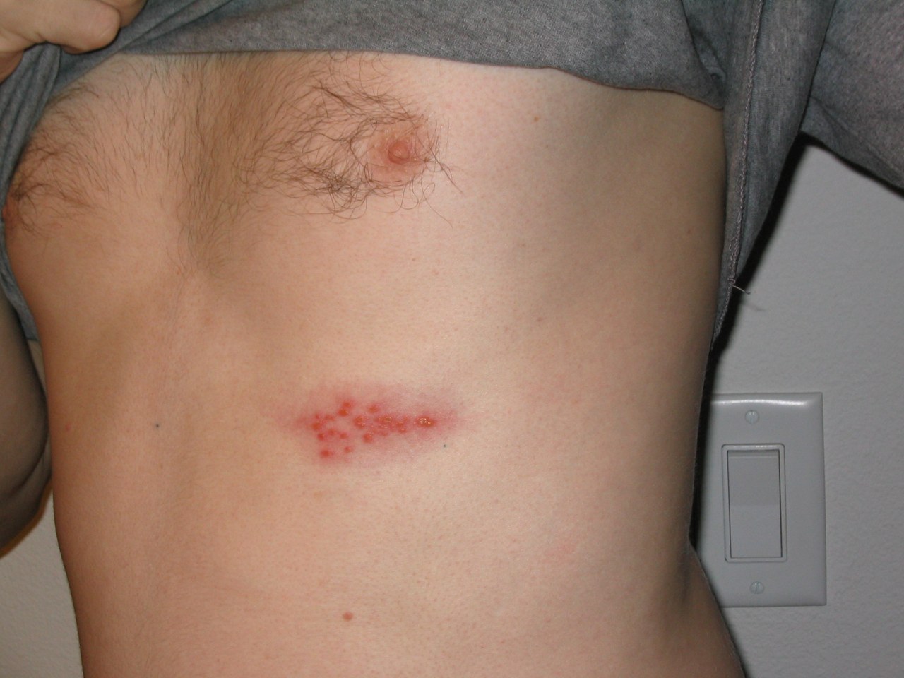
Clinical photograph of a localized cluster of vesicles and redness on the chest.Open the complete image

A real shingles photograph shows grouped lesions on the trunk. Compare the clustered distribution, but do not infer an exact root level or the extent of an eruption from this cropped clinical view.

Preston Hunt. Original image. CC BY 3.0. Resized proportionally and JPEG-reencoded; the photographed field is unchanged.

Herpes zoster classically produces pain and vesicles in one or two adjacent dermatomes, usually on one side without crossing the midline. Atypical and disseminated disease can look different, especially with immunosuppression, and a unilateral band is not automatically a root lesion. [5]

A sensory level, bilateral long-tract signs, new bladder dysfunction, or saddle sensory loss needs urgent spinal evaluation. Stabilize suspected traumatic spinal injury, document the neurologic examination, and obtain urgent imaging appropriate to the suspected cause rather than assigning an exact level from one skin point. [6] [7]

Separate uncertainty about the level from urgency

A localized vesicular eruption gives information that isolated tingling does not. Burning pain may precede the rash, so early pain alone can leave several possibilities open. Once grouped vesicles appear, their distribution and the person's immune status help distinguish a typical localized pattern from a presentation needing broader assessment. [5]

Saddle symptoms concern the perineal region rather than the lateral foot. Ask about new bladder, bowel, or sexual dysfunction and examine the relevant sacral functions when clinically indicated. A normal ankle reflex does not reliably exclude cauda equina or conus compression, because it is not a substitute for evaluating the affected sacral functions. [8]

A patient with fever, severe back pain and evolving neurologic deficits needs assessment for spinal infection and compression. A patient with known cancer, spinal pain and a new sensory level needs urgent assessment for malignant compression. In both cases, recognizing the dangerous pattern is more important than choosing a perfectly precise dermatome before imaging. [7] [9]

These diagrams are study references, not tools for ruling out urgent disease in yourself or another person. A new objective deficit, a sensory level, or bladder symptoms changes the task from memorizing a map to obtaining an appropriate clinical assessment. Preserve the observations in words so that the next examiner can understand what actually changed. [6] [7] [8]

Case 24

A patient develops severe low-back pain, new urinary retention, reduced perianal sensation, and bilateral leg symptoms. Which response is most appropriate?

Show answer and explanations for case 24
  1. A. Assign S3 and arrange routine follow-up (Why this does not fit)

    What makes routine follow-up unsafe despite a possible sacral level?

    New urinary retention signals dysfunction beyond an isolated sensory label.

    Which accompanying findings add a compressive emergency pattern?

    Perianal loss and bilateral leg symptoms indicate multiple sacral or spinal structures.

    Read the full explanation

    A single sacral label understates a time-sensitive compressive syndrome.

  2. B. Treat as isolated pudendal neuropathy (Why this does not fit)

    What region can pudendal neuropathy plausibly affect?

    It can produce perineal sensory and pelvic symptoms in its named territory.

    Which feature requires a broader localization here?

    Bilateral leg symptoms extend beyond an isolated pudendal nerve lesion.

    Read the full explanation

    Pudendal neuropathy does not adequately explain urinary retention with bilateral leg symptoms.

  3. C. Suspect cauda equina or conus compression and obtain emergency assessment (Best answer)

    Which findings define the red-flag cluster?

    Urinary retention, saddle sensory loss, and bilateral leg symptoms form the cluster.

    Can the bedside pattern reliably separate conus from cauda?

    No; the two compressive syndromes have substantial clinical overlap.

    What response follows from either possibility?

    Emergency spinal assessment and imaging are required to protect neurologic function.

    Read the full explanation

    Saddle sensory change, bladder dysfunction, and bilateral leg symptoms are red flags for urgent spinal compression evaluation.

  4. D. Reassure because saddle dermatomes overlap (Why this does not fit)

    Does dermatome overlap reduce the significance of bladder dysfunction?

    No; mapping uncertainty does not neutralize urinary retention and saddle loss.

    What would reassurance risk in this presentation?

    It could delay treatment of time-sensitive cauda equina or conus compression.

    Read the full explanation

    Overlap affects precise mapping but does not neutralize bladder and saddle red flags.

Takeaway: S2-S4 saddle sensory loss with bladder dysfunction requires emergency evaluation for cauda equina or conus compression.

Case sources: [8]

Practice localization

For each patient, choose the localization or next interpretation that explains the whole pattern. The rationale for every option shows which additional finding supports or weakens it.

Case 1

A 32-year-old patient has a narrow band of reduced pinprick sensation at the nipple line after thoracic surgery. Strength and reflexes are normal. Which dermatome is the best initial surface estimate?

Show answer and explanations for case 1
  1. A. C4 (Why this does not fit)

    Where would a C4 sensory change usually appear relative to this band?

    C4 centers on the shoulder cape, far above this thoracic band.

    Do normal strength and reflexes supply a missing cervical clue?

    No; they leave an isolated sensory landmark without a matching cervical motor pattern.

    Read the full explanation

    C4 is used near the shoulder cape, not the nipple line.

  2. B. T4 (Best answer)

    Which trunk landmark lies at the same height as the reported band?

    The nipple line is the customary surface estimate for T4.

    What do normal strength and reflexes contribute to that localization?

    They support a limited sensory finding rather than a broader motor syndrome.

    How precisely should that landmark assign the involved segment?

    Dermatomal overlap makes T4 an initial estimate, not an exact boundary.

    Read the full explanation

    The nipple line is a practical T4 reference point, although individual overlap limits precision.

  3. C. T6 (Why this does not fit)

    Which surface landmark would a T6 band be expected to approach?

    T6 is generally estimated near the xiphoid rather than the nipple.

    How does that expectation compare with this patient's band?

    It places the predicted sensory change several levels below the observed line.

    Read the full explanation

    T6 is closer to the xiphoid and would place the band too low.

  4. D. T10 (Why this does not fit)

    Which familiar trunk landmark is associated with T10?

    T10 is estimated near the umbilicus, well inferior to the nipple line.

    Can normal motor findings overcome that vertical mismatch?

    No; preserved strength does not relocate an umbilical dermatome to the chest.

    Read the full explanation

    T10 is commonly referenced near the umbilicus, well below this finding.

Takeaway: The nipple line is a useful T4 estimate, but no single skin landmark is exact.

Case sources: [1]

Case 2

A patient with midline epigastric numbness points directly over the xiphoid. Which dermatome is the best general landmark?

Show answer and explanations for case 2
  1. A. T4 (Why this does not fit)

    Where should a T4 landmark fall compared with the xiphoid?

    T4 is estimated near the nipple line, superior to the reported numbness.

    What does that height difference do to the T4 proposal?

    It makes T4 too rostral for a point centered directly over the xiphoid.

    Read the full explanation

    T4 is commonly referenced near the nipple line, superior to the xiphoid.

  2. B. T6 (Best answer)

    Which common surface anchor occupies the xiphoid level?

    The xiphoid is generally used as the T6 screening landmark.

    Why is a screening landmark preferable to an exact segment claim?

    Adjacent trunk dermatomes overlap, so a surface point only approximates its level.

    What conclusion follows from location plus that uncertainty?

    The finding supports a T6 estimate while leaving room for individual variation.

    Read the full explanation

    The xiphoid is generally used as a T6 surface landmark in a screening examination.

  3. C. T8 (Why this does not fit)

    Where would T8 lie among the major trunk anchors?

    T8 lies below the xiphoid region and above the umbilical region.

    Does that predicted height match a point directly over the xiphoid?

    No; it shifts the estimate inferior to the patient's marked site.

    Read the full explanation

    T8 lies between the xiphoid and umbilical reference regions and is too inferior here.

  4. D. T10 (Why this does not fit)

    What location would make T10 the better landmark?

    A point near the umbilicus would support T10 more strongly.

    How far is that expected site from the present complaint?

    It is substantially inferior to numbness centered at the xiphoid.

    Read the full explanation

    T10 is the usual umbilical reference and does not fit the xiphoid location.

Takeaway: Use T6 as the general xiphoid anchor while remembering that trunk dermatomes overlap.

Case sources: [1]

Case 4

During a formal ISNCSCI sensory examination after spinal injury, the examiner tests the midpoint of the inguinal ligament. Which designated key sensory point is being tested?

Show answer and explanations for case 4
  1. A. T10 (Why this does not fit)

    Where is the T10 key point placed in the formal map?

    It lies near the umbilical level at the midclavicular line.

    Does that reach the inguinal ligament midpoint being tested?

    No. The inguinal midpoint is inferior to the T10 examination point.

    Read the full explanation

    T10 is tested at the midclavicular line near the umbilical level, not at the inguinal ligament.

  2. B. T12 (Best answer)

    Which examination convention does the question explicitly specify?

    It specifies the standardized ISNCSCI key sensory points, not an unspecified dermatome atlas.

    Which key point is assigned to the inguinal ligament midpoint?

    ISNCSCI assigns that precise point to T12.

    How does the formal L1 location differ?

    L1 is tested inferiorly, midway between the T12 and L2 key points.

    Read the full explanation

    The formal ISNCSCI T12 key sensory point is at the midpoint of the inguinal ligament.

  3. C. L1 (Why this does not fit)

    Where is L1 tested in the specified ISNCSCI convention?

    It is midway between the T12 and L2 key sensory points.

    Why does a broad groin label not settle this question?

    The stem identifies a precise standardized point rather than the entire inguinal sensory region.

    Read the full explanation

    The formal ISNCSCI L1 point is midway between the T12 and L2 sensory points, below the inguinal ligament midpoint.

  4. D. L4 (Why this does not fit)

    Which distal landmark identifies the formal L4 key sensory point?

    The L4 point is over the medial malleolus.

    Does the tested location correspond to that ankle landmark?

    No. The examiner is testing the inguinal ligament, not the medial ankle.

    Read the full explanation

    L4 is tested over the medial malleolus, far from this inguinal examination point.

Takeaway: In the formal ISNCSCI map, the inguinal ligament midpoint is T12; L1 is tested inferiorly between T12 and L2.

Case sources: [1]

Case 5

A patient has numbness over the shoulder cape but normal deltoid strength. Which root-level landmark best matches the sensory region?

Show answer and explanations for case 5
  1. A. C4 (Best answer)

    Which cervical landmark actually covers the shoulder cape?

    C4 commonly supplies the cape and upper acromial sensory region.

    What does normal deltoid strength add to the sensory clue?

    It removes the main motor feature that might instead suggest C5 involvement.

    How should this patch be labeled at the bedside?

    Treat it as a C4 sensory estimate rather than formal C5 testing.

    Read the full explanation

    C4 commonly covers the shoulder cape and upper acromial region.

  2. B. C5 (Why this does not fit)

    Where is the formal C5 key sensory point located?

    C5 is tested at the lateral antecubital fossa, not the shoulder cape.

    Which expected C5 motor finding is also absent?

    Deltoid weakness would support C5, but shoulder abduction remains normal.

    Read the full explanation

    C5 testing is better linked to the lateral antecubital point and elbow flexion, not the cape alone.

  3. C. C6 (Why this does not fit)

    Which sensory distribution would make C6 more persuasive?

    C6 symptoms usually extend along the lateral forearm toward the thumb.

    What motor or reflex partner is missing from this case?

    Wrist-extension weakness or a reduced brachioradialis reflex would support C6.

    Read the full explanation

    C6 favors the lateral forearm and thumb rather than the shoulder cape.

  4. D. T1 (Why this does not fit)

    Where should T1 symptoms appear relative to the shoulder?

    T1 favors the medial forearm rather than the superior shoulder cape.

    Which motor deficit would be expected with meaningful T1 involvement?

    Weak finger abduction would add the intrinsic-hand pattern absent here.

    Read the full explanation

    T1 is associated with the medial forearm and hand intrinsic function.

Takeaway: Shoulder-cape sensation favors C4; do not substitute it for formal C5 testing.

Case sources: [1]

Case 6

During a formal ISNCSCI examination, the examiner must test the C5 key sensory point. Which site is correct?

Show answer and explanations for case 6
  1. A. Skin over the lateral deltoid prominence (Why this does not fit)

    What does the lateral deltoid patch represent clinically?

    It is an axillary nerve territory often checked after shoulder injury.

    Why does that useful patch fail this formal examination task?

    ISNCSCI standardizes C5 sensation at a different reproducible key point.

    Read the full explanation

    That axillary sensory patch is clinically useful but is not the formal C5 key sensory point.

  2. B. Lateral side of the antecubital fossa (Best answer)

    Where must the examiner place the stimulus for C5?

    The formal C5 point lies on the lateral side of the antecubital fossa.

    Why use that site instead of the shoulder cap?

    A standardized point makes serial examinations comparable across clinicians and settings.

    What distinction does this choice preserve?

    It separates formal C5 scoring from the axillary nerve's lateral-deltoid patch.

    Read the full explanation

    ISNCSCI defines the C5 key sensory point at the lateral antecubital fossa.

  3. C. Dorsal thumb at the nail bed (Why this does not fit)

    Which neighboring root is represented at the thumb?

    Thumb sensation is used chiefly to sample the C6 sensory region.

    What error would thumb testing introduce into this score?

    It would record a C6 point while the examiner intends to grade C5.

    Read the full explanation

    The thumb is associated with the C6 key sensory region rather than C5.

  4. D. Middle finger at the distal phalanx (Why this does not fit)

    Which root is sampled at the distal middle finger?

    The middle finger serves as the formal sensory point for C7.

    Why can it not substitute for the requested site?

    Moving two levels caudally would miss the standardized C5 territory entirely.

    Read the full explanation

    The middle finger is used for C7 sensory testing, not C5.

Takeaway: Formal C5 sensation is tested at the lateral antecubital fossa, not the axillary shoulder patch.

Case sources: [1]

Case 7

A patient has neck pain radiating to the thumb, reduced wrist extension, and a diminished brachioradialis reflex. Which root is most likely affected?

Show answer and explanations for case 7
  1. A. C5 (Why this does not fit)

    Which motor territory would a C5 lesion emphasize?

    C5 more strongly affects shoulder abduction and elbow flexion.

    How does the thumb symptom challenge that choice?

    Thumb radiation lies below the expected C5 shoulder-centered sensory pattern.

    Read the full explanation

    C5 more strongly affects shoulder abduction and elbow flexion and does not best explain thumb symptoms.

  2. B. C6 (Best answer)

    Which root connects the radiating pain to the thumb?

    C6 commonly links the lateral forearm and thumb sensory distribution.

    Does the weak movement belong to the same segment?

    Wrist extension is a useful C6-predominant motor test in this comparison.

    Which third finding independently reinforces that level?

    A diminished brachioradialis reflex supplies concordant C6 segmental evidence.

    Read the full explanation

    Thumb and lateral forearm symptoms with wrist extension weakness and reduced brachioradialis reflex support C6.

  3. C. C7 (Why this does not fit)

    What sensory target would make C7 the stronger choice?

    C7 radicular symptoms characteristically favor the middle finger.

    Which motor-reflex pair should accompany that alternative?

    Weak elbow extension and a reduced triceps reflex would support C7.

    Read the full explanation

    C7 favors the middle finger, elbow extension, and the triceps reflex.

  4. D. C8 (Why this does not fit)

    What hand distribution would make C8 more likely?

    C8 favors the little finger and medial hand rather than the thumb.

    Which motor deficit should replace weak wrist extension?

    Finger-flexion weakness would be more characteristic of meaningful C8 involvement.

    Read the full explanation

    C8 favors the little finger and finger flexion rather than wrist extension.

Takeaway: C6 localization strengthens when thumb symptoms, wrist extension weakness, and a matching reflex change agree.

Case sources: [3] [4]

Case 8

A patient has pain from the neck into the middle finger, weak elbow extension, and a reduced triceps reflex. Which root best fits?

Show answer and explanations for case 8
  1. A. C5 (Why this does not fit)

    Where would C5 sensory symptoms usually concentrate?

    C5 findings favor the shoulder region rather than the middle finger.

    Can C5 explain the weak elbow extension?

    No; triceps-driven extension is centered lower, principally at C7.

    Read the full explanation

    C5 does not account for the middle finger pattern or triceps weakness.

  2. B. C6 (Why this does not fit)

    Which digit and movement would point toward C6?

    Thumb symptoms with weak wrist extension would form a stronger C6 pairing.

    Which reflex would then be expected to change?

    The brachioradialis reflex would be more informative than the triceps reflex.

    Read the full explanation

    C6 favors the thumb, wrist extension, and brachioradialis reflex.

  3. C. C7 (Best answer)

    Which root best matches sensation at the middle finger?

    The middle finger is the classic distal sensory landmark for C7.

    Which observed movement tests the same root?

    Elbow extension recruits the triceps and strongly samples C7 function.

    What completes the three-part localization?

    The reduced triceps reflex provides a third concordant C7 finding.

    Read the full explanation

    Middle finger symptoms, triceps weakness, and reduced triceps reflex form a coherent C7 pattern.

  4. D. C8 (Why this does not fit)

    Which sensory region would favor C8 instead?

    C8 symptoms usually involve the little finger and medial hand.

    What weakness should accompany that lower-root pattern?

    Finger flexion would be more affected than the triceps-driven elbow extension.

    Read the full explanation

    C8 is more closely linked to little-finger sensation and finger flexion.

Takeaway: C7 combines middle-finger symptoms with elbow extension weakness and a reduced triceps reflex.

Case sources: [3] [4]

Case 9

A patient reports tingling in the little finger and medial hand after neck rotation. Finger flexion is weak, but finger abduction is preserved. Which root is the best fit?

Show answer and explanations for case 9
  1. A. C6 (Why this does not fit)

    Which digit would a C6 process usually affect?

    C6 radicular sensation favors the thumb, not the little finger.

    What movement would better support that alternative?

    Weak wrist extension would fit C6 better than weak finger flexion.

    Read the full explanation

    C6 centers on the thumb and wrist extension rather than the little finger.

  2. B. C7 (Why this does not fit)

    How would a C7 sensory complaint differ from this one?

    C7 would center symptoms on the middle finger rather than the medial hand.

    Which missing motor feature further opposes C7?

    The stem lacks elbow-extension weakness, a stronger C7 motor correlate.

    Read the full explanation

    C7 better matches the middle finger and elbow extension.

  3. C. C8 (Best answer)

    Which root links little-finger sensation with finger flexion?

    C8 contributes strongly to both the medial hand and finger flexors.

    Why does neck rotation matter in this comparison?

    Provocation from the neck supports a proximal root process over distal neuropathy.

    What does preserved finger abduction help exclude?

    Intact interossei make a predominantly T1 motor lesion less likely.

    Read the full explanation

    Little-finger symptoms with finger flexion weakness favor C8, while preserved interossei make isolated T1 less likely.

  4. D. T1 (Why this does not fit)

    Which bedside movement most clearly samples T1 here?

    Finger abduction tests the interossei and provides a useful T1 comparison.

    How does its preservation affect the T1 proposal?

    Normal abduction contradicts the motor deficit expected from substantial T1 involvement.

    Read the full explanation

    T1 is better represented by finger abduction and adduction, which are preserved here.

Takeaway: C8 favors the little finger and finger flexion; T1 is tested more clearly with interosseous function.

Case sources: [3] [4]

Case 10

A smoker develops medial forearm pain, hand intrinsic weakness, ptosis, and miosis. Which localization is most concerning?

Show answer and explanations for case 10
  1. A. C6 radiculopathy (Why this does not fit)

    Where would a C6 radiculopathy place the sensory complaint?

    C6 would favor the lateral forearm and thumb, not the medial forearm.

    Can a C6 root lesion account for ptosis and miosis?

    No; those sympathetic findings require a lesion beyond an isolated C6 root.

    Read the full explanation

    C6 does not explain medial forearm symptoms, intrinsic hand weakness, or Horner syndrome.

  2. B. C7 radiculopathy (Why this does not fit)

    Which hand landmark and movement would support C7?

    Middle-finger symptoms and triceps weakness would make C7 more coherent.

    What remains unexplained by the C7 proposal?

    The medial forearm distribution and Horner signs remain anatomically discordant.

    Read the full explanation

    C7 would favor the middle finger and triceps rather than this lower plexus pattern.

  3. C. Lower brachial plexus involving T1 near the lung apex (Best answer)

    Which neural region supplies the medial forearm and intrinsic hand?

    The lower brachial plexus carries prominent T1 fibers to those territories.

    What does the accompanying Horner syndrome localize nearby?

    Ptosis and miosis signal interruption of sympathetic fibers near the lung apex.

    Why does smoking increase concern about that combined pattern?

    It raises concern for an apical chest process affecting adjacent plexus and sympathetic structures.

    Read the full explanation

    T1-predominant lower plexus findings plus Horner syndrome raise concern for an apical chest process.

  4. D. Median neuropathy at the wrist (Why this does not fit)

    What distribution should a median lesion at the wrist produce?

    It should affect radial digits and thenar function distal to the wrist.

    Which findings lie outside that lesion's reach?

    Medial forearm pain and Horner syndrome cannot arise from carpal tunnel compression.

    Read the full explanation

    Carpal tunnel syndrome cannot explain medial forearm pain or sympathetic findings.

Takeaway: Medial forearm symptoms, intrinsic weakness, and Horner syndrome suggest a lower plexus lesion near the lung apex.

Case sources: [10]

Case 11

A patient has numbness at the medial malleolus, weak knee extension, and a reduced patellar reflex. Which root contributes most to this pattern?

Show answer and explanations for case 11
  1. A. L2 (Why this does not fit)

    Which proximal findings would make L2 the leading root?

    Hip-flexion weakness and anterior-thigh sensory loss would support L2.

    What current finding lies well below that territory?

    Numbness at the medial malleolus extends beyond the expected L2 distribution.

    Read the full explanation

    L2 contributes to hip flexion and anterior thigh sensation but does not best fit the distal medial leg pattern.

  2. B. L3 (Why this does not fit)

    Which parts of the examination can L3 plausibly explain?

    L3 contributes to quadriceps strength and the patellar reflex arc.

    Which finding shifts the center of gravity one root lower?

    The medial malleolus is a stronger distal sensory landmark for L4.

    Read the full explanation

    L3 contributes to knee extension and the patellar reflex, but the medial malleolus points more strongly to L4.

  3. C. L4 (Best answer)

    Which root supplies the medial-malleolar sensory landmark?

    The medial leg and malleolus are commonly used to estimate L4.

    Does the motor deficit converge on that root?

    Knee-extension weakness supplies an L3-L4 motor finding commonly weighted toward L4.

    What does the reduced knee jerk add?

    Patellar reflex loss provides independent agreement from the same segmental range.

    Read the full explanation

    Medial malleolar sensation, knee extension weakness, and reduced patellar reflex strongly support L4.

  4. D. L5 (Why this does not fit)

    Where would L5 sensory loss be expected instead?

    L5 favors the dorsum of the foot rather than the medial malleolus.

    Which motor-reflex profile would accompany L5?

    Great-toe extension may weaken while routine knee and ankle reflexes remain normal.

    Read the full explanation

    L5 favors the foot dorsum and great-toe extension and lacks a single routine reflex.

Takeaway: L4 is supported by medial malleolar sensory loss plus knee extension and patellar reflex changes.

Case sources: [3] [4]

Case 12

A patient has pain down the lateral leg to the dorsum of the foot, weak great-toe extension, and normal patellar and Achilles reflexes. Which root best fits?

Show answer and explanations for case 12
  1. A. L3 (Why this does not fit)

    Which sensory and motor findings would make L3 plausible?

    Anterior-thigh symptoms with weak knee extension would support L3.

    Why does the present distal pattern oppose that level?

    Pain reaches the dorsal foot and great-toe extension is weak.

    Read the full explanation

    L3 would more often affect anterior thigh sensation and knee extension.

  2. B. L4 (Why this does not fit)

    Where should L4 sensory symptoms concentrate?

    L4 favors the medial leg and medial malleolus.

    Which reflex result also argues against L4 here?

    A normal patellar reflex removes an expected supporting L4 finding.

    Read the full explanation

    L4 favors the medial leg and patellar reflex rather than great-toe extension.

  3. C. L5 (Best answer)

    Which sensory territory links the lateral leg to the dorsal foot?

    That radiating distribution commonly tracks the L5 root.

    Which focused movement tests the same segment?

    Great-toe extension is a useful L5-predominant motor task.

    Why do normal knee and ankle jerks not negate it?

    No single routine deep tendon reflex reliably isolates L5.

    Read the full explanation

    Dorsal foot symptoms and weak great-toe extension support L5; routine reflexes may remain normal.

  4. D. S1 (Why this does not fit)

    Which sensory border would better support S1?

    S1 symptoms usually reach the lateral foot rather than its dorsum.

    What motor-reflex pair should replace the current findings?

    Weak plantar flexion with a reduced Achilles reflex would favor S1.

    Read the full explanation

    S1 favors the lateral foot, plantar flexion, and the Achilles reflex.

Takeaway: L5 often relies on dorsal-foot sensation and great-toe extension because no single routine reflex is definitive.

Case sources: [3] [4]

Case 13

A patient has numbness along the lateral foot, weak single-leg toe raises, and a reduced Achilles reflex. Which root is most likely affected?

Show answer and explanations for case 13
  1. A. L4 (Why this does not fit)

    Which location and movement would favor L4?

    Medial-leg numbness with weak knee extension would support L4.

    Which reflex should fall in that alternative pattern?

    The patellar reflex, rather than the Achilles reflex, would be reduced.

    Read the full explanation

    L4 favors the medial leg, knee extension, and patellar reflex.

  2. B. L5 (Why this does not fit)

    How would an L5 sensory deficit differ?

    L5 more often affects the dorsal foot instead of its lateral border.

    Which weak movement would accompany that territory?

    Great-toe extension would be more informative than single-leg toe raising.

    Read the full explanation

    L5 favors dorsum-of-foot sensation and great-toe extension, often with preserved Achilles reflex.

  3. C. S1 (Best answer)

    Which root maps to the lateral foot?

    The lateral foot is a classic sensory landmark for S1.

    What does failure of a single-leg toe raise test?

    It exposes plantar-flexion weakness, a useful S1 motor correlate.

    Which reflex closes the localization loop?

    A reduced Achilles reflex supplies matching S1-predominant segmental evidence.

    Read the full explanation

    Lateral-foot sensory loss, plantar-flexion weakness, and reduced Achilles reflex form a classic S1 pattern.

  4. D. S3 (Why this does not fit)

    Where should S3 sensory symptoms appear?

    S3 contributes to sacral and saddle regions rather than the lateral foot.

    Can S3 explain the weak toe raise and ankle reflex?

    No; those plantar-flexion and Achilles findings center much more strongly on S1.

    Read the full explanation

    S3 contributes to saddle and pelvic sensory regions rather than the lateral foot.

Takeaway: S1 links the lateral foot with plantar flexion and the Achilles reflex.

Case sources: [3] [4]

Case 14

After prolonged leg crossing, a patient develops foot drop, weak eversion, and numbness in the first dorsal web space. Which localization is most likely?

Show answer and explanations for case 14
  1. A. Deep fibular neuropathy at the ankle (Why this does not fit)

    Which observed sensory patch can a distal deep fibular lesion explain?

    The first dorsal web space belongs to the deep fibular sensory territory.

    Which weak movement falls outside that distal branch?

    Eversion depends mainly on superficial fibular muscles and should remain stronger.

    Read the full explanation

    A distal deep fibular lesion can affect the first web space but would not usually weaken eversion.

  2. B. Common fibular neuropathy at the fibular neck (Best answer)

    Why does prolonged leg crossing identify a vulnerable site?

    It can compress the common fibular nerve against the fibular neck.

    Which two motor branches would be affected there?

    A proximal lesion can weaken both deep-branch dorsiflexion and superficial-branch eversion.

    How does the web-space numbness complete the pattern?

    Deep fibular sensory fibers also travel through the common trunk at that site.

    Read the full explanation

    Compression at the fibular neck explains dorsiflexion and eversion weakness plus deep fibular sensory loss.

  3. C. L4 radiculopathy (Why this does not fit)

    What sensory landmark would better support L4?

    L4 sensory change would favor the medial leg or medial malleolus.

    Why is the leg-crossing mechanism more anatomically specific?

    It directly exposes the common fibular nerve where both weak actions converge.

    Read the full explanation

    L4 does not best explain first-web sensory loss with combined dorsiflexion and eversion weakness.

  4. D. S1 radiculopathy (Why this does not fit)

    Which movement and sensory field would favor S1?

    Weak plantar flexion with lateral-foot numbness would better support S1.

    How does the current foot drop conflict with that prediction?

    Dorsiflexion and eversion fail while the stem supplies no plantar-flexion deficit.

    Read the full explanation

    S1 would more often impair plantar flexion and affect lateral-foot sensation.

Takeaway: The first dorsal web space is deep fibular territory; broader motor testing localizes the lesion at the fibular neck.

Case sources: [4]

Case 16

A patient has nocturnal tingling in the thumb, index, and middle fingers with thenar weakness. The lateral forearm is normal, and neck testing does not reproduce symptoms. Which diagnosis best fits?

Show answer and explanations for case 16
  1. A. C6 radiculopathy (Why this does not fit)

    What territory beyond the thumb would strengthen C6 radiculopathy?

    Symptoms extending through the lateral forearm would support a C6 root pattern.

    Which absent provocation or function further weakens that choice?

    Neck testing is negative, and the stem supplies no wrist-extension or reflex deficit.

    Read the full explanation

    C6 radiculopathy can involve the thumb but often extends into the lateral forearm and may affect wrist extension or reflexes.

  2. B. C7 radiculopathy (Why this does not fit)

    Which isolated digit can overlap with a C7 complaint?

    The middle finger can be involved in a C7 sensory pattern.

    What expected C7 finding is missing from this median-digit syndrome?

    Triceps weakness or reflex change would provide the needed proximal C7 evidence.

    Read the full explanation

    C7 favors the middle finger with triceps findings rather than a classic median hand pattern.

  3. C. Median neuropathy at the carpal tunnel (Best answer)

    What temporal feature strongly favors median compression at the wrist?

    Nocturnal paresthesias are characteristic of carpal tunnel symptom behavior.

    Do the affected digits share the same named nerve?

    The thumb, index, and middle fingers fall within the median sensory distribution.

    How do thenar weakness and normal lateral-forearm sensation refine localization?

    They pair distal median motor involvement with sparing of a proximal root territory.

    Read the full explanation

    Nocturnal median-digit paresthesias and thenar weakness with spared forearm sensation support carpal tunnel syndrome.

  4. D. Ulnar neuropathy at the elbow (Why this does not fit)

    Which digits should an ulnar neuropathy affect most?

    Ulnar symptoms favor the little finger and ulnar half of the ring finger.

    Which weakness would replace the reported thenar deficit?

    Interosseous weakness would be more typical than median-innervated thenar weakness.

    Read the full explanation

    Ulnar neuropathy centers on the little finger and ulnar hand, not the radial three digits.

Takeaway: Median-digit symptoms plus thenar weakness and spared forearm sensation favor carpal tunnel syndrome over a root lesion.

Case sources: [4]

Case 17

A patient leans on the elbows while working and develops little-finger numbness, weak finger abduction, and slowed conduction across the elbow. Which localization is best?

Show answer and explanations for case 17
  1. A. C8 radiculopathy (Why this does not fit)

    Which findings could initially suggest C8?

    Little-finger symptoms and impaired finger flexion can point toward C8.

    What test result localizes the process away from the root?

    Conduction slows focally across the elbow within the ulnar nerve.

    Read the full explanation

    C8 can affect the little finger and finger flexion, but focal conduction slowing across the elbow localizes a named nerve.

  2. B. T1 radiculopathy (Why this does not fit)

    Which observed weakness could T1 plausibly produce?

    T1 fibers contribute strongly to finger abduction through the interossei.

    Why does the electrophysiology override that root-level possibility?

    A focal across-elbow delay identifies a lesion along the named ulnar nerve.

    Read the full explanation

    T1 can weaken interossei, but it does not explain focal ulnar conduction slowing at the elbow.

  3. C. Ulnar neuropathy at the elbow (Best answer)

    Which nerve links the little finger to the weak movement?

    The ulnar nerve supplies medial-hand sensation and the interossei for finger abduction.

    Why is leaning on the elbows relevant?

    Repeated pressure exposes the ulnar nerve at its vulnerable elbow segment.

    What objective finding fixes the lesion at that site?

    Segmental conduction slowing across the elbow directly supports ulnar neuropathy there.

    Read the full explanation

    Ulnar sensory symptoms, interosseous weakness, and focal slowing across the elbow identify ulnar neuropathy.

  4. D. Median neuropathy at the wrist (Why this does not fit)

    Which sensory territory would median neuropathy at the wrist favor?

    Median compression produces symptoms in the radial digits rather than the little finger.

    Which muscle group makes the proposed nerve especially discordant?

    The weak interossei are supplied by the ulnar nerve, not the median nerve.

    Read the full explanation

    Median neuropathy affects radial digits and thenar muscles rather than the little finger and interossei.

Takeaway: Focal conduction and nerve-specific weakness outweigh a rigid wrist-border rule when localizing ulnar neuropathy.

Case sources: [4]

Case 18

After sleeping with an arm over a chair, a patient has wrist and finger extension weakness with numbness over the dorsal first web region. Triceps strength and reflex are preserved. Which lesion is most likely?

Show answer and explanations for case 18
  1. A. C6 radiculopathy (Why this does not fit)

    Which single weak action could overlap with C6 radiculopathy?

    Wrist extension can receive substantial C6 root contribution.

    What broader pattern favors a named nerve instead?

    Finger-extensor weakness plus dorsal radial sensory loss follows the radial nerve more coherently.

    Read the full explanation

    C6 may affect wrist extension but does not explain the complete radial motor and sensory pattern as well.

  2. B. C7 radiculopathy (Why this does not fit)

    Which proximal motor-reflex pair would support C7 radiculopathy?

    Triceps weakness and a reduced triceps reflex would reinforce C7.

    How does their preservation affect that option?

    Normal triceps function places the lesion distal to its radial nerve branches.

    Read the full explanation

    C7 radiculopathy often affects triceps strength or reflex, which are preserved.

  3. C. Radial neuropathy in the spiral groove (Best answer)

    Which nerve carries both the weak extensors and sensory patch?

    The radial nerve supplies wrist and finger extension plus dorsal first-web sensation.

    What does preserved triceps reveal about lesion height?

    The lesion must lie distal to the proximal branches serving the triceps.

    Which common site satisfies both constraints after compression?

    The spiral groove lies distal to triceps branches but proximal to forearm extensors.

    Read the full explanation

    Wrist and finger extension weakness with radial sensory loss and spared triceps localizes distal to the triceps branches.

  4. D. Posterior cord spinal lesion (Why this does not fit)

    Which modality would a posterior cord lesion chiefly disturb?

    Posterior columns carry vibration and joint position rather than radial motor output.

    Why is the distribution incompatible with a spinal column lesion?

    One named nerve's extensor and sensory territory is selectively affected.

    Read the full explanation

    A spinal posterior cord lesion would not selectively produce one radial nerve motor distribution.

Takeaway: Preserved triceps with radial sensory loss and wrist drop localizes a radial lesion near the spiral groove.

Case sources: [4]

Case 19

After pelvic surgery, a patient has weak knee extension, reduced patellar reflex, and anterior thigh plus medial leg numbness. Hip adduction is preserved. Which localization is favored?

Show answer and explanations for case 19
  1. A. L2 radiculopathy (Why this does not fit)

    Which findings would an isolated L2 lesion emphasize?

    Hip-flexion weakness and proximal anterior-thigh sensory change would dominate.

    Which current deficits require lower segmental involvement?

    Patellar reflex loss and medial-leg numbness extend chiefly into L3-L4 territory.

    Read the full explanation

    L2 alone would not explain the patellar reflex loss and medial leg sensory deficit.

  2. B. L3-L4 radiculopathy (Why this does not fit)

    Why can L3-L4 radiculopathy initially mimic this pattern?

    Those roots contribute to quadriceps strength, the knee jerk, and medial-leg sensation.

    Which spared movement argues for a lesion after those roots divide?

    Preserved hip adduction shows the obturator pathway remains functional.

    Read the full explanation

    These roots can mimic the pattern, but preserved hip adduction and the surgical context favor a femoral lesion.

  3. C. Femoral neuropathy (Best answer)

    Which motor deficit identifies the femoral branch?

    Quadriceps weakness produces impaired knee extension in femoral neuropathy.

    How do the reflex and sensory findings stay within that nerve?

    The femoral reflex arc falls, and its saphenous branch supplies the medial leg.

    Why does the surgical history strengthen this localization?

    Pelvic surgery can place the femoral nerve at risk before it enters the thigh.

    Read the full explanation

    Quadriceps weakness, reduced patellar reflex, and saphenous sensory loss after pelvic surgery fit femoral neuropathy.

  4. D. Common fibular neuropathy (Why this does not fit)

    Which deficit should a common fibular lesion produce instead?

    Foot drop from weak dorsiflexion would be expected, not quadriceps weakness.

    Where would its sensory loss occur?

    Symptoms would favor the lateral leg or dorsal foot rather than the medial leg.

    Read the full explanation

    A fibular lesion causes foot drop and dorsal-foot sensory symptoms, not quadriceps weakness.

Takeaway: Quadriceps weakness plus saphenous sensory loss after pelvic surgery favors femoral neuropathy when adduction is spared.

Case sources: [4]

Case 20

A healthy adult develops burning pain followed by grouped vesicles in one unilateral band around the right chest. The eruption occupies two adjacent levels and stops near the midline. Which diagnosis is most likely?

Show answer and explanations for case 20
  1. A. Typical localized herpes zoster (Best answer)

    What sequence links the pain and grouped vesicles?

    Neuropathic pain followed by vesicles is characteristic of zoster reactivation.

    Does the width of the eruption fit localized disease?

    One or two adjacent dermatomes can form a localized zoster band.

    What does stopping near the midline add?

    The unilateral boundary matches the usual distribution of localized zoster.

    Read the full explanation

    Painful vesicles in one or two adjacent unilateral dermatomes are typical of localized zoster.

  2. B. Bilateral thoracic radiculopathy (Why this does not fit)

    Would bilateral thoracic roots create this side-restricted pattern?

    Bilateral radiculopathy should not remain confined to one unilateral chest band.

    Which visible finding is not explained by root compression?

    Grouped vesicles require a cutaneous eruption rather than isolated radicular dysfunction.

    Read the full explanation

    The eruption is unilateral and vesicular, which favors viral reactivation rather than bilateral roots.

  3. C. Contact dermatitis (Why this does not fit)

    What contextual feature would make contact dermatitis more likely?

    A matching external exposure pattern would support contact dermatitis.

    Why does the current geometry point elsewhere?

    A painful narrow band spanning adjacent dermatomes is more characteristic of zoster.

    Read the full explanation

    Contact dermatitis follows an exposure pattern and is less likely to form a painful dermatomal vesicular band.

  4. D. A spinal cord sensory level (Why this does not fit)

    What can a spinal cord sensory level produce at this height?

    It can alter sensation below a boundary on neurologic examination.

    Which finding cannot result from tract interruption alone?

    A cord lesion does not generate grouped vesicles along a unilateral dermatome.

    Read the full explanation

    A cord lesion can alter sensation but does not cause grouped vesicles in a dermatomal band.

Takeaway: Localized zoster typically affects one or two adjacent dermatomes on one side and usually stops at the midline.

Case sources: [5]

Case 21

An immunocompromised patient has a painful unilateral vesicular band plus many vesicles scattered well outside that region. Which interpretation is safest?

Show answer and explanations for case 21
  1. A. The scattered lesions exclude herpes zoster (Why this does not fit)

    Can immunosuppression allow zoster beyond its starting band?

    Yes; impaired host control can permit lesions outside the primary dermatome.

    What does the scattered eruption therefore do to the diagnosis?

    It raises concern for dissemination instead of excluding zoster.

    Read the full explanation

    Immunocompromised patients can develop disseminated zoster beyond the initial dermatomal eruption.

  2. B. This is necessarily bilateral radiculopathy (Why this does not fit)

    Can bilateral radiculopathy produce widespread vesicles?

    No; root dysfunction causes neurologic symptoms, not a disseminated cutaneous eruption.

    Which context better explains lesions beyond the unilateral band?

    Immunocompromise increases concern for atypical or disseminated zoster.

    Read the full explanation

    Radiculopathy does not explain a widespread vesicular eruption.

  3. C. Atypical or disseminated zoster requires urgent clinical assessment (Best answer)

    What feature still reveals the eruption's likely starting pattern?

    A painful unilateral vesicular band remains characteristic of localized zoster onset.

    What changes when many lesions appear beyond that region?

    Extra-dermatomal vesicles raise concern that viral reactivation has disseminated.

    Why is the patient's immune status action-changing?

    Immunocompromise increases complication risk and warrants urgent clinical assessment.

    Read the full explanation

    Lesions outside the primary dermatome in an immunocompromised patient raise concern for disseminated disease.

  4. D. Every lesion must remain within one dermatome before treatment is considered (Why this does not fit)

    Does zoster always remain confined to one dermatome?

    No; adjacent-dermatome and disseminated patterns occur, especially with immunosuppression.

    What harm could a strict boundary requirement cause here?

    It could delay assessment despite widespread lesions in a high-risk patient.

    Read the full explanation

    Zoster can be atypical or disseminated, so a strict one-dermatome requirement is unsafe.

Takeaway: Atypical or disseminated zoster can extend beyond a classic unilateral band, especially with immunosuppression.

Case sources: [5]

Case 22

A patient reports isolated thumb numbness with normal strength and symmetric reflexes. Which conclusion is most defensible?

Show answer and explanations for case 22
  1. A. C6 radiculopathy is proven (Why this does not fit)

    Why does thumb numbness place C6 on the differential?

    The thumb is a common distal sensory landmark for C6.

    What evidence is missing before calling the root proven?

    There is no matching weakness, reflex asymmetry, or broader lateral-forearm distribution.

    Read the full explanation

    Thumb sensation supports C6 but overlap and peripheral nerve territories prevent proof from one point.

  2. B. Median neuropathy is proven (Why this does not fit)

    Why is median neuropathy also anatomically possible?

    The median nerve carries sensation from much of the thumb.

    What broader distal pattern would make it convincing?

    Additional median digits or thenar weakness would strengthen a median-nerve localization.

    Read the full explanation

    The thumb also receives median sensory input, but isolated numbness without a broader pattern is insufficient.

  3. C. The finding is nonanatomic (Why this does not fit)

    Does failure to localize from one point make the symptom nonanatomic?

    No; genuine sensory symptoms can remain anatomically indeterminate early in evaluation.

    What does the overlapping thumb supply explain instead?

    Both a root and a named nerve can plausibly generate the same complaint.

    Read the full explanation

    A real sensory complaint can remain indeterminate even when it does not yet localize securely.

  4. D. C6 and median-nerve hypotheses require corroborating motor, reflex, and distribution findings (Best answer)

    Which two structures overlap at the thumb?

    C6 root fibers reach the thumb through peripheral nerve sensory branches.

    What examination could separate those candidates?

    Test the wider sensory fields, C6 myotomes, median muscles, and segmental reflexes.

    What conclusion is justified before those findings appear?

    The isolated complaint suggests hypotheses but proves neither localization.

    Read the full explanation

    Overlap makes an isolated thumb finding weak; targeted strength, reflex, and sensory testing should arbitrate.

Takeaway: One sensory point suggests candidates; concordant myotomes and reflexes make a root localization credible.

Case sources: [2] [3] [4]

Case 23

A patient with cancer develops back pain, bilateral leg weakness, brisk knees, and a pinprick boundary near the umbilicus. What is the best next interpretation?

Show answer and explanations for case 23
  1. A. Isolated T10 radiculopathy (Why this does not fit)

    Can one T10 root produce brisk weakness in both legs?

    No; a thoracic root lesion does not create bilateral upper motor neuron signs.

    What does the trunk boundary represent instead?

    It functions as a sensory level pointing toward a central spinal process.

    Read the full explanation

    A single root does not explain bilateral weakness and brisk reflexes below a trunk sensory boundary.

  2. B. Possible spinal cord compression requiring urgent MRI (Best answer)

    Which historical feature raises the prior risk of compression?

    Known cancer makes metastatic spinal involvement a serious possibility.

    Which examination findings show threatened cord function?

    Bilateral weakness, brisk knees, and a trunk sensory level indicate long-tract involvement.

    What test must follow that risk pattern promptly?

    Urgent spinal MRI is needed to evaluate possible cord compression.

    Read the full explanation

    Cancer, back pain, a sensory level, and bilateral long-tract signs require urgent imaging for cord compression.

  3. C. A benign normal dermatome variant (Why this does not fit)

    Can normal dermatomal variation explain brisk weak legs?

    No; map variability cannot account for progressive bilateral long-tract deficits.

    How should the sensory boundary affect urgency despite imprecision?

    It strengthens central localization even without naming the exact lesion level.

    Read the full explanation

    Variation does not make progressive bilateral weakness and a sensory level benign.

  4. D. Common fibular neuropathy (Why this does not fit)

    Which territory would common fibular neuropathy affect?

    It would produce distal weakness and sensory loss in one fibular distribution.

    What findings are entirely beyond that nerve's reach?

    A thoracic sensory level and bilateral brisk legs require a central explanation.

    Read the full explanation

    A fibular nerve lesion cannot create a trunk sensory level or bilateral upper motor neuron findings.

Takeaway: A trunk sensory level with bilateral long-tract findings is an urgent cord pattern, not an exact root diagnosis.

Case sources: [6] [7]

Case 25

A febrile patient who injects drugs has severe back pain, progressive leg weakness, and a new thoracic sensory level. Which next step is most appropriate?

Show answer and explanations for case 25
  1. A. Diagnose thoracic zoster without a rash (Why this does not fit)

    Which observed feature might superficially resemble thoracic zoster pain?

    Severe back pain can precede or accompany a dermatomal eruption.

    Which findings make zoster an inadequate explanation?

    Fever, progressive bilateral weakness, and a sensory level indicate deeper spinal disease.

    Read the full explanation

    Zoster does not explain fever, progressive bilateral weakness, and long-tract findings.

  2. B. Obtain urgent spinal MRI for suspected epidural infection and compression (Best answer)

    Which factors raise concern for spinal infection?

    Fever and injection drug use increase concern for an epidural infectious process.

    What shows that neural tissue may already be compressed?

    Progressive leg weakness and a thoracic sensory level indicate evolving cord dysfunction.

    Which test can define both infection and compression urgently?

    Urgent spinal MRI can evaluate the epidural space and threatened cord.

    Read the full explanation

    Fever, risk factors, back pain, weakness, and a sensory level require urgent imaging for epidural abscess.

  3. C. Map the level for one week before imaging (Why this does not fit)

    What can be lost during a week of additional mapping?

    Progressive compression can cause irreversible neurologic injury while evaluation is delayed.

    Does an approximate sensory level justify waiting for precision?

    No; the complete red-flag pattern already warrants immediate imaging.

    Read the full explanation

    Waiting risks permanent injury in a progressive compressive and infectious presentation.

  4. D. Order isolated lower-leg nerve conduction studies first (Why this does not fit)

    What question do lower-leg conduction studies answer?

    They evaluate peripheral nerve function rather than the thoracic spinal canal.

    Why is that the wrong first localization here?

    A trunk level with bilateral weakness points centrally and demands spinal imaging.

    Read the full explanation

    A trunk level and bilateral weakness localize centrally, making peripheral studies an inappropriate first test.

Takeaway: Fever, back pain, progressive weakness, and a sensory level demand urgent spinal imaging.

Case sources: [9]

Case 26

A patient with medial-leg numbness and weak knee extension has a reduced knee jerk. Which segmental description is most accurate?

Show answer and explanations for case 26
  1. A. The patellar reflex is exclusively L2 (Why this does not fit)

    Does L2 contribute anything to the knee-jerk arc?

    L2 contributes fibers but is not the principal segmental emphasis.

    Which paired findings place this pattern lower?

    Medial-leg sensation and knee extension align more strongly with L3-L4.

    Read the full explanation

    L2 can contribute, but it is not the principal segmental pairing for the knee jerk.

  2. B. The patellar reflex is mainly L3-L4 (Best answer)

    Which nerve carries the afferent and efferent knee-jerk arc?

    The patellar reflex travels through the femoral nerve.

    Which segments contribute most strongly to that arc?

    The reflex principally assesses L3-L4, with L4 commonly emphasized.

    Why is that description especially useful in this patient?

    It agrees with medial-leg sensory loss and quadriceps weakness.

    Read the full explanation

    The femoral-mediated patellar reflex principally assesses L3-L4, commonly emphasized at L4.

  3. C. The patellar reflex is mainly L5-S1 (Why this does not fit)

    Which reflex better represents the L5-S1 end of this range?

    The Achilles reflex, especially its S1 component, tests the ankle rather than knee.

    What motor action would replace weak knee extension?

    Plantar-flexion weakness would fit S1 better than quadriceps weakness.

    Read the full explanation

    L5-S1 does not match the quadriceps and patellar reflex arc.

  4. D. The patellar reflex cannot help root localization (Why this does not fit)

    Can the knee jerk contribute to root localization?

    Yes; reflex asymmetry supplies segmental evidence when the arc is affected.

    What prevents it from standing alone?

    Its value increases only when matching sensory and motor findings agree.

    Read the full explanation

    It is useful when interpreted with matching sensory and motor findings.

Takeaway: The patellar reflex is mainly L3-L4 and is strongest evidence when the rest of that pattern agrees.

Case sources: [3] [4]

Case 27

A patient has lateral-foot numbness and weak plantar flexion. The ankle jerk is absent. Which description of that reflex is most accurate?

Show answer and explanations for case 27
  1. A. It is exclusively L5 (Why this does not fit)

    Which routine reflex is poorly suited to isolating L5?

    Neither the patellar nor Achilles reflex serves as a definitive isolated L5 test.

    What L5 findings would be expected outside the reflex examination?

    Dorsal-foot symptoms and weak great-toe extension would support L5.

    Read the full explanation

    L5 is not the principal root for the Achilles reflex.

  2. B. It is mainly S1 with S2 contribution (Best answer)

    Which named nerve carries the Achilles reflex arc?

    The tibial nerve carries the ankle-jerk afferent and efferent limbs.

    Which roots contribute most to that response?

    S1 predominates, with an additional contribution from S2.

    How do this patient's other findings reinforce S1?

    Lateral-foot numbness and weak plantar flexion match the same segment.

    Read the full explanation

    The tibial-mediated Achilles reflex primarily assesses S1 with contribution from S2.

  3. C. It is mainly S3-S4 (Why this does not fit)

    What functions would shift concern toward S3-S4?

    Saddle sensation and pelvic organ function would become more relevant.

    Why do they not fit this ankle pattern?

    They do not principally supply plantar flexion or the Achilles reflex arc.

    Read the full explanation

    S3-S4 are more relevant to pelvic and sacral function than the Achilles reflex.

  4. D. It proves a root lesion by itself (Why this does not fit)

    Can one absent ankle jerk prove radiculopathy?

    No; age, peripheral neuropathy, and local nerve disease can also reduce it.

    What makes the reflex useful in this particular case?

    It aligns with both lateral-foot sensory loss and plantar-flexion weakness.

    Read the full explanation

    An absent ankle reflex can have other causes and must be paired with the sensory and motor examination.

Takeaway: The Achilles reflex is mainly S1 with S2 contribution and is not diagnostic in isolation.

Case sources: [3] [4]

Case 28

A patient has low-back pain, weak great-toe extension, and numbness over the dorsum of the foot. Patellar and Achilles reflexes are symmetric. Which statement is best?

Show answer and explanations for case 28
  1. A. Normal reflexes exclude radiculopathy (Why this does not fit)

    Which routine tendon reflex directly isolates L5?

    No single standard knee or ankle reflex reliably tests L5 alone.

    What follows when both routine reflexes remain symmetric?

    Their preservation cannot exclude an otherwise concordant L5 pattern.

    Read the full explanation

    L5 lacks a single reliable routine deep tendon reflex, so normal knee and ankle jerks do not exclude it.

  2. B. The pattern still supports L5 radiculopathy (Best answer)

    Which sensory distribution supports the suspected root?

    Numbness across the foot dorsum is commonly associated with L5.

    Which motor task independently points to the same root?

    Great-toe extension is a useful motor test weighted toward L5.

    How does low-back pain change the root-versus-nerve comparison?

    It adds a proximal radicular context despite normal routine reflexes.

    Read the full explanation

    Great-toe extension weakness and dorsal-foot symptoms can localize L5 despite normal routine reflexes.

  3. C. The pattern proves deep fibular neuropathy (Why this does not fit)

    Which current findings can deep fibular neuropathy mimic?

    It can weaken great-toe extension and affect the first dorsal web space.

    What prevents that diagnosis from being proven here?

    Radiating low-back pain and a broader dorsal-foot complaint favor proximal evaluation.

    Read the full explanation

    Deep fibular neuropathy would not account for radiating low-back pain or the broader dorsal-foot field.

  4. D. The pattern is most consistent with S1 radiculopathy (Why this does not fit)

    What motor-sensory pair would favor S1?

    An S1 pattern would instead pair plantar-flexion weakness with lateral-foot numbness.

    Which reflex might then become asymmetric?

    The Achilles reflex could decrease in a meaningful S1 lesion.

    Read the full explanation

    S1 favors plantar-flexion weakness, lateral-foot sensory loss, and Achilles change.

Takeaway: Normal routine reflexes do not exclude L5 radiculopathy when sensory and motor findings align.

Case sources: [3] [4]

Case 29

After a surgical-neck humerus fracture, a patient has weak shoulder abduction and a small numb patch over the lateral deltoid. Elbow flexion and the biceps reflex are preserved. Which lesion is favored?

Show answer and explanations for case 29
  1. A. C4 radiculopathy (Why this does not fit)

    Which part of the sensory complaint can resemble C4?

    The superior shoulder cape can overlap a C4 surface territory.

    What makes a root-only explanation less coherent after this fracture?

    Focal deltoid weakness and a small axillary patch follow one injured nerve.

    Read the full explanation

    C4 may affect the shoulder cape but does not explain focal deltoid weakness after this fracture.

  2. B. C5 radiculopathy (Why this does not fit)

    Which observed motor deficit can C5 radiculopathy explain?

    C5 contributes strongly to deltoid-powered shoulder abduction at the root level.

    Which preserved findings argue against a broader C5 root lesion?

    Elbow flexion and the biceps reflex remain intact.

    Read the full explanation

    C5 can weaken deltoid, but preserved biceps function and the fracture mechanism favor a focal axillary lesion.

  3. C. Axillary neuropathy (Best answer)

    Which nerve is vulnerable at the surgical neck?

    The axillary nerve courses around the proximal humerus at that site.

    Do the motor and sensory deficits share that nerve?

    It supplies the deltoid and sensation over the lateral shoulder patch.

    What does preserved biceps function add?

    It spares a separate C5 pathway, strengthening focal axillary localization.

    Read the full explanation

    The surgical-neck mechanism, deltoid weakness, and lateral-shoulder patch localize the axillary nerve.

  4. D. Radial neuropathy (Why this does not fit)

    Which movement should fail with radial neuropathy?

    Wrist or finger extension would weaken in a meaningful radial lesion.

    Why does isolated deltoid weakness contradict that option?

    The deltoid receives axillary rather than radial motor innervation.

    Read the full explanation

    Radial neuropathy would affect extension at the wrist or fingers rather than isolated deltoid function.

Takeaway: An axillary sensory patch plus deltoid weakness after a surgical-neck fracture is not the formal C5 sensory point.

Case sources: [1] [11]

Case 30

A patient has burning pain in a unilateral thoracic band for two days but no rash, weakness, reflex change, or sensory loss on examination. Which statement is most accurate?

Show answer and explanations for case 30
  1. A. The band proves thoracic radiculopathy (Why this does not fit)

    What neurologic evidence would strengthen thoracic radiculopathy?

    Objective segmental sensory loss or other matching root findings would add support.

    Why is the present pain band insufficient by itself?

    A unilateral thoracic band can arise before zoster or from radicular irritation.

    Read the full explanation

    A unilateral band alone is not enough to prove root dysfunction without corroborating neurologic findings.

  2. B. The band proves herpes zoster (Why this does not fit)

    Can pain precede the visible eruption of zoster?

    Yes; neuropathic pain may occur during the prodromal phase.

    What missing feature prevents diagnostic certainty today?

    No vesicular rash has appeared to establish the typical clinical pattern.

    Read the full explanation

    Prodromal zoster is possible, but pain without rash is not specific enough to prove it.

  3. C. Both zoster prodrome and radicular pain remain possible, so follow-up findings matter (Best answer)

    Which two processes can begin with unilateral band-like pain?

    Both early zoster and thoracic radicular irritation can produce that geometry.

    Why does the normal neurologic examination leave them unresolved?

    It supplies no objective root deficit, while absence of rash leaves zoster unconfirmed.

    Which evolution would separate the possibilities?

    A vesicular eruption or new neurologic deficit would materially refine the diagnosis.

    Read the full explanation

    Early zoster and radicular pain can overlap clinically; evolution of rash or neurologic deficits will refine the diagnosis.

  4. D. Crossing the midline is required for zoster (Why this does not fit)

    What midline behavior is usual for localized zoster?

    Typical localized zoster remains unilateral and usually stops near the midline.

    Why is requiring crossover exactly backwards?

    Crossing is not required, and atypical spread does not define ordinary disease.

    Read the full explanation

    Typical zoster is unilateral and usually does not cross the midline, though atypical patterns occur.

Takeaway: A unilateral band is a pattern to investigate, not a standalone diagnosis of zoster or radiculopathy.

Case sources: [4] [5]

Search Bone Wizardry

Quick links